Embed Size (px)
Citation preview
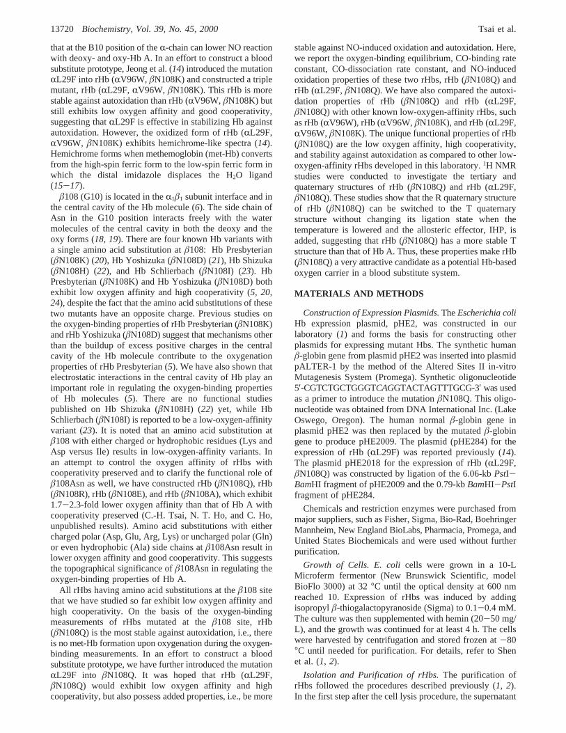
Novel Recombinant Hemoglobin, rHb (âN108Q), with Low Oxygen Affinity, HighCooperativity, and Stability against Autoxidation†
Ching-Hsuan Tsai, Tsuei-Yun Fang,‡ Nancy T. Ho, and Chien Ho*
Department of Biological Sciences, Carnegie Mellon UniVersity, 4400 Fifth AVenue, Pittsburgh, PennsylVania 15213
ReceiVed May 16, 2000; ReVised Manuscript ReceiVed August 3, 2000
ABSTRACT: Using ourEscherichia coliexpression system, we have constructed rHb (âN108Q), a newrecombinant hemoglobin (rHb), with the amino acid substitution located in theR1â1 subunit interface andin the central cavity of the Hb molecule. rHb (âN108Q) exhibits low oxygen affinity, high cooperativity,enhanced Bohr effect, and slower rate of autoxidation of the heme iron atoms from the Fe2+ to the Fe3+
state than other low-oxygen-affinity rHbs developed in our laboratory, e.g., rHb (RV96W) and rHb (RV96W,âN108K). It has been reported by Olson and co-workers [Carver et al. (1992)J. Biol. Chem. 267, 14443-14450; Brantley et al. (1993)J. Biol. Chem. 268, 6995-7010] that the substitution of phenylalanine forleucine at position 29 of myoglobin can inhibit autoxidation in myoglobin and at position 29 of theR-chainof hemoglobin can lower NO reaction in both the deoxy and the oxy forms of human normal adulthemoglobin. Hence, we have further introduced this mutation,RL29F, intoâN108Q. rHb (RL29F,âN108Q)is stabilized against auto- and NO-induced oxidation as compared to rHb (âN108Q), but exhibits loweroxygen affinity at pH below 7.4 and good cooperativity as compared to Hb A. Proton nuclear magneticresonance (NMR) studies show that rHb (âN108Q) has similar tertiary structure around the heme pocketsand quaternary structure in theR1â1 and R1â2 subunit interfaces as compared to those of Hb A. Thetertiary structure of rHb (RL29F,âN108Q) as measured by1H NMR, especially theR-chain heme pocketregion (both proximal and distal histidyl residues), is different from that of CO- and deoxy-Hb A, due tothe amino acid substitution atRL29F. 1H NMR studies also demonstrate that rHb (âN108Q) can switchfrom the R quaternary structure to the T quaternary structure without changing ligation state upon addingan allosteric effector, inositol hexaphosphate, and reducing the temperature. On the basis of its low oxygenaffinity, high cooperativity, and stability against autoxidation, rHb (âN108Q) is considered a potentialcandidate for the Hb-based oxygen carrier in a blood substitute system.
In our laboratory, we have developed an expression systemto produce authentic human normal adult hemoglobin (HbA)1 in good yield in Escherichia coli (1, 2). With thisexpression system, we have designed and expressed mutantHbs with low oxygen affinity and high cooperativity (3-5),which are the desired properties of recombinant (r) Hbs thatmay be used as potential Hb-based oxygen carriers and Hbtherapeutics in a blood substitute system. A unique featureof this class of rHbs is that their R (ligated) quaternarystructure can be switched to the T (unligated) structure,without changing the ligation state of the Hb molecule, bylowering the ambient temperature and/or by adding anallosteric effector, such as inositol hexaphosphate (IHP). rHb
(RV96W) (3) is the first low-oxygen-affinity mutant rHb withhigh cooperativity developed in our laboratory. rHb (RV96W,âN108K) was constructed based onRV96W and a naturallyoccurring low-oxygen-affinity mutant, Hb Presbyterian(âN108K). rHb (RV96W, âN108K) is of particular interestbecause it has good cooperativity and the lowest oxygenaffinity of rHbs studied in our laboratory (5). rHb (RV96W,âN108K) has also been shown to have the greatest tendencyto switch from the R quaternary structure to the T quaternarystructure without changing its ligation state (5).
However, the ease of autoxidation of rHb (RV96W,âN108K) makes it less desirable as an Hb-based oxygencarrier in a blood substitute system. Natural mutant Hbs withlow oxygen affinity are known to exhibit an increased rateof autoxidation (6, 7). The oxidation rate appears to beinversely proportional to the oxygen affinity of Hbs (8). Thiscorrelation between the oxygen affinity and the autoxidationrate poses a serious challenge for engineering Hb-basedoxygen carriers since stability against autoxidation is com-promised by the need for lower oxygen affinity. When Hbis in the extracellular environment of blood vessels, due tothe lack of allosteric effectors, such as 2,3-bisphosphogly-cerate (2,3-BPG), low oxygen affinity with high cooperativityis required for efficient oxygen delivery. It has been reportedby Olson and co-workers (9-13) that the mutation L29F atthe B10 position can inhibit autoxidation in myoglobin and
† This work was supported by research grants from the NationalInstitutes of Health (R01HL-24525, R01HL-58249, and S10RR-11248).C.-H.T. was supported by an Affiliate Student Award (T9902P) fromthe Pennsylvania Affiliate of the American Heart Association.
* Address all correspondence to this author. Telephone: 412-268-3395. Fax: 412-268-7083. E-mail: [email protected].
‡ Present address: Institute of Molecular Biology, Academia Sinica,Taipei, Taiwan.
1 Abbreviations: Hb A, human normal adult hemoglobin; rHb,recombinant hemoglobin; met-Hb, methemoglobin; Mb, myoglobin;NMR, nuclear magnetic resonance; DSS, 2,2-dimethyl-2-silapentane-5-sulfonate; IHP, inositol hexaphosphate; 2,3-BPG, 2,3-bisphospho-glycerate; EDTA, ethylenediaminetetraacetate;k′, CO-binding constant;koff, CO dissociation constant;k′oxy,NO, NO-induced oxidation rateconstant;kauto, autoxidation rate constant.
13719Biochemistry2000,39, 13719-13729
10.1021/bi001116a CCC: $19.00 © 2000 American Chemical SocietyPublished on Web 10/14/2000
that at the B10 position of theR-chain can lower NO reactionwith deoxy- and oxy-Hb A. In an effort to construct a bloodsubstitute prototype, Jeong et al. (14) introduced the mutationRL29F into rHb (RV96W, âN108K) and constructed a triplemutant, rHb (RL29F, RV96W, âN108K). This rHb is morestable against autoxidation than rHb (RV96W, âN108K) butstill exhibits low oxygen affinity and good cooperativity,suggesting thatRL29F is effective in stabilizing Hb againstautoxidation. However, the oxidized form of rHb (RL29F,RV96W, âN108K) exhibits hemichrome-like spectra (14).Hemichrome forms when methemoglobin (met-Hb) convertsfrom the high-spin ferric form to the low-spin ferric form inwhich the distal imidazole displaces the H2O ligand(15-17).
â108 (G10) is located in theR1â1 subunit interface and inthe central cavity of the Hb molecule (6). The side chain ofAsn in the G10 position interacts freely with the watermolecules of the central cavity in both the deoxy and theoxy forms (18, 19). There are four known Hb variants witha single amino acid substitution atâ108: Hb Presbyterian(âN108K) (20), Hb Yoshizuka (âN108D) (21), Hb Shizuka(âN108H) (22), and Hb Schlierbach (âN108I) (23). HbPresbyterian (âN108K) and Hb Yoshizuka (âN108D) bothexhibit low oxygen affinity and high cooperativity (5, 20,24), despite the fact that the amino acid substitutions of thesetwo mutants have an opposite charge. Previous studies onthe oxygen-binding properties of rHb Presbyterian (âN108K)and rHb Yoshizuka (âN108D) suggest that mechanisms otherthan the buildup of excess positive charges in the centralcavity of the Hb molecule contribute to the oxygenationproperties of rHb Presbyterian (5). We have also shown thatelectrostatic interactions in the central cavity of Hb play animportant role in regulating the oxygen-binding propertiesof Hb molecules (5). There are no functional studiespublished on Hb Shizuka (âN108H) (22) yet, while HbSchlierbach (âN108I) is reported to be a low-oxygen-affinityvariant (23). It is noted that an amino acid substitution atâ108 with either charged or hydrophobic residues (Lys andAsp versus Ile) results in low-oxygen-affinity variants. Inan attempt to control the oxygen affinity of rHbs withcooperativity preserved and to clarify the functional role ofâ108Asn as well, we have constructed rHb (âN108Q), rHb(âN108R), rHb (âN108E), and rHb (âN108A), which exhibit1.7-2.3-fold lower oxygen affinity than that of Hb A withcooperativity preserved (C.-H. Tsai, N. T. Ho, and C. Ho,unpublished results). Amino acid substitutions with eithercharged polar (Asp, Glu, Arg, Lys) or uncharged polar (Gln)or even hydrophobic (Ala) side chains atâ108Asn result inlower oxygen affinity and good cooperativity. This suggeststhe topographical significance ofâ108Asn in regulating theoxygen-binding properties of Hb A.
All rHbs having amino acid substitutions at theâ108 sitethat we have studied so far exhibit low oxygen affinity andhigh cooperativity. On the basis of the oxygen-bindingmeasurements of rHbs mutated at theâ108 site, rHb(âN108Q) is the most stable against autoxidation, i.e., thereis no met-Hb formation upon oxygenation during the oxygen-binding measurements. In an effort to construct a bloodsubstitute prototype, we have further introduced the mutationRL29F into âN108Q. It was hoped that rHb (RL29F,âN108Q) would exhibit low oxygen affinity and highcooperativity, but also possess added properties, i.e., be more
stable against NO-induced oxidation and autoxidation. Here,we report the oxygen-binding equilibrium, CO-binding rateconstant, CO-dissociation rate constant, and NO-inducedoxidation properties of these two rHbs, rHb (âN108Q) andrHb (RL29F, âN108Q). We have also compared the autoxi-dation properties of rHb (âN108Q) and rHb (RL29F,âN108Q) with other known low-oxygen-affinity rHbs, suchas rHb (RV96W), rHb (RV96W, âN108K), and rHb (RL29F,RV96W, âN108K). The unique functional properties of rHb(âN108Q) are the low oxygen affinity, high cooperativity,and stability against autoxidation as compared to other low-oxygen-affinity rHbs developed in this laboratory.1H NMRstudies were conducted to investigate the tertiary andquaternary structures of rHb (âN108Q) and rHb (RL29F,âN108Q). These studies show that the R quaternary structureof rHb (âN108Q) can be switched to the T quaternarystructure without changing its ligation state when thetemperature is lowered and the allosteric effector, IHP, isadded, suggesting that rHb (âN108Q) has a more stable Tstructure than that of Hb A. Thus, these properties make rHb(âN108Q) a very attractive candidate as a potential Hb-basedoxygen carrier in a blood substitute system.
MATERIALS AND METHODS
Construction of Expression Plasmids. TheEscherichia coliHb expression plasmid, pHE2, was constructed in ourlaboratory (1) and forms the basis for constructing otherplasmids for expressing mutant Hbs. The synthetic humanâ-globin gene from plasmid pHE2 was inserted into plasmidpALTER-1 by the method of the Altered Sites II in-vitroMutagenesis System (Promega). Synthetic oligonucleotide5′-CGTCTGCTGGGTCAGGTACTAGTTTGCG-3′ was usedas a primer to introduce the mutationâN108Q. This oligo-nucleotide was obtained from DNA International Inc. (LakeOswego, Oregon). The human normalâ-globin gene inplasmid pHE2 was then replaced by the mutatedâ-globingene to produce pHE2009. The plasmid (pHE284) for theexpression of rHb (RL29F) was reported previously (14).The plasmid pHE2018 for the expression of rHb (RL29F,âN108Q) was constructed by ligation of the 6.06-kbPstI-BamHI fragment of pHE2009 and the 0.79-kbBamHI-PstIfragment of pHE284.
Chemicals and restriction enzymes were purchased frommajor suppliers, such as Fisher, Sigma, Bio-Rad, BoehringerMannheim, New England BioLabs, Pharmacia, Promega, andUnited States Biochemicals and were used without furtherpurification.
Growth of Cells. E. colicells were grown in a 10-LMicroferm fermentor (New Brunswick Scientific, modelBioFlo 3000) at 32°C until the optical density at 600 nmreached 10. Expression of rHbs was induced by addingisopropylâ-thiogalactopyranoside (Sigma) to 0.1-0.4 mM.The culture was then supplemented with hemin (20-50 mg/L), and the growth was continued for at least 4 h. The cellswere harvested by centrifugation and stored frozen at-80°C until needed for purification. For details, refer to Shenet al. (1, 2).
Isolation and Purification of rHbs.The purification ofrHbs followed the procedures described previously (1, 2).In the first step after the cell lysis procedure, the supernatant
13720 Biochemistry, Vol. 39, No. 45, 2000 Tsai et al.
from the lysate was left at 30°C overnight (5). Followingthe procedures developed in our laboratory (1, 2), the rHbfraction collected after the Q-Sepharose Fast-Flow column(Pharmacia anion exchanger) was oxidized, reduced, andconverted to the CO form. This Hb solution was then purifiedby eluting from a fast protein liquid chromatography Mono-Scolumn (Pharmacia Cation Exchanger, HR 16/10).
The electrospray ionization mass spectrometric analyseswere performed on a VG Quattro-BQ (Fisons Instruments,VG Biotech, Altrincham, U.K.) as previously described (1).Automated cycles of Edman degradation were performed onan Applied Biosystems gas/liquid-phase sequencer (model470/900A) equipped with an on-line phenylthiohydantoinamino acid analyzer (model 120A) as previously described(1). These two analytical procedures were used to assess thequality of our rHbs. All rHbs used in this study had thecorrect molecular weights and contained less than 1% ofmethionine at the amino termini.
Oxygen-Binding Properties of rHbs.The oxygen dissocia-tion curves of rHbs were measured on a Hemox-Analyzer(TCS Medical Products, Huntington Valley, PA) at 29°Cas a function of pH in 0.1 M sodium phosphate buffer. Theconcentration of Hb used in this study was about 0.1 mM(in terms of heme). The met-Hb reductase system was used(25). A visible absorption spectrum of each sample wasrecorded immediately after the oxygen equilibrium measure-ment, and the met-Hb content was estimated by using theextinction coefficients for Hb reported by Antonini (26).Oxygen equilibrium parameters were derived by fitting theAdair equations to each equilibrium oxygen-binding curveby a nonlinear least-squares procedure.P50, a measure ofoxygen affinity, was obtained at 50% saturation. The Hillcoefficient (nmax), a measure of cooperativity, was determinedfrom the maximum slope of the Hill plot by linear regression.nmax was derived between 60% and 65% oxygen saturation.The accuracy ofP50 measurements (in mmHg) is(5% andthat of nmax is (7%.
Kinetic Measurements. All of the rapid mixing experimentswere performed using an OLIS stopped-flow apparatus(OLIS, Bogart, GA) (with a dead time of≈3 ms). Thetemperature was controlled at 20°C with a circulating waterbath and monitored with a thermometer. To maintainanaerobic conditions in the stopped-flow apparatus, a 10-mL solution of degassed 0.1 M sodium phosphate buffer atpH 8.5 containing 10 mg of dithionite was loaded into thestopped-flow system the day before the kinetic measure-ments. The water bath in the stopped-flow apparatus wasbubbled with Ar gas overnight and during the experiments.Before the samples were loaded, a 20-mL solution ofdegassed 0.1 M sodium phosphate buffer at pH 7.0 was usedto wash the stopped-flow system.
CO-Binding Reaction. For the CO-binding reactions,deoxy-Hb samples (20µM heme before mixing) were reactedwith CO-saturated 0.1 M phosphate buffer at pH 7.0 (1 mMbefore mixing), and the reaction was monitored at 440 nm.The reactions were carried out in the presence and absenceof 50 µM IHP. The time courses were fitted with oneexponential decay to determine the observed rate (kobs). Underthe reaction conditions, the reaction is pseudo-first-order, andthe bimolecular association rate constants were obtained bydividing the observed rate constants by the CO concentration(k′ ) kobs/[CO]).
CO Displacement by NO Reactions. The off-rate of HbCOsamples was determined by mixing HbCO with saturatedNO solution2 (27, 28). A saturated solution of NO was mixedanaerobically with an equal volume of a 20µM (heme)protein solution. The final concentration of NO was 1 mMand of protein was 10µM. The reaction was monitored at440 nm, and the observed rates were determined by fittingthe traces to a single-exponential decay.
NO-Induced Oxidation Reactions. Solutions of oxygen-equilibrated Hb were mixed with anaerobic solutions of NO,and the met-Hb formation was monitored as absorbanceincreases at 402 nm. The value of the bimolecular rateconstant for this reaction is reported to be extremely large,on the order of 10µM-1 s-1 (11), so the concentration ofNO after mixing was kept low (e10 µM) to prevent thereaction from going to completion during the dead time ofthe instrument (3 ms). As a result, very low proteinconcentrations (0.4µM heme after mixing) were requiredto maintain a pseudo-first-order approximation. The low Hbconcentrations resulted in small absorbance changes, and anaveraging of 10-14 traces was necessary to enhance thesignal-to-noise ratio. The time courses were fitted to a single-exponential expression using an iterative nonlinear least-squares algorithm to obtain the observed rate constants. Thebimolecular rate constants were obtained by dividing theobserved rate constants by the NO concentration (k′oxy,NO )kobs/[NO]).
1H NMR InVestigation of rHbs. 1H NMR spectra of rHbswere obtained from Bruker AVANCE DRX-300 andAVANCE DRX-500 NMR spectrometers. All Hb sampleswere in 0.1 M sodium phosphate buffer in 100% water, andthe Hb concentration was about 5% (∼0.8 mM in terms ofheme). The water signal was suppressed by using a jump-and-return pulse sequence (29). Proton chemical shifts arereferenced to the methyl proton resonance of 2,2-dimethyl-2-silapentane-5-sulfonate (DSS) indirectly by using the watersignal, which occurs at 4.76 ppm downfield from that ofDSS at 29°C, as the internal reference.
Autoxidation of rHbs. The autoxidation was recorded bymonitoring the rate of disappearance of the oxy-marker(-2.34 ppm from DSS) (30, 31) on the Bruker AVANCEDRX-300. The autoxidation reaction was carried out inPlasmaLyte buffer (Baxter) in the presence of 5% D2O and5 mM ethylenediaminetetraacetate (EDTA) at pH 7.4 andat 37°C. The HbO2 concentration was 5% (∼0.8 mM).
RESULTS
Functional Studies
Oxygen-Binding Properties of rHbs.Figure 1 showsthe oxygen-binding measurements of rHb (RL29F), rHb(âN108Q), rHb (RL29F, âN108Q), and Hb A in 0.1 Msodium phosphate buffer as a function of pH at 29°C. rHb(âN108Q) exhibits a significantly lower oxygen affinity as
2 Stock solutions of NO were prepared by injecting anaerobic buffersolution (0.1 M phosphate at pH 7.0) into a gas-tight tonometer, whichhad been flushed with 1 atm of NO. The NO gas used had been passedthrough a column of KOH pellets to remove acids (NO2). Anaerobicbuffer solution was prepared by bubbling the solution with argon gasfor at least 30 min. Solutions at lower NO concentration were preparedby dilution of the saturated NO solution with an appropriate volumeof an anaerobic buffer solution.
rHb (âN108Q) Exhibits Stability against Autoxidation Biochemistry, Vol. 39, No. 45, 200013721
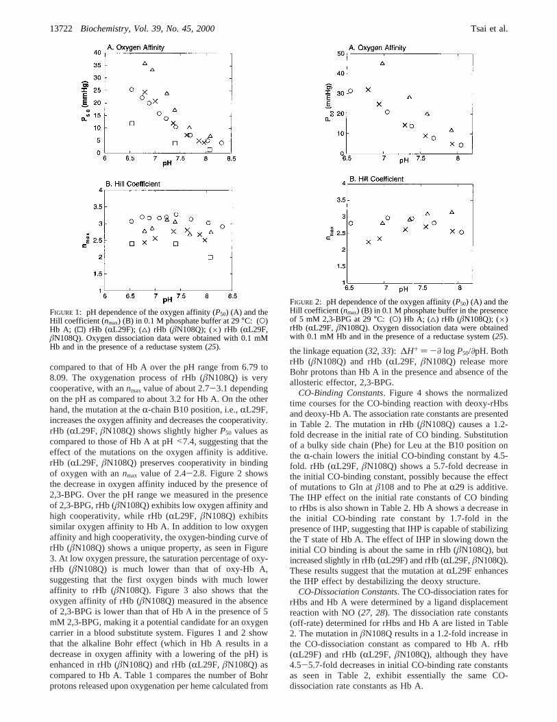
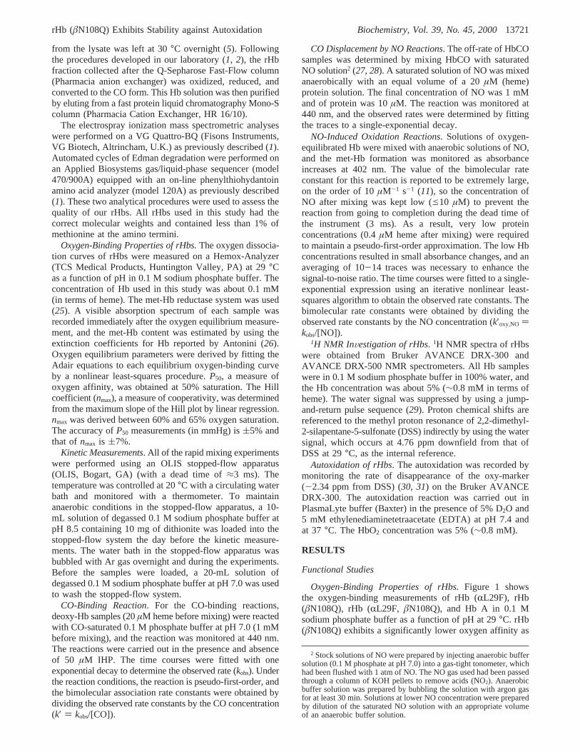
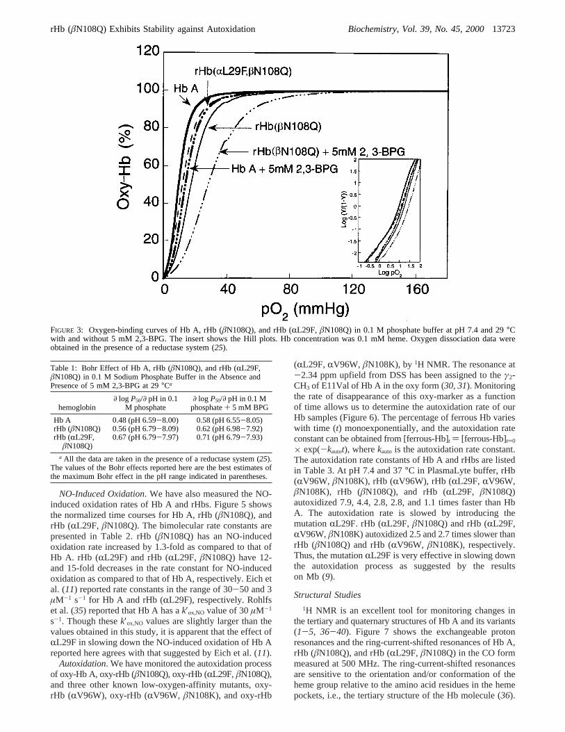
compared to that of Hb A over the pH range from 6.79 to8.09. The oxygenation process of rHb (âN108Q) is verycooperative, with annmax value of about 2.7-3.1 dependingon the pH as compared to about 3.2 for Hb A. On the otherhand, the mutation at theR-chain B10 position, i.e.,RL29F,increases the oxygen affinity and decreases the cooperativity.rHb (RL29F, âN108Q) shows slightly higherP50 values ascompared to those of Hb A at pH<7.4, suggesting that theeffect of the mutations on the oxygen affinity is additive.rHb (RL29F, âN108Q) preserves cooperativity in bindingof oxygen with annmax value of 2.4-2.8. Figure 2 showsthe decrease in oxygen affinity induced by the presence of2,3-BPG. Over the pH range we measured in the presenceof 2,3-BPG, rHb (âN108Q) exhibits low oxygen affinity andhigh cooperativity, while rHb (RL29F, âN108Q) exhibitssimilar oxygen affinity to Hb A. In addition to low oxygenaffinity and high cooperativity, the oxygen-binding curve ofrHb (âN108Q) shows a unique property, as seen in Figure3. At low oxygen pressure, the saturation percentage of oxy-rHb (âN108Q) is much lower than that of oxy-Hb A,suggesting that the first oxygen binds with much loweraffinity to rHb (âN108Q). Figure 3 also shows that theoxygen affinity of rHb (âN108Q) measured in the absenceof 2,3-BPG is lower than that of Hb A in the presence of 5mM 2,3-BPG, making it a potential candidate for an oxygencarrier in a blood substitute system. Figures 1 and 2 showthat the alkaline Bohr effect (which in Hb A results in adecrease in oxygen affinity with a lowering of the pH) isenhanced in rHb (âN108Q) and rHb (RL29F, âN108Q) ascompared to Hb A. Table 1 compares the number of Bohrprotons released upon oxygenation per heme calculated from
the linkage equation (32, 33): ∆H+ ) -∂ log P50/∂pH. BothrHb (âN108Q) and rHb (RL29F, âN108Q) release moreBohr protons than Hb A in the presence and absence of theallosteric effector, 2,3-BPG.
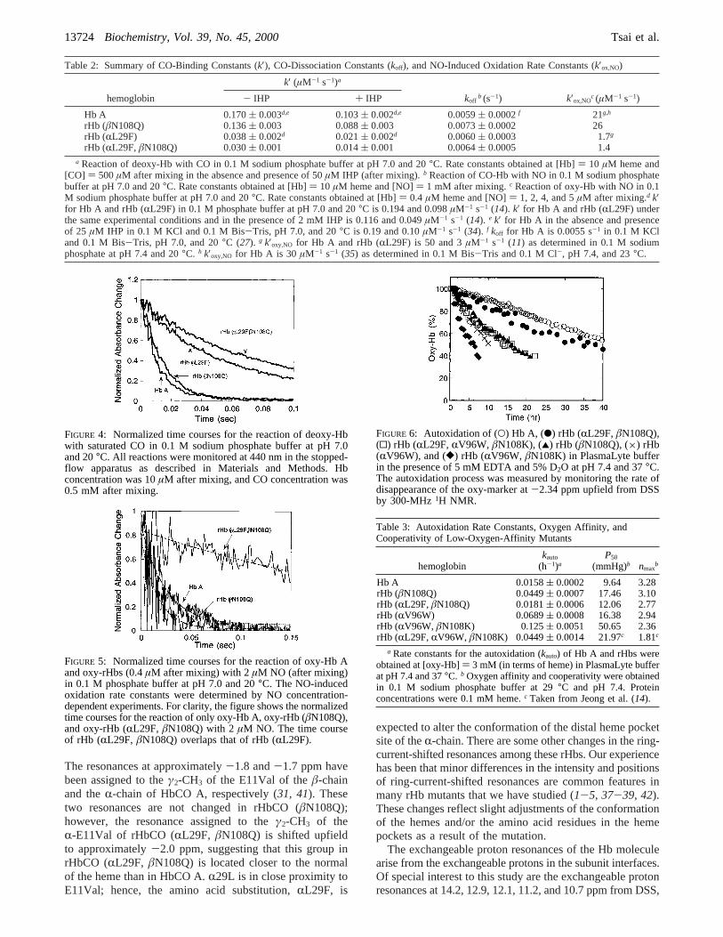
CO-Binding Constants. Figure 4 shows the normalizedtime courses for the CO-binding reaction with deoxy-rHbsand deoxy-Hb A. The association rate constants are presentedin Table 2. The mutation in rHb (âN108Q) causes a 1.2-fold decrease in the initial rate of CO binding. Substitutionof a bulky side chain (Phe) for Leu at the B10 position onthe R-chain lowers the initial CO-binding constant by 4.5-fold. rHb (RL29F, âN108Q) shows a 5.7-fold decrease inthe initial CO-binding constant, possibly because the effectof mutations to Gln atâ108 and to Phe atR29 is additive.The IHP effect on the initial rate constants of CO bindingto rHbs is also shown in Table 2. Hb A shows a decrease inthe initial CO-binding rate constant by 1.7-fold in thepresence of IHP, suggesting that IHP is capable of stabilizingthe T state of Hb A. The effect of IHP in slowing down theinitial CO binding is about the same in rHb (âN108Q), butincreased slightly in rHb (RL29F) and rHb (RL29F,âN108Q).These results suggest that the mutation atRL29F enhancesthe IHP effect by destabilizing the deoxy structure.
CO-Dissociation Constants. The CO-dissociation rates forrHbs and Hb A were determined by a ligand displacementreaction with NO (27, 28). The dissociation rate constants(off-rate) determined for rHbs and Hb A are listed in Table2. The mutation inâN108Q results in a 1.2-fold increase inthe CO-dissociation constant as compared to Hb A. rHb(RL29F) and rHb (RL29F, âN108Q), although they have4.5-5.7-fold decreases in initial CO-binding rate constantsas seen in Table 2, exhibit essentially the same CO-dissociation rate constants as Hb A.
FIGURE 1: pH dependence of the oxygen affinity (P50) (A) and theHill coefficient (nmax) (B) in 0.1 M phosphate buffer at 29°C: (O)Hb A; (0) rHb (RL29F); (4) rHb (âN108Q); (×) rHb (RL29F,âN108Q). Oxygen dissociation data were obtained with 0.1 mMHb and in the presence of a reductase system (25).
FIGURE 2: pH dependence of the oxygen affinity (P50) (A) and theHill coefficient (nmax) (B) in 0.1 M phosphate buffer in the presenceof 5 mM 2,3-BPG at 29°C: (O) Hb A; (4) rHb (âN108Q); (×)rHb (RL29F, âN108Q). Oxygen dissociation data were obtainedwith 0.1 mM Hb and in the presence of a reductase system (25).
13722 Biochemistry, Vol. 39, No. 45, 2000 Tsai et al.
NO-Induced Oxidation. We have also measured the NO-induced oxidation rates of Hb A and rHbs. Figure 5 showsthe normalized time courses for Hb A, rHb (âN108Q), andrHb (RL29F, âN108Q). The bimolecular rate constants arepresented in Table 2. rHb (âN108Q) has an NO-inducedoxidation rate increased by 1.3-fold as compared to that ofHb A. rHb (RL29F) and rHb (RL29F, âN108Q) have 12-and 15-fold decreases in the rate constant for NO-inducedoxidation as compared to that of Hb A, respectively. Eich etal. (11) reported rate constants in the range of 30-50 and 3µM-1 s-1 for Hb A and rHb (RL29F), respectively. Rohlfset al. (35) reported that Hb A has ak′ox,NO value of 30µM-1
s-1. Though thesek′ox,NO values are slightly larger than thevalues obtained in this study, it is apparent that the effect ofRL29F in slowing down the NO-induced oxidation of Hb Areported here agrees with that suggested by Eich et al. (11).
Autoxidation. We have monitored the autoxidation processof oxy-Hb A, oxy-rHb (âN108Q), oxy-rHb (RL29F,âN108Q),and three other known low-oxygen-affinity mutants, oxy-rHb (RV96W), oxy-rHb (RV96W, âN108K), and oxy-rHb
(RL29F,RV96W, âN108K), by1H NMR. The resonance at-2.34 ppm upfield from DSS has been assigned to theγ2-CH3 of E11Val of Hb A in the oxy form (30, 31). Monitoringthe rate of disappearance of this oxy-marker as a functionof time allows us to determine the autoxidation rate of ourHb samples (Figure 6). The percentage of ferrous Hb varieswith time (t) monoexponentially, and the autoxidation rateconstant can be obtained from [ferrous-Hb]t ) [ferrous-Hb]t)0
× exp(-kautot), wherekauto is the autoxidation rate constant.The autoxidation rate constants of Hb A and rHbs are listedin Table 3. At pH 7.4 and 37°C in PlasmaLyte buffer, rHb(RV96W, âN108K), rHb (RV96W), rHb (RL29F, RV96W,âN108K), rHb (âN108Q), and rHb (RL29F, âN108Q)autoxidized 7.9, 4.4, 2.8, 2.8, and 1.1 times faster than HbA. The autoxidation rate is slowed by introducing themutationRL29F. rHb (RL29F, âN108Q) and rHb (RL29F,RV96W, âN108K) autoxidized 2.5 and 2.7 times slower thanrHb (âN108Q) and rHb (RV96W, âN108K), respectively.Thus, the mutationRL29F is very effective in slowing downthe autoxidation process as suggested by the resultson Mb (9).
Structural Studies1H NMR is an excellent tool for monitoring changes in
the tertiary and quaternary structures of Hb A and its variants(1-5, 36-40). Figure 7 shows the exchangeable protonresonances and the ring-current-shifted resonances of Hb A,rHb (âN108Q), and rHb (RL29F, âN108Q) in the CO formmeasured at 500 MHz. The ring-current-shifted resonancesare sensitive to the orientation and/or conformation of theheme group relative to the amino acid residues in the hemepockets, i.e., the tertiary structure of the Hb molecule (36).
FIGURE 3: Oxygen-binding curves of Hb A, rHb (âN108Q), and rHb (RL29F, âN108Q) in 0.1 M phosphate buffer at pH 7.4 and 29°Cwith and without 5 mM 2,3-BPG. The insert shows the Hill plots. Hb concentration was 0.1 mM heme. Oxygen dissociation data wereobtained in the presence of a reductase system (25).
Table 1: Bohr Effect of Hb A, rHb (âN108Q), and rHb (RL29F,âN108Q) in 0.1 M Sodium Phosphate Buffer in the Absence andPresence of 5 mM 2,3-BPG at 29°Ca
hemoglobin∂ log P50/∂ pH in 0.1
M phosphate∂ log P50/∂ pH in 0.1 M
phosphate+ 5 mM BPG
Hb A 0.48 (pH 6.59-8.00) 0.58 (pH 6.55-8.05)rHb (âN108Q) 0.56 (pH 6.79-8.09) 0.62 (pH 6.98-7.92)rHb (RL29F,
âN108Q)0.67 (pH 6.79-7.97) 0.71 (pH 6.79-7.93)
a All the data are taken in the presence of a reductase system (25).The values of the Bohr effects reported here are the best estimates ofthe maximum Bohr effect in the pH range indicated in parentheses.
rHb (âN108Q) Exhibits Stability against Autoxidation Biochemistry, Vol. 39, No. 45, 200013723
The resonances at approximately-1.8 and-1.7 ppm havebeen assigned to theγ2-CH3 of the E11Val of theâ-chainand theR-chain of HbCO A, respectively (31, 41). Thesetwo resonances are not changed in rHbCO (âN108Q);however, the resonance assigned to theγ2-CH3 of theR-E11Val of rHbCO (RL29F, âN108Q) is shifted upfieldto approximately-2.0 ppm, suggesting that this group inrHbCO (RL29F, âN108Q) is located closer to the normalof the heme than in HbCO A.R29L is in close proximity toE11Val; hence, the amino acid substitution,RL29F, is
expected to alter the conformation of the distal heme pocketsite of theR-chain. There are some other changes in the ring-current-shifted resonances among these rHbs. Our experiencehas been that minor differences in the intensity and positionsof ring-current-shifted resonances are common features inmany rHb mutants that we have studied (1-5, 37-39, 42).These changes reflect slight adjustments of the conformationof the hemes and/or the amino acid residues in the hemepockets as a result of the mutation.
The exchangeable proton resonances of the Hb moleculearise from the exchangeable protons in the subunit interfaces.Of special interest to this study are the exchangeable protonresonances at 14.2, 12.9, 12.1, 11.2, and 10.7 ppm from DSS,
Table 2: Summary of CO-Binding Constants (k′), CO-Dissociation Constants (koff), and NO-Induced Oxidation Rate Constants (k′ox,NO)
k′ (µM-1 s-1)a
hemoglobin - IHP + IHP koffb (s-1) k′ox,NO
c (µM-1 s-1)
Hb A 0.170( 0.003d,e 0.103( 0.002d,e 0.0059( 0.0002f 21g,h
rHb (âN108Q) 0.136( 0.003 0.088( 0.003 0.0073( 0.0002 26rHb (RL29F) 0.038( 0.002d 0.021( 0.002d 0.0060( 0.0003 1.7g
rHb (RL29F,âN108Q) 0.030( 0.001 0.014( 0.001 0.0064( 0.0005 1.4a Reaction of deoxy-Hb with CO in 0.1 M sodium phosphate buffer at pH 7.0 and 20°C. Rate constants obtained at [Hb]) 10 µM heme and
[CO] ) 500µM after mixing in the absence and presence of 50µM IHP (after mixing).b Reaction of CO-Hb with NO in 0.1 M sodium phosphatebuffer at pH 7.0 and 20°C. Rate constants obtained at [Hb]) 10 µM heme and [NO]) 1 mM after mixing.c Reaction of oxy-Hb with NO in 0.1M sodium phosphate buffer at pH 7.0 and 20°C. Rate constants obtained at [Hb]) 0.4 µM heme and [NO]) 1, 2, 4, and 5µM after mixing.d k′for Hb A and rHb (RL29F) in 0.1 M phosphate buffer at pH 7.0 and 20°C is 0.194 and 0.098µM-1 s-1 (14). k′ for Hb A and rHb (RL29F) underthe same experimental conditions and in the presence of 2 mM IHP is 0.116 and 0.049µM-1 s-1 (14). e k′ for Hb A in the absence and presenceof 25 µM IHP in 0.1 M KCl and 0.1 M Bis-Tris, pH 7.0, and 20°C is 0.19 and 0.10µM-1 s-1 (34). f koff for Hb A is 0.0055 s-1 in 0.1 M KCland 0.1 M Bis-Tris, pH 7.0, and 20°C (27). g k′oxy,NO for Hb A and rHb (RL29F) is 50 and 3µM-1 s-1 (11) as determined in 0.1 M sodiumphosphate at pH 7.4 and 20°C. h k′oxy,NO for Hb A is 30 µM-1 s-1 (35) as determined in 0.1 M Bis-Tris and 0.1 M Cl-, pH 7.4, and 23°C.
FIGURE 4: Normalized time courses for the reaction of deoxy-Hbwith saturated CO in 0.1 M sodium phosphate buffer at pH 7.0and 20°C. All reactions were monitored at 440 nm in the stopped-flow apparatus as described in Materials and Methods. Hbconcentration was 10µM after mixing, and CO concentration was0.5 mM after mixing.
FIGURE 5: Normalized time courses for the reaction of oxy-Hb Aand oxy-rHbs (0.4µM after mixing) with 2µM NO (after mixing)in 0.1 M phosphate buffer at pH 7.0 and 20°C. The NO-inducedoxidation rate constants were determined by NO concentration-dependent experiments. For clarity, the figure shows the normalizedtime courses for the reaction of only oxy-Hb A, oxy-rHb (âN108Q),and oxy-rHb (RL29F, âN108Q) with 2µM NO. The time courseof rHb (RL29F, âN108Q) overlaps that of rHb (RL29F).
FIGURE 6: Autoxidation of (O) Hb A, (b) rHb (RL29F,âN108Q),(0) rHb (RL29F,RV96W, âN108K), (2) rHb (âN108Q), (×) rHb(RV96W), and ([) rHb (RV96W, âN108K) in PlasmaLyte bufferin the presence of 5 mM EDTA and 5% D2O at pH 7.4 and 37°C.The autoxidation process was measured by monitoring the rate ofdisappearance of the oxy-marker at-2.34 ppm upfield from DSSby 300-MHz1H NMR.
Table 3: Autoxidation Rate Constants, Oxygen Affinity, andCooperativity of Low-Oxygen-Affinity Mutants
hemoglobinkauto
(h-1)aP50
(mmHg)b nmaxb
Hb A 0.0158( 0.0002 9.64 3.28rHb (âN108Q) 0.0449( 0.0007 17.46 3.10rHb (RL29F,âN108Q) 0.0181( 0.0006 12.06 2.77rHb (RV96W) 0.0689( 0.0008 16.38 2.94rHb (RV96W, âN108K) 0.125( 0.0051 50.65 2.36rHb (RL29F,RV96W, âN108K) 0.0449( 0.0014 21.97c 1.81c
a Rate constants for the autoxidation (kauto) of Hb A and rHbs wereobtained at [oxy-Hb]) 3 mM (in terms of heme) in PlasmaLyte bufferat pH 7.4 and 37°C. b Oxygen affinity and cooperativity were obtainedin 0.1 M sodium phosphate buffer at 29°C and pH 7.4. Proteinconcentrations were 0.1 mM heme.c Taken from Jeong et al. (14).
13724 Biochemistry, Vol. 39, No. 45, 2000 Tsai et al.
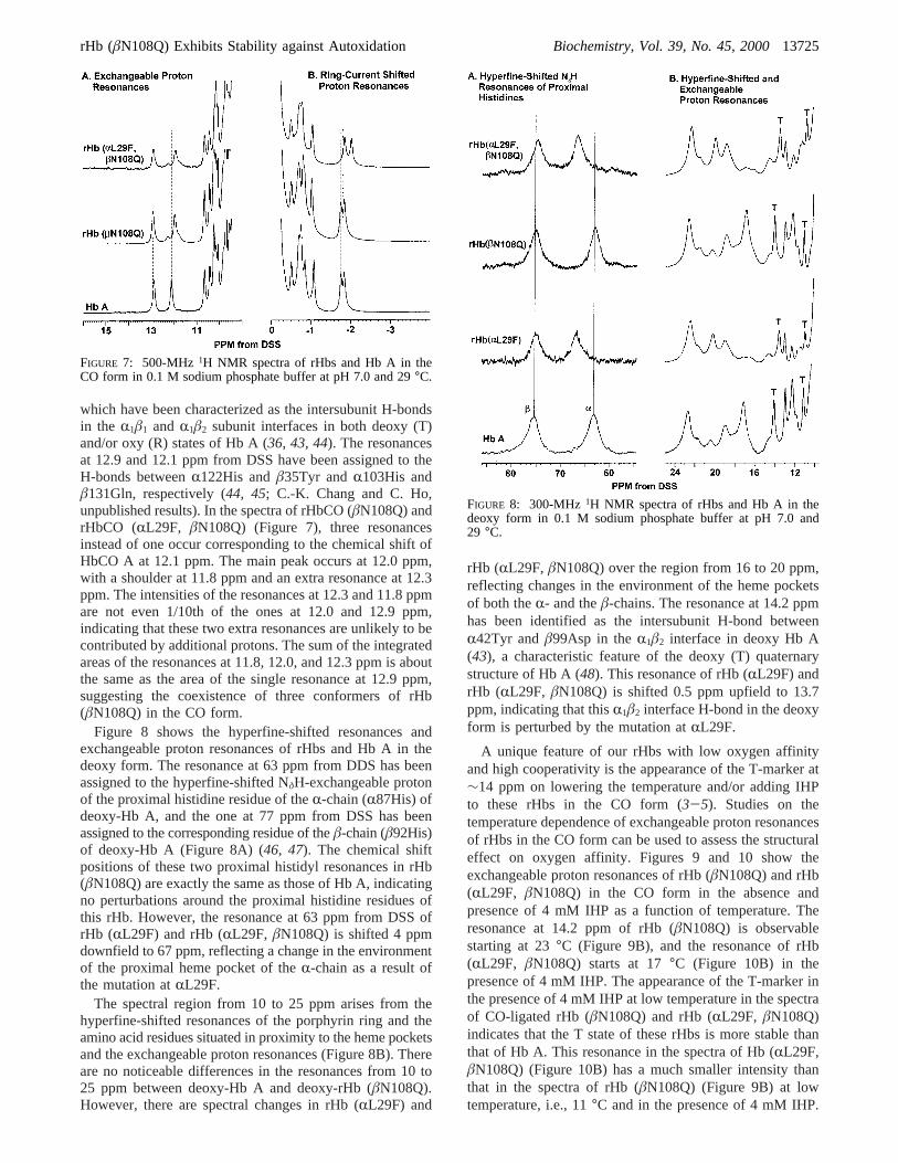
which have been characterized as the intersubunit H-bondsin the R1â1 and R1â2 subunit interfaces in both deoxy (T)and/or oxy (R) states of Hb A (36, 43, 44). The resonancesat 12.9 and 12.1 ppm from DSS have been assigned to theH-bonds betweenR122His andâ35Tyr andR103His andâ131Gln, respectively (44, 45; C.-K. Chang and C. Ho,unpublished results). In the spectra of rHbCO (âN108Q) andrHbCO (RL29F, âN108Q) (Figure 7), three resonancesinstead of one occur corresponding to the chemical shift ofHbCO A at 12.1 ppm. The main peak occurs at 12.0 ppm,with a shoulder at 11.8 ppm and an extra resonance at 12.3ppm. The intensities of the resonances at 12.3 and 11.8 ppmare not even 1/10th of the ones at 12.0 and 12.9 ppm,indicating that these two extra resonances are unlikely to becontributed by additional protons. The sum of the integratedareas of the resonances at 11.8, 12.0, and 12.3 ppm is aboutthe same as the area of the single resonance at 12.9 ppm,suggesting the coexistence of three conformers of rHb(âN108Q) in the CO form.
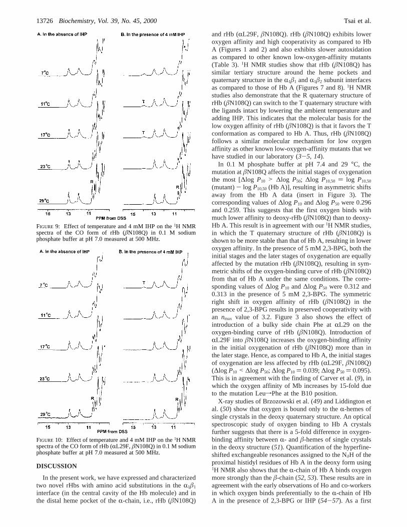
Figure 8 shows the hyperfine-shifted resonances andexchangeable proton resonances of rHbs and Hb A in thedeoxy form. The resonance at 63 ppm from DDS has beenassigned to the hyperfine-shifted NδH-exchangeable protonof the proximal histidine residue of theR-chain (R87His) ofdeoxy-Hb A, and the one at 77 ppm from DSS has beenassigned to the corresponding residue of theâ-chain (â92His)of deoxy-Hb A (Figure 8A) (46, 47). The chemical shiftpositions of these two proximal histidyl resonances in rHb(âN108Q) are exactly the same as those of Hb A, indicatingno perturbations around the proximal histidine residues ofthis rHb. However, the resonance at 63 ppm from DSS ofrHb (RL29F) and rHb (RL29F, âN108Q) is shifted 4 ppmdownfield to 67 ppm, reflecting a change in the environmentof the proximal heme pocket of theR-chain as a result ofthe mutation atRL29F.
The spectral region from 10 to 25 ppm arises from thehyperfine-shifted resonances of the porphyrin ring and theamino acid residues situated in proximity to the heme pocketsand the exchangeable proton resonances (Figure 8B). Thereare no noticeable differences in the resonances from 10 to25 ppm between deoxy-Hb A and deoxy-rHb (âN108Q).However, there are spectral changes in rHb (RL29F) and
rHb (RL29F, âN108Q) over the region from 16 to 20 ppm,reflecting changes in the environment of the heme pocketsof both theR- and theâ-chains. The resonance at 14.2 ppmhas been identified as the intersubunit H-bond betweenR42Tyr andâ99Asp in theR1â2 interface in deoxy Hb A(43), a characteristic feature of the deoxy (T) quaternarystructure of Hb A (48). This resonance of rHb (RL29F) andrHb (RL29F, âN108Q) is shifted 0.5 ppm upfield to 13.7ppm, indicating that thisR1â2 interface H-bond in the deoxyform is perturbed by the mutation atRL29F.
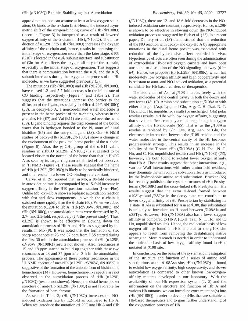
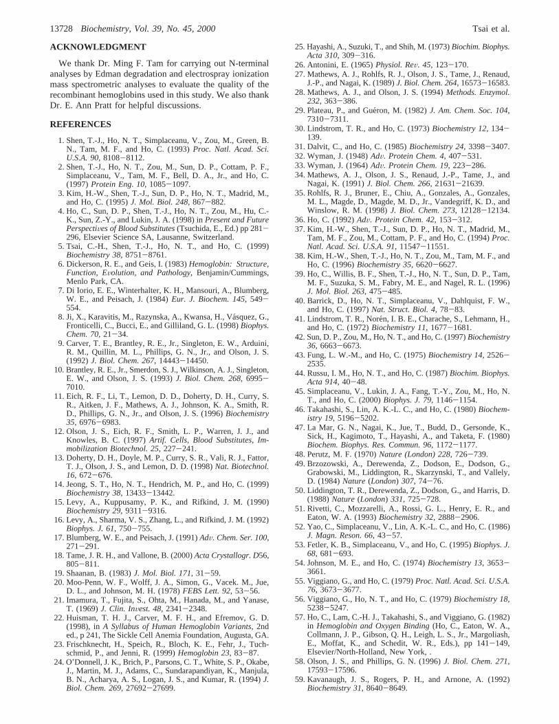
A unique feature of our rHbs with low oxygen affinityand high cooperativity is the appearance of the T-marker at∼14 ppm on lowering the temperature and/or adding IHPto these rHbs in the CO form (3-5). Studies on thetemperature dependence of exchangeable proton resonancesof rHbs in the CO form can be used to assess the structuraleffect on oxygen affinity. Figures 9 and 10 show theexchangeable proton resonances of rHb (âN108Q) and rHb(RL29F, âN108Q) in the CO form in the absence andpresence of 4 mM IHP as a function of temperature. Theresonance at 14.2 ppm of rHb (âN108Q) is observablestarting at 23°C (Figure 9B), and the resonance of rHb(RL29F, âN108Q) starts at 17°C (Figure 10B) in thepresence of 4 mM IHP. The appearance of the T-marker inthe presence of 4 mM IHP at low temperature in the spectraof CO-ligated rHb (âN108Q) and rHb (RL29F, âN108Q)indicates that the T state of these rHbs is more stable thanthat of Hb A. This resonance in the spectra of Hb (RL29F,âN108Q) (Figure 10B) has a much smaller intensity thanthat in the spectra of rHb (âN108Q) (Figure 9B) at lowtemperature, i.e., 11°C and in the presence of 4 mM IHP.
FIGURE 7: 500-MHz 1H NMR spectra of rHbs and Hb A in theCO form in 0.1 M sodium phosphate buffer at pH 7.0 and 29°C.
FIGURE 8: 300-MHz 1H NMR spectra of rHbs and Hb A in thedeoxy form in 0.1 M sodium phosphate buffer at pH 7.0 and29 °C.
rHb (âN108Q) Exhibits Stability against Autoxidation Biochemistry, Vol. 39, No. 45, 200013725
DISCUSSION
In the present work, we have expressed and characterizedtwo novel rHbs with amino acid substitutions in theR1â1
interface (in the central cavity of the Hb molecule) and inthe distal heme pocket of theR-chain, i.e., rHb (âN108Q)
and rHb (RL29F, âN108Q). rHb (âN108Q) exhibits loweroxygen affinity and high cooperativity as compared to HbA (Figures 1 and 2) and also exhibits slower autoxidationas compared to other known low-oxygen-affinity mutants(Table 3).1H NMR studies show that rHb (âN108Q) hassimilar tertiary structure around the heme pockets andquaternary structure in theR1â1 andR1â2 subunit interfacesas compared to those of Hb A (Figures 7 and 8).1H NMRstudies also demonstrate that the R quaternary structure ofrHb (âN108Q) can switch to the T quaternary structure withthe ligands intact by lowering the ambient temperature andadding IHP. This indicates that the molecular basis for thelow oxygen affinity of rHb (âN108Q) is that it favors the Tconformation as compared to Hb A. Thus, rHb (âN108Q)follows a similar molecular mechanism for low oxygenaffinity as other known low-oxygen-affinity mutants that wehave studied in our laboratory (3-5, 14).
In 0.1 M phosphate buffer at pH 7.4 and 29°C, themutation atâN108Q affects the initial stages of oxygenationthe most [∆log P10 > ∆log P50; ∆log P10,50 ) log P10,50
(mutant)- log P10,50(Hb A)], resulting in asymmetric shiftsaway from the Hb A data (insert in Figure 3). Thecorresponding values of∆log P10 and∆log P50 were 0.296and 0.259. This suggests that the first oxygen binds withmuch lower affinity to deoxy-rHb (âN108Q) than to deoxy-Hb A. This result is in agreement with our1H NMR studies,in which the T quaternary structure of rHb (âN108Q) isshown to be more stable than that of Hb A, resulting in loweroxygen affinity. In the presence of 5 mM 2,3-BPG, both theinitial stages and the later stages of oxygenation are equallyaffected by the mutation rHb (âN108Q), resulting in sym-metric shifts of the oxygen-binding curve of rHb (âN108Q)from that of Hb A under the same conditions. The corre-sponding values of∆log P10 and∆log P50 were 0.312 and0.313 in the presence of 5 mM 2,3-BPG. The symmetricright shift in oxygen affinity of rHb (âN108Q) in thepresence of 2,3-BPG results in preserved cooperativity withan nmax value of 3.2. Figure 3 also shows the effect ofintroduction of a bulky side chain Phe atRL29 on theoxygen-binding curve of rHb (âN108Q). Introduction ofRL29F into âN108Q increases the oxygen-binding affinityin the initial oxygenation of rHb (âN108Q) more than inthe later stage. Hence, as compared to Hb A, the initial stagesof oxygenation are less affected by rHb (RL29F, âN108Q)(∆log P10 < ∆log P50; ∆log P10 ) 0.039;∆log P50 ) 0.095).This is in agreement with the finding of Carver et al. (9), inwhich the oxygen affinity of Mb increases by 15-fold dueto the mutation LeufPhe at the B10 position.
X-ray studies of Brzozowski et al. (49) and Liddington etal. (50) show that oxygen is bound only to theR-hemes ofsingle crystals in the deoxy quaternary structure. An opticalspectroscopic study of oxygen binding to Hb A crystalsfurther suggests that there is a 5-fold difference in oxygen-binding affinity betweenR- andâ-hemes of single crystalsin the deoxy structure (51). Quantification of the hyperfine-shifted exchangeable resonances assigned to the NδH of theproximal histidyl residues of Hb A in the deoxy form using1H NMR also shows that theR-chain of Hb A binds oxygenmore strongly than theâ-chain (52, 53). These results are inagreement with the early observations of Ho and co-workersin which oxygen binds preferentially to theR-chain of HbA in the presence of 2,3-BPG or IHP (54-57). As a first
FIGURE 9: Effect of temperature and 4 mM IHP on the1H NMRspectra of the CO form of rHb (âN108Q) in 0.1 M sodiumphosphate buffer at pH 7.0 measured at 500 MHz.
FIGURE 10: Effect of temperature and 4 mM IHP on the1H NMRspectra of the CO form of rHb (RL29F,âN108Q) in 0.1 M sodiumphosphate buffer at pH 7.0 measured at 500 MHz.
13726 Biochemistry, Vol. 39, No. 45, 2000 Tsai et al.
approximation, one can assume at least at low oxygen satur-ation, O2 binds to theR-chain first. Hence, the induced asym-metric shift of the oxygen-binding curve of rHb (âN108Q)(insert in Figure 3) is interpreted as a result of loweredoxygen affinity of theR-chain in rHb (âN108Q). The intro-duction ofRL29F into rHb (âN108Q) increases the oxygenaffinity of the R-chain and, hence, results in increasing theinitial stage of oxygenation more than the later stage.â108(G10) is located in theR1â1 subunit interface, and substitutionof Gln for Asn affects the oxygen affinity of theR-chain,especially in the initial stage of oxygenation. This suggeststhat there is communication between theR1â1 and theR1â2
subunit interfaces during the oxygenation process of the Hbmolecule, as we have suggested previously (5).
The mutations rHb (âN108Q) and rHb (RL29F,âN108Q)have caused 1.2- and 5.7-fold decreases in the initial rate ofCO binding, respectively (Figure 4 and Table 2). Thissuggests that the mutations increase the barrier to thediffusion of the ligand, especially in rHb (RL29F,âN108Q)(58). In deoxy-Hb A, a noncoordinated water molecule ispresent in the heme pocket of theR-chains, whereas in theâ-chains His (E7) and Val (E11) are collapsed over the heme(59). Ligand binding requires the displacement of the boundwater that is hydrogen bonded to the Nε atom of distalhistidine (E7) and the entry of ligand (58). Our 1H NMRstudies of deoxy-rHb (RL29F, âN108Q) show a change inthe environment of the proximal heme pocket of theR-chain.(Figure 8). Also, theγ2-CH3 group of theR-E11 valineresidues in rHbCO (RL29F, âN108Q) is suggested to belocated closer to the normal of the heme than that in HbCOA as seen by its larger ring-current-shifted effect observedin 1H NMR (Figure 7). These results suggest that the hemeof rHb (RL29F,âN108Q) is likely to be sterically hindered,and this results in a lower CO-binding rate constant.
Carver et al. (9) reported that, in Mb, a 10-fold decreasein autoxidation rate is accompanied by a 15-fold increase inoxygen affinity in the B10 position mutation (LeufPhe).Unlike Mb, oxy-Hb A shows a biphasic autoxidation reactionwith fast and slow components, in which theR-chain isoxidized more rapidly than theâ-chain (60). When we addedthe mutationRL29F to Hb A, rHb (RV96W, âN108K), andrHb (âN108Q), the autoxidation rates were decreased by 2-,2.7-, and 2.5-fold, respectively (14; the present study). Thus,RL29F is shown to be effective in slowing down theautoxidation process of Hb A and rHbs as suggested by theresults in Mb (9). It was noted that the formation of twosharp resonances at 23 and 37 ppm from DSS started duringthe first 30 min in the autoxidation process of rHb (RL29F,RV96W, âN108K) (results not shown). Also, resonances at15 and 18 ppm started to build up together with these tworesonances at 23 and 37 ppm after 3 h in theautoxidationprocess. The appearance of these proton resonances in the1H NMR spectra of met-rHb (RL29F, RV96W, âN108K) issuggestive of the formation of the anionic form of bishistidinehemichrome (14). However, hemichrome-like spectra are notobserved in the autoxidation process of rHb (RL29F,âN108Q) (results not shown). Hence, the distal heme pocketstructure of met-rHb (RL29F, âN108Q) is not favorable forthe formation of hemichrome.
As seen in Table 2, rHb (âN108Q) increases the NO-induced oxidation rate by 1.2-fold as compared to Hb A.When we introduce the mutationRL29F into Hb A and rHb
(âN108Q), there are 12- and 18.6-fold decreases in the NO-induced oxidation rate constant, respectively. Hence,RL29Fis shown to be effective in slowing down the NO-inducedoxidation process as suggested by Eich et al. (11). In a recentpaper, Doherty et al. (13) demonstrated that the reductionof the NO reaction with deoxy- and oxy-Hb A by appropriatemutations in the distal heme pocket was associated withreduction of the hypertensive effect recorded in vivo.Hypertensive effects are often seen during the administrationof extracellular Hb-based oxygen carriers and have beenattributed to disruption of the NO signaling pathway (61-64). Hence, we propose rHb (RL29F, âN108K), which hasmoderately low oxygen affinity and high cooperativity andis resistant to auto- and NO-induced oxidation, as a potentialcandidate for Hb-based carriers or therapeutics.
The side chain of Asn atâ108 interacts freely with thewater molecules of the central cavity in both the deoxy andoxy forms (18, 19). Amino acid substitution atâ108Asn witheither charged (Asp, Lys, and Glu, Arg; C.-H. Tsai, N. T.Ho, and C. Ho, unpublished results) or uncharged (Gln) polarresidues results in rHbs with low oxygen affinity, suggestingthat solvation effects can play a role in regulating the oxygenaffinity of the Hb molecule (65). That is, as theâ108Asnresidue is replaced by Gln, Lys, Arg, Asp, or Glu, theelectrostatic interaction between theâ108 residue and thewater molecules in the central cavity of the Hb becomesprogressively stronger. This results in an increase in thestability of the T state. rHb (âN108A) (C.-H. Tsai, N. T.Ho, and C. Ho, unpublished results) and Hb (âN108I) (23),however, are both found to exhibit lower oxygen affinitythan Hb A. These results suggest that other interactions, e.g.,van der Wall interactions with other amino acid residues,may dominate the unfavorable solvation effects as introducedby the hydrophobic amino acid substitution. Brucker (66)has recently published the crystal structures of rHb Presby-terian (âN108K) and the cross-linked rHb Presbyterian. Hisresults suggest that the extra H-bond formed betweenâ108Lys andâ35Tyr in the deoxy state can contribute tolower oxygen affinity of rHb Presbyterian by stabilizing itsT state. If Ala is substituted for Asn atâ108, this substitutionis unlikely to introduce an H-bond betweenâ108Ala andâ35Tyr. However, rHb (âN108A) also has a lower oxygenaffinity as compared to Hb A (C.-H. Tsai, N. T. Ho, and C.Ho, unpublished results). Hence, the molecular basis of lowoxygen affinity found in rHbs mutated at theâ108 siteappears to result from removing the destabilizing nativeasparagine. More research is needed in order to understandthe molecular basis of low oxygen affinity found in rHbsmutated atâ108 site.
In conclusion, on the basis of the systematic investigationof the structure and function of a series of amino acidsubstitutions at theâ108Asn site, rHb (âN108Q) is foundto exhibit low oxygen affinity, high cooperativity, and slowerautoxidation as compared to other known low-oxygen-affinity mutants developed in our laboratory. With theavailability of our Hb expression system (1, 2) and theinformation on the structure and function of Hb A andvarious Hb mutants, we can introduce extra mutation(s) intorHb (âN108Q) in order to develop rHbs that are suitable asHb-based therapeutics and to gain further understanding ofthe oxygenation process of Hb.
rHb (âN108Q) Exhibits Stability against Autoxidation Biochemistry, Vol. 39, No. 45, 200013727
ACKNOWLEDGMENT
We thank Dr. Ming F. Tam for carrying out N-terminalanalyses by Edman degradation and electrospray ionizationmass spectrometric analyses to evaluate the quality of therecombinant hemoglobins used in this study. We also thankDr. E. Ann Pratt for helpful discussions.
REFERENCES
1. Shen, T.-J., Ho, N. T., Simplaceanu, V., Zou, M., Green, B.N., Tam, M. F., and Ho, C. (1993)Proc. Natl. Acad. Sci.U.S.A. 90, 8108-8112.
2. Shen, T.-J., Ho, N. T., Zou, M., Sun, D. P., Cottam, P. F.,Simplaceanu, V., Tam, M. F., Bell, D. A., Jr., and Ho, C.(1997)Protein Eng. 10, 1085-1097.
3. Kim, H.-W., Shen, T.-J., Sun, D. P., Ho, N. T., Madrid, M.,and Ho, C. (1995)J. Mol. Biol. 248, 867-882.
4. Ho, C., Sun, D. P., Shen, T.-J., Ho, N. T., Zou, M., Hu, C.-K., Sun, Z.-Y., and Lukin, J. A. (1998) inPresent and FuturePerspectiVes of Blood Substitutes(Tsuchida, E., Ed.) pp 281-296, Elsevier Science SA, Lausanne, Switzerland.
5. Tsai, C.-H., Shen, T.-J., Ho, N. T., and Ho, C. (1999)Biochemistry 38, 8751-8761.
6. Dickerson, R. E., and Geis, I. (1983)Hemoglobin: Structure,Function, EVolution, and Pathology, Benjamin/Cummings,Menlo Park, CA.
7. Di Iorio, E. E., Winterhalter, K. H., Mansouri, A., Blumberg,W. E., and Peisach, J. (1984)Eur. J. Biochem. 145, 549-554.
8. Ji, X., Karavitis, M., Razynska, A., Kwansa, H., Va´squez, G.,Fronticelli, C., Bucci, E., and Gilliland, G. L. (1998)Biophys.Chem. 70, 21-34.
9. Carver, T. E., Brantley, R. E., Jr., Singleton, E. W., Arduini,R. M., Quillin, M. L., Phillips, G. N., Jr., and Olson, J. S.(1992)J. Biol. Chem. 267, 14443-14450.
10. Brantley, R. E., Jr., Smerdon, S. J., Wilkinson, A. J., Singleton,E. W., and Olson, J. S. (1993)J. Biol. Chem. 268, 6995-7010.
11. Eich, R. F., Li, T., Lemon, D. D., Doherty, D. H., Curry, S.R., Aitken, J. F., Mathews, A. J., Johnson, K. A., Smith, R.D., Phillips, G. N., Jr., and Olson, J. S. (1996)Biochemistry35, 6976-6983.
12. Olson, J. S., Eich, R. F., Smith, L. P., Warren, J. J., andKnowles, B. C. (1997)Artif. Cells, Blood Substitutes, Im-mobilization Biotechnol. 25,227-241.
13. Doherty, D. H., Doyle, M. P., Curry, S. R., Vali, R. J., Fattor,T. J., Olson, J. S., and Lemon, D. D. (1998)Nat. Biotechnol.16, 672-676.
14. Jeong, S. T., Ho, N. T., Hendrich, M. P., and Ho, C. (1999)Biochemistry 38, 13433-13442.
15. Levy, A., Kuppusamy, P. K., and Rifkind, J. M. (1990)Biochemistry 29, 9311-9316.
16. Levy, A., Sharma, V. S., Zhang, L., and Rifkind, J. M. (1992)Biophys. J. 61, 750-755.
17. Blumberg, W. E., and Peisach, J. (1991)AdV. Chem. Ser. 100,271-291.
18. Tame, J. R. H., and Vallone, B. (2000)Acta Crystallogr. D56,805-811.
19. Shaanan, B. (1983)J. Mol. Biol. 171, 31-59.20. Moo-Penn, W. F., Wolff, J. A., Simon, G., Vacek. M., Jue,
D. L., and Johnson, M. H. (1978)FEBS Lett. 92, 53-56.21. Imamura, T., Fujita, S., Ohta, M., Hanada, M., and Yanase,
T. (1969)J. Clin. InVest. 48, 2341-2348.22. Huisman, T. H. J., Carver, M. F. H., and Efremov, G. D.
(1998), inA Syllabus of Human Hemoglobin Variants, 2nded., p 241, The Sickle Cell Anemia Foundation, Augusta, GA.
23. Frischknecht, H., Speich, R., Bloch, K. E., Fehr, J., Tuch-schmid, P., and Jenni, R. (1999)Hemoglobin 23, 83-87.
24. O’Donnell, J. K., Brich, P., Parsons, C. T., White, S. P., Okabe,J., Martin, M. J., Adams, C., Sundarapandiyan, K., Manjula,B. N., Acharya, A. S., Logan, J. S., and Kumar, R. (1994)J.Biol. Chem. 269, 27692-27699.
25. Hayashi, A., Suzuki, T., and Shih, M. (1973)Biochim. Biophys.Acta 310, 309-316.
26. Antonini, E. (1965)Physiol. ReV. 45, 123-170.27. Mathews, A. J., Rohlfs, R. J., Olson, J. S., Tame, J., Renaud,
J.-P., and Nagai, K. (1989)J. Biol. Chem. 264, 16573-16583.28. Mathews, A. J., and Olson, J. S. (1994)Methods. Enzymol.
232, 363-386.29. Plateau, P., and Gue´ron, M. (1982)J. Am. Chem. Soc. 104,
7310-7311.30. Lindstrom, T. R., and Ho, C. (1973)Biochemistry 12, 134-
139.31. Dalvit, C., and Ho, C. (1985)Biochemistry 24, 3398-3407.32. Wyman, J. (1948)AdV. Protein Chem. 4, 407-531.33. Wyman, J. (1964)AdV. Protein Chem. 19, 223-286.34. Mathews, A. J., Olson, J. S., Renaud, J.-P., Tame, J., and
Nagai, K. (1991)J. Biol. Chem. 266, 21631-21639.35. Rohlfs, R. J., Bruner, E., Chiu, A., Gonzales, A., Gonzales,
M. L., Magde, D., Magde, M. D., Jr., Vandegriff, K. D., andWinslow, R. M. (1998)J. Biol. Chem. 273, 12128-12134.
36. Ho, C. (1992)AdV. Protein Chem. 42, 153-312.37. Kim, H.-W., Shen, T.-J., Sun, D. P., Ho, N. T., Madrid, M.,
Tam, M. F., Zou, M., Cottam, P. F., and Ho, C. (1994)Proc.Natl. Acad. Sci. U.S.A. 91, 11547-11551.
38. Kim, H.-W., Shen, T.-J., Ho, N. T., Zou, M., Tam, M. F., andHo, C. (1996)Biochemistry 35, 6620-6627.
39. Ho, C., Willis, B. F., Shen, T.-J., Ho, N. T., Sun, D. P., Tam,M. F., Suzuka, S. M., Fabry, M. E., and Nagel, R. L. (1996)J. Mol. Biol. 263, 475-485.
40. Barrick, D., Ho, N. T., Simplaceanu, V., Dahlquist, F. W.,and Ho, C. (1997)Nat. Struct. Biol. 4, 78-83.
41. Lindstrom, T. R., Nore´n, I. B. E., Charache, S., Lehmann, H.,and Ho, C. (1972)Biochemistry 11, 1677-1681.
42. Sun, D. P., Zou, M., Ho, N. T., and Ho, C. (1997)Biochemistry36, 6663-6673.
43. Fung, L. W.-M., and Ho, C. (1975)Biochemistry 14, 2526-2535.
44. Russu, I. M., Ho, N. T., and Ho, C. (1987)Biochim. Biophys.Acta 914, 40-48.
45. Simplaceanu, V., Lukin, J. A., Fang, T.-Y., Zou, M., Ho, N.T., and Ho, C. (2000)Biophys. J. 79, 1146-1154.
46. Takahashi, S., Lin, A. K.-L. C., and Ho, C. (1980)Biochem-istry 19, 5196-5202.
47. La Mar, G. N., Nagai, K., Jue, T., Budd, D., Gersonde, K.,Sick, H., Kagimoto, T., Hayashi, A., and Taketa, F. (1980)Biochem. Biophys. Res. Commun. 96, 1172-1177.
48. Perutz, M. F. (1970)Nature (London) 228, 726-739.49. Brzozowski, A., Derewenda, Z., Dodson, E., Dodson, G.,
Grabowski, M., Liddington, R., Skarzynski, T., and Vallely,D. (1984)Nature (London) 307, 74-76.
50. Liddington, T. R., Derewenda, Z., Dodson, G., and Harris, D.(1988)Nature (London) 331, 725-728.
51. Rivetti, C., Mozzarelli, A., Rossi, G. L., Henry, E. R., andEaton, W. A. (1993)Biochemistry 32, 2888-2906.
52. Yao, C., Simplaceanu, V., Lin, A. K.-L. C., and Ho, C. (1986)J. Magn. Reson. 66, 43-57.
53. Fetler, K. B., Simplaceanu, V., and Ho, C. (1995)Biophys. J.68, 681-693.
54. Johnson, M. E., and Ho, C. (1974)Biochemistry 13, 3653-3661.
55. Viggiano, G., and Ho, C. (1979)Proc. Natl. Acad. Sci. U.S.A.76, 3673-3677.
56. Viggiano, G., Ho, N. T., and Ho, C. (1979)Biochemistry 18,5238-5247.
57. Ho, C., Lam, C.-H. J., Takahashi, S., and Viggiano, G. (1982)in Hemoglobin and Oxygen Binding(Ho, C., Eaton, W. A.,Collmann, J. P., Gibson, Q. H., Leigh, L. S., Jr., Margoliash,E., Moffat, K., and Schedit, W. R., Eds.), pp 141-149,Elsevier/North-Holland, New York, .
58. Olson, J. S., and Phillips, G. N. (1996)J. Biol. Chem. 271,17593-17596.
59. Kavanaugh, J. S., Rogers, P. H., and Arnone, A. (1992)Biochemistry 31, 8640-8649.
13728 Biochemistry, Vol. 39, No. 45, 2000 Tsai et al.
60. Mansouri, A., and Winterhalter, K. H. (1973)Biochemistry12, 4946-4949.
61. Hess, J. R., MacDonald, V. W., and Brinkley, W. W. (1993)J. Appl. Physiol. 74, 1769-1778.
62. Lee, R., Neya, K., Svizzero., T. A., and Vlahakes, G. J. (1995)J. Appl. Physiol. 79, 236-242.
63. Lieberthal, W., Wolf, E. F., Merrill, E. W., Levinsky, N. G.,and Valeri, C. R. (1987)Life Sci. 41, 2525-2533.
64. Vandegriff, K. D., and Winslow, R. M. (1995) inBloodSubstitutes, Birkhauser, Boston.
65. Colombo, M. F., Rau, D. C., and Parsegian, V. A. (1992)Science 256, 655-659.
66. Brucker, E. A. (2000)Acta Crystallogr. D56, 812-816.
BI001116A
rHb (âN108Q) Exhibits Stability against Autoxidation Biochemistry, Vol. 39, No. 45, 200013729