Embed Size (px)
Citation preview
Aus dem Bereich für Oralmedizin, zahnärztliche Röntgenologie und – Chirurgie des CharitéCentrum für Zahn – Mund – und Kieferheilkunde
der Medizinischen Fakultät Charité – Universitätsmedizin Berlin
DISSERTATION
Oral white lesions due to qat chewing among women in Yemen
zur Erlangung des akademischen Grades Doctor medicinae dentariae (Dr. med. dent.)
vorgelegt der Medizinischen Fakultät Charité – Universitätsmedizin Berlin
von
Jabr Saleh Musleh AL-Sanabani
aus dem Jemen
2
Gutachter: 1. Prof. Dr. A. M. Schmidt-Westhausen
2. Prof. Dr. T. Remmerbach
3. Prof. Dr. J. Jackowski Datum der Promotion: 08. April 2011
3
Contents:
1. INTRODUCTION .........................................................................................................8
1.1. Objectives of the study .............................................................................................9
2. LITERATURE REVIEW.............................................................................................10
2.1. History of qat origin and use ...................................................................................10
2.2. Qat prevalence .......................................................................................................10
2.3. Women and qat use ...............................................................................................11
2.4. The practice of qat chewing....................................................................................11
2.5. Effects of qat chewing on social live in Yemen .......................................................12
2.6. Environmental effects of the growing of qat............................................................12
2.7. Botany of qat ..........................................................................................................13
2.8. Chemistry of qat .....................................................................................................13
2.9. Pharmacology of qat...............................................................................................14
2.10. Systemic effects of qat..........................................................................................15
2.10.1. Effects of qat on CNS ........................................................................................15
2.10.2. Effects of qat on cardiovascular system ............................................................16
2.10.3. Effects of qat on digestive system .....................................................................16
2.10.4. Effects of qat on genitourinary system...............................................................17
2.10.5. Effects of qat on the health of pregnant women and foetus...............................17
2.11. Effect of qat chewing on oral tissue ......................................................................18
2.11.1. Effects of qat chewing on periodontal tissues....................................................18
2.11.2. Effects of qat chewing on hard tissues of the teeth ...........................................18
2.11.3. Effects of qat chewing on temporo-mandibular-joint (TMJ) and facial tissues ...19
2.11.4. Effects of qat chewing on salivary glands ..........................................................19
2.12.1. Histopathological studies ...................................................................................20
2.12.2. Effects of qat chewing on oral mucosa and oral cancer.....................................21
3. MATERIAL AND METHODS ....................................................................................23
3.1. Materials .................................................................................................................23
3.2. Methods..................................................................................................................24
3.3. Clinical examination................................................................................................25
3.4. Statistical analysis ..................................................................................................26
4
4. RESULTS..................................................................................................................27
4.1. Age and qat chewing habit .....................................................................................27
4.2. Residence...............................................................................................................27
4.3. State of oral hygiene...............................................................................................28
4.4. Tooth brushing behaviour, type of brush and brushing frequency ..........................28
4.5. Age of starting chewing and predominant side of chewing.....................................29
4.6. Duration of qat chewing habit / year/, frequency/week and sessions/hours (table 6)
......................................................................................................................................30
4.7. Cigarettes smoking, duration /years and frequency / day.......................................31
4.8. Water-pipe smoking duration/year and frequency/day ...........................................32
4.9. White lesion ............................................................................................................33
4.9.1. White lesion detected among the whole study subjects (table 9) ........................33
4.9.2. White lesions detected among cases (table 10) ..................................................34
4.9.3. White lesions detected among control 1 (table 11)..............................................35
4.9.4. White lesion detected among control 2 (table 12)................................................35
4.9.5. Distribution of white lesions among study subjects and control 1 and control 2
(table 13) .......................................................................................................................36
4.10. Statistical evaluation .............................................................................................36
4.10.1. Correlations: ......................................................................................................36
4.10.2. Multivariate analysis of the risk factors associated with white lesion .................38
5. DISCUSSION ............................................................................................................40
6. ABSTRACT...............................................................................................................46
7. Zusammenfassung..................................................................................................48
8. REFERENCES..........................................................................................................50
9. APPENDICES ...........................................................................................................58
9.1. Figures....................................................................................................................58
9.2. Questionnaire format ..............................................................................................67
10. ACKNOWLEDGMENT............................................................................................69

5
11. Erklärung................................................................................................................70
6
LIST OF TABLES:
Table 1: Distribution of the patients according to age and qat chewing habit ......... 27
Table 2: Distribution of residence town/village ....................................................... 27
Table 3: State of oral hygiene................................................................................. 28
Table 4: Description of tooth brushing behaviour, type of brush and brushing
frequency.................................................................................................. 29
Table 5: Distribution of chewers according to their age of starting chewing and
predominant mouth side of chewing ......................................................... 30
Table 6: Distribution of qat chewers according to duration/year, frequency of
habit per week, average time/ hours......................................................... 31
Table 7: Distribution of cigarette smoking, duration/year and number of
cigarettes/day……………………………………………………………………32
Table 8: Distribution of water-pipe smoking, duration/years and frequency/day.... 33
Table 9: Distribution of white lesion among study subject .…...………………...…...34
Table 10: Distribution of white lesion among cases (chewing site)……………. ...…..34
Table 11: Distribution of white lesion on the opposite side among chewers (control1)
..............................................................................................................…35
Table 12: Distribution of white lesions among control 2 . ……………………………...35
Table 13: Distribution of White lesions among study subjects and controls
1 and 2……………………………………………………………………………36
Table 14: Risk factors associated with white lesions……………………………………39
7
LIST OF FIGURES:
Figure 1 Qat majles ................................................................................................ 58
Figure 2 Women qat majles.................................................................................... 58
Figure 3 Special female session of qat with water-pipe.......................................... 59
Figure 4 Reclining against firm back cushion, resting left forearm on movable
rest, left leg curled and right is perpendicular to the floor ......................... 59
Figure 5 Start qat chewing...................................................................................... 60
Figure 6 Qat chewing left side of the mouth ........................................................... 60
Figure 7 Qat chewing left side of the mouth ........................................................... 61
Figure 8 Qat chewing right side of the mouth ......................................................... 61
Figure 9 Qat chewing, left side of the mouth .......................................................... 62
Figure 10 Qat branch with soft leaves ...................................................................... 62
Figure 11 Qat tree .................................................................................................... 63
Figure 12 Large qat plantation.................................................................................. 63
Figure 13 Qat terraces in Yemen.............................................................................. 64
Figure 14 White lesion on left buccal mucosa .......................................................... 64
Figure 15 White lesions on left buccal mucosa and gingiva ..…………………………65
Figure 16 Duration of qat chewing in years for correlation with white lesions........... 65
Figure 17 Frequency of qat chewing per week for correlation with white oral lesions on
chewing side............................................................................................. 66
Figure 18 Period of each session in hours for correlation with white oral lesions on
chewing side........................................................................................... 66
8
1. INTRODUCTION
The qat plant (Catha edulis Forsk) is a small to large tree, which belongs to the family
Celastraceae. This plant is grown in Yemen and some countries of East Africa including
Ethiopia, Somalia, Kenya, Madagascar, and Tanzania.
The fresh leaves and young shoots of qat are habitually chewed for their amphetamine-
like effect (1). The qat (Syn: Kaht, Khat, Quat, Qat, Mirra, etc) chewers start to chew the
leaves one by one; the juice is swallowed and the chewed materials is kept in the
buccal sulcus either unilaterally or bilaterally for several hours and later expectorated.
This habit is practiced by several millions of peoples in Yemen and some countries of
East Africa. Qat chewing habit is predominant among males however, recently it was
reported that women start to chew qat as an integral part of their social life (2).
Chemical analyses found that there are three groups of alkaloids present in qat:
phenylalkalamine, phenylpentalamine and cathedulins (3). Cathine and cathinone are
the main constituents of qat that have amphetamine-like effects (1). Other components
of qat including amino acids, vitamins, tannin and minerals were reported in different
concentrations (4, 5, 6).
Qat has a negative aspect medically, psychologically, socially and economically. The
qat syndrome is described by Kalix in 1988 as a combination of certain signs and
symptoms as a result of CNS stimulation and sympathomimetic effects of qat (7). The
symptoms of CNS stimulation including: increase in the level of energy, and alertness
with improved ability to communicate. The side effects of qat include increase blood
pressure, tachycardia, loss of appetite, insomnia and some gastrointestinal
disturbances (8, 9, 10, 11).
Qat consumption may induce moderate psychic dependence on withdrawal. Symptoms
include lethargy, mild depression, slight trembling and recurrent bad dreams (8). The
adverse effects of qat chewing on oral health have been reported in some studies.
These effects on hard tissues include attrition, staining and cervical caries (12).
Regarding the oral soft tissues, certain changes such as keratosis and keratotic white
lesion were significantly associated with qat chewing (12, 13, 14, 15, 16). Furthermore,
some investigators found a significant correlation between frequent qat chewing and
oral cancer (17, 18, 19, 20).
On the other hand, several studies failed to report any positive correlation between oral
diseases and qat chewing (14, 21, 22). The conflicting results on the effects of qat
9
chewing on oral health need further studies with proper design and inclusion criteria
aiming to further elaborate the exact effect of qat chewing on oral health.
In Yemeni population, only few studies were conducted among adult males to
investigate the effect of qat chewing on the oral health. Because no previous studies
were carried out on Yemeni women, the present study was conducted to assess the
prevalence of oral white lesion and its potential correlation with qat chewing among
Yemeni women.
1.1. Objectives of the study
1- To determine the presence of white lesion among Yemeni women 2- To assess the correlation between qat chewing and presence of white lesion among Yemeni women. 3- To assess the correlation between smoking, water pipe smoking and their duration and frequency with the presence of white lesion. 4- To assess the correlation between the duration and frequency of qat chewing with the presence of white lesion.
10
2. LITERATURE REVIEW 2.1. History of qat origin and use
Qat is a plant of species Catha Edulis Forsks, which belongs to the family Celastraceae.
This plant is grown in some countries of East Africa including Ethiopia, Somalia, Kenya,
Madagascar, Tanzania, and South countries of Arabia, including south-west of Saudi
Arabia and Yemen Republic. Little information is available regarding the qat mainland
whether it was Ethiopia or Yemen (23). It appears that qat was first used by Ethiopians
for recreation as documented by their chronicle written in the 14th century (24). It was
also probable that qat was first introduced to Yemen in the 15th century where it was
used in a form of a drink of dry leaves among the Sufis during their religious ceremonies
(21). The drink was weak compared to coffee, and then qat use was modified by
chewing its leaves and absorbing its ingredients (25). By the middle of the 16th century,
qat use was common among the upper class only. Because qat alters the mood to
one’s expectations, the opinion of religion was sought (23). The first religion opinion
provided by the famous Islamic scholar Hythemi stated that qat is different from alcohol
or opium and should not be prohibited. Thus, use of qat continued to spread among all
classes of Yemenis during 17th, 18th to 19th centuries (2, 23). In the early 20th century,
the use of qat became wider and was consumed by all social strata. It was expensive
and remained occasional for most of the society. In the 1970s, qat use exploded in
Yemen. By the end of the 20th century, qat use became prevalent all over the country
and in men and women (26). Accordingly, qat production has increased significantly
during the last three decades of the 20th century partially replacing other crops, mostly
grapes and coffee. In thousands of tons, qat production increased from 76.1 in 1991 to
108 in 2000 (27).
2.2. Qat prevalence
Several studies were carried out among Yemeni population to determine the prevalence
of qat chewing habit among males and females. The results of these studies revealed
that the prevalence of qat chewing habit ranged from 60% to 90% in males and from
10% to 77% in females. In 1967, it was reported that 60% of males and 35% females
were qat users (20). In 1972, 80% of adult men in cities and 90% of adult men in
villages were regular qat chewers (28). In 1976, research work was carried out in the
region of Aden. Authors estimated that 50% of the male adult population were qat
11
chewers (29). In mid-1980, Kennedy reported that 80-85% of men and 50-60% of
women were qat chewers (64). In a recent survey from Sana’a and neighbourhood
villages, it was found that 90% of men and 20% of women were regular qat chewers
(30).
2.3. Women and qat use
According to the literature, women did not chew qat in the past. Moreover, no written
accounts exist on when women started chewing it, but it could be assumed that the
habit of qat chewing started to spread among women in the 1930s and 1940s. Al-
Yahya, quoted in Sayem (31), visited Yemen in 1940, and he has stated that the ugliest
habits among women were smoking, ´´hubble-bubble`` (water-pipe) and chewing qat.
Qat consumption has become more prevalent among women since the early 1960s and
until recently, qat gatherings were restricted to rich families. In middle and lower
classes, women rarely chewed (32). They also reported that qat gatherings are familiar
among married and divorced women but never practiced among unmarried girls.
The main reasons for qat chewing among women were the need to cool off and the
desire to rest after an exhausting day (33). Women consumed smaller quantities and
chewed less long hours then men (34). Women now chew qat more than in the past and
the chewing habit has become an integral part of their social and cultural life (2). A
study among 200 students (100 males and 100 females) of Sana’a University found that
70 % and 10 % of males and females, respectively, chewed qat (35). A recent study
among 805 women in Sana’a found that 77 % were chewers and among these only 46
% were daily chewers (31). The motives for chewing were to study, to socialize, for
recreation, no alternatives, and to rest after an exhausting day (31). Qat chewing is no
longer a male emblem as it was traditionally, but has entered the women’s sphere (36).
However, not in all areas do women chew qat, this habit seems to be still a shameful
(aib) habit for women in some parts of the country (26).
2.4. The practice of qat chewing
In Yemen, chewing sessions are a very important part of the culture. Yemenis construct
their houses to provide a warm reception to their qat-chewing guests. A special room
specifically designed for the chewing called mafraj or majles (2) (figures 1, 2). These
rooms are finely decorated and furnished with colourful comfortable mattresses,
12
cushions and arm rests for reclining and rest (26, 31). Qat chewing covers all social
occasions such as weddings, births and other ceremonies (2, 26). Qat sessions always
start in the afternoon between 2:00 and 8:00 pm, and in the gathering room each
person is careful to choose a seat appropriate to his relative status. During chewing,
windows and doors of the room kept tightly closed to create a dense atmosphere of
smoke and heat. Each chewer makes himself comfortable by reclining against the firm
back cushion, resting his left forearm on a moveable arm rest, his left leg is curled back
on the seating cushion, the right is perpendicular to the floor, and he starts chewing in
the left side of the mouth (Figures 3, 4). This might be due to the need of the chewer to
free the right hand to use it when necessary in writing, explaining, etc. Few people may
rest on their right forearms and subsequently chew in the same side of the mouth.
Each chewer opens his plastic bag, picking some branches out, taking some tender
leaves between his fingers, tucking into some side of the mouth and begins to chew.
Chewing continues during conversation and other activities until the cheek grow into a
noticeable ball, this process is called qat chewing (12) (figures 5, 6, 7, 8, 9).
2.5. Effects of qat chewing on social live in Yemen
Qat usually improve social relationship in the society. However, the family relationship
as well as the relationship between parents and children may be affected negatively due
to this habit (31, 32, 37, 38, 39, 40). The effects of qat chewing on the family budget
have been studied (36, 39, 41). It was found that spending for qat accounted for
between 9-20% of the family budget among middle class and reached 50% of the
budget in the poor class. This may result in reducing the share of the income available
for child nutrition and other basic needs and in some cases children are forced to work
or deprived of education (36). Furthermore, the family budget deficit may result in
seeking other illegal sources of income such as bribes and corruption (39).
2.6. Environmental effects of the growing of qat
Due to the high economic importance of qat as a cash crop, farmers tend to use
pesticides and fertilizers heavily on qat trees in order to protect them from pests, to
ensure healthy foliage and thus to increase the yield and income. (42). Most of the side
effects of fertilizers result from the inadequate knowledge among farmers, in the choice
of fertilizers, nutrients combination, rate, method and timing of application, irrigation and
13
water management (42, 43). Intensive agricultural production of qat with nitrogen-based
fertilizers form nitrate, and with some microorganisms in the soil and water leads to the
formation of nitrite, which causes methemoglobinemia (difficulties in blood oxygen
transport system), and which reacts with secondary amines forming nitrosamines in
food. Nitrosamines cause liver damage and hemorrhagic lung lesions in rats (44) and
the N-nitroso compounds are suspected of playing causative role in various forms of
cancer after a long latency period (45). Other environmental hazards come from the
increased use of plastic bags, used by qat merchants to keep qat leaves fresh and to
protect them from drying out. Polychlorinated biphenyls (PCBs) are known as being
contaminants of soil and water (46).
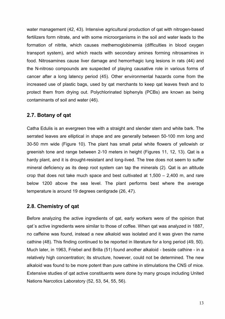
2.7. Botany of qat
Catha Edulis is an evergreen tree with a straight and slender stem and white bark. The
serrated leaves are elliptical in shape and are generally between 50-100 mm long and
30-50 mm wide (Figure 10). The plant has small petal white flowers of yellowish or
greenish tone and range between 2-10 meters in height (Figures 11, 12, 13). Qat is a
hardy plant, and it is drought-resistant and long-lived. The tree does not seem to suffer
mineral deficiency as its deep root system can tap the minerals (2). Qat is an altitude
crop that does not take much space and best cultivated at 1,500 – 2,400 m, and rare
below 1200 above the sea level. The plant performs best where the average
temperature is around 19 degrees centigrade (26, 47).
2.8. Chemistry of qat
Before analyzing the active ingredients of qat, early workers were of the opinion that
qat´s active ingredients were similar to those of coffee. When qat was analyzed in 1887,
no caffeine was found, instead a new alkaloid was isolated and it was given the name
cathine (48). This finding continued to be reported in literature for a long period (49, 50).
Much later, in 1963, Friebel and Brilla (51) found another alkaloid - beside cathine - in a
relatively high concentration; its structure, however, could not be determined. The new
alkaloid was found to be more potent than pure cathine in stimulations the CNS of mice.
Extensive studies of qat active constituents were done by many groups including United
Nations Narcotics Laboratory (52, 53, 54, 55, 56).

14
They reported that the phenylalkylamine fraction of the qat alkaloids contained another
compound in addition to cathine which was called cathinone later. The United Nation
Narcotics Laboratory Reports no. 8 and 9 concluded that cathinone is a biosynthetic
precursor that accumulates in young leaves while in adult leaves it undergoes
enzymatic reduction to the less active compounds cathine and norephedrine. This
finding supports the fact that qat users prefer the young leaves from the tips of the
branches suggesting high concentration and stimulating effect of cathinone (55). The
amount of alkaloids in 40 types of different fresh Yemeni qat was evaluated (57) and
results showed that the concentration of the total cathinone and cathin ranged between
78 to 342 mg out of 100 grams of fresh qat obtained from each type. The amount of
cathinone in four types of fresh qat was recently investigated (58). The amount found
was 343 mg in Nihmi, 323 mg in Sawti, 127 mg in Dholah and 122 mg in Qatabi out of
100 mg of fresh leaves from each type.
Qat also contains a group of alkaloids called cathedulins, so far eleven alkaloids of this
type have been isolated and characterized (58). Flavanoids are another important
ingredient of qat exhibiting a high content of tannins. Tannins are a group of phenolic
compounds and their concentrations vary depending on the type of qat (5). The
concentration of tannins reported by Alles and co-workers ranges between 5.58% and
7.4% (5). Recently, Revri reported a concentration of tannins between 6-11% in five
types of qat (47).
Variations in the tannin content of qat are primarily due to environmental differences
rather than to differences between cultivars. Amount of tannins in qat leaves was shown
to be affected by the amounts of calcium in the leaves, amounts of nitrogen in soil and
the altitude, i. e. the higher the percentage of calcium the lower the tannins´ content. At
the same time the higher the nitrogen content in the soil, the higher the tannins´ content.
Qat also contains a significant percentage of vitamin C (5, 6). In addition to that 17
types of amino acids were isolated from fresh qat leaves.
2.9. Pharmacology of qat
Cathinone which is the main constituent of qat is rapidly absorbed after oral
administration compared to cathine, which is absorbed slowly (9). Cathinone is highly
lipid soluble; this characteristic facilitates its access into the central nervous system (9).
The maximum effects of cathinone were found after 15-30 minutes from its oral
15
administration (59). Cathinone is metabolized in the liver into norephedrine and is
excreted almost exclusively in this form; only about 2% of cathinone absorbed appears
unchanged in the urine (60). The rapid rate of inactivation of cathinone is almost the
same as the rate of absorption during chewing. That is why the level of cathinone in the
blood is limited during qat chewing (9). The sympathomimetic effects of qat are due to
the concurrent action of cathinone and of cathine, whereas its central nervous system
effects are almost entirely due to cathinone. Indeed, cathinone is more potent than
cathine with regard to central nervous stimulation and more lipophilic than cathine (9).
Cathinone is a potent amphetamine-like substance, which shares the pharmacological
profile and features of amphetamine with regard to their central nervous system effects
(61, 62). It is important to recall that amphetamine is an indirectly acting adrenergic
drug; it causes the release of catecholamines in the central nervous system and
prolongs their action (63). Actually, only very few differences between cathinone and
amphetamine are found. The development of tolerance with cathinone is much more
unlikely pronounced in comparison with amphetamine (64). The central nervous system
effects of qat usually do not develop any tolerance (8), except for cases of insomnia
(65) and anorexia (66).
A WHO expert group on drug dependence (67) has extensively revised the previously
reported dependence induced by cathinone. They concluded that cathinone induced
neither physical dependence nor addiction (65, 67, and 68).
2.10. Systemic effects of qat
2.10.1. Effects of qat on CNS
The popularity of qat chewing is mainly due to its major active ingredient cathinone (9).
Its effects in the human body appear within a period of 2-4 hours on average of qat
chewing (11). Qat provides psycho-stimulant effects in the form of moderate euphoria
and mild excitement (7, 69). Also it increases the alertness and energy and produces an
enhanced depth of perception (70, 71); these psycho-stimulant effects are progressively
replaced in many chewers by mild dysphoria, anxiety, reactive depression, insomnia
and anorexia (72, 73). Sometimes chewers try to overcome insomnia with sedatives or
alcohol (7, 8). The simultaneous and excessive effects of smoking may also influence
the symptoms produced by qat chewing (7, 8).
16
2.10.2. Effects of qat on cardiovascular system
The sympathomimetic effect of cathinone and cathine leads to a significant rise of
arterial systolic and diastolic blood pressure and pulse rate (73, 74). The peak effect on
the arterial blood pressure and pulse rate is reached 3 hours after starting to chew,
followed by a decline 1 hour after spitting the leaves out. These changes run parallel
with changes in plasma cathinone levels during and after qat chewing (75). The
cathinone responsibility for the increase in arterial blood pressure and pulse rate during
qat chewing is supported by Brenneisen et al. who observed a similar blood pressure
increase in subjects who have taken a pure cathinone in gelatine capsules orally (60,
76). A study conducted by Hassan et al. revealed that the increase of blood pressure is
due to the stimulant effect of cathinone on beta-one adrenoceptor in the heart (77). Qat
chewing may be a potential cardiovascular risk factor in patients with hypertension and
heart diseases, and may precipitate the occurrence of cerebrovascular accidents and
myocardial infarction in susceptible individuals (78). The aforementioned is supported
by AL-Motarreb who carried out a study on guinea pigs. He found that cathinone can
induce vasoconstriction of the coronary vascular bed (79).
2.10.3. Effects of qat on digestive system
Clinical observation found that qat chewers often complain of symptoms suggestive of
oesophagitis and gastritis. The strongly astringent tannins in qat were blamed as a
causative substance of the digestive disorders (13, 80). Other studies concluded that
the sympathomimetic action of cathinone did indeed delay gastric emptying of a semi-
solid meal (72, 80). Anorexia was noted after qat chewing (72) and this significant
reduction of appetite after qat chewing may be attributed to combined direct central and
gastric effects of cathinone (72). Common complaint among qat chewers is
constipation, which is probably due the astringent properties of the qat tannins (9).
Habitual users try to attenuate this undesirable effect by laxatives or by eating a meal
with high fat content prior to the qat session in order to facilitate intestinal transit (8).
Additionally reports state that chewing qat leaves significantly slows both the orocaecal
transit time (81) and the whole gut transit time (82). The liver has been suspected to be
particularly vulnerable to the harmful effects of qat use (8, 65) and a disturbance in liver
function and architecture has been described in experimental animals both on short-
term (83) and long-term studies (84).
17
2.10.4. Effects of qat on genitourinary system
One of the obvious side-effects of chewing qat leaves in males is temporary difficult
micturition with hesitancy and poor flow. Overall urine flow rates were recently found to
be significantly lower in qat users (85). This effect is probably mediated through
stimulation of beta-one adrenoceptors in the bladder neck by the sympathomimetic
effects of cathinone. These effects were abolished by the beta-one adrenoceptor
blocker indoramin. The consumption of qat is found to induce an increase in libido,
spermatorrhoea and erectie dysfunction (70).
2.10.5. Effects of qat on the health of pregnant women and foetus
In the domain of reproductive health, epidemiological data derived from 1181 deliveries
in Yemen showed that at birth the mean weight of full-term singleton infants from
mothers who chewed qat habitually or occasionally was below average (86). A study on
pregnancy outcome and qat showed a significantly increased incidence of low-birth-
weight full-term infants among the offspring of women who chewed qat during
pregnancy in comparison to those who were non-chewers during pregnancy (87).
Recent evidence has indicated that neonates of mothers who chewed qat during
pregnancy had a significant decrease in all neonatal parameters such as birth weight,
length, and head circumference in comparison with those of mothers who did not chew
qat during pregnancy (88). This effect was found to increase in severity with the
increased frequency and duration of qat chewing during pregnancy. An experimental
study in rats has recently proved that qat can affect intrauterine fetal growth by reducing
total fetal fat and weight and by inducing some changes in the chemical composition of
fetal organs, particularly the liver, heart and kidneys (89). The author attributed that
effect to depletion of carbohydrate material and suppression of DNA and protein
synthesis in the fetal organs. Nursing mothers in Yemen frequently complain of poor
lactation. Some authors believe that this phenomenon may be related to the use of qat
as cathine in qat may inhibit prolactin secretion (65). Interestingly, it has been found that
the breast milk of qat-chewing mothers contains cathine, and this compound could even
be detected in the urine of one breastfed infant (90).
18
2.11. Effect of qat chewing on oral tissue
2.11.1. Effects of qat chewing on periodontal tissues
Earlier studies on periodontal condition among different ethnic groups in Israel found a
high rate of periodontal disease among Yemeni emigrants due to qat chewing (91).
Long term qat chewing was reported to cause stomatitis followed by secondary infection
which might be related to mechanical trauma and chemical content of qat (8). A high
rate of periodontal disease has been observed among Yemeni male qat chewers (65).
On the other hand, significantly deeper periodontal pockets were reported on the
opposite side of chewing compared to chewing sites and were claimed to have
beneficial effects of qat chewing on periodontal tissues (14). Moreover, in Kenya, no
significant differences were found in periodontal health among 131 mirra (mirra is the
name of qat in Kenya) chewers and 199 non mirra chewers. Additionally, significantly
lower lingual plaque and gingivitis scores among mirra chewers than non mirra chewers
were reported (92). The authors concluded that no evidence was found indicating that
chewing is detrimental to periodontal health. The community periodontal index of
treatment needs, clinical attachment loss and calculus index were higher among 1001
Yemeni qat chewers than non chewers. Differences were significant for the 12-24 years
age group while insignificant for those in the 35-44 years age group (93). The same
study also showed that scores of attachment loss were lower among chewers than
scores among non chewers. Recently, a cross sectional hospital study among Yemeni
qat chewers and non chewers revealed an increased risk for a number of periodontal
lesions (12). The study revealed that qat chewing causes many lesions to the
supporting structure of the teeth, namely gingivitis, periodontal pocket formation,
gingival recession, tooth mobility and finally tooth mortality.
2.11.2. Effects of qat chewing on hard tissues of the teeth
Effects of qat chewing habit on the hard tissues of teeth have been reported by many
researchers. The earliest investigation reported that a low rate of dental caries was
observed among Yemeni qat chewers (65). Discolored and missing teeth were reported
among Yemeni qat chewers (8). Low caries rate and universal attrition was reported
among 115 qat chewers examined. The prevalence of dental caries was less than 2% of
all teeth examined. The low prevalence of dental caries among qat chewers was
attributed either to the high contents of fluoride in water or to the amount of fluoride
19
available in qat leaves (14). A recent investigation found no association between
conventional (occlusal) dental caries scores and qat chewing among 325 Yemeni qat
chewers (12). The same study reported a strong association between qat damaging
effects to dental tissues in the forms of attrition, staining and cervical caries. Attrition
and staining were found among 52.6% and 82.8% of 325 qat chewers, respectively.
Cervical caries was found among all chewers consuming crystallized sugar during
chewing (12). Certain types of qat are bitter in taste, therefore many chewers use sweet
drinks or crystallized sugar to compensate the taste during chewing (2, 12, 65).
2.11.3. Effects of qat chewing on temporo-mandibular-joint (TMJ) and facial
tissues
The effects of qat chewing on TMJ pain, clicking and facial asymmetry had been
investigated. Early reports found TMJ-related pain among 40% of 115 qat chewers
examined (14). TMJ related clicking and pain and facial asymmetry among qat chewers
were studied extensively (12). TMJ-related clicking was found among 109 subjects, of
them 27 (24.8%) suffered from pain. Clicking was attributed to the overloading of the
condyle surface due to chewing forces. Obvious facial asymmetry was present among
75.4% of the qat chewers examined which was correlated positively with long duration
of the qat chewing habit (12). This could be attributed to muscle hypertrophy due
functional demand.
2.11.4. Effects of qat chewing on salivary glands
The effects of qat chewing on the dryness of the mouth among chewers have been
documented in many reports (2, 9, 94). The authors substantiated that the aetiology of
xerostomia might be due to the sympathomimetic effects of cathinone or due to the
over-excretion of saliva from salivary glands during chewing. The effects of qat chewing
on saliva and salivary glands were extensively studied (12). The results showed strong
causal relationship between qat chewing and xerostomia, enlargements of major
salivary glands and inflammation and enlargements of parotid duct opening. No clear
effects of qat chewing on salivary viscosity was found, however, there was an obvious
effect on salivary flow after qat chewing, as 67.4% complained of xerostomia for many
hours after qat chewing. The enlargement of major salivary gland might be due to over-
exhaustion of continuous over-secretion for many hours per day for decades. The
20
inflammation found on parotid opening at the site of chewing was probably due to long-
term friction of qat fibres. The folded papilla might be a defence mechanism to prevent
qat particles from entering the duct in similar mechanisms observed in pipers (12).
2.12.1. Histopathological studies
Before histopathological studies were performed of the oral mucosa, information was
obtained from histopathological studies of the upper gastrointestinal tract among qat
chewers. In this study, regular daily qat chewing was not associated with a significant
effect on the esophagus (95). Additionally, mild abnormal growth of gastric mucosal
cells (dysplasia) and abnormal intestinal cells (metaplasia) at the lower esophagus were
higher in qat chewers than non chewers (96). Buccal and gingival mucosa at sites of qat
chewing among Yemeni chewers had been extensively studied histopathologically in
the past 6 years (12, 97, 98). The earliest investigation was done on 42 biopsies divided
into 3 groups. Group 1 included 30 biopsies from sites of chewing (13 were chewers
and cigarette smokers and 17 were chewers and non-smokers). Group 2 included 7
biopsies taken from the non-chewing sites of qat chewers (5 were chewers and
smokers and 2 were chewers and non-smokers). The results revealed that
histopathological changes found on oral mucosa included increased rete ridges,
acanthosis, intercellular edema, orthokeratosis, parakeratosis, epithelial dysplasia,
inflammatory cell infiltrate and increased amount of collagen fibers (12). All results were
statistically significant except for results of epithelial dysplasia and inflammatory cell
infiltrates. Also the distribution of histopathological cases among smokers and non
smokers were statistically not significant (12). Another investigation was done on 40
biopsies divided equally into two groups taken from gingival and buccal mucosa of
chewing and opposite sides (97). Results showed apparent histopathological
differences between the 2 groups and all alterations were similar to those reported
earlier (12). An investigation was done recently to further study different
histopathological effects on oral mucosa among qat chewers due to qat chewing (98).
Results similar to previous those of studies were reported. The authors concluded that
qat chewing caused innocuous histopathological changes at site of chewing without any
malignancy (97, 98).
21
2.12.2. Effects of qat chewing on oral mucosa and oral cancer
Effects of qat chewing on oral mucosa and oral tumours have been investigated by
many authors. Tumours of the oral cavity (including lower maxilla, buccal mucosa and
lateral border of the tongue) were reported in n=74 (0.13%) out of 5757 patients seeking
treatment in a stomatology clinic in Hodeidah, Yemen, over a two years period (17). The
majority of cases were men over 40 years of age who had chewed qat and or used
shammah (snuff) for 20 years or more. No oral cancer has been reported from
comprehensive evaluation of 706 Yemeni qat chewers (335 female and 371 male) in the
age range 15-60 years (13). Mucosal changes or some degrees of oral keratosis were
found at the site of chewing among 50% of qat chewers. However, no cancerous lesion
was diagnosed (14). Occurrence of oral cancer in a study conducted over a two year
period in Asir region was reported (18). Out of 28 head and neck cancers, 10 cases (7
males and 3 females, age ranged 25-40 years) were diagnosed in the mouth mostly in
the anterior two thirds of the tongue and floor of the mouth. No information of shammah
use was provided despite fact that the inhabitants in the area of the study are known to
be shammah dippers. One case of oral verrucous carcinoma with a history of tobacco
chewing, snuff dipping and qat chewing was reported (19). In Kenya, the association
between leukoplakia, smoking, alcohol consumption and qat chewing has been studied
(15). Results showed that qat chewing was not significantly associated with leukoplakia.
17 cases of head and neck carcinoma were diagnosed during a one year period among
chronic qat chewers (99). Of these cases, 10 were also snuff dippers and 5 others were
smokers. In a cross sectional hospital-based study, 431 subjects (325 chewers and 106
non chewers) were recruited. All those 325 subjects who chewed qat for not less than 3
years demonstrated buccal and gingival white lesions. 100% and 90% of mucosal
changes were on buccal and gingival mucosa, respectively (12), no dysplasia or cancer
was reported. Another study reported 342 mild keratotic white lesions among Yemeni
qat chewers (16). Oral white lesions were reported among 47 Yemeni Israeli Jews
above the age of 30 years with duration of qat chewing of more than 3 years. According
to this study, white lesions due to qat chewing were seen at the site of chewing with no
atypical epithelial cells observed. No suggestive premalignant or malignant changes
were identified in the subset of patients (100).
Recently, a study done in vitro to see the effects of qat extracts on oral keratinocytes
and fibroblasts showed that qat extracts inhibited the proliferation of oral keratinocytes
22
and increased expression of stress-sensitive p53 protein and p16 protein after 24 hours.
On fibroblast qat extracts also inhibited the proliferation and increased expression of
p21 protein after 24 hours (101).
There is not enough evidence in the literature that qat chewing alone is carcinogenic or
that it plays an independent direct role in the development of head and neck cancers.
Unfortunately, the frequent combined use of qat and tobacco products makes it difficult
to isolate the contribution each makes to the risk of developing oral cancer. Although
the exact mechanism of any reported carcinogenesis is unknown, available information
suggests that qat chewing should be considered one of the possible confounding risk
factors for oral cancer specific to individuals who practise qat chewing along with
tobacco use (102).
23
3. MATERIAL AND METHODS
3.1. Materials
The aim of this study was to use quantitative epidemiological methods to assess and
document the clinical characteristics for oral white lesions among Yemeni female qat
chewers. So far, these lesions were observed mainly among male chewers and were
hypothesized to be induced by qat chewing.
The design of the study was hospital-based cross sectional. Qat chewing was the
exposure and the white lesions produced considered the outcome or the disease status.
The epidemiological approach was a retrospective cohort, as the exposure was well
ascertained because the qat chewing habit is widespread and socially accepted.
Therefore, the analysis was performed from the exposure to the disease status. The
exposure was ascertained simultaneously with the disease status. The analysis
therefore emphasized the exposure and then the outcome, the disease status. It was
then found appropriate to consider a retrospective cohort analysis.
Subjects of this study were female patients who presented to dental clinics of Al-Thawra
health institution in Sana'a city for dental treatment during the period 2006-2008. Sana'a
is the capital of Yemen which has a population of more than 2 million representing most
ethnic and social strata of Yemenis.
Participation of subjects in the study was according to the following criteria:
Criteria of selection:
• Adult Yemeni healthy qat chewing females aged 20-65 years.
• Continuous qat chewers for at least 5 years.
• Chewers on one side of their mouths only.
Exclusion criteria:
• Bilateral chewers (chewers in both sides of their mouths).
• Shammah users.
• Poor general or mental health.
24
Participants were subjected to a detailed clinical examination of oral mucosal tissues at
chewing sites and opposite sides:
• The chewing sites among female qat chewers were dealt with as cases of the
study.
• The contralateral side of female qat chewers were dealt with as control 1.
• Both sides of female non-qat chewers were dealt with as control 2.
3.2. Methods
Procedure of investigation: The procedure of the investigation started as patients arrived at the dental clinic of the
Althawra Health Institute. In the dental clinic, female patients reported their histories and
main complaints. Before examining the patients, the investigator (JS) introduced himself
and briefly talked with each female patient to obtain her consent and also to assess her
suitability to be selected for the study. If the female patient was found to be an eligible
candidate according to the inclusion criteria, the study commenced using the short
interview questionnaire and the clinical examination sheet (appendix).
Materials of interview:
The interview questionnaire included following information:
• Personal data (name, age, residence and address).
• Information on qat (frequency of habit / week, intraoral site of chewing, age of
starting chewing, duration of the habit /years, average period of each session
/hours).
• Information on cigarettes (cigarettes smoking situation, duration of smoking /
years and frequency of smoking /day).
• Information on water-pipe (water-pipe smoking situation, duration of water-pipe
smoking / years and frequency of water–pipe smoking /day).
• Oral hygiene status (OHS) (use of tooth brush or other oral hygiene aids,
frequency of tooth brushing).
25
3.3. Clinical examination
Participants were subjected to a detailed clinical oral examination including oral hygiene
status and oral mucosa at the site of chewing and at the opposite side.
Oral hygiene status examination included the state of oral hygiene, whether it was good,
fair or poor by assessing the amount of dental plaque collected on teeth. Each
participant was asked if she was a toothbrush user or not, if yes, what type of device
she used for mouth cleaning and how many times a day.
Examination of the oral mucosa started by inspecting buccal mucosa on the internal
surface of the cheek, inspecting buccal mucosa and its continuation or extensions up to
the upper vestibular mucosa and down to lower buccal and lower vestibular mucosa.
Buccal mucosa was reflected sufficiently and inspected carefully for any mucosal
changes in colour and texture on both sides of the mouth.
Gingival mucosa and its extension to alveolar mucosa were also inspected carefully for
any mucosal changes in colour and texture on both sides of the mouth.
White lesion (leukoplakia) was defined as lesion at the site of qat use that could not be
removed and was not clinically suspected as any other white lesion. It was scaled using
the previous scale of white lesions due to qat chewing (16).
Inspection also included the presence of any other mucosal changes or lesions such as
leukoedema, frictional keratosis at the site of chewing and the opposite side.
Careful bimanual palpation of all mucosal surfaces was performed to examine the
underlying structure and to find out if the patient felt any pain. Clinical examination was
carried out using two disposable dental mirrors with the patient lying on a dental chair
using artificial light.
Ethical considerations:
Aim of the study was explained to each subject and only patients who consented
voluntarily to participate were included in this study. Following the clinical examination
all patients were treated free of charge.
26
3.4. Statistical analysis
Data from the questionnaires and case sheets were processed using a statistical
software package (SPSS version 15). The level of significance was set at p< 0.05.
Univariate comparisons between patient and controls were made for the potential risk
factors using the chi square test and OR and their 95 % CI for the nominal and
categorical variables.
A multinomial logistic regression model was used to identify the significant association
of the difference potential risk factors as independent variables with the presence of
white lesion as the dependent variable.
27
4. RESULTS
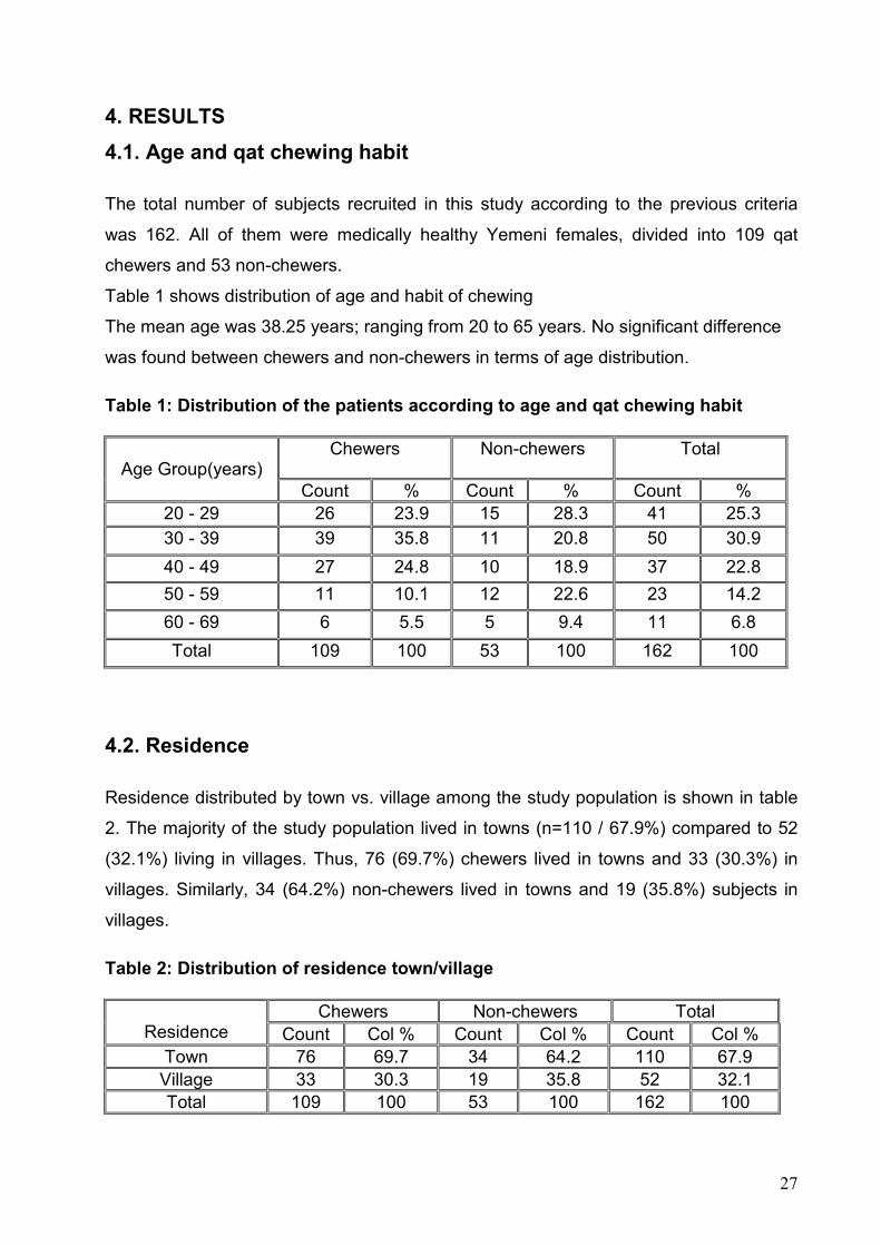
4.1. Age and qat chewing habit
The total number of subjects recruited in this study according to the previous criteria
was 162. All of them were medically healthy Yemeni females, divided into 109 qat
chewers and 53 non-chewers.
Table 1 shows distribution of age and habit of chewing
The mean age was 38.25 years; ranging from 20 to 65 years. No significant difference
was found between chewers and non-chewers in terms of age distribution.
Table 1: Distribution of the patients according to age and qat chewing habit
Chewers Non-chewers Total Age Group(years)
Count % Count % Count % 20 - 29 26 23.9 15 28.3 41 25.3
30 - 39 39 35.8 11 20.8 50 30.9
40 - 49 27 24.8 10 18.9 37 22.8
50 - 59 11 10.1 12 22.6 23 14.2
60 - 69 6 5.5 5 9.4 11 6.8
Total 109 100 53 100 162 100
4.2. Residence
Residence distributed by town vs. village among the study population is shown in table
2. The majority of the study population lived in towns (n=110 / 67.9%) compared to 52
(32.1%) living in villages. Thus, 76 (69.7%) chewers lived in towns and 33 (30.3%) in
villages. Similarly, 34 (64.2%) non-chewers lived in towns and 19 (35.8%) subjects in
villages.
Table 2: Distribution of residence town/village
Chewers Non-chewers Total Residence Count Col % Count Col % Count Col %
Town 76 69.7 34 64.2 110 67.9 Village 33 30.3 19 35.8 52 32.1 Total 109 100 53 100 162 100
28
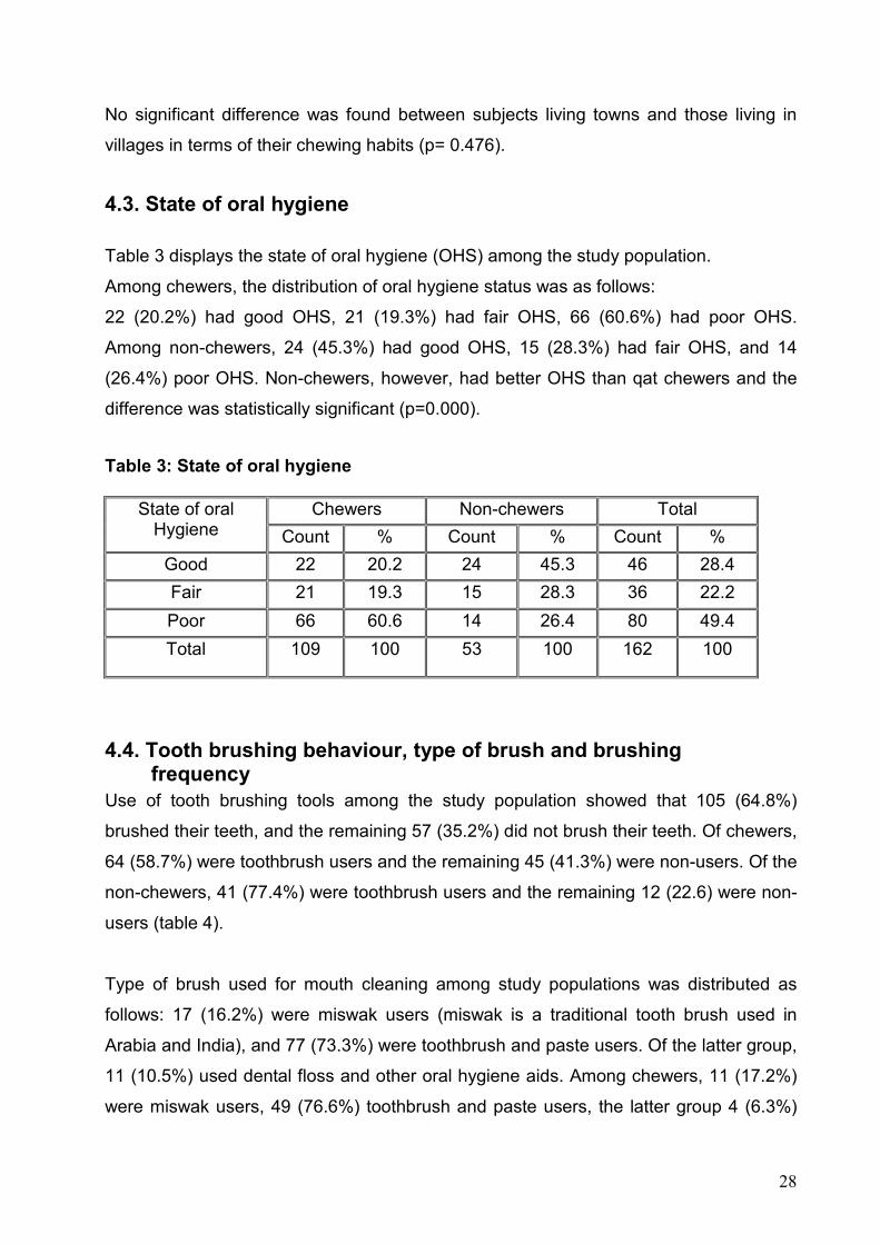
No significant difference was found between subjects living towns and those living in
villages in terms of their chewing habits (p= 0.476).
4.3. State of oral hygiene
Table 3 displays the state of oral hygiene (OHS) among the study population.
Among chewers, the distribution of oral hygiene status was as follows:
22 (20.2%) had good OHS, 21 (19.3%) had fair OHS, 66 (60.6%) had poor OHS.
Among non-chewers, 24 (45.3%) had good OHS, 15 (28.3%) had fair OHS, and 14
(26.4%) poor OHS. Non-chewers, however, had better OHS than qat chewers and the
difference was statistically significant (p=0.000).
Table 3: State of oral hygiene
Chewers Non-chewers Total State of oral Hygiene Count % Count % Count %
Good 22 20.2 24 45.3 46 28.4
Fair 21 19.3 15 28.3 36 22.2
Poor 66 60.6 14 26.4 80 49.4
Total 109 100 53 100 162 100
4.4. Tooth brushing behaviour, type of brush and brushing frequency
Use of tooth brushing tools among the study population showed that 105 (64.8%)
brushed their teeth, and the remaining 57 (35.2%) did not brush their teeth. Of chewers,
64 (58.7%) were toothbrush users and the remaining 45 (41.3%) were non-users. Of the
non-chewers, 41 (77.4%) were toothbrush users and the remaining 12 (22.6) were non-
users (table 4).
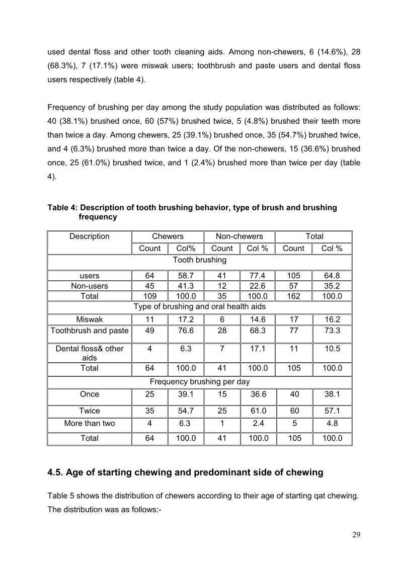
Type of brush used for mouth cleaning among study populations was distributed as
follows: 17 (16.2%) were miswak users (miswak is a traditional tooth brush used in
Arabia and India), and 77 (73.3%) were toothbrush and paste users. Of the latter group,
11 (10.5%) used dental floss and other oral hygiene aids. Among chewers, 11 (17.2%)
were miswak users, 49 (76.6%) toothbrush and paste users, the latter group 4 (6.3%)
29
used dental floss and other tooth cleaning aids. Among non-chewers, 6 (14.6%), 28
(68.3%), 7 (17.1%) were miswak users; toothbrush and paste users and dental floss
users respectively (table 4).
Frequency of brushing per day among the study population was distributed as follows:
40 (38.1%) brushed once, 60 (57%) brushed twice, 5 (4.8%) brushed their teeth more
than twice a day. Among chewers, 25 (39.1%) brushed once, 35 (54.7%) brushed twice,
and 4 (6.3%) brushed more than twice a day. Of the non-chewers, 15 (36.6%) brushed
once, 25 (61.0%) brushed twice, and 1 (2.4%) brushed more than twice per day (table
4).
Table 4: Description of tooth brushing behavior, type of brush and brushing frequency
Chewers Non-chewers Total Description
Count Col% Count Col % Count Col %
Tooth brushing
users 64 58.7 41 77.4 105 64.8 Non-users 45 41.3 12 22.6 57 35.2
Total 109 100.0 35 100.0 162 100.0 Type of brushing and oral health aids
Miswak 11 17.2 6 14.6 17 16.2
Toothbrush and paste 49 76.6 28 68.3 77 73.3
Dental floss& other aids
4 6.3 7 17.1 11 10.5
Total 64 100.0 41 100.0 105 100.0
Frequency brushing per day
Once 25 39.1 15 36.6 40 38.1
Twice 35 54.7 25 61.0 60 57.1
More than two 4 6.3 1 2.4 5 4.8
Total 64 100.0 41 100.0 105 100.0
4.5. Age of starting chewing and predominant side of chewing
Table 5 shows the distribution of chewers according to their age of starting qat chewing.
The distribution was as follows:-
30
34 (31.2%) started chewing at 10-20 years of age,
52 (47.7%) started chewing at 20-30 years of age
23 (21.1%) started chewing at 30-40 years of age
Table 5 also shows the intraoral side which was mainly used to chew.
The distribution was as follow:
16 (14.7%) chewers chew on their right side
93 (85.3%) chewers chew on their left side.
Table 5: Distribution of chewers according to their age of starting chewing and predominant mouth side of chewing
Distribution Count % Age of starting qat chewing (years)
10 - 20 34 31.2 20 - 30 52 47.7 30 - 40 23 21.1 Total 109 100
Oral side of chewing Right 16 14.7 Left 93 85.3
Total 109 100
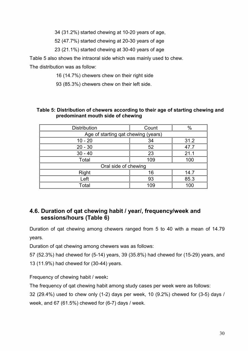
4.6. Duration of qat chewing habit / year/, frequency/week and sessions/hours (Table 6)
Duration of qat chewing among chewers ranged from 5 to 40 with a mean of 14.79
years.
Duration of qat chewing among chewers was as follows:
57 (52.3%) had chewed for (5-14) years, 39 (35.8%) had chewed for (15-29) years, and
13 (11.9%) had chewed for (30-44) years.
Frequency of chewing habit / week:
The frequency of qat chewing habit among study cases per week were as follows:
32 (29.4%) used to chew only (1-2) days per week, 10 (9.2%) chewed for (3-5) days /
week, and 67 (61.5%) chewed for (6-7) days / week.
31
Average period of qat session /hours:
The average periods of each qat chewing session / hours were as follows:
29 (26.6%) chewed qat on average for (1-2) hours / day, 55 (50.5%) chewed qat on
average for (3-5) hours / day, 25 (22.9%) chewed qat on average for (>6) hours / day.
Table 6: Distribution of qat chewers according to duration/years, frequency of habit per week, average time/hours
Distribution Count % Duration of the habit in years
5 - 14 57 52.3 15 - 29 39 35.8 30 - 44 13 11.9 Total 109 100
Frequency of the per week 1 – 2 32 29.4 3 – 5 10 9.2 6 – 7 67 61.5 Total 109 100
Period of each session in hours 1 – 2 29 26.6 3 – 5 55 50.5 > 6 25 22.9
Total 109 100
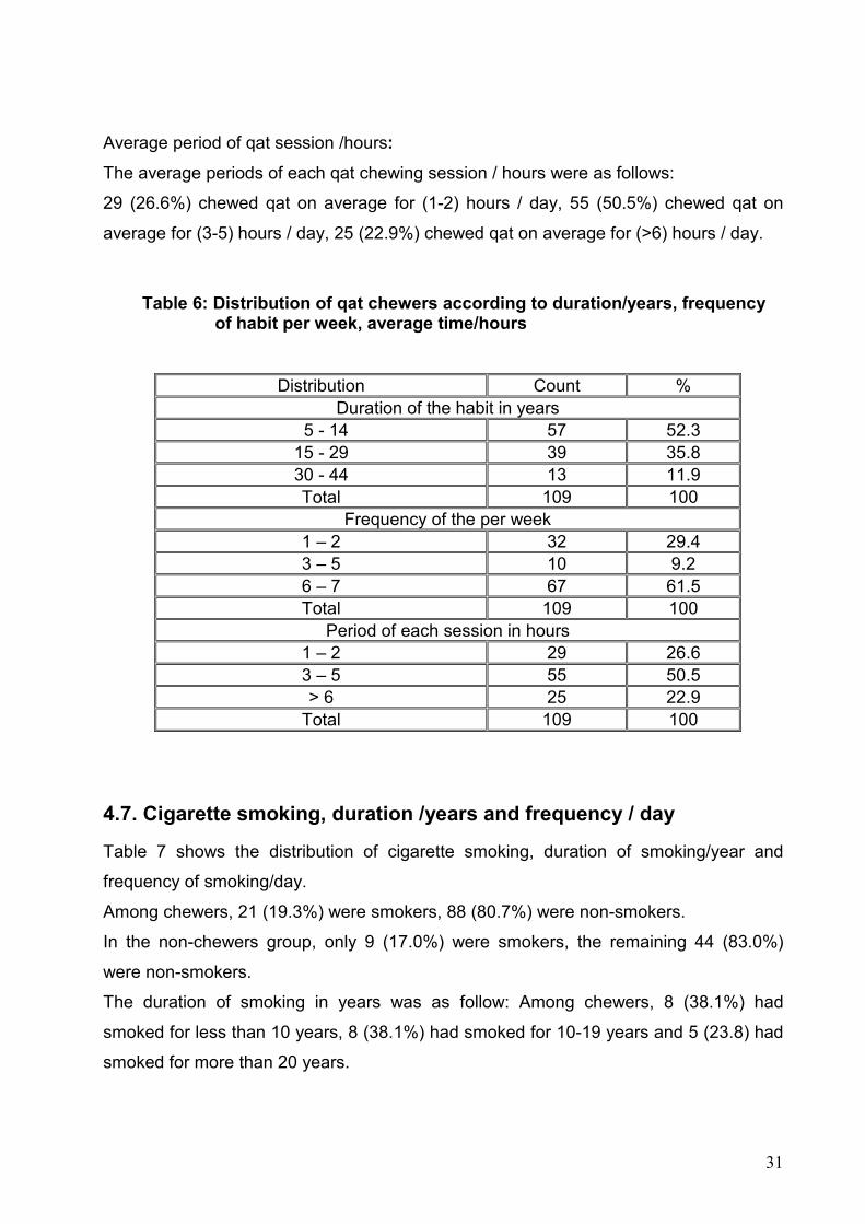
4.7. Cigarette smoking, duration /years and frequency / day
Table 7 shows the distribution of cigarette smoking, duration of smoking/year and
frequency of smoking/day.
Among chewers, 21 (19.3%) were smokers, 88 (80.7%) were non-smokers.
In the non-chewers group, only 9 (17.0%) were smokers, the remaining 44 (83.0%)
were non-smokers.
The duration of smoking in years was as follow: Among chewers, 8 (38.1%) had
smoked for less than 10 years, 8 (38.1%) had smoked for 10-19 years and 5 (23.8) had
smoked for more than 20 years.
32
Among non-chewers, 4 (44.4%) had smoked for less than 10 years, 4 (44.4%) for 10-19
years and 1 (11.1%) for more than 20 years.
The number of cigarettes per day was as follows: Among chewers, 9 (42.9%) had
smoked less than 10 cigarettes per day, 8 (38.1%) less than 20 cigarettes and 4
(19.0%) more than 20 cigarettes per day. Non-chewers were 7 (77.8%) who had
smoked less than 10 cigarettes per day and 2 (22.2%) more than 10 and less than 20
cigarettes per day.
Table 7: Distribution of cigarette smoking, duration/year and number of cigarettes/day
Chewers Non-chewers Total Description Count % Count %
Count %
Smoking habit. Smokers 21 19.3 9 17.0 30 18.5
Non-smokers 88 80.7 44 83.0 132 81.5 Total 109 100 53 100 162 100
Duration of smoking in years. < 10 years 8 38.1 4 44.4 12 40.0
10 – 19 years 8 38.1 4 44.4 12 40.0
> 20 years 5 23.8 1 11.1 6 20.0 Total 21 100 9 100 30 100
Number of cigarette per day. < 10 cigarette
daily 9 42.9 7 77.8 16 53.8
10-20 cigarettes daily
8 38.1 2 22.2 10 33.3
>20 cigarette daily
4 19.0 -- -- 4 13.3
Total 21 100 9 100 30 100
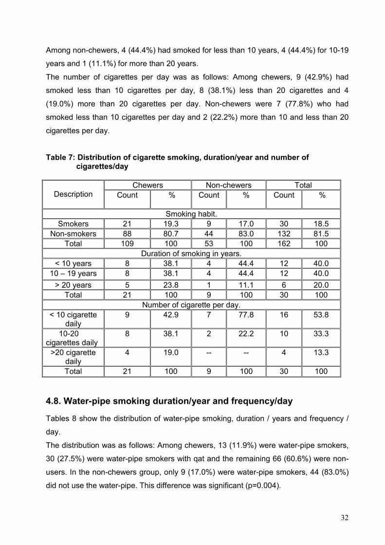
4.8. Water-pipe smoking duration/year and frequency/day
Tables 8 show the distribution of water-pipe smoking, duration / years and frequency /
day.
The distribution was as follows: Among chewers, 13 (11.9%) were water-pipe smokers,
30 (27.5%) were water-pipe smokers with qat and the remaining 66 (60.6%) were non-
users. In the non-chewers group, only 9 (17.0%) were water-pipe smokers, 44 (83.0%)
did not use the water-pipe. This difference was significant (p=0.004).
33
The duration of the water-pipe smoking habit showed that among chewers, 14 (32.6%)
had smoked for less than 10 years, 10 (23.3%) for 10-19 years and the remaining 19
(44.2%) had smoked for more than 20 years.
The frequency of water-pipe smoking per day showed that 22 (51.2%) had smoked only
once a day, 13 (30.2%) had smoked twice and the remaining 8 (18.6%) had smoked
more than two times per day.
Among non-chewers were 6 (66.7%) who had smoked only once a day and 3 (33.3%)
twice a day.
Table 8: Distribution of water-pipe smoking, duration/years and frequency/day
Chewers Non-chewers Total Description
Count % Count % Count %
Water-pipe Smokers Users 13 11.9 9 17.0 22 13.6
Non-users 66 60.6 44 83.0 110 67.9 With Qat only 30 27.5 -- -- 30 18.5
Total 109 100 53 100 162 100 Duration of water-pipe smoking
< 10 years 14 32.6 4 44.4 18 34.6
10 – 19 years 10 23.3 4 44.4 14 26.9 > 20 years 19 44.2 1 11.1 20 38.5
Total 43 100 9 100 52 100
Frequency per day Once 22 51.2 6 66.7 28 53.8 Twice 13 30.2 3 33.3 16 30.8
More than two 8 18.6 -- -- 8 15.4 Total 43 100 9 100 52 100
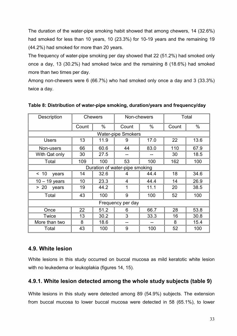
4.9. White lesion
White lesions in this study occurred on buccal mucosa as mild keratotic white lesion
with no leukedema or leukoplakia (figures 14, 15).
4.9.1. White lesion detected among the whole study subjects (table 9)
White lesions in this study were detected among 89 (54.9%) subjects. The extension
from buccal mucosa to lower buccal mucosa were detected in 58 (65.1%), to lower
34
vestibular mucosa in 36 (62.1%), and to upper vestibular mucosa in 3 (5.1%) of the
sides.
On gingival mucosa, white lesion were detected in 31 (34.8%), and extension to
alveolar mucosa was found in 18 (58.1%) of the sides (table 9).
Table 9: distribution of white lesions among study subjects
present absent Total Distribution of white lesion among study subjects count % count % Normal 73 45.1 89 54.9 162 White lesion 89 54.9 73 45.1 162 Lower buccal mucosa 58 65.2 31 34.8 89 Lower vestibular sulcus 36 62.1 22 37.9 58 Upper vestibular sulcus 3 5.1 55 94.8 58 Gingival mucosa 31 34.8 58 65.2 89 Alveolar mucosa 18 58.1 13 41.9 31
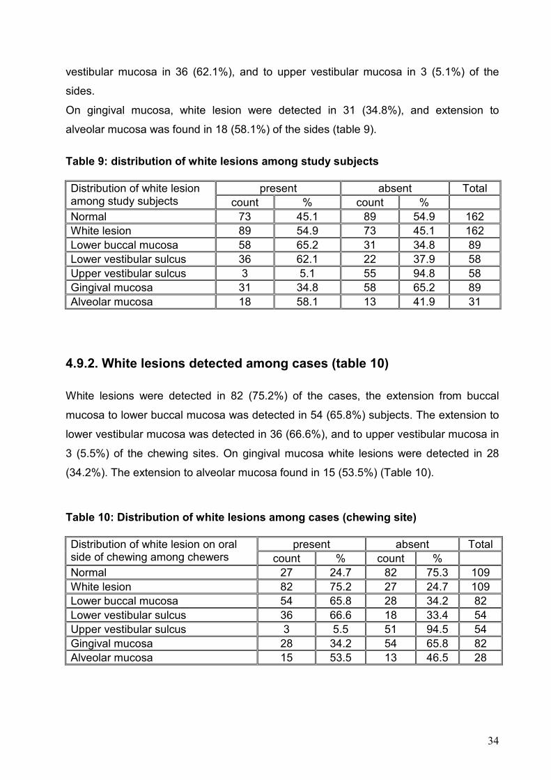
4.9.2. White lesions detected among cases (table 10)
White lesions were detected in 82 (75.2%) of the cases, the extension from buccal
mucosa to lower buccal mucosa was detected in 54 (65.8%) subjects. The extension to
lower vestibular mucosa was detected in 36 (66.6%), and to upper vestibular mucosa in
3 (5.5%) of the chewing sites. On gingival mucosa white lesions were detected in 28
(34.2%). The extension to alveolar mucosa found in 15 (53.5%) (Table 10).
Table 10: Distribution of white lesions among cases (chewing site)
present absent Total Distribution of white lesion on oral side of chewing among chewers count % count % Normal 27 24.7 82 75.3 109 White lesion 82 75.2 27 24.7 109 Lower buccal mucosa 54 65.8 28 34.2 82 Lower vestibular sulcus 36 66.6 18 33.4 54 Upper vestibular sulcus 3 5.5 51 94.5 54 Gingival mucosa 28 34.2 54 65.8 82 Alveolar mucosa 15 53.5 13 46.5 28
35
4.9.3. White lesions detected among control 1 (table 11)
White lesion among control 1 were detected in 6 (5.5%) subjects. The extension from
buccal mucosa to lower buccal mucosa was detected in 3 (50.0%), to lower vestibular
mucosa detected in 1 (33.4%), and to upper vestibular mucosa detected in 1 (33.4%) of
the opposite side. On gingival mucosa, white lesions were detected in 3 (50.0%)
subjects.
Table 11: Distribution of white lesion on the opposite side among chewers (Control 1)
present absent Total Distribution of white lesion on the opposite side among chewers count % count % Normal 103 94.4 6 5.5 109 White lesion 6 5.5 103 94.4 109 Lower buccal mucosa 3 50.0 3 50.0 6 Lower vestibular sulcus 1 33.4 2 66.4 3 Upper vestibular sulcus 1 33.4 2 66.4 3 Gingival mucosa 3 50.0 3 50.0 6 Alveolar mucosa 0 0 3 100 3
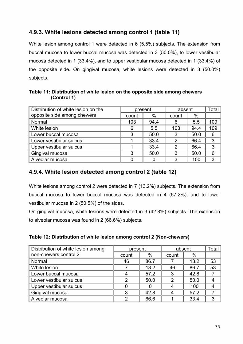
4.9.4. White lesion detected among control 2 (table 12)
White lesions among control 2 were detected in 7 (13.2%) subjects. The extension from
buccal mucosa to lower buccal mucosa was detected in 4 (57.2%), and to lower
vestibular mucosa in 2 (50.5%) of the sides.
On gingival mucosa, white lesions were detected in 3 (42.8%) subjects. The extension
to alveolar mucosa was found in 2 (66.6%) subjects.
Table 12: Distribution of white lesion among control 2 (Non-chewers)
present absent Total Distribution of white lesion among non-chewers control 2 count % count % Normal 46 86.7 7 13.2 53 White lesion 7 13.2 46 86.7 53 Lower buccal mucosa 4 57.2 3 42.8 7 Lower vestibular sulcus 2 50.0 2 50.0 4 Upper vestibular sulcus 0 0 4 100 4 Gingival mucosa 3 42.8 4 57.2 7 Alveolar mucosa 2 66.6 1 33.4 3
36
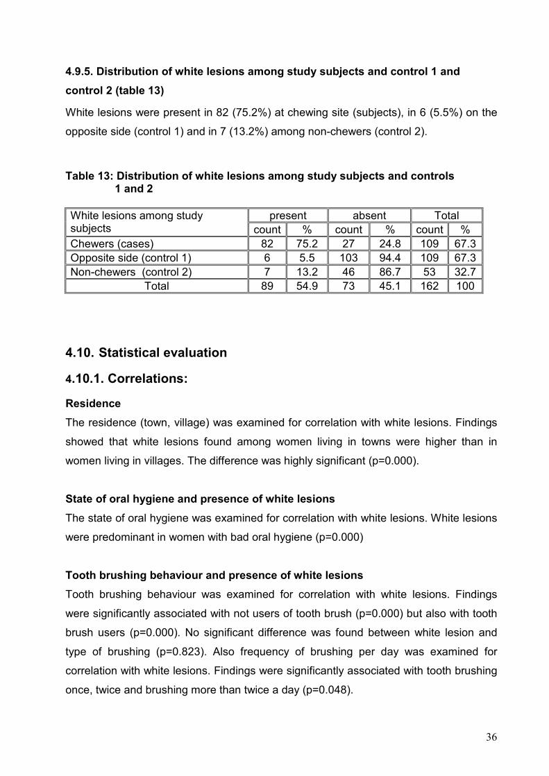
4.9.5. Distribution of white lesions among study subjects and control 1 and
control 2 (table 13)
White lesions were present in 82 (75.2%) at chewing site (subjects), in 6 (5.5%) on the
opposite side (control 1) and in 7 (13.2%) among non-chewers (control 2).
Table 13: Distribution of white lesions among study subjects and controls 1 and 2
present absent Total White lesions among study subjects count % count % count % Chewers (cases) 82 75.2 27 24.8 109 67.3 Opposite side (control 1) 6 5.5 103 94.4 109 67.3 Non-chewers (control 2) 7 13.2 46 86.7 53 32.7
Total 89 54.9 73 45.1 162 100
4.10. Statistical evaluation
4.10.1. Correlations:
Residence
The residence (town, village) was examined for correlation with white lesions. Findings
showed that white lesions found among women living in towns were higher than in
women living in villages. The difference was highly significant (p=0.000).
State of oral hygiene and presence of white lesions
The state of oral hygiene was examined for correlation with white lesions. White lesions
were predominant in women with bad oral hygiene (p=0.000)
Tooth brushing behaviour and presence of white lesions
Tooth brushing behaviour was examined for correlation with white lesions. Findings
were significantly associated with not users of tooth brush (p=0.000) but also with tooth
brush users (p=0.000). No significant difference was found between white lesion and
type of brushing (p=0.823). Also frequency of brushing per day was examined for
correlation with white lesions. Findings were significantly associated with tooth brushing
once, twice and brushing more than twice a day (p=0.048).
37
Age of starting chewing and presence of white lesions
Age of starting of chewing was examined for correlation with white lesion. Findings were
not significant (p=0.339).
Site of chewing and presence of white lesions
White lesions were significantly more frequent at the chewing site (p<0.000).
Duration of qat chewing habit (years), frequency (weeks) sessions (hours) and
presence of white lesions
Duration of qat chewing was examined for correlation with white lesions. White lesions
were significantly more frequent the longer the habit persisted (p=0.000) (figure 16).
White lesions were significantly more frequent the longer qat was used per week (p=
0.031) (figure 17).
Average periods of each session in hours were examined for correlation with white
lesions. Findings were highly significant (p=0.000) (figure 18).
Duration of cigarette smoking (years), frequency (day) and presence of white
lesions
There was a positive correlation between cigarette smoking and white lesions
(p=0.026).
Duration of smoking (in years) was not significantly correlated with the presence of
white lesions (p=0.742).
No significant difference was found between frequency of cigarette smoking (day) and
presence of white lesion (p=0.267)
Duration of water-pipe smoking (years), frequency (day) and presence of white
lesions
Water-pipe smoking was positively associated with white lesions (p=0.000).
Duration of water-pipe smoking (in years) was not significantly correlated with the
presence of white lesions (p=0.084).
38
Frequency of water-pipe smoking per day was examined for correlation with white
lesions. Findings were not statistically significant (p=0.311).
Analysis of white lesions among study group (chewers) and group 2 (non-
chewers)
Differences between white lesions (buccal and gingival) between chewers and non-
chewers were significant (p<0.000).
Analysis of white lesions among study group (chewers) and control 1
(contralateral side of chewing) and group 2 (non-chewers) in terms of presence of
white lesions
White lesions were significantly more frequent among the study group in comparison to
both controls (p=0.009), and (p=0.000).
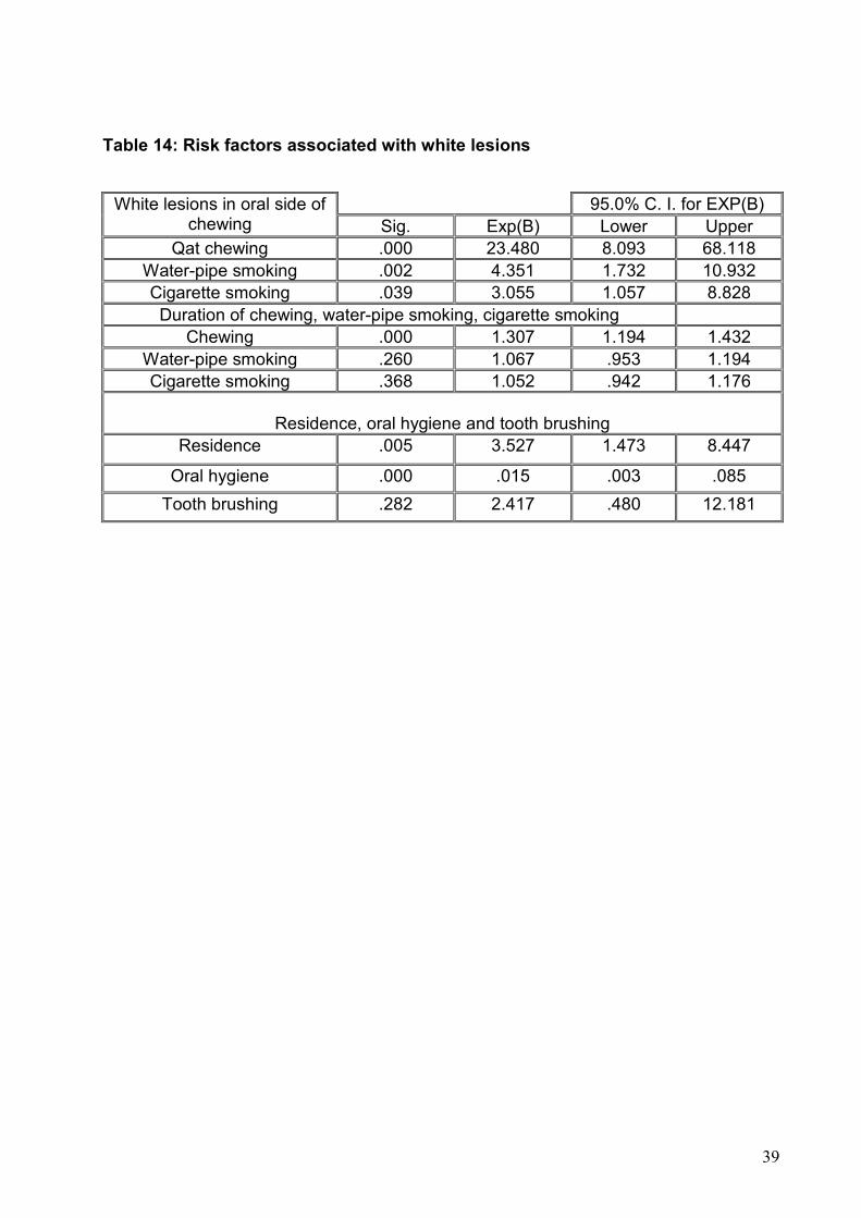
4.10.2 Multivariate analysis of the risk factors associated with white lesion
Table 14 shows the multinomial logistic regression model that was used to assess the
risk factors that were significant at the univariate analysis. The results revealed that the
risk factor that remained significantly associated with white lesion was qat chewing
(p=0.000, OR= 23.480), water-pipe smoking (p=0.002, OR= 4.351) and cigarette
smoking (p= 0.039, OR= 3.055)
When white lesions were correlated with the durations of chewing, water-pipe and
cigarette smoking, the results were highly significant (p=0.000), water-pipe smoking was
not significant (p=0.260), cigarette smoking was not significant (p=0.368).
Residence, oral hygiene, and tooth brushing behaviour were correlated with presence of
white lesions. Results concerning residence were significant (p=0.005), oral hygiene
was highly significant (p=0.000), and tooth brushing behaviour was not significant
(p=0.282).
39
Table 14: Risk factors associated with white lesions
95.0% C. I. for EXP(B) White lesions in oral side of chewing Sig. Exp(B) Lower Upper
Qat chewing .000 23.480 8.093 68.118 Water-pipe smoking .002 4.351 1.732 10.932 Cigarette smoking .039 3.055 1.057 8.828 Duration of chewing, water-pipe smoking, cigarette smoking
Chewing .000 1.307 1.194 1.432 Water-pipe smoking .260 1.067 .953 1.194 Cigarette smoking .368 1.052 .942 1.176
Residence, oral hygiene and tooth brushing
Residence .005 3.527 1.473 8.447
Oral hygiene .000 .015 .003 .085
Tooth brushing .282 2.417 .480 12.181
5. DISCUSSION
In Yemen, qat chewing is a widespread social habit practiced by men, women and
occasionally even children for few hours every day, and in many cases it is a lifetime
habit. The continuous process of chewing qat leaves and keeping them in one side of
the mouth until the cheek grows into a noticeable ball has been explained well in the
literature. The literature of this study also covered qat history, botany, chemistry
pharmacology and other related effects (2, 9, 26, 28, 31, 42, 53 and 64).
The adverse effects of this habit on different human body systems such as CNS,
cardiovascular, digestive, genitourinary, and reproductive health systems were
extensively investigated (7, 63, 74, 75, 82, 89 and 90).
The effects of qat chewing on different oral tissues including periodontal tissues, hard
tissues of teeth, TMJ, muscles of mastication and salivary glands were also extensively
reviewed (8, 12, 14, 91, 92 and 93).
The mechanism by which qat chewing induced white lesions on the oral mucosa might
be due to its mechanical and/or chemical effects. The daily application of qat on the oral
mucosa is expected to cause mechanical and/or chemical irritation leading to an
increase in the thickness and keratinisation as a defence mechanism. Similar changes
were reported under the traumatic focal (frictional) hyperkeratosis leading to increased
thickness and colour changes of the oral mucosa due to continuous trauma (107).
Tannins, the phenolic compound present in qat, have some local effects on the
digestive system, due to their astringent effects (8, 9). Moreover, they are known to
cause an increase in the thickness of mucous membrane of the oropharynx and
esophagus (81, 82). Also, chemical additives in qat green leaves such as chlorinated
hydrocarbon from pesticides used in the treatment of qat plants (42). Both tannins and
qat additives may have caused the changes seen in the oral mucosa.
On oral mucosa, oral keratotic white lesions with no leukoplakia or cancer have been
reported to be due to qat chewing in many reports. Some degree of oral keratosis was
found at the site of qat chewing among 60 Yemeni male chewers (14). Buccal and
gingival white lesions at chewing sites were reported among Yemeni qat chewers who
chewed for 3 years and more (12). 342 mild keratotic white lesions were reported
41
among Yemeni qat chewers (16). Also oral white lesions were reported among Yemeni
Israeli Jew male chewers who had chewed qat for three years and more (100).
So far, all reports were either done on male chewers (14, 100) or included a very narrow
sample of female chewers (12, 16). In the past, Yemeni women considered non-
chewers, later on, few rich women used to chew qat, but the habit was hidden.
Traditionally, chewing among females in Yemen was a shameful habit. Recently,
chewing increased in women, but generally, they spend fewer hours in qat sessions and
consumed smaller quantities than men (31).
This is the first survey conducted among Yemeni women qat chewers, with the objective
of ascertainins a causal relationship between qat chewing and oral mucosal white
lesions by applying a cross sectional study design. The role of cross sectional
epidemiological method in ascertaining evidence for causal relationship is well
established (104). Other advantages of cross sectional design are related to their
usefulness in investigating exposures of fixed characteristics among individuals
belonging to a specific socioeconomic situation or ethnicity. Its success was reported by
many investigators who conducted studies on adverse health and habits relationship
(104).
Qat consumption was the exposure hypothesized to produce an adverse effect on oral
mucosa due to its physical and/or chemical action resulting in disease state among
cases. To control for misclassification of the lesions, the investigator used the non-
chewing sites of the jaw as control side (control 1). Non-chewers were used as second
control. Although the investigator is aware of the fact that non-chewers are the best
control, non-chewers of qat in Yemen are very rare. Because of this, the opposite side
was used as a control side. This procedure minimizes the selection bias.
Subjects, exposed or non-exposed, were selected from patients seeking dental
treatment in Sana’a city. Catchment area for patients who seek dental treatment in
Sana’a city could be the city, surrounding villages or any area of Yemen. Majority of
subjects, however, are expected to be from Sana’a city, thus forces driving both cases
and control due to the same hospital are similar, and this also minimizes selection bias.
Subjects who predominantly use one side of their mouths were selected, bilateral
chewers were excluded. Predominant unilateral chewers may occasionally use other
side, but the adverse effects are always greater on the chewing site. The majority of
42
Yemenis chew on their left sides. This is due to the need of the chewer to have the right
hand free for use when necessary for writing and explaining.
In this study, 162 women had been recruited and divided into 109 chewers and 53 non-
chewers. The mean age was 38.25 years, ranging from 20 to 65 years. No significant
difference was found between qat chewers and control 2 (non-chewers).
The majority of the study population lived in towns 110 (67.9%) compared to 52 (32.1%)
who lived in villages. No significant difference was found between the too groups.
Similar results were reported among Yemeni male chewers (12).
The residence (town, village) was examined for correlation with white lesions. Findings
showed that white lesions found among women living in town were higher than in
women living in village. The deference was highly significant (p=0.000). This result was
expected since women in town usually spend more time in their qat sessions compared
to women in village (31).
The oral hygiene status (OHS) among the study population showed that 46 (28.4%)
subjects had good OHS, 36 (22.2%) had fair OHS and the remaining 80 (49.4%)
subjects had poor OHS. Tooth brushing among the study population was recorded in
105 (64.8%) subjects and the remaining 57 (35.2) did not brush their teeth. Type of
device used for mouth cleaning among study subjects showed that 17 (16.2%) used
miswak, 77 (73.3%) used toothbrush and paste and the remaining 11 (10.5%) used
dental floss and other oral hygiene aids. Forty subjects (38.1%) brushed their teeth
once, 60 (57.1%) twice, and 5 (4.8%) brushed their teeth more than twice a day.
The oral hygiene status was examined for correlation with white lesions. White lesions
were predominant in women with bad oral hygiene (p= 0.000). This is probably due to
the fact that 80 (49.4%) subjects who participated in this study had poor oral hygiene.
Subsequently, superinfection with Candida spp. might have had an influence on the
more whitish character of the lesions.
The presence of white lesions was correlated with residence, oral hygiene, and tooth
brushing behaviour. Results were significant (p=0.003), oral hygiene was highly
significant (p=0.000), and tooth brushing behaviour was not significant (p=0.282).
Similar results regarding residence, oral hygiene, and tooth brushing behaviour was
reported by AL-Sharabi (2002). In the latter study, 74% of subjects lived in town, 66%
had poor oral hygiene status and 48% did not brush their teeth (12).

43
The age of starting chewing among the study population showed that 34 (31.2%)
started chewing at 10-20, 52 (47.7%) at 20-30 and 23 (21.1%) at 30-40 years of age.
Age of starting chewing was examined for correlation with white lesions. Findings were
not significant (p=0.339).
The duration of the chewing habit ranged from 5 to 40 years with a mean of 14.79
years. The majority of chewers in this study chewed on their left side every day and for
3 - 6 hours per day. Similar results regarding the duration of the chewing habit per
years, side of qat chewing in the mouth left or right, number of qat chewing days per
week and also number of hours spent every day per chewing session has been
reported (12, 16).
Results of our study showed that 21 (19.3%) subjects among cases were cigarette
smoker, whereas 43 (39.4%) subjects were water pipe smokers. This result was
expected because Yemeni women prefer water-pipe smoking to cigarette smoking
during qat chewing (31). While male chewers in Yemen prefer cigarette smoking to
water pipe smoking. In one study of male chewers, 55% were cigarette smokers and
7% were water-pipe smokers (12).
The most important result in this study was the detection of white lesions among
Yemeni women due to qat chewing. Among cases, white lesions occurred in 82 (75%)
subjects. White lesions occurred on the internal surface of the cheek at the side of qat
chewing. From this side white lesions extended or continued to be present down along
the lower buccal mucosa in 54 (65.8%) subjects and further continued to the vestibular
sulcus in 36 (33.1%) subjects. Similarly, white lesions on buccal mucosa extended or
continued to be present upward to the upper vestibular sulcus in only 3 (5.5%) subjects.
White lesions present on gingival mucosa among cases were found in 28 (34.2%)
subjects; white lesions present on gingival mucosa extended or continued to be present
on alveolar mucosa in 15 (53.5%) subjects.
Among control 1 (non-chewing site), white lesions were detected in 6 (5.5%) subjects,
extended to lower buccal mucosa in 3 (50.0%) subjects and extended further to lower
vestibular sulcus only in 1 (33.4%) subject. On gingival mucosa, white lesions were
detected in 3 (50.0%) subjects with no extension to alveolar mucosa. The presence of
white lesions on buccal and gingival mucosa on the non-chewing site could be
explained by the fact that some chewers chew a huge amount of qat and due to the
44
movement of chewing some fibres cross to the opposite side causing whitening or white
lesions probably occurred due to the chemical effects of qat.
Among control 2 (non-chewers), white lesions detected in 7 (13.2%) subjects extended
to lower buccal mucosa in 4 (57.2%) subjects and extended further to the lower
vestibular sulcus in only 2 (50.0%) subjects. Gingival white lesions were recorded in 3
(42.8%) subjects, both of them extended to the alveolar mucosa. The presence of white
lesions among non-chewer women could be explained by the fact that some females
still regarded the qat chewing habit as shameful, so they either preferred not to report
about it or these women were probably ex-chewers.
Presence of white lesions among non-chewers had been reported earlier in male
subjects (12). Buccal white lesions were reported in 8.5% and gingival white lesions in
7.5% among non-chewers male subjects (12).
The results of white lesions in this study showed statistically highly significant
differences between white lesions occurring among chewers (75%) when compared to
those occurring in both controls (5.5%) (p=0.000), (13.2%) (p= 0.009). The results of
white lesions reported in this study among Yemeni women qat chewers are in
agreement with many studies done in Yemen on male qat chewers (12, 14, and 16).
Results of this study are also in agreement with the study done in Israel on Yemeni Jew
male chewers (100).
Regarding the etiology of white lesions, many people in the world conceptualized that
qat is a form of tobacco or at least tobacco containing product. Thus, if it is so, it might
be a cause of oral pre-cancer or cancer in Yemen. The only similarity between qat and
tobacco is their alkaloid constituent. While the alkaloid in tobacco is nicotine, the
alkaloid in qat is cathinone, i. e. the two products are obviously different. Tobacco
smoking (cigarette or water-pipe) is well known to induce white lesions on oral mucosa
(103, 105, 106). Tobacco related white lesions are known to develop into malignancy.
Cigarette and water-pipe smoking is practiced frequently by qat chewers during chewing
sessions. The white lesions detected among qat chewers could be due to the effect of
tobacco use and not of qat chewing. White lesions detected in this study could not be
attributed to qat chewing unless this effect of tobacco use is diminished or removed
statistically.
45
In this study, white lesions detected among cases were statistically examined in
correlation with qat chewing, cigarette smoking and water-pipe smoking. Statistical
results of qat chewing were highly significant (p=0.000). In case of cigarette smoking,
p=0.039 and in case of water-pipe smoking, p-value was 0.002. When the multinomial
logistic regression model was used for the three groups, the relative risk (R.R.) or the
odds ratio OR (95% CI) was 23.480 (8.093 - 68.118) in case of qat chewing, 3.055
(1.057 - 8.828) in case of cigarette smoking and 4.351 (1.732 - 10.932) in the case of
water-pipe smoking.
White lesions among chewers were also statistically examined in correlation with the
duration of chewing, cigarette smoking and water-pipe smoking. Statistical results of qat
chewing duration was highly significant (p=0.000). In the case of smoking duration, the
statistical result was not significant with p=0.368. Similar statistical result of water-pipe
duration was not significant with p=0.260. Additionally, all white lesions recorded among
cases (n=82) were found at the site of qat chewing. Differences between white lesions
occurring among chewers in comparison to both controls were statistically highly
significant (p<0.000).
All these statistical results support the hypothesis that white lesions reported in this
study are attributed to qat chewing and not to tobacco. If these lesions were due to any
type of smoking, they will be present anywhere in the mouth and not at the site of
chewing.
In conclusion, qat chewing habit caused buccal and gingival white lesions at side of
chewing among Yemeni women who chew every day for 3 - 6 hours and over a number
of years. These results are reported for the first time.
6. ABSTRACT
BACKGROUND:
Qat chewing habit is widespread and practiced by millions of people in Yemen. The
habit is increasing every year in Kenya, Somalia, Djibouti and Ethiopia. Recently, the
habit is also increasing in Europe, where immigrants from qat consuming countries are
living. An adverse health effect of qat chewing on different body systems and oral
tissues has been reported.
AIM OF THE STUDY:
The aim of this study was to assess the association between habitual qat use and oral
mucosal white lesions among Yemeni women.
STUDY DESIGN:
In a cross sectional hospital study, 162 healthy women were recruited. Subjects were
divided into 109 qat chewers and 53 non qat chewers. Information obtained from the
chewing sides among the 109 qat chewers was analyzed as cases. Two controls were
used in this study. Information obtained from the opposite sides (non-chewing side)
among the 109 qat chewers were analyzed as first control. Information obtained from
both sides of the 53 non qat chewers was analyzed as a second control. Inclusion
criteria were: healthy females aged ≥ 20 years, chewing qat continuously for a minimum
of 5 years and on one side of their mouths only. Bilateral qat chewers, shammah users
and subjects with poor general or mental health were excluded. All women were
interviewed for qat chewing information and examined clinically for oral mucosal white
lesions.
RESULTS:
Among chewers, white lesions were recorded in 82/109 (75.2%) at the chewing site.
White lesions on the opposite side (control 1) were recorded among 6/109 (5.5%)
subjects. White lesions among non-chewers were recorded in 7/53 (13.2%) subjects
(control 2). White lesions were identified primarily on the lower buccal mucosa,
vestibular sulcus, gingival mucosa, alveolar mucosa and upper vestibular mucosa on
the chewing site. White lesions reported among chewers were positively correlated to
47
the side of chewing and to the longer duration of the habit. The difference in the
prevalence of white lesions present among cases and both controls was highly
significant (p<0.000).
CONCLUSION:
Continuous chewing of qat fibres during sessions lasting 3 - 6 hours every day over
decades caused mucosal white lesions, which were recorded on the chewing side
among Yemeni women.
48
7. Zusammenfassung
Orale weiße Läsionen durch das Kauen von Qat bei Frauen im Jemen
Hintergrund:
Das Kauen von Qat ist im Jemen verbreitet und wird von Millionen Menschen
praktiziert. Jedes Jahr steigt die Anzahl der Qat Konsumenten in Kenia, Somalia, Djibuti
und Äthiopien. In den letzten Jahren lässt sich auch in Europa eine Zunahme dieses
Habits durch Immigranten aus Ländern, in denen das Qat Kauen verbreitet ist,
feststellen. Beschrieben wurden bereits Beeinträchtigungen einzelner Körpersysteme
und Veränderungen des oralen Gewebes durch Qat bei Männern. Grund für Letzteres
ist eine chemische und mechanische Irritation der Schleimhaut.
Ziel vorliegender Studie war es, mögliche Zusammenhänge zwischen dem Kauen von
Qat und dem Auftreten oraler weißer Läsionen bei Frauen im Jemen darzustellen.
Material und Methode:
In einer Querschnittsstudie wurden 162 Frauen rekrutiert. Diese wurden in 109 Qat
Konsumentinnen und 53 Nicht-Konsumentinnen eingeteilt. Als Kontrolle diente 1. die
Schleimhaut der gegenüberliegende Seite der Qat Konsumentinnen (Kontrolle 1) und 2.
die Schleimhaut bei Nicht-Konsumentinnen (Kontrolle 2). Einschlusskriterien waren
gesunde weibliche Personen ≥ 20 Jahre, die seit mindestens fünf Jahren regelmäßig
und nur auf einer Seite Qat kauen. Qat-Konsumentinnen, die auf beiden Seiten Qat
kauten, Shammah-Gebraucherinnen sowie Personen mit schlechtem Allgemeinzustand
wurden ausgeschlossen. Qat-Konsumentinnen wurden zu ihrem Habit befragt, alle
Frauen wurden klinisch auf weiße Mundschleimhautläsionen untersucht.
Ergebnisse:
Bei 82/109 Qat-Konsumentinnen (75.2%) wurden weiße Veränderungen auf der
Kauseite festgestellt. Weiße Veränderungen auf der gegenüberliegenden Seite
(Kontrolle 1) wurden bei 6/109 (5.5%) Konsumentinnen beobachtet. Bei Nicht-
Konsumentinnen wurden in 7/53 (13.2%) weiße Veränderungen nachgewiesen
(Kontrolle 2). Weiße Läsionen wurden primär im kaudalen Bereich der
Wangenschleimhaut, im Vestibulum, an der gingivalen Schleimhaut und im Vestibulum
des Oberkiefers auf der Kauseite festgestellt. Es bestand eine signifikante Korrelation
49
zwischen dem Auftreten weißer Läsionen und der Kauseite sowie der Dauer des Habits.
Der Unterschied der Häufigkeit des Auftretens weißer Läsionen zwischen Qat-
Konsumentinnen auf der Kauseite und beiden Kontrollen war hochsignifikant.
(p<0.000).
SCHLUßFOLGERUNG:
Das Kauen von Qat während der täglichen 3 – 6-stündigen Qat-Sitzungen führte bei
jemenitischen Frauen zu weißen Mundschleimhautläsionen auf der Kauseite. Das
Entstehen von weißen Läsionen ist abhängig von der Dauer dieses Habits.
50
8. REFERENCES
1 Kalix P. Catha edulis, a plant that has amphetamine effect. Pharm World Sci 1996; 18 (2): 69 - 73. 2 Kennedy J. (Ed). The flower of paradise. The Institutionalized Use of the Drug Qat in North Yemen, D. Reidel Publishing Co. Dordrecht, Holland 1987; p. 89: 201-244. 3 Al-Motarreb A., Baker K, Broadly KJ. Khat: pharmacological and medical aspect and its social use in Yemen. Phytother Res 2002; 16 (5): 403-413. .
4 Winterfeld K., Bernsmann G. Zur Kenntnis der Inhaltsstoffe von Catha edulis Forskal. Arch Pharm Chem 1960; 293/ 65: 991-1000. 5 Alles GA, Fairchild MD, Jensen M. Chemical pharmacology of catha edulis. J Med Pharm Chem 1961; 3: 323- 52. 6 Mustard MJ. Ascorbic acid content of some miscellaneous tropical and subtropical Plant and plant products. Food Res 1952; 17: 31-35. 7 Kalix P. Khat: A plant with amphetamine effects: J Subst Abuse Treat 1988; 5 (3): 163-169. 8 Halbach H. Medical aspects of the chewing of qat leaves. Bulletin of the World Health Organization 1972; 47(1): 21-9. 9 Kalix P, Braenden O. Pharmacological aspect of chewing of khat leaves. Pharmacol Rev 1985; 37(2):149-64. 10 Al-Bekairi AM, Abulaban FS, Qureshis, et al. The toxicity of catha edulis (Khat) - A review. Fitoterapia 1991; 62 (4): 291-300. 11 Nencini P, Ahmad AM, Elmi AS. Subjective effects of Khat chewing in humans. Drug Alcohol Depend 1986; 18 (1): 97-105. 12 AL-Sharabi A. Oral and para-oral lesions caused by takhazeen alqat (chewing). PhD Thesis. Al-Khrtoum, Sudan: Uni. Khartoum, 2002. 13 Kennedy JG, Teague J, Rokaw W, et al. A medical evaluation of the use of qat in North Yemen. So Sci Med 1983; 17 (12): 783-93. 14 Hill CM and Gibson A. The oral and dental effects of qat chewing. Oral Surg Oral Med Oral Path 1987; 63 (4): 433-436. 15 Macigo FG Mwaniki DL, Guthua SW. The association between oral leukoplakia and use of tobacco, alcohol and khat based on relative risks assessment in Kenya. Eur J Oral Sci 1995; 103 (5): 268-273.
51
16 Ali AA, AL-Sharabi AK, Aguirre JM, et al. A study of 342 oral keratotic white Lesion induced by qat chewing among 2500 Yemeni. J Oral Pathol Med 2004; 33 (6): 368-372. 17 Makki I. Schleimhautkarzinome der Mundhöhle unter besonderer Berücksichtigung des Qat- und Shammah-Abusus. Dissertation, Universität Heidelberg, Deutschland 1975. 18 Soufi HE, Kameswaran M, Malatani T. Khat and oral cancer. J Laryngol Otol 1991; 105 (8): 643-5. 19 Awange DO, Onyango JF. Oral verrucous carcinoma: report of two cases and review of literature. East Afr Med J 1993; 70 (5): 316-318. 20 Mancioli M and Parrinello A. Qat ( catha edulis ). La Clinica Treputica Roma. Clin Ter 1967; 43 (2): 103-172. 21 Schopen A. (Ed ), Traditionelle Heilmittel in Jemen ( German ): Research reports of the ethnology depart. University of Frankfort publication: Franz Steiner Verlag. Wiesbaden, Deutschland 1983; 256: 209-221. 22 Moser C. The flower of paradise. National Geographic Magazine 1917; 32: 173 186. 23 AL-Hipshi A. (Ed), AL-Qat fi aladab ALymani (Arabic), ALyaman algadid Sana’a, Yemen 1972; 3rd issues: p 143-157. 24 Dillmann A. Die Kriegstaten des Königs ´Amda-Sion gegen die Muslim` Sitzungsberichte der königlich Preußischen Akademie der Wissenschaften zu Berlin 1884 . 25 EL-Mahi T. A preliminary study on Khat together with institutional history of coffee as a beverage in relation to khat. WHO, regional office for Eastern Mediterranean. EM/MENT/29/4 March, 1962; p: 2-3. 26 Weir S. (Ed) Qat in Yemen, consumption and social changes. British Museum Publication. London 1985; p 181-185. 27 Ward C, and Gatter P. Qat in Yemen. Document of the World Bank: Middle East and North Africa Region June 2000; p 5-48. 28 Hughes PH. Qat chewing in Yemen. Papers presented at 4th int. council on drug dependence. Lausanne, Switzerland 1973; p: 32-46. 29 Baasher T. Mental health Services in the Demo Republic of Yemen. WHO Document No EM/MENT/81, 1976. 30 National conferences on qat. Toward the formulation of a comprehensive qat policy in the Republic of Yemen. Technical field study. Gender and qat consumption. Sana’a, 6-7 April 2002; p: 67.
52
31 Sayem N. Motivating factors and changes in qat use amongst women. National conference on qat, conference discussing materials Sana’a Yemen, 6- 7 April 2002; p: 36-37. 32 Hijazi I. Qat session. Social study (Arabic), faculty of arts. J Sana’a University 1981; 5: p: 13. 33 Makhloof C. (Ed) Changing veils: women and modernization in north Yemen. Austin: University of Texas Press 1979; p: 18. 34 Dionna L. (Ed), Yemen, the country I had seen. AL-Adab publication Beirut Lebanon, 1984; p: 20-65. 35 Al-Zuby MA. AL-qat Alwai-Tahadhor. (Arabic) J Yemen studies Sana’a Yemen, 1988; 31: 100-124.
36 Al-Zalab A. Qat culture in Yemen. Sociology study (Arabic), PhD Thesis in Sociology, Tunis 1st University library 1998; p: 237. 37 Al-Maqrami A. Qat and development. An empirical study in Yemen society, (MSc thesis) (Arabic). University of Cairo Library, 1982. 38 Al-Raadi MA. (Ed), Alqat alsalwa walbalwa (Arabic), AL-Afif cultural foundation publication Sana’a Yemen, 1992; Book (4): 15-60. 39 Zabara A. and Sabri M. Qat and its adverse effect on health particulary on children in (qat phenomenon and its solutions) (Arabic). AL-Afif cultural foundation publication, Sana’a Yemen, 1997; Book (13): 102-112. 40 Al-Shargi RH. (Ed) The social effects of qat: Qat and women (Arabic) Al-Afif cultural foundation publication. Sana’a Yemen, 1997; Book (13): 44-53. 41 Bamatraf A, and Muharam I. (Ed) The importance of scientific research to qat phenomenon (Arabic). AL-Afif cultural foundation publication, Sana’a Yemen, 1997; Book (13): 12-28. 42 Thabet AA. Joint action of some natural pests control agents and Cypermethrin insecticide against cereals aphids and their Predators. J Pest Control Environ Sci 1999; 7(3): 25-38. 43 AL-Ghashm and Nasher. First workshop on plant protection and production. Annual report of Tehama Develop Authority Ministry of Agriculture: Sana’a Yemen, 1999; p: 1-3. 44 Heath DF, and Magee PN. The decomposition and toxicity of dialkylnitrosamines in rats and some related Compounds. Biochem J 1962; 85 (1): 72-91. 45 Edwards CA, and Thompson AR. (Ed) Pesticides and the soil fauna. Residue Rev 1973; 45: 1-79.
53
46 Bawazir A. and Amin B. Plastic abuse in Yemen. 1st National conference of pests and other chemical on human and environment. Yemen cancer society publications, 14-16 March Sana’a 2000. 47 Revri R. Catha edulis Forsk: Geographical dispersal, botanical, ecological, and agronomical aspects with special references to Yemen Arab Republic. Diss. Univ. Hohenheim, Hohenheim, Germany 1983; 30-119. 48 Fluckiger F, and Georock J. Contributions to the knowledge of catha leaves. Pharma J and Trans 1887; 47: 221-224. 49 Mosso U. Azione fisiologica del principio attivo del Celastrus edulis (Italian). Riv Clin Milano 1891; 30: 65-79. 50 Beitter A. Pharmkognostisch-chemische Untersuchung von Catha edulis. Arch Pharma 1901; 239: 17-33. 51 Friebel H, and Brilla R. Über den zentralerregenden Wirkstoff der frischen Blätter und Zweigspitzen von catha edulis forsk. Naturwissenschaften 1963; 50: 354-355. 52 Wolfes O. Über das Vorkommen von D-nor-iro-Ephedrine in catha edulis. Arch Pharm 1930; 268: 81-83. 53 Cais M, Ginsberg D, Mandelbaum A. Constituents of Catha edulis.Tetrahedron 1975; 31: 2727-2731. 54 Braenden O. Research on the chemical composition of qat research monograph, US Government printing Office, Washington, 1979; p: 320-321. 55 Reports on the botany and chemistry of Khat. United Nations Narcotic Laboratory Reports No. 8 and 9, 1979 56 Szendrei K. The chemistry of khat narcotics. Bull Narc 1980; 32 (3): 5-35 57 Ramadan M, Qirbi A, and Tash F. Evaluation of Cathinone and cathine of different khat in Yemen (Arabic). Yemeni Studies J 1981; 5: 20-28.
58 Afendi AH. Qat composition and health effects (Arabic). Sana’a Alymaniah publishing House 1997; p: 15-40. 59 Maitai CK, Mugera GM. Excretion of the active principle of catha edulis (mira) in human urine. J Pharma Sci 1975; 64 (4): 702-703. 60 Brenneisen R, Geisshüsler S, Schorno X. Metabolism of Cathinone to (-)-norephedrine and (-)-norpseudoephedrine. J Pharma Pharmacol. 1986; 38(4): 298-300. 61 Kalix P. The releasing effect of the isomers of the alkaloid cathinone at central and peripheral catecholamine storage sites. Neuropharmacology 1986; 25 (5): 499-501.
54
62 Gelenon R, Young R, Hauck A, McKinney J. Structure activity studies on amphetamine analogs using drug discrimination methodology. Pharmacol Biochem Behav 1984; 21: 895-901. 63 Goth A. (Ed) Medical pharmacology principals and concepts 9th Edition. The C.V. Mosby Company Saint Louis 1978; p: 310-311. 64 Kennedy J, Teague J, Fairbanks L. Qat use in North Yemen and problems of addication: A study in medical anthropology. Cult Med Psychiatry 1980; 4 (4): 311-344. 65 Luqman W, and Danowski TS. The use of khat (Catha edules) in Yemen. Social and medical observations. Ann Intern Med 1976; 85 (2): 246-249. 66 Nencini P, Johansson C, Schuster C. Sensitization to kappa opiod mechanisms associated with tolerance to the anorectic effects of Cathinone. J Pharma Exp Ther 1988; 245 (1): 147-154. 67 Adam F, Hasselot N. Khat: from traditional usage to the risk of drug addiction. Med Trop (Mars) 1994; 54 (2): 141-144. 68 Giannini AJ, Burge H, Shaheen JM, Price WA. Khat: another drug of abuse. J Psychoactive Drugs 1986; 18 (2): 155-158. 69 Granek M, Shalev A, Weingarten AM. Khat induced hypnologic hallucinations. Acta Psychiatry Scand 1988; 78 (4): 458-461. 70 Kalix P. Pharmacological properties of the stimulant khat. Pharmacol Therap 1990; 48: 397-416. 71 Randall T. Khat abuse fuels Somali conflict drains economy. JAMA 1993; 269(1): 12-15. 72 Heymann TD, Bhupulan A, Zureikat NE, et al. Khat chewing delays gastric emptying of a semi-solid meal. Aliment Pharmacol Ther 1995; 9 (1): 81-83. 73 Hassan NAGM, Gunaid AA, Abdo-Rabbo AA, et al. The effect of qat chewing on blood pressure and heart rate in healthy volunteers. J Trop Doc 2000; 30 (2): 107-108. 74 Nencini P, Ahmed A, Anania M, Moscucci M, and Paroli E. Prolonged anaesthesia induced by cathinone. The role of stress and opioid and nonopioid. Pharmacology 1984; 29 (5): 269-281. 75 Halket JM, Karasu Z, Murray-Lyon IM. Plasma cathinone levels following chewing khat leaves (catha edulis forsks). J Ethno Pharmacology 1995; 49 (2): 111-3. 76 Brenneisen R, Fish H, Kalix P. Amphetamine like effects in humans of the khat alkaloid cathinone. Brit J Clinic Pharmacol 1990; 30 (6): 825-828.
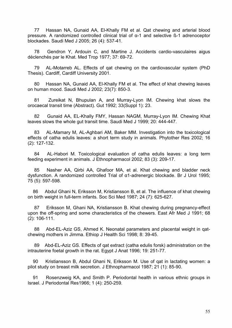
55
77 Hassan NA, Gunaid AA, El-Khally FM et al. Qat chewing and arterial blood pressure. A randomized controlled clinical trial of α-1 and selective ß-1 adrenoceptor blockades. Saudi Med J 2005; 26 (4): 537-41. 78 Gendron Y, Ardouin C, and Martine J. Accidents cardio-vasculaires aigus déclenchés par le Khat. Med Trop 1977; 37: 69-72. 79 AL-Motarreb AL. Effects of qat chewing on the cardiovascular system (PhD Thesis). Cardiff, Cardiff University 2001. 80 Hassan NA, Gunaid AA, El-Khally FM et al. The effect of khat chewing leaves on human mood. Saudi Med J 2002; 23(7): 850-3. 81 Zureikat N, Bhupulan A, and Murray-Lyon IM. Chewing khat slows the orocaecal transit time (Abstract). Gut 1992; 33(Suppl 1): 23. 82 Gunaid AA, EL-Khally FMY, Hassan NAGM, Murray-Lyon IM. Chewing Khat leaves slows the whole gut transit time. Saudi Med J 1999; 20: 444-447. 83 AL-Mamary M, AL-Aghbari AM, Baker MM. Investigation into the toxicological effects of catha edulis leaves: a short term study in animals. Phytother Res 2002; 16 (2): 127-132. 84 AL-Habori M. Toxicological evaluation of catha edulis leaves: a long term feeding experiment in animals. J Ethnopharmacol 2002; 83 (3): 209-17. 85 Nasher AA, Qirbi AA, Ghafoor MA, et al. Khat chewing and bladder neck dysfunction. A randomized controlled Trial of α1-adrenergic blockade. Br J Urol 1995; 75 (5): 597-598. 86 Abdul Ghani N, Eriksson M, Kristiansson B, et al. The influence of khat chewing on birth weight in full-term infants. Soc Sci Med 1987; 24 (7): 625-627. 87 Eriksson M, Ghani NA, Kristiansson B. Khat chewing during pregnancy-effect upon the off-spring and some characteristics of the chewers. East Afr Med J 1991; 68 (2): 106-111. 88 Abd-EL-Aziz GS, Ahmed K. Neonatal parameters and placental weight in qat-chewing mothers in Jimma. Ethiop J Health Sci 1998; 8: 39-45. 89 Abd-EL-Aziz GS. Effects of qat extract (catha edulis forsk) administration on the intrauterine foetal growth in the rat. Egypt J Anat 1996; 19: 251-77. 90 Kristiansson B, Abdul Ghani N, Eriksson M. Use of qat in lactating women: a pilot study on breast milk secretion. J Ethnopharmacol 1987; 21 (1): 85-90. 91 Rosenzweig KA, and Smith P. Periodontal health in various ethnic groups in Israel. J Periodontal Res1966; 1 (4): 250-259.
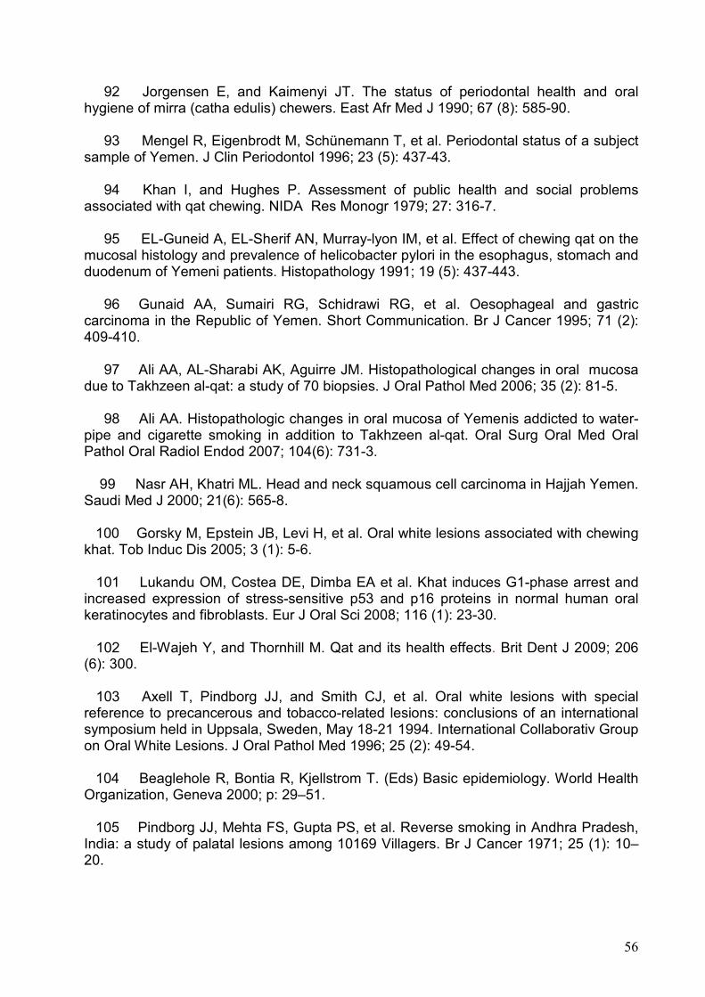
56
92 Jorgensen E, and Kaimenyi JT. The status of periodontal health and oral hygiene of mirra (catha edulis) chewers. East Afr Med J 1990; 67 (8): 585-90. 93 Mengel R, Eigenbrodt M, Schünemann T, et al. Periodontal status of a subject sample of Yemen. J Clin Periodontol 1996; 23 (5): 437-43. 94 Khan I, and Hughes P. Assessment of public health and social problems associated with qat chewing. NIDA Res Monogr 1979; 27: 316-7. 95 EL-Guneid A, EL-Sherif AN, Murray-lyon IM, et al. Effect of chewing qat on the mucosal histology and prevalence of helicobacter pylori in the esophagus, stomach and duodenum of Yemeni patients. Histopathology 1991; 19 (5): 437-443. 96 Gunaid AA, Sumairi RG, Schidrawi RG, et al. Oesophageal and gastric carcinoma in the Republic of Yemen. Short Communication. Br J Cancer 1995; 71 (2): 409-410. 97 Ali AA, AL-Sharabi AK, Aguirre JM. Histopathological changes in oral mucosa due to Takhzeen al-qat: a study of 70 biopsies. J Oral Pathol Med 2006; 35 (2): 81-5. 98 Ali AA. Histopathologic changes in oral mucosa of Yemenis addicted to water-pipe and cigarette smoking in addition to Takhzeen al-qat. Oral Surg Oral Med Oral Pathol Oral Radiol Endod 2007; 104(6): 731-3.
99 Nasr AH, Khatri ML. Head and neck squamous cell carcinoma in Hajjah Yemen. Saudi Med J 2000; 21(6): 565-8.
100 Gorsky M, Epstein JB, Levi H, et al. Oral white lesions associated with chewing khat. Tob Induc Dis 2005; 3 (1): 5-6. 101 Lukandu OM, Costea DE, Dimba EA et al. Khat induces G1-phase arrest and increased expression of stress-sensitive p53 and p16 proteins in normal human oral keratinocytes and fibroblasts. Eur J Oral Sci 2008; 116 (1): 23-30. 102 El-Wajeh Y, and Thornhill M. Qat and its health effects. Brit Dent J 2009; 206 (6): 300. 103 Axell T, Pindborg JJ, and Smith CJ, et al. Oral white lesions with special reference to precancerous and tobacco-related lesions: conclusions of an international symposium held in Uppsala, Sweden, May 18-21 1994. International Collaborativ Group on Oral White Lesions. J Oral Pathol Med 1996; 25 (2): 49-54.
104 Beaglehole R, Bontia R, Kjellstrom T. (Eds) Basic epidemiology. World Health Organization, Geneva 2000; p: 29–51.
105 Pindborg JJ, Mehta FS, Gupta PS, et al. Reverse smoking in Andhra Pradesh, India: a study of palatal lesions among 10169 Villagers. Br J Cancer 1971; 25 (1): 10–20.

57
106 Pindborg JJ, Roed-Petersen B, Renstrup G. Role of smoking in floor of mouth leukoplakias. J Oral Pathol 1972; 1 (1): 22-9.
107 Regezi JA, and Sciubba J. (Ed) Oral pathology; clinical-pathologic correlations. W B Saunders Philadelphia 1993: 2nd edition, p: 93-133.
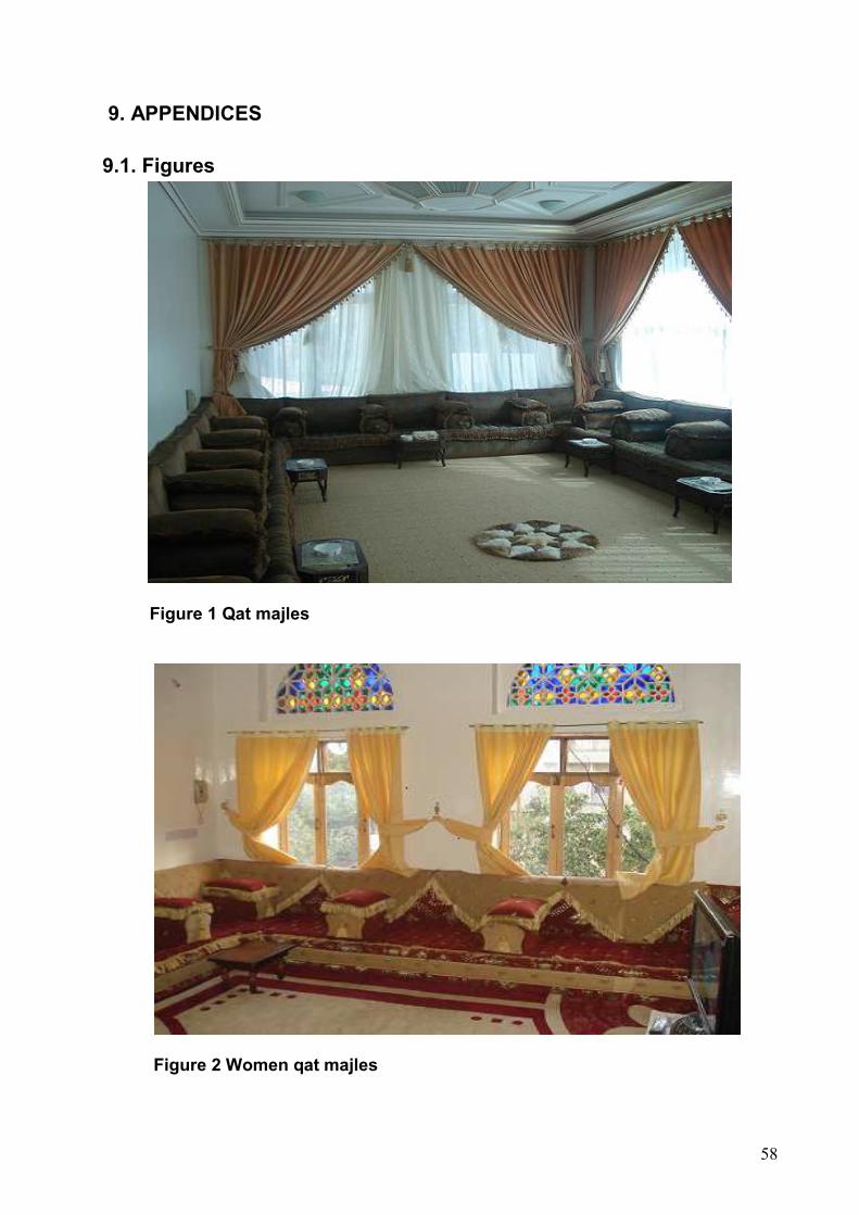
58
9. APPENDICES
9.1. Figures
Figure 1 Qat majles
Figure 2 Women qat majles
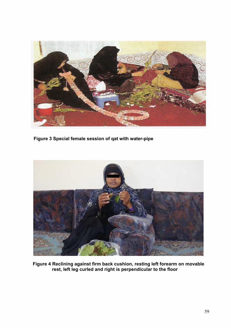
59
Figure 3 Special female session of qat with water-pipe
Figure 4 Reclining against firm back cushion, resting left forearm on movable rest, left leg curled and right is perpendicular to the floor
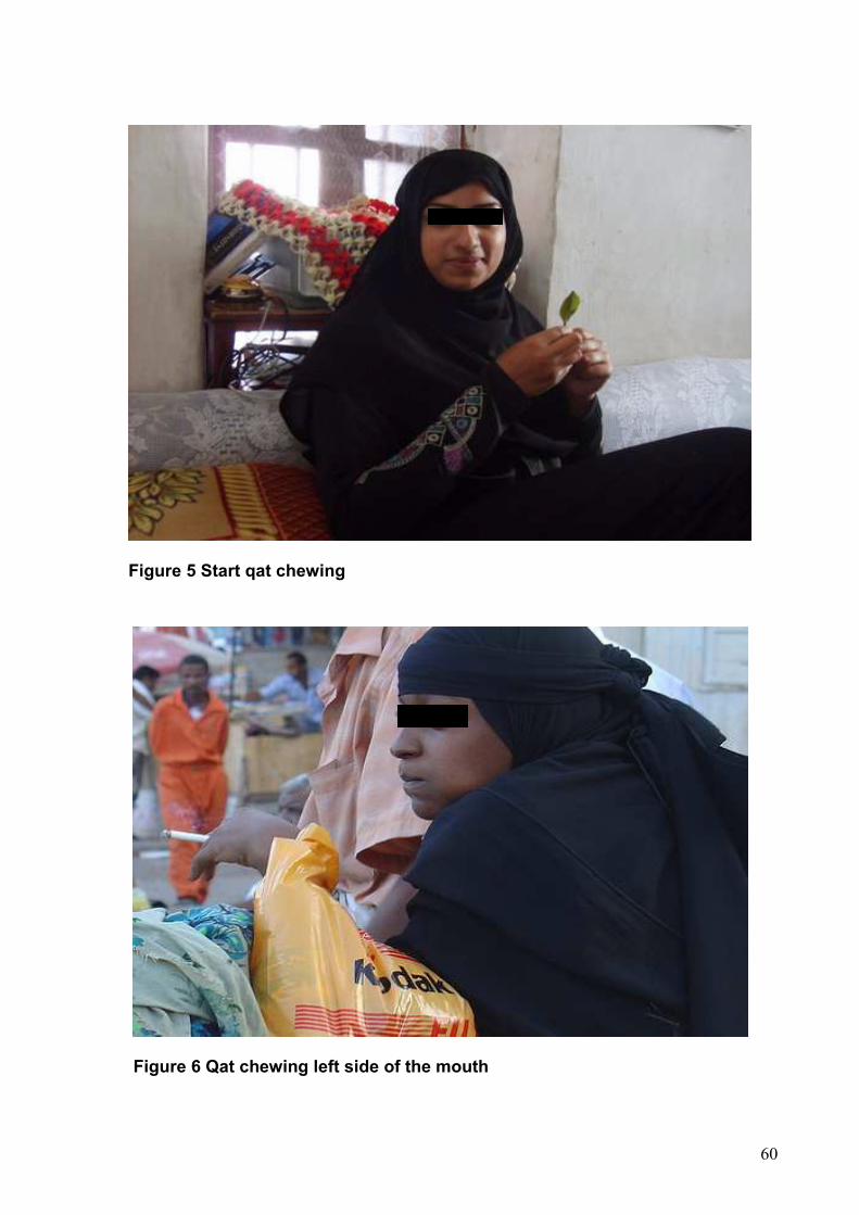
60
Figure 5 Start qat chewing
Figure 6 Qat chewing left side of the mouth
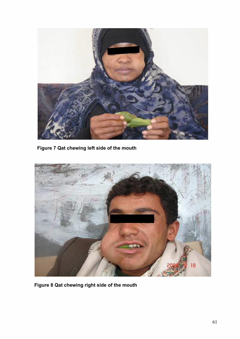
61
Figure 7 Qat chewing left side of the mouth
Figure 8 Qat chewing right side of the mouth
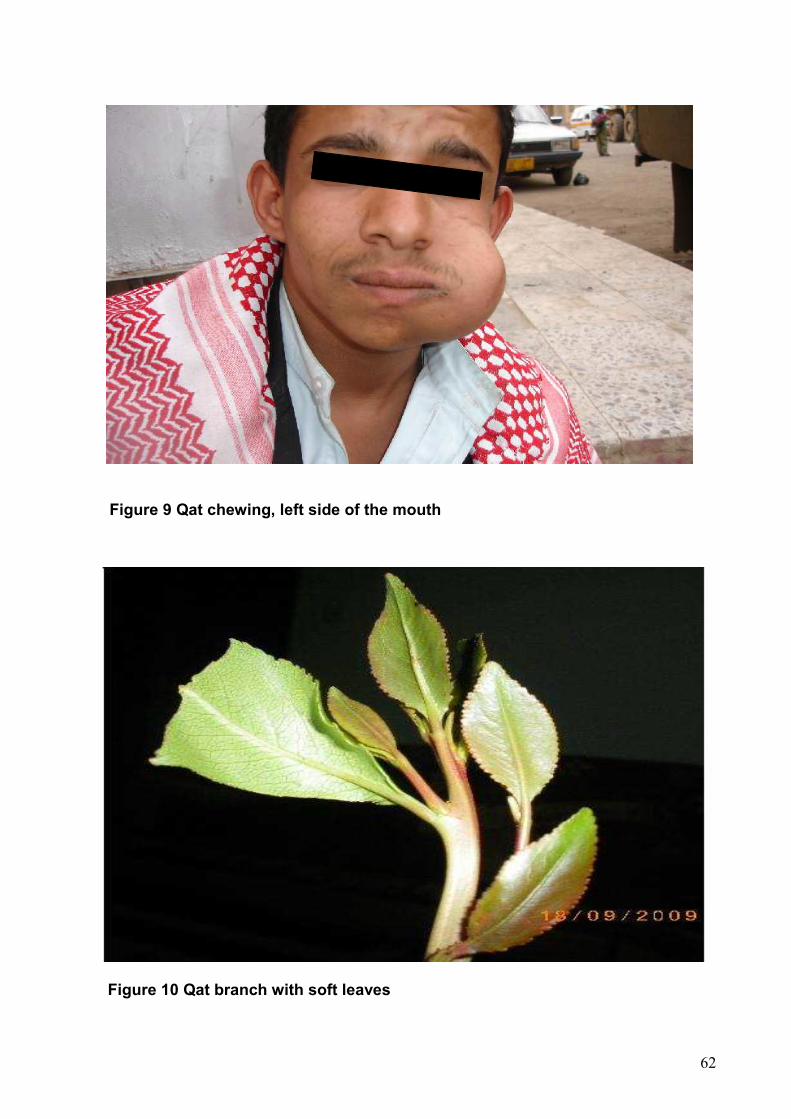
62
Figure 9 Qat chewing, left side of the mouth
Figure 10 Qat branch with soft leaves
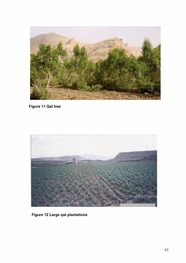
63
Figure 11 Qat tree
fgارع ا_bcت ا_^[\]
Figure 12 Large qat plantations
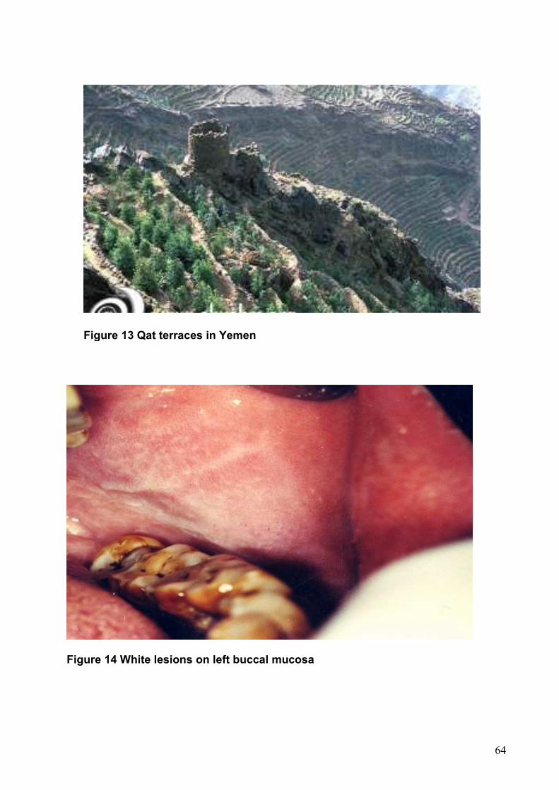
64
Figure 14 White lesions on left buccal mucosa
Figure 13 Qat terraces in Yemen
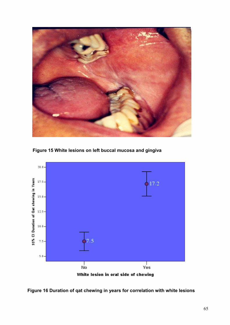
65
Figure 15 White lesions on left buccal mucosa and gingiva
Figure 16 Duration of qat chewing in years for correlation with white lesions
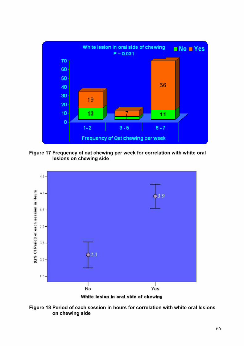
66
Figure 17 Frequency of qat chewing per week for correlation with white oral lesions on chewing side
Figure 18 Period of each session in hours for correlation with white oral lesions
on chewing side
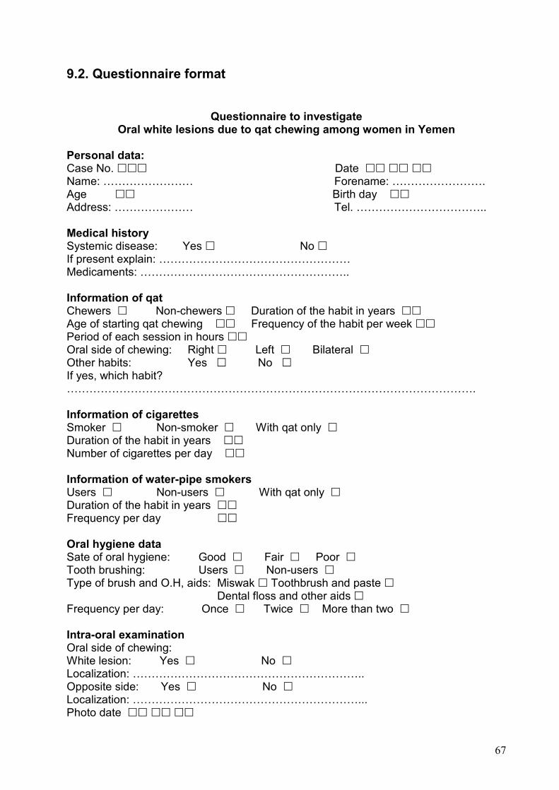
67
9.2. Questionnaire format
Questionnaire to investigate Oral white lesions due to qat chewing among women in Yemen
Personal data: Case No. ��� Date �� �� �� Name: …………………… Forename: ……………………. Age �� Birth day �� Address: ………………… Tel. …………………………….. Medical history Systemic disease: Yes � No � If present explain: …………………………………………… Medicaments: ……………………………………………….. Information of qat Chewers � Non-chewers � Duration of the habit in years �� Age of starting qat chewing �� Frequency of the habit per week �� Period of each session in hours �� Oral side of chewing: Right � Left � Bilateral � Other habits: Yes � No � If yes, which habit? ………………………………………………………………………………………………. Information of cigarettes Smoker � Non-smoker � With qat only � Duration of the habit in years �� Number of cigarettes per day �� Information of water-pipe smokers Users � Non-users � With qat only � Duration of the habit in years �� Frequency per day �� Oral hygiene data Sate of oral hygiene: Good � Fair � Poor � Tooth brushing: Users � Non-users � Type of brush and O.H, aids: Miswak � Toothbrush and paste � Dental floss and other aids � Frequency per day: Once � Twice � More than two � Intra-oral examination Oral side of chewing: White lesion: Yes � No � Localization: …………………………………………………….. Opposite side: Yes � No � Localization: ……………………………………………………... Photo date �� �� ��

68
Therapy The first steps include patient counseling about the harmful side effects of qat to stop or withdraw this habit. Outcome: ……………………………………………………………………………
69
10. ACKNOWLEDGMENT
With much gratitude, I would like to pay tribute to my supervisor Prof. Dr. A. M. Schmidt-
Westhausen at the Charité - Universitätsmedizin Berlin, Department of Oral Medicine,
Dental Radiology and Oral Surgery in Germany. She has played an important role
during the course of this study; her ideas, experience and constructive criticism,
interest, and perspective of the field of white lesions, have always impressed me.
Without her support, this study would not have been possible.
I wish to express my deep gratitude to the local supervisor Dr. Ali Kaid AL-Sharabi,
Associate Professor of the Periodontology Faculty at the Dentistry University of Sana’a,
for his support and guidance during this study.
I owe my particular thanks to everybody who helped me and made my mission easier in
completing this study.
Finally, the special loving thanks go to my wife Hanan for her love and caring support as
well as to my uncle Mr. Mohammed Musleh, his sons Saleh and Hisham and my
brothers, for their support and understanding during these years.
This study was financially supported by a grant from the Faculty of the Dentistry Thamar
University in Yemen (Grant No.147).
70
12. Erklärung
„Ich, Jabr AL-Sanabani, erkläre, dass ich die vorgelegte Dissertation mit dem Thema:
„Oral white lesions due to qat chewing among women in Yemen“ selbst verfasst und
keine anderen als die angegebenen Quellen und Hilfsmittel benutzt, ohne die
(unzulässige) Hilfe Dritter verfasst und auch in Teilen keine Kopien anderer Arbeiten
dargestellt habe.“
Berlin, 22. Juni 2010 Unterschrift