Embed Size (px)
Citation preview
OSPEGASTRONUTRITIONAL BLOCK
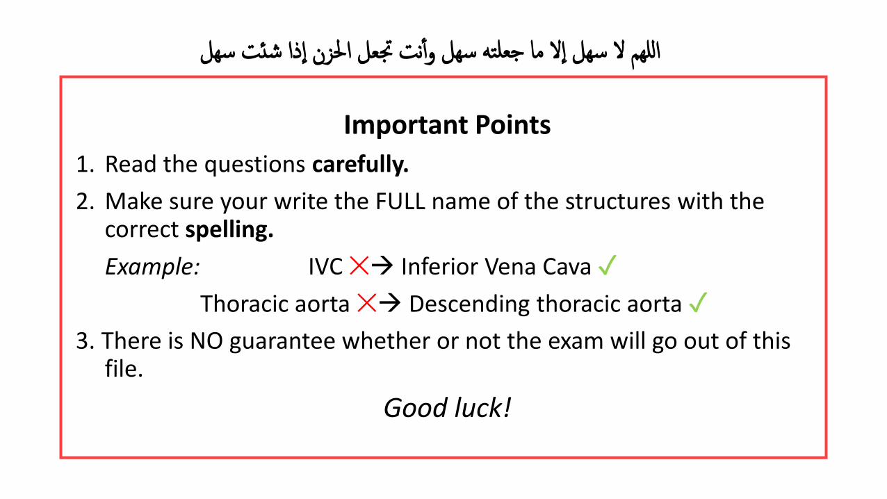
Important Points
1. Read the questions carefully.
2. Make sure your write the FULL name of the structures with the correct spelling.
Example: IVC ✕ Inferior Vena Cava ✓
Thoracic aorta ✕ Descending thoracic aorta ✓
3. There is NO guarantee whether or not the exam will go out of this file.
Good luck!
ذا شئت سهل ال ما جعلته سهل وأ نت جتعل احلزن ا اللهم ال سهل ا
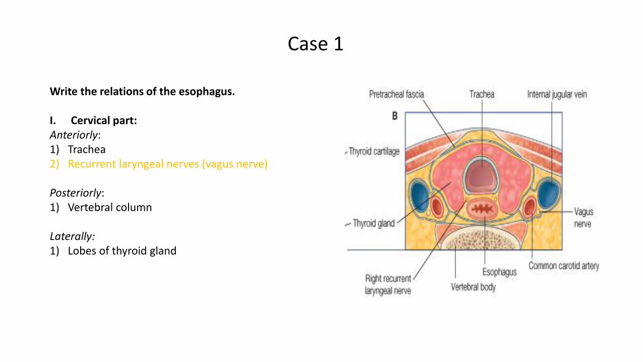
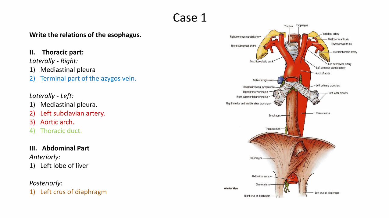
Case 1
Write the relations of the esophagus.
I. Cervical part:Anteriorly:1) Trachea2) Recurrent laryngeal nerves (vagus nerve)
Posteriorly:1) Vertebral column
Laterally:1) Lobes of thyroid gland
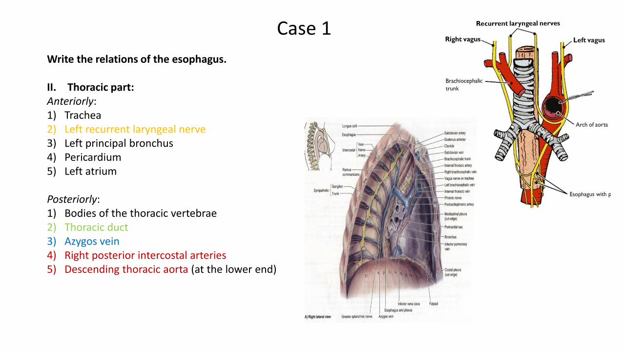
Case 1
Write the relations of the esophagus.
II. Thoracic part:Anteriorly:1) Trachea2) Left recurrent laryngeal nerve3) Left principal bronchus4) Pericardium5) Left atrium
Posteriorly:1) Bodies of the thoracic vertebrae2) Thoracic duct3) Azygos vein4) Right posterior intercostal arteries5) Descending thoracic aorta (at the lower end)
Case 1Write the relations of the esophagus.
II. Thoracic part:Laterally - Right:1) Mediastinal pleura2) Terminal part of the azygos vein.
Laterally - Left:1) Mediastinal pleura.2) Left subclavian artery.3) Aortic arch.4) Thoracic duct.
III. Abdominal PartAnteriorly: 1) Left lobe of liver
Posteriorly:1) Left crus of diaphragm
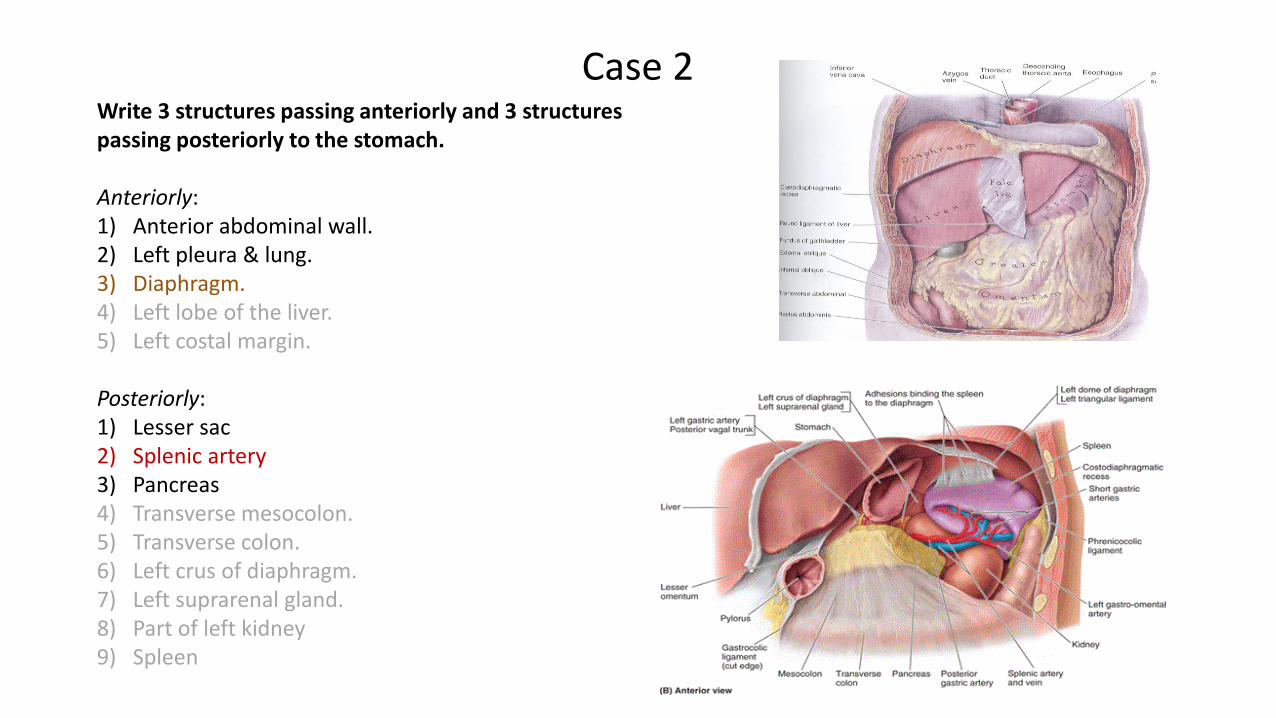
Write 3 structures passing anteriorly and 3 structures passing posteriorly to the stomach.
Anteriorly:1) Anterior abdominal wall.2) Left pleura & lung.3) Diaphragm.4) Left lobe of the liver.5) Left costal margin.
Posteriorly:1) Lesser sac2) Splenic artery3) Pancreas4) Transverse mesocolon.5) Transverse colon.6) Left crus of diaphragm.7) Left suprarenal gland.8) Part of left kidney9) Spleen
Case 2
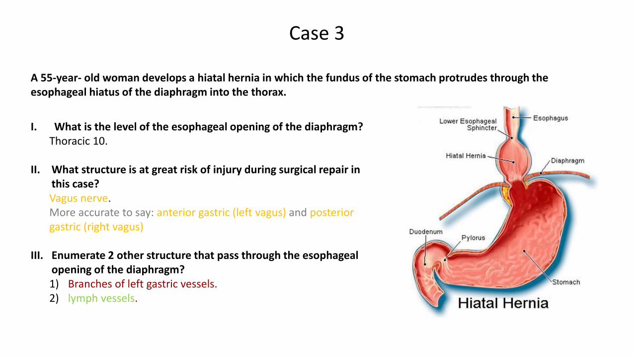
A 55-year- old woman develops a hiatal hernia in which the fundus of the stomach protrudes through the esophageal hiatus of the diaphragm into the thorax.
Case 3
I. What is the level of the esophageal opening of the diaphragm? Thoracic 10.
II. What structure is at great risk of injury during surgical repair in this case?Vagus nerve. More accurate to say: anterior gastric (left vagus) and posterior gastric (right vagus)
III. Enumerate 2 other structure that pass through the esophageal opening of the diaphragm? 1) Branches of left gastric vessels. 2) lymph vessels.
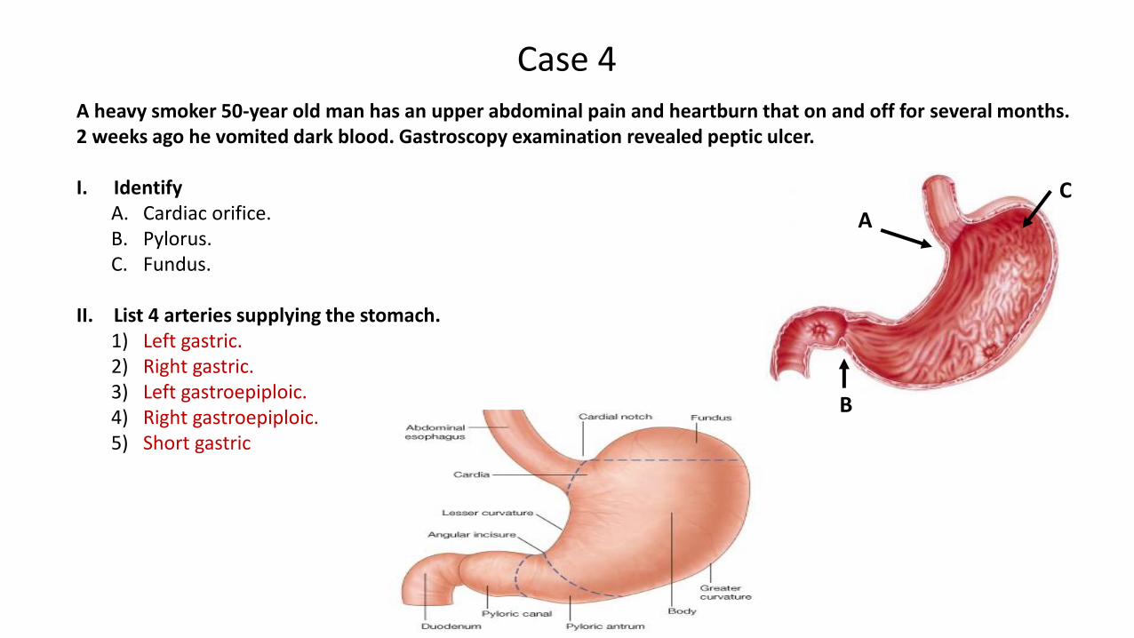
A heavy smoker 50-year old man has an upper abdominal pain and heartburn that on and off for several months. 2 weeks ago he vomited dark blood. Gastroscopy examination revealed peptic ulcer.
I. IdentifyA. Cardiac orifice.B. Pylorus. C. Fundus.
II. List 4 arteries supplying the stomach. 1) Left gastric.2) Right gastric.3) Left gastroepiploic.4) Right gastroepiploic.5) Short gastric
Case 4
A
C
B
This slide was added by Dr. Jameela
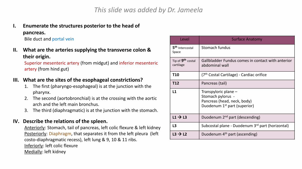
I. Enumerate the structures posterior to the head of pancreas.Bile duct and portal vein
II. What are the arteries supplying the transverse colon & their origin.Superior mesenteric artery (from midgut) and inferior mesenteric artery (from hind gut)
III. What are the sites of the esophageal constrictions?1. The first (pharyngo-esophageal) is at the junction with the
pharynx.2. The second (aortobronchial) is at the crossing with the aortic
arch and the left main bronchus.3. The third (diaphragmatic) is at the junction with the stomach.
IV. Describe the relations of the spleen.Anteriorly: Stomach, tail of pancreas, left colic flexure & left kidneyPosteriorly: Diaphragm, that separates it from the left pleura (left costo-diaphragmatic recess), left lung & 9, 10 & 11 ribs.Inferiorly: left colic flexureMedially: left kidney
Level Surface Anatomy
5th Intercostal Space
Stomach fundus
Tip of 9th costal cartilage
Gallbladder Fundus comes in contact with anterior abdominal wall
T10 (7th Costal Cartilage) - Cardiac orifice
T12 Pancreas (tail)
L1 Transpyloric plane –Stomach pylorus -Pancreas (head, neck, body)Duodenum 1st part (superior)
L1 L3 Duodenum 2nd part (descending)
L3 Subcostal plane - Duodenum 3rd part (horizontal)
L3 L2 Duodenum 4th part (ascending)
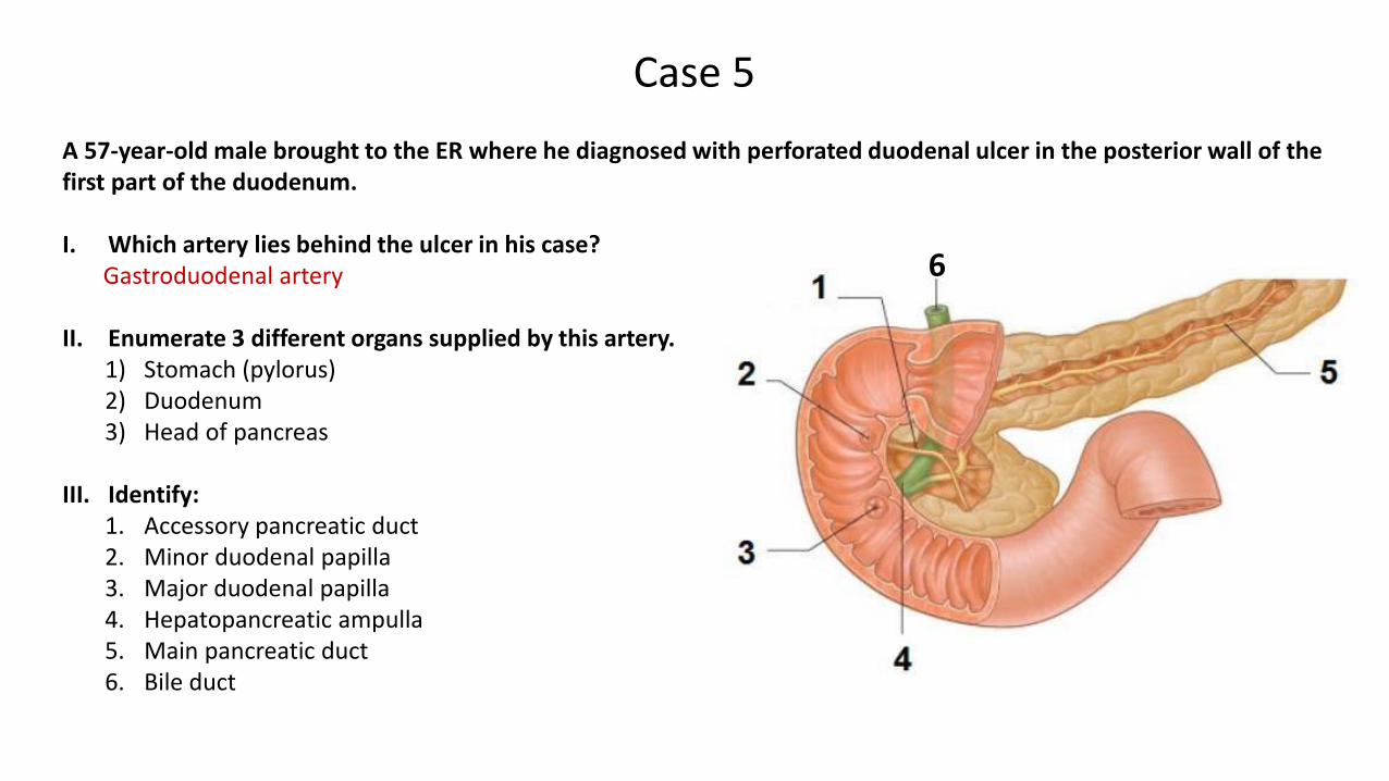
A 57-year-old male brought to the ER where he diagnosed with perforated duodenal ulcer in the posterior wall of the first part of the duodenum.
I. Which artery lies behind the ulcer in his case?Gastroduodenal artery
II. Enumerate 3 different organs supplied by this artery.1) Stomach (pylorus)2) Duodenum3) Head of pancreas
III. Identify:1. Accessory pancreatic duct2. Minor duodenal papilla3. Major duodenal papilla4. Hepatopancreatic ampulla5. Main pancreatic duct6. Bile duct
Case 5
6
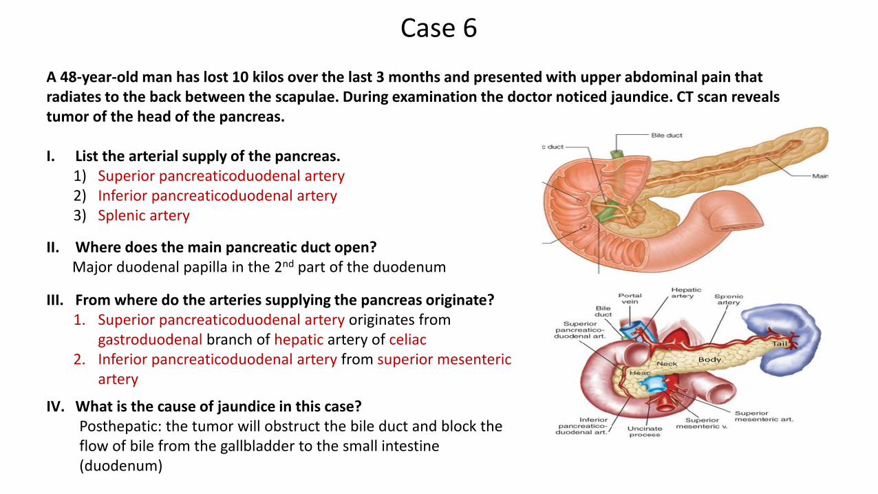
A 48-year-old man has lost 10 kilos over the last 3 months and presented with upper abdominal pain that radiates to the back between the scapulae. During examination the doctor noticed jaundice. CT scan reveals tumor of the head of the pancreas.
I. List the arterial supply of the pancreas.1) Superior pancreaticoduodenal artery2) Inferior pancreaticoduodenal artery3) Splenic artery
II. Where does the main pancreatic duct open?Major duodenal papilla in the 2nd part of the duodenum
Case 6
III. From where do the arteries supplying the pancreas originate?1. Superior pancreaticoduodenal artery originates from
gastroduodenal branch of hepatic artery of celiac2. Inferior pancreaticoduodenal artery from superior mesenteric
artery
IV. What is the cause of jaundice in this case?Posthepatic: the tumor will obstruct the bile duct and block the flow of bile from the gallbladder to the small intestine (duodenum)
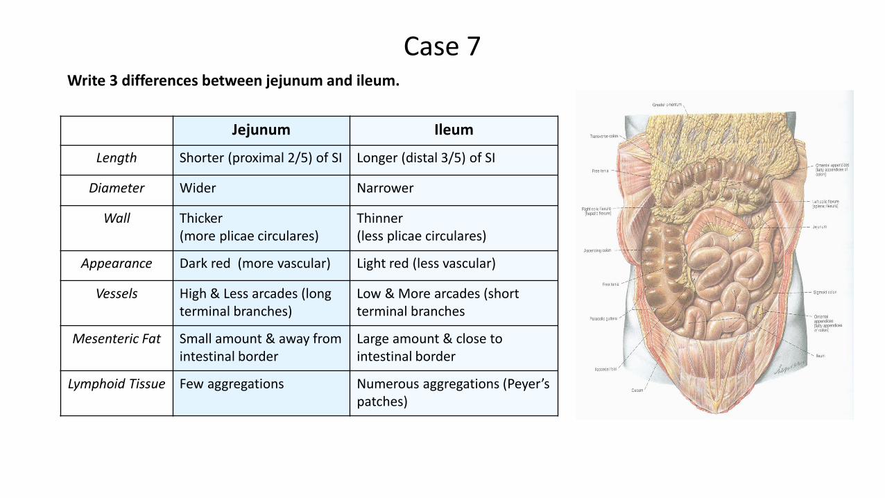
Case 7Write 3 differences between jejunum and ileum.
Jejunum Ileum
Length Shorter (proximal 2/5) of SI Longer (distal 3/5) of SI
Diameter Wider Narrower
Wall Thicker (more plicae circulares)
Thinner (less plicae circulares)
Appearance Dark red (more vascular) Light red (less vascular)
Vessels High & Less arcades (long terminal branches)
Low & More arcades (short terminal branches
Mesenteric Fat Small amount & away from intestinal border
Large amount & close to intestinal border
Lymphoid Tissue Few aggregations Numerous aggregations (Peyer’spatches)
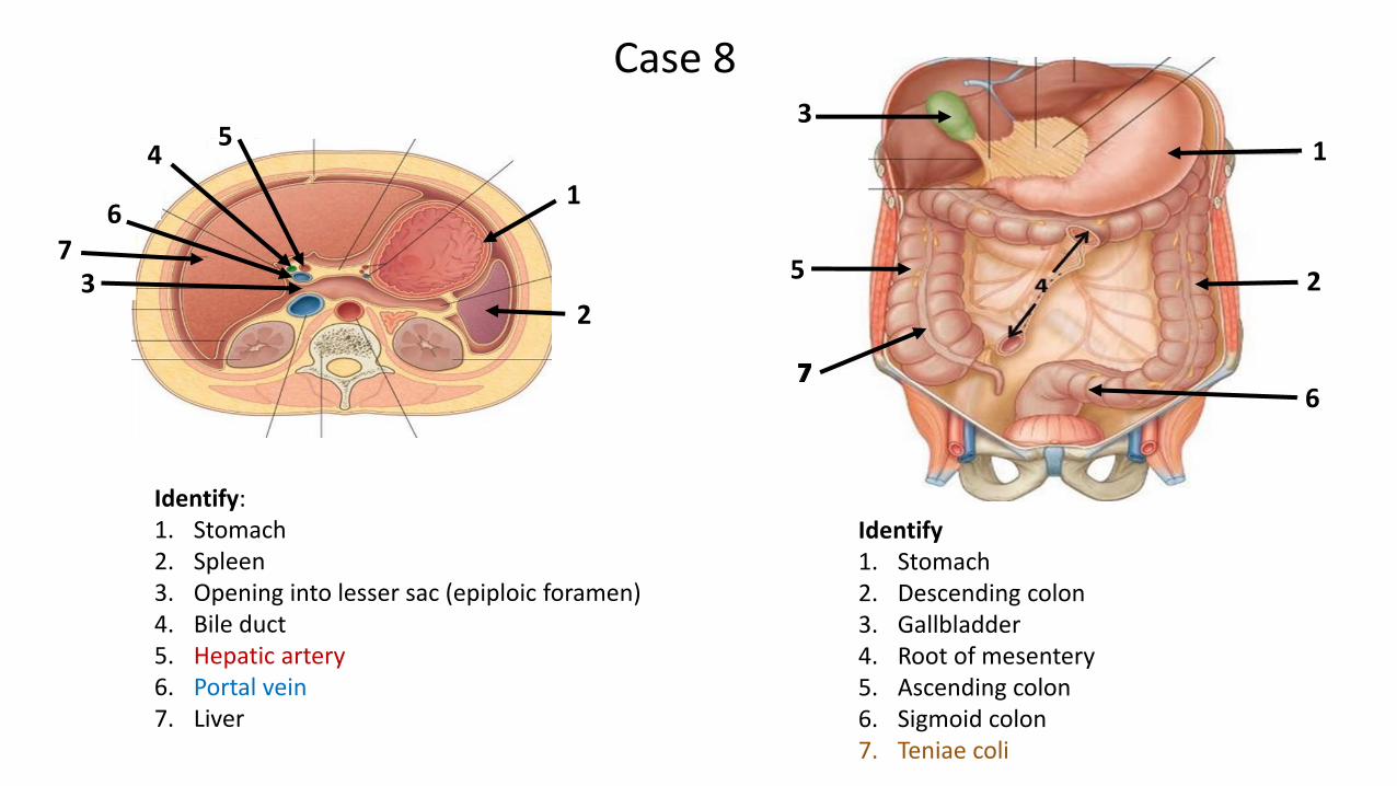
Identify:1. Stomach2. Spleen3. Opening into lesser sac (epiploic foramen)4. Bile duct5. Hepatic artery6. Portal vein7. Liver
32
1
7
45
6
Case 8
Identify1. Stomach2. Descending colon3. Gallbladder4. Root of mesentery5. Ascending colon6. Sigmoid colon7. Teniae coli
6
3
25
1
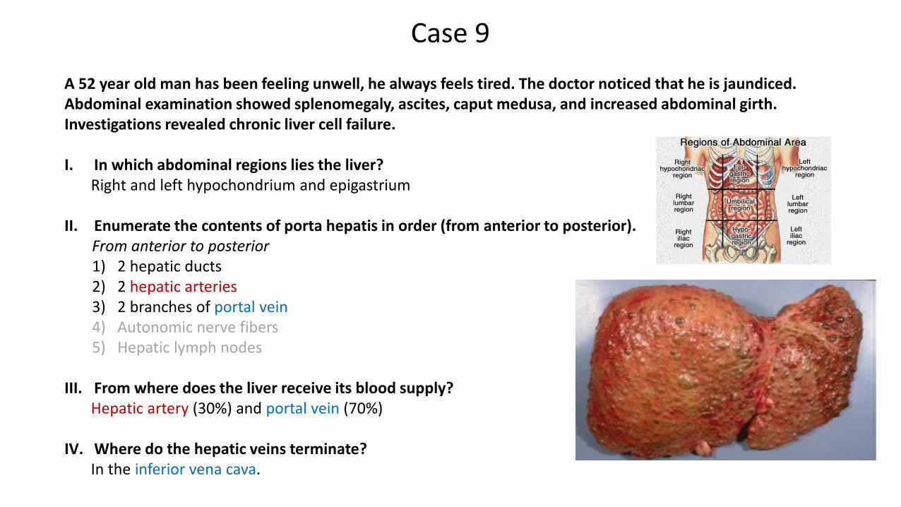
A 52 year old man has been feeling unwell, he always feels tired. The doctor noticed that he is jaundiced. Abdominal examination showed splenomegaly, ascites, caput medusa, and increased abdominal girth. Investigations revealed chronic liver cell failure.
I. In which abdominal regions lies the liver?Right and left hypochondrium and epigastrium
II. Enumerate the contents of porta hepatis in order (from anterior to posterior).From anterior to posterior1) 2 hepatic ducts2) 2 hepatic arteries 3) 2 branches of portal vein4) Autonomic nerve fibers5) Hepatic lymph nodes
III. From where does the liver receive its blood supply?Hepatic artery (30%) and portal vein (70%)
IV. Where do the hepatic veins terminate?In the inferior vena cava.
Case 9
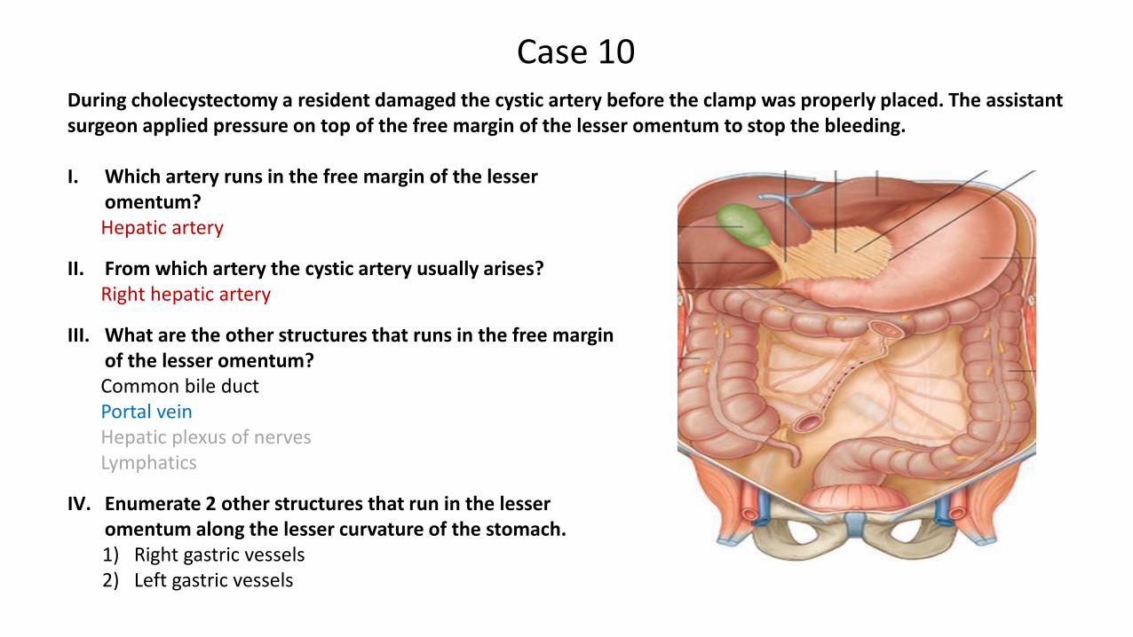
During cholecystectomy a resident damaged the cystic artery before the clamp was properly placed. The assistant surgeon applied pressure on top of the free margin of the lesser omentum to stop the bleeding.
Case 10
I. Which artery runs in the free margin of the lesser omentum?Hepatic artery
II. From which artery the cystic artery usually arises?Right hepatic artery
III. What are the other structures that runs in the free margin of the lesser omentum?Common bile ductPortal veinHepatic plexus of nervesLymphatics
IV. Enumerate 2 other structures that run in the lesser omentum along the lesser curvature of the stomach.1) Right gastric vessels2) Left gastric vessels
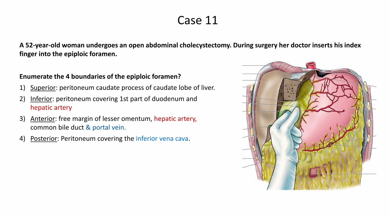
A 52-year-old woman undergoes an open abdominal cholecystectomy. During surgery her doctor inserts his index finger into the epiploic foramen.
Case 11
Enumerate the 4 boundaries of the epiploic foramen?
1) Superior: peritoneum caudate process of caudate lobe of liver.
2) Inferior: peritoneum covering 1st part of duodenum and hepatic artery
3) Anterior: free margin of lesser omentum, hepatic artery,common bile duct & portal vein.
4) Posterior: Peritoneum covering the inferior vena cava.
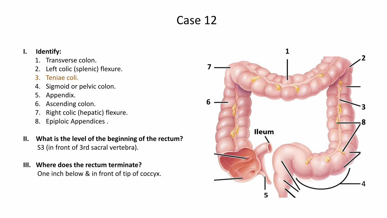
I. Identify:1. Transverse colon.2. Left colic (splenic) flexure.3. Teniae coli.4. Sigmoid or pelvic colon.5. Appendix.6. Ascending colon.7. Right colic (hepatic) flexure.8. Epiploic Appendices .
II. What is the level of the beginning of the rectum? S3 (in front of 3rd sacral vertebra).
III. Where does the rectum terminate?One inch below & in front of tip of coccyx.
Case 12
2
3
4
7
6
1
8
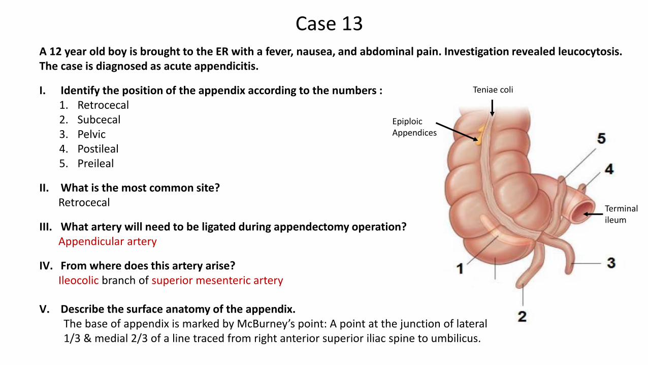
A 12 year old boy is brought to the ER with a fever, nausea, and abdominal pain. Investigation revealed leucocytosis. The case is diagnosed as acute appendicitis.
I. Identify the position of the appendix according to the numbers :1. Retrocecal2. Subcecal3. Pelvic4. Postileal5. Preileal
II. What is the most common site?Retrocecal
III. What artery will need to be ligated during appendectomy operation?Appendicular artery
IV. From where does this artery arise?Ileocolic branch of superior mesenteric artery
Case 13
Terminal ileum
Epiploic Appendices
Teniae coli
V. Describe the surface anatomy of the appendix.The base of appendix is marked by McBurney’s point: A point at the junction of lateral 1/3 & medial 2/3 of a line traced from right anterior superior iliac spine to umbilicus.
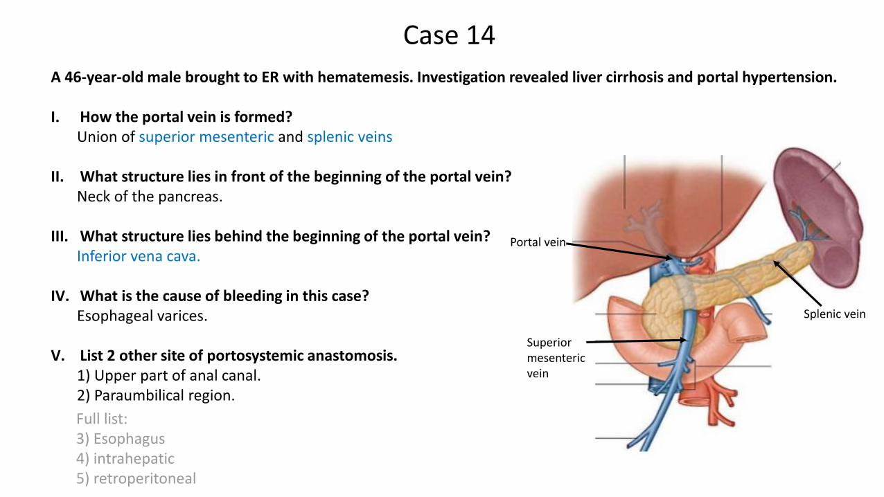
A 46-year-old male brought to ER with hematemesis. Investigation revealed liver cirrhosis and portal hypertension.
I. How the portal vein is formed? Union of superior mesenteric and splenic veins
II. What structure lies in front of the beginning of the portal vein? Neck of the pancreas.
III. What structure lies behind the beginning of the portal vein? Inferior vena cava.
IV. What is the cause of bleeding in this case? Esophageal varices.
V. List 2 other site of portosystemic anastomosis. 1) Upper part of anal canal. 2) Paraumbilical region.
Full list: 3) Esophagus4) intrahepatic 5) retroperitoneal
Case 14
Splenic vein
Portal vein
Superior mesenteric vein
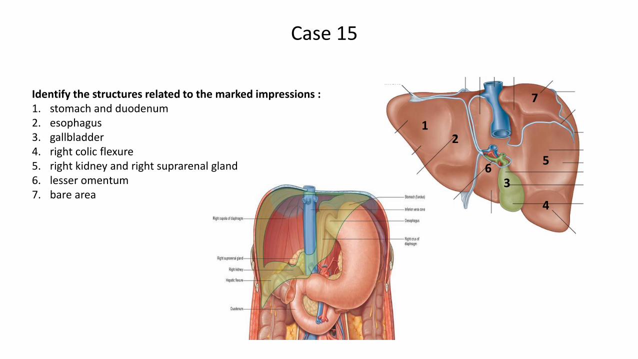
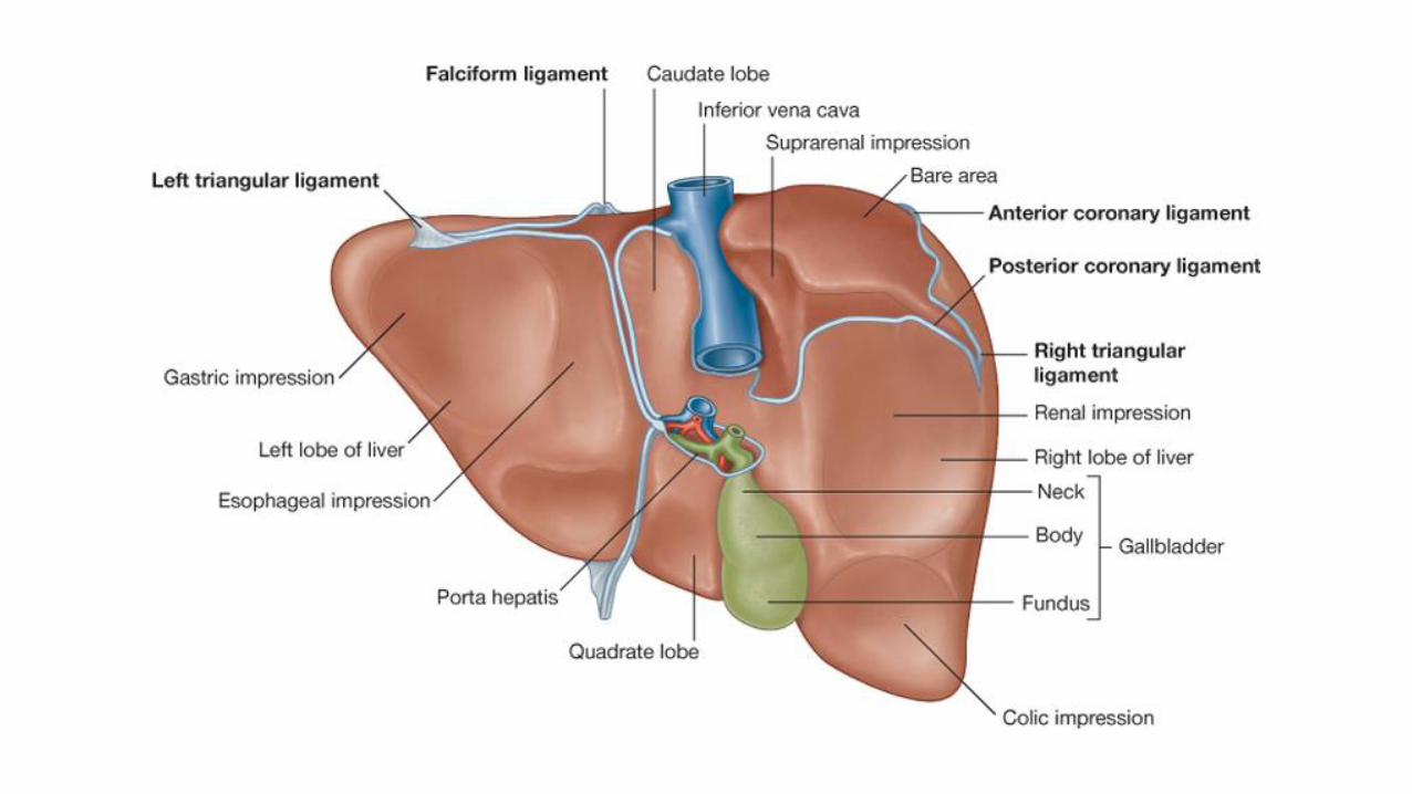
Identify the structures related to the marked impressions :1. stomach and duodenum2. esophagus3. gallbladder 4. right colic flexure 5. right kidney and right suprarenal gland 6. lesser omentum7. bare area
Case 15
1
63
5
4
2
7
Jawaher Abanumy
Nada Aldakheel
@anatomy436
Feedback
Mohammed Ghandour
Mohammed Alyousef
Khalid Aleedan
Done by:
Extra Pictures For PracticePractice Test
References:
1- Team 435
2- Greys Anatomy for Students
Editing File