Embed Size (px)
Citation preview
Proc. Nati. Acad. Sci. USAVol. 91, pp. 3589-3592, April 1994Medical Sciences
Pancreatic beta-cell replication and amelioration of surgicaldiabetes by Reg protein
(Reg gene/diabetes mellitus/Isets of Langerhans/growth factor)
TAKUO WATANABE*, YUTAKA YONEMURAt, HIDETO YONEKURA*, YOSHIHISA SUZUKI*, HIKARI MIYASHITA*,KAZUO SUGIYAMAt, SHIGEKI MORIIZUMI*, MICHIAKI UNNO*, OSAMU TANAKAt, HISATAKE KONDOt,ADRIAN J. BONE§, SHIN TAKASAWA*, AND HIROSHI OKAMOTO*¶Departments of *Biochemistry and *Anatomy, Tohoku University School of Medicine, Sendai 980, Miyagi, Japan; tDepartment of Surgery, KanazawaUniversity School of Medicine, Kanazawa 920, Ishikawa, Japan; and IDepartment of Pharmacy, University of Brighton, Moulsecoomb,Brighton BN2 4GJ, United Kingdom
Communicated by Osamu Hayaishi, December 20, 1993
ABSTRACT We previously Isolated from a rat regenerat-big islet cDNA library a gene named Reg, which Is expressed inregenerating Islets but is not expressed in normal islets. Herewe examined the effect of rat Reg protein on pancreaticbeta-cell replication using both 90% depancreatized rats andisolated Islets. The depancreatized rats that received i.p. ad-ministration ofrecombinant rat Reg protein (1 mg/kg per day)for 2 months showed amelioration of the surgical diabetes, asevidenced by a sigificant decrease in blood glucose with anincreased beta-cell mass in the residual pancreas. In Isolated ratIslets, Reg protein (18-180 nM: 0.3-3 pg/ml) significantlyincreased [3Hlthymidine incorporation into the nuclei of betacells. These results indicate that Reg protein Is a growth factorfor pancreatic beta cells and also suggest that the aministra-tion of Reg protein could be used as another therapeuticapproach for diabetes mellitus.
Pancreatic islets of Langerhans are the only organs of insulinproduction, but they have a limited capacity for regeneration,which predisposes to the development ofdiabetes mellitus. In1984, we found that the administration of poly(ADP-ribose)synthetase inhibitors, such as nicotinamide, to 90% depan-creatized rats induced the regeneration of pancreatic islets(1-3). In screening the regenerating islet-derived cDNA li-brary, we identified a gene, Reg (i.e., regenerating gene),which is expressed in regenerating islets but is not expressedin normal islets (4). The rat Reg cDNA had a single openreading frame that encoded a 165-amino acid protein with a21-amino acid signal peptide. We also isolated the humanREG cDNA and gene, which encoded a 166-amino acidprotein that showed a high degree of homology with the ratReg protein (4-6). Although the Reg protein has been sug-gested to be involved in beta-cell regeneration or growth(4-11), no direct evidence for its role in stimulating thegrowth of pancreatic beta cells had yet been obtained. Herewe demonstrate that recombinant rat Reg protein amelioratesthe diabetes of 909% depancreatized rats with beta-cell pro-liferation and that stimulation by Reg protein causes DNAsynthesis in islet beta cells in culture.
MATERIALS AND METHODSAnimal Experiment. Thirty-seven male Wistar rats (200-
220 g of weight) were 90o depancreatized (1, 2, 4) andmaintained on standard rat chow. Recombinant rat Regprotein, produced in Saccharomyces cerevisiae, purified (12)and dissolved at a concentration of 1 mg/ml in 50 mM aceticacid, was injected i.p. at a dose of 1 mg/kg ofbody weight into
20 rats every day. Seventeen control rats were injected i.p.with 50 mM of acetic acid without Reg protein after the 90%pancreatectomy. In both groups the injections were contin-ued until the 60th postoperative day. Rats were fastedovernight before blood sampling, and the plasma glucoselevel was measured by the glucose oxidase method, usingGlucose Auto and Stat GA-illO (Kyoto Daiichi Instrument,Kyoto). The statistical significance of differences betweenrats injected with Reg protein (n = 20) and those injected withacetic acid (n = 17) was analyzed by using Student's t test.The residual pancreatic tissues were removed 2 months afterthe partial pancreatectomy and fixed in Bouins' solution.Hydrated 5-pm sections of paraffin-embedded pancreatictissues were stained for insulin by the labeled streptavidin-biotin method, using an LSAB kit (Dakopatts, Glostrup,Denmark). After being stained, the relative volumes of betacells were measured by the point-counting method (1, 13).In Vitro Experiment. Pancreatic islets were isolated from
male Wistar rats (body weight 200-250 g) by using densityseparation on a dextran gradient and cultured free-floating inRPMI 1640 medium/10%o fetal calf serum/penicillin G at 100pg/ml/streptomycin at 100 g/ml for 48 hr to allow recoveryfrom the isolation procedure (14). After this initial period,islets were transferred to 24-well culture dishes in groups of50 islets. The islets were cultured in RPMI 1640 medium/2.7mM D-glucose/2% fetal calf serum/penicillin G at 100 ;g/ml/streptomycin at 100 ;g/ml in the presence of increasedconcentrations ofReg protein for 72 hr. During the last 24 hr,the islets were cultured in the above medium to which[methyl-3H]thymidine at 10 puCi/ml (Amersham; 1 Ci = 37GBq) had been added. To estimate the amount of [3H]thy-midine incorporated into newly synthesized DNA, the isletswere washed as described (14) after the culture period andsonicated in 10 mM Tris.HCl/5 mM EDTA. DNA wasprecipitated by the addition of 7% ice-cold trichloroaceticacid and trapped by filtration on a glass-fiber disc (WhatmanGF/C). The discs were dried, and radioactivity was countedafter the addition of scintillation fluid (Packard Ultima GoldF). The DNA content of the islets was measured by aflurometric DNA assay using Hoechst 33258. To estimatelabeling indices for insulin-positive cells, the [3H]thymidine-labeled islets were fixed in 4% paraformaldehyde for 20 minand embedded in OCT compound. Ten-micrometer sectionswere cut with a cryostat and immunostained with anti-porcine insulin guinea pig antiserum (DAKO, 1:500) andVectastain ABC-GO kit (Vector Laboratories). Autoradiog-raphy ofthe immunostained sections was performed by usingKonica NR-M2 emulsion.
lTo whom reprint requests should be addressed at: Department ofBiochemistry, Tohoku University School of Medicine, 2-1 Seiryo-machi, Aoba-ku, Sendai 980, Miyagi, Japan.
3589
The publication costs of this article were defrayed in part by page chargepayment. This article must therefore be hereby marked "advertisement"in accordance with 18 U.S.C. §1734 solely to indicate this fact.
Dow
nloa
ded
by g
uest
on
Mar
ch 6
, 202
0
3590 Medical Sciences: Watanabe et al.
Postoperative days Postoperative days
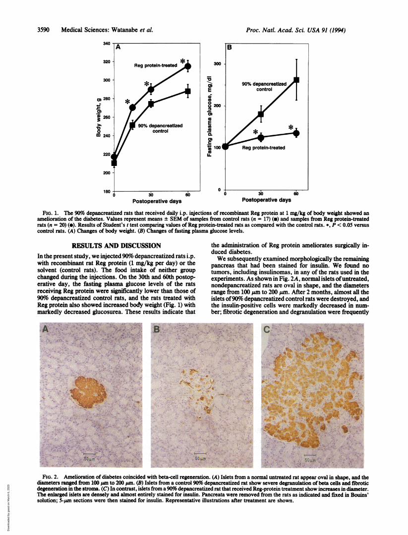
FIG. 1. The 90%o depancreatized rats that received daily i.p. injections of recombinant Reg protein at 1 mg/kg of body weight showed anamelioration of the diabetes. Values represent means ± SEM of samples from control rats (n = 17) (v) and samples from Reg protein-treatedrats (n = 20) (e). Results of Student's t test comparing values ofReg protein-treated rats as compared with the control rats. *, P < 0.05 versuscontrol rats. (A) Changes of body weight. (B) Changes of fasting plasma glucose levels.
RESULTS AND DISCUSSIONIn the present study, we injected 90% depancreatized rats i.p.with recombinant rat Reg protein (1 mg/kg per day) or thesolvent (control rats). The food intake of neither groupchanged during the injections. On the 30th and 60th postop-erative day, the fasting plasma glucose levels of the ratsreceiving Reg protein were significantly lower than those of90% depancreatized control rats, and the rats treated withReg protein also showed increased body weight (Fig. 1) withmarkedly decreased glucosurea. These results indicate thatA *t; p ,. Alp; > ai- ''tv's,me .i; .,.5< ;;7:C ............ *. i . .e@ ^
.r
:. ._'
.. ,. t.a.
-- .:* . s -*s v ''''an
Beiv. , , q , i , -* S , 4P S.
s;^ IS
,v me- .... . ... .... ... A.. j
- >1
; ., + ,.9 Z S *aSef f
+ S 1,
+ ;80 rim § e, +_.
the administration of Reg protein ameliorates surgically in-duced diabetes.We subsequently examined morphologically the remaining
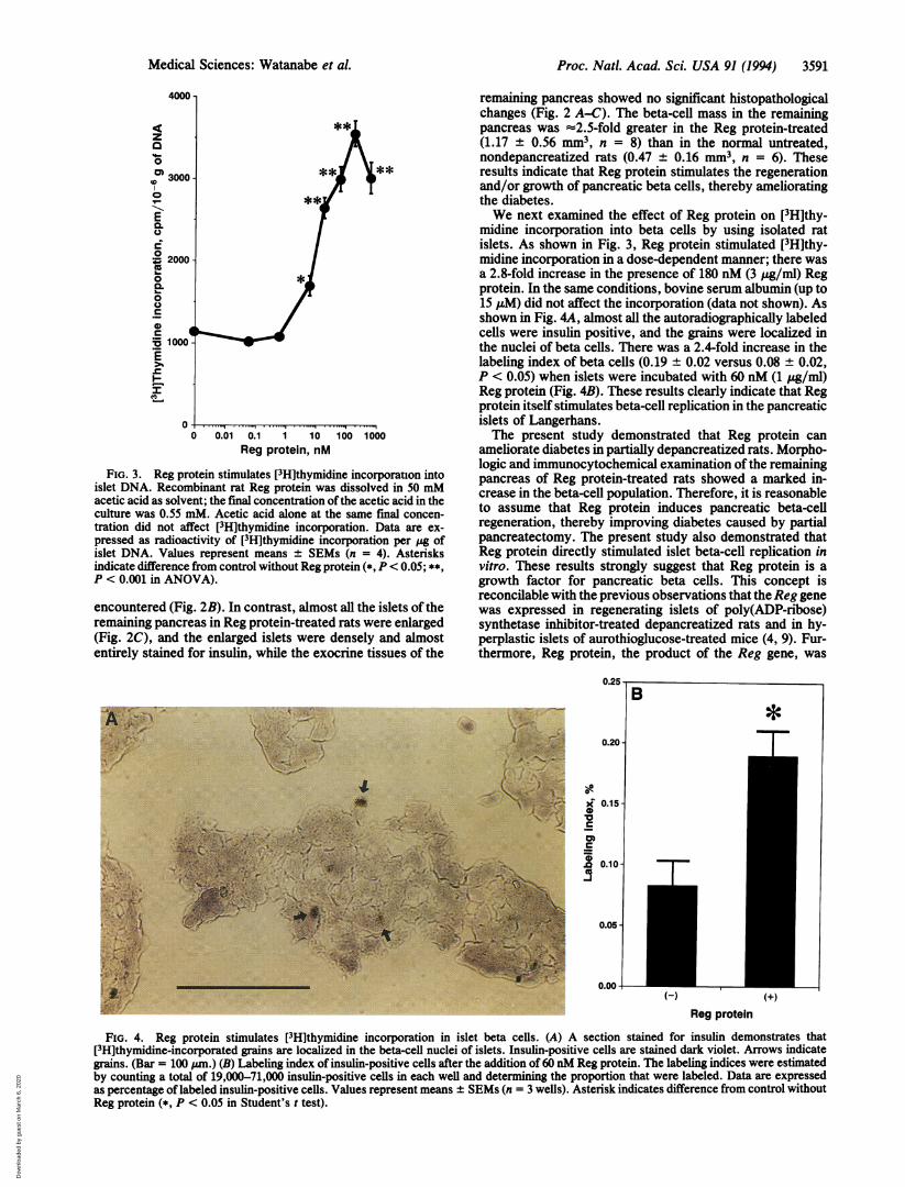
pancreas that had been stained for insulin. We found notumors, including insulinomas, in any of the rats used in theexperiments. As shown in Fig. 2A, normal islets ofuntreated,nondepancreatized rats are oval in shape, and the diametersrange from 100 ,um to 200 )zm. After 2 months, almost all theislets of90% depancreatized control rats were destroyed, andthe insulin-positive cells were markedly decreased in num-ber; fibrotic degeneration and degranulation were frequently
B . * .s a. . w s . r.. . 4 , ...
:.
.e
* 6i>
>
f A, ^.3
s ; Use 4
Age *42 {i '*.. § U
_
oa.i,,'I
sS
: 'ffi Sowm
.. .. W
-~~~- . :} s&:
V
~~~~~~~~~~~A 10
V
w~~~~~5, k0m;; ;s
FIG. 2. Amelioration of diabetes coincided with beta-cell regeneration. (A) Islets from a normal untreated rat appear oval in shape, and thediameters ranged from 100 ,.m to 200 pm. (B) Islets from a control 90%o depancreatized rat show severe degranulation of beta cells and fibroticdegeneration in the stroma. (C) In contrast, islets from a90% depancreatized rat that received Reg-protein treatment show increases in diameter.The enlarged islets are densely and almost entirely stained for insulin. Pancreata were removed from the rats as indicated and fixed in Bouins'solution; 5-,um sections were then stained for insulin. Representative illustrations after treatment are shown.
340
0._
3 260
a00M 240
Proc. Natl. Acad. Sci. USA 91 (1994)
Dow
nloa
ded
by g
uest
on
Mar
ch 6
, 202
0
Proc. Nati. Acad. Sci. USA 91 (1994) 3591
4000 -
<, ~~~**Iz
0
0 3000**-**0 **SE0.0
0= 2000
0.
0
a)
1000
0-..,..........................0 0.01 0.1 1 10 100 1000
Reg protein, nM
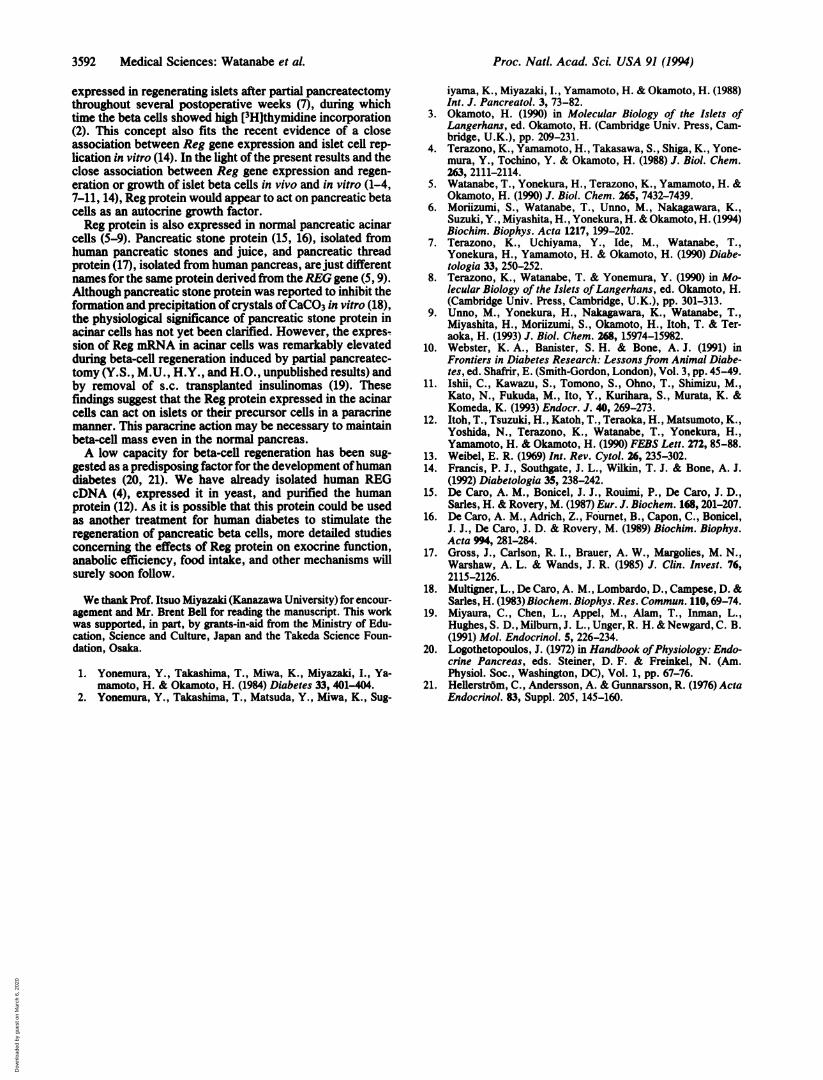
FIG. 3. Reg protein stimulates [3H]thymidine incorporauon intoislet DNA. Recombinant rat Reg protein was dissolved in 50 mMacetic acid as solvent; the final concentration of the acetic acid in theculture was 0.55 mM. Acetic acid alone at the same final concen-tration did not affect [3H]thymidine incorporation. Data are ex-pressed as radioactivity of [3H]thymidine incorporation per Zg ofislet DNA. Values represent means ± SEMs (n = 4). Asterisksindicate difference from control without Reg protein (*, P < 0.05; **,P < 0.001 in ANOVA).
encountered (Fig. 2B). In contrast, almost all the islets of theremaining pancreas in Reg protein-treated rats were enlarged(Fig. 2C), and the enlarged islets were densely and almostentirely stained for insulin, while the exocrine tissues of the
-it
:4
remaining pancreas showed no significant histopathologicalchanges (Fig. 2 A-C). The beta-cell mass in the remainingpancreas was =2.5-fold greater in the Reg protein-treated(1.17 ± 0.56 mm3, n = 8) than in the normal untreated,nondepancreatized rats (0.47 ± 0.16 mm3, n = 6). Theseresults indicate that Reg protein stimulates the regenerationand/or growth of pancreatic beta cells, thereby amelioratingthe diabetes.We next examined the effect of Reg protein on [3H]thy-
midine incorporation into beta cells by using isolated ratislets. As shown in Fig. 3, Reg protein stimulated [3H]thy-midine incorporation in a dose-dependent manner; there wasa 2.8-fold increase in the presence of 180 nM (3 yug/ml) Regprotein. In the same conditions, bovine serum albumin (up to15 pM) did not affect the incorporation (data not shown). Asshown in Fig. 4A, almost all the autoradiographically labeledcells were insulin positive, and the grains were localized inthe nuclei of beta cells. There was a 2.4-fold increase in thelabeling index of beta cells (0.19 ± 0.02 versus 0.08 ± 0.02,P < 0.05) when islets were incubated with 60 nM (1 jtg/ml)Reg protein (Fig. 4B). These results clearly indicate that Regprotein itself stimulates beta-cell replication in the pancreaticislets of Langerhans.The present study demonstrated that Reg protein can
ameliorate diabetes in partially depancreatized rats. Morpho-logic and immunocytochemical examination of the remainingpancreas of Reg protein-treated rats showed a marked in-crease in the beta-cell population. Therefore, it is reasonableto assume that Reg protein induces pancreatic beta-cellregeneration, thereby improving diabetes caused by partialpancreatectomy. The present study also demonstrated thatReg protein directly stimulated islet beta-cell replication invitro. These results strongly suggest that Reg protein is agrowth factor for pancreatic beta cells. This concept isreconcilable with the previous observations that the Reg genewas expressed in regenerating islets of poly(ADP-ribose)synthetase inhibitor-treated depancreatized rats and in hy-perplastic islets of aurothioglucose-treated mice (4, 9). Fur-thermore, Reg protein, the product of the Reg gene, was
0.20 -....A,7-i)+
VP4
X 0.15
0
D 0.10-
0.05 -
0.00
B*p
T17
(H) (+)
Reg protein
FIG. 4. Reg protein stimulates [3H]thymidine incorporation in islet beta cells. (A) A section stained for insulin demonstrates that[3H]thymidine-incorporated grains are localized in the beta-cell nuclei of islets. Insulin-positive cells are stained dark violet. Arrows indicategrains. (Bar = 100 jan.) (B) Labeling index of insulin-positive cells after the addition of 60 nM Reg protein. The labeling indices were estimatedby counting a total of 19,000-71,000 insulin-positive cells in each well and determining the proportion that were labeled. Data are expressedas percentage of labeled insulin-positive cells. Values represent means ± SEMs (n = 3 wells). Asterisk indicates difference from control withoutReg protein (*, P < 0.05 in Student's t test).
Medical Sciences: Watanabe et al.
Dow
nloa
ded
by g
uest
on
Mar
ch 6
, 202
0
3592 Medical Sciences: Watanabe et al.
expressed in regenerating islets after partial pancreatectomythroughout several postoperative weeks (7), during whichtime the beta cells showed high [3H]thymidine incorporation(2). This concept also fits the recent evidence of a closeassociation between Reg gene expression and islet cell rep-lication in vitro (14). In the light of the present results and theclose association between Reg gene expression and regen-eration or growth of islet beta cells in vivo and in vitro (1-4,7-11, 14), Reg protein would appear to act on pancreatic betacells as an autocrine growth factor.Reg protein is also expressed in normal pancreatic acinar
cells (5-9). Pancreatic stone protein (15, 16), isolated fromhuman pancreatic stones and juice, and pancreatic threadprotein (17), isolated from human pancreas, are just differentnames for the same protein derived from theREG gene (5, 9).Although pancreatic stone protein was reported to inhibit theformation and precipitation of crystals ofCaCO3 in vitro (18),the physiological significance of pancreatic stone protein inacinar cells has not yet been clarified. However, the expres-sion of Reg mRNA in acinar cells was remarkably elevatedduring beta-cell regeneration induced by partial pancreatec-tomy (Y.S., M.U., H.Y., and H.O., unpublished results) andby removal of s.c. transplanted insulinomas (19). Thesefindings suggest that the Reg protein expressed in the acinarcells can act on islets or their precursor cells in a paracrinemanner. This paracrine action may be necessary to maintainbeta-cell mass even in the normal pancreas.A low capacity for beta-cell regeneration has been sug-
gested as a predisposing factor for the development ofhumandiabetes (20, 21). We have already isolated human REGcDNA (4), expressed it in yeast, and purified the humanprotein (12). As it is possible that this protein could be usedas another treatment for human diabetes to stimulate theregeneration of pancreatic beta cells, more detailed studiesconcerning the effects of Reg protein on exocrine function,anabolic efficiency, food intake, and other mechanisms willsurely soon follow.
We thank Prof. Itsuo Miyazaki (Kanazawa University) for encour-agement and Mr. Brent Bell for reading the manuscript. This workwas supported, in part, by grants-in-aid from the Ministry of Edu-cation, Science and Culture, Japan and the Takeda Science Foun-dation, Osaka.
1. Yonemura, Y., Takashima, T., Miwa, K., Miyazaki, I., Ya-mamoto, H. & Okamoto, H. (1984) Diabetes 33, 401-404.
2. Yonemura, Y., Takashima, T., Matsuda, Y., Miwa, K., Sug-
iyama, K., Miyazaki, I., Yamamoto, H. & Okamoto, H. (1988)Int. J. Pancreatol. 3, 73-82.
3. Okamoto, H. (1990) in Molecular Biology of the Islets ofLangerhans, ed. Okamoto, H. (Cambridge Univ. Press, Cam-bridge, U.K.), pp. 209-231.
4. Terazono, K., Yamamoto, H., Takasawa, S., Shiga, K., Yone-mura, Y., Tochino, Y. & Okamoto, H. (1988) J. Biol. Chem.263, 2111-2114.
5. Watanabe, T., Yonekura, H., Terazono, K., Yamamoto, H. &Okamoto, H. (1990) J. Biol. Chem. 265, 7432-7439.
6. Moriizumi, S., Watanabe, T., Unno, M., Nakagawara, K.,Suzuki, Y., Miyashita, H., Yonekura, H. & Okamoto, H. (1994)Biochim. Biophys. Acta 1217, 199-202.
7. Terazono, K., Uchiyama, Y., Ide, M., Watanabe, T.,Yonekura, H., Yamamoto, H. & Okamoto, H. (1990) Diabe-tologia 33, 250-252.
8. Terazono, K., Watanabe, T. & Yonemura, Y. (1990) in Mo-lecular Biology of the Islets ofLangerhans, ed. Okamoto, H.(Cambridge Univ. Press, Cambridge, U.K.), pp. 301-313.
9. Unno, M., Yonekura, H., Nakagawara, K., Watanabe, T.,Miyashita, H., Moriizumi, S., Okamoto, H., Itoh, T. & Ter-aoka, H. (1993) J. Biol. Chem. 268, 15974-15982.
10. Webster, K. A., Banister, S. H. & Bone, A. J. (1991) inFrontiers in Diabetes Research: Lessons from Animal Diabe-tes, ed. Shafrir, E. (Smith-Gordon, London), Vol. 3, pp. 45-49.
11. Ishii, C., Kawazu, S., Tomono, S., Ohno, T., Shimizu, M.,Kato, N., Fukuda, M., Ito, Y., Kurihara, S., Murata, K. &Komeda, K. (1993) Endocr. J. 40, 269-273.
12. Itoh, T., Tsuzuki, H., Katoh, T., Teraoka, H., Matsumoto, K.,Yoshida, N., Terazono, K., Watanabe, T., Yonekura, H.,Yamamoto, H. & Okamoto, H. (1990) FEBS Lett. 272, 85-88.
13. Weibel, E. R. (1969) Int. Rev. Cytol. 26, 235-302.14. Francis, P. J., Southgate, J. L., Wilkin, T. J. & Bone, A. J.
(1992) Diabetologia 35, 238-242.15. De Caro, A. M., Bonicel, J. J., Rouimi, P., De Caro, J. D.,
Sarles, H. & Rovery, M. (1987) Eur. J. Biochem. 168, 201-207.16. De Caro, A. M., Adrich, Z., Fournet, B., Capon, C., Bonicel,
J. J., De Caro, J. D. & Rovery, M. (1989) Biochim. Biophys.Acta 994, 281-284.
17. Gross, J., Carlson, R. I., Brauer, A. W., Margolies, M. N.,Warshaw, A. L. & Wands, J. R. (1985) J. Clin. Invest. 76,2115-2126.
18. Multigner, L., De Caro, A. M., Lombardo, D., Campese, D. &Sarles, H. (1983) Biochem. Biophys. Res. Commun. 110, 69-74.
19. Miyaura, C., Chen, L., Appel, M., Alam, T., Inman, L.,Hughes, S. D., Milburn, J. L., Unger, R. H. & Newgard, C. B.(1991) Mol. Endocrinol. 5, 226-234.
20. Logothetopoulos, J. (1972) in Handbook ofPhysiology: Endo-crine Pancreas, eds. Steiner, D. F. & Freinkel, N. (Am.Physiol. Soc., Washington, DC), Vol. 1, pp. 67-76.
21. Hellerstr6m, C., Andersson, A. & Gunnarsson, R. (1976) ActaEndocrinol. 83, Suppl. 205, 145-160.
Proc. Natl. Acad. Sci. USA 91 (1994)
Dow
nloa
ded
by g
uest
on
Mar
ch 6
, 202
0