Embed Size (px)
Citation preview

Y.Y.O. Vet. F;ık. Derg. 1996, 7(1-2)' 58-61
Parathyroid eel! Variants May be Induced by Different Fixatives II, An Electro" Microscopic Study
Mehmet KANTER
YQzancO Yıl Onivtırsilesi Veterintır FekO!teai. Histolojl ve Embriyoloji Anabilim Dalı. Van. TÜRKIYE
Geliş Tarihi: 14 Hazıroın 1996
Farklı Tespit Sıvılarının Paratiroid Hücre Çeşitlilı!)! Üzerine Etkisi II. Elektron Mikroskobik Inceleme
SummOlry; In t~ i~ study. the ro~s of the different combination of a kje~ydos on the u!trulructure of parathyroid (PT) eclis were ınvestigated . Parat~yroid glands of rats were rOled by perfusion with diffcrcnt cornblnııllons of aldehydes in sodium phosphate buffer. and po,t (ıxed osmium tetro~ide (0804) In the same buffer. The specimens, parentllynıa] cells ol PT glands, fixed in 2.5% gluıarakkthyde (GA) were unnoım cells with m:ırkedly enlôırgcd intereellular spııceıı (1$), tOl'tuous plaYf1a membranes, aledro~nse cytoplasmic matrix containlng $.Iighlly dilliiiid ciSlemae of rough endoplnmıc retieuk.ım (RER), large or smail GoIgi mmplex (Ge), many mito<:tıondria and fe.... secıelory granules (SG). PT glands fixııd with <1 % paralomıaldehyde (PFA) icd to inlermııdi~te aılls with elııd:ronlucenl cytoplnmle matrix oonlıılolng mmedly diıaıed cistema ol RER. large or smail Ge, law mitochondira and few SG. However, lhe use ol r,xativı:ıs (;Onaist oll %G/I. _ 2% formaldehyt!e (FA) !ed only to lighl cells with highty electron-Iuconl cytoplıısmic matıiıl <:ontaining disrupted membranes of RER or Ge. The resu lts thus show that parathyroid ee ll vıırlanls arise during improper immersion or perfusi<ın fııration and t"~t differenı combinations of aldehydes lira importıınl factors provoklng parathyroid eell ~oriants.
Kay WordS: Rat, parath)'foid glalld, par.ılhyroid eeıı ~ariants, diffarent rıxatlves
Özet: SU ça.lı~mada, farklı aldehi! kombinasyonlarının, paratireid hOcrelerinin ince yapısı Olerine etkisi incelendi. Sıçan paratiroid bezltori, sodyum fosfat lamponu varlı9ında fa~1ı aldeM kombinasyonları kullanılarak perfOZYOi\-fiksıısyoıı metodu ile ön tespite, osmik as it kuııanımı Ile de ikinci tespitı bbi tutulduıar. Elektron mikroskobik incelemelerde; % 2.5 glutaraldeM ile tesp it edilı:ın doku ömekı.erinde p~ratiroid dokusu pıır8nkima! hücrelerinin Oniform hOcre yapısında oldugu gOrOldO. Bu hOcrelerde, inter~IIOler mesafenin oldukça genı" hücra zarının kallantılı, s~oplôızmik matriksin yogun, granOllu endoplazmik retillulumun halil dilale, büyük ~e kGçOk Golgı kompleksieri ile çok sayıda mitokandrlyon ve ;ız sayıda satgr granllllerin'" ~arlıgr dikkati çekti. Poıraformı1ldeMin %,4'liIk solusyonu ile lesp~ edilen paralirOId dokusunun intennediyer hücrelerden olu~UOu ve bu hOcrolerde 5itoplavnik mlllril<.sin açık, granOIlO cndoplazmik reıikulumun şlckIetli dilıı!e, bUyOk ve kOçilk GoIgI kompleksieri ile az sayıda mitokondriyon ve s~tgı 9ranO~rtııln ~arl~1 belirtendi. "41 GA·%2 FA ile lesprt edilen doku örneklerinde ise, paratiroie! dokusunun sadece açık hoa-e~rden oluştutju ve bu hOcrclerde ııitoplııımik malriksln oldukÇII açık, granullii endoplazmik relikulum v(l Golgl kompleksi membranlannın parçııtıınmı, oldllğu gozlendi. Bu çal ı şmada, paratiroicl hOere çaşilliHginin. yetersiz immersiyon -veya peı1Qzyon- fiksasyon esnasında o l uşmuş olablleCC9i v(l fa~1ı eldehit kombinasyonlarının bu hiicre ı;:e;itliliOin.ııe onemli rol oynadı!,!! sonucuna varıldı.
Anahtar Kelimeler: Sıçan , paratiroid bezi, p91eliroid hiicre ~iltiliOi, IMdı filc.satif
Introduction
Lighl and dark cells of paralhyroid (PT) glands are believed to represent different functional stages of PT cells which undergo cydiC morphological changes in the ccurse of parathyroid hormon secretion (1,2,8). This idea was put forward, first bacause of Ihe occurence of so ealled dark and lighl cells differing in the stainability of the cytoplasm, in the size and distribution of organeUes, and in the course of the plasma membrane (8,12). Second, it was found that the appearance of PT ceııs varie<! in relaıion lo the calcium concenlralion used for stimulalion or suppression secretory activity of PT cells (9). Stöeckel and Porte (11), however, have suggesled that the different morphology in rat PT cells is the resun of fixation artefacts rather than biologir.at events. This possibility may be supported by [hı... 111 'ng of a more uniform electron density in paralhyroiJ glands fixed by perfusion than in those fıxed by immerslon (10). Comparison of immersion
fixed PT glands wilh those fixed by perfusion of Mongolian gerbils (4), rals (6,14), dogs (10,15), mice and cats (15) revealed strong evidence lo assume thal PT cell variants anse during immersion fixalien.
The aim of this study was undertaken to invesligate the roles of Ihe different combination of aldehydes on the ultraslructure of PT cells in rats using vascular penusion lechnique.
Materials and Methods
Six adult rats (200-300g) kept on a standard diet were deeply anesthetized by intraperiloneal injedion of Kelalar (65mglkg). Five thousand units of sodium heparin dissolved in 1 ml saline was injected directly into the teft venlride. The righl atrium and the apex of the heart were eul, and a cannula was inserted through the left ventricla into the aorta. Physiological saiine proceeded the now of fixalives: aı 2,5 % gluteraldehyde, bı 4 % paraformaldehyde,

Y.Y.Ü. Vet. Fak. Derg. 1996,7(1-2): 58-61
c) 1 % gluteraldehyde + 2 % formaldehyde; respectively in 0.1 M sodium phosphate buffer (pH 7.2). The flow rate was maintained at approximately 6 ml/min for 15 min. for both the saline and the fixative solutions. Following perfusion, PT glands were carefully removed and immersed in the same fixatives for 1 h at 4 oC, washed in the same buffer for 1 h at 4 oC, and post-fixed with 1 % 0504 in sodium phosphate buffer for 1 h at 4 oC. The tissues were then dehydrated in a graded series of ethanol starting at 70% each step for 10 min and, after !wo changes in propylene oxide. The tissues were embedded in Araidite. Ultrathin sections were stained with Mg-uranyl acetate and lead citrate for the electron microscopic (Zeiss EM-10) examination.
Results
PT cell variants in rats as revealed by perfusion fixation with different combinations of aldehydes in same buffers are summarized in Table 1. The specimens, parenchymal cells of PT glands, fixed in 2.5% GA were uniform cells with markedly enlarged iS, tortuous plasma membranes, electrondense cytoplasmic matrix containing slightly dilated cisternae of RER, large or smail GC, many mitochondria and few SG. (figure 1). PT glands fixed with 4% PFA led to intermediate cells with electron-Iucent cytoplasmic matrix containing markedly dilated cisterna of RER, large or smail GC, few mitochondira and few SG. (figure 2). However, the use of fixatives consist of 1%GA - 2%FA led only to light cells with highly electron-Iucent cytoplasmic matrix containing disrupted membranes of RER or GC. (figure 3).
Tablo 1. PT cell variants in rats as revealed by perfusion fixation with different combinations of aldehydes in same buffers Fixatives Buffers PT eell variants %2.5GA Sodium uniform cells electron-dense cytoplasmic matrix, slightly dilated cisternae of RER,
phosphate large or smail GC, many mitochondria, few SG, markedly enlarged LS, tortuous olasma membranes, foeally free ribosomes.
%4 PFA Sodium intermediate cells electron-Iucent cytoplasmic matrix, markedly dilated cisterna of RER, i ohosohate larae or smail GC, few mitochondira, few SG.
%1 GA - Sodium light cells highly electron-Iucent cytoplasmic matrix, disrupted membranes of %2FA i ohosohate RERorGC.
Figure 1. Rat PT cells fixed by perfusion with 2.5% GA in 0.1 M sodium-phosphate and postfixed with 1 % Os04 in 0.1 M sodium-phosphate. Uniform cells with markedly enlarged intercellular spaces (IS), tortuous plasma membranes, electron-dense cytoplasmic matrix containing slightly dilated cisternae of rough endoplasmic reticulum (RER), large or smail Golgi complex (GC), many mitochondria and few secretory granules (SG). (EM x 13000).
59
Y.Y.Ü. Vet. fak. Derg. 1996. 7(1-2): 58-61
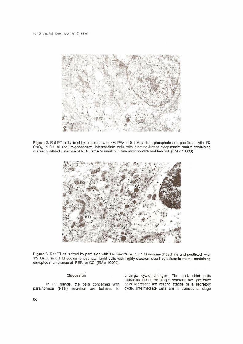
Figure 2. Rat PT cells fixed by perfusion with 4% PFA in 0. 1 M sodium-phosphate and postfxed with 1 % Os04 in 0.1 M sodium-phosphate. Intermediate cells with electron-Iucent cytoplasmic matrix containing markedly dilated cisternae of RER, large or smail GC, few mitochondira and few SG. (EM x 13000).
Figure 3. Rat PT cells fixed by perfusion with 1% GA-2%FA in 0.1 M sodium-phosphate and postfixed with 1 % Os04 in 0.1 M sodium-phosphate. Light cells with highly electron-Iucent cytoplasmic matrix containing disrupted membranes of RER or GC. (EM x 10000).
Discussion
In PT glands, the cells concerned with parathormon (PTH) secretion are believed to
60
undergo cyclic changes. The dark chief cells represent the active stages whereas the light chief cells represent the resting stages of a secretory cycle. Intermediate cells are in transitional stage

Y.Y.O. Vet. Fak. Derg. 1996, 7(1-2): 58-61
either from active to resting stages or vice versa (1,2,8,9). In normal PT glands, the dark cells are characterized by prominent rough endoplasmic retikulum (RER) and Golgi apparatus, an increased tortuosily of the plasma membrane. In light cells RER, Golgi apparatus, secretory granules and the tortuosily of the plasma membrane are reduced (1,2,7,8,12).
Interestingly, Stöeckel and Porte (11) mentioned that the differences in PT cell morphology mav arise during fı xation. Lever (5), who fırst described Ihe ultrastructure of PT ceııs, argued that many of the cell membranes lack inlegrily. Comparalive studies have revealed that PT cell varianls which regularly oceured in immerison-fixed samples were abseni in perfusion-fıxed PT glands (4,10,14,15) or occured only in perfusion-fixed glands in areas of incomplete fixation (10).
The concept of the existence of a secretory cycle in PT cells was recently called inlo question by the work of Larsson el aL. (4). They demonstrated Ihat, in PT glands of Mongolian gerbils, the occurrence of lighl chief cells and atrophic ce lls depended on improper fıxation .
PT cell variants occurred in all species examined when tissue was fıxed in 2.5% GA. Diversily in PT cell morphology was, however, largely avoided by immersion in mixtures containing 1 %GA and 1.5%-2% FA and 2.5-5% acrolein (AC) in bovine, feline and murine PT glands (13). In an attempl to clarify the formation of PT cell variants in dogs, cats, mice and rats (13) and in cattle, sheep, goaı, horse and pig (3), it was found that aldehydes and buffers play important roles.
In this study, the specimens, parenchymal cells of PT glands, fixed in 2.5% GA were uniform cells wilh markedly enlarged IS, tortuous plasma membranes, electron dense malrix containing slighlly dilaled cisternae of RER, large or smail GC, many milochondria and few SG. PT glands fıxed wilh 4% PFA led to intermediate cells wilh electron lucent matrix containing markedly dilated cisterna of RER, large or smail GC, few mitochondira and few SG. However, the use of fixatives consist of 1 %GA -2%FA led only to light cells with highly electronlucent matrix containing disrupted membranes of RER or GC. The results support the idea, arising after examination of perfusion-fixed parathyroid tissue, that parathyroid cell variants occur during improper fixation rather than that express functional diversily.
References
1. Altenahr, T.B. (1972). Ultrastruetural pathology of parathyroid glands. Curr. Top. Pathol. 56, 1-54.
2. Capen, C.C. (1980). Parathyroid hormone, caleitonin, and eholecalciferol: the calcium regulaling hormones. In: MeDonaid, Veterinary endoerinolgy and
reproduetions; 3rd ed., 62-114. Lea and Febiger Philadelphia
3. Kanter, M. (1993). Die Ullrastruklur der Epilhelkörperehen von Rind, Sehaf, Ziege, Pferd und Schwein und ihre Beeinflussbarkeit durch Fixalionsmedien (Doktorarbeit, Zürich).
4. Larsson, H.O., Lorentzon , R. and Boquist, L. (1984). Strueture of parathyroid glands , as revealed by different melhods of fıxalion . A quanlitative lig ht- and electron microscopic study in unlreated Mongolian gerbils. Cell Tiss. Res. 235, 51-58.
5. Lever, J.O. (1957). Fine sıruelural appearanees in the ral parathyroid . J. Anat. 91,73-81 .
6. Moreira, J.E. and Gonçalves, R.P. (1985). Ullrastruetural ehangings of Ihe rat parathyroid gland under various fıxalion melhods. Anat. Anz. 158, 413-423.
7. Nilsson, O. (1977). Studies on the ultrastructure of the human parathyroid glands in various palhologieal eonditions. Aela. Path ol. Mierobiol. Scand., A. Suppl. 263, 1-88.
8. Roth, S.I. and Capen, C.C. (1974). Ultrastruetural and funetional correlations of the paralhyroid gland. Int. Rev. Exp. Pathol. 13, 161-221.
9. Roth, S.I. and Raishz, L.G. (1966). The eourse and reversibility of the ealeium effeet on the ultraslrueture of the rat parathyroid gland in organ culture. Lab. Invest. 15,1187-1211.
10. Seloguli, T. (1.977) . Electron mieroseopic sludies of parathyroid gl and of senile dogs. Am. J. Anat. 148, 65-84.
11. Stöeekel, M.E. and Porte, A. (1966). Observations ullrastrueturales sur la paralhyroid des souris. i. Etude chez la souris normale. Z. Zellforsch. Mikrosk. Anat. 73, 448-502.
12. Thiele, J. : Human paralhyroid gland (1977). A freeze fracture and Ihin section study. Curr. Top. Path ol. 65,31-80.
13. Wild, P. Kellner, S.J . and Schraner, EM. (1987). Parathyroid eel i variants mav be provoked during immersion fıxalion . Histoehem. 87, 263-271.
14. Wild, P. Manser, E (1985). Ultrastructural morphometry in parathyroid cells in rals of differenl ages. Cell Tissue. Res. 240, 585-591.
15. Wild, P. , Sehraner, EM., Augsburger, H., Beglinger, R., Pfısler, R. (1986): Ultraslructural allerations in mammalian parathyroid glands induced by fıxation . Acla Anat. 126,87-96.
61