Embed Size (px)
Citation preview

RESEARCH ARTICLE
Passive immunotherapy of tauopathy targetingpSer413-tau: a pilot study in miceTomohiro Umeda1, Hiroshi Eguchi2, Yuichi Kunori2, Yoichi Matsumoto2, Taizo Taniguchi3, HiroshiMori1 & Takami Tomiyama1
1Department of Neuroscience, Osaka City University Graduate School of Medicine, Osaka, Japan2Teijin Institute for Bio-medical Research, Teijin Pharma Limited, Hino, Japan3Faculty of Pharmaceutical Sciences, Himeji Dokkyo University, Himeji, Japan
Correspondence
Takami Tomiyama, Department of
Neuroscience, Osaka City University Graduate
School of Medicine, 1-4-3 Asahimachi,
Abeno-ku, Osaka 545-8585, Japan. Tel: +81-
6-6645-3921; Fax: +81-6-6645-3922; E-mail:
Funding Information
This study was supported by funding from
Teijin Pharma Limited.
Received: 20 May 2014; Revised: 9
December 2014; Accepted: 11 December
2014
Annals of Clinical and Translational
Neurology 2015; 2(3): 241–255
doi: 10.1002/acn3.171
Abstract
Objective: Cellular inclusions of hyperphosphorylated tau are a hallmark of
tauopathies, which are neurodegenerative disorders that include Alzheimer’s
disease (AD). Active and passive immunization against hyperphosphorylated
tau has been shown to attenuate phenotypes in model mice. We developed new
monoclonal antibodies to hyperphosphorylated tau and sought high therapeutic
efficacy for future clinical use. Methods: Using more than 20 antibodies, we
investigated which sites on tau are phosphorylated early and highly in the
tauopathy mouse models tau609 and tau784. These mice display tau hyper-
phosphorylation, synapse loss, memory impairment at 6 months, and tangle
formation and neuronal loss at 15 months. We generated mouse monoclonal
antibodies to selected epitopes and examined their effects on memory and tau
pathology in aged tau609 and tau784 mice by the Morris water maze and by
histological and biochemical analyses. Results: Immunohistochemical screening
revealed that pSer413 is expressed early and highly. Monoclonal antibodies to
pSer413 and to pSer396 (control) were generated. These antibodies specifically
recognized pathological tau in AD brains but not normal tau in control brains
according to Western blots. Representative anti-pSer413 and anti-pSer396 anti-
bodies were injected intraperitoneally into 10–11- or 14-month-old mice once a
week at 0.1 or 1 mg/shot 5 times. The anti-pSer413 antibody significantly
improved memory, whereas the anti-pSer396 antibodies showed less effect. The
cognitive improvement paralleled a reduction in the levels of tau hyperphosph-
orylation, tau oligomer accumulation, synapse loss, tangle formation, and
neuronal loss. Interpretation: These results indicate that pSer413 is a promising
target in the treatment of tauopathy.
Introduction
Neuronal and glial inclusions of hyperphosphorylated tau
aggregates are hallmarks of tauopathies, which are neuro-
degenerative disorders that include Alzheimer’s disease
(AD), Pick’s disease, corticobasal degeneration, progres-
sive supranuclear palsy, argyrophilic grain disease, and
frontotemportal dementia and parkinsonism linked to
chromosome 17 (FTDP-17).1 FTDP-17 is an inherited
tauopathy, and a number of exonic and intronic muta-
tions in the tau gene have been identified. Many mouse
models of tauopathies have been generated by introducing
the human tau gene with or without mutations.2 We pre-
viously generated tau transgenic (Tg) mice expressing
both three-repeat and four-repeat human tau by inserting
tau intronic sequences into both sides of tau exon 10 in
the transgene.3 These mice, originally referred to as lines
609 and 784 and hereafter termed tau609 and tau784,
dominantly express four-repeat human tau in adult age
by the presence of the FTDP-17-related tau intron
10 + 16C ? T mutation. They exhibited abnormal tau
phosphorylation, synapse loss, and memory impairment
at 6 months, and neurofibrillary tangle (NFT) formation
and neuronal loss at 24 months. More recently, we found
ª 2015 The Authors. Annals of Clinical and Translational Neurology published by Wiley Periodicals, Inc on behalf of American Neurological Association.
This is an open access article under the terms of the Creative Commons Attribution-NonCommercial-NoDerivs License, which permits use and
distribution in any medium, provided the original work is properly cited, the use is non-commercial and no modifications or adaptations are made.
241
that these mice start to display NFTs and neuronal loss at
15 months in layer II/III of the entorhinal cortex (EC-II/
III) and cingulated cortex.
Active and passive immunization against hyperphosph-
orylated tau has been shown to attenuate phenotypes in
model mice. For example, active immunization with tau
partial peptides phosphorylated at Ser396/404,4–7 Ser202/
Thr205, Thr212/Ser214, Thr231,8 or Ser4229 decreased the
level of hyperphosphorylated tau and rescued motor/cog-
nitive dysfunction. Immunization with human paired
helical filaments (PHFs) composed of hyperphosphorylat-
ed tau aggregates also reduced NFTs.10 Meanwhile, some
studies cautioned that active tau immunization may cause
neuroinflammation in the brain.11,12 Thus, passive immu-
nization would seem safer than active immunization, as
the former only compensates humoral immunity, whereas
the latter activates both humoral and cellular immunity
making it difficult to manage adverse effects. Additionally,
passive immunization with PHF-1 (anti-pSer396/404) or
MC1 (anti-pathological conformation) antibody decreased
the level of hyperphosphorylated tau and improved motor
function.13–15 These passive immunization studies,
however, lean to the prevention rather than therapy of
tauopathy, as they used young mice before or just after
the disease onset. To evaluate clinical efficacy, immuniza-
tion should be performed in aged mice with overt neuro-
pathology.
For future clinical use in the treatment of tauopathy,
we decided to develop new monoclonal antibodies to hy-
perphosphorylated tau with higher therapeutic efficacy
than those of existing anti-tau monoclonal antibodies.
To determine the target epitopes, we initially studied
which sites on tau are phosphorylated early and highly
in our model mice, tau609 and tau784. Immunohisto-
chemical screening with more than 20 commercially
available antibodies revealed that Ser413 is such a site.
We generated mouse monoclonal antibodies to pSer413
and to pSer396, our control, and compared their effects
in aged tau609 and tau784 mice. Our results indicate
that pSer413 is a promising target in the treatment of
tauopathy.
Materials and Methods
Immunohistochemical screening for targetepitopes
Antibodies used in the immunohistochemical screening
for target epitopes are listed in Table S1. Brain sections
were prepared from tau609, tau784, and non-Tg mice at
6 and 24 months.3 Immunohistochemical staining was
performed as described previously3 but the pretreatment
of sections under an acidic condition was not performed.
Peptide synthesis
Tau partial peptides used in immunization and antibody
screening are listed in Table S2.
Generation of polyclonal antibody
Anti-pSer413 polyclonal antibody was generated by
immunizing a rabbit with the pSer413 peptide, as detailed
in Data S1.
Generation of monoclonal antibodies
Anti-pSer413 and anti-pSer396/404 monoclonal antibod-
ies were generated by immunizing mice with the corre-
sponding peptides, as detailed in Data S1.
SPR analysis
The binding affinity of monoclonal antibodies was evalu-
ated by surface plasmon resonance (SPR), as detailed in
Data S1.
Western blot analysis with AD samples
Brain samples were obtained by autopsy from patients
with AD and from nondemented control subjects with
informed consent. The samples were processed according
to the methods of Berger et al.16 and subjected to
Western blot to study the specificity of the monoclonal
antibodies. Details are given in Data S1.
Passive immunization
Passive immunization was performed in male tau609 and
tau784 mice. These two lines showed no apparent differ-
ences in the levels of tau expression or tauopathy pheno-
types. Monoclonal antibodies were diluted in the buffers
indicated in the figure legends. Anti-Pseudomonas aeru-
ginosa lipopolysaccharide (LPS) mouse monoclonal anti-
bodies 4C10F4 and 6F11B6 were generated at Teijin
Pharma Limited, whereas anti-Shiga like-toxin II (Stx2)
mouse monoclonal antibody 11F11 was obtained from
ATCC. These antibodies were used as the control. Mice
were divided into two groups (n = 8–10 each) so that the
mean body weights were not significantly different
between groups. One group was exposed to anti-pSer413
or anti-pSer396 antibody and the other to the control
antibody. Anti-pSer413 (Ta1505) and anti-pSer396 (Ta4)
antibodies were injected intraperitoneally into 14-month-
old tau784 mice once a week at 1 mg/shot 5 times,
whereas another anti-pSer396 antibody (Ta9) was injected
into age-matched tau609 mice in the same protocol. In
242 ª 2015 The Authors. Annals of Clinical and Translational Neurology published by Wiley Periodicals, Inc on behalf of American Neurological Association.
Passive Immunotherapy Targeting pSer413-tau T. Umeda et al.

additional experiments, Ta1505 and Ta9 antibodies were
injected into 10–11-month-old tau784 mice once a week
at 0.1 mg/shot 5 times. The buffer used for antibody dilu-
tion was injected into age-matched non-Tg littermates
(n = 8–10) as a normal control. Mice were subjected to
behavioral tests in the week after the last injection. All
animal experiments were approved by the ethics commit-
tee of Osaka City University (Osaka, Japan) and were per-
formed in accordance with the Guide for Animal
Experimentation, Osaka City University.
Behavioral tests
Spatial reference memory in antibody-treated mice was
assessed using the Morris water maze as described previ-
ously17 except for intertrial intervals of 5 min. Locomotor
activities of the mice were examined by an open-field test.17
Histological analysis
After the behavioral tests, antibody-treated mice were
divided into two groups: one (n = 5–6) for histological
analysis and the other (n = 3–4) for Western blot
analysis. Brain sections were prepared and immunohis-
tochemical and Gallyas silver staining were performed
as described previously.3 PHF-1 antibody was a kind
gift from Dr. Peter Davies, Albert Einstein College of
Medicine, whereas AT8 (Thermo Scientific, Waltham,
MA), anti-tau oligomer (T22; EMD Millipore, Teme-
cula, CA),18 antisynaptophysin (SVP-38; Sigma-Aldrich,
St. Louis, MO), and anti-NeuN (Chemicon, Temecula,
CA) antibodies were purchased. Tau hyperphosphoryla-
tion and tau oligomer accumulation were evaluated by
quantifying the staining intensities of phospho-tau-posi-
tive and T22-positive areas in each photograph of the
hippocampal CA2-3 region. Analysis was performed
using NIH ImageJ software (National Institutes of
Health; http://rsb.info.nih.gov/nih-image/) on individual
sections (3 sections per animal). Synapse loss was
assessed by quantifying synaptophysin fluorescence
intensities in the apical dendritic-somata field
(30 9 60 lm) of the hippocampal CA3 region using
NIH ImageJ software (2 sections per animal). The level
of NFT formation was determined by counting Gallyas
silver positive cells in an area (220 9 160 lm) of the
EC-II/III region, whereas neuronal loss was estimated
by counting NeuN-positive cells in an area within
1000 lm along the EC-II (3 sections per animal).
Western blot analysis
For Western blot analysis, anti-pSer396 antibody-treated
brain samples were obtained from 15-month-old mice
(n = 3–4) after the behavioral tests, whereas anti-pSer413
antibody-treated brain samples were newly prepared by
injecting anti-pSer413 and control 11F11 antibodies into
12-month-old tau784 mice (n = 4 each) once a week at
1 mg/shot 5 times. The separate preparations were carried
out because we had used up anti-pSer413 antibody-trea-
ted 15-month-old brain samples in different experiments
not shown here. Brain tissues were homogenized and
fractionated into Tris buffered saline (TBS)-, sarkosyl-,
and GuHCl-soluble fractions as described previously3 with
minor modification. That is, sarkosyl-insoluble precipi-
tates were dissolved in four volumes of tissue weight of
4 mol/L GuHCl. After centrifugation, the supernatants
were dialyzed against TBS using a Slide-A-Lyzed G2 Dial-
ysis Cassette with 20K cut-off membrane (Thermo Scien-
tific). The TBS-soluble and dialyzed GuHCl-soluble
fractions were subjected to Western blot with anti-tau
and anti-actin (Sigma-Aldrich) antibodies, as described
previously.3 To measure tau oligomers, brain homogen-
ates were centrifuged at 13,000g and 4°C for 15 min. The
supernatants were collected as total tau extracts. Both
TBS-soluble fractions and total tau extracts were subjected
to Western blot with human tau-specific Tau12 antibody
(Abcam, Cambridge, UK).
Statistical analysis
All data are given as mean � SEM. Comparisons of
means between two groups were performed using
Student’s t-test, whereas comparisons of means among
more than two groups were performed using analysis of
variance (ANOVA) or two-factor repeated measures
ANOVA (for the behavioral tests), followed by Fisher’s
protected least significant difference test. Differences with
a P value of <0.05 were considered significant.
Results
Immunohistochemical screening for targetepitopes
In our previous study, both tau609 and tau784 mice
began to exhibit abnormal tau phosphorylation in hippo-
campal mossy fibers from 6 months and in neuronal cell
bodies of the hippocampus and cerebral cortex from
18 months,3 suggesting the hippocampal mossy fibers are
most susceptible to tau hyperphosphorylation in these
mice. Therefore, we focused on this region during immu-
nohistochemical screening for target epitopes. We first
examined 24-month-old mouse brains. Of the more than
20 commercially available antibodies, 13 antibodies clearly
stained the hippocampal mossy fibers (Table 1). None of
the 13 antibodies, except AT100, showed positive staining
ª 2015 The Authors. Annals of Clinical and Translational Neurology published by Wiley Periodicals, Inc on behalf of American Neurological Association. 243
T. Umeda et al. Passive Immunotherapy Targeting pSer413-tau
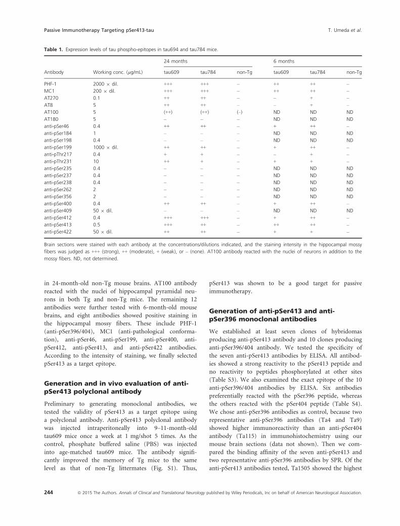
in 24-month-old non-Tg mouse brains. AT100 antibody
reacted with the nuclei of hippocampal pyramidal neu-
rons in both Tg and non-Tg mice. The remaining 12
antibodies were further tested with 6-month-old mouse
brains, and eight antibodies showed positive staining in
the hippocampal mossy fibers. These include PHF-1
(anti-pSer396/404), MC1 (anti-pathological conforma-
tion), anti-pSer46, anti-pSer199, anti-pSer400, anti-
pSer412, anti-pSer413, and anti-pSer422 antibodies.
According to the intensity of staining, we finally selected
pSer413 as a target epitope.
Generation and in vivo evaluation of anti-pSer413 polyclonal antibody
Preliminary to generating monoclonal antibodies, we
tested the validity of pSer413 as a target epitope using
a polyclonal antibody. Anti-pSer413 polyclonal antibody
was injected intraperitoneally into 9–11-month-old
tau609 mice once a week at 1 mg/shot 5 times. As the
control, phosphate buffered saline (PBS) was injected
into age-matched tau609 mice. The antibody signifi-
cantly improved the memory of Tg mice to the same
level as that of non-Tg littermates (Fig. S1). Thus,
pSer413 was shown to be a good target for passive
immunotherapy.
Generation of anti-pSer413 and anti-pSer396 monoclonal antibodies
We established at least seven clones of hybridomas
producing anti-pSer413 antibody and 10 clones producing
anti-pSer396/404 antibody. We tested the specificity of
the seven anti-pSer413 antibodies by ELISA. All antibod-
ies showed a strong reactivity to the pSer413 peptide and
no reactivity to peptides phosphorylated at other sites
(Table S3). We also examined the exact epitope of the 10
anti-pSer396/404 antibodies by ELISA. Six antibodies
preferentially reacted with the pSer396 peptide, whereas
the others reacted with the pSer404 peptide (Table S4).
We chose anti-pSer396 antibodies as control, because two
representative anti-pSer396 antibodies (Ta4 and Ta9)
showed higher immunoreactivity than an anti-pSer404
antibody (Ta115) in immunohistochemistry using our
mouse brain sections (data not shown). Then we com-
pared the binding affinity of the seven anti-pSer413 and
two representative anti-pSer396 antibodies by SPR. Of the
anti-pSer413 antibodies tested, Ta1505 showed the highest
Table 1. Expression levels of tau phospho-epitopes in tau694 and tau784 mice.
Antibody Working conc. (lg/mL)
24 months 6 months
tau609 tau784 non-Tg tau609 tau784 non-Tg
PHF-1 2000 9 dil. +++ +++ – ++ ++ –
MC1 200 9 dil. +++ +++ – ++ ++ –
AT270 0.1 ++ ++ – – + –
AT8 5 ++ ++ – – + –
AT100 5 (++) (++) (–) ND ND ND
AT180 5 – – – ND ND ND
anti-pSer46 0.4 ++ ++ – + ++ –
anti-pSer184 1 – – – ND ND ND
anti-pSer198 0.4 – – – ND ND ND
anti-pSer199 1000 9 dil. ++ ++ – + ++ –
anti-pThr217 0.4 + + – – + –
anti-pThr231 10 ++ + – + + –
anti-pSer235 0.4 – – – ND ND ND
anti-pSer237 0.4 – – – ND ND ND
anti-pSer238 0.4 – – – ND ND ND
anti-pSer262 2 – – – ND ND ND
anti-pSer356 2 – – – ND ND ND
anti-pSer400 0.4 ++ ++ – + ++ –
anti-pSer409 50 9 dil. – – – ND ND ND
anti-pSer412 0.4 +++ +++ – + ++ –
anti-pSer413 0.5 +++ ++ – ++ ++ –
anti-pSer422 50 9 dil. ++ ++ – + + –
Brain sections were stained with each antibody at the concentrations/dilutions indicated, and the staining intensity in the hippocampal mossy
fibers was judged as +++ (strong), ++ (moderate), + (weak), or – (none). AT100 antibody reacted with the nuclei of neurons in addition to the
mossy fibers. ND, not determined.
244 ª 2015 The Authors. Annals of Clinical and Translational Neurology published by Wiley Periodicals, Inc on behalf of American Neurological Association.
Passive Immunotherapy Targeting pSer413-tau T. Umeda et al.

binding affinity with the lowest KD value to the antigen
peptide (Table S5). On the other hand, the two anti-
pSer396 antibodies showed a higher binding affinity to
their antigen peptide than did the anti-pSer413 antibod-
ies. We therefore chose Ta1505 as a representative anti-
pSer413 antibody for further evaluation.
Specificity of monoclonal antibodies topathological tau in AD brains
We examined whether our monoclonal antibodies specifi-
cally recognized pathological tau in AD brains. Initially, we
compared four samples from AD patients and two samples
from nondemented control subjects (Table S6) for patho-
logical tau species by Western blot with human tau-specific
G2 antibody.3 The control brains showed only monomers
of tau with molecular sizes of 45–65 kDa, whereas AD
brains exhibited not only monomers but also dimers and/
or trimers with molecular sizes of 120–220 kDa (Fig. 1).
Two AD samples and one control sample were selected for
the analysis of the specificity of the monoclonal antibodies.
Anti-human tau monoclonal antibody Tau12 recognized
all tau species in both AD and control brains, whereas
Ta1505, Ta4, and Ta9 antibodies recognized only tau spe-
cies in AD brains but not in control brains.
In vivo evaluation of monoclonal antibodies
Effects on memory
We compared the effects of monoclonal antibodies on
memory in tau609 and tau784 mice. Ta1505 and control
4C10F4 antibodies were injected intraperitoneally into 14-
month-old tau784 mice once a week at 1 mg/shot 5
times. Ta1505 antibody significantly improved the mem-
ory of Tg mice to the same level as non-Tg littermates,
whereas 4C10F4 antibody did not (Fig. 2A). Probe trials
revealed that memory retention was significantly
improved by Ta1505 injection to a level similar to that in
non-Tg littermates (Fig. 2B). No differences in locomotor
activities were observed among the three groups (not
shown). Next, Ta4 and control 4C10F4 antibodies were
administered to 14-month-old tau784 mice once a week
at 1 mg/shot 5 times. Ta4 antibody showed a tendency to
improve the memory of Tg mice, but the difference
between Ta4- and 4C10F4-treated groups was not signifi-
cant (Fig. 2C). In probe trials, Ta4 antibody showed no
significant improvement in memory retention (Fig. 2D).
We repeated the experiments with another anti-pSer396
antibody, Ta9. Ta9 and control 6F11B6 antibodies were
injected into 14-month-old tau609 mice once a week at
1 mg/shot 5 times. Ta9 antibody significantly improved
the memory of Tg mice, whereas 6F11B6 antibody did
not (Fig. S2A). While the effect of Ta9 antibody was
weaker than that of Ta1505 antibody, memory retention
was significantly improved by Ta9 injection to a level
similar to that in non-Tg littermates (Fig. S2B).
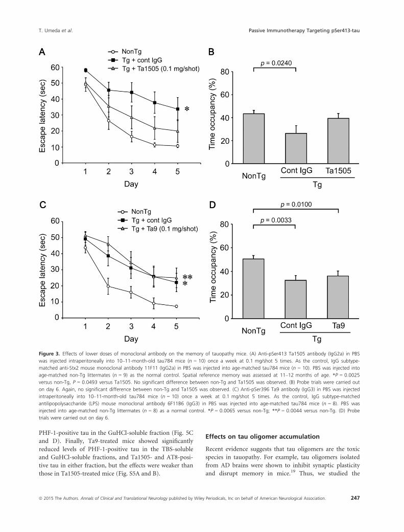
The effects of monoclonal antibodies were further stud-
ied at a lower dose. Ta1505 and control 11F11 antibodies
were injected into 10–11-month-old tau784 mice once a
week at 0.1 mg/shot 5 times. Even at such a low dose,
Ta1505 antibody significantly improved the memory of Tg
mice to a level between that of 11F11-injected Tg mice and
PBS-injected non-Tg littermates (Fig. 3A and B). Next, Ta9
and control 6F11B6 antibodies were administered to 10–11-month-old tau784 mice once a week at 0.1 mg/shot 5
times. In contrast to Ta1505 antibody, Ta9 antibody
showed no significant effect on memory (Fig. 3C and D).
Effects on tau hyperphosphorylation
Brain sections prepared from 15-month-old antibody-
treated mice (at 1 mg/shot) were stained with Ta1505,
PHF-1, and AT8 antibodies. Compared with control anti-
body-treated mice, Ta1505-treated mice showed a signifi-
cant reduction in Ta1505-, PHF-1-, and AT8-positive
staining in the hippocampal mossy fibers (Fig. 4A and
C). In contrast, Ta4- and Ta9-treated mice exhibited a
significant reduction in PHF-1-positive staining but little
in Ta1505- and AT8-staining (Fig. 4B and C and Fig. S3A
and B). We also examined brain sections of 11–12-month-old antibody-treated mice (at 0.1 mg/shot).
Ta1505-treated mice showed an apparent reduction in
Ta1505- and PHF-1-positive staining (Fig. S4A and C),
whereas Ta9-treated mice exhibited no effect (Fig. S4B
and C).
Figure 1. Specificity of monoclonal antibodies to pathological tau in
Alzheimer’s disease (AD) brains. Four samples from AD patients (no.
4, 24, 32, 89) and two samples from nondemented control subjects
(no. 31, 33) were stained with G2 antibody, a rabbit polyclonal
antibody specific to human tau.3 M, six isoforms of recombinant
human tau. Two AD (no. 32, 89) samples and one control (no. 31)
sample were selected for the analysis of the specificity of monoclonal
antibodies. Tau12, anti-human tau antibody; Ta1505, anti-pSer413
antibody; Ta4 and Ta9, anti-pSer396 antibodies; 11F11, anti-Stx2
antibody.
ª 2015 The Authors. Annals of Clinical and Translational Neurology published by Wiley Periodicals, Inc on behalf of American Neurological Association. 245
T. Umeda et al. Passive Immunotherapy Targeting pSer413-tau

TBS-soluble and GuHCl-soluble (sarkosyl-insoluble)
brain fractions prepared from 13- and 15-month-old anti-
body-treated mice (at 1 mg/shot) were subjected to Wes-
tern blot with G2, pool-2 (anti-human/mouse tau),3
Ta1505, PHF-1, and AT8 antibodies. Ta1505-treated mice
showed significantly lower levels of total (i.e., pool-2-posi-
tive) and Ta1505-, PHF-1-, and AT8-positive tau than did
control antibody-treated mice in the TBS-soluble and
GuHCl-soluble fractions (Fig. 5A and B). In contrast,
Ta4-treated mice exhibited a significant reduction only of
Figure 2. Effects of monoclonal antibodies on the memory of tauopathy mice. (A) Anti-pSer413 Ta1505 antibody (IgG2a) in 0.1 mol/L citrate
buffer (pH5.0) was injected intraperitoneally into 14-month-old tau784 mice (n = 9) once a week at 1 mg/shot 5 times. As the control,
antilipopolysaccharide (LPS) mouse monoclonal antibody 4C10F4 (IgG2b) in the same buffer was injected into age-matched tau784 mice (n = 9).
The buffer used for the antibody dilution was injected into age-matched non-Tg littermates (n = 8) as the normal control. Spatial reference
memory was assessed at 15 months of age by the Morris water maze. *P = 0.0110 versus non-Tg, P = 0.0152 versus Ta1505. No significant
difference between non-Tg and Ta1505 was observed. (B) Probe trials with the platform removed were carried out on day 6. Each bar represents
the mean time occupancy in the target quadrant for 30 sec. Again, no significant difference between non-Tg and Ta1505 was observed. (C) Anti-
pSer396 Ta4 antibody (IgG2b) in PBS was injected intraperitoneally into 14-month-old tau784 mice (n = 9) once a week at 1 mg/shot 5 times. As
the control, IgG subtype-matched 4C10F4 antibody in PBS was injected into age-matched tau784 mice (n = 8). PBS was injected into age-
matched non-Tg littermates (n = 9) as the normal control. *P = 0.0254 versus non-Tg. (D) Probe trials were carried out on day 5. No significant
differences among three groups were observed.
246 ª 2015 The Authors. Annals of Clinical and Translational Neurology published by Wiley Periodicals, Inc on behalf of American Neurological Association.
Passive Immunotherapy Targeting pSer413-tau T. Umeda et al.
PHF-1-positive tau in the GuHCl-soluble fraction (Fig. 5C
and D). Finally, Ta9-treated mice showed significantly
reduced levels of PHF-1-positive tau in the TBS-soluble
and GuHCl-soluble fractions, and Ta1505- and AT8-posi-
tive tau in either fraction, but the effects were weaker than
those in Ta1505-treated mice (Fig. S5A and B).
Effects on tau oligomer accumulation
Recent evidence suggests that tau oligomers are the toxic
species in tauopathy. For example, tau oligomers isolated
from AD brains were shown to inhibit synaptic plasticity
and disrupt memory in mice.19 Thus, we studied the
Figure 3. Effects of lower doses of monoclonal antibody on the memory of tauopathy mice. (A) Anti-pSer413 Ta1505 antibody (IgG2a) in PBS
was injected intraperitoneally into 10–11-month-old tau784 mice (n = 10) once a week at 0.1 mg/shot 5 times. As the control, IgG subtype-
matched anti-Stx2 mouse monoclonal antibody 11F11 (IgG2a) in PBS was injected into age-matched tau784 mice (n = 10). PBS was injected into
age-matched non-Tg littermates (n = 9) as the normal control. Spatial reference memory was assessed at 11–12 months of age. *P = 0.0025
versus non-Tg, P = 0.0493 versus Ta1505. No significant difference between non-Tg and Ta1505 was observed. (B) Probe trials were carried out
on day 6. Again, no significant difference between non-Tg and Ta1505 was observed. (C) Anti-pSer396 Ta9 antibody (IgG3) in PBS was injected
intraperitoneally into 10–11-month-old tau784 mice (n = 10) once a week at 0.1 mg/shot 5 times. As the control, IgG subtype-matched
antilipopolysaccharide (LPS) mouse monoclonal antibody 6F11B6 (IgG3) in PBS was injected into age-matched tau784 mice (n = 8). PBS was
injected into age-matched non-Tg littermates (n = 8) as a normal control. *P = 0.0065 versus non-Tg; **P = 0.0044 versus non-Tg. (D) Probe
trials were carried out on day 6.
ª 2015 The Authors. Annals of Clinical and Translational Neurology published by Wiley Periodicals, Inc on behalf of American Neurological Association. 247
T. Umeda et al. Passive Immunotherapy Targeting pSer413-tau
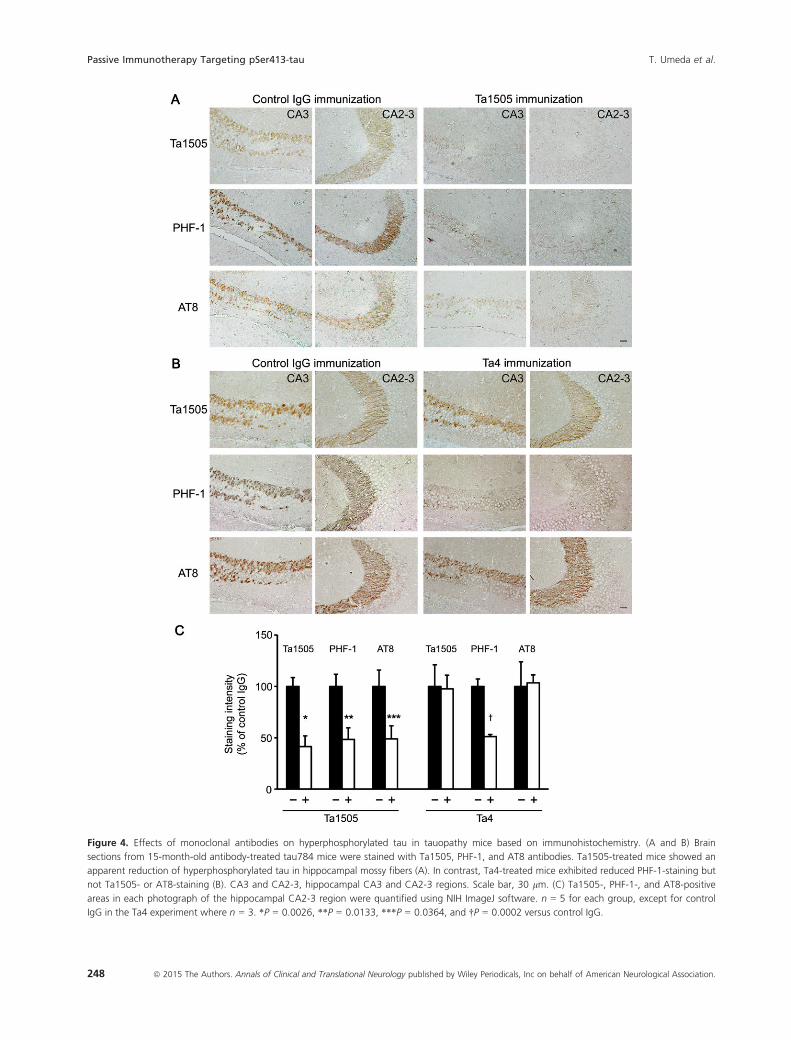
Figure 4. Effects of monoclonal antibodies on hyperphosphorylated tau in tauopathy mice based on immunohistochemistry. (A and B) Brain
sections from 15-month-old antibody-treated tau784 mice were stained with Ta1505, PHF-1, and AT8 antibodies. Ta1505-treated mice showed an
apparent reduction of hyperphosphorylated tau in hippocampal mossy fibers (A). In contrast, Ta4-treated mice exhibited reduced PHF-1-staining but
not Ta1505- or AT8-staining (B). CA3 and CA2-3, hippocampal CA3 and CA2-3 regions. Scale bar, 30 lm. (C) Ta1505-, PHF-1-, and AT8-positive
areas in each photograph of the hippocampal CA2-3 region were quantified using NIH ImageJ software. n = 5 for each group, except for control
IgG in the Ta4 experiment where n = 3. *P = 0.0026, **P = 0.0133, ***P = 0.0364, and †P = 0.0002 versus control IgG.
248 ª 2015 The Authors. Annals of Clinical and Translational Neurology published by Wiley Periodicals, Inc on behalf of American Neurological Association.
Passive Immunotherapy Targeting pSer413-tau T. Umeda et al.
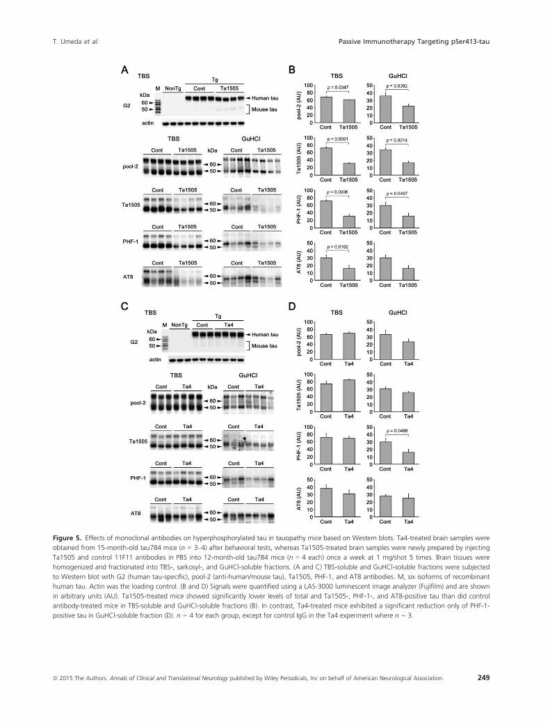
Figure 5. Effects of monoclonal antibodies on hyperphosphorylated tau in tauopathy mice based on Western blots. Ta4-treated brain samples were
obtained from 15-month-old tau784 mice (n = 3–4) after behavioral tests, whereas Ta1505-treated brain samples were newly prepared by injecting
Ta1505 and control 11F11 antibodies in PBS into 12-month-old tau784 mice (n = 4 each) once a week at 1 mg/shot 5 times. Brain tissues were
homogenized and fractionated into TBS-, sarkosyl-, and GuHCl-soluble fractions. (A and C) TBS-soluble and GuHCl-soluble fractions were subjected
to Western blot with G2 (human tau-specific), pool-2 (anti-human/mouse tau), Ta1505, PHF-1, and AT8 antibodies. M, six isoforms of recombinant
human tau. Actin was the loading control. (B and D) Signals were quantified using a LAS-3000 luminescent image analyzer (Fujifilm) and are shown
in arbitrary units (AU). Ta1505-treated mice showed significantly lower levels of total and Ta1505-, PHF-1-, and AT8-positive tau than did control
antibody-treated mice in TBS-soluble and GuHCl-soluble fractions (B). In contrast, Ta4-treated mice exhibited a significant reduction only of PHF-1-
positive tau in GuHCl-soluble fraction (D). n = 4 for each group, except for control IgG in the Ta4 experiment where n = 3.
ª 2015 The Authors. Annals of Clinical and Translational Neurology published by Wiley Periodicals, Inc on behalf of American Neurological Association. 249
T. Umeda et al. Passive Immunotherapy Targeting pSer413-tau

involvement of tau oligomers in our mice by immunohis-
tochemical staining with tau oligomer-specific T22 anti-
body. Both tau609 and tau784 mice displayed an
apparent accumulation of tau oligomers in hippocampal
neurons at 6 months (Fig. S6A), at which age these mice
showed synaptic and cognitive dysfunction.3 The effects
of antibodies on tau oligomer accumulation were exam-
ined with 15-month-old brain sections. Ta1505 signifi-
cantly reduced the levels of tau oligomers, but Ta4 and
Ta9 did not (Fig. S6B and C). This finding was confirmed
by Western blot with Tau12 antibody. The levels of tau
oligomers in both TBS-soluble brain fractions and total
tau extracts were significantly decreased by Ta1505 injec-
tion (Fig. S6D and E).
Effects on synapse loss, NFT formation, andneuronal loss
We next examined synapse loss in 15-month-old anti-
body-treated mice. Ta1505-treated mice showed signifi-
cant recovery in synaptophysin levels in the hippocampal
CA3 region (Fig. 6A and B), whereas Ta4-treated mice
did not (Fig. 6C and D). Ta9-treated mice showed only
slight recovery of synapses (Fig. S7A and B).
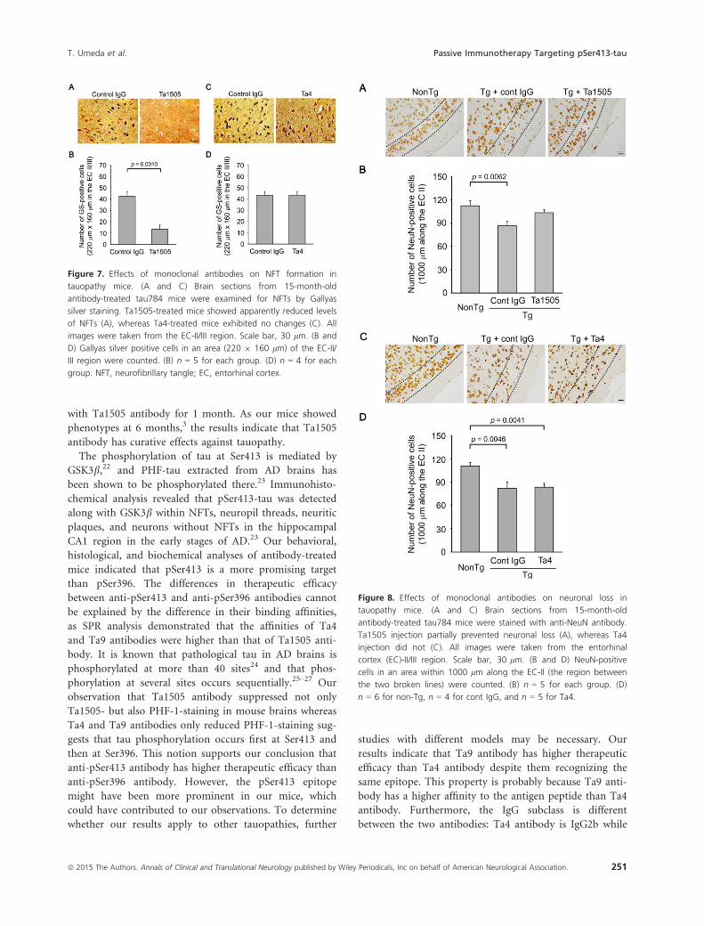
We also assessed NFTs and neuronal loss in 15-month-
old antibody-treated mice, as our mice start to display
NFTs and neuronal loss at this age. Ta1505-treated mice
displayed significantly decreased levels of NFTs in the
EC-II/III region (Fig. 7A and B), whereas Ta4- and Ta9-
treated mice showed little changes (Fig. 7C and D and
Fig. S8A and B). Furthermore, Ta1505 partially prevented
neuronal loss in the EC-II (Fig. 8A and B), whereas Ta4
and Ta9 did not (Fig. 8C and D and Fig. S9A and B).
Discussion
In this study, we generated new monoclonal antibodies to
hyperphosphorylated tau and showed their therapeutic
effects in our model mice, tau609 and tau784. While
many have previously reported passive immunization
with anti-tau monoclonal antibodies, these studies exam-
ined mostly preventative not therapeutic measures. For
example, Boutajangout et al.13 injected PHF-1 antibody
to 2–3-month-old JNPL3 mice for 3 months and
observed its beneficial effects at 5–7 months. As JNPL3
mice show phenotypes at 6.5 months in hemizygotes and
4.5 months in homozygotes,20 this treatment is preven-
tive. Similarly, Chai et al.14 administered PHF-1 and MC1
antibodies to 2-month-old JNPL3 and P301S mice for
3–4 months, but since P301S mice show phenotypes at
5–6 months in homozygotes,21 this treatment is also pre-
ventive. D’Abramo et al.15 injected MC1 antibody into
3- or 7-month-old JNPL3 mice for 4 months. They
described the immunization of 3-month-old mice as pre-
ventive and that of 7-month-old mice as therapeutic.
Here, we treated 14-month-old tau609 and tau784 mice
Figure 6. Effects of monoclonal antibodies on synapse loss in
tauopathy mice. (A and C) Brain sections from 15-month-old antibody-
treated tau784 mice were stained with antisynaptophysin antibody.
Ta1505-treated mice showed apparent recovery of synaptophysin
levels (A), whereas Ta4-treated mice did not (C). All images were taken
from the hippocampal CA3 region. Scale bar, 30 lm. (B and D)
Synaptophysin fluorescence intensity in the apical dendritic-somata
field (30 9 60 lm, rectangle) of the hippocampal CA3 region was
quantified and is shown in arbitrary units (AU). (B) n = 5 for each
group. (D) n = 6 for non-Tg, n = 4 for cont IgG, and n = 5 for Ta4.
250 ª 2015 The Authors. Annals of Clinical and Translational Neurology published by Wiley Periodicals, Inc on behalf of American Neurological Association.
Passive Immunotherapy Targeting pSer413-tau T. Umeda et al.
with Ta1505 antibody for 1 month. As our mice showed
phenotypes at 6 months,3 the results indicate that Ta1505
antibody has curative effects against tauopathy.
The phosphorylation of tau at Ser413 is mediated by
GSK3b,22 and PHF-tau extracted from AD brains has
been shown to be phosphorylated there.23 Immunohisto-
chemical analysis revealed that pSer413-tau was detected
along with GSK3b within NFTs, neuropil threads, neuritic
plaques, and neurons without NFTs in the hippocampal
CA1 region in the early stages of AD.23 Our behavioral,
histological, and biochemical analyses of antibody-treated
mice indicated that pSer413 is a more promising target
than pSer396. The differences in therapeutic efficacy
between anti-pSer413 and anti-pSer396 antibodies cannot
be explained by the difference in their binding affinities,
as SPR analysis demonstrated that the affinities of Ta4
and Ta9 antibodies were higher than that of Ta1505 anti-
body. It is known that pathological tau in AD brains is
phosphorylated at more than 40 sites24 and that phos-
phorylation at several sites occurs sequentially.25–27 Our
observation that Ta1505 antibody suppressed not only
Ta1505- but also PHF-1-staining in mouse brains whereas
Ta4 and Ta9 antibodies only reduced PHF-1-staining sug-
gests that tau phosphorylation occurs first at Ser413 and
then at Ser396. This notion supports our conclusion that
anti-pSer413 antibody has higher therapeutic efficacy than
anti-pSer396 antibody. However, the pSer413 epitope
might have been more prominent in our mice, which
could have contributed to our observations. To determine
whether our results apply to other tauopathies, further
studies with different models may be necessary. Our
results indicate that Ta9 antibody has higher therapeutic
efficacy than Ta4 antibody despite them recognizing the
same epitope. This property is probably because Ta9 anti-
body has a higher affinity to the antigen peptide than Ta4
antibody. Furthermore, the IgG subclass is different
between the two antibodies: Ta4 antibody is IgG2b while
Figure 8. Effects of monoclonal antibodies on neuronal loss in
tauopathy mice. (A and C) Brain sections from 15-month-old
antibody-treated tau784 mice were stained with anti-NeuN antibody.
Ta1505 injection partially prevented neuronal loss (A), whereas Ta4
injection did not (C). All images were taken from the entorhinal
cortex (EC)-II/III region. Scale bar, 30 lm. (B and D) NeuN-positive
cells in an area within 1000 lm along the EC-II (the region between
the two broken lines) were counted. (B) n = 5 for each group. (D)
n = 6 for non-Tg, n = 4 for cont IgG, and n = 5 for Ta4.
Figure 7. Effects of monoclonal antibodies on NFT formation in
tauopathy mice. (A and C) Brain sections from 15-month-old
antibody-treated tau784 mice were examined for NFTs by Gallyas
silver staining. Ta1505-treated mice showed apparently reduced levels
of NFTs (A), whereas Ta4-treated mice exhibited no changes (C). All
images were taken from the EC-II/III region. Scale bar, 30 lm. (B and
D) Gallyas silver positive cells in an area (220 9 160 lm) of the EC-II/
III region were counted. (B) n = 5 for each group. (D) n = 4 for each
group. NFT, neurofibrillary tangle; EC, entorhinal cortex.
ª 2015 The Authors. Annals of Clinical and Translational Neurology published by Wiley Periodicals, Inc on behalf of American Neurological Association. 251
T. Umeda et al. Passive Immunotherapy Targeting pSer413-tau

Ta9 antibody is IgG3. These differences may have differ-
ently affected the antibody efficacy in vivo.
Recent studies suggest that soluble tau oligomers, rather
than insoluble tau aggregates in NFTs, are the toxic species
in tauopathy. In a conditional mouse model of tauopathy
(rTg4510), suppression of tau expression resulted in the
recovery of memory and halting of neuronal loss despite
the continued accumulation of NFTs.28 In this mouse
model, two forms of tau multimers (140 and 170 kDa)
were identified as pathological tau species and had levels
that correlated with memory loss.16 These oligomers were
also detected in JNPL3 mice, patients with AD and FTDP-
17,16 and our tauopathy mice (Fig. S6). Studies with tau
oligomer-specific antibodies revealed that tau oligomers
(primarily dimers and trimers) are markedly elevated in
AD brains and are detected in neuropil threads and pretan-
gles.18,29 Additionally, a biochemical study demonstrated
that hyperphosphorylated tau oligomers accumulate at
synapses in AD brains.30 Although tau is primarily a cyto-
plasmic protein, recent evidence indicates that tau is
secreted into the extracellular space depending on neuronal
activity.31 It may be that tau oligomers are released from
affected neurons and that these oligomers impair synaptic
function. In fact, tau oligomers isolated from AD brains
were shown to inhibit synaptic plasticity in hippocampal
brain slices, and when injected into the hippocampus of
wild-type mice, brain-derived tau oligomers, but not PHFs,
disrupted memory and induced the aggregation and prop-
agation of endogenous mouse tau.19 These findings suggest
that extracellular tau oligomers contribute to the intercel-
lular propagation of tau pathology, which is consistent
with a hypothesis for the pathogenesis.32–34
If so, the depletion of extracellular tau oligomers could
prevent the progression of tauopathy. Yanamandra et al.35
developed new anti-tau monoclonal antibodies that block
the seeding activity of tau aggregates in vitro and tested
them in P301S mice, a different mouse line36 from the
one described above. Intracerebroventricular infusion of
the antibodies from 6 months of age for 3 months
reduced hyperphosphorylated tau and improved memory.
Castillo-Carranza et al.37 generated a new monoclonal
antibody to tau oligomers and tested it in JNPL3 mice. A
single intravenous or intracerebroventricular injection of
the antibody into 8-month-old mice reversed both loco-
motor and memory deficits 4–6 days after injection with-
out affecting NFTs or tau monomers. These two studies
demonstrate that passive immunization targeting extracel-
lular tau aggregates/oligomers has curative effects against
tauopathy even after the disease onset. Because our
Ta1505 antibody strongly reacted with tau oligomers in
AD brains (Fig. 1B) and decreased the levels of tau oligo-
mers in our tauopathy mice (Fig. S6), it is likely that it
exhibited therapeutic effects by sequestering extracellular
tau oligomers. Peripherally administered antibodies have
been shown to enter the brain and be detected within
neurons.4 The entry likely occurs via endocytosis, which
allows the internalized antibodies to localize within the
endosomes/lysosomes isolated from tau in the cytoplasm.
Nevertheless, intracellular pathological tau, including tau
oligomers, was cleared by immunization in many studies
including ours. The clearance of extracellular tau by pas-
sive immunization may shift the equilibrium between the
extracellular and intracellular tau pools, leading to the
removal of intracellular tau.37 Another plausible mecha-
nism to the clearance of pathological tau could be
lysosomes absorbing tau aggregates in the cytoplasm (via,
e.g., autophagy), where internalized antibodies may bind
to and unravel tau aggregates for degradation by
lysosomal enzymes.4
Our goal is to develop new monoclonal antibodies with
higher therapeutic efficacy than existing antibodies for
future clinical use in the treatment of tauopathy. To this
end, a selected mouse monoclonal antibody should be
humanized to avoid immunological rejection. The proper-
ties of our Ta1505 antibody suggest that it could be used
as a prototype. To evaluate the antibodies, we used
tau609 and tau784 mice that express only human tau.
However, if we consider the antibody for the treatment of
AD, one of the most studied tauopathies, animal models
showing both amyloid and tau pathology38 would be
required for more precise evaluation of the clinical effi-
cacy. Whether passive immunization with an anti-tau
antibody is effective at improving memory and tau
pathology even in the presence of amyloid pathology
remains to be studied.
Acknowledgments
This study was supported by funding from Teijin Pharma
Limited. We thank Naoko Namiki, Shinobu Tani, Naomi
Sakama, Reina Fujita and Maiko Mori for technical assis-
tance, Naruhiko Sahara for discussion, and Peter Kara-
giannis for reading the manuscript.
Author Contributions
Y. K., Y. M., T. Ta., H. M., and T. To. conceived and/or
designed the study, T. U., H. E. and T. To. performed the
experiments, T. U., H. E., Y. K., Y. M., and T. To. ana-
lyzed the data, and T. To. wrote the manuscript.
Conflict of Interest
This study was supported by funding from Teijin Pharma
Limited, developed the anti-pSer413 and anti-pSer396
antibodies tested in this study. Dr. Eguchi, Dr. Kunori
252 ª 2015 The Authors. Annals of Clinical and Translational Neurology published by Wiley Periodicals, Inc on behalf of American Neurological Association.
Passive Immunotherapy Targeting pSer413-tau T. Umeda et al.

and Dr. Matsumoto, has a patent WO 2013/180238 A1
pending. Dr. Mori and Dr. Tomiyama reports grants
from Teijin Pharma Limited, during the conduct of the
study. In addition, Dr. Mori and Dr. Tomiyama has a
patent WO2013180238 A1 pending. Dr. Umeda reports
grants from Teijin Pharma Limited, during the conduct
of the study.
References
1. Lee VM, Goedert M, Trojanowski JQ. Neurodegenerative
tauopathies. Annu Rev Neurosci 2001;24:1121–1159.
2. Frank S, Clavaguera F, Tolnay M. Tauopathy models and
human neuropathology: similarities and differences. Acta
Neuropathol 2008;115:39–53.3. Umeda T, Yamashita T, Kimura T, et al.
Neurodegenerative disorder FTDP-17-related tau intron
10 + 16C ? T mutation increases tau exon 10 splicing
and causes tauopathy in transgenic mice. Am J Pathol
2013;183:211–225.
4. Asuni AA, Boutajangout A, Quartermain D, Sigurdsson
EM. Immunotherapy targeting pathological tau conformers
in a tangle mouse model reduces brain pathology with
associated functional improvements. J Neurosci
2007;27:9115–9129.5. Boutajangout A, Quartermain D, Sigurdsson EM.
Immunotherapy targeting pathological tau prevents
cognitive decline in a new tangle mouse model. J Neurosci
2010;30:16559–16566.6. Bi M, Ittner A, Ke YD, et al. Tau-targeted immunization
impedes progression of neurofibrillary histopathology in
aged P301L tau transgenic mice. PLoS One 2011;6:e26860.
7. Theunis C, Crespo-Biel N, Gafner V, et al. Efficacy and
safety of a liposome-based vaccine against protein tau,
assessed in tau.P301L mice that model tauopathy. PLoS
One 2013;8:e72301.
8. Boimel M, Grigoriadis N, Lourbopoulos A, et al. Efficacy
and safety of immunization with phosphorylated tau
against neurofibrillary tangles in mice. Exp Neurol
2010;224:472–485.9. Troquier L, Caillierez R, Burnouf S, et al. Targeting
phospho-Ser422 by active tau immunotherapy in the
THYTau22 mouse model: a suitable therapeutic approach.
Curr Alzheimer Res 2012;9:397–405.10. Ando K, Kabova A, Stygelbout V, et al. Vaccination with
sarkosyl insoluble PHF-tau decrease neurofibrillary tangles
formation in aged tau transgenic mouse model: a pilot
study. J Alzheimers Dis 2014;40 Suppl 1:S135–145.11. Rosenmann H, Grigoriadis N, Karussis D, et al.
Tauopathy-like abnormalities and neurologic deficits in
mice immunized with neuronal tau protein. Arch Neurol
2006;63:1459–1467.12. Rozenstein-Tsalkovich L, Grigoriadis N, Lourbopoulos A,
et al. Repeated immunization of mice with phos-
phorylated-tau peptides causes neuroinflammation. Exp
Neurol 2013;248:451–456.
13. Boutajangout A, Ingadottir J, Davies P, Sigurdsson EM.
Passive immunization targeting pathological phospho-tau
protein in a mouse model reduces functional decline and
clears tau aggregates from the brain. J Neurochem
2011;118:658–667.
14. Chai X, Wu S, Murray TK, et al. Passive immunization
with anti-tau antibodies in two transgenic models:
reduction of tau pathology and delay of disease
progression. J Biol Chem 2011;286:34457–34467.
15. d’Abramo C, Acker CM, Jimenez HT, Davies P. Tau
passive immunotherapy in mutant P301L mice: antibody
affinity versus specificity. PLoS One 2013;8:e62402.
16. Berger Z, Roder H, Hanna A, et al. Accumulation of
pathological tau species and memory loss in a conditional
model of tauopathy. J Neurosci 2007;27:3650–3662.
17. Tomiyama T, Matsuyama S, Iso H, et al. A mouse model
of amyloid beta oligomers: their contribution to synaptic
alteration, abnormal tau phosphorylation, glial activation,
and neuronal loss in vivo. J Neurosci 2010;30:
4845–4856.18. Lasagna-Reeves CA, Castillo-Carranza DL, Sengupta U,
et al. Identification of oligomers at early stages of tau
aggregation in Alzheimer’s disease. FASEB J 2012;26:
1946–1959.19. Lasagna-Reeves CA, Castillo-Carranza DL, Sengupta U,
et al. Alzheimer brain-derived tau oligomers
propagate pathology from endogenous tau. Sci Rep
2012;2:700.
20. Lewis J, McGowan E, Rockwood J, et al. Neurofibrillary
tangles, amyotrophy and progressive motor disturbance in
mice expressing mutant (P301L) tau protein. Nat Genet
2000;25:402–405.21. Allen B, Ingram E, Takao M, et al. Abundant tau filaments
and nonapoptotic neurodegeneration in transgenic mice
expressing human P301S tau protein. J Neurosci
2002;22:9340–9351.22. Ishiguro K, Omori A, Takamatsu M, et al.
Phosphorylation sites on tau by tau protein kinase I, a
bovine derived kinase generating an epitope of paired
helical filaments. Neurosci Lett 1992;148:202–206.
23. Shiurba RA, Ishiguro K, Takahashi M, et al.
Immunocytochemistry of tau phosphoserine 413 and tau
protein kinase I in Alzheimer pathology. Brain Res
1996;737:119–132.
24. Hanger DP, Anderton BH, Noble W. Tau
phosphorylation: the therapeutic challenge for
neurodegenerative disease. Trends Mol Med 2009;15:112–119.
25. Augustinack JC, Schneider A, Mandelkow EM, Hyman BT.
Specific tau phosphorylation sites correlate with severity of
neuronal cytopathology in Alzheimer’s disease. Acta
Neuropathol 2002;103:26–35.
ª 2015 The Authors. Annals of Clinical and Translational Neurology published by Wiley Periodicals, Inc on behalf of American Neurological Association. 253
T. Umeda et al. Passive Immunotherapy Targeting pSer413-tau
26. Li T, Paudel HK. Glycogen synthase kinase 3beta
phosphorylates Alzheimer’s disease-specific Ser396 of
microtubule-associated protein tau by a sequential
mechanism. Biochemistry 2006;45:3125–3133.
27. Bertrand J, Plouffe V, S�en�echal P, Leclerc N. The pattern
of human tau phosphorylation is the result of priming and
feedback events in primary hippocampal neurons.
Neuroscience 2010;168:323–334.28. Santacruz K, Lewis J, Spires T, et al. Tau suppression in a
neurodegenerative mouse model improves memory
function. Science 2005;309:476–481.
29. Patterson KR, Remmers C, Fu Y, et al. Characterization of
prefibrillar tau oligomers in vitro and in Alzheimer
disease. J Biol Chem 2011;286:23063–23076.30. Tai HC, Serrano-Pozo A, Hashimoto T, et al. The synaptic
accumulation of hyperphosphorylated tau oligomers in
Alzheimer disease is associated with dysfunction of the
ubiquitin-proteasome system. Am J Pathol 2012;181:1426–1435.
31. Yamada K, Holth JK, Liao F, et al. Neuronal activity
regulates extracellular tau in vivo. J Exp Med
2014;211:387–393.32. Frost B, DiamondMI. Prion-likemechanisms in
neurodegenerative diseases. Nat Rev Neurosci 2010;11:155–159.33. Brundin P, Melki R, Kopito R. Prion-like transmission of
protein aggregates in neurodegenerative diseases. Nat Rev
Mol Cell Biol 2010;11:301–307.
34. Goedert M, Clavaguera F, Tolnay M. The propagation of
prion-like protein inclusions in neurodegenerative diseases.
Trends Neurosci 2010;33:317–325.35. Yanamandra K, Kfoury N, Jiang H, et al. Anti-tau
antibodies that block tau aggregate seeding in vitro
markedly decrease pathology and improve cognition in
vivo. Neuron 2013;80:402–414.36. Yoshiyama Y, Higuchi M, Zhang B, et al. Synapse loss and
microglial activation precede tangles in a P301S tauopathy
mouse model. Neuron 2007;53:337–351.
37. Castillo-Carranza DL, Sengupta U, Guerrero-Mu~noz MJ,
et al. Passive immunization with tau oligomer monoclonal
antibody reverses tauopathy phenotypes without affecting
hyperphosphorylated neurofibrillary tangles. J Neurosci
2014;34:4260–4272.
38. Umeda T, Maekawa S, Kimura T, et al. Neurofibrillary
tangle formation by introducing wild-type human tau into
APP transgenic mice. Acta Neuropathol 2014;127:685–698.
Supporting Information
Additional Supporting Information may be found in the
online version of this article:
Data S1. Materials and methods.
Table S1. Antibodies used in immunohistochemical
screening for target epitopes.
Table S2. Tau partial peptides used in immunization and
antibody screening. The peptides were synthesized and
purified by HPLC at Medical & Biological Laboratories
(MBL, Nagoya, Japan), GL Biochem (Shanghai, China)
and Bio-Synthesis Inc. (Lewisville, TX). Underlines repre-
sent additional amino acids coupled onto the tau peptides.
Table S3. The specificity of anti-pSer413 monoclonal anti-
bodies. The reactivity of the antibodies to tau partial pep-
tides phosphorylated at different sites was determined by
ELISA with plates coated with each peptide.
Table S4. The specificity of anti-pSer396/404 monoclonal
antibodies. The reactivity of the antibodies to tau partial
peptides phosphorylated at Ser396 and/or Ser404 was
determined by ELISA with plates coated with each
peptide.
Table S5. The binding affinity of anti-pSer413 and anti-
pSer396 monoclonal antibodies. Binding affinity of anti-
bodies to the pSer413(Im) or pSer396/pSer404 peptide
was determined by SPR using Biacore 3000. No bindings
of the antibodies to the NonP-Ser413(L) or NonP-Ser396/
pSer404 peptide were detected. The negative control
mouse monoclonal antibody MAB003 showed no signifi-
cant interaction with the pSer413(Im), NonP-Ser413(L),
pSer396/pSer404, or NonP-Ser396/pSer404 peptide.
Table S6. Human brain samples.
Figure S1. Effects of anti-pSer413 polyclonal antibody on
the memory of tauopathy mice. The antibody was diluted
in PBS and injected intraperitoneally into 9–11-month-
old tau609 mice (n = 10) once a week at 1 mg/shot 5
times. As the control, PBS was injected into age-matched
tau609 mice (n = 9) and non-Tg littermates (n = 8).
Three days after the last injection, we started behavioral
tests to examine spatial reference memory using the Mor-
ris water maze. *P = 0.0071 versus non-Tg, P = 0.0029
versus pSer413. No significant difference between non-Tg
and pSer413 was observed. (B) Probe trials with the plat-
form removed were carried out on day 5.
Figure S2. Effects of anti-pSer396 Ta9 antibody on the
memory of tauopathy mice. (A) Ta9 antibody (IgG3) in
20 mmol/L citrate buffer (pH6.0) was injected intraperi-
toneally into 14-month-old tau609 (n = 10) once a week
at 1 mg/shot 5 times. As the control, IgG subtype-
matched 6F11B6 antibody (IgG3) in the same buffer was
injected into age-matched tau609 mice (n = 9). The buf-
fer used for the antibody dilution was injected into age-
matched non-Tg littermates (n = 10) as the normal con-
trol. *P = 0.0003 versus non-Tg, P = 0.0402 versus Ta9,
**P = 0.0460 versus non-Tg. (B) Probe trials were carried
out on day 6.
Figure S3. Effects of anti-pSer396 Ta9 antibody on hyper-
phosphorylated tau in tauopathy mice based on immuno-
histochemistry. (A) Brain sections from 15-month-old
antibody-treated tau609 mice were stained with Ta1505,
254 ª 2015 The Authors. Annals of Clinical and Translational Neurology published by Wiley Periodicals, Inc on behalf of American Neurological Association.
Passive Immunotherapy Targeting pSer413-tau T. Umeda et al.

PHF-1, and AT8 antibodies. Ta9-treated mice showed
reduced PHF-1-staining but not Ta1505- or AT8-staining
in hippocampal mossy fibers. Scale bar, 30 lm. (B)
Ta1505-, PHF-1-, and AT8-positive areas in each photo-
graph of the hippocampal CA2-3 region were quantified
using NIH ImageJ software. n = 5 for each group.
*P = 0.0351 versus control IgG.
Figure S4. Effects of lower doses of monoclonal anti-
bodies on hyperphosphorylated tau in tauopathy mice
based on immunohistochemistry. (A and B) Brain sec-
tions from 11–12-month-old antibody-treated (at
0.1 mg/shot) tau784 mice were stained with Ta1505,
PHF-1, and AT8 antibodies. Ta1505-treated mice
showed an apparent reduction of hyperphosphorylated
tau in hippocampal mossy fibers (A). In contrast, Ta9-
treated mice exhibited no effects (B). Scale bar, 30 lm.
(C) Ta1505-, PHF-1-, and AT8-positive areas in each
photograph of the hippocampal CA2-3 region were
quantified using NIH ImageJ software. In Ta1505 exper-
iments, n = 5 for control IgG, n = 6 for Ta1505 in
Ta1505- and PHF-1-staining, and n = 4 for each group
in AT8-staining. *P = 0.0467, **P = 0.0430 versus con-
trol IgG. In Ta9 experiments, n = 4 for control IgG and
n = 5 for Ta9 in all staining.
Figure S5. Effects of anti-pSer396 Ta9 antibody on hyper-
phosphorylated tau in tauopathy mice based on Western
blots. Ta9-treated brain samples were obtained from 15-
month-old tau609 mice (n = 4) after behavioral tests. (A)
TBS-soluble and GuHCl-soluble fractions were subjected
to Western blot with G2 (human tau-specific), pool-2
(anti-human/mouse tau), Ta1505, PHF-1, and AT8 anti-
bodies. M, 6 isoforms of recombinant human tau. Actin
was the loading control. (B) Signals were quantified using
a LAS-3000 luminescent image analyzer and are shown in
arbitrary units (AU). Ta9-treated mice showed signifi-
cantly reduced levels of PHF-1-positive tau in TBS-soluble
and GuHCl-soluble fractions. n = 4 for both control IgG
and Ta9.
Figure S6. Effects of monoclonal antibodies on tau oligo-
mer accumulation in tauopathy mice. (A) Brain sections
from 4-, 6-, and 8-month-old non-Tg, tau609, and
tau784 mice were stained with tau oligomer-specific T22
antibody. All images were taken from the hippocampal
CA2-3 region. Scale bar, 30 lm. (B) Brain sections from
15-month-old antibody-treated tau784 and tau609 mice
were stained with T22 antibody. Ta1505- and, to a lesser
extent, Ta9-treated mice showed reduced levels of tau
oligomers, but Ta4-treated mice did not. (C) T22-positive
areas in each photograph were quantified using NIH Ima-
geJ software. n = 5 for each group, except for control IgG
in the Ta4 experiment where n = 3. (D) TBS-soluble
brain fractions and total tau extracts were subjected to
Western blot with Tau12 antibody. M, six isoforms of
recombinant human tau. (E) Signals of tau oligomers
(arrows in D) were quantified using a LAS-3000 lumines-
cent image analyzer and are shown in arbitrary units
(AU). Ta1505 significantly decreased the levels of tau
oligomers in both TBS-soluble fractions and total tau
extracts, but Ta4 and Ta9 did not. n = 4 for each group,
except for control IgG in the Ta4 experiment where
n = 3.
Figure S7. Effects of anti-pSer396 Ta9 antibody on syn-
apse loss in tauopathy mice. (A) Brain sections from 15-
month-old antibody-treated tau609 mice were stained
with antisynaptophysin antibody. All images were taken
from the hippocampal CA3 region. Scale bar, 30 lm. (B)
Synaptophysin fluorescence intensity in the apical den-
dritic-somata field (30 9 60 lm, rectangle) of the hippo-
campal CA3 region was quantified and is shown in
arbitrary units (AU). Ta9-treated mice showed slight
recovery of synaptophysin levels. n = 4 for non-Tg, n = 5
for control IgG and Ta9.
Figure S8. Effects of anti-pSer396 Ta9 antibody on NFT
formation in tauopathy mice. (A) Brain sections from 15-
month-old antibody-treated tau609 mice were examined
for NFTs by Gallyas silver staining. All images were taken
from the EC-II/III region. Scale bar, 30 lm. (B) Gallyas
silver positive cells in an area (220 9 160 lm) of the EC-
II/III region were counted. Ta9 exhibited little effect on
NFT formation. n = 5 for each group.
Figure S9. Effects of anti-pSer396 Ta9 antibody on neuro-
nal loss in tauopathy mice. (A) Brain sections from
15-month-old antibody-treated tau609 mice were stained
with anti-NeuN antibody. All images were taken from the
EC-II/III region. Scale bar, 30 lm. (B) NeuN-positive cells
in an area within 1000 lm along the EC-II (the region
between the two broken lines) were counted. Ta9 exhibited
no effects on neuronal loss. n = 5 for each group.
ª 2015 The Authors. Annals of Clinical and Translational Neurology published by Wiley Periodicals, Inc on behalf of American Neurological Association. 255
T. Umeda et al. Passive Immunotherapy Targeting pSer413-tau