Embed Size (px)
Citation preview

lable at ScienceDirect
Biomaterials 30 (2009) 2716–2723
Contents lists avai
Biomaterials
journal homepage: www.elsevier .com/locate/biomater ia ls
Periodontal regeneration with multi-layered periodontal ligament-derivedcell sheets in a canine model
Takanori Iwata a, Masayuki Yamato a, Hiroaki Tsuchioka b,1, Ryo Takagi a, Shigeki Mukobata c,Kaoru Washio a, Teruo Okano a,*, Isao Ishikawa a
a Institute of Advanced Biomedical Engineering and Science, Tokyo Women’s Medical University, 8-1 Kawada-cho, Shinjuku-ku, Tokyo 162-8666, Japanb Section of Periodontology, Department of Hard Tissue Engineering, Tokyo Medical and Dental University Graduate School, 1-5-45 Yushima, Bunkyo-ku, Tokyo 113-8549, Japanc CellSeed Inc., 33-8 Wakamatsu-cho, Shinjuku-ku, Tokyo 162-0056, Japan
a r t i c l e i n f o
Article history:Received 29 December 2008Accepted 19 January 2009Available online 7 February 2009
Keywords:Temperature-responsive culture surfacesPeriodontal ligament cellsb-tricalcium phosphate
* Corresponding author. Tel.: þ81 3 5367 9945x620E-mail address: [email protected] (T. Oka
1 Present address: Tsuchioka Dental Clinic, 4-7-3-2shi, Chiba, 272-0023, Japan.
0142-9612/$ – see front matter � 2009 Elsevier Ltd.doi:10.1016/j.biomaterials.2009.01.032
a b s t r a c t
Periodontal regeneration has been challenged with chemical reagents and/or biological approaches,however, there is still no sufficient technique that can regenerate complete periodontium, includingalveolar bone, cementum, and well-oriented collagen fibers. The purpose of this study was to examinemulti-layered sheets of periodontal ligament (PDL)-derived cells for periodontal regeneration. CaninePDL cells were isolated enzymatically and expanded in vitro. The cell population contained cells capableof making single cell-derived colonies at an approximately 20% frequency. Expression of mRNA ofperiodontal marker genes, S100 calcium binding protein A4 and periostin, was observed. Alkalinephosphatase activity and gene expression of both osteoblastic/cementoblastic and periodontal markerswere upregulated by osteoinductive medium. Then, three-layered PDL cell sheets supported with wovenpolyglycolic acid were transplanted to dental root surfaces having three-wall periodontal defects in anautologous manner, and bone defects were filled with porous b-tricalcium phosphate. Cell sheettransplantation regenerated both new bone and cementum connecting with well-oriented collagenfibers, while only limited bone regeneration was observed in control group where cell sheet trans-plantation was eliminated. These results suggest that PDL cells have multiple differentiation properties toregenerate periodontal tissues comprising hard and soft tissues. PDL cell sheet transplantation shouldprove useful for periodontal regeneration in clinical settings.
� 2009 Elsevier Ltd. All rights reserved.
1. Introduction
Periodontitis is inflammation around teeth with alveolar boneloss, and can lead to tooth loss. Recent studies indicate thatperiodontitis is associated with not only oral diseases but alsoa number of systemic diseases such as adverse pregnancyoutcomes, cardiovascular disease, stroke, pulmonary disease, anddiabetes [1]. Once bone loss occurred, connective tissues betweenalveolar bone and tooth root, Sharpey’s fibers, are degenerated andnever regenerate spontaneously. Then, epithelial down-growthprogresses into periodontal pockets between them. These are oftenobserved even after the pocket reduction procedures were
1; fax: þ81 3 5359 6046.no).F Minami-Yawata, Ichikawa-
All rights reserved.
performed [2]. Periodontal regeneration, which needs regenerationof both hard and soft tissues, has been the challenge for decadesbecause of the complexity of periodontal tissues. A number oftreatments including guided tissue regeneration, bone grafting andenamel matrix derivatives were examined in animal models as wellas clinical settings, however satisfied regeneration was hardlyobserved [3,4]. Recent studies suggest that stromal cells capable ofregeneration of cementum, bone, and periodontal ligament (PDL)exist in the periodontal tissue [5,6]. Transplantation of PDL-derivedcells is reported to regenerate periodontal tissue in animal models[7–9].
We also reported that PDL cell sheet can regenerate cementumand periodontal ligament in rats [10–12] and dogs [13]. In the firststudy, human PDL cells cultured in Dulbecco’s Modified EagleMedium (D-MEM) with ascorbic acid (AA) enhanced periodontalregeneration in an athymic rat model [10]. Following study showedhuman PDL cells cultured in a-Minimum Essential Medium (a-MEM) with osteoinductive reagents, which contained not only AA

AL
P a
ctiv
ity
(n
mo
l/µ
g D
NA
/m
in
)
C
0
0.2
0.4
0.6
0.8
1
1.2
Dog1 Dog2 Dog3 Dog4
* ** *
Relative g
en
e exp
ressio
n / B
2M
induction(-)
induction(+)0
2
4
6
8
10
Dog1 Dog2 Dog3 Dog4
BSP
0
1
2
3
4
Dog1 Dog2 Dog3 Dog4
POSTN
0
2
4
6
Dog1 Dog2 Dog3 Dog4
OCN
0
2
4
6
Dog1 Dog2 Dog3 Dog4
S100A4
D
A B C
400
200
(bp)A
E F G
B
Fig. 1. Morphology and characteristics of canine periodontal ligament (PDL)-derived cells and construction of three-layered cell sheets. (A) Morphology of a single cell-derivedcolony of canine PDL cells. Cells were spread at the clonal density and pictures were taken after 7-day culture period. (bar, 500 mm) (B) Messenger RNA expressions of GAPDH (A),S100A4 (B), and periostin (C) in cultures of canine PDL cells. (C) ALP activity of individual canine PDL cells with (þ) or without (�) induction of osteoinductive supplements (100 mg/ml AA, 10 mM bGP, and 10 nM Dex) for 5 days. Data are means� SD of three culture wells. *Statistically significant difference (p< 0.05). (D) Effect of osteoinductive supplements onthe gene expression of osteoblastic/cementoblastic and periodontal markers. After individual canine PDL cells were treated with (þ) or without (�) osteoinductive supplements(100 mg/ml AA, 10 mM bGP, and 10 nM Dex), the gene expression at day 5 was analyzed by real-time PCR. Data are means� SD of three independent experiments. (E) Morphology ofcanine PDL cell sheet just before the transplantation. Cells were cultured in osteoinductive medium for 5 days. (bar, 500 mm) (F) Macro image of canine three-layered PDL cell sheetssupported by woven PGA. (G) Hematoxylin and eosin staining of three-layered PDL cell sheets. (bar, 50 mm).
T. Iwata et al. / Biomaterials 30 (2009) 2716–2723 2717

T. Iwata et al. / Biomaterials 30 (2009) 2716–27232718
but also b-glycerophosphate (bGP) and dexamethasone (Dex),induced thicker cementum than that cultured in a-MEM only [12],suggesting osteoinductive reagents might be effective to createthick cementum on denuded root surfaces. Another study alsodemonstrated that three-layered human PDL cell sheets cultured inosteoinductive medium regenerated thick cementum includingSharpey’s fibers in a rat back model [11]. In a canine model, weshowed periodontal regeneration using a monolayered canine PDLcell sheet reinforced with hyaluronan. However its success rate was60% (3 out of 5) and bone regeneration was limited [13].
In the present study, we utilized and modified cell sheet-basedperiodontal regeneration using three-layered cell sheets derivedfrom PDL tissue. Canine PDL cells are characterized, expanded invitro, and transplanted into periodontal defects with porous b-tri-calcium phosphate (bTCP) in dogs, then histometric measurementsand radio-graphic examinations were performed after 6-week-healing period.
2. Materials and methods
All experimental protocols were approved by the animal welfare committee ofTokyo Women’s Medical University.
2.1. PDL cell culture
Four beagle dogs (10 kg, male) were intramuscularly injected with 0.04 mg/kgatropine and 15 mg/kg ketamine for anesthetic premedication, then subjected tointravenous injection of 2.5 mg/kg propofol. An endotracheal tube was inserted andanesthesia was maintained with sevoflurane and nitrous oxide inhalation. Localanesthesia was performed with 2% lidocaine hydrochloride containing epinephrineat a concentration of 1:80000 (Xylocaine, Fujisawa, Osaka, Japan). All mandibularpremolars of four dogs were extracted and the wounds were sutured 5 weeks beforetransplantation. Extracted teeth were rinsed with a-MEM (Invitrogen, Carlsbad, CA)containing 100 units/ml penicillin and 100 mg/ml streptomycin (Invitrogen) for3 min 5 times. PDL tissues were gently separated from the surface of the mid-third ofroot, then dispersed with 3 mg/mL collagenase type I (Worthington Biochem,Freehold, NJ) in a-MEM for 45 min at 37 �C. Single cell suspensions were obtained bypassing cells through a 70-mm strainer (Falcon, BD Labware, Franklin Lakes, NJ), andseeded at the clonal density, which was less than 1000 cells per 35-mm Primaria�culture dish (Falcon). Single cell-derived colonies were cultured in completemedium (a-MEM supplemented with 100 units/ml penicillin and 100 mg/ml strep-tomycin containing 10% fetal bovine serum (Moregate Biotech, QueenslandAustralia) at 37 �C in a humidified atmosphere of 95% air and 5% CO2. The cells wereharvested with 0.25% trypsin and 1 mM ethylenediamine tetraacetic acid (EDTA,Invitrogen) at 37 �C for 3 min, and then counted with a hemocytometer to deter-mine the number of cells at passage 0.
2.2. Colony-forming assay
Cells at passage 1 were plated at a density of 100 cells/60 cm2 dish, and culturedin complete medium for 14 days, and stained with 0.5% crystal violet in methanol for5 min as previously described by Nimura et al.[14]. The cells were washed twicewith distilled water, and the number of colonies was counted. Colonies less than2 mm in diameter and/or faintly stained were ignored.
2.3. Alkaline phosphatase (ALP) activity
Cells were plated onto 96-well plates at a density of 1�104 cells/well andcultured in complete medium for 48 h, then medium was changed to completemedium with or without osteoinductive supplements, 100 mg/ml AA (WAKO, Tokyo,Japan), 10 mM bGP (Sigma–Aldrich, St. Louis, MO), and 10 nM Dex (Wako). After 5additional days of culture, cells were washed once with Dulbecco’s phosphatebuffered saline (Invitrogen), and ALP activity was evaluated after the incubationwith 10 mM p-nitrophenylphosphate as a substrate in 100 mM 2-amino-2-methyl-1,3-propanediol-HCl buffer (pH 10.0) containing 5 mM MgCl2 for 5 min at 37 �C. Theaddition of NaOH quenched the reaction, and the absorbance at 405 nm was read ona plate reader (Bio-Rad Model 450, Hercules, CA, USA) and normalized by DNAcontent.
2.4. Isolation of RNA and the polymerase chain reaction (PCR)
Total RNA was isolated with QIA shredder and RNeasy Mini Kit (QIAGEN, Valencia,CA) according to the manufacture’s instruction. Thereafter, cDNA was synthesizedfrom 1 mg of the total RNA using Superscript 3 (Invitrogen). The PCR reactions withAccuPrime� SuperMix I (Invitrogen) proceeded as follows: 2 min of denaturation at
95 �C, followed by 30 cycles consisting of denaturation at 95 �C for 15 s, primerannealing at 55 �C for 30 s, and elongation at 68 �C for 30 s. The final elongation was at68 �C for 5 min. PCR primers were designed based upon canine mRNA sequences inthe GenBank database. The primer pairs were as follows: GAPDH (GenBank Accessionno. NM_001003142, 336 bp; sense 50-TTACTCCTTGGAGGCCATGT, antisense 50-AACGGGAAGCTCACTGGCAT), S100A4 NP_001003161, 184 bp; sense 50-GGA-GAAGGCTCTGGATGTGA, antisense 50-TCTGTTGCTGTCCAAGTTGC), periostinXM_534490, 245 bp, sense 50-TCAAAGAAATCCCCATGACTG, antisense 50-CACAGG-CACTCCATCAATGA). The PCR products were resolved by electrophoresis on a 2%agarose gel stained with ethidium bromide.
2.5. Quantitative real-time polymerase chain reaction (PCR)
Cells were plated onto 35 mm dishes at a density of 3�104 cells/dish. After 2-day period, the medium was replaced to complete medium with or withoutosteoinductive supplements, 100 mg/ml AA, 10 mM bGP, and 10 nM Dex. Additional 5days of culture, total RNA was isolated with QIA shredder and RNeasy Plus Mini Kit(QIAGEN) according to the manufacture’s instruction. Thereafter, cDNA wassynthesized from 500 ng of the total RNA using SuperScript� VILO� cDNA SynthesisKit (Invitrogen). Real-time PCR was performed in triplicate using the specificprimers-probe for bone sialoprotein (BSP, Applied Biosystems, Foster City, CA. ABIassay number: Cf02689784_m1), osteocalcin (OCN, Cf02623891_g1), S100 calciumbinding protein A4 (S100A4, Cf02622009_m1), periostin (POSTN, Cf02633583_m1),or beta-2-microglobulin (B2M, Cf02659077_m1), and analyzed by the AppliedBiosystems 7300 Real-Time PCR System (Applied Biosystems). The mean foldchanges in gene expression relative to B2M were calculated using the valuesobtained from untreated canine PDL cells of each dog as a calibrator by means of2�DDCT method [15]. All analyses were done in triplicate and the results wereconfirmed by two independent experiments.
2.6. Cell sheet transplantation
After 5-week period of healing and cell culture, 3-wall infrabony defects(5� 5� 4 mm in depth, mesio-distal width, and bucco-lingual width, respectively)were created surgically on the mesial side of bilateral mandibular first molars(Fig. 2A and B). Root cementum was removed completely with curettes and condi-tioning with 19% EDTA (Sigma–Aldrich) was performed for 2 min to enhance the cellattachment [16]. Canine PDL cells at passage 3 were seeded on temperature-responsive culture dishes (35 mm in diameter, UpCell�, CellSeed, Tokyo, Japan) ata cell density of 3�104 cells/dish, and cultured in complete medium supplementwith 100 mg/mL AA for 3 days. We chose this initial density because these cells weresometimes detached from the dish surfaces when they reached overconfluency.Then, culture medium was changed to osteoinductive medium supplement with100 mg/ml AA, 10 mM bGP, and 10 nM Dex, and cells were cultured additional 2 daysto reach overconfluency. For cell sheet harvest, temperature was reduced to roomtemperature, then culture medium was aspirated and a wet sheet of woven poly-glycolic acid (PGA) (Neoveil�, PGA Felt-Sheet Type, 0.15 mm in thickness: Gunze,Tokyo, Japan) was placed on the apical surface of cells. In the present study, wovenPGA approved for clinical use by Ministry of Health, Labour and Welfare of Japan wasemployed as a carrier, because of its advantages of cell sheet handling andbiocompatibility. Since PGA sheets stuck cell sheets detached from the dish surfaceswithin several seconds, PGA sheets together with cell sheets were harvested bypeeling them from the dishes with forceps. This procedure was repeated two moretimes, and finally layering of three PDL cell sheets was achieved within 2 min (seeSupplementary video). Three-layered was chosen based on a previous study [11] andthe thickness of transplant to avoid necrosis [17]. Three-layered PDL cell sheetssupported by PGA sheets were trimmed to the size of defects, and applied to theexposed root surfaces in the experimental group, while only PGA sheets wereapplied in the control group. Infrabony defects were filled with porous bTCP(Osferion�, Olympus Terumo Biomaterials, Tokyo, Japan) (Fig. 2C). Finally, gingivalflaps were re-positioned and sutured. Post-surgical managements involved antibi-otics (Penicillin G, 200,000 units) daily for 3 days, a soft diet, and topical applicationof 2% solution of chlorhexidine (Hibitane� concentrate, Sumitomo, Osaka, Japan)twice a week until the end of experiments. The sutures were removed 2 weeks aftersurgery.
2.7. Quantitative and histological evaluation of periodontal tissue regeneration
At 6 weeks after transplantation, all animals were sacrificed. Surgical sites weredissected, fixed in 10% neutral buffered formalin (Wako), and subjected to micro-computed tomography (mCT) analyses with SkyScan 1076 (Skyscan, Kontich,Belgium). Then, samples were decalcified in Plank-Rychro solution (WAKO) for 6weeks, routinely processed into 6-mm thick paraffin-embedded sections, stainedwith hematoxylin–eosin (HE) or Azan, and observed under a microscope (EclipseE800, Nikon, Tokyo, Japan). Sections were also observed by polarizing microscopyand differential interference contrast microscopy. Histometric and morphometricanalyses were performed with a software (Photoshop, Adobe, San Jose, CA). Threesections approximately 60 mm apart were selected from the most central area ofdefects, identified by micro-CT analysis and the size of the root canal. The mean
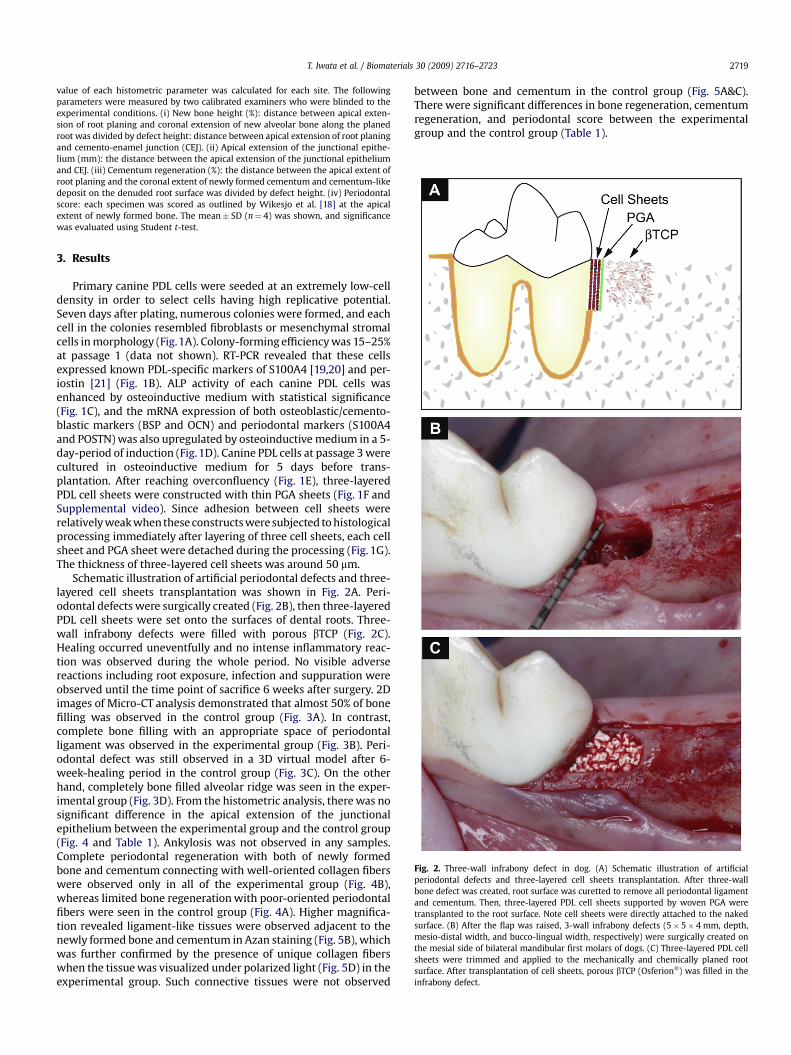
Fig. 2. Three-wall infrabony defect in dog. (A) Schematic illustration of artificialperiodontal defects and three-layered cell sheets transplantation. After three-wallbone defect was created, root surface was curetted to remove all periodontal ligamentand cementum. Then, three-layered PDL cell sheets supported by woven PGA weretransplanted to the root surface. Note cell sheets were directly attached to the nakedsurface. (B) After the flap was raised, 3-wall infrabony defects (5� 5� 4 mm, depth,mesio-distal width, and bucco-lingual width, respectively) were surgically created onthe mesial side of bilateral mandibular first molars of dogs. (C) Three-layered PDL cellsheets were trimmed and applied to the mechanically and chemically planed rootsurface. After transplantation of cell sheets, porous bTCP (Osferion�) was filled in theinfrabony defect.
T. Iwata et al. / Biomaterials 30 (2009) 2716–2723 2719
value of each histometric parameter was calculated for each site. The followingparameters were measured by two calibrated examiners who were blinded to theexperimental conditions. (i) New bone height (%): distance between apical exten-sion of root planing and coronal extension of new alveolar bone along the planedroot was divided by defect height: distance between apical extension of root planingand cemento-enamel junction (CEJ). (ii) Apical extension of the junctional epithe-lium (mm): the distance between the apical extension of the junctional epitheliumand CEJ. (iii) Cementum regeneration (%): the distance between the apical extent ofroot planing and the coronal extent of newly formed cementum and cementum-likedeposit on the denuded root surface was divided by defect height. (iv) Periodontalscore: each specimen was scored as outlined by Wikesjo et al. [18] at the apicalextent of newly formed bone. The mean� SD (n¼ 4) was shown, and significancewas evaluated using Student t-test.
3. Results
Primary canine PDL cells were seeded at an extremely low-celldensity in order to select cells having high replicative potential.Seven days after plating, numerous colonies were formed, and eachcell in the colonies resembled fibroblasts or mesenchymal stromalcells in morphology (Fig.1A). Colony-forming efficiency was 15–25%at passage 1 (data not shown). RT-PCR revealed that these cellsexpressed known PDL-specific markers of S100A4 [19,20] and per-iostin [21] (Fig. 1B). ALP activity of each canine PDL cells wasenhanced by osteoinductive medium with statistical significance(Fig. 1C), and the mRNA expression of both osteoblastic/cemento-blastic markers (BSP and OCN) and periodontal markers (S100A4and POSTN) was also upregulated by osteoinductive medium in a 5-day-period of induction (Fig.1D). Canine PDL cells at passage 3 werecultured in osteoinductive medium for 5 days before trans-plantation. After reaching overconfluency (Fig. 1E), three-layeredPDL cell sheets were constructed with thin PGA sheets (Fig. 1F andSupplemental video). Since adhesion between cell sheets wererelatively weak when these constructs were subjected to histologicalprocessing immediately after layering of three cell sheets, each cellsheet and PGA sheet were detached during the processing (Fig. 1G).The thickness of three-layered cell sheets was around 50 mm.
Schematic illustration of artificial periodontal defects and three-layered cell sheets transplantation was shown in Fig. 2A. Peri-odontal defects were surgically created (Fig. 2B), then three-layeredPDL cell sheets were set onto the surfaces of dental roots. Three-wall infrabony defects were filled with porous bTCP (Fig. 2C).Healing occurred uneventfully and no intense inflammatory reac-tion was observed during the whole period. No visible adversereactions including root exposure, infection and suppuration wereobserved until the time point of sacrifice 6 weeks after surgery. 2Dimages of Micro-CT analysis demonstrated that almost 50% of bonefilling was observed in the control group (Fig. 3A). In contrast,complete bone filling with an appropriate space of periodontalligament was observed in the experimental group (Fig. 3B). Peri-odontal defect was still observed in a 3D virtual model after 6-week-healing period in the control group (Fig. 3C). On the otherhand, completely bone filled alveolar ridge was seen in the exper-imental group (Fig. 3D). From the histometric analysis, there was nosignificant difference in the apical extension of the junctionalepithelium between the experimental group and the control group(Fig. 4 and Table 1). Ankylosis was not observed in any samples.Complete periodontal regeneration with both of newly formedbone and cementum connecting with well-oriented collagen fiberswere observed only in all of the experimental group (Fig. 4B),whereas limited bone regeneration with poor-oriented periodontalfibers were seen in the control group (Fig. 4A). Higher magnifica-tion revealed ligament-like tissues were observed adjacent to thenewly formed bone and cementum in Azan staining (Fig. 5B), whichwas further confirmed by the presence of unique collagen fiberswhen the tissue was visualized under polarized light (Fig. 5D) in theexperimental group. Such connective tissues were not observed
between bone and cementum in the control group (Fig. 5A&C).There were significant differences in bone regeneration, cementumregeneration, and periodontal score between the experimentalgroup and the control group (Table 1).

Fig. 3. Micro-CT analysis of periodontal defects after 6-week period of healing. (A, B) 2D image of the 3-wall defect transplanted with bTCP (A) or three-layered PDL cell sheets/bTCP(B). Triangle indicates cement-enamel junction (CEJ) of bilateral mandibular first molars of dog. (bar, 1 mm) (C, D) 3D virtual model of the 3-wall defect transplanted with bTCP (C)or three-layered PDL cell sheets/bTCP (D). After the 6 week of healing period, micro-CT analysis was performed and images were reconstructed with VG max software.
T. Iwata et al. / Biomaterials 30 (2009) 2716–27232720
4. Discussion
In the present study, we successfully expanded primary caninePDL cells in vitro and fabricated transplantable constructscomposing three-layered PDL cell sheets and a PGA sheet asa supporter. Because of the limited initial adhesion of primarycanine PDL cells [13], Primaria� culture dish was utilized topromote cell adhesion. By reducing seeding cell density as reportedpreviously [14] with some modifications, highly proliferative PDLcells were selected. In the present study, primary PDL cells wereenzymatically isolated. These cells were higher in proliferativecapacity and smaller in cell size than those cells isolated by anexplant method, implying that enzymatic digestion can facilitaterapid cell expansion with stromal or progenitor phenotypes (datanot shown). Despite intensive research efforts, no definitivemarkers specific to PDL cells have not been shown. Among thesecandidates reported previously, gene expression of S100A4 [19,20]and periostin [21] were examined and both genes were expressedby isolated PDL cells in the present study. PDL tissues are veryunique because they are never calcified spontaneously even theyare located in a close gap between two types of hard tissues, boneand cementum. Duarte et al. proposed that S100A4 can be a nega-tive regulator of mineralization in PDL tissues [19,20]. Periostin thatis known to be strongly expressed in PDL tissues [21] is suggested tobe essential for the integrity and function of PDL [22].
In addition, we investigated the differentiation capacity ofcanine PDL cells. We confirmed the capacities of osteoblastic/cementoblastic differentiation in each canine PDL cells from theresults of both ALP activity and gene expression, suggesting culture
of 5-day-period with osteoinductive medium can induce cytodif-ferentiation of each canine PDL cells with varying degrees.Considering the previous studies which demonstrated that PDLcells cultured with osteoinductive medium could regenerate newlyformed cementum-like tissues [11,12], we cultured PDL cells withosteoinductive medium then transplanted into periodontal defects.
In this study, we first utilized a poly (vinylidine difluoride)membrane for the transfer of cell sheets onto teeth root surfacesand the membrane was recovered after the transfer as previouslydescribed [23]. In this case, transplanted cell sheets were hardlyseen on the teeth root surfaces, because the colour was similaramong cell sheets and root surfaces. This shortcoming mighthamper reliable and reproducible transplantation of cell sheets,since the attachment of cell sheets onto hard tissue surfaces isweaker than that onto soft tissues. Therefore, we considered to usehyaluronan membrane [13], that were co-transplanted with cellsheets in the next study. We performed periodontal regenerationwith monolayered PDL cell sheet combined with hyaluronan ina canine dehiscence defects. However, the success rate was 60% (3of 5 dogs) and authors concluded that it may be due to the low cellsheet stability on the denuded root surface [13]. Thus, we needa definite method for delivery and stability of cell sheet transplantwith a safe biomaterial which is approved for clinical settings. Inaddition, this hyaluronan membrane was fabricated in house, andwasn’t approved for clinical use by Ministry of Health, Labour andWelfare of Japan. Since our final goal is its clinical application, wefinally selected a commercially available material, PGA membrane,which is already approved for clinical use. The PGA membranefacilitated quick fabrication of transplantable constructs of three-
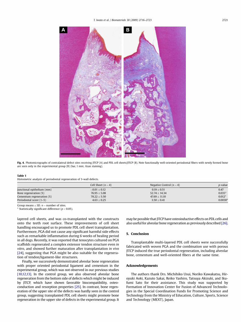
Table 1Histometric analysis of periodontal regeneration of 3-wall defects.
Cell Sheet (n¼ 4) Negative Control (n¼ 4) p-value
Junctional epithelium (mm) �0.01� 0.12 0.19� 0.51 0.47Bone regeneration (%) 76.95� 5.08 52.74� 14.34 0.035*Cementum regeneration (%) 78.22� 5.50 47.69� 11.10 0.012*Periodontal score (1–5) 4.63� 0.25 3.50� 0.41 0.0030*
Group means� SD. n¼ number of sites.* Statistically significant difference (p< 0.05).
Fig. 4. Photomicrographs of contralateral defect sites receiving bTCP (A) and PDL cell sheets/bTCP (B). Note functionally well-oriented periodontal fibers with newly formed boneare seen only in the experimental group (B) (bar, 1 mm; Azan staining).
T. Iwata et al. / Biomaterials 30 (2009) 2716–2723 2721
layered cell sheets, and was co-transplanted with the constructsonto the teeth root surface. These improvements of cell sheethandling encouraged us to promote PDL cell sheet transplantation.Furthermore, PGA did not cause any significant harmful side effectssuch as remarkable inflammation during 6 weeks of healing periodin all dogs. Recently, it was reported that tenocytes cultured on PGAscaffolds regenerated a complex extensor tendon structure even invitro, and showed further maturation after transplantation in vivo[24], suggesting that PGA might be also suitable for the regenera-tion of tendon/ligament-like structures.
Finally, we successively demonstrated alveolar bone regenerationwith proper oriented periodontal ligament and cementum in theexperimental group, which was not observed in our previous studies[10,12,13]. In the control group, we also observed alveolar boneregeneration from the bottom side of defects which might be inducedby bTCP, which have shown favorable biocompatibility, osteo-conduction and resorption properties [25]. In contrast, bone regen-eration of the upper site of the defects was hardly seen in the controlgroup, suggesting transplanted PDL cell sheets might promote boneregeneration in the upper site of defects in the experimental group. It
may be possible that bTCP have osteoinductive effects on PDL cells andalso useful for alveolar bone regeneration as previously described [26].
5. Conclusion
Transplantable multi-layered PDL cell sheets were successfullyfabricated with woven PGA and the combination use with porousbTCP induced the true periodontal regeneration, including alveolarbone, cementum and well-oriented fibers at the same time.
Acknowledgements
The authors thank Drs. Michihiko Usui, Noriko Kawakatsu, Hir-oyuki Aoki, Kazuto Sakai, Reiko Yashiro, Tatsuya Akizuki, and Iku-fumi Sato for their assistance. This study was supported byFormation of Innovation Center for Fusion of Advanced Technolo-gies in the Special Coordination Funds for Promoting Science andTechnology from the Ministry of Education, Culture, Sports, Scienceand Technology (MEXT), Japan.

Fig. 5. Higher magnification of Fig. 4 (A, C: bTCP, B, D: PDL cell sheets/bTCP). Complete periodontal regeneration, functionally well-oriented periodontal fibers connecting with bothnewly formed cementum and bone (arrowhead), is observed in the experimental group by Azan staining (B) and by polarized light (D). In contrast, such well-oriented fibers are notobserved in the control group (A and C) (bar, 500 mm).
T. Iwata et al. / Biomaterials 30 (2009) 2716–27232722
Appendix
Figures with essential colour discrimination. Parts of themajority of the figures in this article are difficult to interpret inblack and white. The full colour images can be found in the on-lineversion, at doi:10.1016/j.biomaterials.2009.01.032.
Appendix. Supplementary material
Supplementary material can be found, in the online version, atdoi:10.1016/j.biomaterials.2009.01.032.
References
[1] Pihlstrom BL, Michalowicz BS, Johnson NW. Periodontal diseases. Lancet2005;366:1809–20.
[2] Caton J, Nyman S, Zander H. Histometric evaluation of periodontal surgery. II.Connective tissue attachment levels after four regenerative procedures. J ClinPeriodontol 1980;7:224–31.
[3] Esposito M, Grusovin MG, Coulthard P, Worthington HV. Enamel matrixderivative (Emdogain) for periodontal tissue regeneration in intrabonydefects. Cochrane Database Syst Rev 2005. CD003875.
[4] Bartold PM, Xiao Y, Lyngstaadas SP, Paine ML, Snead ML. Principles andapplications of cell delivery systems for periodontal regeneration. Periodon-tology 2000;2006(41):123–35.
[5] Seo BM, Miura M, Gronthos S, Bartold PM, Batouli S, Brahim J, et al. Investi-gation of multipotent postnatal stem cells from human periodontal ligament.Lancet 2004;364:149–55.
[6] Nagatomo K, Komaki M, Sekiya I, Sakaguchi Y, Noguchi K, Oda S, et al. Stem cellproperties of human periodontal ligament cells. J Periodont Res 2006;41:303–10.
[7] Nakahara T, Nakamura T, Kobayashi E, Kuremoto K, Matsuno T, Tabata Y, et al.In situ tissue engineering of periodontal tissues by seeding with periodontalligament-derived cells. Tissue Eng 2004;10:537–44.
[8] Liu Y, Zheng Y, Ding G, Fang D, Zhang C, Bartold PM, et al. Periodontal ligamentstem cell-mediated treatment for periodontitis in miniature swine. Stem Cells2008;26:1065–73.
[9] Sonoyama W, Liu Y, Fang D, Yamaza T, Seo BM, Zhang C, et al. Mesenchymal stemcell-mediated functional tooth regeneration in swine. PLoS ONE 2006;1:e79.
[10] Hasegawa M, Yamato M, Kikuchi A, Okano T, Ishikawa I. Human periodontalligament cell sheets can regenerate periodontal ligament tissue in an athymicrat model. Tissue Eng 2005;11:469–78.
[11] Flores MG, Hasegawa M, Yamato M, Takagi R, Okano T, Ishikawa I. Cementum–periodontal ligament complex regeneration using the cell sheet technique.J Periodont Res 2008;43:364–71.
[12] Flores MG, Yashiro R, Washio K, Yamato M, Okano T, Ishikawa I. Periodontalligament cell sheet promotes periodontal regeneration in athymic rats. J ClinPeriodontol 2008;35:1066–72.
[13] Akizuki T, Oda S, Komaki M, Tsuchioka H, Kawakatsu N, Kikuchi A, et al.Application of periodontal ligament cell sheet for periodontal regeneration:a pilot study in beagle dogs. J Periodont Res 2005;40:245–51.
[14] Nimura A, Muneta T, Koga H, Mochizuki T, Suzuki K, Makino H, et al. Increasedproliferation of human synovial mesenchymal stem cells with autologoushuman serum: comparisons with bone marrow mesenchymal stem cells andwith fetal bovine serum. Arthritis Rheum 2008;58:501–10.
[15] Livak KJ, Schmittgen TD. Analysis of relative gene expression data using real-timequantitative PCR and the 2(T) (-Delta Delta C) method. Methods 2001;25:402–8.
[16] Maruyama H, Aoki A, Sasaki KM, Takasaki AA, Iwasaki K, Ichinose S, et al. Theeffect of chemical and/or mechanical conditioning on the Er:YAG laser-treatedroot cementum: analysis of surface morphology and periodontal ligamentfibroblast attachment. Lasers Surg Med 2008;40:211–22.
[17] Shimizu T, Sekine H, Yang J, Isoi Y, Yamato M, Kikuchi A, et al. Polysurgery ofcell sheet grafts overcomes diffusion limits to produce thick, vascularizedmyocardial tissues. FASEB J 2006;20:708–10.
[18] Wikesjo UM, Sorensen RG, Kinoshita A, Jian Li X, Wozney JM. Periodontalrepair in dogs: effect of recombinant human bone morphogenetic protein-12(rhBMP-12) on regeneration of alveolar bone and periodontal attachment.J Clin Periodontol 2004;31:662–70.
[19] Duarte WR, Iimura T, Takenaga K, Ohya K, Ishikawa I, Kasugai S. Extracellularrole of S100A4 calcium-binding protein in the periodontal ligament. BiochemBiophys Res Commun 1999;255:416–20.
[20] Kato C, Kojima T, Komaki M, Mimori K, Duarte WR, Takenaga K, et al. S100A4inhibition by RNAi up-regulates osteoblast related genes in periodontal liga-ment cells. Biochem Biophys Res Commun 2005;326:147–53.
[21] Horiuchi K, Amizuka N, Takeshita S, Takamatsu H, Katsuura M, Ozawa H, et al.Identification and characterization of a novel protein, periostin, withrestricted expression to periosteum and periodontal ligament and increased

T. Iwata et al. / Biomaterials 30 (2009) 2716–2723 2723
expression by transforming growth factor beta. J Bone Miner Res 1999;14:1239–49.
[22] Rios HF, Ma D, Xie Y, Giannobile WV, Bonewald LF, Conway SJ, et al. Periostin isessential for the integrity and function of the periodontal ligament duringocclusal loading in mice. J Periodontol 2008;79:1480–90.
[23] Ohki T, Yamato M, Murakami D, Takagi R, Yang J, Namiki H, et al. Treatment ofoesophageal ulcerations using endoscopic transplantation of tissue-engi-neered autologous oral mucosal epithelial cell sheets in a canine model. Gut2006;55:1704–10.
[24] Wang B, Liu W, Zhang Y, Jiang Y, Zhang WJ, Zhou G, et al. Engineering ofextensor tendon complex by an ex vivo approach. Biomaterials 2008;29:2954–61.
[25] Gaasbeek RD, Toonen HG, van Heerwaarden RJ, Buma P. Mechanism of boneincorporation of beta-TCP bone substitute in open wedge tibial osteotomy inpatients. Biomaterials 2005;26:6713–9.
[26] Yamauchi K, Takahashi T, Funaki K, Yamashita Y. Periosteal expansion osteo-genesis using highly purified beta-tricalcium phosphate blocks: a pilot studyin dogs. J Periodontol 2008;79:999–1005.