Embed Size (px)
Citation preview

Yoshinori Nishino
Laboratory of Coherent X-ray Optics,Research Institute for Electronic Science,
Hokkaido University
XDL2011 報告
Workshop 1-Diffraction Microscopy, Holography and Ptychographyusing Coherent Beams

Outline
• ワークショップ概要• 回折顕微法• ホログラフィー• タイコグラフィー• Cross-correlation Analysis / Fluctuation Microscopy• ワークショップの内容
• ワークショップでの私の発表

Workshop 1-Diffraction Microscopy, Holography and Ptychography using Coherent Beams
Organizers: Janos Kirz (Lawrence Berkeley National Lab)Qun Shen (National Synchrotron Light Source II)Darren Dale (Cornell University)
Purpose: The purpose of the workshop is to assess the state-of-the-art in the use of coherent hard x-ray beams for high-resolution imaging. Diffraction microscopy, Fourier transform holography and ptychography are making rapid strides, but have yet to realize their full potential due to current limitations of spatial and temporal coherence of hard x-ray beams. We are especially interested in exploring what might be most feasible with a Energy Recovery Linac (ERL) and Ultimate Storage Ring (USR) x-ray sources.
http://erl.chess.cornell.edu/gatherings/2011_Workshops/details1.htm

Description:Coherence-based imaging experiments are limited mainly by the lack of coherent X-ray sources. Diffraction microscopy and related techniques have started to extend the resolution in X-ray microscopy beyond the limitations of X-ray focusing elements such as zone plates or multilayer Laue Lenses. Calculations indicate that these techniques should be limited only by radiation damage, and/or the diffraction limit. While using short pulse “flash” illumination at X-ray FELs can yield sufficient scattered intensity to image a sample with a single shot, the power in that shot vaporizes the specimen. Hence FELs are not ideal for imaging techniques requiring multiple exposures, for example ptychography and tomography. ERL and USR sources are being conceived to deliver intense, spatially coherent, hard X-ray beams with a quasi-continuous time structure. Sufficient temporal coherence may be provided by the undulator source directly, possibly further filtered using a monochromator. The ideal X-ray energy will depend on the sample, but in general soft X-rays offer higher scattering cross sections and higher coherent flux, but higher energies provide greater penetration through the specimen, and hence the Born approximation (generally assumed in the reconstruction process) is better satisfied. The increased penetration of harder x-rays will also allow greater emphasis on studies of samples in complex environments. The expected high coherent flux may provide the opportunity to study time-sequence series of dynamic phenomena.

For materials where the resolution is not limited by radiation damage, these techniques will provide new capabilities to analyze and visualize porosity, inclusions, defects, and other buried structures. These new imaging techniques can be used in combination with spectroscopy to create a sensitive tool highlighting elemental constituents. Work by Ian Robinson’s group has provided a unique new tool to investigate the morphology, strain fields and defects in nanocrystals. This approach makes use of the fine speckles around Bragg peaks, and requires the short wavelength and penetrating power of high-energy X-rays.
For biological samples, such as cells, tissue sections, etc., one can use cryogenic freezing to preserve the morphology of the sample while imaging with a resolution of about 10 nm. High-pressure cryogenic freezing techniques are under development at Cornell to prepare such specimens while minimizing structural perturbations due to the expansion of water on freezing.
Fourier transform holography techniques provide a quick and easy route to a reconstruction at a resolution given by the size of the source of the reference beam. Multiple reference beams, or uniformly redundant arrays have been used to increase the strength of the reference beam. Can this technique be extended to 3D?
Ptychography has been shown to provide a robust “engine” for 2D image reconstruction without the need for finite support. The first 3D reconstruction using this technique was recently demonstrated; the potential impact and limitations of extending this technique to 3D should be explored.

The premise of the workshop is that it is technologically feasible to extend the resolution in X-ray microscopy well beyond the limitations in X-ray optics, and optics-based instruments. The workshop will examine this premise from the standpoint of what might be needed to bring these techniques to routine use.
The workshop will bring together both practitioners of the various coherence-based imaging techniques and scientists from various fields using alternative techniques to analyze samples, to assess the scientific needs, to discuss the technical challenges, distill the best approaches, and to establish the requirements for a successful program.
Some of the technical challenges that need to be addressed could include:
. Suppression of harmonics form the source
. Dynamic range in the detector
. Precision positioning, alignment and rotation of the sample
. Efficient data handling, assembly and reconstruction algorithms
. Pre-and post-exposure assessment of sample quality, suitability, and radiation damage
. Understanding the effects of background from Compton scattering and other sources
. Effects due to the missing data where either a beam-stop is used, or where the substrate interferes with complete 360 degree tomographic data collection
. In the case of diffraction microscopy, determining the finite support, and minimizing the effect of the specimen mount

The specific purpose of this workshop is to assess and discuss the new scientific opportunities based on coherent imaging that will be opened up by ERLs and USRs. The intent of the workshop is not to focus on experiments that have already been done, but rather to brainstorm and discuss experiments that you would like to do, but are impractical with existing sources. The talks are intended to be short, future-oriented, and each followed by ample discussion time. The goal is to present innovative ideas, new approaches, and out-of-the-box thinking. The workshop will include a poster session where latest results may be presented and augmented by handouts focusing especially on the new science that might be expected from ERLs or USRs.
[ Invitation Letter より ]

プログラム(1日目)
イントロダクションDon Bilderback (Cornell University)
"Energy Recovery Linac (ERL) and Ultimate Storage Ring (USR) Properties "Sol Gruner (Cornell University)
"X-ray Detectors: State-of-the-art & Future Possibilities "Qun Shen (National Synchrotron Light Source II)
"New Opportunities with Hard X-ray Diffraction Limited Sources “
回折顕微法、ホログラフィーGarth Williams (Linac Coherent Light Source)
"Coherent Imaging Without a Laser: getting the most bang for your electrons"Jim Fienup (University of Rochester)
"X-ray Coherent Diffractive Imaging with an Extended Reference"Stefano Marchesini (Lawrence Berkeley National Laboratory) ALS COSMIC
"High-efficiency Fourier Holography with Uniformly Redundant Arrays “
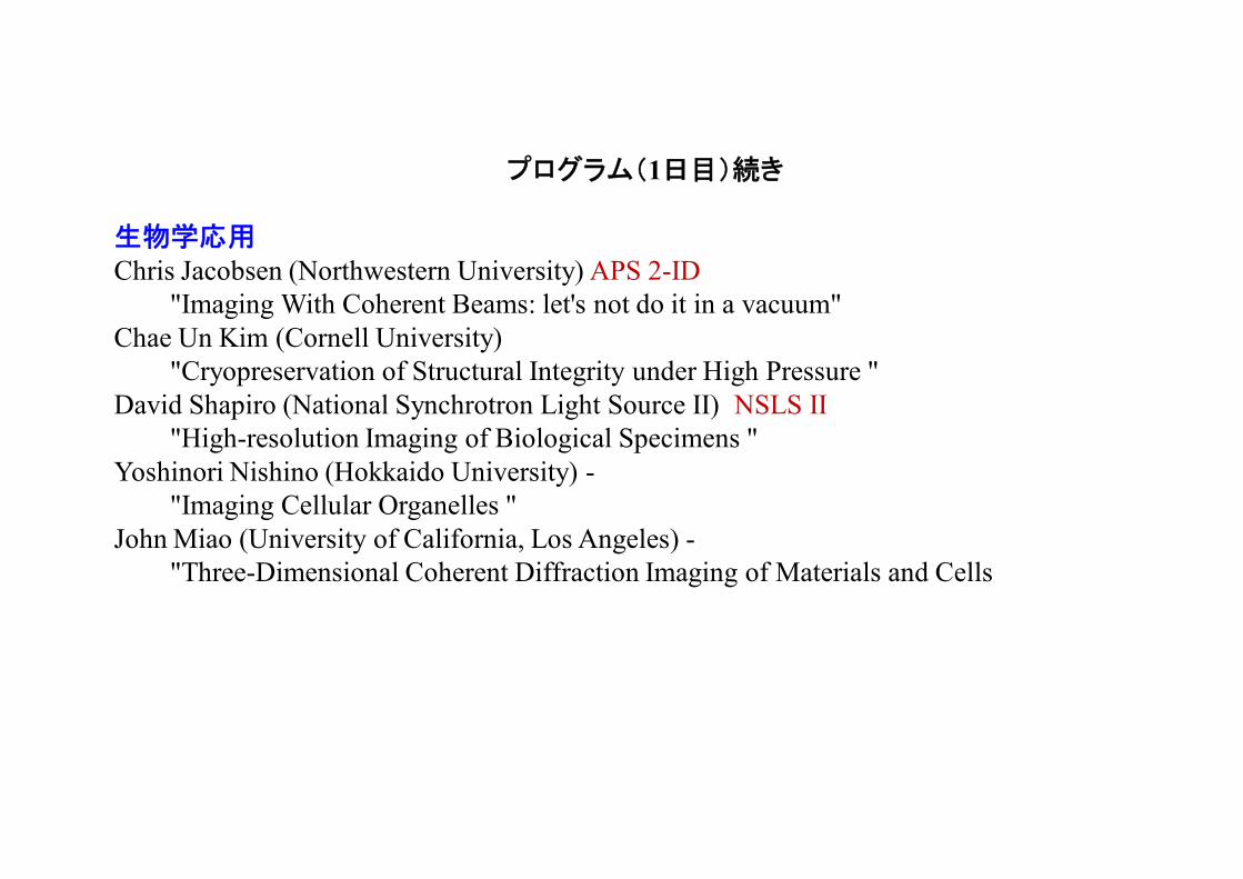
プログラム(1日目)続き
生物学応用Chris Jacobsen (Northwestern University) APS 2-ID
"Imaging With Coherent Beams: let's not do it in a vacuum"Chae Un Kim (Cornell University)
"Cryopreservation of Structural Integrity under High Pressure "David Shapiro (National Synchrotron Light Source II) NSLS II
"High-resolution Imaging of Biological Specimens "Yoshinori Nishino (Hokkaido University) -
"Imaging Cellular Organelles "John Miao (University of California, Los Angeles) -
"Three-Dimensional Coherent Diffraction Imaging of Materials and Cells

プログラム(2日目)
材料科学応用、タイコグラフィーIvan Vartaniants (Deutsches Elektronen-Synchrotron)
"Coherent Diffractive Imaging and Determining Structural Properties from Cross-correlation Analysis "
Ross Harder (Advanced Photon Source) APS ID-34-C"Probing Strain and Defects in Single Crystals with Coherent X-ray Diffraction"
Pierre Thibault (Technische Universität München) SLS cSAXS"Ptychography in 2D and 3D"
Harald Ade (North Carolina State University) "Spectromicroscopy, Resonant Scattering, Possible Extensions to PtychographicImaging"
Oleg Shpyrko (University of California, San Diego) "Magnetic Domains and Dynamics"
Ian McNulty (Advanced Photon Source)"Resonant Coherent X-ray Imaging"
Breakout sessions and summary writing, roundup workshop

X-ray Diffraction Microscopy
Diffraction Pattern (log scale)l=17 Å
Reconstructed Imagespatial resolution: 75 nm
Gold dots (~100 nm diameter, 80 nm thick)on silicon nitride membrane
“Extending the methodology of x-ray crystallography to allow imaging of micrometer-sized non-crystalline specimens”
J. Miao, P. Charalambous, J. Kirz & D. Sayre, Nature 400, 342 (1999).
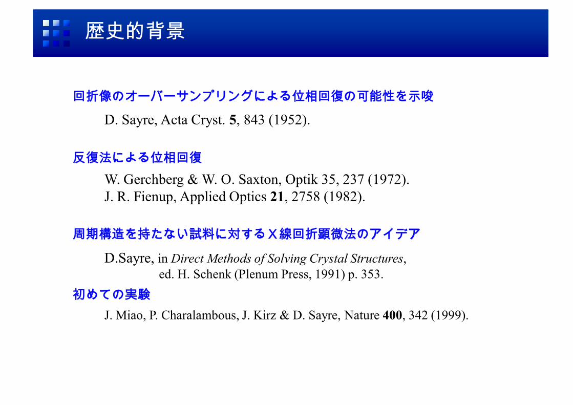
歴史的背景
W. Gerchberg & W. O. Saxton, Optik 35, 237 (1972).J. R. Fienup, Applied Optics 21, 2758 (1982).
反復法による位相回復
周期構造を持たない試料に対するX線回折顕微法のアイデア
D.Sayre, in Direct Methods of Solving Crystal Structures,ed. H. Schenk (Plenum Press, 1991) p. 353.
回折像のオーバーサンプリングによる位相回復の可能性を示唆
D. Sayre, Acta Cryst. 5, 843 (1952).
初めての実験J. Miao, P. Charalambous, J. Kirz & D. Sayre, Nature 400, 342 (1999).

Oversamping
Diffraction Pattern (log scale) Image
no-density region around image
oversampling

反復法による位相回復
otherwise,'0','
1 rrrr
rnn
nnn
Sr
Hybrid Input-Output (HIO) Algorithm
S: Support, : between 0.5 and 1
R.W. Gerchberg and W.O. Saxton, Optik (Stuttgart) 35, 237 (1972).J.R. Fienup, Appl. Opt. 21, 2758 (1982).

Simulation of the HIO algorithm
Sample Image Calculated Diffraction Pattern
Iterative Image Reconstruction Reconstructed Image after 5000 Iterations

ナノ結晶中のひずみ場分布の可視化
電子密度+ひずみ場
電子密度
M.A. Pfeifer et al., Nature 442, 63 (2006)
空間分解能: 45 nm
SiO2基板との界面で最大0.5Åの変形
34-ID-C, APS
波長1.38Å
鉛のナノ結晶中のひずみ場の可視化
ロンドン大学 ユニバーシティー・カレッジ のIan Robinsonらのグループが米国APSで実験を行っている

Ptychography
Rodenberg et al., PRL 98, 034801 (2007)
P.Thibault, et al., Science 321, 379 (2008)

Ptychography
K. Giewekemeyer et al., PNAS 107, 529 (2010)
Unstained and unsliced freeze-dried Deinococcus radiodurans
M. Dierolf et al., Nature 467, 436 (2010)

HERALDO (holography with extended reference by autocorrelation linear differential operation)
M. Guizar-Sicairos and J. R. Fienup, Optics Express 15, 17592 (2007).
without ReferenceDiffraction Microscopy
Point ReferenceFourier TransformHolography
Rectangular-Hole ReferenceHolography by Podorov, Pavlov & Paganin
Obscuring-Tip Reference (as an example)HERALDO
Lensless Imaging
Iterative phase-retrievalfor reconstruction Non-Iterative Reconstruction

Femtosecond Snapshot imaging with EUV FEL at SCSS
Test pattern milled by FIB~ 800 nm thick Au depositedon 100 nm thick Si3N4 membrane
Horizontal Slit:width: ~ 0.9 µm, length: ~ 112 µm
Vertical Slit:width: ~ 1.2 µm, length: ~ 57 µm
Scanning Ion Microscope (SIM) Image
Sample (SPring-8 logo)
Horizontal Reference Slit
Vertical Reference Slit
Holography with Extended Reference by Autocorrelation Linear Differential Operation (HERALDO)
50µm
Y. Nishino et al., Appl. Phys. Express, 3, 102701 (2010).
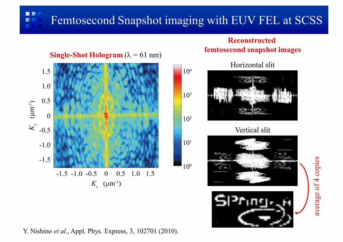
Femtosecond Snapshot imaging with EUV FEL at SCSS
Vertical slit
Horizontal slitSingle-Shot Hologram ( = 61 nm)
Reconstructed femtosecond snapshot images
aver
age
of 4
cop
ies
Y. Nishino et al., Appl. Phys. Express, 3, 102701 (2010).

Holograpy
“Massively parallel X-ray holography”,S. Marchesini et al., Nature Photonics 2, 560 (2008).
W. F. Schlotter et al., App. Phys. Lett. 89, 163112 (2006)
L-M. Stadler et al.,Phys. Rev. Lett. 100, 245503 (2008).

X-ray cross correlation analysis (XCCA)
P. Wochner et al., PNAS 106, 11511 (2009)
hidden local symmetries in disordered matter

Fluctuation Microscopy
V. Elser: arXiv:1007.3777v1
D. K. Saldin et al., PRL 106, 115501 (2011)
TEM image of randomly oriented 90×25 nm gold nanorods
Reconstructed Image of gold nanorods
“The cross-correlation scheme has a disadvantage relative to the single-shot scheme both because of restrictions in the processing and the smaller number of photons scattered per particle.”
Z. Kam, Macromolecules (1977)

プログラム(1日目)
イントロダクションDon Bilderback (Cornell University)
"Energy Recovery Linac (ERL) and Ultimate Storage Ring (USR) Properties "Sol Gruner (Cornell University)
"X-ray Detectors: State-of-the-art & Future Possibilities "Qun Shen (National Synchrotron Light Source II)
"New Opportunities with Hard X-ray Diffraction Limited Sources “
回折顕微法、ホログラフィーGarth Williams (Linac Coherent Light Source)
"Coherent Imaging Without a Laser: getting the most bang for your electrons"Jim Fienup (University of Rochester)
"X-ray Coherent Diffractive Imaging with an Extended Reference"Stefano Marchesini (Lawrence Berkeley National Laboratory) ALS COSMIC
"High-efficiency Fourier Holography with Uniformly Redundant Arrays “

プログラム(1日目)続き
生物学応用Chris Jacobsen (Northwestern University) APS 2-ID
"Imaging With Coherent Beams: let's not do it in a vacuum"Chae Un Kim (Cornell University)
"Cryopreservation of Structural Integrity under High Pressure "David Shapiro (National Synchrotron Light Source II) NSLS II
"High-resolution Imaging of Biological Specimens "Yoshinori Nishino (Hokkaido University) -
"Imaging Cellular Organelles "John Miao (University of California, Los Angeles) -
"Three-Dimensional Coherent Diffraction Imaging of Materials and Cells

Scanning X-ray Microscopy
Freshwater flagellate Cryptomonas
Scanning Zernike
C. Holzner et al., Nature Physics 6, 883 (2010)
X-ray fluorescence microtomography
freshwater diatom Cyclotella meneghiniana
M. D. de Jonge et al, PNAS 107, 15676 (2010)

プログラム(2日目)
材料科学応用、タイコグラフィーIvan Vartaniants (Deutsches Elektronen-Synchrotron)
"Coherent Diffractive Imaging and Determining Structural Properties from Cross-correlation Analysis "
Ross Harder (Advanced Photon Source) APS ID-34-C"Probing Strain and Defects in Single Crystals with Coherent X-ray Diffraction"
Pierre Thibault (Technische Universität München) SLS cSAXS"Ptychography in 2D and 3D"
Harald Ade (North Carolina State University) "Spectromicroscopy, Resonant Scattering, Possible Extensions to PtychographicImaging"
Oleg Shpyrko (University of California, San Diego) "Magnetic Domains and Dynamics"
Ian McNulty (Advanced Photon Source)"Resonant Coherent X-ray Imaging"
Breakout sessions and summary writing, roundup workshop
Yoshinori Nishino
Laboratory of Coherent X-ray Optics,Research Institute for Electronic Science,
Hokkaido University
Imaging Cellular Organelles

Laboratory of Coherent X-ray Optics
ProfessorYoshinori Nishino
Laboratory of Coherent X-ray Optics,Research Institute for Electronic Science,Hokkaido University
since April, 2010
Secretary
Undergraduate Students
・ Kei Soeta・ Chie Nagase・ Arata Mori・ Kiyo Ssaki
Assistant ProfessorMarcus Newton
Assistant ProfessorTakashi Kimura

Outline
• Imaging Chromosome• Discussions
– Average Structure / Individual Structure– Focusing X-rays
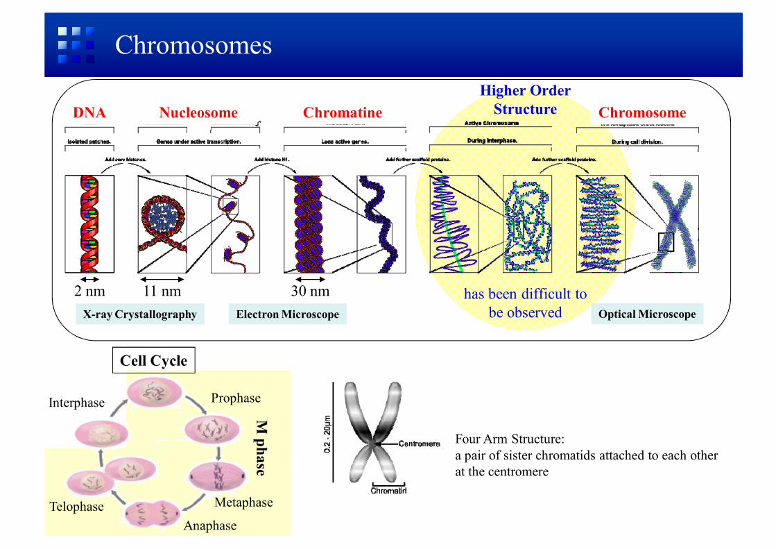
DNA
2 nm
Nucleosome
11 nm
Chromatine Chromosome
30 nm
Chromosomes
Four Arm Structure:a pair of sister chromatids attached to each other at the centromere
Cell Cycle
Interphase
M phase
Prophase
Metaphase
AnaphaseTelophase
X-ray Crystallography Electron Microscope Optical Microscope
Higher OrderStructure
has been difficult tobe observed

BL29XUL, SPring-8
Coherent Diffraction Pattern
Sample
Unstained Human Chromosome
Fourier Transform
Phase Retrieval
Coherent X-ray Diffraction
Sample ImageReconstruction
without the aid of Lenses
Y. Nishino, Y. Takahashi, N. Imamoto, T. Ishikawa, and K. Maeshima, Phys. Rev. Lett. 102, 018101 (2009).
Physics Today, 62 (2), 20 (2009).Nature 457, 238 (2009).News Articles:
Synchrotron Radiation
SPring-8 (Harima, Japan)

2D Observation of unstained human chromosome
K. Maeshima & U. K. Laemmli, Developmental Cell 4, 467 (2003)
helically folded axial structure
Immunofluorescence Microscope Image
E. Boy de la Tour & U.K. Laemmli, Cell 55, 937 (1988)
First observation of axial structure in unstained chromosome
bar: 1 µm
Y. Nishino et al., Phys. Rev. Lett. 102, 018101 (2009).
condensin antibody (red)
spatial resolution = 38 nm

3D observation of unstained human chromosome
spatial resolution = 120 nmscale bars = 500 nm
First observation of a cellular organelle in 3D by using hard X-rays
centromere
centromere
• highest electron densityaround centromere
• consistent with2D reconstruction
• high densitynear axis
• wavy (helical) structurewas not observed
Y. Nishino et al., Phys. Rev. Lett. 102, 018101 (2009).

Spatial Resolution
S. Marchesini et al., Optics Express 11, 2344 (2003).
our 3D reconstruction(close to feature-destroying dose line)
Estimated Dose
• Single diffraction: 4 × 10⁸ Gy• 3D diffraction: 2 × 10¹⁰ Gy
For higher spatial resolution
• cooling the sample cryogenically• optimizing the dose• improving the phase retrieval method
M. R. Howells et al., J. Electron Spectrosc. Relat. Phenom. 170, 4 (2009) .

Average Structure / Individual Structure
Classical X-ray Methods
• X-ray Crystallography• Small-Angle X-ray Scattering (SAXS)
Average Structure
Less Radiation Dose to Each Individual Biological Object→ Higher Resolution
• for bio-molecules difficult crystallize• nearly physiological conditions
Solution Scattering of Biomolecules

Diffraction Microscopy Instrument at SPring-8
pinhole(20 µm)
guardslit 2
Sample beamstop CCD
detector
guardslit 1
• CCD Detector:Princeton Instruments PI-LCX 13001300 × 1340 PixelsPixel Size: 20 m × 20 mDirect IlluminationDeep Depletion
Hard X-ray Beamline BL29XULUSAXSMeasurement

DNA
2 nm
Nucleosome
11 nm
Chromatine Chromosome
30 nm
Chromosomes
Four Arm Structure:a pair of sister chromatids attached to each other at the centromere
Cell Cycle
Interphase
M phase
Prophase
Metaphase
AnaphaseTelophase
X-ray Crystallography Electron Microscope Optical Microscope
Higher OrderStructure
has been difficult tobe observed
?

Fluctuation Microscopy
V. Elser: arXiv:1007.3777v1
D. K. Saldin et al., PRL 106, 115501 (2011)
TEM image of randomly oriented 90×25 nm gold nanorods
Reconstructed Image of gold nanorods
“The cross-correlation scheme has a disadvantage relative to the single-shot scheme both because of restrictions in the processing and the smaller number of photons scattered per particle.”
Z. Kam, Macromolecules, 10, 927 (1977)

CC
Pulsed Coherent X-ray Solution Scattering
Averaging
• Ensemble Average
• Time Average
Concentric Scattering Pattern(1 Dimensional Data)
Unaveraged Data Acquisition
• Illuminating a Small Number ofMoleculses
• Ultrashort Pulse Duration
Conventional SAXS
Solution Scattering with XFEL
Speckled Coherent Scattering Pattern(2-Dimensinal Data)

X-ray Focusing down to world’s smallest 7 nm
20 keV X-rays
H. Mimura et al., Nature Physics 6, 122 (2009).

1 km-long beam line
Beamline Requirements
Light Source
Upstream Mirror
DownstreamMirror
Focal Point
Geometrical Demagnification= (Lens-to-Focus)/(Source-to-Lens)
• Diffraction Limited Source both Vertically and Horizontally
• Long Beamline• Stability
ERL or URL:Coherently Illuminate Focusing Mirrors without Loss
Example:Source Size: ~10 µmFocal Size: ~10 nmWorking Distance ~100 mm
↓Beamline Length ~100 m
wave optics
focal spot size ~N.A.λ

Adaptive Optical System for Hard-X-ray Focusing
Iterative phase-retrieval method using the intensity profiles around the beam waist
H. Yumoto, et al. , RSI 77, 063712 (2006).
in situ determination of the wavefront error of X-ray focusing mirror
20 platinum/carbon bilayers
H. Mimura et al., Nature Physics 6, 122 (2009).
T. Kimura et al., JJAP 48, 072503 (2009).
Adaptive optics to compensate figure error of the focusing mirror

Scanning X-ray Fluorescence Microscope
S. Matsuyama et al., X-Ray Spectrom. 38, 89 (2009).
SPring-8BL29XU EH2
M. Shimura et al.,Cancer Research 65, 4998 (2005).

Collaborators
• Hokkaido Univ.: T. Kimura, M. Newton
• National Institute of Genetics: K. Maeshima
• Osaka Univ.: Y. Takahashi, S. Matsuyama, K. Yamauchi
•Univ. Tokyo: H. Mimura
• National Center for Global Health and Medicine: M. Shimura
• JASRI: Y. Joti
• Univ. Tokyo: S. Takeuchi
•RIKEN SPring-8: Y. Bessho, T. Ishikawa

Summary
• Imaging Chromosome• X-ray diffraction microscopy enable high-contrastimaging for relatively thick samples.
• 2D & 3D observation of unstained human chromosome
• Average Structure / Individual Structure• SAXS• cross-correlation
• Focusing X-ray• X-ray focusing down to 7 nm
- long beamline- in situ determination of figure error of X-ray focusing mirror- Adaptive optics to compensate figure error
![Ⅳ 環境側面の報告 - Hitachi Metals · Ⅳ 環境側面の報告. 45. 日立金属グループcsr 活動報告2017[詳細活動報告] Ⅳ 環境側面の報告. 1. 環境マネジメント](https://img.pdfslide.tips/doc/110x75/5ec9a6db7c3455151d652bab/a-cfe-hitachi-metals-a-cfe-45-cefffcsr.jpg)
![[報告] 電影欣賞報告 - 奧圖瑪塔](https://img.pdfslide.tips/doc/110x75/58edf5eb1a28ab8c708b469b/-58edf5eb1a28ab8c708b469b.jpg)