Embed Size (px)
Citation preview
lable at ScienceDirect
Biomaterials 30 (2009) 5707–5719
Contents lists avai
Biomaterials
journal homepage: www.elsevier .com/locate/biomateria ls
Poly(u-pentadecalactone-co-butylene-co-succinate) nanoparticlesas biodegradable carriers for camptothecin delivery
Jie Liu a,b, Zhaozhong Jiang a, Shengmin Zhang b, W. Mark Saltzman a,*
a Department of Biomedical Engineering, Yale University, 55 Prospect Street, MEC 414, New Haven, CT 06511-8260, USAb Advanced Biomaterials and Tissue Engineering Center, Huazhong University of Science and Technology, Wuhan 430074, China
a r t i c l e i n f o
Article history:Received 16 April 2009Accepted 30 June 2009Available online 25 July 2009
Keywords:CamptothecinPolymer nanoparticleHydrophobicControlled releaseAntitumor effect
* Corresponding author. Tel.: þ1 203 432 4262; faxE-mail address: [email protected] (W.M. Sa
0142-9612/$ – see front matter � 2009 Elsevier Ltd.doi:10.1016/j.biomaterials.2009.06.061
a b s t r a c t
In this study, we show that degradable particles of a hydrophobic polymer can effectively deliver drugs totumors after i.v. administration. Free-standing nanoparticles with diameters of 100–300 nm weresuccessfully fabricated from highly hydrophobic, biodegradable poly(u-pentadecalactone-co-butylene-co-succinate) (PPBS) copolyesters. PPBS copolymers with various compositions (20–80 mol% PDL unitcontents) were synthesized via copolymerization of u-pentadecalactone (PDL), diethyl succinate (DES),and 1,4-butanediol (BD) using Candida antarctica lipase B (CALB) as the catalyst. Camptothecin (CPT, 12–22%) was loaded into PPBS nanoparticles with high encapsulation efficiency (up to 96%) using a modifiedoil-in-water single emulsion technique. The CPT-loaded nanoparticles had a zeta potential of about�10 mV. PPBS particles were non-toxic in cell culture. Upon encapsulation, the active lactone form of CPTwas remarkably stabilized and no lactone-to-carboxylate structural conversion was observed for CPT-loaded PPBS nanoparticles incubated in both phosphate-buffered saline (PBS, pH¼ 7.4) and DMEMmedium for at least 24 h. In PBS at 37 �C, CPT-loaded PPBS nanoparticles showed a low burst CPT release(20–30%) within the first 24 h followed by a sustained, essentially complete, release of the remainingdrug over the subsequent 40 days. Compared to free CPT, CPT-loaded PPBS nanoparticles showeda significant enhancement of cellular uptake, higher cytotoxicity against Lewis lung carcinoma and 9L celllines in vitro, a longer circulation time, and substantially better antitumor efficacy in vivo. These resultsdemonstrate the potential of PPBS nanoparticles as long-term stable and effective drug delivery systemsin cancer therapy.
� 2009 Elsevier Ltd. All rights reserved.
1. Introduction
Current chemotherapy is far from satisfactory: drug treatmentsoften have limited effectiveness and patients suffer from seriousside effects. Drug delivery devices have been studied extensivelyover the past few decades, including degradable polymer matrices,polymeric nanoparticles, liposomes, and micelles [1,2].The use ofnano-sized particles in cancer therapy is particularly exciting, asthese materials can increase drug solubility and stability, as well asimprove pharmacological effect by passively delivering chemo-therapeutic agents to tumor sites via enhanced permeability andretention (EPR) effect [3–5].
Camptothecin (CPT) is a natural plant alkaloid extracted fromCamptotheca acuminate (a tree grown in China), which has showna broad spectrum of antitumor activity against various types of
: þ1 203 432 0030.ltzman).
All rights reserved.
solid tumors [6]. However, effective delivery of CPT to tumor targetsis extremely challenging due to its insolubility in water, structuralinstability, and high toxicity to normal tissue cells. Under physio-logical conditions, i.e. at pH equal to or above 7, CPT undergoeslactone ring-opening hydrolysis to form the inactive carboxylateform as shown in Scheme 1 [7].
Additionally, human serum albumin in the blood has a highaffinity for binding to the carboxylate form of CPT, thus driving theabove lactone–carboxylate equilibrium toward the formation of theinactive carboxylate form [8]. As a result, the potency of the drug isreduced substantially when administrated to humans. Because ofits toxic side effects, CPT, like most other antitumor drugs, needs tobe frequently administered with limited doses to achieve desirabledrug efficacy. Effective drug delivery methods providing sustainedrelease of controllable amount of drugs over a prolonged period oftime would be obviously advantageous for administration of thesekinds of drugs [9].
To address these problems in CPT delivery, several differentapproaches have been taken to improve drug delivery efficiency
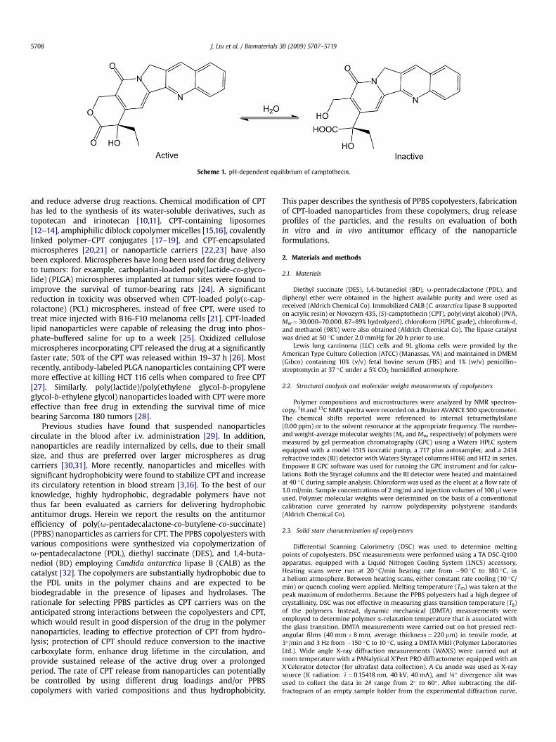
Scheme 1. pH-dependent equilibrium of camptothecin.
J. Liu et al. / Biomaterials 30 (2009) 5707–57195708
and reduce adverse drug reactions. Chemical modification of CPThas led to the synthesis of its water-soluble derivatives, such astopotecan and irinotecan [10,11]. CPT-containing liposomes[12–14], amphiphilic diblock copolymer micelles [15,16], covalentlylinked polymer–CPT conjugates [17–19], and CPT-encapsulatedmicrospheres [20,21] or nanoparticle carriers [22,23] have alsobeen explored. Microspheres have long been used for drug deliveryto tumors: for example, carboplatin-loaded poly(lactide-co-glyco-lide) (PLGA) microspheres implanted at tumor sites were found toimprove the survival of tumor-bearing rats [24]. A significantreduction in toxicity was observed when CPT-loaded poly(3-cap-rolactone) (PCL) microspheres, instead of free CPT, were used totreat mice injected with B16-F10 melanoma cells [21]. CPT-loadedlipid nanoparticles were capable of releasing the drug into phos-phate-buffered saline for up to a week [25]. Oxidized cellulosemicrospheres incorporating CPT released the drug at a significantlyfaster rate; 50% of the CPT was released within 19–37 h [26]. Mostrecently, antibody-labeled PLGA nanoparticles containing CPT weremore effective at killing HCT 116 cells when compared to free CPT[27]. Similarly, poly(lactide)/poly(ethylene glycol-b-propyleneglycol-b-ethylene glycol) nanoparticles loaded with CPT were moreeffective than free drug in extending the survival time of micebearing Sarcoma 180 tumors [28].
Previous studies have found that suspended nanoparticlescirculate in the blood after i.v. administration [29]. In addition,nanoparticles are readily internalized by cells, due to their smallsize, and thus are preferred over larger microspheres as drugcarriers [30,31]. More recently, nanoparticles and micelles withsignificant hydrophobicity were found to stabilize CPT and increaseits circulatory retention in blood stream [3,16]. To the best of ourknowledge, highly hydrophobic, degradable polymers have notthus far been evaluated as carriers for delivering hydrophobicantitumor drugs. Herein we report the results on the antitumorefficiency of poly(u-pentadecalactone-co-butylene-co-succinate)(PPBS) nanoparticles as carriers for CPT. The PPBS copolyesters withvarious compositions were synthesized via copolymerization ofu-pentadecalactone (PDL), diethyl succinate (DES), and 1,4-buta-nediol (BD) employing Candida antarctica lipase B (CALB) as thecatalyst [32]. The copolymers are substantially hydrophobic due tothe PDL units in the polymer chains and are expected to bebiodegradable in the presence of lipases and hydrolases. Therationale for selecting PPBS particles as CPT carriers was on theanticipated strong interactions between the copolyesters and CPT,which would result in good dispersion of the drug in the polymernanoparticles, leading to effective protection of CPT from hydro-lysis; protection of CPT should reduce conversion to the inactivecarboxylate form, enhance drug lifetime in the circulation, andprovide sustained release of the active drug over a prolongedperiod. The rate of CPT release from nanoparticles can potentiallybe controlled by using different drug loadings and/or PPBScopolymers with varied compositions and thus hydrophobicity.
This paper describes the synthesis of PPBS copolyesters, fabricationof CPT-loaded nanoparticles from these copolymers, drug releaseprofiles of the particles, and the results on evaluation of bothin vitro and in vivo antitumor efficacy of the nanoparticleformulations.
2. Materials and methods
2.1. Materials
Diethyl succinate (DES), 1,4-butanediol (BD), u-pentadecalactone (PDL), anddiphenyl ether were obtained in the highest available purity and were used asreceived (Aldrich Chemical Co). Immobilized CALB (C. antarctica lipase B supportedon acrylic resin) or Novozym 435, (S)-camptothecin (CPT), poly(vinyl alcohol) (PVA,Mw¼ 30,000–70,000, 87–89% hydrolyzed), chloroform (HPLC grade), chloroform-d,and methanol (98%) were also obtained (Aldrich Chemical Co). The lipase catalystwas dried at 50 �C under 2.0 mmHg for 20 h prior to use.
Lewis lung carcinoma (LLC) cells and 9L glioma cells were provided by theAmerican Type Culture Collection (ATCC) (Manassas, VA) and maintained in DMEM(Gibco) containing 10% (v/v) fetal bovine serum (FBS) and 1% (w/v) penicillin–streptomycin at 37 �C under a 5% CO2 humidified atmosphere.
2.2. Structural analysis and molecular weight measurements of copolyesters
Polymer compositions and microstructures were analyzed by NMR spectros-copy. 1H and 13C NMR spectra were recorded on a Bruker AVANCE 500 spectrometer.The chemical shifts reported were referenced to internal tetramethylsilane(0.00 ppm) or to the solvent resonance at the appropriate frequency. The number-and weight-average molecular weights (Mn and Mw, respectively) of polymers weremeasured by gel permeation chromatography (GPC) using a Waters HPLC systemequipped with a model 1515 isocratic pump, a 717 plus autosampler, and a 2414refractive index (RI) detector with Waters Styragel columns HT6E and HT2 in series.Empower II GPC software was used for running the GPC instrument and for calcu-lations. Both the Styragel columns and the RI detector were heated and maintainedat 40 �C during sample analysis. Chloroform was used as the eluent at a flow rate of1.0 ml/min. Sample concentrations of 2 mg/ml and injection volumes of 100 ml wereused. Polymer molecular weights were determined on the basis of a conventionalcalibration curve generated by narrow polydispersity polystyrene standards(Aldrich Chemical Co).
2.3. Solid state characterization of copolyesters
Differential Scanning Calorimetry (DSC) was used to determine meltingpoints of copolyesters. DSC measurements were performed using a TA DSC-Q100apparatus, equipped with a Liquid Nitrogen Cooling System (LNCS) accessory.Heating scans were run at 20 �C/min heating rate from �90 �C to 180 �C, ina helium atmosphere. Between heating scans, either constant rate cooling (10 �C/min) or quench cooling were applied. Melting temperature (Tm) was taken at thepeak maximum of endotherms. Because the PPBS polyesters had a high degree ofcrystallinity, DSC was not effective in measuring glass transition temperature (Tg)of the polymers. Instead, dynamic mechanical (DMTA) measurements wereemployed to determine polymer a-relaxation temperature that is associated withthe glass transition. DMTA measurements were carried out on hot pressed rect-angular films (40 mm� 8 mm, average thickness¼ 220 mm) in tensile mode, at3�/min and 3 Hz from �150 �C to 10 �C, using a DMTA MkII (Polymer LaboratoriesLtd.). Wide angle X-ray diffraction measurements (WAXS) were carried out atroom temperature with a PANalytical X’Pert PRO diffractometer equipped with anX’Celerator detector (for ultrafast data collection). A Cu anode was used as X-raysource (K radiation: l¼ 0.15418 nm, 40 kV, 40 mA), and ¼� divergence slit wasused to collect the data in 2q range from 2� to 60� . After subtracting the dif-fractogram of an empty sample holder from the experimental diffraction curve,
J. Liu et al. / Biomaterials 30 (2009) 5707–5719 5709
the amorphous and crystalline contributions in the resulting diffractogram werecalculated by a fitting method using the Fityk software. The degree of crystallinity(cc) was evaluated as the ratio of the crystalline peak areas to the total area underthe scattering curve.
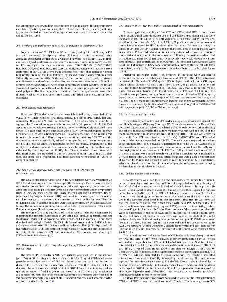
2.4. Synthesis and purification of poly(PDL-co-butylene-co-succinate) (PPBS)
Polymerizations of PDL, DES, and BD were catalyzed by 10 wt-% Novozym 435(vs. total monomer) in diphenyl ether (200 wt-% vs. total monomer) usinga parallel synthesizer connected to a vacuum line with the vacuum (�0.2 mmHg)controlled by a digital vacuum regulator. The monomer molar ratios of PDL to DESto BD employed for five reactions were 0.20:0.80:0.80, 0.35:0.65:0.65,0.50:0.50:0.50, 0.65:0.35:0.35, and 0.79:0.21:0.21, respectively. All reactions wereperformed at 95 �C using a two-stage process: first stage oligomerization under600 mmHg pressure for 18 h followed by second stage polymerization under2.0 mmHg pressure for 48 h. At the end of the reactions, each product mixturewas dissolved in chloroform and the resultant chloroform solution was filtered toremove the enzyme catalyst. After being concentrated under vacuum, the filtratewas added dropwise to methanol while stirring to cause precipitation of a whitesolid polymer. The five copolymers obtained from the synthesizer were thenfiltered, washed with methanol three times, and dried under vacuum at 30 �Covernight.
2.5. PPBS nanoparticle fabrication
Blank and CPT-loaded nanoparticles were fabricated using a modified oil-in-water (o/w) single emulsion technique. Briefly, 100 mg of PPBS copolymer and,optionally, 10 mg of CPT were co-dissolved in 2 ml of methylene chloride ina glass tube. The resultant organic solution was added dropwise to 4 ml of 5% PVAaqueous solution while vortexing. The mixture was subsequently sonicated threetimes (10 s each time) at 38% amplitude with a TMX 400 sonic disruptor (Tekmar,Cincinnati, OH) to yield a homogeneous oil-in-water emulsion. This emulsion wasimmediately poured into 100 ml of aqueous solution containing 0.3% PVA and thewhole mixture was magnetically stirred in an open beaker at room temperaturefor 3 h. This process allows nanoparticles to form via gradual evaporation of themethylene chloride solvent. The nanoparticles formed by this method werecollected by centrifugation at 11,000g for 15 min, washed three times withdeionized water, re-suspended in 5 ml of aqueous solution containing 5% treha-lose, and dried on a lyophilizer. The dried particles were stored at �20 �C inairtight containers.
2.6. Nanoparticle characterization and measurement of CPT contentsin nanoparticles
The surface morphology and size of PPBS nanoparticles were analyzed using anXL30 ESEM scanning electron microscope (FEI Company). Particle samples weremounted on an aluminum stub using carbon adhesive tape and sputter-coated witha mixture of gold and palladium (60:40) in an argon atmosphere under low pressureusing a Dynavac Mini Coater. The image-analysis application program, ImageJ(developed by Wayne Rasband, NIH), was used to measure particle diameters,calculate average particle sizes, and determine particle size distributions. The sizesof nanoparticles in aqueous medium were also determined by dynamic light scat-tering. The surface zeta-potential values of particles were measured with a Zeta-Potential Analyzer (Brookhaven Instruments Corp.).
The amount of CPT drug encapsulated in PPBS nanoparticles was determined bymeasuring the intrinsic fluorescence of CPT using a SpectraMax spectrofluorometer(Molecular Devices). In a typical example, CPT-loaded nanoparticles (3 mg) weredissolved in dimethyl sulfoxide (DMSO, 1 ml). A small amount of the DMSO solution(10 ml) was mixed with PBS (1.0 ml), sodium dodecyl sulfate (SDS, 10 ml), and 1 Nhydrochloric acid (10 ml). The resultant mixture had a pH value of 3. The fluorescenceintensity of the extracted CPT was measured at 428 nm emission wavelength(370 nm excitation wavelength).
2.7. Determination of in vitro drug release profiles of CPT-encapsulated PPBSnanoparticles
The rates of CPT release from PPBS nanoparticles were evaluated in PBS solution(pH¼ 7.4) at 37 �C using membrane dialysis. Briefly, 3 mg of CPT-loaded nano-particles were added to 2 ml of PBS and the mixture was sonicated for 20 s todisperse the particles. The nanoparticle suspension was then placed in a Piercedialysis tube with molecular weight cutoff at 10,000 Da, and the tube was subse-quently immersed in fresh PBS (20 ml) and incubated at 37 �C on a rotary shaker setat a speed of 100 rpm. The liquid medium was completely replaced with fresh PBS atvarious preset intervals. The amount of CPT released was measured according to themethod described in Section 2.6.
2.8. Stability of CPT free drug and CPT encapsulated in PPBS nanoparticles
To investigate the stability of free CPT and CPT-loaded PPBS nanoparticlesunder physiological conditions, free CPT and CPT-loaded PPBS nanoparticles wereincubated in PBS (pH 7.4, 37 �C) or DMEM (pH 7.4, 37 �C) with 10% FBS. For free CPT,at different time intervals (0.5, 1, 2, 4, 6, 24 h), 20 ml aliquots were withdrawn andimmediately analyzed by HPLC to determine the ratio of lactone to carboxylateforms of CPT. For the CPT-loaded PPBS nanoparticles, 3 mg of nanoparticles weresuspended in PBS or DMEM and put into a dialysis tube, which was subsequentlyimmersed and incubated in the same medium following the method as describedin Section 2.7. Then 200 ml nanoparticle suspension were withdrawn at varioustime intervals and centrifuged at 16,000 rpm. The obtained nanoparticles werelyophilized, dissolved in DMSO and appropriately diluted with PBS (pH 7.4), thenimmediately analyzed by HPLC to measure the ratio of lactone to carboxylate formsof CPT.
Analytical procedures using HPLC reported in literature were adopted todetermine the lactone to carboxylate form ratio of CPT [33]. The HPLC instrumentconsisted of a Shimadzu SIL-10A system (Kyoto, Japan) with a Ascentis C18 sepa-ration column (15 cm� 4.6 mm, 5 mm). Mixed solvent, 50 mM phosphate buffer (pH6.0)–acetonitrile–tetrahydrofuran (THF) (80:20:2, v/v), was used as the mobilephase that was maintained at 30 �C and pumped at a flow rate of 1.0 ml/min. Thedetection was performed using a fluorescence detector (Shimadzu RF-10A, Kyoto,Japan) with an excitation wavelength of 370 nm and emission wavelength of450 nm. The CPT standards in carboxylate, lactone, and mixed carboxylate/lactoneforms were prepared by dilution of a CPT stock solution (1 mg/ml in DMSO) to PBSwith appropriate pH (10.0, 3.0, or 7.4, respectively).
2.9. In vitro cytotoxicity studies
The cytotoxicity of free CPT and CPT-loaded nanoparticles was tested against LLCand 9L cells using an MTS assay (Promega, WI). The cells were seeded in 96-well flat-bottomed microplates (BD Falcon) at a density of 4000 cells per well. After allowingthe cells to adhere overnight, the culture medium was removed and 100 ml of themedium containing an appropriate amount of drug (0.001–100 mM) was added toeach well. Free CPT was dissolved in 1:9 (v/v) DMSO/DMEM and CPT-loadednanoparticles were suspended in DMEM. The cells were exposed to variousconcentrations of CPT or CPT-loaded nanoparticles at 37 �C for 24–72 h. At the end ofthe incubation period, drug-containing medium was removed and the cells werethoroughly rinsed three times with cold PBS. Then, 100 ml of fresh medium and 20 mlof the MTS reagent were added to each well, and the microplates were incubated at37 �C in darkness for 2 h. After the incubation, the plates were placed on a rotationalshaker for 10–15 min and allowed to cool to room temperature. MTS absorbance,which is related to the number of metabolically-active cells, was measured usinga microplate reader (Molecular Devices).
2.10. Cellular uptake measurements
Flow cytometry was used to study the drug-associated intracellular fluores-cence of monolayer cultures. One milliliter of suspended cells at a density of5�105 cells/ml was seeded in each well of 12-well tissue culture plates (BDFalcon) and allowed to attach overnight. The cells were then exposed to variousconcentrations (0–100 mM) of free CPT or CPT-loaded nanoparticles for 2 h. For thenanoparticles, the drug concentration was calculated using the total amount ofCPT in the particles. After incubation, the drug-containing medium was removedand the cells were thoroughly rinsed twice with cold PBS. Subsequently, thetreated cells were harvested using trypsin (0.05%), transferred to centrifuge tubes,and centrifuged for 5 min at 1500 rpm. Upon removal of the supernatant, the cellswere re-suspended in 0.5 ml of FACS buffer, transferred to round-bottom poly-styrene test tubes (BD Falcon, 12�75 mm), and kept in the dark at 4 �C untilanalysis. Analytical flow cytometry was performed using a BD LSR-II instrument(Becton Dickinson, San Jose, CA) and data were collected and processed using BDFACSDiva software (Becton Dickinson). Signals for forward and side light scatter(excitation at 355 nm, fluorescence emission at 450/50 nm) were collected from20,000 cells.
The ratio of carboxylate/lactone forms of CPT in the cells were also quantitatedby HPLC. LLC cells (1�106) were incubated in DMEM containing 50 mM CPT whichwas added using either free CPT or CPT-loaded nanoparticles. At different timeintervals (0.5, 2, and 4 h), the cells were washed three times with ice-cold PBS (1 mleach time), harvested using trypsin (0.05%), and then centrifuged at 1500 rpm for5 min at 4 �C. Upon removal of the supernatant, the cells were suspended in 200 mlof PBS (pH 7.4) and disrupted by vigorous sonication. The resulting, sonicatedmixture was frozen with liquid N2, followed by rapid thawing. This process wasrepeated for three times. Subsequently, 200 ml of DMSO was added to the cell lysatesolution to dissolve CPT and/or CPT nanoparticles, This cell lysate solution was thencentrifuged at 16,000 rpm for 5 min at 4 �C and the supernatant was analyzed byHPLC according to the method described in Section 2.8 to determine the ratio of CPTlactone/carboxylate forms in the solution.
Confocal laser scanning microscopy was used to visualize the internalization ofCPT-loaded PPBS nanoparticles with cultured LLC cells. LLC cells were grown to 50%
Scheme 2. Two-stage process for copolymerization of PDL, DES, and BD.
J. Liu et al. / Biomaterials 30 (2009) 5707–57195710
confluency. After incubated for 2 h with free CPT or CPT nanoparticles, the cells werewashed and fixed, and the cell cytoskeletons were labeled with Texas-Red phalloi-din. Images were obtained with a Leica TCS SP5 Spectral Confocal Microscope usinga 63� objective.
2.11. In vivo antitumor activity evaluation
All animal care and studies were approved by Yale’s Institutional Animal Careand Use Committee (IACUC). The antitumor activity of CPT-loaded nanoparticles wasevaluated in mice bearing subcutaneous tumors of Lewis lung carcinoma (LLC). LLCcells (1�106 cells, 0.1 ml) were transplanted into C57BL/6 male mice (6 week old,Charles River Laboratories) subcutaneously, and drug treatments were started after7 days, a time when the average tumor volume reached approximately 100 mm3.Forty mice were divided into five groups with eight animals in each group. Theaverage size and size variation of the tumors in all groups were comparable. CPT indifferent formulations was injected into the tail vein once on day 7 or three day 11,and day 16 after tumor cell implantation. Free CPT was formulated as a PBS solutioncontaining DMSO (10%, v/v) and Tween-80 (5%, v/v); CPT-loaded nanoparticles weresuspended in PBS (pH 7.4). Mice were divided into five groups with each groupreceiving formulations as follows: (i) PBS (pH 7.4); (ii) blank 50% PDL-PPBS nano-particles; (iii) free CPT at 10 mg/kg dose; (iv) CPT-encapsulated 50% PDL-PPBSnanoparticles as a single injection at 10 mg CPT/kg; (v) CPT-encapsulated 50% PDL-PPBS nanoparticles in multiple doses: 10 mg CPT/kg� 1 followed by 5 mg CPT/kg� 2. The tumor volumes and body weights of the mice were measured andrecorded. Tumor volume was calculated as follows: volume¼ 1/2LW2, where L is thelong diameter and W is the short diameter of a tumor. Animals were euthanized
Table 1Characterization of the isolated poly(PDL-co-butylene-co-succinate) copolymers.
PDL/DES/BD (molar ratio) Polymer yield PDL/succinate/butylene unit ratioa P
0.20:0.80:0.80 94% 0.20:0.80:0.80 20.35:0.65:0.65 92% 0.35:0.65:0.65 30.50:0.50:0.50 95% 0.50:0.50:0.50 50.65:0.35:0.35 94% 0.65:0.35:0.35 60.79:0.21:0.21 93% 0.79:0.21:0.21 7
a Calculated from the proton NMR spectra.b Molar ratio of PDL units vs. total (PDLþ succinate) units in the polymer chains.c Peak temperature of main relaxation determined by DMTA (dynamic mechanical md Melting point measured by DSC (differential scanning calorimetry).e Crystallinity degree from WAXS (wide angle X-ray diffraction measurements).
when tumor size exceeded 2000 mm3, body weight loss exceeded 20%, or othersignals of sickness, such as breathing problems, failure to eat and drink, lethargy orabnormal posture, were observed.
2.12. Studies on CPT biodistribution in tumor-bearing mice
Six week old male C57BL/6 mice, with body weight between 18 and 22 g(Charles River Laboratories), were used to measure CPT biodistribution. Mice wereallowed free access to food and water throughout the duration of the experimentsand were kept in a 12 h alternating light cycle. At 1 week after subcutaneoustransplantation of 1�106 LLC cells, when tumor size reached approximately100 mm3, tumor-bearing mice were injected via a lateral tail vein with free CPTsolution or CPT-loaded nanoparticles at a dose of 10 mg/kg of CPT. At 2 h, 24 h, and48 h after injection, mice were sacrificed via carbon dioxide inhalation and 0.5–1 mlwhole blood was collected via cardiac puncture. Plasma was obtained by centrifu-gation of the blood at 3000 rpm for 5 min. One hundred microliters of plasma wasadded to 0.1 ml of 0.1% acetic acid followed by mixing. Methylene chloride (0.2 ml)was added to the resultant mixture, stirred vigorously for 1 min using a vortexmixer, and then centrifuged at 12,000 rpm for 5 min. The organic phase wascollected for fluorescence measurement. The heart, liver, spleen, lung, kidney andtumor were excised, weighed, and frozen at �20 �C. The tissues were homogenizedin acidified DMSO. The fluorescence of the extracted CPT in DMSO was measured aspreviously described. The recovery ratios of CPT were 93%, 96%, 95%, 98%, 95%, and96% for heart, liver, spleen, lung, kidney and tumor, respectively. For the plasma, theextraction efficiency was 72%. These recovery ratios were used for data correction.
DL unit contentb (mol%) Copolymer Tac (�C) Tm
d (�C) cce (%)
Mw Mw/Mn
0 103 000 3.8 �37 92 475 101 000 3.4 �35 75, 56 520 85 400 3.5 �33 64 545 100 000 3.5 �29 72 599 91 100 2.3 �23 81 64
easurements).
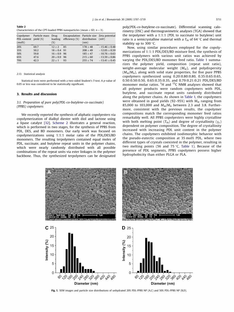
Table 2Characteristics of the CPT-loaded PPBS nanoparticles (mean� SD; n¼ 3).
CopolymerPDL content(mol%)
Particle massyield (%)
Drugloading(%)
Encapsulationefficiency (%)
Particle sizedistribution(nm)
Zeta potential(mV)
20% 69.7 12� 2 85 178� 48 �15.46� 0.4835% 50.2 18� 0.4 91 208� 49 �12.05� 0.5950% 59.8 16� 0.9 96 185� 47 �10.76� 0.6365% 47.6 20� 0.9 96 215� 60 �13.59� 0.8679% 42.3 22� 1 93 255� 74 �13.41� 0.45
J. Liu et al. / Biomaterials 30 (2009) 5707–5719 5711
2.13. Statistical analysis
Statistical tests were performed with a two-sided Student’s T-test. A p-value of0.05 or less was considered to be statistically significant.
3. Results and discussion
3.1. Preparation of pure poly(PDL-co-butylene-co-succinate)(PPBS) copolymers
We recently reported the synthesis of aliphatic copolyesters viacopolymerization of dialkyl diester with diol and lactone usinga lipase catalyst [32]. Scheme 2 illustrates a general reaction,which is performed in two stages, for the synthesis of PPBS fromPDL, DES, and BD monomers. Our early work was focused oncopolymerizations using 1:1:1 molar ratio of the PDL/DES/BDmonomers. The resulting terpolymers contained equal moles ofPDL, succinate, and butylene repeat units in the polymer chains,which were nearly randomly distributed with all possiblecombinations of the repeat units via ester linkages in the polymerbackbone. Thus, the synthesized terpolymers can be designated
Fig. 1. SEM images and particle size distributions of unhydra
poly(PDL-co-butylene-co-succinate). Differential scanning calo-rimetry (DSC) and thermogravimetric analyses (TGA) showed thatthe terpolymer with a 1:1:1 (PDL to succinate to butylene) unitratio is a semicrystalline material with a Tm of 64 �C and thermalstability up to 300 �C.
Now, using similar procedures employed for the copoly-merizations of 1:1:1 PDL/DES/BD mixture feed, the synthesis ofPPBS copolymers with various unit ratios was achieved byvarying the PDL/DES/BD monomer feed ratio. Table 1 summa-rizes the polymer yield, composition (repeat unit ratio),weight-average molecular weight (Mw), and polydispersity(Mw/Mn), along with solid state properties, for five pure PPBScopolymers synthesized using 0.20:0.80:0.80, 0.35:0.65:0.65,0.50:0.50:0.50, 0.65:0.35:0.35, and 0.79:0.21:0.21 PDL/DES/BDmonomer molar ratios. 1H and 13C NMR analyses showed thatall polymer products were random copolymers with PDL,butylene, and succinate repeat units randomly distributedalong the polymer chains. As shown in Table 1, the copolymerswere obtained in good yields (92–95%) with Mw ranging from85,000 to 103,000 and Mw/Mn between 2.3 and 3.8. Further-more, consistent with the previous results, the copolymercompositions match the corresponding monomer feed ratiosremarkably well. All PPBS copolyesters were highly crystallinewith both melting point (Tm) and degree of crystallinity (cc)dependent on polymer composition. The degree of crystallinityincreased with increasing PDL unit content in the polymerchains. The copolymers exhibited isodimorphic behavior withthe pseudo-eutectic composition at 35 mol% PDL, where twodifferent types of crystals coexisted in the polymer, resulting intwo melting points (56 and 75 �C, Table 1). Because of thepresence of PDL segments, PPBS copolymers possess higherhydrophobicity than either PLGA or PLA.
ted 20% PDL-PPBS NP (A,C) and 50% PDL-PPBS NP (B,D).
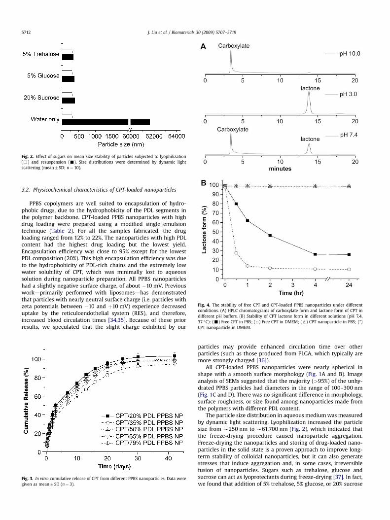
Fig. 4. The stability of free CPT and CPT-loaded PPBS nanoparticles under differentconditions. (A) HPLC chromatograms of carboxylate form and lactone form of CPT indifferent pH buffers. (B) Stability of CPT lactone form in different solutions (pH 7.4,37 �C): (-) Free CPT in PBS; (B) Free CPT in DMEM; (6) CPT nanoparticle in PBS; (*)CPT nanoparticle in DMEM.
Fig. 2. Effect of sugars on mean size stability of particles subjected to lyophilization(,) and resuspension (-). Size distributions were determined by dynamic lightscattering (mean� SD; n¼ 10).
J. Liu et al. / Biomaterials 30 (2009) 5707–57195712
3.2. Physicochemical characteristics of CPT-loaded nanoparticles
PPBS copolymers are well suited to encapsulation of hydro-phobic drugs, due to the hydrophobicity of the PDL segments inthe polymer backbone. CPT-loaded PPBS nanoparticles with highdrug loading were prepared using a modified single emulsiontechnique (Table 2). For all the samples fabricated, the drugloading ranged from 12% to 22%. The nanoparticles with high PDLcontent had the highest drug loading but the lowest yield.Encapsulation efficiency was close to 95% except for the lowestPDL composition (20%). This high encapsulation efficiency was dueto the hydrophobicity of PDL-rich chains and the extremely lowwater solubility of CPT, which was minimally lost to aqueoussolution during nanoparticle preparation. All PPBS nanoparticleshad a slightly negative surface charge, of about �10 mV. Previousworkdprimarily performed with liposomesdhas demonstratedthat particles with nearly neutral surface charge (i.e. particles withzeta potentials between �10 and þ10 mV) experience decreaseduptake by the reticuloendothelial system (RES), and therefore,increased blood circulation times [34,35]. Because of these priorresults, we speculated that the slight charge exhibited by our
Fig. 3. In vitro cumulative release of CPT from different PPBS nanoparticles. Data weregiven as mean� SD (n¼ 3).
particles may provide enhanced circulation time over otherparticles (such as those produced from PLGA, which typically aremore strongly charged [36]).
All CPT-loaded PPBS nanoparticles were nearly spherical inshape with a smooth surface morphology (Fig. 1A and B). Imageanalysis of SEMs suggested that the majority (>95%) of the unhy-drated PPBS particles had diameters in the range of 100–300 nm(Fig. 1C and D). There was no significant difference in morphology,surface roughness, or size found among nanoparticles made fromthe polymers with different PDL content.
The particle size distribution in aqueous medium was measuredby dynamic light scattering. Lyophilization increased the particlesize from w250 nm to w61,700 nm (Fig. 2), which indicated thatthe freeze-drying procedure caused nanoparticle aggregation.Freeze-drying the nanoparticles and storing of drug-loaded nano-particles in the solid state is a proven approach to improve long-term stability of colloidal nanoparticles, but it can also generatestresses that induce aggregation and, in some cases, irreversiblefusion of nanoparticles. Sugars such as trehalose, glucose andsucrose can act as lyoprotectants during freeze-drying [37]. In fact,we found that addition of 5% trehalose, 5% glucose, or 20% sucrose
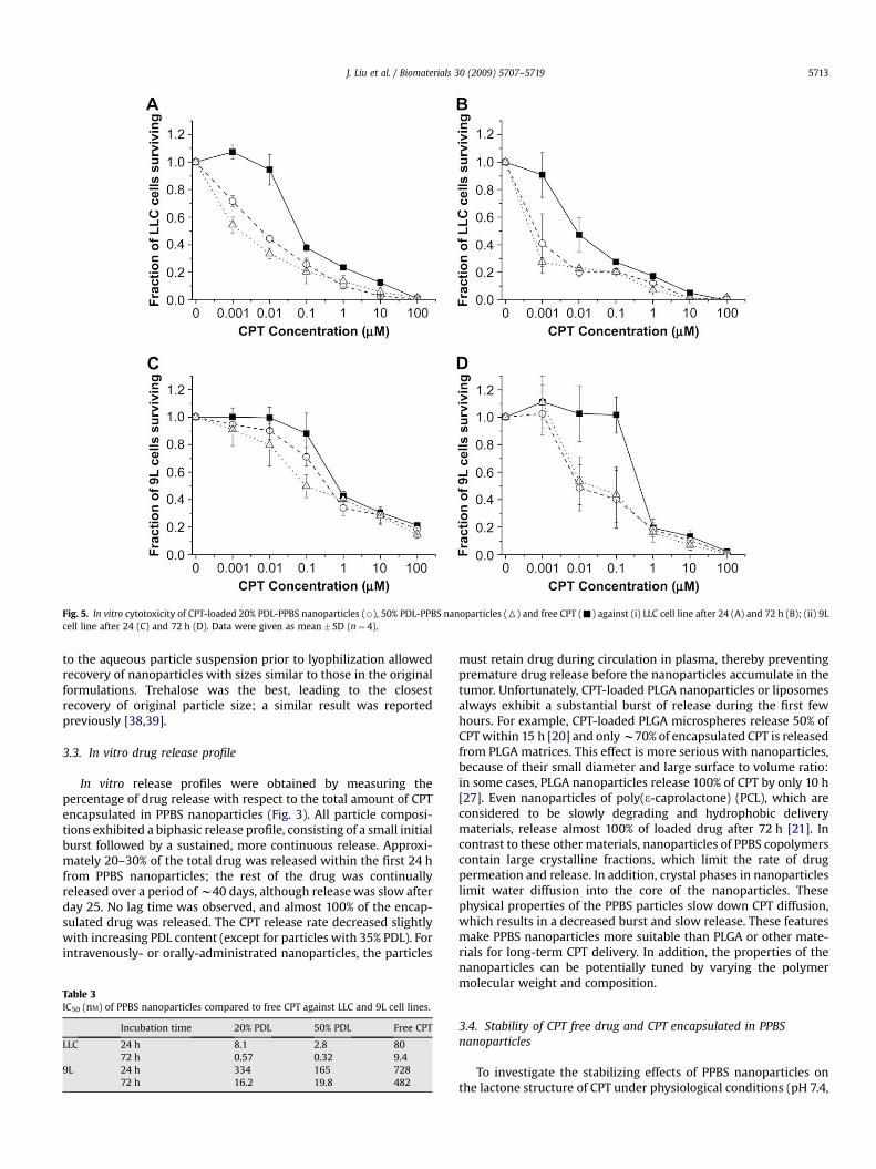
Fig. 5. In vitro cytotoxicity of CPT-loaded 20% PDL-PPBS nanoparticles (B), 50% PDL-PPBS nanoparticles (6) and free CPT (-) against (i) LLC cell line after 24 (A) and 72 h (B); (ii) 9Lcell line after 24 (C) and 72 h (D). Data were given as mean� SD (n¼ 4).
J. Liu et al. / Biomaterials 30 (2009) 5707–5719 5713
to the aqueous particle suspension prior to lyophilization allowedrecovery of nanoparticles with sizes similar to those in the originalformulations. Trehalose was the best, leading to the closestrecovery of original particle size; a similar result was reportedpreviously [38,39].
3.3. In vitro drug release profile
In vitro release profiles were obtained by measuring thepercentage of drug release with respect to the total amount of CPTencapsulated in PPBS nanoparticles (Fig. 3). All particle composi-tions exhibited a biphasic release profile, consisting of a small initialburst followed by a sustained, more continuous release. Approxi-mately 20–30% of the total drug was released within the first 24 hfrom PPBS nanoparticles; the rest of the drug was continuallyreleased over a period of w40 days, although release was slow afterday 25. No lag time was observed, and almost 100% of the encap-sulated drug was released. The CPT release rate decreased slightlywith increasing PDL content (except for particles with 35% PDL). Forintravenously- or orally-administrated nanoparticles, the particles
Table 3IC50 (nM) of PPBS nanoparticles compared to free CPT against LLC and 9L cell lines.
Incubation time 20% PDL 50% PDL Free CPT
LLC 24 h 8.1 2.8 8072 h 0.57 0.32 9.4
9L 24 h 334 165 72872 h 16.2 19.8 482
must retain drug during circulation in plasma, thereby preventingpremature drug release before the nanoparticles accumulate in thetumor. Unfortunately, CPT-loaded PLGA nanoparticles or liposomesalways exhibit a substantial burst of release during the first fewhours. For example, CPT-loaded PLGA microspheres release 50% ofCPT within 15 h [20] and only w70% of encapsulated CPT is releasedfrom PLGA matrices. This effect is more serious with nanoparticles,because of their small diameter and large surface to volume ratio:in some cases, PLGA nanoparticles release 100% of CPT by only 10 h[27]. Even nanoparticles of poly(3-caprolactone) (PCL), which areconsidered to be slowly degrading and hydrophobic deliverymaterials, release almost 100% of loaded drug after 72 h [21]. Incontrast to these other materials, nanoparticles of PPBS copolymerscontain large crystalline fractions, which limit the rate of drugpermeation and release. In addition, crystal phases in nanoparticleslimit water diffusion into the core of the nanoparticles. Thesephysical properties of the PPBS particles slow down CPT diffusion,which results in a decreased burst and slow release. These featuresmake PPBS nanoparticles more suitable than PLGA or other mate-rials for long-term CPT delivery. In addition, the properties of thenanoparticles can be potentially tuned by varying the polymermolecular weight and composition.
3.4. Stability of CPT free drug and CPT encapsulated in PPBSnanoparticles
To investigate the stabilizing effects of PPBS nanoparticles onthe lactone structure of CPT under physiological conditions (pH 7.4,
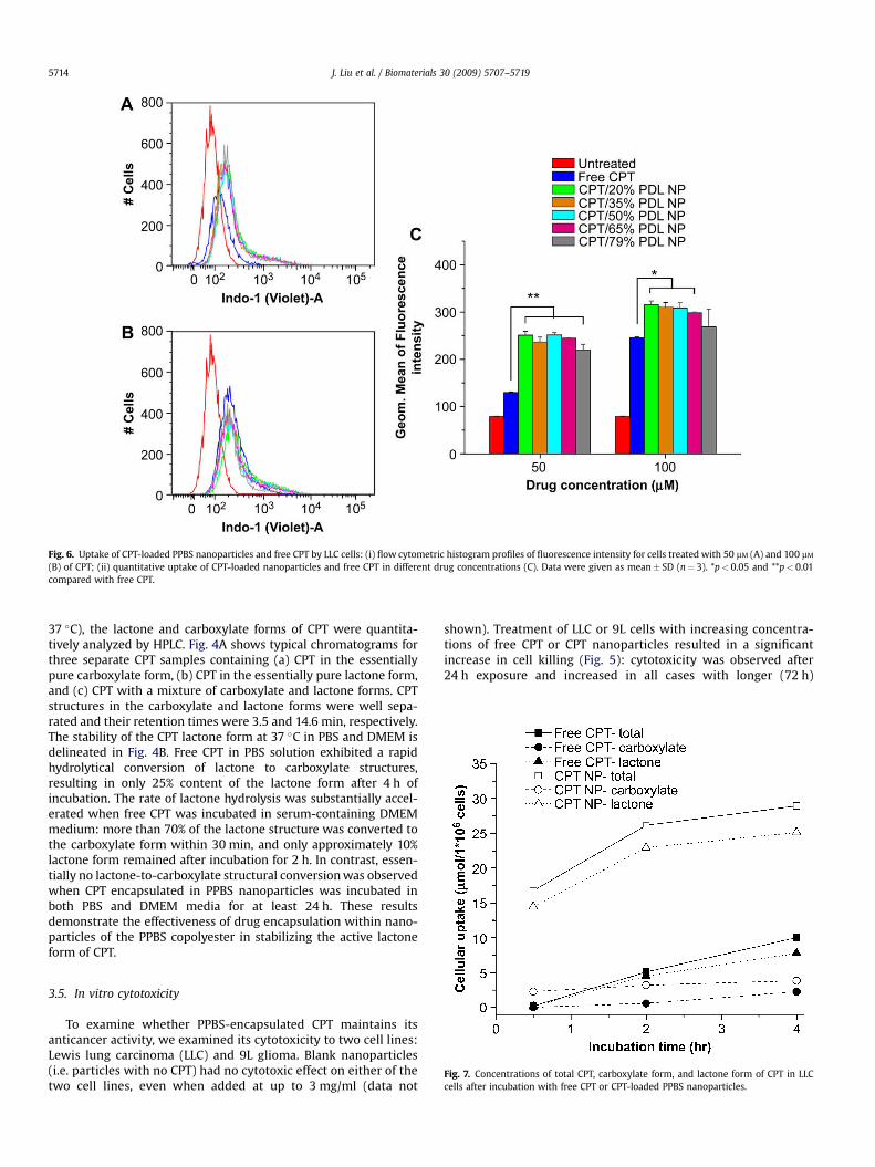
Fig. 6. Uptake of CPT-loaded PPBS nanoparticles and free CPT by LLC cells: (i) flow cytometric histogram profiles of fluorescence intensity for cells treated with 50 mM (A) and 100 mM
(B) of CPT; (ii) quantitative uptake of CPT-loaded nanoparticles and free CPT in different drug concentrations (C). Data were given as mean� SD (n¼ 3). *p< 0.05 and **p< 0.01compared with free CPT.
J. Liu et al. / Biomaterials 30 (2009) 5707–57195714
37 �C), the lactone and carboxylate forms of CPT were quantita-tively analyzed by HPLC. Fig. 4A shows typical chromatograms forthree separate CPT samples containing (a) CPT in the essentiallypure carboxylate form, (b) CPT in the essentially pure lactone form,and (c) CPT with a mixture of carboxylate and lactone forms. CPTstructures in the carboxylate and lactone forms were well sepa-rated and their retention times were 3.5 and 14.6 min, respectively.The stability of the CPT lactone form at 37 �C in PBS and DMEM isdelineated in Fig. 4B. Free CPT in PBS solution exhibited a rapidhydrolytical conversion of lactone to carboxylate structures,resulting in only 25% content of the lactone form after 4 h ofincubation. The rate of lactone hydrolysis was substantially accel-erated when free CPT was incubated in serum-containing DMEMmedium: more than 70% of the lactone structure was converted tothe carboxylate form within 30 min, and only approximately 10%lactone form remained after incubation for 2 h. In contrast, essen-tially no lactone-to-carboxylate structural conversion was observedwhen CPT encapsulated in PPBS nanoparticles was incubated inboth PBS and DMEM media for at least 24 h. These resultsdemonstrate the effectiveness of drug encapsulation within nano-particles of the PPBS copolyester in stabilizing the active lactoneform of CPT.
Fig. 7. Concentrations of total CPT, carboxylate form, and lactone form of CPT in LLCcells after incubation with free CPT or CPT-loaded PPBS nanoparticles.
3.5. In vitro cytotoxicity
To examine whether PPBS-encapsulated CPT maintains itsanticancer activity, we examined its cytotoxicity to two cell lines:Lewis lung carcinoma (LLC) and 9L glioma. Blank nanoparticles(i.e. particles with no CPT) had no cytotoxic effect on either of thetwo cell lines, even when added at up to 3 mg/ml (data not
shown). Treatment of LLC or 9L cells with increasing concentra-tions of free CPT or CPT nanoparticles resulted in a significantincrease in cell killing (Fig. 5): cytotoxicity was observed after24 h exposure and increased in all cases with longer (72 h)
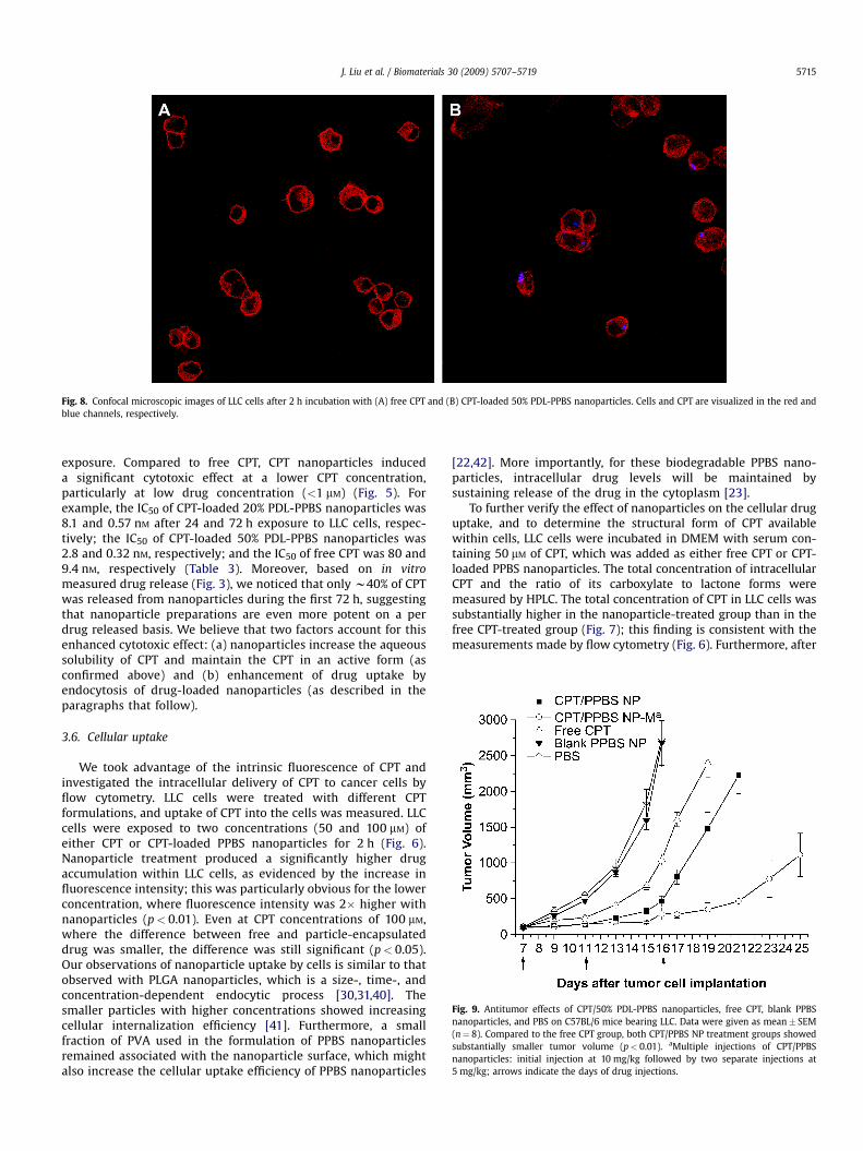
Fig. 8. Confocal microscopic images of LLC cells after 2 h incubation with (A) free CPT and (B) CPT-loaded 50% PDL-PPBS nanoparticles. Cells and CPT are visualized in the red andblue channels, respectively.
Fig. 9. Antitumor effects of CPT/50% PDL-PPBS nanoparticles, free CPT, blank PPBSnanoparticles, and PBS on C57BL/6 mice bearing LLC. Data were given as mean� SEM(n¼ 8). Compared to the free CPT group, both CPT/PPBS NP treatment groups showedsubstantially smaller tumor volume (p< 0.01). aMultiple injections of CPT/PPBSnanoparticles: initial injection at 10 mg/kg followed by two separate injections at5 mg/kg; arrows indicate the days of drug injections.
J. Liu et al. / Biomaterials 30 (2009) 5707–5719 5715
exposure. Compared to free CPT, CPT nanoparticles induceda significant cytotoxic effect at a lower CPT concentration,particularly at low drug concentration (<1 mM) (Fig. 5). Forexample, the IC50 of CPT-loaded 20% PDL-PPBS nanoparticles was8.1 and 0.57 nM after 24 and 72 h exposure to LLC cells, respec-tively; the IC50 of CPT-loaded 50% PDL-PPBS nanoparticles was2.8 and 0.32 nM, respectively; and the IC50 of free CPT was 80 and9.4 nM, respectively (Table 3). Moreover, based on in vitromeasured drug release (Fig. 3), we noticed that only w40% of CPTwas released from nanoparticles during the first 72 h, suggestingthat nanoparticle preparations are even more potent on a perdrug released basis. We believe that two factors account for thisenhanced cytotoxic effect: (a) nanoparticles increase the aqueoussolubility of CPT and maintain the CPT in an active form (asconfirmed above) and (b) enhancement of drug uptake byendocytosis of drug-loaded nanoparticles (as described in theparagraphs that follow).
3.6. Cellular uptake
We took advantage of the intrinsic fluorescence of CPT andinvestigated the intracellular delivery of CPT to cancer cells byflow cytometry. LLC cells were treated with different CPTformulations, and uptake of CPT into the cells was measured. LLCcells were exposed to two concentrations (50 and 100 mM) ofeither CPT or CPT-loaded PPBS nanoparticles for 2 h (Fig. 6).Nanoparticle treatment produced a significantly higher drugaccumulation within LLC cells, as evidenced by the increase influorescence intensity; this was particularly obvious for the lowerconcentration, where fluorescence intensity was 2� higher withnanoparticles (p< 0.01). Even at CPT concentrations of 100 mM,where the difference between free and particle-encapsulateddrug was smaller, the difference was still significant (p< 0.05).Our observations of nanoparticle uptake by cells is similar to thatobserved with PLGA nanoparticles, which is a size-, time-, andconcentration-dependent endocytic process [30,31,40]. Thesmaller particles with higher concentrations showed increasingcellular internalization efficiency [41]. Furthermore, a smallfraction of PVA used in the formulation of PPBS nanoparticlesremained associated with the nanoparticle surface, which mightalso increase the cellular uptake efficiency of PPBS nanoparticles
[22,42]. More importantly, for these biodegradable PPBS nano-particles, intracellular drug levels will be maintained bysustaining release of the drug in the cytoplasm [23].
To further verify the effect of nanoparticles on the cellular druguptake, and to determine the structural form of CPT availablewithin cells, LLC cells were incubated in DMEM with serum con-taining 50 mM of CPT, which was added as either free CPT or CPT-loaded PPBS nanoparticles. The total concentration of intracellularCPT and the ratio of its carboxylate to lactone forms weremeasured by HPLC. The total concentration of CPT in LLC cells wassubstantially higher in the nanoparticle-treated group than in thefree CPT-treated group (Fig. 7); this finding is consistent with themeasurements made by flow cytometry (Fig. 6). Furthermore, after
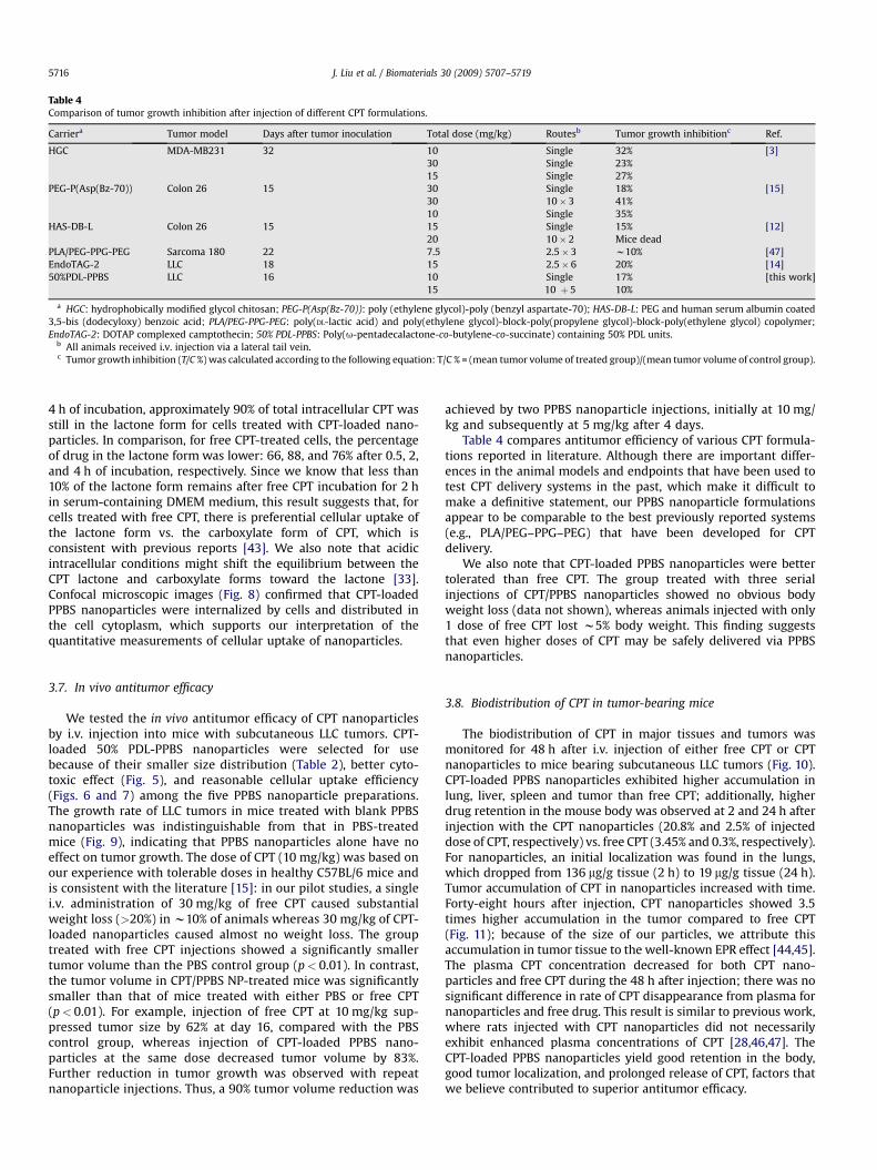
Table 4Comparison of tumor growth inhibition after injection of different CPT formulations.
Carriera Tumor model Days after tumor inoculation Total dose (mg/kg) Routesb Tumor growth inhibitionc Ref.
HGC MDA-MB231 32 10 Single 32% [3]30 Single 23%15 Single 27%
PEG-P(Asp(Bz-70)) Colon 26 15 30 Single 18% [15]30 10� 3 41%10 Single 35%
HAS-DB-L Colon 26 15 15 Single 15% [12]20 10� 2 Mice dead
PLA/PEG-PPG-PEG Sarcoma 180 22 7.5 2.5� 3 w10% [47]EndoTAG-2 LLC 18 15 2.5� 6 20% [14]50%PDL-PPBS LLC 16 10 Single 17% [this work]
15 10 þ 5 10%
a HGC: hydrophobically modified glycol chitosan; PEG-P(Asp(Bz-70)): poly (ethylene glycol)-poly (benzyl aspartate-70); HAS-DB-L: PEG and human serum albumin coated3,5-bis (dodecyloxy) benzoic acid; PLA/PEG-PPG-PEG: poly(DL-lactic acid) and poly(ethylene glycol)-block-poly(propylene glycol)-block-poly(ethylene glycol) copolymer;EndoTAG-2: DOTAP complexed camptothecin; 50% PDL-PPBS: Poly(u-pentadecalactone-co-butylene-co-succinate) containing 50% PDL units.
b All animals received i.v. injection via a lateral tail vein.c Tumor growth inhibition (T/C %) was calculated according to the following equation: T/C % = (mean tumor volume of treated group)/(mean tumor volume of control group).
J. Liu et al. / Biomaterials 30 (2009) 5707–57195716
4 h of incubation, approximately 90% of total intracellular CPT wasstill in the lactone form for cells treated with CPT-loaded nano-particles. In comparison, for free CPT-treated cells, the percentageof drug in the lactone form was lower: 66, 88, and 76% after 0.5, 2,and 4 h of incubation, respectively. Since we know that less than10% of the lactone form remains after free CPT incubation for 2 hin serum-containing DMEM medium, this result suggests that, forcells treated with free CPT, there is preferential cellular uptake ofthe lactone form vs. the carboxylate form of CPT, which isconsistent with previous reports [43]. We also note that acidicintracellular conditions might shift the equilibrium between theCPT lactone and carboxylate forms toward the lactone [33].Confocal microscopic images (Fig. 8) confirmed that CPT-loadedPPBS nanoparticles were internalized by cells and distributed inthe cell cytoplasm, which supports our interpretation of thequantitative measurements of cellular uptake of nanoparticles.
3.7. In vivo antitumor efficacy
We tested the in vivo antitumor efficacy of CPT nanoparticlesby i.v. injection into mice with subcutaneous LLC tumors. CPT-loaded 50% PDL-PPBS nanoparticles were selected for usebecause of their smaller size distribution (Table 2), better cyto-toxic effect (Fig. 5), and reasonable cellular uptake efficiency(Figs. 6 and 7) among the five PPBS nanoparticle preparations.The growth rate of LLC tumors in mice treated with blank PPBSnanoparticles was indistinguishable from that in PBS-treatedmice (Fig. 9), indicating that PPBS nanoparticles alone have noeffect on tumor growth. The dose of CPT (10 mg/kg) was based onour experience with tolerable doses in healthy C57BL/6 mice andis consistent with the literature [15]: in our pilot studies, a singlei.v. administration of 30 mg/kg of free CPT caused substantialweight loss (>20%) in w10% of animals whereas 30 mg/kg of CPT-loaded nanoparticles caused almost no weight loss. The grouptreated with free CPT injections showed a significantly smallertumor volume than the PBS control group (p< 0.01). In contrast,the tumor volume in CPT/PPBS NP-treated mice was significantlysmaller than that of mice treated with either PBS or free CPT(p< 0.01). For example, injection of free CPT at 10 mg/kg sup-pressed tumor size by 62% at day 16, compared with the PBScontrol group, whereas injection of CPT-loaded PPBS nano-particles at the same dose decreased tumor volume by 83%.Further reduction in tumor growth was observed with repeatnanoparticle injections. Thus, a 90% tumor volume reduction was
achieved by two PPBS nanoparticle injections, initially at 10 mg/kg and subsequently at 5 mg/kg after 4 days.
Table 4 compares antitumor efficiency of various CPT formula-tions reported in literature. Although there are important differ-ences in the animal models and endpoints that have been used totest CPT delivery systems in the past, which make it difficult tomake a definitive statement, our PPBS nanoparticle formulationsappear to be comparable to the best previously reported systems(e.g., PLA/PEG–PPG–PEG) that have been developed for CPTdelivery.
We also note that CPT-loaded PPBS nanoparticles were bettertolerated than free CPT. The group treated with three serialinjections of CPT/PPBS nanoparticles showed no obvious bodyweight loss (data not shown), whereas animals injected with only1 dose of free CPT lost w5% body weight. This finding suggeststhat even higher doses of CPT may be safely delivered via PPBSnanoparticles.
3.8. Biodistribution of CPT in tumor-bearing mice
The biodistribution of CPT in major tissues and tumors wasmonitored for 48 h after i.v. injection of either free CPT or CPTnanoparticles to mice bearing subcutaneous LLC tumors (Fig. 10).CPT-loaded PPBS nanoparticles exhibited higher accumulation inlung, liver, spleen and tumor than free CPT; additionally, higherdrug retention in the mouse body was observed at 2 and 24 h afterinjection with the CPT nanoparticles (20.8% and 2.5% of injecteddose of CPT, respectively) vs. free CPT (3.45% and 0.3%, respectively).For nanoparticles, an initial localization was found in the lungs,which dropped from 136 mg/g tissue (2 h) to 19 mg/g tissue (24 h).Tumor accumulation of CPT in nanoparticles increased with time.Forty-eight hours after injection, CPT nanoparticles showed 3.5times higher accumulation in the tumor compared to free CPT(Fig. 11); because of the size of our particles, we attribute thisaccumulation in tumor tissue to the well-known EPR effect [44,45].The plasma CPT concentration decreased for both CPT nano-particles and free CPT during the 48 h after injection; there was nosignificant difference in rate of CPT disappearance from plasma fornanoparticles and free drug. This result is similar to previous work,where rats injected with CPT nanoparticles did not necessarilyexhibit enhanced plasma concentrations of CPT [28,46,47]. TheCPT-loaded PPBS nanoparticles yield good retention in the body,good tumor localization, and prolonged release of CPT, factors thatwe believe contributed to superior antitumor efficacy.
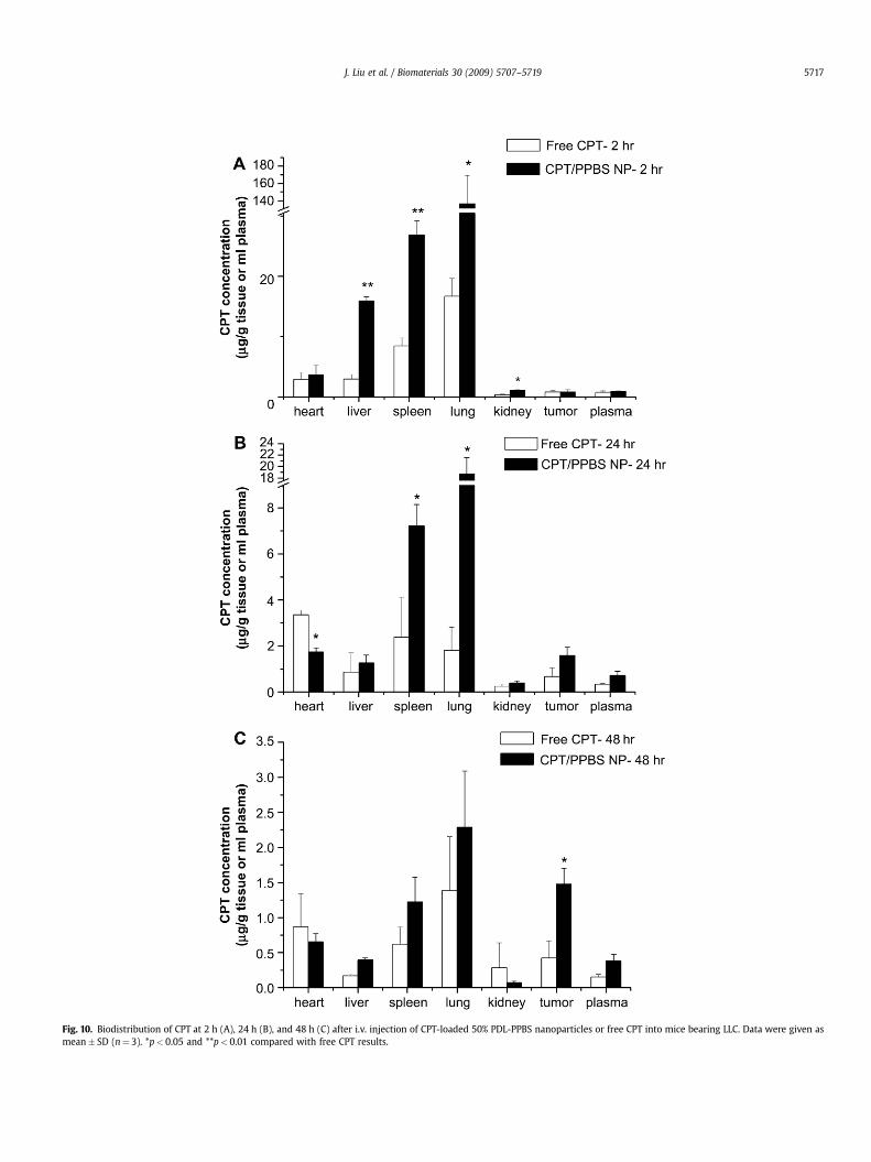
Fig. 10. Biodistribution of CPT at 2 h (A), 24 h (B), and 48 h (C) after i.v. injection of CPT-loaded 50% PDL-PPBS nanoparticles or free CPT into mice bearing LLC. Data were given asmean� SD (n¼ 3). *p< 0.05 and **p< 0.01 compared with free CPT results.
J. Liu et al. / Biomaterials 30 (2009) 5707–5719 5717
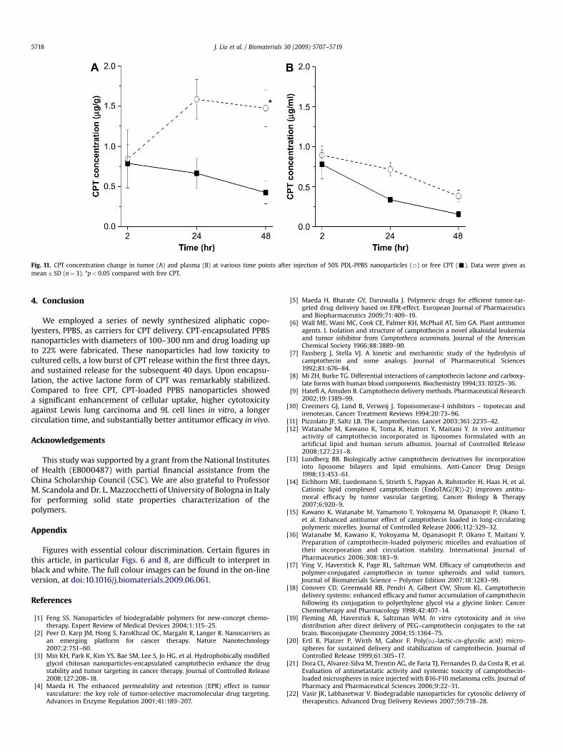
Fig. 11. CPT concentration change in tumor (A) and plasma (B) at various time points after injection of 50% PDL-PPBS nanoparticles (B) or free CPT (-). Data were given asmean� SD (n¼ 3). *p< 0.05 compared with free CPT.
J. Liu et al. / Biomaterials 30 (2009) 5707–57195718
4. Conclusion
We employed a series of newly synthesized aliphatic copo-lyesters, PPBS, as carriers for CPT delivery. CPT-encapsulated PPBSnanoparticles with diameters of 100–300 nm and drug loading upto 22% were fabricated. These nanoparticles had low toxicity tocultured cells, a low burst of CPT release within the first three days,and sustained release for the subsequent 40 days. Upon encapsu-lation, the active lactone form of CPT was remarkably stabilized.Compared to free CPT, CPT-loaded PPBS nanoparticles showeda significant enhancement of cellular uptake, higher cytotoxicityagainst Lewis lung carcinoma and 9L cell lines in vitro, a longercirculation time, and substantially better antitumor efficacy in vivo.
Acknowledgements
This study was supported by a grant from the National Institutesof Health (EB000487) with partial financial assistance from theChina Scholarship Council (CSC). We are also grateful to ProfessorM. Scandola and Dr. L. Mazzocchetti of University of Bologna in Italyfor performing solid state properties characterization of thepolymers.
Appendix
Figures with essential colour discrimination. Certain figures inthis article, in particular Figs. 6 and 8, are difficult to interpret inblack and white. The full colour images can be found in the on-lineversion, at doi:10.1016/j.biomaterials.2009.06.061.
References
[1] Feng SS. Nanoparticles of biodegradable polymers for new-concept chemo-therapy. Expert Review of Medical Devices 2004;1:115–25.
[2] Peer D, Karp JM, Hong S, FaroKhzad OC, Margalit R, Langer R. Nanocarriers asan emerging platform for cancer therapy. Nature Nanotechnology2007;2:751–60.
[3] Min KH, Park K, Kim YS, Bae SM, Lee S, Jo HG, et al. Hydrophobically modifiedglycol chitosan nanoparticles-encapsulated camptothecin enhance the drugstability and tumor targeting in cancer therapy. Journal of Controlled Release2008;127:208–18.
[4] Maeda H. The enhanced permeability and retention (EPR) effect in tumorvasculature: the key role of tumor-selective macromolecular drug targeting.Advances in Enzyme Regulation 2001;41:189–207.
[5] Maeda H, Bharate GY, Daruwalla J. Polymeric drugs for efficient tumor-tar-geted drug delivery based on EPR-effect. European Journal of Pharmaceuticsand Biopharmaceutics 2009;71:409–19.
[6] Wall ME, Wani MC, Cook CE, Palmer KH, McPhail AT, Sim GA. Plant antitumoragents. I. Isolation and structure of camptothecin a novel alkaloidal leukemiaand tumor inhibitor from Camptotheca acuminata. Journal of the AmericanChemical Society 1966;88:3889–90.
[7] Fassberg J, Stella VJ. A kinetic and mechanistic study of the hydrolysis ofcamptothecin and some analogs. Journal of Pharmaceutical Sciences1992;81:676–84.
[8] Mi ZH, Burke TG. Differential interactions of camptothecin lactone and carboxy-late forms with human blood components. Biochemistry 1994;33:10325–36.
[9] Hatefi A, Amsden B. Camptothecin delivery methods. Pharmaceutical Research2002;19:1389–99.
[10] Creemers GJ, Lund B, Verweij J. Topoisomerase-I inhibitors – topotecan andirenotecan. Cancer Treatment Reviews 1994;20:73–96.
[11] Pizzolato JF, Saltz LB. The camptothecins. Lancet 2003;361:2235–42.[12] Watanabe M, Kawano K, Toma K, Hattori Y, Maitani Y. In vivo antitumor
activity of camptothecin incorporated in liposomes formulated with anartificial lipid and human serum albumin. Journal of Controlled Release2008;127:231–8.
[13] Lundberg BB. Biologically active camptothecin derivatives for incorporationinto liposome bilayers and lipid emulsions. Anti-Cancer Drug Design1998;13:453–61.
[14] Eichhorn ME, Luedemann S, Strieth S, Papyan A, Ruhstorfer H, Haas H, et al.Cationic lipid complexed camptothecin (EndoTAG((R))-2) improves antitu-moral efficacy by tumor vascular targeting. Cancer Biology & Therapy2007;6:920–9.
[15] Kawano K, Watanabe M, Yamamoto T, Yokoyama M, Opanasopit P, Okano T,et al. Enhanced antitumor effect of camptothecin loaded in long-circulatingpolymeric micelles. Journal of Controlled Release 2006;112:329–32.
[16] Watanabe M, Kawano K, Yokoyama M, Opanasopit P, Okano T, Maitani Y.Preparation of camptothecin-loaded polymeric micelles and evaluation oftheir incorporation and circulation stability. International Journal ofPharmaceutics 2006;308:183–9.
[17] Ying V, Haverstick K, Page RL, Saltzman WM. Efficacy of camptothecin andpolymer-conjugated camptothecin in tumor spheroids and solid tumors.Journal of Biomaterials Science – Polymer Edition 2007;18:1283–99.
[18] Conover CD, Greenwald RB, Pendri A, Gilbert CW, Shum KL. Camptothecindelivery systems: enhanced efficacy and tumor accumulation of camptothecinfollowing its conjugation to polyethylene glycol via a glycine linker. CancerChemotherapy and Pharmacology 1998;42:407–14.
[19] Fleming AB, Haverstick K, Saltzman WM. In vitro cytotoxicity and in vivodistribution after direct delivery of PEG–camptothecin conjugates to the ratbrain. Bioconjugate Chemistry 2004;15:1364–75.
[20] Ertl B, Platzer P, Wirth M, Gabor F. Poly(D,L-lactic-co-glycolic acid) micro-spheres for sustained delivery and stabilization of camptothecin. Journal ofControlled Release 1999;61:305–17.
[21] Dora CL, Alvarez-Silva M, Trentin AG, de Faria TJ, Fernandes D, da Costa R, et al.Evaluation of antimetastatic activity and systemic toxicity of camptothecin-loaded microspheres in mice injected with B16-F10 melanoma cells. Journal ofPharmacy and Pharmaceutical Sciences 2006;9:22–31.
[22] Vasir JK, Labhasetwar V. Biodegradable nanoparticles for cytosolic delivery oftherapeutics. Advanced Drug Delivery Reviews 2007;59:718–28.
J. Liu et al. / Biomaterials 30 (2009) 5707–5719 5719
[23] Panyam J, Labhasetwar V. Sustained cytoplasmic delivery of drugs withintracellular receptors using biodegradable nanoparticles. Molecular Phar-maceutics 2004;1:77–84.
[24] Emerich DF, Winn SR, Snodgrass P, LaFreniere D, Agostino M, Wiens T, et al.Injectable chemotherapeutic microspheres and glioma II: enhanced survivalfollowing implantation into deep inoperable tumors. Pharmaceutical Research2000;17:776–81.
[25] Yang SC, Lu LF, Cai Y, Zhu JB, Liang BW, Yang CZ. Body distribution in mice ofintravenously injected camptothecin solid lipid nanoparticles and targetingeffect on brain. Journal of Controlled Release 1999;59:299–307.
[26] Kumar V, Kang JC, Hohl RJ. Improved dissolution and cytotoxicity of camp-tothecin incorporated into oxidized-cellulose microspheres prepared by spraydrying. Pharmaceutical Development and Technology 2001;6:459–67.
[27] McCarron PA, Marouf WM, Quinn DJ, Fay F, Burden RE, Olwill SA, et al. Anti-body targeting of camptothecin-loaded PLGA nanoparticles to tumor cells.Bioconjugate Chemistry 2008;19:1561–9.
[28] Kunii R, Onishi H, Ueki KI, Koyama KI, Machida Y. Particle characteristics andbiodistribution of camptothecin-loaded PLA/(PEG–PPG–PEG) nanoparticles.Drug Delivery 2008;15:3–10.
[29] Park J, Fong P, Lu J, Russell K, Booth C, Satzman W. PEGylated PLGA nanoparticlesfor the improved delivery of doxorubicin. Nanomedicine: Nanotechnology,Biology and Medicine 2009.
[30] Desai MP, Labhasetwar V, Walter E, Levy RJ, Amidon GL. The mechanism ofuptake of biodegradable microparticles in Caco-2 cells is size dependent.Pharmaceutical Research 1997;14:1568–73.
[31] Cartiera MS, Johnson KM, Rajendran V, Caplan MJ, Saltzman WM. The uptakeand intracellular fate of PLGA nanoparticles in epithelial cells. Biomaterials2009;30:2790–8.
[32] Jiang ZZ. Lipase-catalyzed synthesis of aliphatic polyesters via copolymeri-zation of lactone, dialkyl diester, and diol. Biomacromolecules 2008;9:3246–51.
[33] Sano K, Yoshikawa M, Hayasaka S, Satake K, Ikegami Y, Yoshida H, et al. Simplenon-ion-paired high-performance liquid chromatographic method for simul-taneous quantitation of carboxylate and lactone forms of 14 new campto-thecin derivatives. Journal of Chromatography B – Analytical Technologies inthe Biomedical and Life Sciences 2003;795:25–34.
[34] Levchenko TS, Rammohan R, Lukyanov AN, Whiteman KR, Torchilin VP.Liposome clearance in mice: the effect of a separate and combined presence ofsurface charge and polymer coating. International Journal of Pharmaceutics2002;240:95–102.
[35] Li SD, Huang L. Pharmacokinetics and biodistribution of nanoparticles.Molecular Pharmaceutics 2008;5:496–504.
[36] Cu Y, Saltzman WM. Controlled surface modification with poly(ethylene)glycol enhances diffusion of PLGA nanoparticles in human cervicalmucus. Molecular Pharmaceutics 2009;6:173–81.
[37] Abdelwahed W, Degobert G, Stainmesse S, Fessi H. Freeze-drying of nano-particles: formulation, process and storage considerations. Advanced DrugDelivery Reviews 2006;58:1688–713.
[38] Chacon M, Molpeceres J, Berges L, Guzman M, Aberturas MR. Stability andfreeze-drying of cyclosporine loaded poly(D,L lactide-glycolide) carriers.European Journal of Pharmaceutical Sciences 1999;8:99–107.
[39] Quintanar-Guerrero D, Ganem-Quintanar A, Allemann E, Fessi H, Doelker E.Influence of the stabilizer coating layer on the purification and freeze-dryingof poly(D,L-lactic acid) nanoparticles prepared by an emulsion–diffusiontechnique. Journal of Microencapsulation 1998;15:107–19.
[40] Panyam J, Labhasetwar V. Dynamics of endocytosis and exocytosis of poly(D,L-lactide-co-glycolide) nanoparticles in vascular smooth muscle cells. Pharma-ceutical Research 2003;20:212–20.
[41] Qaddoumi MG, Ueda H, Yang J, Davda J, Labhasetwar V, Lee VHL. The char-acteristics and mechanisms of uptake of PLGA nanoparticles in rabbitconjunctival epithelial cell layers. Pharmaceutical Research 2004;21:641–8.
[42] Prabha S, Labhasetwar V. Critical determinants in PLGA/PLA nanoparticle-mediated gene expression. Pharmaceutical Research 2004;21:354–64.
[43] Kobayashi K, Bouscarel B, Matsuzaki Y, Ceryak S, Kudoh S, Fromm H. pH-dependent uptake of irinotecan and its active metabolite, SN-38, by intestinalcells. International Journal of Cancer 1999;83:491–6.
[44] Gao XH, Cui YY, Levenson RM, Chung LWK, Nie SM. In vivo cancer targetingand imaging with semiconductor quantum dots. Nature Biotechnology2004;22:969–76.
[45] Wong HL, Bendayan R, Rauth AM, Xue HY, Babakhanian K, Wu XY. A mech-anistic study of enhanced doxorubicin uptake and retention in multidrugresistant breast cancer cells using a polymer–lipid hybrid nanoparticle system.Journal of Pharmacology and Experimental Therapeutics 2006;317:1372–81.
[46] Loh JP, Ahmed AE. Determination of camptothecin in biological-fluidsusing reversed-phase high-performance liquid-chromatography withfluorescence detection. Journal of Chromatography-Biomedical Applica-tions 1990;530:367–76.
[47] Kunii R, Onishi H, Machida Y. Preparation and antitumor characteristics ofPLA/(PEG–PPG–PEG) nanoparticles loaded with camptothecin. EuropeanJournal of Pharmaceutics and Biopharmaceutics 2007;67:9–17.