Embed Size (px)
Citation preview

BRAINA JOURNAL OF NEUROLOGY
Prospective 10-year surveillance of human priondiseases in JapanIchiro Nozaki,1,2 Tsuyoshi Hamaguchi,1 Nobuo Sanjo,3,4 Moeko Noguchi-Shinohara,1 Kenji Sakai,1
Yosikazu Nakamura,4,5 Takeshi Sato,4,6 Tetsuyuki Kitamoto,4,7 Hidehiro Mizusawa,3,4
Fumio Moriwaka,4,8 Yusei Shiga,4,9 Yoshiyuki Kuroiwa,4,10 Masatoyo Nishizawa,4,11
Shigeki Kuzuhara,4,12 Takashi Inuzuka,4,13 Masatoshi Takeda,4,14 Shigetoshi Kuroda,4,15
Koji Abe,4,16 Hiroyuki Murai,4,17 Shigeo Murayama,4,18 Jun Tateishi,4,19 Ichiro Takumi,4,20
Susumu Shirabe,4,21 Masafumi Harada,4,22 Atsuko Sadakane5 and Masahito Yamada1,4
1 Department of Neurology and Neurobiology of Aging, Kanazawa University Graduate School of Medical Science, Kanazawa, Japan
2 Department of Neurology, Noto General Hospital, Nanao, Japan
3 Department of Neurology and Neurological Science, Graduate School, Tokyo Medical and Dental University, Tokyo, Japan
4 Creutzfeldt–Jakob Disease Surveillance Committee, Japan
5 Department of Public Health, Jichi Medical University, Shimotsuke, Japan
6 Department of Neurology, Higashi Yamato Hospital, Higashiyamato, Japan
7 Department of Prion Protein Research, Division of CJD Science and Technology, Tohoku University Graduate School of Medicine, Sendai, Japan
8 Department of Communication Disorders, Health Sciences University of Hokkaido Graduate School of Psychological Science, Ishikari, Japan
9 Department of Neurology, Aoba Neurosurgical Clinic, Sendai, Japan
10 Department of Neurology, Yokohama City University School of Medicine, Yokohama, Japan
11 Department of Neurology, Brain Research Institute, Niigata University, Niigata, Japan
12 Department of Neurology, National Center Hospital of Neurology and Psychiatry, Tokyo, Japan
13 Department of Neurology and Geriatrics, Gifu University Graduate School of Medicine, Gifu, Japan
14 Department of Psychiatry, Osaka University Graduate School of Medicine, Suita, Japan
15 Department of Neuropsychiatry, Graduate School of Medicine, Dentistry and Pharmaceutical Sciences, Okayama University, Okayama, Japan
16 Department of Neurology, Graduate School of Medicine, Dentistry and Pharmaceutical Science, Okayama University, Okayama, Japan
17 Department of Neurology, Iizuka Hospital, Fukuoka, Japan
18 Department of Neuropathology, Tokyo Metropolitan Institute of Gerontology, Tokyo, Japan
19 Harukaze Healthcare Service Institution, Fukuoka, Japan
20 Department of Neurosurgery, Nippon Medical School Musashi Kosugi Hospital, Kawasaki, Japan
21 Centre of Health and Community Medicine, Nagasaki University, Nagasaki, Japan
22 Department of Medical Imaging, Institute of Health Biosciences, the University of Tokushima Graduate School, Tokushima, Japan
Correspondence to: Masahito Yamada,
Department of Neurology and Neurobiology of Ageing,
Kanazawa University Graduate School of Medical Science,
13-1 Takara-machi,
Kanazawa 920-8640, Japan
E-mail: [email protected]
We analysed the epidemiological data and clinical features of patients with prion diseases that had been registered by the
Creutzfeldt-Jakob Disease Surveillance Committee, Japan, over the past 10 years, since 1999. We obtained information on 1685
Japanese patients suspected as having prion diseases and judged that 1222 patients had prion diseases, consisting of definite
(n = 180, 14.7%) and probable (n = 1029, 84.2%) cases, except for dura mater graft-associated Creutzfeldt–Jakob disease which
also included possible cases (n = 13, 1.1%). They were classified into 922 (75.5%) with sporadic Creutzfeldt–Jakob disease, 216
doi:10.1093/brain/awq216 Brain 2010: 133; 3043–3057 | 3043
Received May 1, 2010. Revised June 14, 2010. Accepted June 15, 2010. Advance Access publication September 20, 2010
� The Author (2010). Published by Oxford University Press on behalf of the Guarantors of Brain. All rights reserved.
For Permissions, please email: [email protected]
Dow
nloaded from https://academ
ic.oup.com/brain/article-abstract/133/10/3043/321900 by guest on 15 April 2019

(17.7%) with genetic prion diseases, 81 (6.6%) with acquired prion diseases, including 80 cases of dura mater graft-associated
Creutzfeldt–Jakob disease and one case of variant Creutzfeldt–Jakob disease, and three cases of unclassified Creutzfeldt–Jakob
disease (0.2%). The annual incidence rate of prion disease ranged from 0.65 in 1999 to 1.10 in 2006, with an average of 0.85,
similar to European countries. Although methionine homozygosity at codon 129 polymorphism of the prion protein gene was
reported to be very common (93%) in the general Japanese population, sporadic Creutzfeldt–Jakob disease in Japan was
significantly associated with codon 129 homozygosity (97.5%), as reported in western countries. In sporadic Creutzfeldt–
Jakob disease, MM1 type (Parchi’s classification) is the most common, as in western countries. Among atypical sporadic
Creutzfeldt–Jakob disease cases, the MM2 type appeared most common, probably related to the very high proportion of
methionine allele in the Japanese population. As for iatrogenic Creutzfeldt–Jakob disease, only dura mater graft-associated
Creutzfeldt–Jakob disease cases were reported in Japan and, combined with the data from previous surveillance systems, the
total number of dura mater graft-associated Creutzfeldt–Jakob disease was 138, comprising the majority of worldwide dura
mater graft-associated Creutzfeldt–Jakob disease patients. Regarding genetic prion diseases, the most common mutation of
prion protein gene was V180I (41.2%), followed by P102L (18.1%), E200K (17.1%) and M232R (15.3%), and this distribution
was quite different from that in Europe. In particular, V180I and M232R were quite rare mutations worldwide. Patients with
V180I or M232R mutations rarely had a family history of prion diseases, indicating that a genetic test for sporadic cases is
necessary to distinguish these from sporadic Creutzfeldt–Jakob disease. In conclusion, our prospective 10-year surveillance
revealed a frequent occurrence of dura mater graft-associated Creutzfeldt–Jakob disease, and unique phenotypes of sporadic
Creutzfeldt–Jakob disease and genetic prion diseases related to the characteristic distribution of prion protein gene mutations
and polymorphisms in Japan, compared with those in western countries.
Keywords: prion disease; dura mater graft-associated Creutzfeldt–Jakob disease; 14-3-3 protein; periodic synchronous wave com-plexes; codon 129 or 219 polymorphism
Abbreviations: PrP = prion protein; PSWC = periodic synchronous wave complex; WHO = World Health Organization
IntroductionPrion diseases are a fatal human transmissible spongiform enceph-
alopathy (Prusiner, 1998) and are classified into sporadic, genetic
and acquired forms; the most common being sporadic
Creutzfeldt–Jakob disease, which is of unknown aetiology. The
overall annual mortality rate of sporadic Creutzfeldt–Jakob disease
is �1.5 per million worldwide (Ladogana et al., 2005). The genetic
form (i.e. genetic prion disease) is defined as a prion disease with
causative mutations in the human prion protein (PrP) gene (PrP)
and/or a relevant family history, including Gerstmann–Straussler–
Scheinker disease, fatal familial insomnia and genetic Creutzfeldt–
Jakob disease (Kovacs et al., 2005). The acquired forms are
transmitted among humans (i.e. iatrogenic Creutzfeldt–Jakob
disease or Kuru) or from animals to humans, particularly bovine
to human (i.e. variant Creutzfeldt–Jakob disease). To date,
4400 patients with iatrogenic Creutzfeldt–Jakob disease have
been reported, including transmission via infectious PrP-
contaminated neurosurgical instruments, deep brain electrodes,
human pituitary growth hormone, human cadaveric dura mater
grafts, corneal transplantation or blood transfusion (Brown et al.,
2006). In particular, 450% of patients with cadaveric dura
mater graft-associated Creutzfeldt–Jakob disease have occurred
in Japan (Brown et al., 2006). Although our previous case-control
study revealed that medical procedures before the onset of
prion diseases did not influence the onset of sporadic
Creutzfeldt–Jakob disease, there was a problem that surgical treat-
ments were performed on some patients with sporadic
Creutzfeldt–Jakob disease after the onset of prion diseases
(Hamaguchi et al., 2009a, b). Since the first identification of vari-
ant Creutzfeldt–Jakob disease in the UK in 1996, 212 patients
have been reported worldwide, including in Canada, France,
Ireland, Italy, Japan, Portugal, Saudi Arabia, Spain, the
Netherlands and the USA as well as the UK [The National
Creutzfeldt–Jakob Disease Surveillance Unit (NCJDSU) (http://
www.cjd.ed.ac.uk/vcjdworld.htm)].
From the viewpoint of public health, identification of the inci-
dence of human and animal prion diseases is essential to prevent
disease transmission. Various Creutzfeldt–Jakob disease surveil-
lance systems have been established since the 1990s in many
countries, including Australia, Austria, Belgium, Canada,
Catalonia, China, France, Germany, Ireland, Italy, Slovakia,
Spain, Switzerland, the Netherlands, the UK, the USA and Japan
(Will et al., 1998; Nakamura et al., 1999; Collins et al., 2002;
Glatzel et al., 2002; Puopolo et al., 2003; Horan et al.,
2004; Pocchiari et al., 2004; Sanchez-Valle et al., 2004;
Ladogana et al., 2005; de Pedro-Cuesta et al., 2006;
Van Everbroeck et al., 2006; Heinemann et al., 2007; Shi et al.,
2008; Klug et al., 2009; Holman et al., 2010).
Although detailed data from the nationwide surveillance of
human prion diseases have been published by European countries,
Australia and the USA (Ladogana et al., 2005; de Pedro-Cuesta
et al., 2006; Heinemann et al., 2007; Klug et al., 2009; Holman
et al., 2010), large-scale prospective data, comparable to those
from western countries, have never been reported from Asia.
In Japan, the current Creutzfeldt–Jakob disease surveillance
system was established in 1999 (Noguchi-Shinohara et al., 2007)
and prospective nationwide surveillance has been ongoing for
3044 | Brain 2010: 133; 3043–3057 I. Nozaki et al.
Dow
nloaded from https://academ
ic.oup.com/brain/article-abstract/133/10/3043/321900 by guest on 15 April 2019
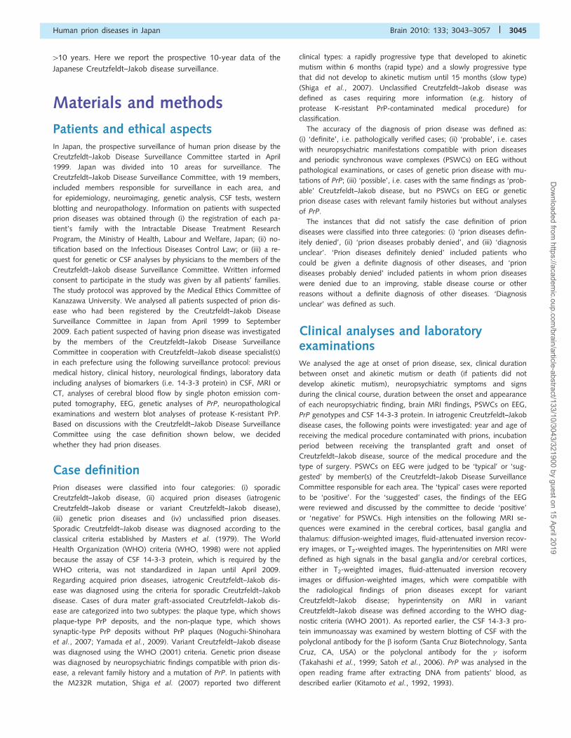
410 years. Here we report the prospective 10-year data of the
Japanese Creutzfeldt–Jakob disease surveillance.
Materials and methods
Patients and ethical aspectsIn Japan, the prospective surveillance of human prion disease by the
Creutzfeldt–Jakob Disease Surveillance Committee started in April
1999. Japan was divided into 10 areas for surveillance. The
Creutzfeldt–Jakob Disease Surveillance Committee, with 19 members,
included members responsible for surveillance in each area, and
for epidemiology, neuroimaging, genetic analysis, CSF tests, western
blotting and neuropathology. Information on patients with suspected
prion diseases was obtained through (i) the registration of each pa-
tient’s family with the Intractable Disease Treatment Research
Program, the Ministry of Health, Labour and Welfare, Japan; (ii) no-
tification based on the Infectious Diseases Control Law; or (iii) a re-
quest for genetic or CSF analyses by physicians to the members of the
Creutzfeldt–Jakob disease Surveillance Committee. Written informed
consent to participate in the study was given by all patients’ families.
The study protocol was approved by the Medical Ethics Committee of
Kanazawa University. We analysed all patients suspected of prion dis-
ease who had been registered by the Creutzfeldt–Jakob Disease
Surveillance Committee in Japan from April 1999 to September
2009. Each patient suspected of having prion disease was investigated
by the members of the Creutzfeldt–Jakob Disease Surveillance
Committee in cooperation with Creutzfeldt–Jakob disease specialist(s)
in each prefecture using the following surveillance protocol: previous
medical history, clinical history, neurological findings, laboratory data
including analyses of biomarkers (i.e. 14-3-3 protein) in CSF, MRI or
CT, analyses of cerebral blood flow by single photon emission com-
puted tomography, EEG, genetic analyses of PrP, neuropathological
examinations and western blot analyses of protease K-resistant PrP.
Based on discussions with the Creutzfeldt–Jakob Disease Surveillance
Committee using the case definition shown below, we decided
whether they had prion diseases.
Case definitionPrion diseases were classified into four categories: (i) sporadic
Creutzfeldt–Jakob disease, (ii) acquired prion diseases (iatrogenic
Creutzfeldt–Jakob disease or variant Creutzfeldt–Jakob disease),
(iii) genetic prion diseases and (iv) unclassified prion diseases.
Sporadic Creutzfeldt–Jakob disease was diagnosed according to the
classical criteria established by Masters et al. (1979). The World
Health Organization (WHO) criteria (WHO, 1998) were not applied
because the assay of CSF 14-3-3 protein, which is required by the
WHO criteria, was not standardized in Japan until April 2009.
Regarding acquired prion diseases, iatrogenic Creutzfeldt–Jakob dis-
ease was diagnosed using the criteria for sporadic Creutzfeldt–Jakob
disease. Cases of dura mater graft-associated Creutzfeldt–Jakob dis-
ease are categorized into two subtypes: the plaque type, which shows
plaque-type PrP deposits, and the non-plaque type, which shows
synaptic-type PrP deposits without PrP plaques (Noguchi-Shinohara
et al., 2007; Yamada et al., 2009). Variant Creutzfeldt–Jakob disease
was diagnosed using the WHO (2001) criteria. Genetic prion disease
was diagnosed by neuropsychiatric findings compatible with prion dis-
ease, a relevant family history and a mutation of PrP. In patients with
the M232R mutation, Shiga et al. (2007) reported two different
clinical types: a rapidly progressive type that developed to akinetic
mutism within 6 months (rapid type) and a slowly progressive type
that did not develop to akinetic mutism until 15 months (slow type)
(Shiga et al., 2007). Unclassified Creutzfeldt–Jakob disease was
defined as cases requiring more information (e.g. history of
protease K-resistant PrP-contaminated medical procedure) for
classification.
The accuracy of the diagnosis of prion disease was defined as:
(i) ‘definite’, i.e. pathologically verified cases; (ii) ‘probable’, i.e. cases
with neuropsychiatric manifestations compatible with prion diseases
and periodic synchronous wave complexes (PSWCs) on EEG without
pathological examinations, or cases of genetic prion disease with mu-
tations of PrP; (iii) ‘possible’, i.e. cases with the same findings as ‘prob-
able’ Creutzfeldt–Jakob disease, but no PSWCs on EEG or genetic
prion disease cases with relevant family histories but without analyses
of PrP.
The instances that did not satisfy the case definition of prion
diseases were classified into three categories: (i) ‘prion diseases defin-
itely denied’, (ii) ‘prion diseases probably denied’, and (iii) ‘diagnosis
unclear’. ‘Prion diseases definitely denied’ included patients who
could be given a definite diagnosis of other diseases, and ‘prion
diseases probably denied’ included patients in whom prion diseases
were denied due to an improving, stable disease course or other
reasons without a definite diagnosis of other diseases. ‘Diagnosis
unclear’ was defined as such.
Clinical analyses and laboratoryexaminationsWe analysed the age at onset of prion disease, sex, clinical duration
between onset and akinetic mutism or death (if patients did not
develop akinetic mutism), neuropsychiatric symptoms and signs
during the clinical course, duration between the onset and appearance
of each neuropsychiatric finding, brain MRI findings, PSWCs on EEG,
PrP genotypes and CSF 14-3-3 protein. In iatrogenic Creutzfeldt–Jakob
disease cases, the following points were investigated: year and age of
receiving the medical procedure contaminated with prions, incubation
period between receiving the transplanted graft and onset of
Creutzfeldt–Jakob disease, source of the medical procedure and the
type of surgery. PSWCs on EEG were judged to be ‘typical’ or ‘sug-
gested’ by member(s) of the Creutzfeldt–Jakob Disease Surveillance
Committee responsible for each area. The ‘typical’ cases were reported
to be ‘positive’. For the ‘suggested’ cases, the findings of the EEG
were reviewed and discussed by the committee to decide ‘positive’
or ‘negative’ for PSWCs. High intensities on the following MRI se-
quences were examined in the cerebral cortices, basal ganglia and
thalamus: diffusion-weighted images, fluid-attenuated inversion recov-
ery images, or T2-weighted images. The hyperintensities on MRI were
defined as high signals in the basal ganglia and/or cerebral cortices,
either in T2-weighted images, fluid-attenuated inversion recovery
images or diffusion-weighted images, which were compatible with
the radiological findings of prion diseases except for variant
Creutzfeldt–Jakob disease; hyperintensity on MRI in variant
Creutzfeldt–Jakob disease was defined according to the WHO diag-
nostic criteria (WHO 2001). As reported earlier, the CSF 14-3-3 pro-
tein immunoassay was examined by western blotting of CSF with the
polyclonal antibody for the b isoform (Santa Cruz Biotechnology, Santa
Cruz, CA, USA) or the polyclonal antibody for the � isoform
(Takahashi et al., 1999; Satoh et al., 2006). PrP was analysed in the
open reading frame after extracting DNA from patients’ blood, as
described earlier (Kitamoto et al., 1992, 1993).
Human prion diseases in Japan Brain 2010: 133; 3043–3057 | 3045
Dow
nloaded from https://academ
ic.oup.com/brain/article-abstract/133/10/3043/321900 by guest on 15 April 2019
Neuropathological examinations andwestern blot analyses of proteaseK-resistant PrPBrain sections were stained with routine techniques; immunohisto-
chemistry was performed with mouse monoclonal antibody 3F4
(Senetek, MD Heights, MO, USA) (Kitamoto et al., 1992). Frozen
brain tissues were homogenized and western blot analyses of protease
K-resistant PrP were performed with 3F4 as described earlier (Shimizu
et al., 1999).
Statistical analysisDifferences were assessed among three types of prion diseases (spor-
adic Creutzfeldt–Jakob disease, dura mater graft-associated
Creutzfeldt–Jakob disease and genetic prion diseases), between sub-
types of sporadic Creutzfeldt–Jakob disease according to Parchi’s clas-
sification (Parchi et al., 1999) and between PrP mutation groups of
genetic prion disease with respect to age at onset or clinical duration
by using one-factor ANOVA, and by positive findings of laboratory
studies using the chi-square test or Fisher’s exact probability test.
Statistical significance was defined as P50.05. Statistical analyses
were performed using StatView� J-7.5 (Abacus Concepts, Berkeley,
CA, USA). Crude, age- and sex-specific incidence rates were calculated
using denominator population data for 2005 provided by the Statistics
Bureau, the Ministry of Internal Affairs and Communications, Japan
(http://www.stat.go.jp/english/data/kokusei/2005/poj/mokuji.htm).
Age-adjusted incidence rate by sex was calculated by the direct
method using population data for 2005. Data on the number of
deaths from prion diseases were obtained by Vital Statistics of Japan
(Statistics and Information Department, Ministers Secretariat, Ministry
of Health, Labour and Welfare, 2009) and the mortality rate was
calculated using denominator population data for 2005.
Results
Overall characteristics and classificationof prion diseaseThe Creutzfeldt–Jakob Disease Surveillance Committee obtained
information on 1685 patients suspected as having prion disease
during the 10 years between April 1999 and September 2009.
After the surveillance, 1324 patients were judged to have prion
diseases, including definite (n = 180, 13.6%), probable (n = 1029,
77.7%) or possible (n = 115, 8.7%) cases. There were 264 patients
with ‘prion disease definitely denied’ or ‘prion disease probably
denied’ (Table 1) and 61 patients with ‘diagnosis unclear’.
We analysed the characteristics and laboratory data of only def-
inite and probable cases, except for dura mater graft-associated
Creutzfeldt–Jakob disease. As patients with plaque-type dura
mater graft-associated Creutzfeldt–Jakob disease show mostly pro-
gressive ataxia without PSWCs and do not satisfy the criteria of
‘probable’ (Noguchi-Shinohara et al., 2007; Yamada et al., 2009),
possible cases were included in the analysis.
Consequently, 1222 patients were analysed and classified
into 922 (75.5%) with sporadic Creutzfeldt–Jakob disease,
216 (17.7%) with genetic prion diseases, 81 (6.6%) with acquired
prion diseases, including 80 cases of dura mater graft-associated
Creutzfeldt–Jakob disease and 1 case of variant Creutzfeldt–Jakob
disease, and 3 cases of unclassified Creutzfeldt–Jakob disease
(0.2%) (Table 2). There were no iatrogenic Creutzfeldt–Jakob
disease cases other than dura mater graft-associated Creutzfeldt–
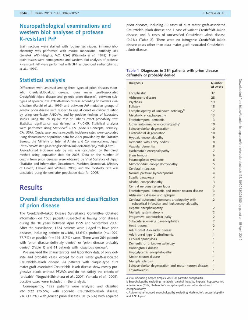
Jakob disease.
Table 1 Diagnoses in 264 patients with prion diseasedefinitely or probably denied
Diagnosis Numberof cases
Encephalitisa 32
Alzheimer’s disease 28
Psychosis 19
Epilepsy 18
Encephalopathy of unknown aetiologyb 17
Metabolic encephalopathy 13
Frontotemporal dementia 13
Other autoimmune encephalopathyc 10
Spinocerebellar degeneration 10
Corticobasal degeneration 10
Hypoxic encephalopathy 9
Dementia with Lewy bodies 8
Vascular dementia 8
Hashimoto’s encephalopathy 7
Brain tumour 6
Paraneoplastic syndrome 6
Mitochondrial encephalomyopathy 5
Cerebral infarction 4
Normal pressure hydrocephalus 4
Spastic paraplegia 4
Alcohol encephalopathy 3
Central nervous system lupus 3
Frontotemporal dementia and motor neuron disease 3
Alzheimer’s disease and epilepsy 2
Cerebral autosomal dominant arteriopathy withsubcortical infarction and leukoencephalopathy
2
Hepatic encephalopathy 2
Multiple system atrophy 2
Progressive supranuclear palsy 2
Subacute sclerosing panencephalitis 2
Head trauma 2
Adult-onset Alexander disease 1
Adult-onset type 2 citrullinemia 1
Cervical spondylosis 1
Dementia of unknown aetiology 1
Huntington’s disease 1
Hypoglycemic encephalopathy 1
Motor neuron disease 1
Multiple sclerosis 1
Spinocerebellar degeneration and motor neuron disease 1
Thyrotoxicosis 1
a Viral (including herpes simplex virus) or parasitic encephalitis.
b Encephalopathy excluding metabolic, alcohol, hepatic, hypoxic, hypoglycemic,autoimmune (CNS, Hashimoto’s encephalopathy and others)-inducedencephalopathy.c Autoimmune-induced encephalopathy excluding Hashimoto’s encephalopathyand CNS lupus.
3046 | Brain 2010: 133; 3043–3057 I. Nozaki et al.
Dow
nloaded from https://academ
ic.oup.com/brain/article-abstract/133/10/3043/321900 by guest on 15 April 2019
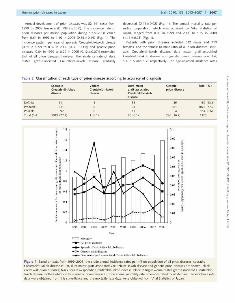
Annual development of prion diseases was 82–141 cases from
1999 to 2008 (mean� SD 108.8� 20.0). The incidence rate of
prion diseases per million population during 1999–2008 varied
from 0.64 in 1999 to 1.10 in 2006 (0.85� 0.16) (Fig. 1). The
incidence pattern per year of sporadic Creutzfeldt–Jakob disease
[0.55 in 1999 to 0.87 in 2006 (0.66� 0.11)] and genetic prion
diseases [0.04 in 1999 to 0.20 in 2005 (0.12� 0.07)] resembled
that of all prion diseases; however, the incidence rate of dura
mater graft-associated Creutzfeldt–Jakob disease gradually
decreased (0.41� 0.02) (Fig. 1). The annual mortality rate per
million population, which was obtained by Vital Statistics of
Japan, ranged from 0.88 in 1999 and 2000 to 1.59 in 2008
(1.15� 0.23) (Fig. 1).
Patients with prion diseases included 512 males and 710
females, and the female to male ratio of all prion diseases, spor-
adic Creutzfeldt–Jakob disease, dura mater graft-associated
Creutzfeldt–Jakob disease and genetic prion diseases was 1.4,
1.4, 1.6 and 1.2, respectively. The age-adjusted incidence rates
Figure 1 Based on data from 1999–2008, the crude annual incidence rates per million population of all prion diseases, sporadic
Creutzfeldt–Jakob disease (CJD), dura mater graft-associated Creutzfeldt–Jakob disease and genetic prion diseases are shown. Black
circles = all prion diseases; black squares = sporadic Creutzfeldt–Jakob disease; black triangles = dura mater graft-associated Creutzfeldt–
Jakob disease; dotted white circles = genetic prion diseases. Crude annual mortality rate is demonstrated by white bars. The incidence rate
data were obtained from this surveillance and the mortality rate data were obtained from Vital Statistics of Japan.
Table 2 Classification of each type of prion disease according to accuracy of diagnosis
SporadicCreutzfeldt–Jakobdisease
VariantCreutzfeldt–Jakobdisease
Dura matergraft-associatedCreutzfeldt–Jakobdisease
Geneticprion disease
Total (%)
Definite 111 1 33 35 180 (13.6)
Probable 811 0 34 181 1026 (77.7)
Possible 97 0 13 4 114 (8.6)
Total (%) 1019 (77.2) 1 (0.1) 80 (6.1) 220 (16.7) 1320
Human prion diseases in Japan Brain 2010: 133; 3043–3057 | 3047
Dow
nloaded from https://academ
ic.oup.com/brain/article-abstract/133/10/3043/321900 by guest on 15 April 2019
per million per year for females were higher than those for males
in all prion diseases (male 0.79; female 0.88), sporadic
Creutzfeldt–Jakob disease (male 0.62; female 0.71) and dura
mater graft-associated Creutzfeldt–Jakob disease (male 0.048;
female 0.074), except for genetic prion diseases (male 0.16;
female 0.14).
The age at onset in all prion diseases ranged from 15 to 94
(mean� SD 66.9� 11.4 years). The age at onset of dura mater
graft-associated Creutzfeldt–Jakob disease was significantly
younger than for sporadic Creutzfeldt–Jakob disease and genetic
prion diseases (P50.001) (Table 3).
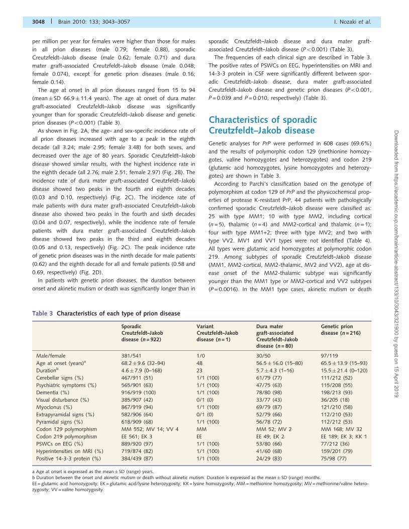
As shown in Fig. 2A, the age- and sex-specific incidence rate of
all prion diseases increased with age to a peak in the eighth
decade (all 3.24; male 2.95; female 3.48) for both sexes, and
decreased over the age of 80 years. Sporadic Creutzfeldt–Jakob
disease showed similar results, with the highest incidence rate in
the eighth decade (all 2.76; male 2.51; female 2.97) (Fig. 2B). The
incidence rate of dura mater graft-associated Creutzfeldt–Jakob
disease showed two peaks in the fourth and eighth decades
(0.03 and 0.10, respectively) (Fig. 2C). The incidence rate of
male patients with dura mater graft-associated Creutzfeldt–Jakob
disease also showed two peaks in the fourth and sixth decades
(0.04 and 0.07, respectively), while the incidence rate of female
patients with dura mater graft-associated Creutzfeldt–Jakob
disease showed two peaks in the third and eighth decades
(0.05 and 0.13, respectively) (Fig. 2C). The peak incidence rate
of genetic prion diseases was in the ninth decade for male patients
(0.62) and the eighth decade for all and female patients (0.58 and
0.69, respectively) (Fig. 2D).
In patients with genetic prion diseases, the duration between
onset and akinetic mutism or death was significantly longer than in
sporadic Creutzfeldt–Jakob disease and dura mater graft-
associated Creutzfeldt–Jakob disease (P50.001) (Table 3).
The frequencies of each clinical sign are described in Table 3.
The positive rates of PSWCs on EEG, hyperintensities on MRI and
14-3-3 protein in CSF were significantly different between spor-
adic Creutzfeldt–Jakob disease, dura mater graft-associated
Creutzfeldt–Jakob disease and genetic prion diseases (P50.001,
P = 0.039 and P = 0.010, respectively) (Table 3).
Characteristics of sporadicCreutzfeldt–Jakob diseaseGenetic analyses for PrP were performed in 608 cases (69.6%)
and the results of polymorphic codon 129 (methionine homozy-
gotes, valine homozygotes and heterozygotes) and codon 219
(glutamic acid homozygotes, lysine homozygotes and heterozy-
gotes) are shown in Table 3.
According to Parchi’s classification based on the genotype of
polymorphism at codon 129 of PrP and the physicochemical prop-
erties of protease K-resistant PrP, 44 patients with pathologically
confirmed sporadic Creutzfeldt–Jakob disease were classified as:
25 with type MM1; 10 with type MM2, including cortical
(n = 5), thalamic (n = 4) and MM2-cortical and thalamic (n = 1);
four with type MM1+2; three with type MV2; and two with
type VV2. MV1 and VV1 types were not identified (Table 4).
All types were glutamic acid homozygotes at polymorphic codon
219. Among subtypes of sporadic Creutzfeldt–Jakob disease
(MM1, MM2-cortical, MM2-thalamic, MV2 and VV2), age at dis-
ease onset of the MM2-thalamic subtype was significantly
younger than the MM1 type or MM2-cortical and VV2 subtypes
(P = 0.0016). In the MM1 type cases, akinetic mutism or death
Table 3 Characteristics of each type of prion disease
SporadicCreutzfeldt–Jakobdisease (n = 922)
VariantCreutzfeldt–Jakobdisease (n = 1)
Dura matergraft-associatedCreutzfeldt–Jakobdisease (n = 80)
Genetic priondisease (n = 216)
Male/female 381/541 1/0 30/50 97/119
Age at onset (years)a 68.2� 9.6 (32–94) 48 56.5� 16.0 (15–80) 65.5� 13.9 (15–93)
Durationb 4.6� 7.9 (0–168) 23 5.7� 4.3 (1–16) 15.5� 21.4 (0–120)
Cerebellar signs (%) 467/911 (51) 1/1 (100) 61/79 (77) 111/212 (52)
Psychiatric symptoms (%) 565/901 (63) 1/1 (100) 47/75 (63) 115/208 (55)
Dementia (%) 916/919 (100) 1/1 (100) 78/80 (98) 198/213 (93)
Visual disturbance (%) 385/907 (42) 0/1 (0) 33/77 (43) 36/205 (18)
Myoclonus (%) 867/919 (94) 1/1 (100) 69/79 (87) 121/210 (58)
Extrapyramidal signs (%) 582/906 (64) 0/1 (0) 52/79 (66) 112/210 (53)
Pyramidal signs (%) 618/909 (68) 1/1 (100) 56/78 (72) 112/212 (53)
Codon 129 polymorphism MM 552; MV 14; VV 4 MM MM 52; MV 2 MM 168; MV 32
Codon 219 polymorphism EE 561; EK 3 EE EE 49; EK 2 EE 189; EK 3; KK 1
PSWCs on EEG (%) 889/920 (97) 1/1 (100) 53/80 (66) 77/212 (36)
Hyperintensities on MRI (%) 719/874 (82) 1/1 (100) 41/60 (68) 159/201 (79)
Positive 14-3-3 protein (%) 384/439 (87) 1/1 (100) 24/29 (83) 75/98 (77)
a Age at onset is expressed as the mean� SD (range) years.
b Duration between the onset and akinetic mutism or death without akinetic mutism. Duration is expressed as the mean� SD (range) months.EE = glutamic acid homozygosity; EK = glutamic acid/lysine heterozygosity; KK = lysine homozygosity; MM = methionine homozygosity; MV = methionine/valine hetero-zygosity; VV = valine homozygosity.
3048 | Brain 2010: 133; 3043–3057 I. Nozaki et al.
Dow
nloaded from https://academ
ic.oup.com/brain/article-abstract/133/10/3043/321900 by guest on 15 April 2019
occurred significantly earlier than in MM2-cortical, MM2-thalamic
and MV2 subtypes (P50.001). Frequencies of positive PSWCs
were lower in MM2-cortical, MM2-thalamic, MV2 and VV2 sub-
types compared with the MM1 type. The positive rate of CSF
14-3-3 protein in the MM1 type was higher than in
MM2-cortical, MM2-thalamic and MV2 subtypes.
Hyperintensities on MRI were identified in all but patients with
the MM2-thalamic subtype.
Characteristics of variantCreutzfeldt–Jakob diseaseOnly one patient with a history of a short stay in the UK, France
and other European countries developed variant Creutzfeldt–Jakob
disease; this case has already been reported (Yamada, 2006).
Although the clinical features in the early stage were compatible
with variant Creutzfeldt–Jakob disease (Will et al., 2004), the pa-
tient showed PSWCs on EEG in the late stage, leading to the
diagnosis of probable sporadic Creutzfeldt–Jakob disease. At poly-
morphic codons 129 and 219 the patient was homozygotic for
methionine and glutamic acid, respectively. The patient developed
akinetic mutism at 23 months and died 42 months after onset;
autopsy confirmed the diagnosis of variant Creutzfeldt–Jakob dis-
ease (Table 3).
Characteristics of duramater graft-associatedCreutzfeldt–Jakob diseaseBesides the 80 patients with dura mater graft-associated
Creutzfeldt–Jakob disease identified by this surveillance system,
58 patients with dura mater graft-associated Creutzfeldt–Jakob
disease had already been reported by previous surveillance
systems (Yamada et al., 2009); therefore, the total number of
dura mater graft-associated Creutzfeldt–Jakob disease patients
was 138. The source of cadaveric dura mater was Lyodura�
(B. Braun, Germany) in all dura mater graft-associated
Creutzfeldt–Jakob disease cases in which the brand name could
be identified.
Figure 2 Based on data from 1999–2008, age- and sex-specific annual incidence per million population of all prion diseases (A), sporadic
Creutzfeldt–Jakob disease (B), dura mater graft-associated Creutzfeldt–Jakob disease (C), and genetic prion diseases (D) is shown. Dotted
white triangles = all patients; black squares = male patients; dashed white circles = female patients.
Human prion diseases in Japan Brain 2010: 133; 3043–3057 | 3049
Dow
nloaded from https://academ
ic.oup.com/brain/article-abstract/133/10/3043/321900 by guest on 15 April 2019

The medical conditions for which dura mater grafts were used in
neurosurgery included meningioma (21.0%), hemifacial spasm
(13.8%) and acoustic neurinoma (12.3%) (Table 5). The propor-
tion of non-life-threatening conditions, such as hemifacial spasm
(13.8%) and trigeminal neuralgia (5%), were relatively high
(Table 5). Dura mater grafts were implanted during 1975–93,
and most patients (112 cases, 81.2%) received them during
1983–87 (Fig. 3A); in May 1987 the procedures for the collection
and processing of grafts were revised by the company (Sato et al.,
1997). It was reported that one of the patients with dura mater
graft-associated Creutzfeldt–Jakob disease, who received dura
mater grafts after May 1987, received a graft that had been
produced before May 1987. The incubation period (i.e. duration
from implantation of dura mater grafts to dura mater
graft-associated Creutzfeldt–Jakob disease onset) ranged from
1 to 30 years (mean� SD 11.8� 5.5 years) (Fig. 3B). The year
of disease onset ranged from 1985 to 2008, and many patients
(83 cases, 60.1%) developed dura mater graft-associated
Creutzfeldt–Jakob disease during 1993–2001, with a peak in
1995 (Fig. 3C).
Genetic analyses for PrP were performed in 58 cases (73.1%).
The polymorphic PrP codon 129 included 52 methionine homozy-
gotes (96.3%) and two heterozygotes (3.7%), and for codon 219,
49 glutamic acid homozygotes (96.1%) and two heterozygotes
(3.9%) (Table 3).
Among 33 patients with definite dura mater graft-associated
Creutzfeldt–Jakob disease, 29 patients had sufficient pathological
data to be categorized as either plaque type (n = 14, 48%) or
non-plaque type (n = 15, 52%).Tab
le4
Char
acte
rist
ics
of
each
subty
pe
of
spora
dic
Cre
utz
feld
t–Ja
kob
dis
ease
MM
1(n
=25)
MM
1+
2(n
=4)
MM
2-c
ort
ical
(n=
5)
MM
2-t
hal
amic
(n=
4)
MM
2-c
ort
ical
and
thal
amic
(n=
1)
MV
2( n
=3)
VV
2(n
=2)
Mal
e/fe
mal
e11/1
41/3
2/3
3/1
1/0
3/0
1/1
Age
atonse
ta67.2�
5.5
(57–7
7)
66.3�
4.8
(62–7
3)
66.8�
7.3
(57–7
4)
52.8�
8.3
(43–6
1)
65
62.0�
5.3
(58–6
8)
72
(69–7
5)
Dura
tion
b3.1�
2.7
(0–1
4)
7.5�
5.4
(3–1
4)
24.7�
15.1
(10–5
0)
18.5�
6.7
(13–2
8)
11
26.5
(12–4
1)
6
Codon
219
poly
morp
his
mEE
23
EE3
EE5
EE3
EEEE
3EE
2
PSW
Cs
on
EEG
(%)
23/2
5(9
2)
4/4
(100)
2/4
(40)
0/4
(0)
0/1
(0)
0/3
(0)
0/2
(0)
Hyp
erin
tensi
ties
on
MR
I(%
)25/2
5(1
00)
4/4
(100)
5/5
(100)
0/4
(0)
1/1
(100)
3/3
(100)
2/2
(100)
Posi
tive
14-3
-3pro
tein
(%)
15/1
6(9
4)
3/4
(75)
2/5
(40)
1/3
(33)
0/1
(0)
0/1
(0)
2/2
(100)
aA
ge
atonse
tis
expre
ssed
asth
em
ean�
SD(r
ange)
year
s.b
Dura
tion
bet
wee
nth
eonse
tan
dak
inet
icm
utism
or
dea
thw
ithout
akin
etic
mutism
.D
ura
tion
isex
pre
ssed
asth
em
ean�
SD(r
ange)
month
s.
Table 5 Medical conditions leading to use of dura matergrafts
Medical conditions No. ofcases
Meningioma 29
Hemifacial spasm 19
Acoustic neurinoma 17
Subarachonoid haemorrhage 11
Cerebral/cerebellar haemorrhage 8
Arteriovenous malformation 7
Trigeminal neuralgia 7
Brain aneurysm 6
Epidural/subdural haematoma 6
Trauma 6
Arnold-Chiari malformation 5
Ependymoma 2
Epidermoid 2
Glioma 2
Hemangioblastoma 2
Spinal cord tumour 2
Arachonoid cyst 1
Osteoma 1
Ossification of the posterior longitudinal ligament 1
Pituitary adenoma 1
Teratoma 1
Brain tumour with unknown details 2
3050 | Brain 2010: 133; 3043–3057 I. Nozaki et al.
Dow
nloaded from https://academ
ic.oup.com/brain/article-abstract/133/10/3043/321900 by guest on 15 April 2019

Characteristics of genetic prion diseasesThe distribution and frequencies of PrP mutations associated with
genetic prion diseases in Japan are shown in Table 6. The most
common mutation was V180I, followed by P102L, E200K, M232R
and P105L. The characteristics associated with the mutations are
shown in Table 7. The characteristics of relatively frequent muta-
tions in Japan (P102L, P105L, V180I, E200K and M232R) were as
follows: patients with P102L, P105L and E200K showed relatively
high penetrance (Table 7), whereas only 2.2% of patients with
V180I mutation and no patients with M232R mutation had a
positive family history (Table 7).
P102L and P105L cases showed onset at a relatively young
age and a Gerstmann–Straussler–Scheinker disease phenotype
with slow progression, while V180I cases presented with
onset in old age and with slow progression in spite of the
Creutzfeldt–Jakob disease type pathology (Mutsukura et al.,
2009) (Table 7). Thirty-one M232R patients with sufficient
clinical data were classified into 19 with rapid type and 12 with
slow type.
P102L and P105L showed low positive rates in PSWCs on EEG
and hyperintensities on MRI. V180I and the slow type of M232R
presented with a low positive rate of PSWCs on EEG, but a high
positive rate of hyperintensities on MRI.
DiscussionThis study revealed the epidemiological and clinical characteristics
of prion diseases in Japan over a 10-year period. Nationwide sur-
veillance data of prion diseases have been reported from European
countries, Australia and the USA (Horan et al., 2004; Sanchez-
Valle et al., 2004; Ladogana et al., 2005; de Pedro-Cuesta
et al., 2006; Van Everbroeck et al., 2006; Heinemann et al.,
2007; Klug et al., 2009; Holman et al., 2010). Although a study
of Chinese patients with prion diseases has been reported (Shi
et al., 2008), it did not accurately reflect the incidence rate in
China, as the number of patients with prion diseases was quite
small compared with that estimated for the population in China.
Figure 3 The time when cadaveric dura mater grafts were implanted is shown by year, with black bars indicating patients with dura mater
graft-associated Creutzfeldt–Jakob disease (A). The incubation period (i.e. duration from implantation of dura mater grafts to onset of
dura mater graft-associated Creutzfeldt–Jakob disease) is shown by year, with black bars indicating patients with dura mater
graft-associated Creutzfeldt–Jakob disease (B). The year when dura mater graft-associated Creutzfeldt–Jakob disease developed in
patients is shown by black bars indicating patients with dura mater graft-associated Creutzfeldt–Jakob disease (C).
Human prion diseases in Japan Brain 2010: 133; 3043–3057 | 3051
Dow
nloaded from https://academ
ic.oup.com/brain/article-abstract/133/10/3043/321900 by guest on 15 April 2019
Our study is therefore the largest report of prospective prion dis-
ease surveillance from Asia.
Incidence rate of prion diseasesThe crude annual mortality rate per million of prion diseases in
Japan was 0.89–1.58 (mean 1.18), which was obtained from Vital
Statistics of Japan and is similar to that in European countries,
Australia, Canada and the USA (1.0–1.5 per million) (Ladogana
et al., 2005; Klug et al., 2009; Holman et al., 2010). On the other
hand, the annual incidence rate of prion diseases per million popu-
lation in Japan obtained by our surveillance was 0.65–1.10 (mean
0.85), which was lower than the mortality rate obtained by Vital
Statistics of Japan, because our surveillance rate was not 100%.
The annual incidence rate of sporadic Creutzfeldt–Jakob disease in
Japan was 0.55–0.87 per million (mean 0.66), which was slightly
lower than in Germany (0.8–1.6) and in European countries,
Australia and Canada (1.39 as overall annual mortality rate)
(Ladogana et al., 2005; Heinemann et al., 2007). The reasons
for the lower incidence rate of sporadic Creutzfeldt–Jakob disease
in Japan may be explained by the lower autopsy rate in Japan
compared with other western countries, and the limited sensitivity
of the diagnostic criteria by Masters et al. (1979) for probable
sporadic Creutzfeldt–Jakob disease. When possible sporadic
Creutzfeldt–Jakob disease cases were included in the number of
sporadic Creutzfeldt–Jakob disease cases, the annual incidence
rate per million increased to 0.57–0.95 (mean 0.73), but it was
still lower than those of other western countries.
Interestingly, in Japan, female predominance was identified in
age-adjusted incidence rates of prion diseases, sporadic
Creutzfeldt–Jakob disease, and dura mater graft-associated
Creutzfeldt–Jakob disease, but not for genetic prion diseases. An
excess of females has been reported for Creutzfeldt–Jakob disease,
sporadic Creutzfeldt–Jakob disease or genetic cases (Collins et al., Tab
le7
Char
acte
rist
ics
of
gen
etic
pri
on
dis
ease
s
Inse
rtio
n( n
=3)
P102L
(n=
39)
P105L
(n=
5)
D178N
-129M
(n=
3)
D178N
-129V
(n=
1)
V180I
(n=
89)
E200K
(n=
37)
V203I
(n=
2)
R208H
(n=
1)
M232R
(n=
33)
V180I+
M232R
(n=
1)
Mal
e/fe
mal
e2/1
16/2
34/1
2/1
1/0
35/5
415/2
22/0
0/1
18/1
50/1
Age
atonse
ta49.5�
21.7
(26–5
5)
54.0�
12.4
(22–7
5)
41.6�
8.0
(31–5
1)
52.3�
5.7
(46–5
7)
74
76.1�
7.4
(44–9
3)
58.5�
9.8
(31–7
7)
73
74
64.2�
12.5
(15–8
1)
74
Posi
tive
fam
ilyhis
tory
(%)
1(3
3)
29
(74)
4(8
0)
None
None
2(2
)17
(46)
None
None
None
None
Dura
tion
b20.0�
21.4
(3–4
4)
36.7�
30.1
(3–9
6)
99.7�
23.5
(74–1
20)
10.7�
3.2
(7–1
3)
24
13.3�
10.9
(1–5
8)
3.9�
3.6
(1–1
4)
5(4
–6)
38.0�
8.7
(0–3
2)
1
Codon
129
poly
morp
his
mM
M2
MM
29;
MV
3M
V4
MM
3M
V1
MM
65;
MV
22
MM
34
MM
2M
M1
MM
30;
MV
2M
M1
Codon
219
poly
morp
his
mEE
1;
KK
1EE
31;
EK1
EE4
EE3
EE1
EE81
EE33;
EK1
EE2
EE1
EE31;
EK1
EE1
PSW
Cs
on
EEG
(%)
2/3
(67)
7/3
7(1
9)
0/5
(0)
0/3
(0)
0/1
(0)
10/8
8(1
1)
34/3
7(9
2)
2/2
(100)
1/1
(100)
20/3
2(6
3)
1/1
(100)
Hyp
erin
tensi
ties
on
MR
I(%
)1/2
(50)
14/3
6(3
9)
0/5
(0)
0/3
(0)
0/1
(0)
84/8
4(1
00)
31/3
5(8
9)
2/2
(100)
1/1
(100)
26/3
1(8
4)
0/1
(0)
Posi
tive
14-3
-3pro
tein
(%)
0/1
(0)
6/1
0(6
0)
1/2
(50)
1/1
(100)
1/1
(100)
35/4
5(7
8)
11/1
2(9
2)
1/1
(100)
1/1
(100)
18/2
3(7
8)
0/1
(0)
EE=
glu
tam
icac
idhom
ozy
gosi
ty;
EK=
glu
tam
icac
id/l
ysin
ehet
erozy
gosi
ty;
KK
=ly
sine
hom
ozy
gosi
ty;
MM
=m
ethio
nin
ehom
ozy
gosi
ty;
MV
=m
ethio
nin
e/va
line
het
erozy
gosi
ty;
PSW
Cs=
per
iodic
synch
ronous
wav
eco
mple
xes;
VV
=va
line
hom
ozy
gosi
ty.
aA
ge
atonse
tis
expre
ssed
asth
em
ean�
SD(r
ange)
year
s.
bD
ura
tion
bet
wee
nth
eonse
tan
dak
inet
icm
utism
or
dea
thw
ithout
akin
etic
mutism
.D
ura
tion
isex
pre
ssed
asth
em
ean�
SD(r
ange)
month
s.
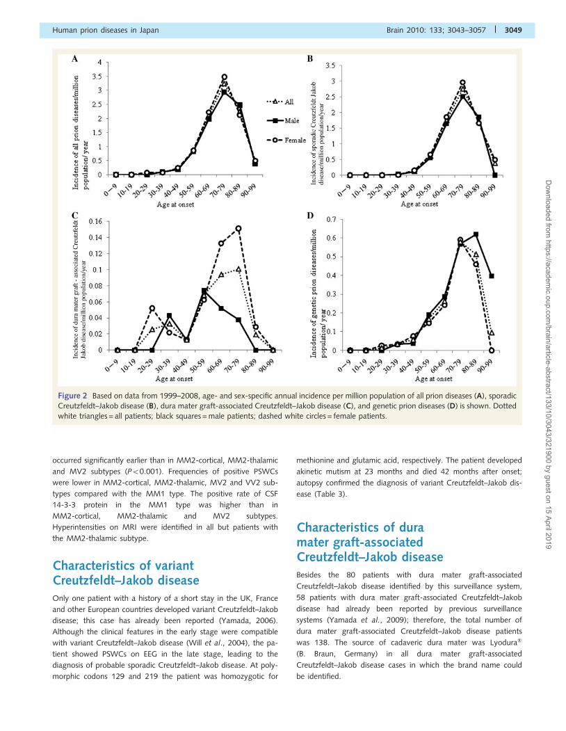
Table 6 Comparison of the distribution in genetic priondisease between Japan and EUROCJD
Japan(n = 216) (%)
EUROCJDa
(n = 425) (%)
Insertion 3 (1.4) 42 (9.9)
P102L 39 (18.1) 24 (5.6)
P105L 5 (2.3) 0 (0)
A117V-129V 0 (0) 12 (2.8)
D178N-129M 3 (1.4) 64 (15.1)
D178N-129V 1 (0.5) 16 (3.8)
V180I 89 (41.2) 1 (0.2)
E200K 37 (17.1) 175 (41.2)
V203I 2 (0.9) 5 (1.2)
R208H 1 (0.5) 2 (0.5)
V210I 0 (0) 69 (16.2)
M232R 33 (15.3) 0 (0)
V180I+M232R 1 (0.5) 0 (0)
Other mutations 0 (0) 15 (3.5)
a European Creutzfeldt–Jakob Disease Surveillance Network; Kovacs et al., 2005.
3052 | Brain 2010: 133; 3043–3057 I. Nozaki et al.
Dow
nloaded from https://academ
ic.oup.com/brain/article-abstract/133/10/3043/321900 by guest on 15 April 2019

2002, 2006; Kovacs et al., 2005; Holman et al., 2010); however,
the average age-adjusted incidence rate of Creutzfeldt–Jakob
disease was reported to be similar for males and females in
Australia (Collins et al., 2002) and higher for males than for
females in the USA (Holman et al., 2010). Further data corrected
by the age distribution of gender in the general population of each
country are essential to clarify gender difference.
The age- and sex-specific incidence rate of sporadic Creutzfeldt–
Jakob disease in Japan was similar to that of sporadic Creutzfeldt–
Jakob disease in European countries, Australia and Canada
(Ladogana et al., 2005; Heinemann et al., 2007), showing a
decreased incidence rate over the age of 80 years. The reason
for this remains unknown and requires further study. The age-
and sex-specific incidence rate of patients with dura mater
graft-associated Creutzfeldt–Jakob disease showed two peaks, re-
flecting two peaks in the age at dura mater transplantation (male,
second and fifth decade; female, second and sixth decade) (data
not shown). The incidence pattern of genetic prion diseases
peaked in old age, similar to sporadic Creutzfeldt–Jakob disease,
and seemed to be influenced by onset in old age in a high pro-
portion of patients with the V180I mutation.
Types of prion diseasesThe proportions of sporadic Creutzfeldt–Jakob disease and genetic
prion diseases were almost identical to those in European coun-
tries, except for Slovakia, in which the percentage of patients with
genetic prion diseases was 70% (Ladogana et al., 2005).
The proportion of iatrogenic Creutzfeldt–Jakob disease was
relatively high in Japan compared with other European countries
because of the large number of patients with dura mater
graft-associated Creutzfeldt–Jakob disease. Of the 196 (62.7%)
worldwide dura mater graft-associated Creutzfeldt–Jakob
disease cases, 123 had been identified in Japan up to
2006 (Brown et al., 2006). In France, the proportion of iatrogenic
Creutzfeldt–Jakob disease was also high (8.7%) (de Pedro-Cuesta
et al., 2006), but most iatrogenic Creutzfeldt–Jakob disease cases
were induced by contaminated human growth hormone (Brown
et al., 2006). Worldwide, contaminated human growth hormone-
associated Creutzfeldt–Jakob disease was the most common
iatrogenic Creutzfeldt–Jakob disease, except for dura mater
graft-associated Creutzfeldt–Jakob disease (Brown et al., 2006),
although there were no cases of human growth hormone-
associated Creutzfeldt–Jakob disease in Japan. Although there
was only one case of variant Creutzfeldt–Jakob disease in the
past 10 years in Japan, the number of variant Creutzfeldt–Jakob
disease cases was 212 cases worldwide, in particular 172 (81.3%)
in the UK and 25 (11.8%) in France, up to March 2010 (NCJDSU)
(http://www.cjd.ed.ac.uk/vcjdworld.htm).
PrP polymorphisms in prion diseasesThe genotype distribution at codon 129 of PrP in sporadic
Creutzfeldt–Jakob disease in Japan revealed a higher proportion
of methionine homozygotes (96.8%) than in European countries,
Australia and Canada (67.2%) (Ladogana et al., 2005), whereas
the proportion of methionine homozygotes at codon 129 in
sporadic Creutzfeldt–Jakob disease was 100% (150/150) in
Korea (Jeong et al., 2005) and 97.0 % (131/135) in China (Shi
et al., 2008), similar to Japan.
The general Japanese population also presented with a
higher frequency of codon 129 homozygosity (methionine homo-
zygotes: 0.92, heterozygotes: 0.08, valine homozygotes: 0)
than European countries (methionine homozygotes: 0.37–0.49,
heterozygotes: 0.42–0.49, valine homozygotes: 0.08–0.15)
(Collinge et al., 1991; Doh-ura et al., 1991; Zimmermann et al.,
1999; Nurmi et al., 2003; Mitrova et al., 2005; Georgsson et al.,
2006; Dyrbye et al., 2008). The proportion of methionine
homozygotes at codon 129 in the general population
was 94.3% (499/529) in Korea (Jeong et al., 2004) and
97.6% (200/205) among Han Chinese (Yu et al., 2004), also
similar to Japan.
Genetic predisposition to sporadic Creutzfeldt–Jakob disease in
codon 129 homozygosity (methionine homozygotes or valine
homozygotes) was revealed in the UK (Palmer et al., 1991), but
this predisposition was not previously identified in a small number
of Japanese sporadic Creutzfeldt–Jakob disease patients (n = 21;
methionine homozygotes or valine homozygotes: 0.95, heterozy-
gotes: 0.05) because of the high frequency of methionine homo-
zygotes at codon 129 in the general Japanese population (Doh-ura
et al., 1991). Using data on codon 129 polymorphisms (n = 645;
methionine homozygotes: 0.93, heterozygotes: 0.07, valine homo-
zygotes: 0; M allele: 0.97, V allele: 0.03) in the general Japanese
population obtained from combining previous data (Doh-ura
et al., 1991; Ohkubo et al., 2003) as a control, we assessed
whether homozygosity, methionine homozygosity or the M allele
at codon 129 was associated with sporadic Creutzfeldt–Jakob dis-
ease in Japan, and found a significant association (P50.001,
P = 0.004 and P = 0.019, respectively). This is the first report show-
ing that codon 129 homozygosity is a risk for sporadic
Creutzfeldt–Jakob disease in Asia, as reported in western coun-
tries. In addition, methionine homozygosity and the M allele
were not associated with sporadic Creutzfeldt–Jakob disease in
the UK (Palmer et al., 1991). In dura mater graft-associated
Creutzfeldt–Jakob disease, neither homozygosity (methionine
homozygotes or valine homozygotes: 0.96), methionine homozy-
gosity (0.96) nor the M allele (0.98) at codon 129 were signifi-
cantly different from the general Japanese population. Genetic
prion diseases had a significantly higher proportion of codon
129 heterozygosity and the V allele than the general population
(both P50.001). This seemed to be related to the higher propor-
tion of codon 129 heterozygosity in V180I cases (methionine
homozygotes: 0.75, heterozygotes: 0.25), which is the most
common genetic prion disease in Japan, although the V180I mu-
tation was located on the allele with methionine at codon 129 in
all cases investigated.
It was previously reported that heterozygosity at codon 219 was
found in the general Japanese population (glutamic acid homozy-
gotes: 0.88, heterozygotes: 0.12), while the K allele was not found
in 85 Japanese patients with sporadic Creutzfeldt–Jakob disease
(Shibuya et al., 1998). The frequencies of the K allele (0.0027)
and heterozygous genotype (heterozygotes: 0.0053) at codon 219
in sporadic Creutzfeldt–Jakob disease patients in this study were
significantly lower than in the general Japanese population
Human prion diseases in Japan Brain 2010: 133; 3043–3057 | 3053
Dow
nloaded from https://academ
ic.oup.com/brain/article-abstract/133/10/3043/321900 by guest on 15 April 2019

(n = 566; glutamic acid homozygotes: 0.86, heterozygotes: 0.14,
lysine homozygotes: 0; E allele: 0.93, K allele: 0.07) (Kitamoto
et al., 1994; Ohkubo et al., 2003) (P50.001). The frequencies
of the K allele (0.013) and heterozygous genotype (0.016) at
codon 219 in genetic prion diseases were also significantly lower
than in the general Japanese population (both P50.001), while
in dura mater graft-associated Creutzfeldt–Jakob disease, the fre-
quencies of the K allele (0.02) and heterozygosity (0.04) were not
significantly different from the general population.
Sporadic Creutzfeldt–Jakob diseaseThe very high frequency of PSWCs in sporadic Creutzfeldt–Jakob
disease (97%), compared with the data of western countries
(Collins et al., 2006), is related to the application of the diagnostic
criteria by Masters et al. (1979) and the low autopsy rate in Japan.
Regarding the subtypes according to Parchi’s classification (Parchi
et al., 1999), the MM1 type was the most common (25/44,
56.8%), characterized by typical Creutzfeldt–Jakob disease fea-
tures: rapid clinical course, positive PSWCs and CSF 14-3-3 protein
and typical MRI findings (Table 4). Among atypical cases other
than the MM1 type, the proportion of the MM2 type was rela-
tively high (10/44, 22.7%) compared with Europe, the USA (12/
300, 4.0%) (Parchi et al., 1999) and Germany (12/243, 4.9%)
(Heinemann et al., 2007). MM2 type cases included cortical
(50%), thalamic (40%) and combined (cortical and thalamic)
forms (10%). Our results were influenced by the bias that atypical
cases might have been more selectively autopsied to confirm the
diagnosis; however, the relatively high proportion of the MM2
type in Japanese patients with sporadic Creutzfeldt–Jakob disease
reflected the high proportion of the methionine homozygote
genotype in the Japanese population.
Clinical characteristics of each sporadic Creutzfeldt–Jakob dis-
ease subtype (MM1, MM2-cortical, MM2-thalamic, MV2 and
VV2) were almost the same as in previous reports (Parchi et al.,
1999; Collins et al., 2006), except for the higher frequency of
extrapyramidal signs (72%) in the MM1 type [7% in a previous
report (Parchi et al., 1999)] and the lower frequency of pyramidal
signs (0%) in MM2-cortical subtype [83% in previous reports
(Parchi et al., 1999; Krasnianski et al., 2006)]. The deficiency of
pyramidal or other neurological signs in the MM2-cortical subtype
would lead to difficulties in the clinical diagnosis of MM2-type
sporadic Creutzfeldt–Jakob disease on the basis of the current
sporadic Creutzfeldt–Jakob disease criteria, although cortical
hyperintensities on MRI suggest the diagnosis (Hamaguchi et al.,
2005). In this study, the age at onset of the MM2-thalamic sub-
type was younger with a longer duration than the MM1 type, and
neither PSWCs on EEG nor hyperintensities on MRI were identified
in the MM2-thalamic subtype.
Dura mater graft-associatedCreutzfeldt–Jakob diseaseWorldwide, the majority of patients with dura mater graft–
associated Creutzfeldt–Jakob disease have been reported
from Japan (Belay et al., 2005; Brown et al., 2006;
Noguchi-Shinohara et al., 2007; Nakamura et al., 2008; Yamada
et al., 2009). In Japan, all dura mater graft-associated Creutzfeldt–
Jakob disease cases in which the origin of the dural grafts could be
identified were recipients of Lyodura�, as previously reported
(Yamada et al., 2009). In Japan, the import of Lyodura� was
approved in 1973 and then prohibited in 1997. The mean incu-
bation period of dura mater graft-associated Creutzfeldt–Jakob
disease (11.8 years) was shorter than human growth
hormone-associated Creutzfeldt–Jakob disease (20.5 years)
(Belay et al., 2005). The longest incubation period of dura
mater graft-associated Creutzfeldt–Jakob disease was 30 years,
and the year when the patient received implantation (1975) was
also the earliest among previous reports (Nakamura et al., 2008;
Yamada et al., 2009). Regarding the medical conditions in which
patients received the implantation of cadaveric dura mater grafts,
non-life-threatening conditions such as hemifacial spasm and
trigeminal pain were relatively common, because recipients
with lethal conditions might have died before dura mater
graft-associated Creutzfeldt–Jakob disease developed. Clinical dur-
ation (from onset to akinetic mutism or death) of dura mater
graft-associated Creutzfeldt–Jakob disease was longer than that
of sporadic Creutzfeldt–Jakob disease, and positive rates of
PSWCs on EEG and hyperintensities on MRI were lower than
those of sporadic Creutzfeldt–Jakob disease (Table 3). These find-
ings can be explained by the fact that dura mater graft-associated
Creutzfeldt–Jakob disease presented with two distinct clinico-
pathological subtypes, i.e. ‘plaque’ and ‘non-plaque’ types: in con-
trast to the non-plaque type with classic Creutzfeldt–Jakob disease
features, the plaque type shows relatively slow progression and no
or late occurrence of PSWCs on EEG (Noguchi-Shinohara et al.,
2007; Yamada et al., 2009). When patients with negative PSWCs
and dura mater graft-associated Creutzfeldt–Jakob disease were
combined with those with plaque-type dura mater graft-associated
Creutzfeldt–Jakob disease, and patients with positive PSWCs and
dura mater graft-associated Creutzfeldt–Jakob disease with those
with non-plaque type dura mater graft-associated Creutzfeldt–
Jakob disease, one-third of patients with dura mater graft-
associated Creutzfeldt–Jakob disease could have ‘plaque type’
(data not shown), which was almost the same as in previous
reports (Noguchi-Shinohara et al., 2007; Yamada et al., 2009).
Genetic prion diseasesAs shown in Table 6, the proportion of PrP mutations was quite
different from those of EUROCJD, the European Creutzfeldt–
Jakob Disease Surveillance Network (Kovacs et al., 2005). The
V180I mutation was the most common in Japan but is very rare
in Europe (only one case in France). Conversely, the most
common mutation in Europe was E200K, which was the third
most common in Japan. Additionally, the V210I mutation was
the second most common mutation in Europe but was not identi-
fied in Japan.
In China, the following 10 genetic prion diseases cases have
been reported; three D178N-129M cases and one case each of
S97N, G114V, T188K, F198V, E200K, R208C and M232R
(Shi et al., 2008; Zheng et al., 2009); in Korea, three genetic
prion disease cases (D178N-129M, E200K and M232R) have
been identified (Choi et al., 2009). The V180I mutation was not
3054 | Brain 2010: 133; 3043–3057 I. Nozaki et al.
Dow
nloaded from https://academ
ic.oup.com/brain/article-abstract/133/10/3043/321900 by guest on 15 April 2019
identified in China or Korea but, conversely, S97N, G114V,
T188K, F198V and R208C mutations were not identified
in Japan or Korea. Despite the similar ethnic background
of East Asia, the distribution of genetic prion diseases in Japan
might be different from China and Korea; however, the number
of patients reported from China and Korea (Shi et al., 2008;
Choi et al., 2009) is too small to reach a conclusion, requiring a
larger study in the future.
V180I and M232R mutations were common in Japan but rare
in European countries. Interestingly, patients with V180I or
M232R mutations had no or rare family histories; therefore,
they would have been misdiagnosed with sporadic Creutzfeldt–
Jakob disease if genetic analysis had not been performed.
Previous reports also showed no family history in cases of V180I
or M232R mutations (Bratosiewicz et al., 2001; Jin et al., 2004;
Shiga et al., 2007; Zheng et al., 2008; Choi et al., 2009).
These findings suggest that V180I and M232R might be
polymorphisms, but not pathogenic mutations. Compared
with the genotypes of PrP in the general Japanese population
(n = 466; isoleucine allele at codon 180:0; arginine at
codon 232:0) (Ohkubo et al., 2003), both V180I and M232R
mutations had significantly higher proportions of overall prion
disease with PrP (n = 881) (both P50.001), indicating that
V180I and M232R are not simple polymorphisms, but are disease
related.
Age at disease onset of patients with the V180I mutation
was older than that of sporadic Creutzfeldt–Jakob disease
(P50.001), and patients with the V180I mutation had a longer
clinical duration (P50.001) and lower rate of positive PSWCs
on EEG (P50.001) than those with sporadic Creutzfeldt–Jakob
disease (Tables 3 and 7). Similar findings were reported by Jin
et al. (2004), who mentioned that MRI findings in V180I revealed
characteristic hyperintensities in medial regions, posterior to
the parieto-occipital sulci in occipital lobes on T2-weighted,
fluid-attenuated inversion recovery, and diffusion weighted
images. The following characteristics of V180I cases appeared
to be similar to the MM2-cortical type: longer duration, hyperin-
tensities on MRI and a lower rate of PSWCs; therefore, genetic
analysis for PrP is necessary for a differential diagnosis.
The M232R mutation was the fourth most common in
Japan, but is rare worldwide. Outside Japan, only three cases
(Polish, Chinese and Korean) have been identified (Bratosiewicz
et al., 2001; Zheng et al., 2008; Choi et al., 2009). As the
M232R mutation has been identified mainly in Asian countries,
this mutation may be particular to an Asian ethnic background.
M232R cases included two clinical subtypes, slow and rapid, as
reported previously (Shiga et al., 2007). The proportion of the
slow type was higher (39%) than reported earlier (25%) (Shiga
et al., 2007). While rapid-type patients with the M232R mutation
present with clinical and laboratory findings, similar to MM1 type
sporadic Creutzfeldt–Jakob disease, patients with the slow-type
M232R mutation have atypical features similar to MM2-cortical
type sporadic Creutzfeldt–Jakob disease. Further, M232R cases
with a much longer clinical duration may be misdiagnosed as
other neurodegenerative diseases if genetic examination of PrP is
not performed.
In conclusion, the incidence rate of prion diseases was similar to
that of western countries, but dura mater graft-associated
Creutzfeldt–Jakob disease was frequent in Japan. Genetic differ-
ences, such as codon 129 and 219 polymorphisms and mutations
in PrP, show some differences in the phenotypes of prion diseases
between Japan and western countries.
AcknowledgementsThe authors thank Creutzfeldt–Jakob disease specialists in the pre-
fectures, doctors-in-chief and Creutzfeldt–Jakob disease patients
and families for providing clinical information about patients.
FundingThe Creutzfeldt-Jakob Disease Surveillance Committee belongs to
the Research Group on Prion Disease and Slow Virus Infection,
funded by the Ministry of Health, Labour and Welfare of Japan.
This work was supported in part by a Health and Labour Sciences
Research Grant for Research on Measures for Intractable Diseases
(Prion Disease and Slow Virus Infections) from the Ministry of
Health, Labour and Welfare of Japan.
ReferencesBelay ED, Schonberger LB. The public health impact of prion diseases.
Annu Rev Public Health 2005; 26: 191–212.
Brandel JP, Heath CA, Head MW, Levavasseur E, Knight R, Laplanche JL,
et al. Variant Creutzfeldt-Jakob disease in France and the United
Kingdom: evidence for the same agent strain. Ann Neurol 2009; 65:
249–56.
Bratosiewicz J, Liberski PP, Kulczycki J, Kordek R. Codon 129 polymorph-
ism of the PRNP gene in normal Polish population and in
Creutzfeldt-Jakob disease, and the search for new mutations in
PRNP gene. Acta Neurobiol Exp 2001; 61: 151–6.Brown P, Brandel JP, Preece M, Sato T. Iatrogenic Creutzfeldt-Jakob
disease: the waning of an era. Neurology 2006; 67: 389–93.
Choi BY, Kim SY, Seo SY, An SS, Kim S, Park SE, et al. Mutations at
codons 178, 200-129, and 232 contributed to the inherited prion
diseases in Korean patients. BMC Infect Dis 2009; 9: 132.
Collinge J, Palmer MS, Dryden AJ. Genetic predisposition to iatrogenic
Creutzfeldt-Jakob disease. Lancet 1991; 337: 1441–2.
Collins S, Boyd A, Lee JS, Lewis V, Fletcher A, McLean CA, et al.
Creutzfeldt–Jakob disease in Australia 1970-1999. Neurology 2002;
59: 1365–71.
Collins SJ, Sanchez-Juan P, Masters CL, Klug GM, van Duijn C,
Poleggi A, et al. Determinants of diagnostic investigation sensitivities
across the clinical spectrum of sporadic Creutzfeldt-Jakob disease.
Brain 2006; 129: 2278–87.
de Pedro-Cuesta J, Glatzel M, Almazan J, Stoeck K, Mellina V,
Puopolo M, et al. Human transmissible spongiform encephalopathies
in eleven countries: diagnostic pattern across time, 1993-2002.
BMC Public Health 2006; 6: 278.Doh-ura K, Kitamoto T, Sakaki Y, Tateishi J. CJD discrepancy. Nature
1991; 353: 801–2.
Dyrbye H, Broholm H, Dziegiel MH, Laursen H. The M129V polymorph-
ism of codon 129 in the prion gene (PRNP) in the Danish population.
Eur J Epidemiol 2008; 23: 23–7.
Human prion diseases in Japan Brain 2010: 133; 3043–3057 | 3055
Dow
nloaded from https://academ
ic.oup.com/brain/article-abstract/133/10/3043/321900 by guest on 15 April 2019
Georgsson G, Tryggvason T, Jonasdottir AD, Gudmundsson S,
Thorgeirsdottir S. Polymorphism of PRNP codons in the normal
Icelandic population. Acta Neurol Scand 2006; 113: 419–25.
Glatzel M, Rogivue C, Ghani A, Streffer JR, Amsler L, Aguzzi A.
Incidence of Creutzfeldt-Jakob disease in Switzerland. Lancet 2002;
360: 139–41.
Hamaguchi T, Kitamoto T, Sato T, Mizusawa H, Nakamura Y,
Noguchi M, et al. Clinical diagnosis of MM2-type sporadic
Creutzfeldt-Jakob disease. Neurology 2005; 64: 643–8.Hamaguchi T, Noguchi-Shinohara M, Nozaki I, Nakamura Y, Sato T,
Kitamoto T, et al. Medical procedures and risk for sporadic
Creutzfeldt-Jakob disease, Japan, 1999-2008. Emerg Infect Dis
2009a; 15: 265–71.
Hamaguchi T, Noguchi-Shinohara M, Nozaki I, Nakamura Y, Sato T,
Kitamoto T, et al. The risk of iatrogenic Creutzfeldt-Jakob disease
through medical and surgical procedures. Neuropathology 2009b;
29: 625–31.
Heinemann U, Krasnianski A, Meissner B, Varges D, Kallenberg K,
Schulz-Schaeffer WJ, et al. Creutzfeldt-Jakob disease in Germany: a
prospective 12-year surveillance. Brain 2007; 130: 1350–9.Holman RC, Belay ED, Christensen KY, Maddox RA, Minino AM,
Folkema AM, et al. Human prion diseases in the United States. PLoS
One 2010; 5: e8521.Horan G, Keohane C, Molloy S, Howley R, Harney M, Heffernan J, et al.
Creutzfeldt-Jakob disease in Ireland: epidemiological aspects
1980–2002. Eur Neurol 2004; 51: 132–7.
Jeong BH, Nam JH, Lee YJ, Lee KH, Jang MK, Carp RI, et al.
Polymorphisms of the prion protein gene (PRNP) in a Korean popula-
tion. J Hum Genet 2004; 49: 319–24.
Jeong BH, Lee KH, Kim NH, Jin JK, Kim JI, Carp RI, et al. Association of
sporadic Creutzfeldt-Jakob disease with homozygous genotypes at
PRNP codons 129 and 219 in the Korean population. Neurogenetics
2005; 6: 229–32.
Jin K, Shiga Y, Shibuya S, Chida K, Sato Y, Konno H, et al. Clinical
features of Creutzfeldt-Jakob disease with V180I mutation.
Neurology 2004; 62: 502–5.Kitamoto T, Ohta M, Doh-ura K, Hitoshi S, Terao Y, Tateishi J. Novel
missense variants of prion protein in Creutzfeldt-Jakob disease or
Gerstmann-Straussler syndrome. Biochem Biophys Res Commun
1993; 191: 709–14.
Kitamoto T, Shin RW, Doh-ura K, Tomokane N, Miyazono M,
Muramoto T, et al. Abnormal isoform of prion proteins accumulates
in the synaptic structures of the central nervous system in patients
with Creutzfeldt-Jakob disease. Am J Pathol 1992; 140: 1285–94.
Kitamoto T, Tateishi J. Human prion diseases with variant prion protein.
Philos Trans R Soc Lond B Biol Sci 1994; 343: 391–8.
Klug GM, Boyd A, Lewis V, McGlade A, Stehmann C, Masters CL, et al.
Surveillance of Creutzfeldt-Jakob disease in Australia: 2009 update.
Commun Dis Intell 2009; 33: 188–91.Kovacs GG, Puopolo M, Ladogana A, Pocchiari M, Budka H,
van Duijn C, et al. EUROCJD. Genetic prion disease: the EUROCJD
experience. Hum Genet 2005; 118: 166–74.
Krasnianski A, Meissner B, Schulz-Schaeffer W, Kallenberg K, Bartl M,
Heinemann U, et al. Clinical features and diagnosis of the MM2
cortical subtype of sporadic Creutzfeldt-Jakob disease. Arch Neurol
2006; 63: 876–80.
Ladogana A, Puopolo M, Croes EA, Budka H, Jarius C, Collins S, et al.
Mortality from Creutzfeldt-Jakob disease and related disorders in
Europe, Australia, and Canada. Neurology 2005; 64: 1586–91.
Masters CL, Harris JO, Gajdusek DC, Gibbs CJ Jr, Bernoulli C, Asher DM.
Creutzfeldt-Jakob disease: patterns of worldwide occurrence and the
significance of familial and sporadic clustering. Ann Neurol 1979; 5:
177–88.Mitrova E, Mayer V, Jovankovieova V, Slivarichova D, Wsolova L.
Creutzfeldt-Jakob disease risk and PRNP codon 129 polymorphism:
necessity to revalue current data. Eur J Neurol 2005; 12: 998–1001.
Mutsukura K, Satoh K, Shirabe S, Tomita I, Fukutome T, Morikawa M,
et al. Familial Creutzfeldt-Jakob disease with a V180I mutation:
comparative analysis with pathological findings and diffusion-weighted
images. Dement Geriatr Cogn Disord 2009; 28: 550–7.
Nakamura Y, Yanagawa H, Hoshi K, Yoshino H, Urata J, Sato T.
Incidence rate of Creutzfeldt-Jakob disease in Japan. Int J Epidemiol
1999; 28: 130–4.
Nakamura Y, Uehara R, Watanabe M, Sadakane A, Yamada M,
Mizusawa H, et al. CDC. Update: Creutzfeldt-Jakob disease associated
with cadaveric dura mater grafts-Japan, 1978-2008. MMWR Morb
Mortal Wkly Rep 2008; 57: 1152–4.Noguchi-Shinohara M, Hamaguchi T, Kitamoto T, Sato T, Nakamura Y,
Mizusawa H, et al. Clinical features and diagnosis of dura mater graft
associated Creutzfeldt Jakob disease. Neurology 2007; 69: 360–7.Nurmi MH, Bishop M, Strain L, Brett F, McGuigan C, Hutchison M, et al.
The normal population distribution of PRNP codon 129 polymorphism.
Acta Neurol Scand 2003; 108: 374–8.
Ohkubo T, Sakasegawa Y, Asada T, Kinoshita T, Goto Y, Kimura H, et al.
Absence of association between codon 129/219 polymorphisms of the
prion protein gene and Alzheimer’s disease in Japan. Ann Neurol 2003;
54: 553–4.
Palmer MS, Dryden AJ, Hughes JT, Collinge J. Homozygous prion protein
genotype predisposes to sporadic Creutzfeldt-Jakob disease. Nature
1991; 352: 340–2.
Parchi P, Giese A, Capellari S, Brown P, Schulz-Schaeffer W, Windl O,
et al. Classification of sporadic Creutzfeldt-Jakob disease based on
molecular and phenotypic analysis of 300 subjects. Ann Neurol
1999; 46: 224–33.
Pocchiari M, Puopolo M, Croes EA, Budka H, Gelpi E, Collins S, et al.
Predictors of survival in sporadic Creutzfeldt-Jakob disease and other
human transmissible spongiform encephalopathies. Brain 2004; 127:
2348–59.
Prusiner SB. Prions. Proc Natl Acad Sci USA 1998; 95: 13363–83.
Puopolo M, Ladogana A, Almonti S, Daude N, Bevivino S, Petraroli R,
et al. Mortality trend from sporadic Creutzfeldt-Jakob disease (CJD) in
Italy, 1993–2000. J Clin Epidemiol 2003; 56: 494–9.
Sanchez-Valle R, Nos C, Yague J, Graus F, Domınguez A, Saiz A. Catalan
Collaborative Study Group for CJD. Clinical and genetic features of
human prion diseases in Catalonia: 1993–2002. Eur J Neurol 2004;
11: 649–55.Sato T, Hoshi K, Yoshino H, Urata J, Nakamura Y, Yanagawa H, et al.
Centers for Disease Control and Prevention (CDC). Creutzfeldt-Jakob
disease associated with cadaveric dura mater grafts – Japan,
January 1979-May 1996. MMWR Morb Mortal Wkly Rep 1997; 46:
1066–9.
Satoh K, Shirabe S, Eguchi H, Tsujino A, Eguchi K, Satoh A, et al. 14-3-3
protein, total tau and phosphorylated tau in cerebrospinal fluid of
patients with Creutzfeldt-Jakob disease and neurodegenerative disease
in Japan. Cell Mol Neurobiol 2006; 26: 45–52.Shi Q, Gao C, Zhou W, Zhang BY, Chen JM, Tian C, et al. Surveillance
for Creutzfeldt-Jakob disease in China from 2006 to 2007. BMC Public
Health 2008; 8: 360.
Shibuya S, Higuchi J, Shin RW, Tateishi J, Kitamoto T. Codon 219 Lys
allele of PRNP is not found in sporadic Creutzfeldt-Jakob disease. Ann
Neurol 1998; 43: 826–8.
Shiga Y, Satoh K, Kitamoto T, Kanno S, Nakashima I, Sato S, et al. Two
different clinical phenotypes of Creutzfeldt-Jakob disease with a
M232R substitution. J Neurol 2007; 254: 1509–17.
Shimizu S, Hoshi K, Muramoto T, Homma M, Ironside JW, Kuzuhara S,
et al. Creutzfeldt-Jakob disease with florid-type plaques after cadaveric
dura mater grafting. Arch Neurol 1999; 56: 357–62.
Statistics and Information Department, Ministers Secretariat, Ministry of
Health, Labour and Welfare. Vital Statistics of Japan-2008. Tokyo:
Health and Welfare Statistics Association; 2009.Takahashi H, Iwata T, Kitagawa Y, Takahashi RH, Sato Y,
Wakabayashi H, et al. Increased levels of epsilon and gamma
isoforms of 14-3-3 proteins in cerebrospinal fluid in patients with
Creutzfeldt-Jakob disease. Clin Diagn Lab Immunol 1999; 6: 983–5.
Van Everbroeck B, Michotte A, Sciot R, Godfraind C, Deprez M,
Quoilin S, et al. Increased incidence of sporadic Creutzfeldt-Jakob
3056 | Brain 2010: 133; 3043–3057 I. Nozaki et al.
Dow
nloaded from https://academ
ic.oup.com/brain/article-abstract/133/10/3043/321900 by guest on 15 April 2019

disease in the age groups between 70 and 90 years in Belgium. Eur JEpidemiol 2006; 21: 443–7.
WHO. Global surveillance, diagnosis and therapy of human transmissible
spongiform encephalopathies. Report of a WHO consultation; Geneva;
9-11 February 1998. (http://www.who.int/csr/resources/publications/bse/WHO_EMC_ZDI_98_9/en/).
WHO. The revision of the surveillance case definition for variant
Creutzfeldt-Jakob disease (vCJD). Report of a WHO consultation;
Edinburgh;17 May 2001. (http://www.who.int/csr/resources/publica-tions/bse/WHO_CDS_CSR_EPH_2001_5/en/).
Will RG, Alperovitch A, Poser S, Pocchiari M, Hofman A, Mitrova E, et al.
Descriptive epidemiology of Creutzfeldt-Jakob disease in six Europeancountries, 1993–1995. EU Collaborative Study Group for CJD.
Ann Neurol 1998; 43: 763–7.
Will RG, Ward HJ. Clinical features of variant Creutzfeldt-Jakob disease.
Curr Top Microbiol Immunol 2004; 284: 121–32.Yamada M, Variant CJD. Working Group, Creutzfeldt-Jakob Disease
Surveillance Committee, Japan. The first Japanese case of variant
Creutzfeldt-Jakob disease showing periodic electroencephalogram.
Lancet 2006; 367: 874.
Yamada M, Noguchi-Shinohara M, Hamaguchi T, Nozaki I, Kitamoto T,
Sato T, et al. Dura mater graft-associated Creutzfeldt-Jakob
disease in Japan: clinicopathological and molecular character-
ization of the two distinct subtypes. Neuropathology 2009; 29:
609–18.
Yu SL, Jin L, Sy MS, Mei FH, Kang SL, Sun GH, et al. Polymorphisms
of the PRNP gene in Chinese populations and the identification of a
novel insertion mutation. Eur J Hum Genet 2004; 12: 867–70.
Zheng L, Longfei J, Jing Y, Xinqing Z, Haiqing S, Haiyan L, et al. PRNP
mutations in a series of apparently sporadic neurodegenerative
dementias in China. Am J Med Genet B Neuropsychiatr Genet 2008;
147B: 938–44.
Zimmermann K, Turecek PL, Schwarz HP. Genotyping of the prion pro-
tein at codon 129. Acta Neuropathol 1999; 97: 355–8.
Human prion diseases in Japan Brain 2010: 133; 3043–3057 | 3057
Dow
nloaded from https://academ
ic.oup.com/brain/article-abstract/133/10/3043/321900 by guest on 15 April 2019


![AdministrationofFK506fromLateStageofDiseaseProlongsSurvival … · 75% of human prion diseases and leads to death through rap-idly progressing dementia and akinetic mutism [1, 2]](https://img.pdfslide.tips/doc/110x75/5f5d06697c86da04f679d9a8/administrationoffk506fromlatestageofdiseaseprolongssurvival-75-of-human-prion-diseases.jpg)