Embed Size (px)
Citation preview
Proteasome Dysfunction Activates Autophagy and theKeap1-Nrf2 Pathway*
Received for publication, May 10, 2014, and in revised form, July 14, 2014 Published, JBC Papers in Press, July 21, 2014, DOI 10.1074/jbc.M114.580357
Shun Kageyama‡§, Yu-shin Sou§, Takefumi Uemura¶, Satoshi Kametaka¶, Tetsuya Saito‡�, Ryosuke Ishimura‡�,Tsuguka Kouno§, Lynn Bedford**, R. John Mayer**, Myung-Shik Lee‡‡, Masayuki Yamamoto§§, Satoshi Waguri¶,Keiji Tanaka�, and Masaaki Komatsu‡§1
From the ‡Department of Biochemistry, School of Medicine, Niigata University, Chuo-ku, Niigata 951-8510, Japan, the §ProteinMetabolism Project and �Laboratory of Protein Metabolism, Tokyo Metropolitan Institute of Medical Science, Setagaya-ku, Tokyo156-8506, Japan, the ¶Department of Anatomy and Histology, Fukushima Medical University School of Medicine, Hikarigaoka,Fukushima 960-1295, Japan, the **Laboratory of Intracellular Proteolysis, School of Biomedical Sciences, University ofNottingham, School of Life Sciences, Queen’s Medical Centre, Nottingham NG7 2UH, United Kingdom, the ‡‡Department ofMedicine, Samsung Medical Center, Gangnam-gu, Seoul 135-710, Korea, and the §§Department of Medical Biochemistry, TohokuUniversity Graduate School of Medicine, Aoba-ku, Sendai 980-8575, Japan
Background: Malfunctions in the ubiquitin-proteasome system cause accumulation of non-functional, potentially toxicprotein aggregates.Results: The protein aggregates activate Nrf2 and are then excluded by autophagy in vivo.Conclusion: Both Nrf2 and autophagy serve as in vivo cellular adaptations to impaired proteasome.Significance: Cells contain networks of cellular defense mechanisms against defective proteostasis.
The ubiquitin-proteasome system and autophagy are cru-cially important for proteostasis in cells. These pathways areinterdependent, and dysfunction in either pathway causes accu-mulation of ubiquitin-positive aggregates, a hallmark of humanpathological conditions. To elucidate in vivo compensatoryaction(s) against proteasomal dysfunction, we developed micewith reduced proteasome activity in their livers. The mutantmice exhibited severe liver damage, accompanied by formationof aggregates positive for ubiquitin and p62/Sqstm1, an adaptorprotein for both selective autophagy and the anti-oxidativeKeap1-Nrf2 pathway. These aggregates were selectivelyentrapped by autophagosomes, and pathological features of liv-ers with impaired proteasome activity were exacerbated bysimultaneous suppression of autophagy. In contrast, concomi-tant loss of p62/Sqstm1 had no apparent effect on the liverpathology though p62/Sqstm1 was indispensable for the aggre-gates formation. Furthermore, defective proteasome functionled to transcriptional activation of the Nrf2, which served as aphysiological adaptation. Our in vivo data suggest that cells con-tain networks of cellular defense mechanisms against defectiveproteostasis.
The 26S proteasome, in collaboration with the sophisticatedubiquitination system used for selection of target proteins, isresponsible for degrading unnecessary or damaged proteins.
Malfunctions in this pathway cause accumulation of non-func-tional, potentially toxic protein aggregates (1–3). The macroau-tophagy (hereafter referred to as autophagy) system serves as asupplier of molecular building blocks under starved conditionsand also contributes to cellular renovation during cell differen-tiation (4, 5). Defects in this process can cause amino acid insuf-ficiency, which impairs protein synthesis during adaptation tostarvation, as well as energy production essential for cell sur-vival and development (4, 5). Even under nutrient-rich condi-tions, autophagy occurs constitutively at low levels to mediateglobal turnover of cytoplasmic materials (6, 7).
Dysfunctions of autophagy coupled to the ubiquitin systemhave been directly linked to human conditions such as Parkin-son disease and inflammatory disorders. Autophagy contrib-utes to selective removal of aggregated proteins (aggrephagy),unnecessary or damaged mitochondria (mitophagy), andinvading bacteria (xenophagy); these processes are usuallymediated by ubiquitin signaling (8 –10). When the ubiquitin-proteasome system is impaired due to accumulation of certainaggregation-prone proteins related to neurodegenerativedisease, autophagy is responsible for eliminating ubiquitin-pos-itive protein aggregates (11–13). In response to loss of mito-chondrial membrane potential, the E3 ligase Parkin translo-cates to damaged mitochondria in a PINK1-dependentmanner; once it is localized to mitochondria, it ubiquitinatesouter membrane proteins, thereby inducing mitophagy (14,15). Parkinson disease-related mutations of Parkin and PINK1prevent induction of mitophagy, resulting in persistence ofdamaged mitochondria, which may play a role in the pathogen-esis of Parkinson disease (14, 15). Invading bacteria in the cyto-sol and/or ruptured endosomal membranes are ubiquitinatedby E3s, including Parkin (16) and LRSAM1 (17), which medi-ates autophagic sequestration of microbes to restrict theirgrowth. Ubiquitin- and LC3-binding adaptor proteins, includ-
* This work was supported by Grant-in-aid for Scientific Research on Innova-tive Areas 25111006 (to M. K. and S. W.), Funding Program for Next Gener-ation World Leading Researchers Grant LS132 (to M. K.), the Takeda Sci-ence Foundation (to T. K. and M. K.), and a Global Research Laboratorygrant (to M-S. L. and M. K.).
1 To whom correspondence should be addressed: Dept. of Biochemistry,School of Medicine, Niigata University, Chuo-ku, Niigata 951-8510, Japan.Tel.: 81-25-277-2077; Fax: 81-25-277-0757, E-mail: [email protected].
THE JOURNAL OF BIOLOGICAL CHEMISTRY VOL. 289, NO. 36, pp. 24944 –24955, September 5, 2014© 2014 by The American Society for Biochemistry and Molecular Biology, Inc. Published in the U.S.A.
24944 JOURNAL OF BIOLOGICAL CHEMISTRY VOLUME 289 • NUMBER 36 • SEPTEMBER 5, 2014
by guest on May 28, 2019
http://ww
w.jbc.org/
Dow
nloaded from
ing p62/Sqstm1 (hereafter referred to as p62) (18), neighbor ofBRCA1 gene 1 (Nbr1)2 (19), NDP52 (20), and optineurin (21),are translocated to these ubiquitinated cargos; this process isassumed to mediate sequestration of ubiquitinated cargos intoautophagosomes. Among them, p62 and Nbr1 have been iden-tified as major components of many types of aggregates orinclusions observed in various human diseases, including neu-rodegenerative diseases, liver disorders, and hepatocellular car-cinomas (19, 22). But significance of such adaptor proteins onthe aggregates, particularly in vivo, remains unclear.
The Keap1-Nrf2 pathway, one of the major cellular defensemechanisms against oxidative and electrophilic stresses (23,24), is activated during selective autophagy (25–28). Under nor-mal conditions, the transcription factor Nrf2 (nuclear factorerythroid 2-related factor 2) is constitutively degraded throughthe ubiquitin-proteasome pathway; its binding partner, Keap1(kelch-like ECH-associated protein 1), is an adaptor of the ubiq-uitin ligase complex that targets Nrf2. Exposure to electro-philes, reactive oxygen species, and nitric oxide instigates mod-ification of the cysteine residues of Keap1, leading to itsinactivation. As a result, Nrf2 is stabilized, and it subsequentlytranslocates to the nucleus to induce the transcription ofnumerous cytoprotective genes through heterodimerizationwith small Maf proteins (23, 24). p62 also regulates the Keap1-
2 The abbreviations used are: Nbr1, neighbor of BRCA1 gene 1; Keap1, kelch-like ECH-associated protein 1; Nqo1, NAD(P)H dehydrogenase quinone 1;Nrf2, nuclear factor erythroid 2-related factor 2; P, postnatal day.
B
Rpt2f/f
Rpt2f/f;Alb
302010
5
10
15
(day)
Bod
y w
eigh
t (g)
20
0
***
D F
Rpt2
Actin
Rpt2f/f
Rpt2f/f ;A
lb
Rpt2f/f
Rpt2f/f ;A
lb
Rpt2f/f
Rpt2f/f ;A
lb
P20 P30 P40
Ubiquitin
- 51
(kDa)
- 51
- 14
- 19
- 191
- 97
- 64
- 39 AST ALT ALP
0
200
600
400
Rpt2f/f
Rpt2f/f ;A
lb0
800
400
1000
Rpt2f/f
Rpt2f/f ;A
lb0
500
1500
1000
2000
Rpt2f/f
Rpt2f/f ;A
lb
IU/L
*** *** ***
600
200
Rpt2f/f
Rpt2f/f ;A
lb
4
8
0
Live
r wei
ght/b
ody
wei
ght (
%)
6
2
30251510 20
Rpt2f/f
Rpt2f/f;Alb
Rpt2f/f
Rpt2f/f;Alb
0
15000
30000
20000
0
2000010000
500010000
30251510 20
60000
40000
No SDS
0.025% SDS25000
50000
30000
P20
Pro
teas
ome
activ
ity (A
FU)
P30
30251510 20
Rpt2f/f
Rpt2f/f;Alb
Rpt2f/fRpt2f/f;Alb
0
30000
0
20000
10000
10000
20000
30251510 20
40000
30000No SDS
0.025% SDS
Pro
teas
ome
activ
ity (A
FU)
P40
30251510 20
Rpt2f/fRpt2f/f;Alb
Rpt2f/fRpt2f/f;Alb
0
30000
5000040000
0
20000
10000
1000020000
30251510 20
40000
30000No SDS
0.025% SDSP
rote
asom
e ac
tivity
(AFU
)
Rpt2f/f Rpt2f/f;Alb
CV
P
P
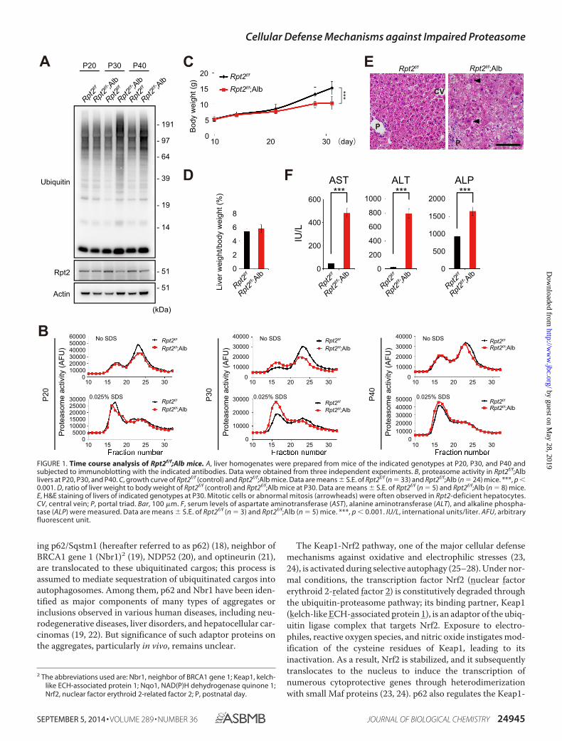
FIGURE 1. Time course analysis of Rpt2f/f;Alb mice. A, liver homogenates were prepared from mice of the indicated genotypes at P20, P30, and P40 andsubjected to immunoblotting with the indicated antibodies. Data were obtained from three independent experiments. B, proteasome activity in Rpt2f/f;Alblivers at P20, P30, and P40. C, growth curve of Rpt2f/f (control) and Rpt2f/f;Alb mice. Data are means � S.E. of Rpt2f/f (n � 33) and Rpt2f/f;Alb (n � 24) mice. ***, p �0.001. D, ratio of liver weight to body weight of Rpt2f/f (control) and Rpt2f/f;Alb mice at P30. Data are means � S.E. of Rpt2f/f (n � 5) and Rpt2f/f;Alb (n � 8) mice.E, H&E staining of livers of indicated genotypes at P30. Mitotic cells or abnormal mitosis (arrowheads) were often observed in Rpt2-deficient hepatocytes.CV, central vein; P, portal triad. Bar, 100 �m. F, serum levels of aspartate aminotransferase (AST), alanine aminotransferase (ALT), and alkaline phospha-tase (ALP) were measured. Data are means � S.E. of Rpt2f/f (n � 3) and Rpt2f/f;Alb (n � 5) mice. ***, p � 0.001. IU/L, international units/liter. AFU, arbitraryfluorescent unit.
Cellular Defense Mechanisms against Impaired Proteasome
SEPTEMBER 5, 2014 • VOLUME 289 • NUMBER 36 JOURNAL OF BIOLOGICAL CHEMISTRY 24945
by guest on May 28, 2019
http://ww
w.jbc.org/
Dow
nloaded from
Nrf2 pathway via a noncanonical mechanism (25–28). Underconditions of selective autophagy, Ser403 of the ubiquitin-asso-ciated domain of p62 is initially phosphorylated by casein
kinase 2 or TANK-binding kinase 1, which promotes the trans-location of p62 to cargos positive for ubiquitin (29, 30). Subse-quently, Ser351 of the Keap1-interacting region of p62 is phos-
Rpt2f/f;Alb Atg7f/f;AlbC D
- 51
- 14
- 19
- 191
- 97- 64
- 39
p62
Actin
Atg7
Ubiquitin
Rpt2
Total Sol. Insol.
Contro
l
Rpt2f/f ;A
lb
Atg7f/f ;A
lb
Rpt2f/f ;A
lb
Atg7f/f ;A
lb
Rpt2f/f ;A
lb
Atg7f/f ;A
lb
Contro
l
Contro
l
- 51
- 51
- 64
(kDa)
Rpt2f/f ;A
lb
a’ d’
c’
b’ e’
Con
trol
Rpt2f/f ;A
lb
p62 Ubiquitin Merged
Atg7f/f ;A
lb
Cellular Defense Mechanisms against Impaired Proteasome
24946 JOURNAL OF BIOLOGICAL CHEMISTRY VOLUME 289 • NUMBER 36 • SEPTEMBER 5, 2014
by guest on May 28, 2019
http://ww
w.jbc.org/
Dow
nloaded from
phorylated, followed by sequestration of Keap1 on the cargos.As a result, Nrf2 is stabilized; as in the canonical pathway, itthen translocates into the nucleus to induce its cytoprotectivetarget genes (25, 26). The ubiquitinated autophagic cargos,together with phosphorylated p62 and the Keap1 complex, aredegraded by autophagy, leading to elimination of cytotoxiccomponents (27). However, the physiological role of the cou-pling between the Keap1-Nrf2 system and selective autophagyin vivo has been not yet determined. In this study, we developedgenetically modified mice with decreased 26S proteasomeactivity, which accumulate aggregate structures positive forboth ubiquitin and p62 in their cells, and found that protea-some-dysfunction activates selective autophagy and the Keap1-Nrf2 pathway, both of which serve as cellular defensemechanisms.
EXPERIMENTAL PROCEDURES
Mice—Rpt2flox/flox mice (31) were cross-bred with albumin-Cre transgenic mice (32) to generate Rpt2flox/flox;Alb-Cre mice.Atg7flox/flox, p62flox/flox, and the Nrf2-knock-out mice used inthis study were described previously (33–35). Mice werehoused in specific pathogen-free facilities, and the EthicsReview Committee for Animal Experimentation of the TokyoMetropolitan Institute of Medical Science approved the exper-imental protocols.
Immunoblot Analysis—Immunoblots were carried out asdescribed previously (26). Antibodies against p62 (Progen Bio-technik, GP62-C), ubiquitin (Santa Cruz Biotechnology, Inc.,P4D1), Keap1 (Proteintech Group, Inc.), Nqo1 (Abcam, Inc.),Nrf2 (Santa Cruz Biotechnology, Inc., H-300), LC3B (Cell Sig-naling Technology, catalog no. 2775), Nbr1 (ProteinExpressCo., Ltd.), GFP (Invitrogen), actin (Chemicon Intl., Inc.,MAB1501R), and lamin B (Santa Cruz Biotechnology, Inc.,M-20) were purchased from the indicated suppliers. Anti-phosphorylated p62 polyclonal antibody was raised in rabbitsusing the peptide Cys�KEVDP(pS)TGELQSL as an antigen(26). The rabbit polyclonal antibodies against Atg7 and Rpt2were described previously (36, 37).
Assay of Proteasome Activity—Peptidase activity was mea-sured using a fluorescent peptide substrate, succinyl-Leu-Leu-Val-Tyr-7-amido-4-methylcoumarin (Suc-LLVY-MCA), asdescribed previously (38).
Histological Examination—Fixation and embedding proce-dures for immunohistochemistry were described previously(39). Briefly, mouse livers were quickly excised, cut into smallpieces, and then fixed by immersion in 4% paraformalde-hyde/4% sucrose in 0.1 M phosphate buffer, pH 7.4 (PB). Afterrinsing, samples were embedded in paraffin (for H&E staining),or in OCT compound (for immunofluorescence). For immuno-fluorescence microscopy, sections were blocked and then incu-
bated for 2–3 days at 4 °C with the following primary antibod-ies: guinea pig polyclonal antibody against p62 (Progen), rabbitpolyclonal antibody against ubiquitin (DAKO), or rabbit poly-clonal antibody against Keap1 (Proteintech Group). Immuno-fluorescence images were taken with an FV1000 laser scanningconfocal microscope equipped with a UPlanSApo 40� numer-ical aperture 1.3 oil objective lens (Olympus). After imageacquisition, contrast and brightness were adjusted using Pho-toshop CS4.
Electron Microscopy and Immunoelectron Microscopy—Forconventional electron microscopy, livers were excised and fixedby immersion in 0.1 M PB containing 2% paraformaldehyde and2% glutaraldehyde. Fixed samples were post-fixed with 1%OsO4, embedded in Epon812, and sectioned. Immunoelectronmicroscopy was carried out on ultrathin cryosections, asdescribed previously (39). In brief, livers were fixed by cardiacperfusion with 0.1 M PB containing 4% paraformaldehyde and4% sucrose and then frozen in PB with 2.3 M sucrose and 20%polyvinylpyrrolidone. Ultrathin sections were mounted onFormvar carbon-coated nickel grids, blocked with 1% bovineserum albumin in PBS, incubated with anti-ubiquitin (DAKO)and anti-p62 (Progen) antibodies, and then incubated with col-loidal gold-conjugated secondary antibodies.
Quantitative Real-time PCR—Using the Transcriptor First-Strand cDNA Synthesis Kit (Roche Applied Science), cDNA wassynthesized from 1 �g of total RNA. Quantitative PCR was per-formed using LightCycler� 480 Probes Master mix (RocheApplied Science) on a LightCycler� 480 (Roche Applied Science).Signals were normalized against that of �-glucuronidase (Gus).The sequences of the primers used were as follows: Nqo1 (left),AGCGTTCGGTATTACGATCC; Nqo1 (right), AGTACAA-TCAGGGCTCTTCTCG.
Statistical analysis—Values, including those displayed in thegraphs, are means � S.E. Statistical analysis was performedusing the unpaired t test (Welch test). p values less than 0.05denoted statistical significance.
RESULTS
Generation of Mice with Decreased Proteasome Activity—Toinvestigate aggrephagy in vivo, we crossbred mice bearing aconditional knock-out of Rpt2, one of six ATPases of the 19Sregulatory particle of the 26S proteasome (Rpt2flox/flox) (31),with albumin-Cre (Alb-Cre) transgenic mice (32). TheRpt2flox/flox;Alb-Cre (Rpt2f/f;Alb) mice were viable at birth andindistinguishable in appearance from their littermates. InRpt2f/f;Alb mice, levels of Rpt2 protein in the liver started todecrease at postnatal day (P)30 and recovered at P40 (Fig. 1A);ubiquitinated proteins accumulated significantly in the liver atP30 (Fig. 1A). The chymotryptic activities of the 26S and 20Sproteasomes (measured using Suc-LLVY-MCA as a substrate)
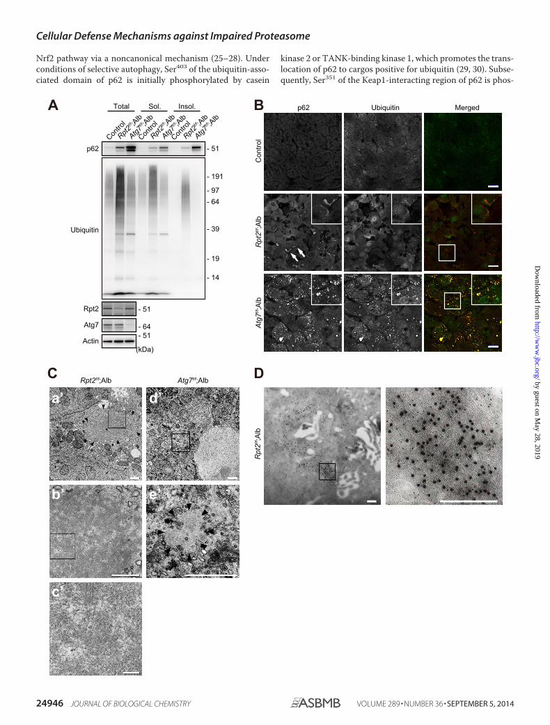
FIGURE 2. Characterization of ubiquitinated aggregates in Rpt2f/f;Alb livers. A, liver homogenates were prepared from mice of the indicated genotypes atP30. Total, soluble (Sol.), and insoluble (Insol.) fractions were subjected to immunoblotting with the indicated antibodies. Data were obtained from threeindependent experiments. B, liver cryosections from mice of the indicated genotypes at P30 were double-immunostained with p62 and ubiquitin antibodies.A portion of each image is magnified and shown in the inset. Arrows indicate large pleomorphic aggregated structures. Merged images are shown at the rightcolumn (red, p62; green, ubiquitin). Bars, 20 �m. C, electron micrographs of hepatocytes of the indicated genotypes. The boxed regions in a�, b�, and d� areenlarged and shown in b�, c�, and e�, respectively. Arrowheads indicate aggregated structures. Bars, a�, 1 �m; b� and c�, 0.5 �m; d�, 0.1 �m. D, immunoelectronmicrographs showing double labeling of ubiquitin (12-nm colloidal gold particles) and p62 (6-nm colloidal gold particles) in hepatocytes of Rpt2f/f;Alb mice atP30. The boxed region is enlarged and shown at the right. Bars, 0.2 �m.
Cellular Defense Mechanisms against Impaired Proteasome
SEPTEMBER 5, 2014 • VOLUME 289 • NUMBER 36 JOURNAL OF BIOLOGICAL CHEMISTRY 24947
by guest on May 28, 2019
http://ww
w.jbc.org/
Dow
nloaded from
in extracts from Rpt2f/f;Alb livers at P20 were comparable withthose in age-matched control livers (Fig. 1B). The activity of the26S proteasome decreased dramatically at P30 and recovered at
P40, whereas the activity of the 20S proteasome increased atP30 only (Fig. 1B). Consistent with these kinetics, growth retar-dation was observed as early as at P30 (Fig. 1C). Although the
C
Con
trol
Rpt2f/f ;A
lb
Leup
eptin
p62
p62 (p-S351)
Keap1
Actin
LC3
Rpt2
Ubiquitin
DMSOLe
upep
tin
DMSOLe
upep
tin
DMSOLe
upep
tin
DMSOLe
upep
tin
DMSOLe
upep
tin
DMSOLe
upep
tinCon
trol
Rpt2f/f ;A
lb
Contro
l
Rpt2f/f ;A
lb
Contro
l
Rpt2f/f ;A
lb
Total Sol. Insol.
- 51
- 51
- 14
(kDa)
- 51
- 51
- 64
- 14- 19
- 191
- 97- 64
- 39
LC3 Ubiquitin Merged
DM
SO
Leup
eptin
Con
trol
DM
SO
Leup
eptin
Rpt2f/f ;A
lb
Cellular Defense Mechanisms against Impaired Proteasome
24948 JOURNAL OF BIOLOGICAL CHEMISTRY VOLUME 289 • NUMBER 36 • SEPTEMBER 5, 2014
by guest on May 28, 2019
http://ww
w.jbc.org/
Dow
nloaded from
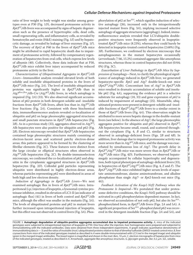
ratio of liver weight to body weight was similar among geno-types even at P30 (Fig. 1D), decreased proteasome activity inRpt2f/f;Alb livers was accompanied by signs of hepatic degener-ation such as the presence of hypertrophic cells, dead cells,small regenerating cells, and inflammatory cells, as revealed byhematoxylin and eosin (H&E) staining (Fig. 1E) and by hepato-cytic damage, as revealed by leakage of liver enzymes (Fig. 1F).The recovery of Rpt2 at P40 in the livers of Rpt2f/f;Alb micemight be attributed to rapid hepatocytic death due to impair-ment of proteasome activity, followed by compensatory regen-eration of hepatocytes from oval cells, which express low levelsof albumin (40). Collectively, these data indicate that at P30,Rpt2f/f;Alb mice exhibit liver injury accompanied by reducedproteasome activity in the liver.
Characterization of Ubiquitinated Aggregates in Rpt2f/f;AlbLivers—Immunoblot analysis revealed elevated levels of bothsoluble and insoluble ubiquitinated proteins in the livers ofRpt2f/f;Alb mice (Fig. 2A). The level of insoluble ubiquitinatedproteins was significantly higher in Rpt2f/f;Alb than inAtg7flox/flox;Alb-Cre (Atg7f/f;Alb) livers, in which autophagy isimpaired (Fig. 2A) (33). We also observed significant accumu-lation of p62 protein in both detergent-soluble and -insolublefractions from Rpt2f/f;Alb livers, albeit less than in Atg7f/f;Albliver fractions (Fig. 2A). Consistent with these biochemicaldata, immunofluorescence analysis revealed co-localization ofubiquitin and p62 on large pleomorphic aggregated structuresand small punctate structures in Rpt2f/f;Alb hepatocytes (Fig.2B). As in a previous study (33), aggregates observed in Atg7f/f;Alb hepatocytes were also positive for ubiquitin and p62 (Fig.2B). Electron microscopy revealed that Rpt2f/f;Alb hepatocytescontained large pleomorphic structures mainly consisting ofelectron-lucent areas and scattered patchy electron-denseareas; this pattern appeared to be formed by the clustering offibrillar elements (Fig. 2C). These features were distinct fromthe large circular or elliptical structures often observed inAtg7f/f;Alb hepatocytes (Fig. 2C). By double-immunoelectronmicroscopy, we confirmed the co-localization of p62 and ubiq-uitin in the cytoplasmic aggregated structures in Rpt2f/f;Albhepatocytes (Fig. 2D). Colloidal gold particles representingubiquitin were distributed in highly electron-dense areas,whereas particles representing p62 were distributed in areas ofboth high and low electron density.
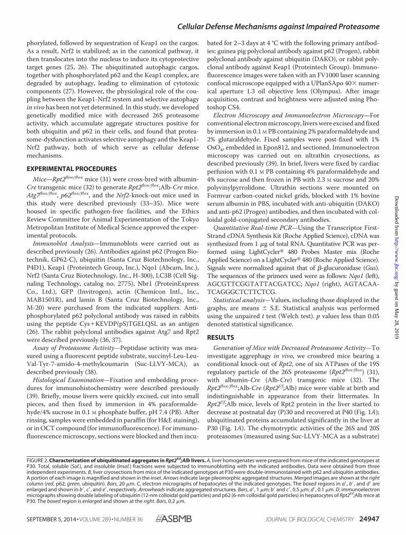
Induction of Aggrephagy in Rpt2f/f;Alb Livers—We nextexamined autophagic flux in livers of Rpt2f/f;Alb mice. Intra-peritoneal (i.p.) injection of leupeptin, a lysosomal cysteine pro-teinase inhibitor, resulted in elevation of LC3-II (an indicator ofautophagic flux) (41) in livers of both control and Rpt2f/f;Albmice, although the effect was smaller in the mutants (Fig. 3A).The levels of ubiquitinated proteins and p62 in mutant liversfurther increased upon intraperitoneal injection of leupeptin,but this effect was not observed in control livers (Fig. 3A). Phos-
phorylation of p62 at Ser351, which signifies induction of selec-tive autophagy (26), increased only in the intraperitoneallyinjected mutant livers (Fig. 3A), implying increased selectiveautophagy of aggregate structures (aggrephagy). Indeed, immu-nofluorescence analysis revealed that LC3/ubiquitin double-positive structures were frequently observed in leupeptin-treated Rpt2f/f;Alb mice (8.23%), whereas they could barely bedetected in leupeptin-treated control hepatocytes (2.68%) (Fig.3B). Furthermore, we confirmed by electron microscopy thatautophagosomes in the mutant hepatocytes occasionally(arrowheads; 7/46, 15.2%) contained aggregate-like amorphousstructures, whereas those in control hepatocytes did not (0/64;0%) (Fig. 3C).
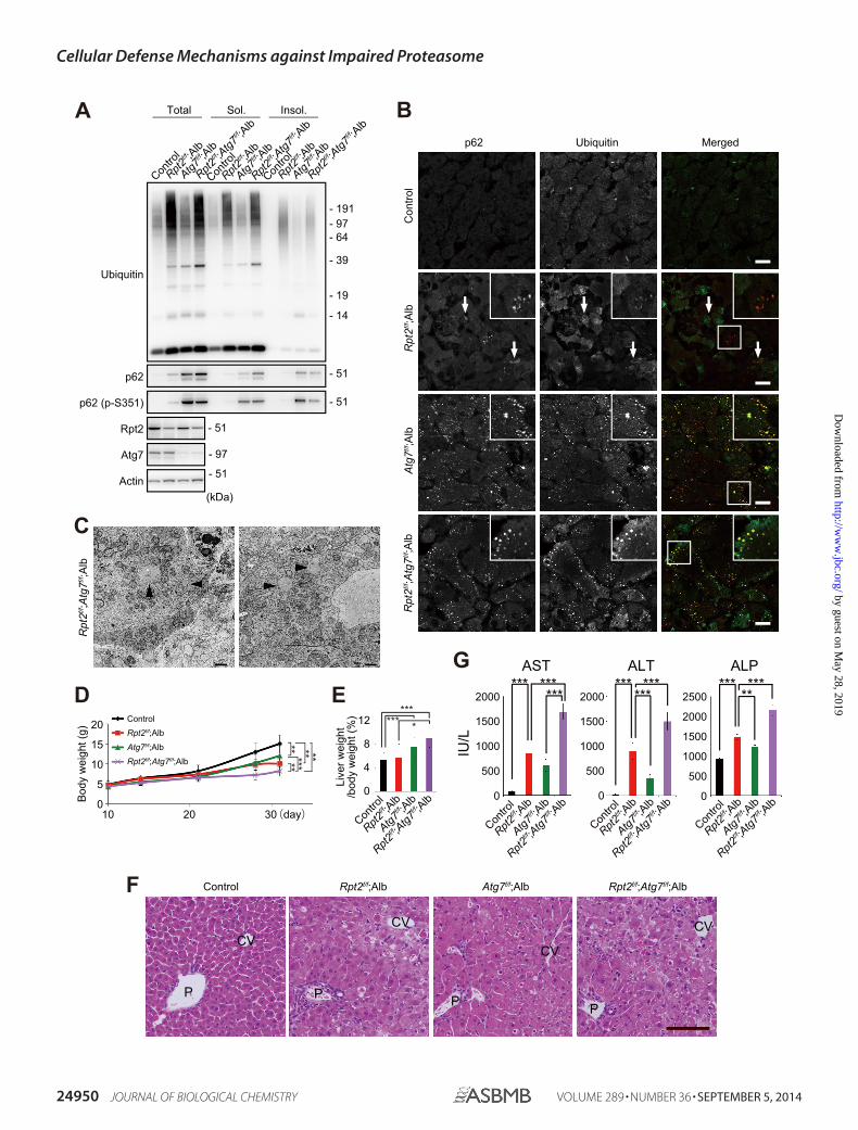
Exacerbation of Liver Pathology in Rpt2f/f;Alb Mice by Sup-pression of Autophagy—Next, to clarify the physiological signif-icance of autophagy induced in Rpt2f/f;Alb liver, we generatedhepatocyte-specific Rpt2 and Atg7 double-knock-out mice(Rpt2f/f;Atg7f/f;Alb). Concomitant loss of Atg7 in Rpt2f/f;Albmice resulted in dramatic accumulation of soluble and insolu-ble p62 (Fig. 4A), supporting the evidence p62 is a selectivesubstrate for autophagy (18, 33) and gene expression of p62 isinduced by impairment of autophagy (25). Meanwhile, ubiq-uitinated proteins were present in detergent-soluble and -insol-uble fractions of Rpt2f/f;Atg7f/f;Alb livers to a similar extent inthose of Rpt2f/f;Alb livers (Fig. 4A). This observation might beattributed to more severe hepatic damage in the double-mutantlivers (see below). In the absence of Atg7, the large pleomorphicaggregates positive for ubiquitin and p62 observed in Rpt2f/f;Alb hepatocytes became small, round, and scattered through-out the cytoplasm (Fig. 4, B and C), similar to structuresobserved in autophagy-deficient livers (Figs. 2B and 4B). Inaddition, liver damage due to impaired proteasome activity wasmore severe than in Atg7f/f;Alb mice, and the damage was exac-erbated by simultaneous loss of Atg7. The growth delay inRpt2f/f;Atg7f/f;Alb mice was more severe than in Rpt2f/f;Alb orAtg7f/f;Alb mice (Fig. 4D). Furthermore, we observed hepato-megaly accompanied by cellular hypertrophy and degenera-tion, both typical phenotypes of autophagy-deficient livers (33),in hepatocytes of Rpt2f/f;Atg7f/f;Alb mice (Fig. 4, E and F). TheRpt2f/f;Atg7f/f;Alb mice exhibited higher serum levels of aspar-tate aminotransferase, alanine aminotransferase, and alkalinephosphatase than single Atg7- or Rpt2-knock-out mice (Fig.4G).
Feedback Activation of the Keap1-Nrf2 Pathway when theProteasome Is Impaired—We postulated that under protea-some-defective conditions, the Keap1-Nrf2 pathway should beactivated in a p62 phosphorylation-dependent manner. In fact,we observed accumulation of not only p62, but also its Ser351-phosphorylated form, in Rpt2f/f;Alb livers (Figs. 2A and 5A). Asignificant proportion of Ser351-phosphorylated p62 was recov-ered in the detergent-insoluble fraction (Figs. 2A and 5A), and
FIGURE 3. Autophagic degradation of ubiquitin-positive aggregates accumulated due to impaired proteasome activity. A, mice of the indicatedgenotypes were subjected to intraperitoneal injection of leupeptin at P30. One hour after the injection, liver homogenates were prepared and subjected toimmunoblotting with the indicated antibodies. Data were obtained from three independent experiments. A graph indicates quantitative densitometry ofimmunoblotting data (n � 3) and the ratios of insoluble (Insol.) ubiquitinated proteins relative to that of dimethyl sulfoxide (DMSO)-treated control mice. B, livercryosections from mice of the indicated genotypes treated as described in A were double-immunostained with LC3 (red in the merged image) and ubiquitin(green in the merged image) antibodies. The boxed region is magnified and shown in the inset. Bars, 10 �m. C, electron micrographs of hepatocytes from miceof the indicated genotypes, treated as described in A. Arrowheads, aggregate-like amorphous structures. G, glycogen granules. Bar, 0.2 �m. Sol., soluble.
Cellular Defense Mechanisms against Impaired Proteasome
SEPTEMBER 5, 2014 • VOLUME 289 • NUMBER 36 JOURNAL OF BIOLOGICAL CHEMISTRY 24949
by guest on May 28, 2019
http://ww
w.jbc.org/
Dow
nloaded from
C
F
Actin
Atg7
p62
Rpt2
Ubiquitin
p62 (p-S351)
Contro
l
Rpt2f/f ;A
lb
Atg7f/f ;A
lb
Rpt2f/f ;Atg7f/f ;A
lb
Contro
l
Rpt2f/f ;A
lb
Atg7f/f ;A
lb
Rpt2f/f ;Atg7f/f ;A
lb
Contro
l
Rpt2f/f ;A
lb
Atg7f/f ;A
lb
Rpt2f/f ;Atg7f/f ;A
lbTotal Sol. Insol.
- 51
- 51
- 51
- 51
- 97
- 14
- 19
- 191- 97- 64
- 39
(kDa)
Rpt2f/f ;Atg7f/f ;A
lb
D
Bod
y w
eigh
t (g)
ControlRpt2f/f;Alb
Atg7f/f;Alb
Rpt2f/f;Atg7f/f;Alb
302010 (day)0
5
10
15
20
*******
** **
E
Contro
l
Rpt2f/f ;A
lb
Atg7f/f ;A
lb
Rpt2f/f ;Atg7f/f ;A
lb
4
8
12
0
Live
r wei
ght
/bod
y w
eigh
t (%
) ****** *
G
IU/L
ALT ALP
Contro
l
Rpt2f/f ;A
lb
Atg7f/f ;A
lb
Rpt2f/f ;Atg7f/f ;A
lb0
1000
500
2000
1500
0
1500
500
2000
1000
0
2500
2000
1000
AST***
1500
500
Contro
l
Rpt2f/f ;A
lb
Atg7f/f ;A
lb
Rpt2f/f ;Atg7f/f ;A
lb
Contro
l
Rpt2f/f ;A
lb
Atg7f/f ;A
lb
Rpt2f/f ;Atg7f/f ;A
lb
************
********* **
Con
trol
Rpt2f/f ;A
lb
p62 Ubiquitin Merged
Rpt2f/f ;Atg7f/f ;A
lbAtg7f/f ;A
lb
Control Rpt2f/f;Alb Rpt2f/f;Atg7f/f;AlbAtg7f/f;Alb
P P P P
CVCV
CV
CV
Cellular Defense Mechanisms against Impaired Proteasome
24950 JOURNAL OF BIOLOGICAL CHEMISTRY VOLUME 289 • NUMBER 36 • SEPTEMBER 5, 2014
by guest on May 28, 2019
http://ww
w.jbc.org/
Dow
nloaded from
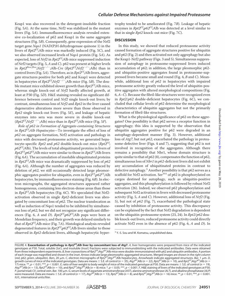
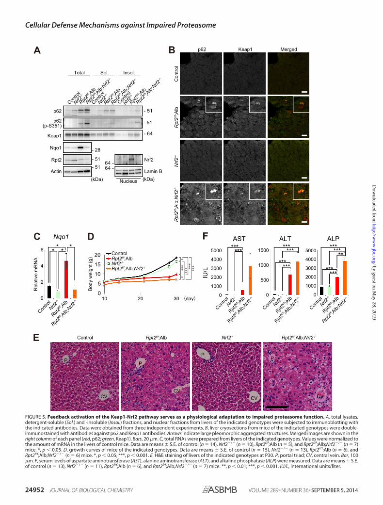
Keap1 was also recovered in the detergent-insoluble fraction(Fig. 5A). At the same time, Nrf2 was stabilized in the mutantlivers (Fig. 5A). Immunofluorescence analysis revealed exten-sive co-localization of p62 and Keap1 in the same aggregatestructures (Fig. 5B). Consequently, gene expression of the Nrf2target gene Nqo1 (NAD(P)H dehydrogenase quinone 1) in thelivers of Rpt2f/f;Alb mice was markedly induced (Fig. 5C), andwe also observed increased levels of Nqo1 protein (Fig. 5A). Asexpected, loss of Nrf2 in Rpt2f/f;Alb mice suppressed inductionof Nrf2 targets (Fig. 5, A and C). p62 was present at higher levelsin Rpt2flox/flox;Nrf2�/�;Alb-Cre (Rpt2f/f;Nrf2�/�;Alb) than incontrol livers (Fig. 5A). Therefore, as in Rpt2f/f;Alb livers, aggre-gate structures positive for both p62 and Keap1 were detectedin hepatocytes of Rpt2f/f;Nrf2�/�;Alb mice (Fig. 5B). The dou-ble mutant mice exhibited slower growth than Rpt2f/f;Alb mice,whereas single knock-out of Nrf2 hardly affected growth, atleast at P30 (Fig. 5D). H&E staining revealed no significant dif-ference between control and Nrf2 single knock-out livers; bycontrast, simultaneous loss of Nrf2 and Rpt2 in the liver causeddegenerative alterations more severe than those observed inRpt2 single knock-out livers (Fig. 5E), and leakage of hepaticenzymes into sera was more severe in double knock-out(Rpt2f/f;Nrf2�/�;Alb) mice than in Rpt2f/f;Alb mice (Fig. 5F).
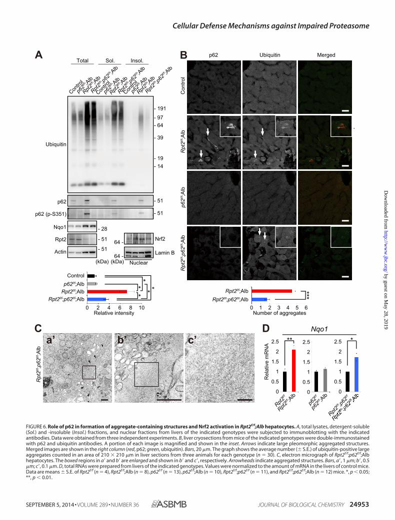
Role of p62 in Formation of Aggregate-containing Structuresin Rpt2f/f;Alb Hepatocytes—To investigate the effect of loss ofp62 on aggregate formation, Nrf2 activation and pathology inmice with decreased proteasome activity, we generated hepa-tocyte-specific Rpt2 and p62 double-knock-out mice (Rpt2f/f;p62f/f;Alb). The levels of total ubiquitinated proteins in livers ofRpt2f/f;p62f/f;Alb mice were similar to those in Rpt2f/f;Alb livers(Fig. 6A). The accumulation of insoluble ubiquitinated proteinsin Rpt2f/f;Alb mice was dramatically suppressed by loss of p62(Fig. 6A). Although the number of aggregates was reduced bydeletion of p62, we still occasionally detected large pleomor-phic aggregates positive for ubiquitin, even in Rpt2f/f;p62f/f;Albhepatocytes, by immunofluorescence staining (Fig. 6B). In elec-tron micrographs, the aggregated structures appeared ratherhomogeneous, containing less electron-dense areas than thosein Rpt2f/f;Alb hepatocytes (Fig. 6C). We speculated that Nrf2-activation observed in single Rpt2-deficient livers was abro-gated by concomitant loss of p62. The nuclear translocation aswell as induction of Nqo1 tended to be inhibited by simultane-ous loss of p62, but we did not recognize any significant differ-ences (Fig. 6, A and D). Rpt2f/f;p62f/f;Alb pups were born atMendelian frequency, and their growth was delayed similarly tothat of Rpt2f/f;Alb mice (Fig. 7A). Histological analysis revealeddegenerated features in Rpt2f/f;p62f/f;Alb livers similar to thoseobserved in Rpt2-deficient livers, although hepatocytic hyper-
trophy tended to be ameliorated (Fig. 7B). Leakage of hepaticenzymes in Rpt2f/f;p62f/f;Alb was detected at a level similar tothat in single Rpt2 knock-out mice (Fig. 7C).
DISCUSSION
In this study, we showed that reduced proteasome activitycaused formation of aggregate structures positive for ubiquitinand p62 (Fig. 2) and then activated not only aggrephagy but alsothe Keap1-Nrf2 pathway (Figs. 3 and 5). Simultaneous suppres-sion of autophagy in proteasome-suppressed livers inducedaccumulation of p62; in addition, the large pleomorphic p62-and ubiquitin-positive aggregates found in proteasome-sup-pressed livers became small and round (Fig. 4, B and C). Mean-while, additional loss of p62 in hepatocytes with impairedproteasome activity greatly reduced the level of ubiquitin-pos-itive aggregates with altered morphological compositions (Fig.6, A–C). Because the fibril-like structures were recognized evenin Rpt2/p62 double-deficient hepatocytes (Fig. 6C), we con-cluded that cellular levels of p62 determine the morphologicalcharacteristics of ubiquitin aggregates but not the primarilyformation of fibril-like structures.
What is the physiological significance of p62 on these aggre-gates? One possibility is that p62 serves a receptor function inaggrephagy; this idea is supported by the observation thatubiquitin aggregates positive for p62 were degraded in anautophagy-dependent manner (Fig. 3). However, additionalloss of Atg7, but not p62, exacerbated the pathology in protea-some-defective liver (Figs. 4 and 7), suggesting that p62 is notinvolved in recognition of the aggregates. Although thereremains a possibility that Nbr1, whose domain structure isquite similar to that of p62 (8), compensates the function of p62,simultaneous loss of Nbr1 in p62-deficient livers did not exhibitany accumulation of ubiquitinated proteins in contrast todefective autophagy.3 Another possibility is that p62 serves as ascaffold for Nrf2 activation. Ser351 of p62 is phosphorylated oncargos destined for autophagy, such as ubiquitin-positiveaggregates, and this phosphorylation is followed by robust Nrf2activation (26). Indeed, we observed p62 phosphorylation andsubsequent Nrf2 activation in livers with decreased proteasomeactivity (Fig. 5, A and C). However, additional loss of Nrf2 (Fig.5), but not of p62 (Fig. 7), exacerbated the pathological statecaused by inhibition of proteasome activity. This discrepancycan be explained by the fact that Nrf2 degradation is dependenton the ubiquitin-proteasome system (23, 24). In Rpt2/p62 dou-ble knock-out livers, reduced proteasome activity could directlyactivate Nrf2 even in the absence of p62 (Fig. 6, A and D). In
3 Y.-S. Sou and M. Komatsu, unpublished data.
FIGURE 4. Exacerbation of pathology in Rpt2f/f;Alb liver by concomitant loss of Atg7. A, liver homogenates were prepared from mice of the indicatedgenotypes at P30. Total, soluble (Sol.), and insoluble (Insol.) fractions were subjected to immunoblotting with the indicated antibodies. Data were obtainedfrom three independent experiments. B, liver cryosections of Rpt2f/f;Atg7f/f;Alb mice were double-immunostained with p62 and ubiquitin antibodies. A portionof each image was magnified and shown in the inset. Arrows indicate large pleomorphic aggregated structures. Merged images are shown in the right column(red, p62; green, ubiquitin). Bars, 20 �m. C, electron micrographs of Rpt2f/f;Atg7f/f;Alb hepatocytes. Arrowheads indicate aggregated structures. Bar, 1 �m. D,growth curves of mice of the indicated genotypes. Data are means � S.E. of control (n � 35), Atg7f/f;Alb (n � 22), Rpt2f/f;Alb (n � 10), and Rpt2f/f;Atg7f/f;Alb (n �17) mice. **, p � 0.01; ***, p � 0.001. E, ratio of liver weight to body weight of mice of the indicated genotype at P30. Data are means � S.E. of control (n � 19),Atg7f/f;Alb (n � 13), Rpt2f/f;Alb (n � 10), and Rpt2f/f;Atg7f/f;Alb (n � 8) mice. *, p � 0.05; ***, p � 0.001. F, H&E staining of livers of the indicated genotypes at P30.P, portal triad; CV, central vein. Bar, 100 �m. G, serum levels of aspartate aminotransferase (AST), alanine aminotransferase (ALT), and alkaline phosphatase (ALP)were measured. Data are means � S.E. of control (n � 17), Atg7f/f;Alb (n � 13), Rpt2f/f;Alb (n � 8), and Rpt2f/f;Atg7f/f;Alb (n � 16) mice. **, p � 0.01; ***, p � 0.001.IU/L, international units/liter.
Cellular Defense Mechanisms against Impaired Proteasome
SEPTEMBER 5, 2014 • VOLUME 289 • NUMBER 36 JOURNAL OF BIOLOGICAL CHEMISTRY 24951
by guest on May 28, 2019
http://ww
w.jbc.org/
Dow
nloaded from
C
Contro
l
Rpt2f/f ;A
lb
Rpt2f/f ;A
lb;Nrf2-/-
Nrf2-/-
0
2
6
4
Nqo1
Rel
ativ
e m
RN
A
* ** *
DControlRpt2f/f;Alb
302010
5
10
15
(day)
Bod
y w
eigh
t (g)
20
0
Rpt2f/f;Alb;Nrf2-/-Nrf2-/- ***
*
* *** ******
F AST ALT ALP
IU/L
0
1000
3000
2000
5000
4000
0
500
1500
1000
0
2000
5000
3000
Contro
l
Rpt2f/f ;A
lb
Rpt2f/f ;A
lb;Nrf2-/-
4000
Nrf2-/-
Contro
l
Rpt2f/f ;A
lb
Rpt2f/f ;A
lb;Nrf2-/-
Nrf2-/-
Contro
l
Rpt2f/f ;A
lb
Rpt2f/f ;A
lb;Nrf2-/-
Nrf2-/-
********
******
******
******
******
1000
E
BA
- 51- 51
- 28
(kDa)Actin
Nqo1
p62
Rpt2
p62(p-S351)
Contro
l
Nrf2-/-
Rpt2f/f ;A
lb
Rpt2f/f ;A
lb;Nrf2-/-
Contro
l
Nrf2-/-
Rpt2f/f ;A
lb
Rpt2f/f ;A
lb;Nrf2-/-
Contro
l
Nrf2-/-
Rpt2f/f ;A
lb
Rpt2f/f ;A
lb;Nrf2-/-
Total Sol. Insol.
- 51
- 51
Keap1 - 64
Nrf2
Lamin B
Nucleus
64 -64 -
(kDa)C
ontro
lRpt2f/f ;A
lbRpt2f/f ;A
lb;Nrf2
-/-Nrf2
-/-
p62 Keap1 Merged
Control Rpt2f/f;Alb Rpt2f/f;Alb;Nrf2-/-Nrf2-/-
PP
PP
CV CVCV
CV
FIGURE 5. Feedback activation of the Keap1-Nrf2 pathway serves as a physiological adaptation to impaired proteasome function. A, total lysates,detergent-soluble (Sol.) and -insoluble (Insol.) fractions, and nuclear fractions from livers of the indicated genotypes were subjected to immunoblotting withthe indicated antibodies. Data were obtained from three independent experiments. B, liver cryosections from mice of the indicated genotypes were double-immunostained with antibodies against p62 and Keap1 antibodies. Arrows indicate large pleomorphic aggregated structures. Merged images are shown in theright column of each panel (red, p62; green, Keap1). Bars, 20 �m. C, total RNAs were prepared from livers of the indicated genotypes. Values were normalized tothe amount of mRNA in the livers of control mice. Data are means � S.E. of control (n � 14), Nrf2�/� (n � 10), Rpt2f/f;Alb (n � 5), and Rpt2f/f;Alb;Nrf2�/� (n � 7)mice. *, p � 0.05. D, growth curves of mice of the indicated genotypes. Data are means � S.E. of control (n � 15), Nrf2�/� (n � 13), Rpt2f/f;Alb (n � 6), andRpt2f/f;Alb;Nrf2�/� (n � 6) mice. *, p � 0.05; ***, p � 0.001. E, H&E staining of livers of the indicated genotypes at P30. P, portal triad; CV, central vein. Bar, 100�m. F, serum levels of aspartate aminotransferase (AST), alanine aminotransferase (ALT), and alkaline phosphatase (ALP) were measured. Data are means � S.E.of control (n � 13), Nrf2�/� (n � 11), Rpt2f/f;Alb (n � 6), and Rpt2f/f;Alb;Nrf2�/� (n � 7) mice. **, p � 0.01; ***, p � 0.001. IU/L, international units/liter.
Cellular Defense Mechanisms against Impaired Proteasome
24952 JOURNAL OF BIOLOGICAL CHEMISTRY VOLUME 289 • NUMBER 36 • SEPTEMBER 5, 2014
by guest on May 28, 2019
http://ww
w.jbc.org/
Dow
nloaded from
A B
DC
Con
trol
Rpt2f/f ;A
lb
p62 Ubiquitin Merged
Rpt2f/f ;p62
f/f;A
lbp62f/f ;A
lb
Rpt2f/f ;p62
f/f;A
lb
a’ b’ c’
- 51
- 51
- 14- 19
- 191
- 97- 64
- 39
Actin
p62
Rpt2
Ubiquitin
p62 (p-S351)
Contro
l
p62f/f ;A
lb
Rpt2f/f ;A
lb
Rpt2f/f ;p62f/f ;A
lb
Contro
l
p62f/f ;A
lb
Rpt2f/f ;A
lb
Rpt2f/f ;p62f/f ;A
lb
Contro
l
p62f/f ;A
lb
Rpt2f/f ;A
lb
Rpt2f/f ;p62f/f ;A
lbTotal Sol. Insol.
- 51
- 51
(kDa)
Nrf2
Lamin B(kDa)
64 -
64 -Nuclear
Nqo1 - 28
Rel
ativ
e m
RN
A
p62f/f ;A
lbp62f/f
Rpt2f/f ;A
lb
Rpt2f/f
Rpt2f/f ;p62f/f
0
1
2.5
1.5
0.5
2
0
1
2.5
1.5
0.5
2
0
1.5
2.5
0.5
** *Nqo1
2
1
**
* * *
100 2 4 6 8Relative intensity
Control
Rpt2f/f;Albp62f/f;Alb
Rpt2f/f;p62f/f;Alb ***Rpt2f/f;Alb
Rpt2f/f;p62f/f;Alb
Number of aggregates60 421 3 5
FIGURE 6. Role of p62 in formation of aggregate-containing structures and Nrf2 activation in Rpt2f/f;Alb hepatocytes. A, total lysates, detergent-soluble(Sol.) and -insoluble (Insol.) fractions, and nuclear fractions from livers of the indicated genotypes were subjected to immunoblotting with the indicatedantibodies. Data were obtained from three independent experiments. B, liver cryosections from mice of the indicated genotypes were double-immunostainedwith p62 and ubiquitin antibodies. A portion of each image is magnified and shown in the inset. Arrows indicate large pleomorphic aggregated structures.Merged images are shown in the right column (red, p62; green, ubiquitin). Bars, 20 �m. The graph shows the average number (� S.E.) of ubiquitin-positive largeaggregates counted in an area of 210 � 210 �m in liver sections from three animals for each genotype (n � 30). C, electron micrograph of Rpt2f/f;p62f/f;Albhepatocytes. The boxed regions in a� and b� are enlarged and shown in b� and c�, respectively. Arrowheads indicate aggregated structures. Bars, a�, 1 �m; b�, 0.5�m; c�, 0.1 �m. D, total RNAs were prepared from livers of the indicated genotypes. Values were normalized to the amount of mRNA in the livers of control mice.Data are means � S.E. of Rpt2f/f (n � 4), Rpt2f/f;Alb (n � 8), p62f/f (n � 13), p62f/f;Alb (n � 10), Rpt2f/f;p62f/f (n � 11), and Rpt2f/f;p62f/f;Alb (n � 12) mice. *, p � 0.05;**, p � 0.01.
Cellular Defense Mechanisms against Impaired Proteasome
SEPTEMBER 5, 2014 • VOLUME 289 • NUMBER 36 JOURNAL OF BIOLOGICAL CHEMISTRY 24953
by guest on May 28, 2019
http://ww
w.jbc.org/
Dow
nloaded from

other words, the effect of phosphorylated p62 on Nrf2 activa-tion might be hidden by the robust activation of Nrf2 thatoccurs in response to impairment of the ubiquitin-proteasomesystem. In conclusion, our data show for the first time that bothelimination of aggregate structures by autophagy and activationof Nrf2 under proteasome-defective conditions serve as physi-ological adaptations to impaired proteasome function in vivo.
Acknowledgments—We thank Y. Yang (Tokyo Metropolitan Instituteof Medical Science) for excellent technical assistance and K. Kannoand A. Yabashi (Fukushima Medical University School of Medicine)for assistance with histological studies.
REFERENCES1. Goldberg, A. L. (2003) Protein degradation and protection against mis-
folded or damaged proteins. Nature 426, 895– 8992. Finley, D. (2009) Recognition and processing of ubiquitin-protein conju-
gates by the proteasome. Annu. Rev. Biochem. 78, 477–5133. Murata, S., Yashiroda, H., and Tanaka, K. (2009) Molecular mechanisms
of proteasome assembly. Nat. Rev. Mol. Cell Biol. 10, 104 –1154. Mizushima, N., and Levine, B. (2010) Autophagy in mammalian develop-
ment and differentiation. Nat Cell Biol. 12, 823– 8305. Mizushima, N., and Komatsu, M. (2011) Autophagy: renovation of cells
and tissues. Cell 147, 728 –7416. Rubinsztein, D. C. (2006) The roles of intracellular protein-degradation
pathways in neurodegeneration. Nature 443, 780 –7867. Rubinsztein, D. C., Mariño, G., and Kroemer, G. (2011) Autophagy and
aging. Cell 146, 682– 6958. Kirkin, V., McEwan, D. G., Novak, I., and Dikic, I. (2009) A role for ubiq-
uitin in selective autophagy. Mol. Cell 34, 259 –2699. Youle, R. J., and Narendra, D. P. (2011) Mechanisms of mitophagy. Nat.
Rev. Mol. Cell Biol. 12, 9 –1410. Deretic, V., and Levine, B. (2009) Autophagy, immunity, and microbial
adaptations. Cell Host Microbe 5, 527–54911. Pandey, U. B., Nie, Z., Batlevi, Y., McCray, B. A., Ritson, G. P., Nedelsky,
N. B., Schwartz, S. L., DiProspero, N. A., Knight, M. A., Schuldiner, O.,Padmanabhan, R., Hild, M., Berry, D. L., Garza, D., Hubbert, C. C., Yao,T. P., Baehrecke, E. H., and Taylor, J. P. (2007) HDAC6 rescues neurode-generation and provides an essential link between autophagy and the UPS.Nature 447, 859 – 863
12. Iwata, A., Christianson, J. C., Bucci, M., Ellerby, L. M., Nukina, N., Forno,L. S., and Kopito, R. R. (2005) Increased susceptibility of cytoplasmic overnuclear polyglutamine aggregates to autophagic degradation. Proc. Natl.Acad. Sci. U.S.A. 102, 13135–13140
13. Iwata, A., Riley, B. E., Johnston, J. A., and Kopito, R. R. (2005) HDAC6 andmicrotubules are required for autophagic degradation of aggregated hun-tingtin. J. Biol. Chem. 280, 40282– 40292
14. Narendra, D., Tanaka, A., Suen, D. F., and Youle, R. J. (2008) Parkin isrecruited selectively to impaired mitochondria and promotes their au-tophagy. J. Cell Biol. 183, 795– 803
15. Narendra, D. P., Jin, S. M., Tanaka, A., Suen, D. F., Gautier, C. A., Shen, J.,Cookson, M. R., and Youle, R. J. (2010) PINK1 is selectively stabilized onimpaired mitochondria to activate Parkin. PLoS Biol. 8, e1000298
16. Manzanillo, P. S., Ayres, J. S., Watson, R. O., Collins, A. C., Souza, G., Rae,C. S., Schneider, D. S., Nakamura, K., Shiloh, M. U., and Cox, J. S. (2013)The ubiquitin ligase parkin mediates resistance to intracellular pathogens.Nature 501, 512–516
17. Huett, A., Heath, R. J., Begun, J., Sassi, S. O., Baxt, L. A., Vyas, J. M.,Goldberg, M. B., and Xavier, R. J. (2012) The LRR and RING domainprotein LRSAM1 is an E3 ligase crucial for ubiquitin-dependent au-tophagy of intracellular Salmonella typhimurium. Cell Host Microbe 12,
B
AST ALT ALP
IU/L
0
500
1500
1000
2500
2000
0
400
800
600
0
200
500
300
Contro
l
Rpt2f/f ;A
lb
400
p62f/f ;A
lb
*******
100
Rpt2f/f ;p62f/f ;A
lb
Contro
l
Rpt2f/f ;A
lb
p62f/f ;A
lb
Rpt2f/f ;p62f/f ;A
lb
Contro
l
Rpt2f/f ;A
lb
p62f/f ;A
lb
Rpt2f/f ;p62f/f ;A
lb
200
**
***
**
********
*** ***
Bod
y w
eigh
t (g)
Controlp62f/f;Alb
Rpt2f/f;AlbRpt2f/f;p62f/f;Alb
302010 (day)0
5
10
15
20
********* ***
Control Rpt2f/f;Alb Rpt2f/f;p62f/f;Albp62f/f;Alb
P
CV
P
CV
P
CV
P
CV
FIGURE 7. Pathology in Rpt2f/f;Alb liver by concomitant loss of p62. A, growth curves of mice of the indicated genotypes. Data are means � S.E. of control(n � 44), p62f/f;Alb (n � 9), Rpt2f/f;Alb (n � 6), and Rpt2f/f;p62f/f;Alb (n � 15) mice. ***, p � 0.001. B, H&E staining of livers of the indicated genotypes at P30. P,portal triad; CV, central vein. Bar, 100 �m. C, serum levels of aspartate aminotransferase (AST), alanine aminotransferase (ALT), and alkaline phosphatase (ALP)were measured. Data are means � S.E. of control (n � 31), p62f/f;Alb (n � 5), Rpt2f/f;Alb (n � 5), and Rpt2f/f;p62f/f;Alb (n � 12) mice. *, p � 0.05; **, p � 0.01; ***,p � 0.001.
Cellular Defense Mechanisms against Impaired Proteasome
24954 JOURNAL OF BIOLOGICAL CHEMISTRY VOLUME 289 • NUMBER 36 • SEPTEMBER 5, 2014
by guest on May 28, 2019
http://ww
w.jbc.org/
Dow
nloaded from
778 –79018. Bjørkøy, G., Lamark, T., Brech, A., Outzen, H., Perander, M., Overvatn, A.,
Stenmark, H., and Johansen, T. (2005) p62/SQSTM1 forms protein aggre-gates degraded by autophagy and has a protective effect on huntingtin-induced cell death. J. Cell Biol. 171, 603– 614
19. Kirkin, V., Lamark, T., Sou, Y. S., Bjørkøy, G., Nunn, J. L., Bruun, J. A.,Shvets, E., McEwan, D. G., Clausen, T. H., Wild, P., Bilusic, I., Theurillat,J. P., Øvervatn, A., Ishii, T., Elazar, Z., Komatsu, M., Dikic, I., and Johansen,T. (2009) A role for NBR1 in autophagosomal degradation of ubiquiti-nated substrates. Mol. Cell 33, 505–516
20. Thurston, T. L., Ryzhakov, G., Bloor, S., von Muhlinen, N., and Randow, F.(2009) The TBK1 adaptor and autophagy receptor NDP52 restricts theproliferation of ubiquitin-coated bacteria. Nat. Immunol. 10, 1215–1221
21. Wild, P., Farhan, H., McEwan, D. G., Wagner, S., Rogov, V. V., Brady, N. R.,Richter, B., Korac, J., Waidmann, O., Choudhary, C., Dötsch, V., Bumann,D., and Dikic, I. (2011) Phosphorylation of the autophagy receptor op-tineurin restricts Salmonella growth. Science 333, 228 –233
22. Zatloukal, K., Stumptner, C., Fuchsbichler, A., Heid, H., Schnoelzer, M.,Kenner, L., Kleinert, R., Prinz, M., Aguzzi, A., and Denk, H. (2002) p62 Is acommon component of cytoplasmic inclusions in protein aggregation dis-eases. Am. J. Pathol. 160, 255–263
23. Hayes, J. D., and McMahon, M. (2009) NRF2 and KEAP1 mutations: per-manent activation of an adaptive response in cancer. Trends Biochem. Sci34, 176 –188
24. Jaramillo, M. C., and Zhang, D. D. (2013) The emerging role of the Nrf2-Keap1 signaling pathway in cancer. Genes Dev. 27, 2179 –2191
25. Komatsu, M., Kurokawa, H., Waguri, S., Taguchi, K., Kobayashi, A.,Ichimura, Y., Sou, Y. S., Ueno, I., Sakamoto, A., Tong, K. I., Kim, M.,Nishito, Y., Iemura, S., Natsume, T., Ueno, T., Kominami, E., Motohashi,H., Tanaka, K., and Yamamoto, M. (2010) The selective autophagy sub-strate p62 activates the stress responsive transcription factor Nrf2 throughinactivation of Keap1. Nat. Cell Biol. 12, 213–223
26. Ichimura, Y., Waguri, S., Sou, Y. S., Kageyama, S., Hasegawa, J., Ishimura,R., Saito, T., Yang, Y., Kouno, T., Fukutomi, T., Hoshii, T., Hirao, A.,Takagi, K., Mizushima, T., Motohashi, H., Lee, M. S., Yoshimori, T.,Tanaka, K., Yamamoto, M., and Komatsu, M. (2013) Phosphorylation ofp62 activates the Keap1-Nrf2 pathway during selective autophagy. Mol.Cell 51, 618 – 631
27. Taguchi, K., Fujikawa, N., Komatsu, M., Ishii, T., Unno, M., Akaike, T.,Motohashi, H., and Yamamoto, M. (2012) Keap1 degradation by au-tophagy for the maintenance of redox homeostasis. Proc. Natl. Acad. Sci.U.S.A. 109, 13561–13566
28. Bae, S. H., Sung, S. H., Oh, S. Y., Lim, J. M., Lee, S. K., Park, Y. N., Lee, H. E.,Kang, D., and Rhee, S. G. (2013) Sestrins activate Nrf2 by promoting p62-dependent autophagic degradation of Keap1 and prevent oxidative liverdamage. Cell Metab. 17, 73– 84
29. Matsumoto, G., Wada, K., Okuno, M., Kurosawa, M., and Nukina, N.(2011) Serine 403 phosphorylation of p62/SQSTM1 regulates selectiveautophagic clearance of ubiquitinated proteins. Mol. Cell 44, 279 –289
30. Pilli, M., Arko-Mensah, J., Ponpuak, M., Roberts, E., Master, S., Mandell,M. A., Dupont, N., Ornatowski, W., Jiang, S., Bradfute, S. B., Bruun, J. A.,Hansen, T. E., Johansen, T., and Deretic, V. (2012) TBK-1 promotes au-
tophagy-mediated antimicrobial defense by controlling autophagosomematuration. Immunity 37, 223–234
31. Bedford, L., Hay, D., Devoy, A., Paine, S., Powe, D. G., Seth, R., Gray, T.,Topham, I., Fone, K., Rezvani, N., Mee, M., Soane, T., Layfield, R., Shep-pard, P. W., Ebendal, T., Usoskin, D., Lowe, J., and Mayer, R. J. (2008)Depletion of 26S proteasomes in mouse brain neurons causes neurode-generation and Lewy-like inclusions resembling human pale bodies.J. Neurosci. 28, 8189 – 8198
32. Postic, C., Shiota, M., Niswender, K. D., Jetton, T. L., Chen, Y., Moates,J. M., Shelton, K. D., Lindner, J., Cherrington, A. D., and Magnuson, M. A.(1999) Dual roles for glucokinase in glucose homeostasis as determined byliver and pancreatic beta cell-specific gene knock-outs using Cre recom-binase. J. Biol. Chem. 274, 305–315
33. Komatsu, M., Waguri, S., Koike, M., Sou, Y. S., Ueno, T., Hara, T., Miz-ushima, N., Iwata, J., Ezaki, J., Murata, S., Hamazaki, J., Nishito, Y., Iemura,S., Natsume, T., Yanagawa, T., Uwayama, J., Warabi, E., Yoshida, H., Ishii,T., Kobayashi, A., Yamamoto, M., Yue, Z., Uchiyama, Y., Kominami, E.,and Tanaka, K. (2007) Homeostatic levels of p62 control cytoplasmic in-clusion body formation in autophagy-deficient mice. Cell 131, 1149 –1163
34. Harada, H., Warabi, E., Matsuki, T., Yanagawa, T., Okada, K., Uwayama, J.,Ikeda, A., Nakaso, K., Kirii, K., Noguchi, N., Bukawa, H., Siow, R. C., Mann,G. E., Shoda, J., Ishii, T., and Sakurai, T. (2013) Deficiency of p62/seques-tosome 1 causes hyperphagia due to leptin resistance in the brain. J. Neu-rosci. 33, 14767–14777
35. Itoh, K., Chiba, T., Takahashi, S., Ishii, T., Igarashi, K., Katoh, Y., Oyake, T.,Hayashi, N., Satoh, K., Hatayama, I., Yamamoto, M., and Nabeshima, Y.(1997) An Nrf2/small Maf heterodimer mediates the induction of phase IIdetoxifying enzyme genes through antioxidant response elements.Biochem. Biophys. Res. Commun. 236, 313–322
36. Tanida, I., Mizushima, N., Kiyooka, M., Ohsumi, M., Ueno, T., Ohsumi, Y.,and Kominami, E. (1999) Apg7p/Cvt2p: a novel protein-activating en-zyme essential for autophagy. Mol. Biol. Cell 10, 1367–1379
37. Kaneko, T., Hamazaki, J., Iemura, S., Sasaki, K., Furuyama, K., Natsume,T., Tanaka, K., and Murata, S. (2009) Assembly pathway of the Mamma-lian proteasome base subcomplex is mediated by multiple specific chap-erones. Cell 137, 914 –925
38. Komatsu, M., Waguri, S., Chiba, T., Murata, S., Iwata, J., Tanida, I., Ueno,T., Koike, M., Uchiyama, Y., Kominami, E., and Tanaka, K. (2006) Loss ofautophagy in the central nervous system causes neurodegeneration inmice. Nature 441, 880 – 884
39. Waguri, S., and Komatsu, M. (2009) Biochemical and morphological de-tection of inclusion bodies in autophagy-deficient mice. Methods Enzy-mol. 453, 181–196
40. Wang, E. Y., Yeh, S. H., Tsai, T. F., Huang, H. P., Jeng, Y. M., Lin, W. H.,Chen, W. C., Yeh, K. H., Chen, P. J., and Chen, D. S. (2011) Depletion ofbeta-catenin from mature hepatocytes of mice promotes expansion ofhepatic progenitor cells and tumor development. Proc. Natl. Acad. Sci.U.S.A. 108, 18384 –18389
41. Kabeya, Y., Mizushima, N., Ueno, T., Yamamoto, A., Kirisako, T., Noda,T., Kominami, E., Ohsumi, Y., and Yoshimori, T. (2000) LC3, a mamma-lian homologue of yeast Apg8p, is localized in autophagosome mem-branes after processing. EMBO J. 19, 5720 –5728
Cellular Defense Mechanisms against Impaired Proteasome
SEPTEMBER 5, 2014 • VOLUME 289 • NUMBER 36 JOURNAL OF BIOLOGICAL CHEMISTRY 24955
by guest on May 28, 2019
http://ww
w.jbc.org/
Dow
nloaded from
Masayuki Yamamoto, Satoshi Waguri, Keiji Tanaka and Masaaki KomatsuRyosuke Ishimura, Tsuguka Kouno, Lynn Bedford, R. John Mayer, Myung-Shik Lee, Shun Kageyama, Yu-shin Sou, Takefumi Uemura, Satoshi Kametaka, Tetsuya Saito,
Proteasome Dysfunction Activates Autophagy and the Keap1-Nrf2 Pathway
doi: 10.1074/jbc.M114.580357 originally published online July 21, 20142014, 289:24944-24955.J. Biol. Chem.
10.1074/jbc.M114.580357Access the most updated version of this article at doi:
Alerts:
When a correction for this article is posted•
When this article is cited•
to choose from all of JBC's e-mail alertsClick here
http://www.jbc.org/content/289/36/24944.full.html#ref-list-1
This article cites 41 references, 13 of which can be accessed free at
by guest on May 28, 2019
http://ww
w.jbc.org/
Dow
nloaded from


![Review ArticleInternational Journal of Alzheimer’s Disease 3 [57–59], it is now understood that the main function of Keap1 is to serve as an adapter for the Cullin3/Ring Box 1](https://img.pdfslide.tips/doc/110x75/60d2e83d298ce44822433153/review-article-international-journal-of-alzheimeras-disease-3-57a59-it-is.jpg)
![PDFѹËõÆ÷ · Diabetes Complications 2015; 29(2): 222-229. [13]Singh K, Agrawal NK, Gupta SK, Mohan G, Chatuwedi S, Singh K. Increased expression of endosomal members of toll-](https://img.pdfslide.tips/doc/110x75/5f33a5bc99af1a5a681d325a/pdf-diabetes-complications-2015-292-222-229-13singh-k-agrawal.jpg)