Embed Size (px)
Citation preview
Instructions for use
Title Endosomal escape and the knockdown efficiency of liposomal-siRNA by the fusogenic peptide shGALA
Author(s) Sakurai, Yu; Hatakeyama, Hiroto; Sato, Yusuke; Akita, Hidetaka; Takayama, Kentaro; Kobayashi, Sachiko; Futaki,Shiroh; Harashima, Hideyoshi
Citation Biomaterials, 32(24): 5733-5742
Issue Date 2011-08
Doc URL http://hdl.handle.net/2115/46877
Type article (author version)
File Information Bio32-24_5733-5742.pdf
Hokkaido University Collection of Scholarly and Academic Papers : HUSCAP
Title
Endosomal Escape and the Knockdown Efficiency of Liposomal-siRNA by the Fusogenic Peptide shGALA.
Author
Yu Sakurai1, Hiroto Hatakeyama1, Yusuke Sato1, Hidetaka Akita1, Kentaro Takayama2, Sachiko Kobayashi2, Shiroh Futaki2,
Hideyoshi Harashima1, *
1 Faculty of Pharmaceutical Sciences, Hokkaido University, Kita-12, Nishi-6, Kita-ku, Sapporo 060-0812, Japan
2 Institute for Chemical Research, Kyoto University, Uji, Kyoto 611-0011, Japan
*To whom correspondence should be addressed.
Hideyoshi Harashima, Ph.D.,Professor of Laboratory for Molecular Design of Pharmaceutics, Faculty of
Pharmaceutical Sciences, Hokkaido University, Kita-12, Nishi-6, Sapporo 060-0812, Japan.
Tel.: 81-11-706-3919; Fax: 81-11-706-4879;
E-mail: [email protected]
We wish it to be known that the first two authors should be regarded as joint First Authors.
Abstract
An siRNA that specifically silences the expression of mRNA is a potential therapeutic agent for dealing with many
diseases including cancer. However, the poor cellular uptake and bioavailability of siRNA remains a major obstacle to
clinical development. For efficient delivery to tumor tissue, the pharmacokinetics and intracellular trafficking of siRNA
must be rigorously controlled. To address this issue, we developed a liposomal siRNA carrier, a multi-functional nano
device (MEND). We describe herein an approach for systemic siRNA delivery to tumors by combining the MEND system
with shGALA, a fusogenic peptide. In cultured cell experiments, shGALA-modification enhanced the endosomal escape of
siRNA encapsulated in a polyethylene glycol modified MEND (PEG-MEND), resulting in an 82% knockdown of the target
gene. In vivo systemic administration clarified that the shGALA-modified MEND (shGALA-MEND) showed 58% gene
silencing in tumor tissues at a dose of 4 mg of siRNA/kg body weight. In addition, a significant inhibition of tumor growth
was observed only for the shGALA-MEND and no somatic or hepatic toxicity was observed. Given the above data, this
peptide-modified delivery system, a shGALA-MEND has great potential for the systemic delivery of therapeutic siRNA
aimed at cancer therapy.
1. Introduction
RNA interference (RNAi) with small interfering RNA (siRNA) holds great potential for the treatment of many diseases[1,
2]. The advantage of RNAi lies on its high specificity and potent gene silencing ability, associated with the fact that it can
potentially target any gene. However, the systemic administration of siRNA for the clinical applications is prohibited[3].
This is because siRNA cannot pass through the plasma membrane due to its hydrophilicity and large size, and as a result,
siRNA cannot be taken up by target cells. In addition, when administered into the blood circulation without appropriate
chemical modification, siRNA is immediately excreted via the urine or degradated by the action of Ribonuclease
(RNase)[4], which is abundant in all body fluids[5]. Thus, a number of attempts to develop various types of carrier systems
for siRNA delivery, such as lipoplexes and polyplexes have been reported [6-8]. We recently reported on the development of
a unique siRNA delivery system, a multi-functional envelope type nano device (MEND)[9].
It is generally accepted that the higher permeability of tumor vessels is due to the presence of thin-walled vascular
endothelial cells that are adjacent to tumor tissues. This results in the poor removal of interstitial fluid within tumors and an
increase in the passive accumulation of macromolecules within tumors, a situation that is referred to as the enhanced
permeability and retention (EPR) effect[10, 11]. The EPR effect allows siRNA carriers to transit to tumor tissues. However,
the EPR effect is holds only in cases involving macromolecules that have a long systemic retention time. PEGylation is
currently the most popular strategy for enabling siRNA carriers to circulate in the bloodstream for a long period and to
accumulate in tumor tissues[12]. However, PEG-modification causes a marked decrease in the endosomal escape of nano
carriers after being taken up by cells via endocytosis, which results in the loss of efficiency in transferring its cargo to the
cytosol[13, 14]. Namely, PEG-modification is associated with conflicting issues, namely, the improvement of
pharmacokinetics and the deterioration of intracellular traficking of carriers[15].
To address this dilemma, many groups have developed a functional PEG, such as a PEG that is cleavable in acidic [16] or
reducing conditions [17, 18]. We previously developed the PEG-lipid cleavable by matrix metalloproteinase, which is
highly excreted from cancer cells and reported the systemic administration with PPD modified MEND (PPD-MEND)
demonstrated a significant marker gene silencing in tumor tissue. [19]
In this study, aimed at developing the new delivery system to overcome the contradiction even with PEG ,we proposed an
approach involving the use of a fusogenic peptide, GALA (WEAALAEALAEALAEHLAEALAEALEALAA) [20]. A 30
amino acid GALA contains a glutamic acid-alanine-leucine-alanine sequence that is repeated 4 times. Since the carboxyl
groups of glutamic acid are negatively charged at a physiological pH, electric repulsion between these groups results in
GALA with a random coil structure. In contrast, at an acidic pH (around 5.5) protonation of the side chain of carboxyl
groups of the glutamic acids dissipates electric repulsion. As a result, the GALA structure changes into an -helix, a
structure that tends to induce membrane fusion[21]. In other words, while GALA-modification has no effect on the
membrane stability of MENDs at a physiologic pH, GALA on the surface of a MEND has the ability to induce membrane
fusion between the MEND and the endosomal membrane in endosome, the pH of which is ~5.5. In a previous study, we
applied the GALA-modified MEND to siRNA delivery in vitro[22] and in an in vivo intratumoral injection model[23].
However, when injected into the blood circulation, the GALA-modified PEG-MEND (GALA-MEND) was eliminated
rapidly. We assumed that this decline was caused by the recognision by biomaclomolecules. Therefore we developed a new
shorter version GALA (shGALA), aimed to be masked by the aqueous layer formed by PEG-modification onto the MEND
and result in avoiding recognition (Fig. 1A). In this report, we report on an evaluation of the effects of
shGALA-modification on endosomal escape and the knockdown effect of PEG-MEND both in vitro and in vivo.
2. Materials & Methods
2.1. Materials
Anti-ACTB siRNA (21-mer, sense strand: 5’-CUG ACU UGA GAC CAG UUG AdTdT-3’) and anti-luciferase (luc)
siRNA (21-mer, sense strand: 5’-GCG CUG CUG GUG CCA ACC GdTdT-3’) were obtained from Sigma (St. Louis,
MO, U. S. A.). Stearyl-octaarginine (STR-R8) was synthesized as described previously[24].
1,2-dioleoyl-sn-glycero-3-phosphoethanolamine (DOPE), 1,2-dioleoyl-3-trimethylammonium-propane (DOTAP),
cholesterol (chol) and distearoyl-sn-glycero-3-phoshoethanolamine-N-[methoxy (polyethylene glycol)-2000] (PEG-DSPE)
were purchased from AVANTI Polar Lipids (Alabaster, AL, U. S. A.). Cholesterol-GALA and -shGALA were synthesized as
described previously[25]. Dulbecco’s modified Eagle medium (DMEM) and fetal bovine serum (FBS), were purchased
from Invitrogen (Carlsbad, CA). DEPC-treated water and G418 were obtained from Nacalai tesque (Kyoto, Japan). HeLa
human cerviacal carcinoma cells and HT1080 human fibrosarcoma cells were obtained from the RIKEN Cell Bank
(Tsukuba, Japan). [3H]-cholesteryl hexadecyl ether (CHE) and [-32P]-adenosine 5'-triphosphate (ATP) were purchased from
PerkinElmer Life Sciences, Japan (Tokyo, Japan). All other chemicals were commercially available reagent-grade products.
2.2 Expreimental animals
Male BALB/cAJcl nude mice and male ICR mice were purchased from CLEA (Tokyo, Japan) and Japan SLC (Shizuoka,
Japan), respectively. The experimental protocols were reviewed and approved by the Hokkaido University Animal Care
Committee in accordance with the guidelines for the care and use of laboratory animals.
2.3. Preaparation and characterization of siRNA-encapsulated MENDs
siRNA (0.4 mg/mL) was complexed with STR-R8 (0.2 mg/mL)[9], at a nitrogen/phosphate ratio of 1.7, in 350 L of 10
mM HEPES buffer (pH 7.4). A lipid film was formed by the evaporation of a chloroform/EtOH (1/1) solution containing
DOTAP, DOPE, chol and PEG-DSPE (745 nmol total lipids in 30:40:30:1 molar ratio). The lipid films for the MENDs

modified with cholesteryl-GALA or shGALA were prepared by evaporation using predetermined amounts of chol-GALA or
-shGALA with the other lipids. The siRNA/STR-R8 complex was added to the lipid film, followed by incubation for 10 min
at room temperature to hydrate the lipids. To encapsulate the siRNA/STR-R8 complex with the lipid, the lipid film was then
sonicated for approximately 1 min in a bath-type sonicator. The prepared MEND was then incubated at 60°C for 1 hr with
DSPE-PEG at 10 mol% total lipid. The average diameter and zeta-potential of the complexed siRNA and MENDs were
determined using a Zetasizer Nano ZS ZEN3600 (MALVERN Instrument, Worchestershire, UK)
2.4. Stability of siRNA encapsulated in MENDs in serum
siRNA duplexes and MENDs were incubated at 37°C at a 1:1 volume ratio with fetal FBS diluted in HEPES buffer and
theincubation mixtures were then extracted with phenol-chlorofolm-isoamyl alcohol (Nacalai tesque). Aliquots containing
66.7 ng of siRNA of each of the samples were subjected to 20% TBE-PAGE and visualized by staining with 1 g/mL
ethidium bromide.
2.5. Cell culture and in vitro silencing effect of MENDs
HT1080 and HeLa cells stably expressing luciferase (HeLa-luc) were cultured in cell-culture dishes (Corning)
containing culture medium supplemented with 10% FBS, penicillin (100 U/mL), streptomycin (100 mg/mL) at
37°C in an atomosphere of 5% CO2 and 95% humidity, and supplemented with G418 in the case of HeLa-luc. For
comparing normal GALA and shGALA, 1 × 10⁵ cells were seeded in 6-well plates, and then grown to 70-80%
confluence . Cells were treated with 1 mL of the prepared MENDs at 480 nM. The medium was replaced with fresh
DMEM after 3 hr. Cells were harvested 24 hr after transfection. The RNAi effect was calculated based on luciferase
activity, as previously reported[23]. The silencing effect was calculated as a percentage using the following equation for
evaluating ACTB-mRNA expression, at one day before transfection, 1 × 10⁵ cells were seeded in 6-well plates, and
then were grown to 70-80% confluence. The cells were treated with 1 mL of the prepared MENDs at a concentration
of 480 nM. The medium was replaced with fresh DMEM after 3 hr and the cells were harvested 24 hr after
transfection. Total RNA (1 g) isolated with RNeasy (Qiagen) was reverse transcribed using a High Capacity
RNA-to-cDNA kit (ABI) according to the manufacturer’s protocol. A quantitative PCR analysis was performed on 2 ng of
cDNA using Power SYBR Green Master Mix (ABI) and Prism7500 (ABI). All reactions used a volume of 25-L. The
primers for human ACTB were (forward) 5’-CAT TCC AAA TAT GAG ATG CG-3 and (reverse) 5’-AAA GTA TTA AGG
CGA AGA TTA-3’ and for human topoisomerase (TOP1), (forward) 5’-CCA CAG ACC GAG AGG AAA AT-3’ and
(reverse) 5’-TTC CTC TTC ACA GAA CTC TG-3’.
The PCR parameters consisted of a primary denaturing at 95°C × 10 min, followed by 40 cycles of PCR at 95°C × 15 sec,
60°C × 1 min. The relative amount of target gene was normalized to TOP1 mRNA. Specificity was verified by 2% TBE
agarose gel electrophoresis and melting curve analyses.
2.6. In vitro uptake of the siRNA-loaded MENDs
24 hr prior to transfection, 2 × 10⁵ HT1080 cells per well were seeded in a 6-well plate. The cells were incubated with the
MENDs for 2 hr at 37°C, and then washed with PBS containing heparin (20 U/mL). The samples were kept in ice and
analyzed on a FACSCalibur flow cytometer (BD Biosciences). The results were analyzed using Cell-Quest (BD
Biosciences).
2.7. Endosomal escape enhancement of shGALA modification
To evaluate the intracellular trafficking of the MEND, 2 × 10⁵ HT1080 cells were seeded on a glassbottom dish (Iwaki,
Osaka, Japan) in 2 ml of culture medium 1 day before transfection. To visualize the endosomal escape process of the siRNA
encapsulated by the MENDs, the siRNA was partially replaced with cy5-labeled siRNA (50% of total siRNA). A 1 ml
aliquot of the labeled MEND solution in DMEM with 10% FBS (corresponding to 6.2 g of siRNA) was added to the cells,
followed by incubation 1hr. The medium was then replaced with fresh medium containing 10% FBS and the cells were
incubated for another 1 hr. To stain the nuclei and endosomes/lysosomes, the cells were incubated with culture medium
containing 5 g/ml of Hoechst33342 for 10 min at room temperature and with culture medium containing 1 g/ml of
Lysotracker Green for 30 min at 37°C, 5% CO2, respectively. After washing the cells with 1 ml of PBS, phenol red free
DMEM was added to the glass bottom dish and microscopic observations were performed. A series of images were obtained
using a Nikon A1 camera equipped with a water immersion objective lens (Plan Apo 60× 1.20 PFS WI).
2.8. [32P]-labeled siRNA synthesis
The siRNA was labeled at the 5’-end by treatment with T4 polynucleotide kinase (Takara Shuzo Co. Ltd.) and [-32P]-ATP
(Perkin Elmer) in 50 L of reaction mixture[26]. Thus, a stock solution of siRNA (3.25 L, 15 M), DEPC-treated water
(29.75 L), 10× T4 polynucleotide kinase buffer (5 L), 2× polynucleotide kinase (2 L, 10 units/L), and [-32P]-ATP (2
L10 TBq/mmol) were incubated at 37°C for 3 hr. The reaction mixture was then placed in the upper chamber of an
Amicon Ultra (MWCO 10,000; Millipore Corp.). After adding 450 L of DEPC-treated water, the solution was centrifuged
(15000 × g) and this procedure was repeated 8 times. The material remaining in the upper chamber contained the
32P-labeled siRNA.
2.9. Evaluation of blood concentration of MENDs
We measured the concentration of the MENDs in blood as previously reported[27]. Briefly, the MEND, with siRNA
incorporated, was partially replaced with synthesized [32P]-labeled siRNA and the lipid envelope was further labeled with
[3H]-CHE. The RI-labeled MENDs were administered to 4-week-old ICR mice via the tail vein. Blood samples were
collected at various time points, solubilized in 1 mL of Soluene-350 (Perkin-Elmer Life Science), and then bleached by
treatment with H2O2. The radioactivity was determined by an LSC-6100 (ALOKA) scintillation counter. The blood
concentration is represented as the % of injected dose per mL blood (%ID/mL blood).
2.10. Observation of the in vivo tumor delivery of shGALA-MEND
Tumor-bearing mice were prepared by the subcutaneous injection of HT1080 cells (106 cells/mouse) into the right
flank of male BALB/c nude mice. Tumor volume was calculated using the equation a × b × b/2, where a denotes the
major axis and b, the minor axis. Mice with tumor sizes of around 500 mm3 were intravenously injected with the
shGALA-MEND preparation at a dose of 4 mg siRNA/kg body weight, in which the envelope was labeled with rhodamine-DOPE.
Mice were sacrificed and tumors were dissected 24 hr after administration. Extirpated tumor tissues were fixed in 4%
paraformaldehyde and embedded in OCT compound (Sakura Finetek Japan). The tissue samples were sectioned 10 m thick and
imaged using a Zeiss LSM510 META with a Plan-Neofluar 20×/0.5.
2.11. In vivo silencing and anti-tumor effect of MENDs in tumor tissue
When the tumor volume reached 350-500 mm3, MENDs were injected into the tumor-bearing mice via the tail vain at
a dose of 4 mg siRNA/mouse 4 times each day. Tumor volumes were measured at daily intervals and 24 hr after the
4th injection the tumors were excised. Tumor samples were homogenized using a PreCellys (bertin technologies,
France), and the homogeneate was then subjected to total RNA extraction and mRNA expression analysis, as
described above.
2.12. Toxicological testing
Mice were intravenously injected with hepes buffer and anti-ACTB siRNA encapsulated MENDs at a dose of 4 mg
siRNA/kg body weight four or one time daily. Blood samples were collected at various time points and allowed to stand at
4°C for coagulation. Serum was obtained by centrifuging the coagulated blood at 10,000 rpm at 4°C for 10 min. Cytokine
levels were determined using an Enzyme linked Immunosorbent assay (ELISA) kit for IL-6 (R&D, Minneapolis, MN) and
an IFN- kit (PBL Biomedical Laboratories). Serum alanine transaminase (ALT) and asparate amino transferase (AST)
activities were determined using a commercially available kit (Wako Chemicals, Osaka, Japan).
2.13. Statistic analysis

Comparisons between multiple treatments were made using one-way analysis of variance (ANOVA), followed by
Student-Newman-Keuls test. Pair-wise comparisons between treatments were made using a two-taial Student t-test. A
p-value of <0.05 was considered to be significant.
3. Results
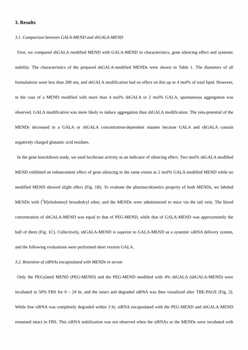
3.1. Comparison between GALA-MEND and shGALA-MEND
First, we compared shGALA modified MEND with GALA-MEND in characteristics, gene silencing effect and systemic
stability. The characteristics of the prepared shGALA-modified MENDs were shown in Table 1. The diameters of all
formulations were less than 200 nm, and shGALA modification had no effect on this up to 4 mol% of total lipid. However,
in the case of a MEND modified with more than 4 mol% shGALA or 2 mol% GALA, spontaneous aggregation was
observed. GALA modification was more likely to induce aggregation than shGALA modification. The zeta-potential of the
MENDs decreased in a GALA or shGALA concentration-dependent manner because GALA and shGALA contain
negatively charged glutamic acid residues.
In the gene knockdown study, we used luciferase activity as an indicator of silencing effect. Two mol% shGALA modified
MEND exhibited an enhancement effect of gene silencing to the same extent as 2 mol% GALA modified MEND while no
modified MEND showed slight effect (Fig. 1B). To evaluate the pharmacokinetics property of both MENDs, we labeled
MENDs with [3H]cholesteryl hexadedcyl ether, and the MENDs were administered to mice via the tail vein. The blood
concentration of shGALA-MEND was equal to that of PEG-MEND, while that of GALA-MEND was approximately the
half of them (Fig. 1C). Collectively, shGALA-MEND is superior to GALA-MEND as a systemic siRNA delivery system,
and the following evaluations were performed short version GALA.
3.2. Retention of siRNAs encapsulated with MENDs in serum
Only the PEGylated MEND (PEG-MEND) and the PEG-MEND modified with 4% shGALA (shGALA-MEND) were
incubated in 50% FBS for 0 – 24 hr, and the intact and degraded siRNA was then visualized after TBE-PAGE (Fig. 2).
While free siRNA was completely degraded within 3 hr, siRNA encapsulated with the PEG-MEND and shGALA-MEND
remained intact in FBS. This siRNA stabilization was not observed when the siRNAs or the MENDs were incubated with
the non-ionic detergent triton.
3.2. Enhancement of gene silencing in vitro by shGALA-modification
To evaluate the enhancement in endosomal escape by shGALA, gene silencing activities were measured by quantification
of ACTB-mRNA with the reverse transcription-quantitative polymerase chain reaction (RT-qPCR) 24 hr after siRNA
transfection with the MENDs (Fig. 3). In the case of no shGALA-modification, the PEG-MEND encapsulating anti-ACTB
siRNA showed no knockdown effect compared with the MENDs encapsulating anti-luc siRNA. Meanwhile, the gene
silencing effect of the PEG-MEND increased dependent on the extent of shGALA-modification. A shGALA modification of
4% was considered to be optimal, in terms of both the knockdown efficiency and the characteristics of the MEND.
To verify that the increase in gene silencing resulted from endosomal escape by shGALA, the amount of cellular uptake
and the distribution of siRNA in HT1080 cells was analyzed by flowcytometry and con-focal laser scanning microscopy
(CLSM). The uptake of siRNA was nearly equal when the cells were transfected with shGALA-MEND and PEG-MEND,
but the distribution of siRNA in the cells was completely different (Fig. 4). In the case of shGALA-MEND, much less
siRNA was colocalized with endosomes (Fig. 4C), compared to PEG-MEND (Fig. 4D).
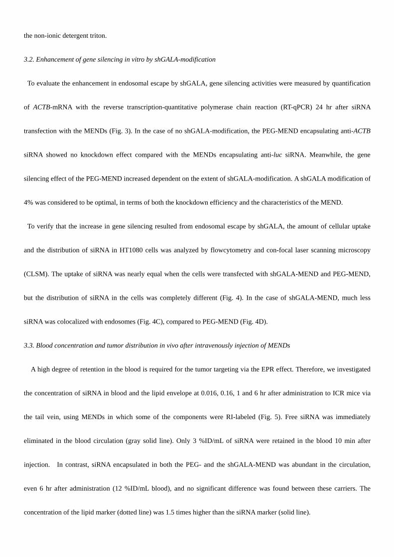
3.3. Blood concentration and tumor distribution in vivo after intravenously injection of MENDs
A high degree of retention in the blood is required for the tumor targeting via the EPR effect. Therefore, we investigated
the concentration of siRNA in blood and the lipid envelope at 0.016, 0.16, 1 and 6 hr after administration to ICR mice via
the tail vein, using MENDs in which some of the components were RI-labeled (Fig. 5). Free siRNA was immediately
eliminated in the blood circulation (gray solid line). Only 3 %ID/mL of siRNA were retained in the blood 10 min after
injection. In contrast, siRNA encapsulated in both the PEG- and the shGALA-MEND was abundant in the circulation,
even 6 hr after administration (12 %ID/mL blood), and no significant difference was found between these carriers. The
concentration of the lipid marker (dotted line) was 1.5 times higher than the siRNA marker (solid line).

We next examined the tumor distribution of the shGALA-MEND 6 hr after intravenous injection in the tumor-bearing
mice. The shGALA-MEND labeled with rhodamine-DOPE was injected, and tumor tissues were collected and imaged after
two hours (Fig. 6). The signals derived from the rhodamine-lipids and hoechst33342 are pseudocolored in red and blue,
respectively. A large number of rhodamine signals were observed.
3.4. The gene silencing effect in tumor tissue and therapeutic effect conferred by the injection of MENDs
We evaluated the gene silencing effect and subsequent anti-tumor effect induced by shGALA-MEND injection in
tumor-bearing mice in vivo. It has been already showed that the knockdown of ACTB mRNA led to the inhibition of growth
of HT1080 cells in vitro (data not shown). When tumor volumes reached 150 – 300 mm3 after the subcutaneous inoculation
of HT1080 cells, we performed continuous injections of 4 per day, each day at a dose of 4 mg siRNA/kg body weight.
ACTB-mRNA expression in tumor tissue was measured by RT-qPCR analysis 24 hr after the final injection. While the
injection of the PEG-MEND showed no gene silencing, the shGALA-MEND injection showed a significant knockdown
effect (Fig. 6A) and tumor growth was strongly inhibited (Fig. 7C, E) compared with non treatment. In addition, to verify
that this knockdown effect was sequence-specific, a shGALA-MEND encapsulating anti-luc siRNA was injected into
tumor-bearing mice. As a result, the anti-luc shGALA-MEND failed to induce gene silencing (Fig. 7C), nor were any
significant therapeutic effects observed (Fig. 7D).
3. 5. Toxicological test
We performed a toxicological analysis of the MENDs. In initial experiments, we monitored chronological changes in
body weight after 4 administrations at a dose of 4 mg siRNA/kg body weight (Fig. 8A). Although the body weight of all of
the experimental groups decreased gradually, these differences were not significant. Second, the liver could be damaged by
the accumulation of lipid components because liposomal carriers tend to accumulate in the reticuloendothelial system (RES),
such as the liver and spleen (Fig. S1E, F). Therefore, we evaluated the hepatic toxicity of the MENDs by measuring the
activities of liver escape enzymes, such as ALT and AST in serum. As a result, no increase was observed in the groups that
had been injected with the MENDs over 24 hr after injection (Fig. 8). Finally, the induction of inflammatory cytokines IL-6
and IFN- by the administered MENDs was determined by ELISA since it was previously reported that inflammatory
cytokines are produced, as the result of the injection of an siRNA carrier [28]. Although both types of the MENDs showed
evidence for cytokine induction, serum cytokine levels decreased to the nontreatment level 24 hr after the administration
(Fig. 9). Moreover, shGALA modification suppressed this induction, and IL-6 was reduced particularly significantly
compared with PEG-MEND (Fig. 9A).
4. Discussion
In this report, we report on attempts to control both intracellular trafficking and biodistribution by preparing a PEGylated
MEND containing siRNA with shGALA, a pH-sensitive fusogenic peptide (Fig. 10). The aqueous layer formed by dense
PEGylation on the surface of liposomes minimizes interactions with serum proteins and cell surfaces, allowing for a longer
circulation half-life and accumulation in tumor tissue. Therefore, the PEG-MEND showed a higher blood concentration than
the bare MEND (Fig 1C). However, the GALA modified PEG-MEND (GALA-MEND) was immediately excreted. It was
reported that the length of GALA in -helical form was 4.5 nm long and the thickness of the aqueous layer formed by
PEG2000 was around 3.0 nm [29, 30]. This suggests that the head of the GALA was not concealed and is recognized by
biomolecules, such as serum proteins (Fig. 1A). We speculated that this decline was caused by the recognition of GALA by
serum proteins, due to the GALA not being masked. Based on this hypothesis, we designed a new shorter version of GALA,
namely, shGALA (Fig. 1A). The shGALA showed an enhancement effect on gene silencing activity, but the
shGALA-MEND circulated stably in the blood (Figs. 1C, 5). We studied the utility of the shGALA-MEND for in vivo
siRNA delivery.
The accumulation of siRNA carriers in tumor tissue via the EPR effect requires that the particle has an appropriate
diameter (<200 nm) and that it is stable in serum. It was reported that the extent of accumulation of a PEGylated carrier
depends on its size and stability in the bloodstream of the carrier[31]. The diameter of all of the PEG modified MENDs
prepared in this study were less than 200 nm (Table 1), which suggests that the PEGylated MENDs could pass through
leaky neovessels and accumulate in tumor tissue. Over 4% shGALA-modification resulted in the formation of aggregates.
This is because the minority of shGALA molecules was likely in an -helix structure and induced membrane fusion even at
physiological pH. This would be expected to induce aggregation under conditions where much of the shGALA was
modified, since the shGALA structure is present in the form of an equilibrium mixture of an -helix and a random coil. In
FBS, the MENDs stably retained siRNA for over 24 hr in contrast to the immediate degradation of free siRNA (Fig. 2).
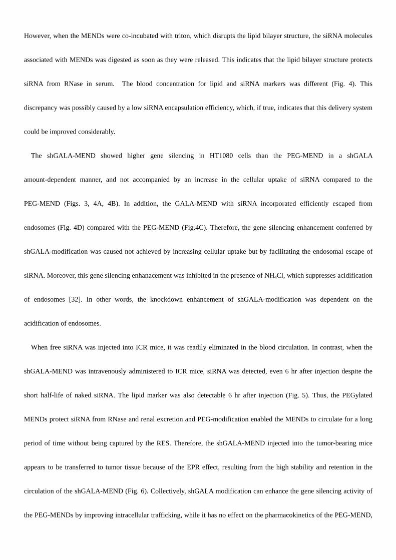
However, when the MENDs were co-incubated with triton, which disrupts the lipid bilayer structure, the siRNA molecules
associated with MENDs was digested as soon as they were released. This indicates that the lipid bilayer structure protects
siRNA from RNase in serum. The blood concentration for lipid and siRNA markers was different (Fig. 4). This
discrepancy was possibly caused by a low siRNA encapsulation efficiency, which, if true, indicates that this delivery system
could be improved considerably.
The shGALA-MEND showed higher gene silencing in HT1080 cells than the PEG-MEND in a shGALA
amount-dependent manner, and not accompanied by an increase in the cellular uptake of siRNA compared to the
PEG-MEND (Figs. 3, 4A, 4B). In addition, the GALA-MEND with siRNA incorporated efficiently escaped from
endosomes (Fig. 4D) compared with the PEG-MEND (Fig.4C). Therefore, the gene silencing enhancement conferred by
shGALA-modification was caused not achieved by increasing cellular uptake but by facilitating the endosomal escape of
siRNA. Moreover, this gene silencing enhanacement was inhibited in the presence of NH4Cl, which suppresses acidification
of endosomes [32]. In other words, the knockdown enhancement of shGALA-modification was dependent on the
acidification of endosomes.
When free siRNA was injected into ICR mice, it was readily eliminated in the blood circulation. In contrast, when the
shGALA-MEND was intravenously administered to ICR mice, siRNA was detected, even 6 hr after injection despite the
short half-life of naked siRNA. The lipid marker was also detectable 6 hr after injection (Fig. 5). Thus, the PEGylated
MENDs protect siRNA from RNase and renal excretion and PEG-modification enabled the MENDs to circulate for a long
period of time without being captured by the RES. Therefore, the shGALA-MEND injected into the tumor-bearing mice
appears to be transferred to tumor tissue because of the EPR effect, resulting from the high stability and retention in the
circulation of the shGALA-MEND (Fig. 6). Collectively, shGALA modification can enhance the gene silencing activity of
the PEG-MENDs by improving intracellular trafficking, while it has no effect on the pharmacokinetics of the PEG-MEND,
unlike the original GALA-modification (Fig. 1B). In summary, shGALA is a useful device for the in vivo delivery of
siRNA.
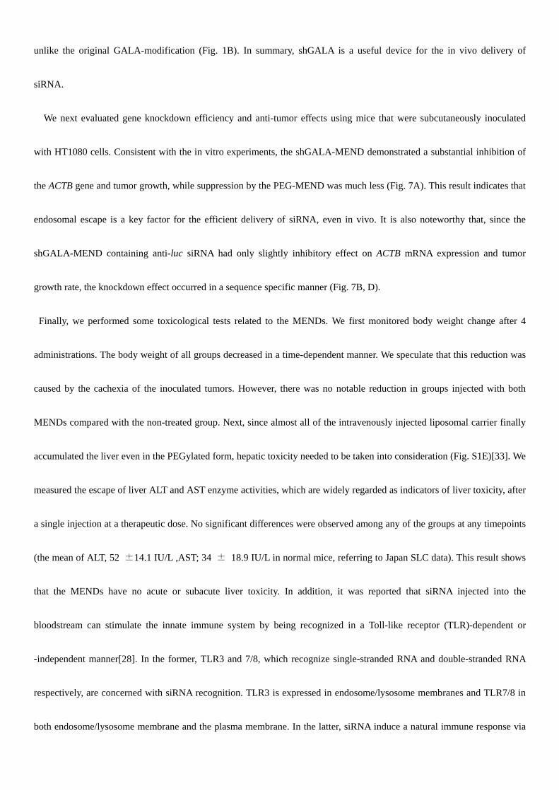
We next evaluated gene knockdown efficiency and anti-tumor effects using mice that were subcutaneously inoculated
with HT1080 cells. Consistent with the in vitro experiments, the shGALA-MEND demonstrated a substantial inhibition of
the ACTB gene and tumor growth, while suppression by the PEG-MEND was much less (Fig. 7A). This result indicates that
endosomal escape is a key factor for the efficient delivery of siRNA, even in vivo. It is also noteworthy that, since the
shGALA-MEND containing anti-luc siRNA had only slightly inhibitory effect on ACTB mRNA expression and tumor
growth rate, the knockdown effect occurred in a sequence specific manner (Fig. 7B, D).
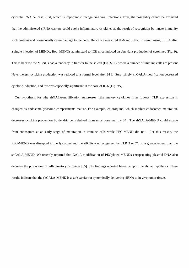
Finally, we performed some toxicological tests related to the MENDs. We first monitored body weight change after 4
administrations. The body weight of all groups decreased in a time-dependent manner. We speculate that this reduction was
caused by the cachexia of the inoculated tumors. However, there was no notable reduction in groups injected with both
MENDs compared with the non-treated group. Next, since almost all of the intravenously injected liposomal carrier finally
accumulated the liver even in the PEGylated form, hepatic toxicity needed to be taken into consideration (Fig. S1E)[33]. We
measured the escape of liver ALT and AST enzyme activities, which are widely regarded as indicators of liver toxicity, after
a single injection at a therapeutic dose. No significant differences were observed among any of the groups at any timepoints
(the mean of ALT, 52 ±14.1 IU/L ,AST; 34 ± 18.9 IU/L in normal mice, referring to Japan SLC data). This result shows
that the MENDs have no acute or subacute liver toxicity. In addition, it was reported that siRNA injected into the
bloodstream can stimulate the innate immune system by being recognized in a Toll-like receptor (TLR)-dependent or
-independent manner[28]. In the former, TLR3 and 7/8, which recognize single-stranded RNA and double-stranded RNA
respectively, are concerned with siRNA recognition. TLR3 is expressed in endosome/lysosome membranes and TLR7/8 in
both endosome/lysosome membrane and the plasma membrane. In the latter, siRNA induce a natural immune response via
cytosolic RNA helicase RIGI, which is important in recognizing viral infections. Thus, the possibility cannot be excluded
that the administered siRNA carriers could evoke inflammatory cytokines as the result of recognition by innate immunity
such proteins and consequently cause damage to the body. Hence we measured IL-6 and IFN-in serum using ELISA after
a single injection of MENDs. Both MENDs administerd to ICR mice induced an abundant production of cytokines (Fig. 9).
This is because the MENDs had a tendency to transfer to the spleen (Fig. S1F), where a number of immune cells are present.
Nevertheless, cytokine production was reduced to a normal level after 24 hr. Surprisingly, shGALA-modification decreased
cytokine induction, and this was especially significant in the case of IL-6 (Fig. 9A).
Our hypothesis for why shGALA-modification suppresses inflammatory cytokines is as follows. TLR expression is
changed as endosome/lysosome compartments mature. For example, chloroquine, which inhibits endosomes maturation,
decreases cytokine production by dendric cells derived from mice bone marrow[34]. The shGALA-MEND could escape
from endosomes at an early stage of maturation in immune cells while PEG-MEND did not. For this reason, the
PEG-MEND was disrupted in the lysosome and the siRNA was recognized by TLR 3 or 7/8 to a greater extent than the
shGALA-MEND. We recently reported that GALA-modification of PEGylated MENDs encapsulating plasmid DNA also
decrease the production of inflammatory cytokines [35]. The findings reported herein support the above hypothesis. These
results indicate that the shGALA-MEND is a safe carrier for systemically delivering siRNA to in vivo tumor tissue.
5. Conclusion
The results of the present study indicate that a strategy that involves the control of both the pharmacokinetics and the
intracellular trafficking of siRNA via combining MENDs with a fusogenic peptide shGALA can be valuable. In the in vitro
study, shGALA-modification conferred membrane fusogenic ability and improved the endosomal escape of the
PEG-MEND as was originally postulated. Therefore, siRNA can avoid lysosomal degradation and can be efficiently
delivered by the shGALA-MEND. The in vivo systemic administration of the shGALA-MEND also demonstrated a
substantial gene silencing in tumor tissue and an inhibitory effect against tumor growth without any remarkable toxicity.
Collectively, a new siRNA delivery system, the shGALA-MEND offers a promising approach for cancer therapy.
Acknowledgmendts
We thank M. S. Feather for his helpful advice in writing the English manuscript. This work was supported in part by
Grants-in-Aid for Young Scientists (B) and by Grant for Industrial Technology Research from New Energy and Industrial
Technology Development Organization (NEDO).
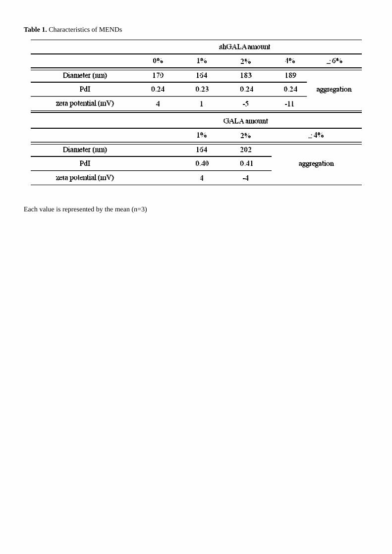
Table 1. Characteristics of MENDs
Each value is represented by the mean (n=3)
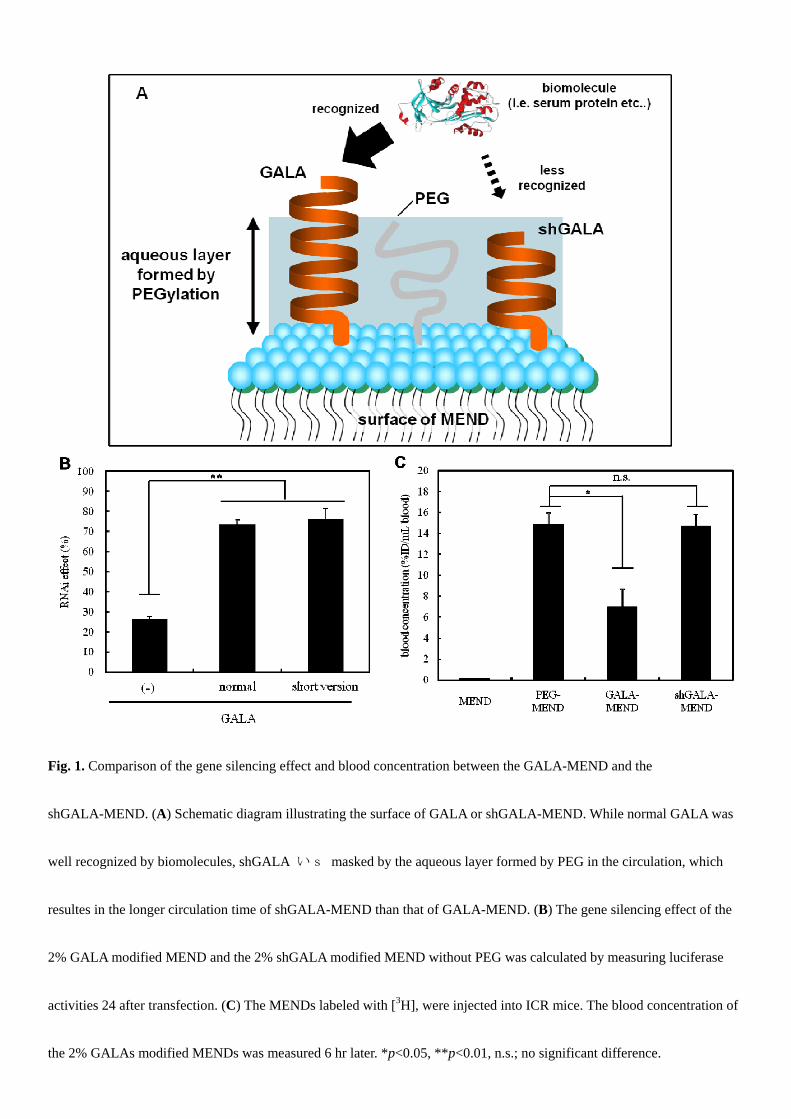
Fig. 1. Comparison of the gene silencing effect and blood concentration between the GALA-MEND and the
shGALA-MEND. (A) Schematic diagram illustrating the surface of GALA or shGALA-MEND. While normal GALA was
well recognized by biomolecules, shGALA いs masked by the aqueous layer formed by PEG in the circulation, which
resultes in the longer circulation time of shGALA-MEND than that of GALA-MEND. (B) The gene silencing effect of the
2% GALA modified MEND and the 2% shGALA modified MEND without PEG was calculated by measuring luciferase
activities 24 after transfection. (C) The MENDs labeled with [3H], were injected into ICR mice. The blood concentration of
the 2% GALAs modified MENDs was measured 6 hr later. *p<0.05, **p<0.01, n.s.; no significant difference.
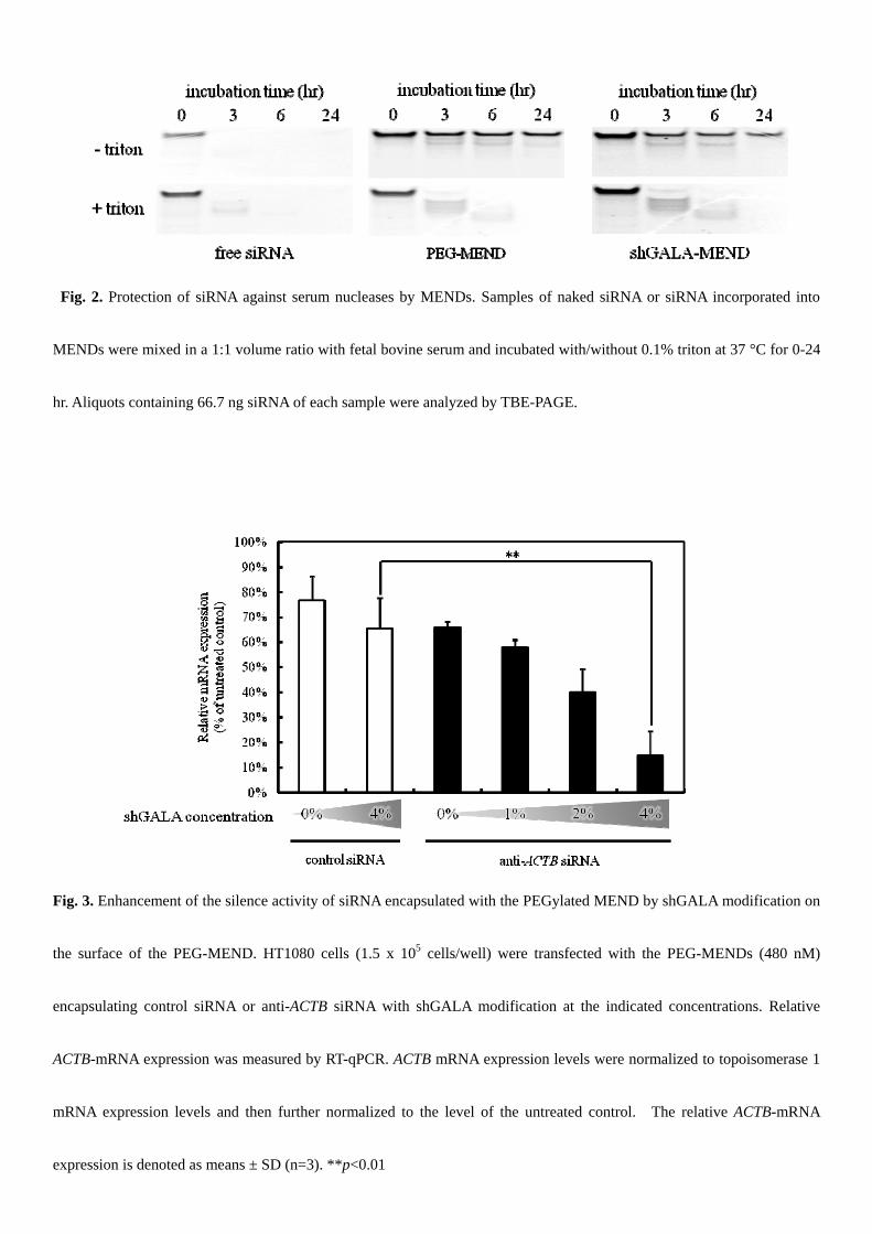
Fig. 2. Protection of siRNA against serum nucleases by MENDs. Samples of naked siRNA or siRNA incorporated into
MENDs were mixed in a 1:1 volume ratio with fetal bovine serum and incubated with/without 0.1% triton at 37 °C for 0-24
hr. Aliquots containing 66.7 ng siRNA of each sample were analyzed by TBE-PAGE.
Fig. 3. Enhancement of the silence activity of siRNA encapsulated with the PEGylated MEND by shGALA modification on
the surface of the PEG-MEND. HT1080 cells (1.5 x 10⁵ cells/well) were transfected with the PEG-MENDs (480 nM)
encapsulating control siRNA or anti-ACTB siRNA with shGALA modification at the indicated concentrations. Relative
ACTB-mRNA expression was measured by RT-qPCR. ACTB mRNA expression levels were normalized to topoisomerase 1
mRNA expression levels and then further normalized to the level of the untreated control. The relative ACTB-mRNA
expression is denoted as means ± SD (n=3). **p<0.01
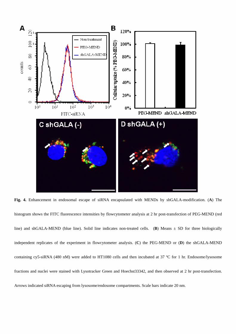
Fig. 4. Enhancement in endosomal escape of siRNA encapsulated with MENDs by shGALA-modification. (A) The
histogram shows the FITC fluorescence intensities by flowcytometer analysis at 2 hr post-transfection of PEG-MEND (red
line) and shGALA-MEND (blue line). Solid line indicates non-treated cells. (B) Means ± SD for three biologically
independent replicates of the experiment in flowcytometer analysis. (C) the PEG-MEND or (D) the shGALA-MEND
containing cy5-siRNA (480 nM) were added to HT1080 cells and then incubated at 37 ºC for 1 hr. Endosome/lysosome
fractions and nuclei were stained with Lysotracker Green and Hoechst33342, and then observed at 2 hr post-transfection.
Arrows indicated siRNA escaping from lysosome/endosome compartments. Scale bars indicate 20 nm.
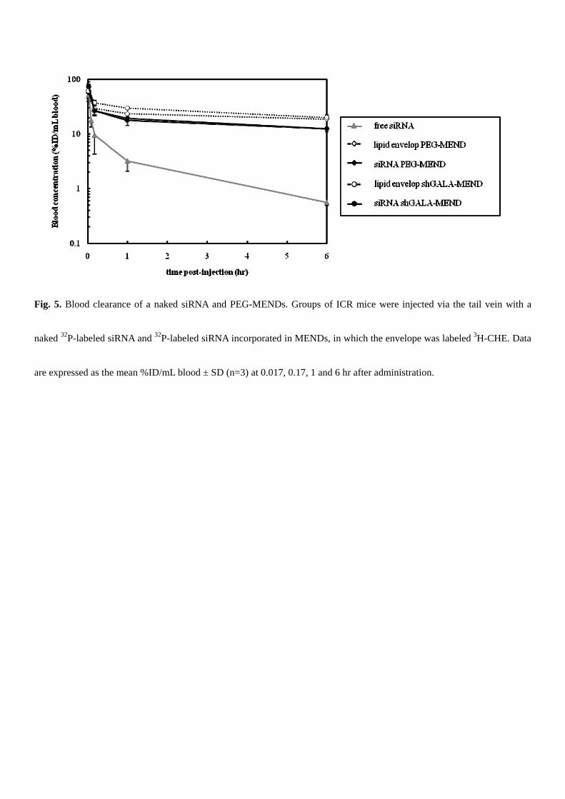
Fig. 5. Blood clearance of a naked siRNA and PEG-MENDs. Groups of ICR mice were injected via the tail vein with a
naked 32P-labeled siRNA and 32P-labeled siRNA incorporated in MENDs, in which the envelope was labeled 3H-CHE. Data
are expressed as the mean %ID/mL blood ± SD (n=3) at 0.017, 0.17, 1 and 6 hr after administration.
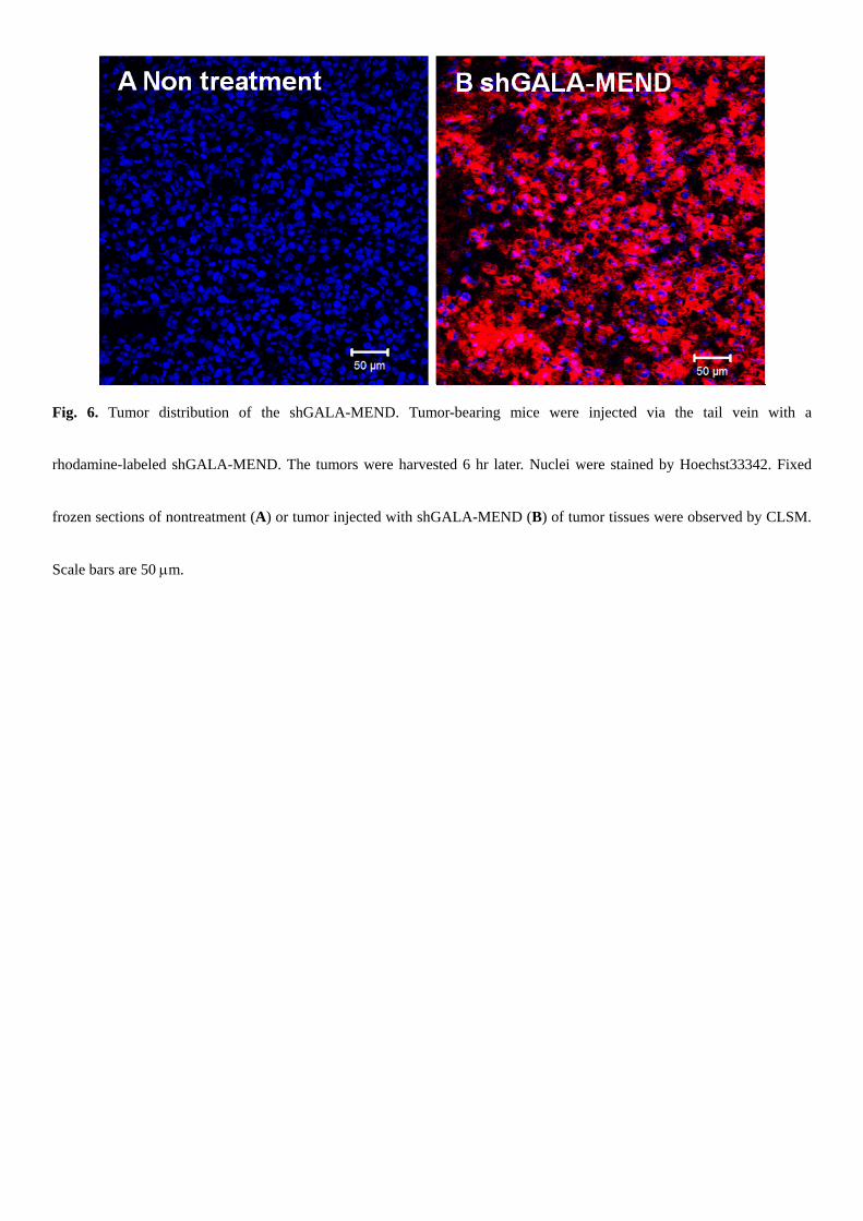
Fig. 6. Tumor distribution of the shGALA-MEND. Tumor-bearing mice were injected via the tail vein with a
rhodamine-labeled shGALA-MEND. The tumors were harvested 6 hr later. Nuclei were stained by Hoechst33342. Fixed
frozen sections of nontreatment (A) or tumor injected with shGALA-MEND (B) of tumor tissues were observed by CLSM.
Scale bars are 50 m.
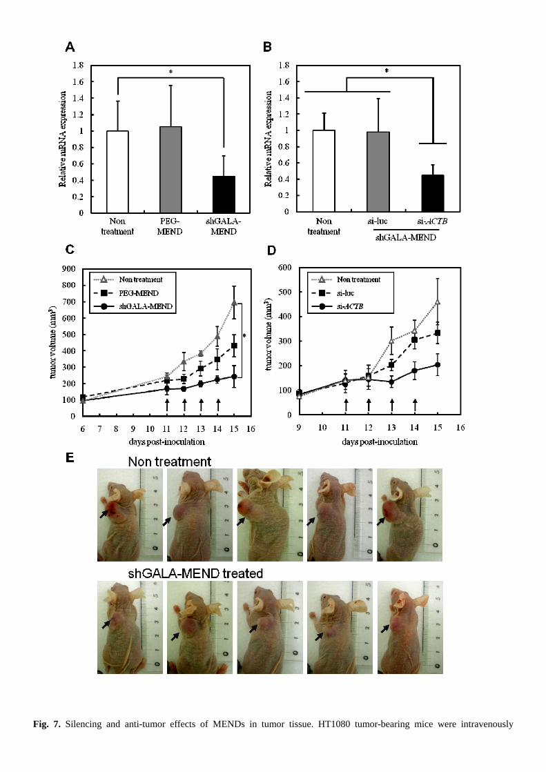
Fig. 7. Silencing and anti-tumor effects of MENDs in tumor tissue. HT1080 tumor-bearing mice were intravenously
administrated with MENDs at a dose of 4 mg/kg siRNA/kg body weight 4 times each day. (A, B) Tumor tissues were
collected 24 hr after the last administration and the ACTB mRNA level in tumor tissue was quantified by RT-qPCR. The
data are represented as the mean ± SD (n=5). *p<0.05. (C, D) Time-dependent changes in tumor volume were analysed.
Curves show the mean value ± SE of tumor size in a group of 5-8 mice and arrows indicates injection of MENDs. (E)
Tumor xenografts non-treated or treated with shGALA-MEND were imaged on day 15.
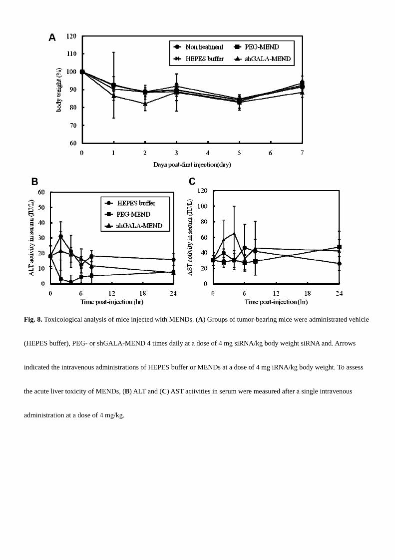
Fig. 8. Toxicological analysis of mice injected with MENDs. (A) Groups of tumor-bearing mice were administrated vehicle
(HEPES buffer), PEG- or shGALA-MEND 4 times daily at a dose of 4 mg siRNA/kg body weight siRNA and. Arrows
indicated the intravenous administrations of HEPES buffer or MENDs at a dose of 4 mg iRNA/kg body weight. To assess
the acute liver toxicity of MENDs, (B) ALT and (C) AST activities in serum were measured after a single intravenous
administration at a dose of 4 mg/kg.
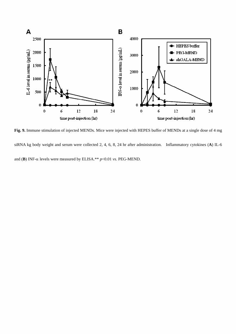
Fig. 9. Immune stimulation of injected MENDs. Mice were injected with HEPES buffer of MENDs at a single dose of 4 mg
siRNA kg body weight and serum were collected 2, 4, 6, 8, 24 hr after administration. Inflammatory cytokines (A) IL-6
and (B) INF- levels were measured by ELISA.** p<0.01 vs. PEG-MEND.
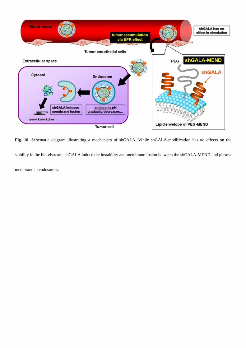
Fig. 10. Schematic diagram illustrating a mechanism of shGALA. While shGALA-modification has no effects on the
stability in the bloodstream, shGALA induce the instability and membrane fusion between the shGALA-MEND and plasma
membrane in endosomes.