Embed Size (px)
Citation preview
Copyright © 2016 American Scientific PublishersAll rights reservedPrinted in the United States of America
R E S E A R CH AR T I C L E
Quantum MatterVol. 5, 1–6, 2016
Synthesis and Structural Characterization ofNi1−xCdxFe2O4: Experiment and Theory
Anurag Srivastava2�∗, Hari Shankar Tewari1, and Aniruddha Mukherjee1
1Department of Pure and Applied Physics, Guru Ghasidas Vishwavidyalaya, Bilaspur2Advanced Materials Research Group, CNT Lab, ABV-Indian Institute of Information Technology and Management, Gwalior
We have synthesized Ni1−xCdxFe2O4, using auto combustion technique with particles size in nanometer(29–33 nm) range. The material was calcined at 800 �C for 6 hours and sintered at three different temperatures.The XRD analysis indicates the formation of single phase materials with six prominent peaks of Ni ferrite andone extra due to Cadmium doping, whereas FT-IR analysis shows absorption band as well as narrow bandin fundamental vibration region (400–700 cm−1) and few microscopic characteristics (density, porosity, size,shape). The experimental structural parameters have been verified through density functional theory basedab-initio analysis using revised PBE type parameterized generalized gradient approximation. The ab-initio resultalso confirms the metallic behavior of Ni1−xCdxFe2O4 analyzed through its band structure and density of statesprofiles.
Keywords: Magnetic Material, Sintering, X-ray Diffraction, FTIR, Ab-Initio Calculations, Band-Structure.
1. INTRODUCTIONThe development of ferrite was first reported in 1946 by Snoeckat Philips Laboratory in Holland, since then a number ofresearchers have shown interest in synthesizing as well as char-acterizing mixed ferrites materials. Spinel ferrites of general for-mula M2+Fe3+2 O4, (M
2+ especially transition metal ions, likeNi2+, Co2+, Zn2+ etc.) possess special magnetic and electricalproperties with high chemical stability and mechanical hard-ness, are important magnetic materials, suitable for operatingat higher frequency region, extensively used in electromagneticdevices1–3 such as memories, sensors and microwaves, as wellas in our modern information technology applications.4 Ni fer-rites are in demand for magnetic recording devices, magnetooptical recording and electronic devices. Magnetic interactionand distribution of cations among tetrahedral (A) and octahe-dral (B) sites have significant effects on the structural as wellas electrical and magnetic properties. The Cd doped Ni fer-rites are the magnetic materials characterized by maximum per-meability, minimum hysteresis losses, temperature variation ofproperties between prescribed limits and low core losses at highfrequencies, depending on their microstructure (density, poros-ity, size, shape) as well as macro structure (crystal structureand elemental composition). Ni–Cd ferrites with nanometer sizecan be synthesized through various methods, where in all themethods, the reagents are mixed together at molecular levels
∗Author to whom correspondence should be addressed.
that form homogeneous materials. Recently, experimental aswell as theoretical studies have been reported on Ni–Co–Mn,5
Ni–Zn,6 Mn–Zn,7 Ni–Cu–Zn,8 Ni–Cr9, Ni–Co,10 Ni–Co–Cd,11
Ni–Cu–Cd,12 and Co–Cd13 by using various techniques. All thesereports confirms the single phase cubic spinel structure of ferritematerials. However, not much work has been reported so far onNi–Cd ferrite. Hence looking to the importance of this categoryof material, we thought it pertinent to explore the properties ofthis material through its synthesis, characterization and theoret-ical validation. Recently, our group has performed experimentalas well as ab-initio based theoretical analysis of variety of nanos-tructured materials.14–17 In present manuscript we have reportedNi–Cd ferrite prepared by auto combustion method, character-ization by XRD and FT-IR analysis and verified the structuralparameters and electronic behavior of these materials using adensity functional theory based ab-initio approach.
2. METHODOLOGYOne of the most popular and efficient method of material prepa-ration is combustion synthesis (CS) is generally of two types3—propagating mode (heated locally), and simultaneous mode (uni-formly heating). Based on the property of matched oxidation andreduction between the fuel and the oxidizer, the extremely safecombustion synthesis can be controlled through initial param-eters. Depending upon the fuel and the oxidizer the tempera-ture rises rapidly and as a result of auto combustion, highly
Quantum Matter Vol. 5, No. 3, 2016 2164-7615/2016/5/001/006 doi:10.1166/qm.2016.1329 1
R ES E A R CH AR T I C L E Quantum Matter 5, 1–6, 2016
pure and crystalline nano size materials are formed. Reactionbetween the mixture of nitrates and highly pure glycine resultsa self-sustained exothermic process. This technique has asso-ciated advantages of getting soft and fine crystalline materialswith high surface area and high purity at very low temperature(<400 �C).
The calculation of molar ratio (stoichiometric fuel to oxidantratio) has a significant role in this redox based combustion syn-thesis. In the present price of work, the nitrates6�7�18�19 of thematerials are taken as oxidizer and glycine [NH2CH2COOH] asfuel. The stoichiometric ratio (�) have been calculated using fol-lowing relation,
� = (∑�summesion of oxidizing element in specific formula�
×valency)× (
�−1�∑
�coefficient of reducing element
specific formula�×valency)−1
Where, valency of the starting materials for (M = +2, N = 0,O =−2, H =+1, c =+4),
The ratio of the total nitrate to glycine (�) is calculated fromthe oxidation number of the starting materials and the valencesof the composition of the starting materials.
� = ���M× �+2��+ �N × �0��+ �O× �−2���
+ ��Fe× �+3��+ �O× �−2����
× ���+4�×C + �−2�×O+ �0�×N + �+1�×H��−1
= 409
= 444
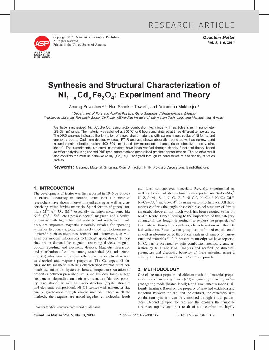
The flow chart of our present experimental setup for the syn-thesis of Ni1−xCdxFe2O4 �00 ≤ x ≤ 06� ferrites is given inFigure 1. Here nitrates of the precursor materials have been takenas oxidizer and glycine as reducing agent or fuel. First of all,stoichiometric amount (�) of nickel, cadmium and ferric nitratesalts were taken in a beaker and heated at 70 �C for 15–20 minuntil it get totally melt. Then the glycine is added to the mixtureand heated till it melts so as to get a homogeneous mixture. Thetotal mixture is further heated at higher temperature (320–350 �C,
Fig. 1. Flow chart representing the experimental steps for Ni1−xCdxFe2O4 Ferrite synthesis.
depending upon x) for 15 min so that the following reaction5
takes place results combustion.
�1−x�Ni�NO3�2+xCd�NO3�2+2Fe�NO3�3
+ 40
9NH2CH2COOH= Ni1−xCdxFe2O4+
100
9H2O
+ 809CO2+ 56
9N2
The structural parameter of Ni–Cd ferrite materials of differentcompositions have been calculated experimentally and to verifythe same we have used a well known density functional theorybased ab-initio tool ATK-VNL.20 For the present computation,a generalized gradient approximation (GGA)21�22 scheme hasbeen used as exchange correlation with revised Perdew, Burke,Ernzerhof23 type parameterization employed with double zetadouble polarized type of basis sets. The calculation has been per-formed on a sufficient k mesh for the inverse structure unit cellcontaining 14 atoms and the optimized structure is a face cen-tered cubic (a= b= c). The obtained theoretical lattice parameteris in close agreement to our experimental results. Further, thetheoretical analysis has been extended to understand the behaviorof synthesized Ni–Cd ferrite in respect of its commercial appli-cations, the electronic properties24�25 in terms of band structureand density of states (DOS) computation have been analyzed anddiscussed in results section separately.
3. RESULTS AND DISCUSSIONThe present section discusses the Powder X-ray Diffraction(XRD) and Fourier Transform Infra-Red (FT-IR) spectroscopyresults of Ni–Cd Ferrite powdered sample followed by the theo-retical verification of structural parameters and electronic prop-erties using ab-initio approach applied through Atomistix ToolKit-Virtual Nano Lab (ATK-VNL) code.
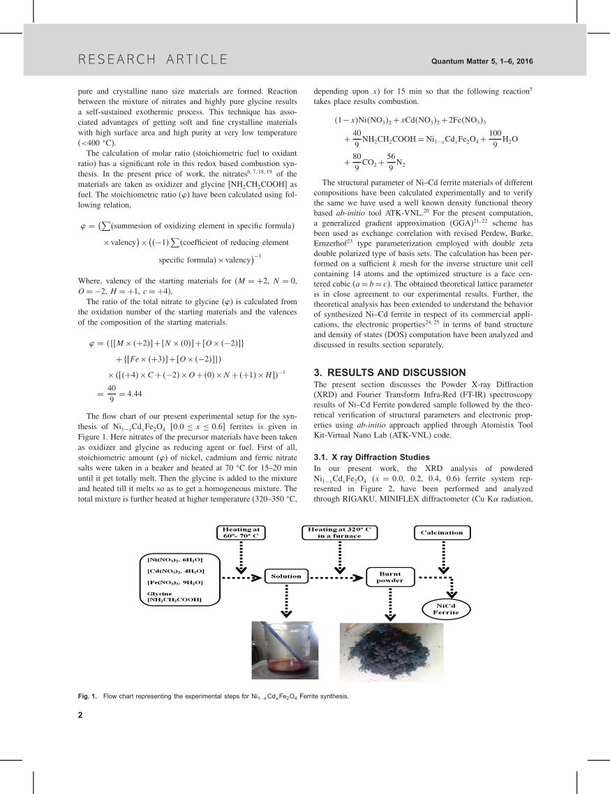
3.1. X ray Diffraction StudiesIn our present work, the XRD analysis of powderedNi1−xCdxFe2O4 (x = 00, 0.2, 0.4, 0.6) ferrite system rep-resented in Figure 2, have been performed and analyzedthrough RIGAKU, MINIFLEX diffractometer (Cu K radiation,
2
R E S E A R CH A R T I C L EQuantum Matter 5, 1–6, 2016
20 30 40 50 60 70 80
X=0.6
X=0.4
X=0.2
X=0.0
rela
tive
inte
nsity
rela
tive
inte
nsity
2θ in degree in degree
Fig. 2. XRD patterns of Ni1−xCdxFe2O4 Ferrite as a function of CdComposition (x).
2� = 20�–80�). Where, the most intense peak, representing (220),(311), (400), (422), (333), (440) planes27 are for Ni ferrite, andan additional peak (533) is due to substitutional doping of Cd.The lattice parameters ‘aexp’ for all the samples have been deter-mined using Eq. (1), represents the most prominent peak (311)of the XRD pattern and tabulated in Table I as a function of theCd composition. The larger ionic radii of Cd2+ (0.97 Å) to thatof Ni2+ (0.69 Å) and Fe3+ (0.645 Å) results an increase in thelattice parameter as well as X ray density and the crystalline sizeof Ni–Cd Ferrite shown in Figure 3. The Ni–Cd ferrites havebeen calcined at 800 �C temperature for 6 hrs and sintered atdifferent temperatures at 1050 �C, 1100 �C, 1150 �C, for 6 hrs.
aexp = dhkl
√�h2+k2 + l2�
�
sin �
√�h2+k2+ l2�
(1)
Where hkl are the Miller indices and dhkl the inter plannerspacing. The average crystalline size (D in nm) in the directionperpendicular to (hkl) plane of reflexes have been estimated byusing Scherrer Eq. (2),
D = k�
cos �(2)
Where, k= 09 is the Scherrer constant, proposed by Klug andAlexander,28 � = 1540562 Å and the full wave half maximaof the diffraction peaks at an angle � for corrected instrumentbroadening (in radian respectively). The (311) peak has beenchosen for calculation as the most suitable for crystalline size
Table I. Lattice parameter (a), density (�the), crystallite size ofNi1−xCdxFe2O4.
Lattice parameter (Å)Cd Crystallite Density (�the)concentration (x) Exp Theory Others�1� size (nm) in gm/cm3
0.0 8.338 8.333 8.34 29.25 5.3700.2 8.343 8.340 – 27.78 5.6060.4 8.361 8.346 – 29.94 5.8130.6 8.379 8.367 – 32.94 5.985
0.0 0.1 0.2 0.3 0.4 0.5 0.6
8.34
8.35
8.36
8.37
8.38
28.5
30.0
31.5
33.0
34.5
Latti
ce p
aram
eter
Cd composition(x)
Cry
stal
lite
size
Fig. 3. Lattice parameter and crystallite size of Ni1−x Cdx Fe2O4 as a func-tion of Cd composition (x).
distribution. The theoretical density (X ray density) has been cal-culated using the following relation
�the =ZM
Na3= 8M
Na3(3)
Here, Z�=8� represent the number of molecules per unit cell ofthe spinel lattice, M the molecular weight of the ferrite, N and a
(aexp� be the respective Avogadro’s number and lattice parameter.Generally all Ni2+ ions occupy octahedral B sites but the Cd2+
ions are preferred to occupy the tetrahedral A sites. The pre-ferred cation distribution2�29–32 of the sample Ni1−xCdxFe2O4 is[Cd2+x Fe3+1−x]A [Ni2+1−x Fe3+1+x]BO
2−4 . Here, the first square bracket
indicates tetrahedral A sites and the second one is octahedral Bsite. The Cd2+ ion has a zero magnetic moment33 whereas Fe3+
and Ni2+ has the magnetic moment of 5 �B and 2 �B respec-tively. Addition of Cd2+ ion at A site create a loss of magneticneighborhood of Fe3+ ions and the spin may become uncoupledconsequently more field is need to align the magnetic momentsin the direction of applied magnetic field.The theoretical lattice parameter (ath) have been computed by
total energy minimization of the optimized structure using ATK-VNL19 code and have a good agreement with the obtained exper-imental values as given in Table I.
3.2. Density, Porosity and Densification ParameterThe densification parameter as shown in Figure 4 described interms of apparent porosity and bulk density as a function of firing(sintering) temperature is calculated using the relation (4)
= �T −�O
�the−�O
(4)
The apparent porosity of the investigated object decreases from1050 �C to 1150 �C in another observation the bulk densitydecreases with increase in Cd concentration but increases withthe firing temperature. The increase of the bulk density withincrease in the firing temperature is due to the fact of decreas-ing porosity (Table II) and the formation of Ni–Cd ferrite phasewhere the reactant has high densities. A characteristic path ofthe sintering process is shrinkage of the samples, are measuredfor the sintered material compared to the original unfired (greenbody) powder compact.
3

R ES E A R CH AR T I C L E Quantum Matter 5, 1–6, 2016
1040 1060 1080 1100 1120 1140 116042
44
46
48
50
52
54
56
58
2.60
2.65
2.70
2.75
2.80
2.85
2.90
2.95
3.00
3.05
3.10
Bul
k de
nsity
(gm
/cc)
sintering temperature (ºC)
poro
sity
X = 0.0 X = 0.2 X = 0.4 X = 0.6
Fig. 4. Porosity and bulk density of Ni1−xCdx Fe2 O4 as a function of sin-tering temperature.
In the present work, increase of sintering temperature accel-erates the linear shrinkage and increases densification. Furtherincrease in sintering temperature leads to shrinkage and reachingto maximum value at 1150 �C. During shrinkage, small poresmerge first and further increase in temperature results continu-ous shrinkage. On the other hand, due to increase in temperature,some micro-pores were merged together forming macro-poresin the pore size distribution. High temperature treatment resultsslower rate shrinkage of large pores than small pores, eventhough both are associated at a grain boundary. Thus sinteringtemperature has a significant effect on bulk density as well asporosity of the materials.
3.3. FTIR AnalysisIR spectrum represents the molecular absorption and transmis-sion, creating a molecular fingerprint of the sample. FT-IRanalysis was used to identify unknown materials as well as todetermine the quality or consistency of a sample and the amountof components in a mixture. The spectra were recorded on aSHIMADZU-FTIR 8400S equipment using KBr as reference ina wave number region of 350 to 4000 cm−1. The ratio of KBrand samples were taken as 95:5 in a cylindrical die and measuredat room temperature. Figure 5 shows the recorded spectra in 400to 800 Cm−1 range of Ni–Cd ferrite system. The inspection ofthe spectra shows absorption band and a narrow band in thatrange. It is due to the fact for these classes of compounds that
Table II. Bulk density (�T ), densification parameter (�) and poros-ity (P) of Ni1−xCdxFe2O4.
Cd Temperature �0 in �T in Porosityconcentration (x) in �C gm/cm3 gm/cm3 � (P )
0.0 1050 2.766 2.766 0.000 48.491100 2.766 2.872 0.041 46.511150 2.766 3.050 0.109 43.20
0.2 1050 2.705 2.705 0.000 51.471100 2.705 2.722 0.006 51.451150 2.705 2.778 0.025 50.45
0.4 1050 2.674 2.674 0.000 53.991100 2.674 2.681 0.002 53.881150 2.674 2.690 0.005 53.72
0.6 1050 2.637 2.637 0.000 55.941100 2.637 2.648 0.003 55.761150 2.637 2.684 0.014 55.16
400 450 500 550 600 650 700 750 8000
10
20
30
40
50
60
tran
smita
nce
(%)
wave number cm–1
X=0.0X=0.0 X=0.4X=0.4 X=0.2X=0.2 X=0.6X=0.6
Fig. 5. FT-IR spectra of Ni1−xCdxFe2O4 at four different Cd compositionsat room temperature.
the absorption in that range is not restricted but occur in spectraof most metallic oxide.34 The reason of arising of these bandsare due to lattice vibration of the oxide ions against the cations.A gradual increase in absorption at higher frequency is observeddue to electronic transition.
The IR spectra have been used to locate the band positions, asgiven in Table III. The higher frequency band is observed around590 cm−1 and lower frequency around 410 cm−1 but a narrowband is also observed at 460 cm−1. The bands in 400–700 cm−1
region are assigned to the fundamental vibration of the ions ofthe ferrite crystal. It is necessary to consider the vibrational spec-tra of the periodic structure for the analysis of such spectra. Bytaking into consideration this vibrational problem, a crystal canbe classified according to the continuity of bonding as (1) contin-uously bonded, (2) discontinuously bonded and (3) intermediate.
Since Ni2+ ions occupied in octahedral B sites so the substitu-tion of Cd2+ ion in the system decreases the amount of Ni2+ ionand transforms Fe3+ ion from B site to A site, shifts the bandposition toward lower wave number. The estimation of force con-stant of the tetrahedral site (Kt) and octahedral site (Ko) havebeen performed for these two vibrational band by employing themethod suggested by Waldron34 as given by,
Kt = 762×Mt ×v2t ×10−7 Nm−1
Ko = 1062× �Mo/2�×v2o ×10−7 Nm−1
Where, Mt and Mo represent the molecular weight of thecations occupying tetrahedral and octahedral sites respectively.Table III contains the estimated values of Kt and Ko. The tetra-hedral force constant gradually increases with Cd concentrationwhere as octahedral force constant decreases in this ferrite sys-tem. Addition of Cd2+ content in tetrahedral site transform Fe3+
Table III. Absorption band frequency and force constant ofNi1−xCdxFe2O4.
Cd concentration vt v vo Kt Ko
(x) cm−1 cm−1 cm−1 102 N/m 102 N/m
0.0 586 461 423 1.46 1.080.2 588 462 410 1.79 1.020.4 593 463 401 2.10 0.970.6 595 464 400 2.42 0.95
4
R E S E A R CH A R T I C L EQuantum Matter 5, 1–6, 2016
ion from tetrahedral to octahedral site results a charge imbalanceon the system increase tetrahedral force constant.
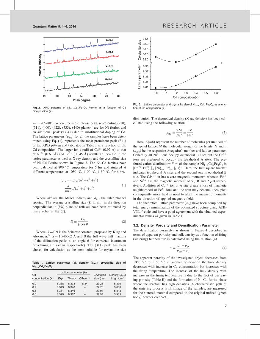
3.4. Band Structure and Density of State AnalysisIn order to verify the nature of Ni1−xCdxFe2O4 �00 ≤ x ≤ 06�we have performed the density functional theory based
(a)
–6 –4 –2 0 2 4 60
20
40
60
80
100
120
DO
S (e
V–1
)Energy (eV)
x=0.0(e)
(b)
DO
S (e
V–1
)
–6 –4 –2 0 2 4 60
10
20
30
40
50
60
70
80
Energy (eV)
x=0.2(f)
(c)
DO
S (e
V–1
)
Energy (eV)–6 –4 –2 0 2 4 6
0
10
20
30
40
50
60
70x=0.4
(g)
(d)
DO
S (e
V–1
)
Energy (eV)
–6 –4 –2 0 2 4 60
10
20
30
40
50
60x=0.6
(h)
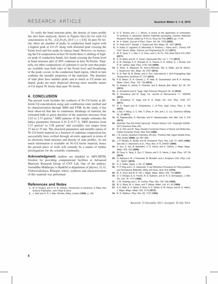
Fig. 6. Band structure and DOS profile of Ni1−xCdxFe2O4 at different Cd concentration (x).
computation to analyze the band structure and density of state(DOS). Figures 6(a)–(d) shows the band structure of the facecentered cubic Ni–Cd ferrite, where, number of bands are over-lapping and crossing the Fermi level showing metallic behavior.On substitutional doping of 20% Cd, few energy levels are cross-ing the Fermi level and in turn show the metallicity.
5
R ES E A R CH AR T I C L E Quantum Matter 5, 1–6, 2016
To verify the band structure plots, the density of states profilehas also been analyzed, shown in Figures 6(e)–(h) for each Cdconcentration in Ni1−xCdxFe2O4 �00 ≤ x ≤ 06�. In pure Ni fer-rite, there are number of peaks in conduction band region witha highest peak at 4.0 eV along with distorted peak crossing theFermi level and few peaks in valence band. However, on increas-ing the Cd composition in host Ni-ferrite there is shifting of high-est peak of conduction bands, few bands crossing the Fermi levelin band structure plot of 20% cadmium in host Ni-Ferrite. Simi-larly, for other compositions of cadmium it can be seen that peaksare available near both sides of the Fermi level and broadeningof the peaks occurs in the conduction band region, which in turnconfirms the metallic properties of the materials. The densitiesof state plots have number peaks and as much as Cd atoms aredoped, peaks are more dispersed showing more metallic natureof Cd doped Ni ferrite than pure Ni ferrite.
4. CONCLUSIONThe present work includes the synthesis of Ni–Cd ferrite at dif-ferent Cd concentration using auto combustion route method andits characterization through XRD and FTIR. In the study, it hasbeen observed that due to continuous shrinkage of material, theestimated bulk or green densities of the materials increases from2.63 to 2.73 gm/cm.3 XRD patterns of the sample estimates thelattice parameters between 8.34 Å–8.37 Å, XRD densities from5.37 gm/cm3 to 5.98 gm/cm3 and crystallite size ranges from27 nm to 33 nm. The structural parameters and metallic nature ofNi–Cd ferrite material as a function of cadmium composition hassuccessfully been verified through ab-initio approach in terms ofits electronic band structure and density of state profiles. As notmuch information is available on Ni–Cd ferrite material, hencethe present piece of work will certainly be a matter of furtherinvestigations for the scientific community.
Acknowledgment: Authors are thankful to ABV-IIITM,Gwalior for providing computational facilities at AdvancedMaterials Research Group of CNT Lab. One of the authors,Aniruddha Mukherjee is thankful to department of physics, G. G.Vishwavidyalaya, Bilaspur, where, synthesis and characterizationof this material was performed.
References and Notes1. W. D. Kingery and H. K. B. Uhlman, Introduction to Ceramics, A Wiley Inter
Science Publication, John Wiley & Sons.2. J. Smit and H. P. J. Wijn, Ferrites, Wiley, London (1959), p. 140.
3. D. E. Burkes and J. J. Moore, A review of the application of combustionof synthesis in advanced ceramic materials processing, Ceramic MaterialsResearch Trends, edited by P. B Lin, Nova Sci Pub (2007), pp. 11-46.
4. M. A. Gabal, Journal of Phys. Chem. Sol. 64, 1375 (2003).5. H. Arabi and F. Ganjali, J. Supercond. Nov. Magn. 26, 1031 (2013).6. A. Sutka, S. Lagzdina, G. Mezinskis, A. Pludons, I. Vitina, and L. Timma, IOP
Conf. Series: Mater. Science and Engineering 25, 012 (2011).7. M. R. Syue, F. J. Wei, C. S. Chou, and C. M. Fu, Thin Solid Films 519, 8303
(2011).8. K. M. Batoo and M. S. Ansari, Nanoscale Res. Let. 7, 112 (2012).9. S. M. Patange, S. E. Shirsath, B. G. Toksha, S. S. Jadhav, S. J. Shukla, and
K. M. Jadhav, Appl. Phys. A 95, 429 (2009).10. K. Khan, A. Maqsood, M. Anis-ur-Rehman, Md. A. Malik, and M. Akram,
J. Supercond. Nov. Magn. 25, 2707 (2012).11. N. D. Patil, M. B. Shelar, and V. Puri, International J. Self Propagating High
Temperature Synthesis 21, 212 (2012).12. P. B. Belavi, G. N. Chavan, L. R. Naik, R. Somashekar, and R. K. Kotnala,
Mater. Chem. Phys. 132, 138 (2012).13. S. Singhal, S. Jauhar, K. Chandra, and S. Bansal, Bull. Mater. Sci. 36, 107
(2013).14. A. Srivastava and N. Tyagi, High Pressure Research 32, 43 (2012).15. A. Srivastava and N. Tyagi, Int. Journal of Nanoscience 11, 1250035
(2012).16. A. Srivastava, N. Tyagi, and R. K. Singh, Am. Inst. Phys. 1349, 317
(2011).17. H. S. Tewari and S. Chakraborty, J. of Pure. Appl. Indus. Phys. 2, 142
(2012).18. J. Bai, F. Meng, C. C. Wei, Y. Zhao, H. Tan, and J. C. Liu, Ceramics–Silikáty
55, 20 (2011).19. M. Raghasudha, D. Ravinder, and P. Veerasomaiah, Adv. Mat. Lett. 4, 910
(2013).20. Atomistix Tool Kit-Virtual NanoLab: Tutorial Version 13.8. Copyright ©2008–
2013 Quantum Wise A/S.21. R. G. Parr and W. Yang, Density Functional Theory of Atoms and Molecules,
Oxford University Press, New York (1989).22. I. N. Levine, Quantum Chemistry, 5th edn., Prentice Hall, Upper Saddle River,
New Jersey (2000), pp. 581–584.23. J. P. Perdew, K. Burke, and M. Ernzerhof, Phys. Rev. Lett. 77, 3865 (1996);
See also V. Staroverov et al., Phys. Rev. A 74, 044501 (2006).24. X. Zuo, S. Yan, B. Barbiellini, V. G. Harris, and C. Vittoria, J. Magn. Magn.
Mater. 303, 432 (2006).25. M. Feng, A. Yang, X. Zuo, C. Vittoria, and V. G. Harris, J. Appl. Phys. 107, 09
(2010).26. G. Nabiyouni, M. J. Fesharaki, M. Mozafari, and J. Amighian, Chin. Phys. Lett.
27, 126401 (2010).27. A. A. Sattar, Egypt. J. Sol. 27 (2004).28. H. P. Klug and L. E. Alexander, X ray Diffraction Procedure for Polycrystalline
and Amorphous Materials, Wiley and Sons, New York (1974).29. M. A. Amer and M. E. Hiti, J. Magn. Magn. Mater. 234, 118 (2001).30. U. V. Chhaya, B. S. Trivedi, R. G. Kulkarni, and D. R. S. Somayajulu, J. Mat.
Sci. Lett. 18, 1177 (1999).31. J. M. Hastings and L. M. Corliss, Phys. Rev. 104, 328 (1956).32. M. U. Rana, M. U. Islam, and T. Abbas, Mater. Lett. 41, 52 (1999).33. S. K. Nath, K. H. Maria, S. Noor, S. S. Sikder, S. M. Hoque, and M. A. Hakim,
J. Magn. Magn. Mater. 324, 2116 (2012).34. R. D. Waldron, Phys. Rev. 99, 1727 (1955).
Received: 22 December 2013. Accepted: 30 July 2014.
6