Embed Size (px)
Citation preview
RACGP Ear wax management
Skye Poulton
Stephanie Yau
Daniel Anderson
Daniel Bennett
Background
Ear syringing is a very common practice among general practitioners (GPs). It is used by many asthe treatment of choice for cerumen (ear wax), and is usually effective and safe. However,complications from syringing are an increasingly common reason for presentation to ear, nose andthroat (ENT) specialists and medicolegal complaints against GPs.
Objective
The aim of this article is to provide GPs with the knowledge to safely manage cerumen.
Discussion
Ear wax is an important part of a normal functioning ear. It is mostly asymptomatic and requires notreatment. Softening ear drops may be necessary to help the ear fulfil its selfcleaning function, andshould be considered firstline treatment. Syringing can be safely performed if this fails by taking athorough history and examination to exclude contraindications, gaining patient consent and ensuringthe appropriate use of equipment. Referral to an ENT clinic for manual removal with microsuctioningmay be necessary in the event of contraindications, complications or failure.
Cerumen impaction (also known as ear wax) is a common presentation to a general practitioner(GP). Ear syringing is often the treatment of choice, and most GPs see a minimum of two patients perweek for ear syringing. It is considered by most to be effective and safe. However, 1 in 1000 patientsexperience major complications following syringing. As a consequence, one in five medicolegalcases involving GPs are related to ear syringing.
This article will discuss the composition of ear wax, why it accumulates, treatment options andpotential complications, to assist GPs in safely managing this condition.
Ear anatomy
A knowledge and understanding of normal ear anatomy is essential in the correct management ofimpacted cerumen with the use of ear syringing. The following is a brief review of the external earand tympanic membrane anatomy.
1
1
1
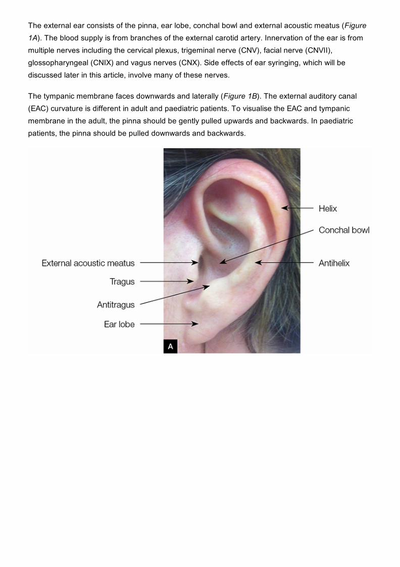
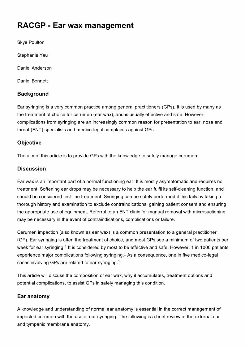
The external ear consists of the pinna, ear lobe, conchal bowl and external acoustic meatus (Figure1A). The blood supply is from branches of the external carotid artery. Innervation of the ear is frommultiple nerves including the cervical plexus, trigeminal nerve (CNV), facial nerve (CNVII),glossopharyngeal (CNIX) and vagus nerves (CNX). Side effects of ear syringing, which will bediscussed later in this article, involve many of these nerves.
The tympanic membrane faces downwards and laterally (Figure 1B). The external auditory canal(EAC) curvature is different in adult and paediatric patients. To visualise the EAC and tympanicmembrane in the adult, the pinna should be gently pulled upwards and backwards. In paediatricpatients, the pinna should be pulled downwards and backwards.
Figure 1. Ear anatomyA. External earB. Left tympanic membrane
What is ear wax?
Ear wax is the product of ceruminous and sebaceous gland secretions, mixed with exfoliatedsquamous epithelium. It forms a protective film, has antibacterial properties and provides lubricationto the ear canal. It is the ear’s selfcleaning mechanism, and traps dust and dirt, which are thenexcreted from the ear canal in a medial to lateral direction. This is aided by epithelial migration andmovement of the jaw.
Why does ear wax accumulate?
When the selfcleaning mechanism is disrupted, wax accumulates and can become impacted.Narrowing or obstruction of the ear canal, due to anatomical variations or infectious or dermatologicaldiseases, can interfere with the normal migratory process.
Irritation from foreign objects placed in the ear (eg cotton tips, hearing aids and ear plugs) can causechronic changes to the skin of the ear canal and impair normal epithelial migration. Cotton buds alsotend to push cerumen deeper into the ear canal, and hearing aids and ear plugs obstruct the earcanal and contribute to cerumen accumulation over time. Furthermore, the ceruminous glandsatrophy with age and produce a drier wax that migrates more slowly. Cerumen impaction is presentin up to 57% of older patients, compared with 5% of younger, healthy adults.
Indications for treatment
2,3
4
4
5
6
Cerumen accumulation is normal and does not require treatment unless it is symptomatic. Wax isusually described as impacted if it obscures visualisation of the tympanic membrane and isassociated with symptoms. Common symptoms include conductive hearing loss, ear pain, itching orfullness, dizziness, tinnitus or reflex cough. Consensus is that symptomatic cerumen generallywarrants removal, although, notably, in up to onethird of cases, the cerumen will clear within fivedays without treatment. Removal is also often indicated to allow view of the tympanic membrane fordiagnostic purposes or to allow audiometry.
Treatment options
The firstline option for treatment of symptomatic patients is cerumenolytics. The use ofcerumenolytic agents, namely eardrops such as docusate sodium, increases the likelihood ofcerumen clearance, compared with no treatment. However, there is no significant difference ineffectiveness between waterbased or oilbased drops. Patients should be discouraged fromusing cotton buds in the canal for cleaning. There is a very limited role for the use of cotton buds onthe outer ear only. Patients with hard impaction or ear canal disease may require irrigation or manualremoval under microscope by a trained doctor or an ear, nose and throat (ENT) specialist. The useof a cerumenolytic will improve success of subsequent irrigation.
How to safely syringe ear wax
Irrigation, or ear syringing, should be performed only after taking a full history, doing an earexamination and explaining the potential complications to the patient. It is also important to ensureappropriate assembly and use of equipment.
Gentle irrigation of the ear canal can be performed with a large syringe (20 mL) and warm water. Theuse of sterile water or saline as opposed to tap water or bacteriostatic agent (eg dilute hydrogenperoxide) can decrease the risk of infection.
Direct visualisation of the ear canal is not necessary for safe and effective syringing. The tip of thesyringe should not pass the outer onethird of the ear canal (approximately 8 mm) – the use of arounded nozzle may assist with this. The jet of water should be aimed towards the edge of thecerumen to enable the debris to flow out of the ear canal. Cease immediately if the patientexperiences pain or if bleeding occurs. Mechanical jet irrigators are available and some allow bettercontrol of water pressure and direction of spray (Figure 2). After syringing, examine the externalcanal and tympanic membrane. Document the patient’s consent, procedure, and pre and postexamination findings.
7
8
9
10
10
2,10
2,11
12
9
9
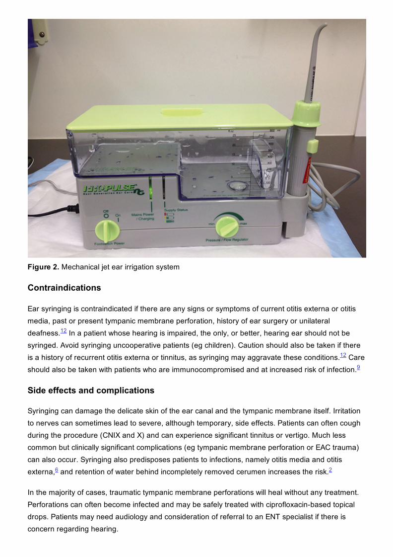
Figure 2. Mechanical jet ear irrigation system
Contraindications
Ear syringing is contraindicated if there are any signs or symptoms of current otitis externa or otitismedia, past or present tympanic membrane perforation, history of ear surgery or unilateraldeafness. In a patient whose hearing is impaired, the only, or better, hearing ear should not besyringed. Avoid syringing uncooperative patients (eg children). Caution should also be taken if thereis a history of recurrent otitis externa or tinnitus, as syringing may aggravate these conditions. Careshould also be taken with patients who are immunocompromised and at increased risk of infection.
Side effects and complications
Syringing can damage the delicate skin of the ear canal and the tympanic membrane itself. Irritationto nerves can sometimes lead to severe, although temporary, side effects. Patients can often coughduring the procedure (CNIX and X) and can experience significant tinnitus or vertigo. Much lesscommon but clinically significant complications (eg tympanic membrane perforation or EAC trauma)can also occur. Syringing also predisposes patients to infections, namely otitis media and otitisexterna, and retention of water behind incompletely removed cerumen increases the risk.
In the majority of cases, traumatic tympanic membrane perforations will heal without any treatment.Perforations can often become infected and may be safely treated with ciprofloxacinbased topicaldrops. Patients may need audiology and consideration of referral to an ENT specialist if there isconcern regarding hearing.
12
12
9
6 2
Other options for mechanical removal
Other options for mechanical removal include microsuction, with or without curettage, undermicroscope. Unfortunately, this is usually only available in dedicated ENT outpatient clinics.Microsuctioning is generally well tolerated, safe and efficacious. It also has the advantage of notexposing the ear to moisture and thus has fewer contraindications and is associated with a lowerfrequency of infections. Discomfort due to noise is the greatest complaint. However, there is noclinical evidence that this affects hearing.
When to refer?
Referral to an ENT specialist is rarely needed for cerumen management alone. Referrals should bemade in the following situations:
pain or bleeding on syringingfailure to remove cerumen after multiple attempts of syringing, preceded by waxsoftening dropspersistence of symptoms despite successful removal of cerumenchronic cerumen impactioncontraindications, especially perforated tympanic membrane, prior ear surgery or cerumenimpaction in the only or better hearing earabnormal tissue in the ear canalany other concerns.
Key points
Patients should be educated about the ear’s selfcleaning mechanism and discouraged from usinganything to remove wax from the ear canal.Patients should first be encouraged to use eardrops to soften wax and facilitate selfcleaning forsymptomatic wax accumulation.Ear syringing can be performed by trained staff if firstline treatment fails.Patients should be referred for specialist ENT assessment if there are any contraindications tosyringing, syringing fails to remove wax or resolve symptoms, or for any other concerns.
Authors
Skye Poulton BHlthSc (Nutr), MBBS, Junior House Officer, Ipswich Hospital, Ipswich, [email protected]
Stephanie Yau MBBS, ENT Principal House Officer, Townsville Hospital, QLD
Daniel Anderson BAppSc (EXSS), MSpMed, MBBS, ENT Registrar, Ipswich Hospital, Ipswich, QLD
Daniel Bennett MBBS FRACS, ENT Consultant, Ipswich Hospital, Ipswich, QLD
Competing interests: None.Provenance and peer review: Not commissioned, externally peer reviewed.
13
9
14
References
1. Bird S. The potential pitfalls of ear syringing: Minimising the risks. Aust Fam Physician2003;32:150–51. Search PubMed
2. Burton MJ, Doree C. Ear drops for the removal of ear wax (review). Cochrane Database Syst Rev2009;(1):CD004326. Search PubMed
3. Kelly KE, Mohs DC. The external auditory canal: Anatomy and physiology. Otolaryngol Clin NorthAm 1996;29:725–39. Search PubMed
4. Jabor MA, Amedee RG. Cerumen impaction. J La State Med Soc 1997;149:358–62. SearchPubMed
5. Meador JA. Cerumen impaction in the elderly. J Gerontol Nurs 1995;21:43–45. Search PubMed6. McCarter DF, Courtney AU, Pollart SM. Cerumen impaction. Am Fam Physician 2007;75:1523–28.Search PubMed
7. Browing GG. Ear wax. Clinical Evidence 2008;504:1–20. Search PubMed8. Mitka M. Cerumen removal guidelines wax practical. JAMA 2008;300:1506. Search PubMed9. Roland PS, Smith TL, Schwartz SR, et al. Clinical practice guideline: Cerumen impaction.Otolaryngol Head Neck Surg 2008;139:1–20. Search PubMed
10. Keane EM, Wilson H, McGrane D, Coakley D, Walsh JB. Use of solvents to disperse ear wax. Br JClin Pract 1995;49:71–72. Search PubMed
11. Clegg AJ, Loveman E, Gospodarevskaya E, et al. The safety and effectiveness of differentmethods of earwax removal: A systematic review and economic evaluation. Health TechnolAssess 2010;14:1–192. Search PubMed
12. Bird S. Ear syringing: Minimising the risks. Aust Fam Physician 2008;37:359–60. Search PubMed13. Prowse SJ, Milla O. Aural microsuction for wax impaction: Survey of efficacy and patient
perception. J Laryngol Otol 2014;128:621–25. Search PubMed14. Snelling JD, Smithard A, Waddell A. Noise levels generated within the external auditory canal
during microsuction aural toilet and the effect on hearing: A prospective controlled series. ClinOtolaryngol 2009;34:21–25. Search PubMed