Embed Size (px)
Citation preview
61
REVIEW ARTICLE SPINE SURGERY AND RELATED RESEARCH
Recent advances in magnetic resonance neuroimaging of lumbarnerve to clinical applications: A review of clinical studies utilizingDiffusion Tensor Imaging and Diffusion-weighted magneticresonance neurography
Yawara Eguchi1), Hirohito Kanamoto2), Yasuhiro Oikawa3), Munetaka Suzuki1), Hajime Yamanaka1), Hiroshi Tamai1),
Tatsuya Kobayashi1), Sumihisa Orita2), Kazuyo Yamauchi2), Miyako Suzuki2), Kazuhide Inage2), Yasuchika Aoki4),
Atsuya Watanabe4), Takeo Furuya2), Masao Koda2), Kazuhisa Takahashi2) and Seiji Ohtori2)
1) Department of Orthopedic Surgery, National Hospital Organization Shimoshizu National Hospital, Japan2) Department of Orthopedic Surgery, Graduate School of Medicine, Chiba University, Japan3) Division of Orthopaedic Surgery, Chiba Children’s Hospital, Japan4) Department of Orthopaedic Surgery, Eastern Chiba Medical Center, Japan
Abstract:Much progress has been made in neuroimaging with Magnetic Resonance neurography and Diffusion Tensor Imaging
(DTI) owing to higher magnetic fields and improvements in pulse sequence technology. Reports on lumbar nerve DTI have
also increased considerably.
Many studies have shown that the use of DTI in lumbar nerve lesions, such as lumbar foraminal stenosis and lumbar disc
herniation, makes it possible to capture images of interruptions of tractography at stenotic sties, enabling the diagnosis of
stenosis. DTI can also reveal significant decreases in fractional anisotropy (FA) with significant increases in apparent diffu-
sion coefficient (ADC) values in compression lesions.
FA values have higher accuracy than ADC values. Furthermore, strong correlations exist between FA values and indica-
tions of neurological severity, including the Japanese Orthopedic Association (JOA) score, the Oswestry Disability Index
(ODI), and the Roland-Morris Disability Questionnaire (RDQ) in patients with lumbar disc herniation-induced radiculopa-
thy.
Most lumbar DTI has become 3T; 3T MRI has made it possible to take high-resolution DTI measurements in a short pe-
riod of time. However, increased motion artifacts in the magnetic susceptibility effect lead to signal irregularities and image
distortion. In the future, high-resolution DTI with reduced field-of-view may become useful in clinical applications, since
visualization of nerve lesions and quantification of DTI parameters could allow more accurate diagnoses of lumbar nerve
dysfunctions. Future translational studies will be necessary to successfully bring MR neuroimaging of lumbar nerve into
clinical use.
Keywords:magnetic resonance imaging, diffusion tensor imaging, diffusion-weighted MR neurography, lumbar nerve, lumbar foram-
inal stenosis, lumbar disc herniation
Spine Surg Relat Res 2017; 1(2): 61-71
dx.doi.org/10.22603/ssrr.1.2016-0015
Introduction
With the rapid aging of our society, the number of pa-
tients with spinal disorders continues to rise. In the United
States alone, 25 million patients complain of lumbar pain,
leading to medical costs of over 100 billion dollars annually.
Intervertebral disc lesions and lumbar nerve root disorders
often cause lower back pain. Pain signals are transmitted
from the local site to the peripheral nerves, via the spinal
cord to the brain, where they are recognized as pain. In re-
Corresponding author: Yawara Eguchi, [email protected]
Received: October 26, 2016, Accepted: December 30, 2016
Copyright Ⓒ 2017 The Japanese Society for Spine Surgery and Related Research
Spine Surg Relat Res 2017; 1(2): 61-71 dx.doi.org/10.22603/ssrr.1.2016-0015
62
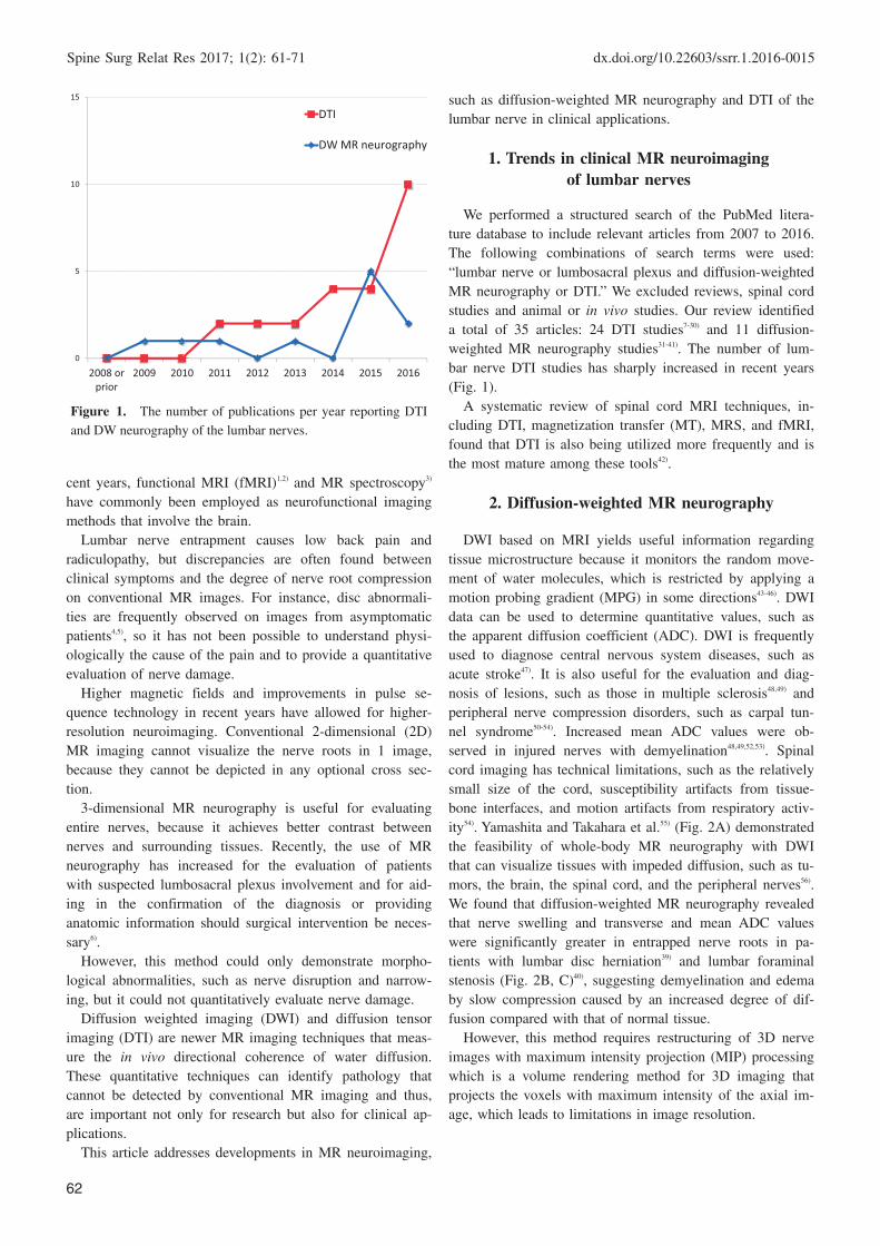
Figure 1. The number of publications per year reporting DTI
and DW neurography of the lumbar nerves.
0
5
10
15
2008 orprior
2009 2010 2011 2012 2013 2014 2015 2016
DTI
DW MR neurography
cent years, functional MRI (fMRI)1,2) and MR spectroscopy3)
have commonly been employed as neurofunctional imaging
methods that involve the brain.
Lumbar nerve entrapment causes low back pain and
radiculopathy, but discrepancies are often found between
clinical symptoms and the degree of nerve root compression
on conventional MR images. For instance, disc abnormali-
ties are frequently observed on images from asymptomatic
patients4,5), so it has not been possible to understand physi-
ologically the cause of the pain and to provide a quantitative
evaluation of nerve damage.
Higher magnetic fields and improvements in pulse se-
quence technology in recent years have allowed for higher-
resolution neuroimaging. Conventional 2-dimensional (2D)
MR imaging cannot visualize the nerve roots in 1 image,
because they cannot be depicted in any optional cross sec-
tion.
3-dimensional MR neurography is useful for evaluating
entire nerves, because it achieves better contrast between
nerves and surrounding tissues. Recently, the use of MR
neurography has increased for the evaluation of patients
with suspected lumbosacral plexus involvement and for aid-
ing in the confirmation of the diagnosis or providing
anatomic information should surgical intervention be neces-
sary6).
However, this method could only demonstrate morpho-
logical abnormalities, such as nerve disruption and narrow-
ing, but it could not quantitatively evaluate nerve damage.
Diffusion weighted imaging (DWI) and diffusion tensor
imaging (DTI) are newer MR imaging techniques that meas-
ure the in vivo directional coherence of water diffusion.
These quantitative techniques can identify pathology that
cannot be detected by conventional MR imaging and thus,
are important not only for research but also for clinical ap-
plications.
This article addresses developments in MR neuroimaging,
such as diffusion-weighted MR neurography and DTI of the
lumbar nerve in clinical applications.
1. Trends in clinical MR neuroimagingof lumbar nerves
We performed a structured search of the PubMed litera-
ture database to include relevant articles from 2007 to 2016.
The following combinations of search terms were used:
“lumbar nerve or lumbosacral plexus and diffusion-weighted
MR neurography or DTI.” We excluded reviews, spinal cord
studies and animal or in vivo studies. Our review identified
a total of 35 articles: 24 DTI studies7-30) and 11 diffusion-
weighted MR neurography studies31-41). The number of lum-
bar nerve DTI studies has sharply increased in recent years
(Fig. 1).
A systematic review of spinal cord MRI techniques, in-
cluding DTI, magnetization transfer (MT), MRS, and fMRI,
found that DTI is also being utilized more frequently and is
the most mature among these tools42).
2. Diffusion-weighted MR neurography
DWI based on MRI yields useful information regarding
tissue microstructure because it monitors the random move-
ment of water molecules, which is restricted by applying a
motion probing gradient (MPG) in some directions43-46). DWI
data can be used to determine quantitative values, such as
the apparent diffusion coefficient (ADC). DWI is frequently
used to diagnose central nervous system diseases, such as
acute stroke47). It is also useful for the evaluation and diag-
nosis of lesions, such as those in multiple sclerosis48,49) and
peripheral nerve compression disorders, such as carpal tun-
nel syndrome50-54). Increased mean ADC values were ob-
served in injured nerves with demyelination48,49,52,53). Spinal
cord imaging has technical limitations, such as the relatively
small size of the cord, susceptibility artifacts from tissue-
bone interfaces, and motion artifacts from respiratory activ-
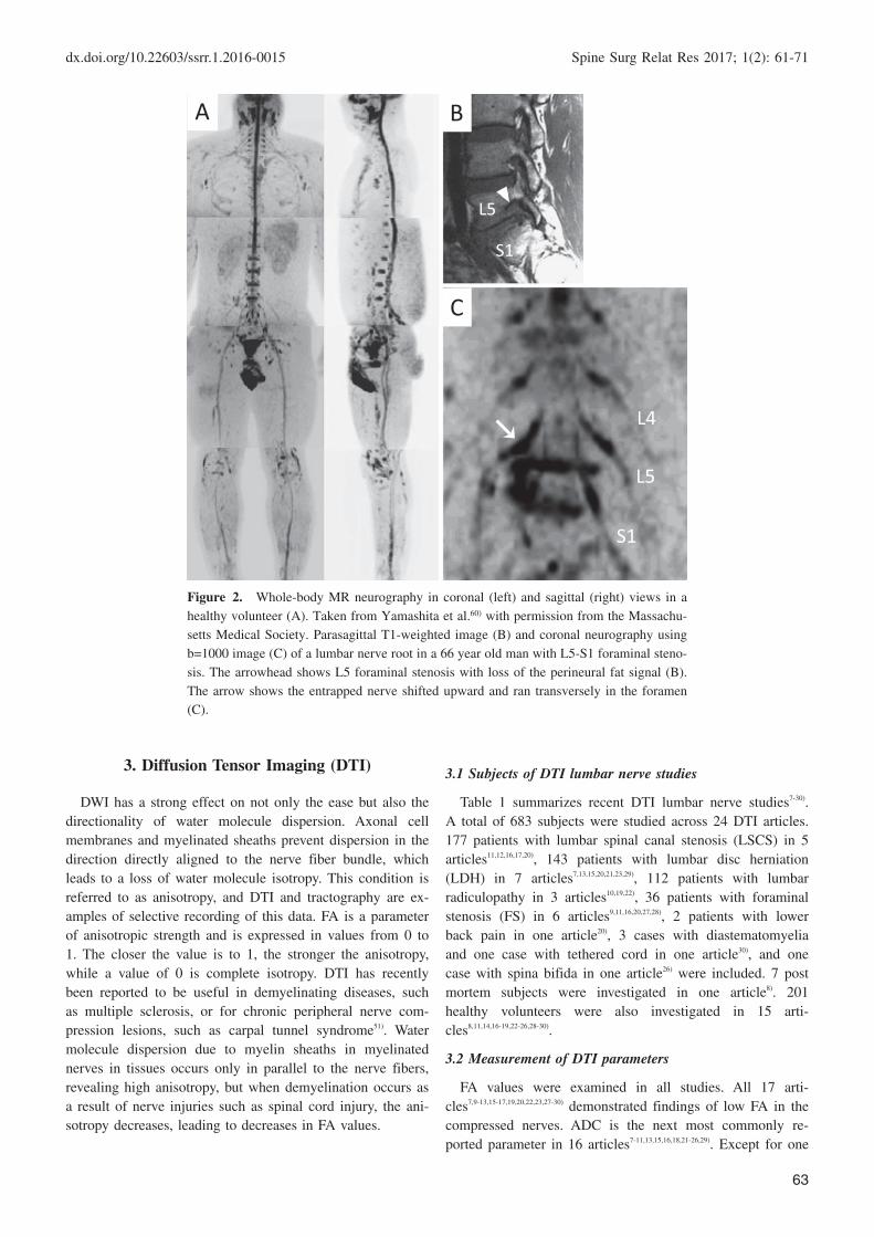
ity54). Yamashita and Takahara et al.55) (Fig. 2A) demonstrated
the feasibility of whole-body MR neurography with DWI
that can visualize tissues with impeded diffusion, such as tu-
mors, the brain, the spinal cord, and the peripheral nerves56).
We found that diffusion-weighted MR neurography revealed
that nerve swelling and transverse and mean ADC values
were significantly greater in entrapped nerve roots in pa-
tients with lumbar disc herniation39) and lumbar foraminal
stenosis (Fig. 2B, C)40), suggesting demyelination and edema
by slow compression caused by an increased degree of dif-
fusion compared with that of normal tissue.
However, this method requires restructuring of 3D nerve
images with maximum intensity projection (MIP) processing
which is a volume rendering method for 3D imaging that
projects the voxels with maximum intensity of the axial im-
age, which leads to limitations in image resolution.
dx.doi.org/10.22603/ssrr.1.2016-0015 Spine Surg Relat Res 2017; 1(2): 61-71
63
Figure 2. Whole-body MR neurography in coronal (left) and sagittal (right) views in a
healthy volunteer (A). Taken from Yamashita et al.60) with permission from the Massachu-
setts Medical Society. Parasagittal T1-weighted image (B) and coronal neurography using
b=1000 image (C) of a lumbar nerve root in a 66 year old man with L5-S1 foraminal steno-
sis. The arrowhead shows L5 foraminal stenosis with loss of the perineural fat signal (B).
The arrow shows the entrapped nerve shifted upward and ran transversely in the foramen
(C).
L5
S1
L4
L5
S1
A B
C
3. Diffusion Tensor Imaging (DTI)
DWI has a strong effect on not only the ease but also the
directionality of water molecule dispersion. Axonal cell
membranes and myelinated sheaths prevent dispersion in the
direction directly aligned to the nerve fiber bundle, which
leads to a loss of water molecule isotropy. This condition is
referred to as anisotropy, and DTI and tractography are ex-
amples of selective recording of this data. FA is a parameter
of anisotropic strength and is expressed in values from 0 to
1. The closer the value is to 1, the stronger the anisotropy,
while a value of 0 is complete isotropy. DTI has recently
been reported to be useful in demyelinating diseases, such
as multiple sclerosis, or for chronic peripheral nerve com-
pression lesions, such as carpal tunnel syndrome51). Water
molecule dispersion due to myelin sheaths in myelinated
nerves in tissues occurs only in parallel to the nerve fibers,
revealing high anisotropy, but when demyelination occurs as
a result of nerve injuries such as spinal cord injury, the ani-
sotropy decreases, leading to decreases in FA values.
3.1 Subjects of DTI lumbar nerve studies
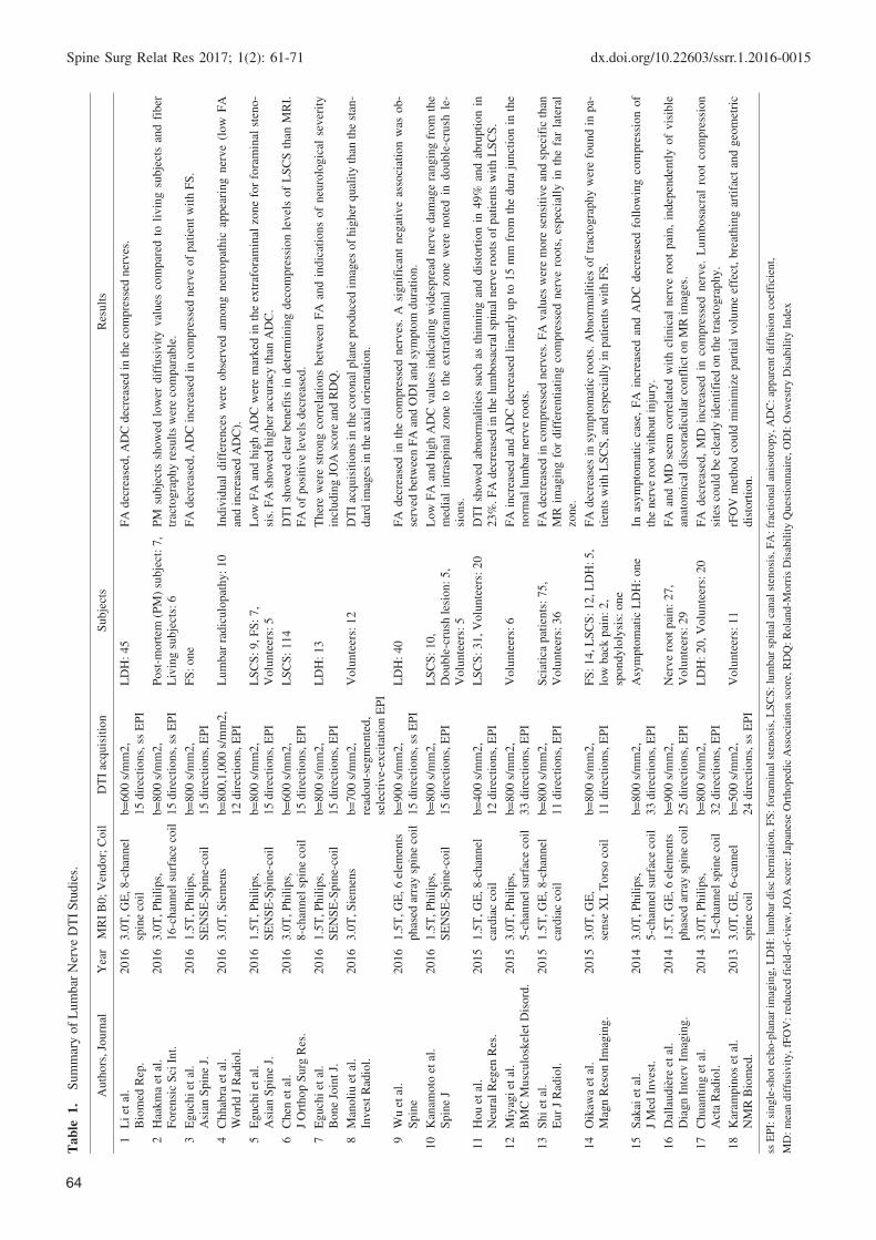
Table 1 summarizes recent DTI lumbar nerve studies7-30).
A total of 683 subjects were studied across 24 DTI articles.
177 patients with lumbar spinal canal stenosis (LSCS) in 5
articles11,12,16,17,20), 143 patients with lumbar disc herniation
(LDH) in 7 articles7,13,15,20,21,23,29), 112 patients with lumbar
radiculopathy in 3 articles10,19,22), 36 patients with foraminal
stenosis (FS) in 6 articles9,11,16,20,27,28), 2 patients with lower
back pain in one article20), 3 cases with diastematomyelia
and one case with tethered cord in one article30), and one
case with spina bifida in one article26) were included. 7 post
mortem subjects were investigated in one article8). 201
healthy volunteers were also investigated in 15 arti-
cles8,11,14,16-19,22-26,28-30).
3.2 Measurement of DTI parameters
FA values were examined in all studies. All 17 arti-
cles7,9-13,15-17,19,20,22,23,27-30) demonstrated findings of low FA in the
compressed nerves. ADC is the next most commonly re-
ported parameter in 16 articles7-11,13,15,16,18,21-26,29). Except for one
Spine Surg Relat Res 2017; 1(2): 61-71 dx.doi.org/10.22603/ssrr.1.2016-0015
64
Tab
le 1
. S
um
mar
y o
f L
um
bar
Ner
ve
DT
I S
tud
ies.
Auth
ors
, Jo
urn
alY
ear
MR
I B
0;
Ven
dor;
Coil
DT
I ac
quis
itio
nS
ubje
cts
Res
ult
s
1
Li
et a
l.
Bio
med
Rep
.
2016
3.0
T, G
E, 8-c
han
nel
spin
e co
il
b=
600 s
/mm
2,
15 d
irec
tions,
ss
EP
I
LD
H:
45
FA
dec
reas
ed, A
DC
dec
reas
ed i
n t
he
com
pre
ssed
ner
ves
.
2
Haa
km
a et
al.
Fore
nsi
c S
ci I
nt.
2016
3.0
T, P
hil
ips,
16-c
han
nel
surf
ace
coil
b=
800 s
/mm
2,
15 d
irec
tions,
ss
EP
I
Post
-mort
em (
PM
) su
bje
ct:
7,
Liv
ing s
ubje
cts:
6
PM
subje
cts
show
ed l
ow
er d
iffu
sivit
y v
alues
com
par
ed t
o l
ivin
g s
ubje
cts
and f
iber
trac
togra
phy r
esult
s w
ere
com
par
able
.
3
Eguch
i et
al.
Asi
an S
pin
e J.
2016
1.5
T, P
hil
ips,
SE
NS
E-S
pin
e-co
il
b=
800 s
/mm
2,
15 d
irec
tions,
EP
I
FS
: one
FA
dec
reas
ed, A
DC
incr
ease
d i
n c
om
pre
ssed
ner
ve
of
pat
ient
wit
h F
S.
4
Chhab
ra e
t al
.
Worl
d J
Rad
iol.
2016
3.0
T, S
iem
ens
b=
800,1
,000 s
/mm
2,
12 d
irec
tions,
EP
I
Lum
bar
rad
iculo
pat
hy:
10
Indiv
idual
dif
fere
nce
s w
ere
obse
rved
am
ong n
euro
pat
hic
appea
ring n
erve
(low
FA
and i
ncr
ease
d A
DC
).
5
Eguch
i et
al.
Asi
an S
pin
e J.
2016
1.5
T, P
hil
ips,
SE
NS
E-S
pin
e-co
il
b=
800 s
/mm
2,
15 d
irec
tions,
EP
I
LS
CS
: 9, F
S:
7,
Volu
nte
ers:
5
Low
FA
and h
igh A
DC
wer
e m
arked
in t
he
extr
afora
min
al z
one
for
fora
min
al s
teno-
sis.
FA
show
ed h
igher
acc
ura
cy t
han
AD
C.
6
Chen
et
al.
J O
rthop S
urg
Res
.
2016
3.0
T, P
hil
ips,
8-c
han
nel
spin
e co
il
b=
600 s
/mm
2,
15 d
irec
tions,
EP
I
LS
CS
: 114
DT
I sh
ow
ed c
lear
ben
efit
s in
det
erm
inin
g d
ecom
pre
ssio
n l
evel
s of
LS
CS
than
MR
I.
FA
of
posi
tive
level
s dec
reas
ed.
7
Eguch
i et
al.
Bone
Join
t J.
2016
1.5
T, P
hil
ips,
SE
NS
E-S
pin
e-co
il
b=
800 s
/mm
2,
15 d
irec
tions,
EP
I
LD
H:
13
Ther
e w
ere
stro
ng c
orr
elat
ions
bet
wee
n F
A a
nd i
ndic
atio
ns
of
neu
rolo
gic
al s
ever
ity
incl
udin
g J
OA
sco
re a
nd R
DQ
.
8
Man
oli
u e
t al
.
Inves
t R
adio
l.
2016
3.0
T, S
iem
ens
b=
700 s
/mm
2,
read
out-
segm
ente
d,
sele
ctiv
e-ex
cita
tion E
PI
Volu
nte
ers:
12
DT
I ac
quis
itio
ns
in t
he
coro
nal
pla
ne
pro
duce
d i
mag
es o
f hig
her
qual
ity t
han
the
stan
-
dar
d i
mag
es i
n t
he
axia
l ori
enta
tion.
9
Wu e
t al
.
Spin
e
2016
1.5
T, G
E, 6 e
lem
ents
phas
ed a
rray
spin
e co
il
b=
900 s
/mm
2,
15 d
irec
tions,
ss
EP
I
LD
H:
40
FA
dec
reas
ed i
n t
he
com
pre
ssed
ner
ves
. A
sig
nif
ican
t neg
ativ
e as
soci
atio
n w
as o
b-
serv
ed b
etw
een F
A a
nd O
DI
and s
ym
pto
m d
ura
tion.
10
Kan
amoto
et
al.
Spin
e J
2016
1.5
T, P
hil
ips,
SE
NS
E-S
pin
e-co
il
b=
800 s
/mm
2,
15 d
irec
tions,
EP
I
LS
CS
: 10,
Double
-cru
sh l
esio
n:
5,
Volu
nte
ers:
5
Low
FA
and h
igh A
DC
val
ues
indic
atin
g w
ides
pre
ad n
erve
dam
age
rangin
g f
rom
the
med
ial
intr
aspin
al z
one
to t
he
extr
afora
min
al z
one
wer
e note
d i
n d
ouble
-cru
sh l
e-
sions.
11
Hou e
t al
.
Neu
ral
Reg
en R
es.
2015
1.5
T, G
E, 8-c
han
nel
card
iac
coil
b=
400 s
/mm
2,
12 d
irec
tions,
EP
I
LS
CS
: 31, V
olu
nte
ers:
20
DT
I sh
ow
ed a
bnorm
alit
ies
such
as
thin
nin
g a
nd d
isto
rtio
n i
n 4
9%
and a
bru
pti
on i
n
23%
. F
A d
ecre
ased
in t
he
lum
bosa
cral
spin
al n
erve
roots
of
pat
ients
wit
h L
SC
S.
12
Miy
agi
et a
l.
BM
C M
usc
ulo
skel
et D
isord
.
2015
3.0
T, P
hil
ips,
5-c
han
nel
surf
ace
coil
b=
800 s
/mm
2,
33 d
irec
tions,
EP
I
Volu
nte
ers:
6F
A i
ncr
ease
d a
nd A
DC
dec
reas
ed l
inea
rly u
p t
o 1
5 m
m f
rom
the
dura
junct
ion i
n t
he
norm
al l
um
bar
ner
ve
roots
.
13
Shi
et a
l.
Eur
J R
adio
l.
2015
1.5
T, G
E, 8-c
han
nel
card
iac
coil
b=
800 s
/mm
2,
11 d
irec
tions,
EP
I
Sci
atic
a pat
ients
: 75,
Volu
nte
ers:
36
FA
dec
reas
ed i
n c
om
pre
ssed
ner
ves
. F
A v
alues
wer
e m
ore
sen
siti
ve
and s
pec
ific
than
MR
im
agin
g f
or
dif
fere
nti
atin
g c
om
pre
ssed
ner
ve
roots
, es
pec
iall
y i
n t
he
far
late
ral
zone.
14
Oik
awa
et a
l.
Mag
n R
eson I
mag
ing.
2015
3.0
T, G
E,
sense
XL
Tors
o c
oil
b=
800 s
/mm
2,
11 d
irec
tions,
EP
I
FS
: 14, L
SC
S:
12, L
DH
: 5,
low
bac
k p
ain:
2,
spondylo
lysi
s: o
ne
FA
dec
reas
es i
n s
ym
pto
mat
ic r
oots
. A
bnorm
alit
ies
of
trac
togra
phy w
ere
found i
n p
a-
tien
ts w
ith L
SC
S, an
d e
spec
iall
y i
n p
atie
nts
wit
h F
S.
15
Sak
ai e
t al
.
J M
ed I
nves
t.
2014
3.0
T, P
hil
ips,
5-c
han
nel
surf
ace
coil
b=
800 s
/mm
2,
33 d
irec
tions,
EP
I
Asy
mpto
mat
ic L
DH
: one
In a
sym
pto
mat
ic c
ase,
FA
incr
ease
d a
nd A
DC
dec
reas
ed f
oll
ow
ing c
om
pre
ssio
n o
f
the
ner
ve
root
wit
hout
inju
ry.
16
Dal
laudiè
re e
t al
.
Dia
gn I
nte
rv I
mag
ing.
2014
1.5
T, G
E, 6 e
lem
ents
phas
ed a
rray
spin
e co
il
b=
900 s
/mm
2,
25 d
irec
tions,
EP
I
Ner
ve
root
pai
n:
27,
Volu
nte
ers:
29
FA
and M
D s
eem
corr
elat
ed w
ith c
linic
al n
erve
root
pai
n,
indep
enden
tly o
f vis
ible
anat
om
ical
dis
cora
dic
ula
r co
nfl
ict
on M
R i
mag
es.
17
Chuan
ting e
t al
.
Act
a R
adio
l.
2014
3.0
T, P
hil
ips,
15-c
han
nel
spin
e co
il
b=
800 s
/mm
2,
32 d
irec
tions,
EP
I
LD
H:
20, V
olu
nte
ers:
20
FA
dec
reas
ed,
MD
incr
ease
d i
n c
om
pre
ssed
ner
ve.
Lum
bosa
cral
root
com
pre
ssio
n
site
s co
uld
be
clea
rly i
den
tifi
ed o
n t
he
trac
togra
phy.
18
Kar
ampin
os
et a
l.
NM
R B
iom
ed.
2013
3.0
T, G
E, 6-c
annel
spin
e co
il
b=
500 s
/mm
2,
24 d
irec
tions,
ss
EP
I
Volu
nte
ers:
11
rFO
V m
ethod c
ould
min
imiz
e par
tial
volu
me
effe
ct,
bre
athin
g a
rtif
act
and g
eom
etri
c
dis
tort
ion.
ss E
PI:
sin
gle
-shot
echo-p
lanar
im
agin
g, L
DH
: lu
mbar
dis
c her
nia
tion, F
S:
fora
min
al s
tenosi
s, L
SC
S:
lum
bar
spin
al c
anal
ste
nosi
s, F
A:
frac
tional
anis
otr
opy, A
DC
: ap
par
ent
dif
fusi
on c
oef
fici
ent,
MD
: m
ean d
iffu
sivit
y, fF
OV
: re
duce
d f
ield
-of-
vie
w, JO
A s
core
: Ja
pan
ese
Ort
hoped
ic A
ssoci
atio
n s
core
, R
DQ
: R
ola
nd-M
orr
is D
isab
ilit
y Q
ues
tionnai
re, O
DI:
Osw
estr
y D
isab
ilit
y I
ndex
dx.doi.org/10.22603/ssrr.1.2016-0015 Spine Surg Relat Res 2017; 1(2): 61-71
65
Auth
ors
, Jo
urn
alY
ear
MR
I B
0;
Ven
dor;
Coil
DT
I ac
quis
itio
nS
ubje
cts
Res
ult
s
19
Budzi
k e
t al
.
Eur
Rad
iol.
2013
3.0
Phil
ips,
32 c
han
nel
card
iac
coil
b=
700 s
/mm
2,
15 d
irec
tions,
ss
EP
I
Volu
nte
ers:
8rF
OV
im
ages
of
lum
bar
ner
ves
wer
e as
sess
ed.
20
van
der
Jag
t et
al.
Neu
roim
age.
2012
3.0
T, P
hil
ips,
16-c
han
nel
surf
ace
coil
b=
800 s
/mm
2,
15 d
irec
tions,
ss
EP
I
Spin
a bif
ida:
one,
Volu
nte
ers:
10
Mea
sure
of
FA
, M
D,
AD
and R
D v
alues
in h
ealt
hy v
olu
nte
ers.
Abnorm
al d
iffu
sion
findin
gs
are
vis
ual
ized
in t
he
spin
a bif
ida
21
Kit
amura
et
al.
Spin
e
2012
3.0
T, G
E,
sense
XL
tors
o c
oil
b=
800 s
/mm
2,
11 d
irec
tions,
EP
I
Far
-out
s yndro
me:
one
FA
dec
reas
ed i
n c
om
pre
ssed
ner
ve
of
pat
ient
wit
h F
ar-o
ut
syndro
me.
22
Eguch
i et
al.
AJN
R
2011
3.0
T, G
E,
sense
XL
Tors
o c
oil
b=
800 s
/mm
2,
11 d
irec
tions,
EP
I
FS
: 8, V
olu
nte
ers:
8F
A d
ecre
ased
in c
om
pre
ssed
ner
ve
of
pat
ient
wit
h F
S.
23
Bal
bi
et a
l.
Eur
Rad
iol.
2011
1.5
T, P
hil
ips,
sense
spin
e co
il
b=
900 s
/mm
2,
25 d
irec
tions,
ss
EP
I
LD
H:
19, V
olu
nte
ers:
19
FA
dec
reas
ed, M
D i
ncr
ease
d i
n c
om
pre
ssed
ner
ve.
24
Fil
ippi
et a
l.
Eur
Rad
iol.
2010
3.0
T, P
hil
ips,
15-c
han
nel
spin
e co
il
b=
400 s
/mm
2,
ssh E
PI
Tet
her
ed c
ord
: one,
Dia
stem
atom
yel
ia:
3,
Volu
nte
ers:
6
FA
dec
reas
ed i
n t
he
teth
ered
cord
s.
ss E
PI:
sin
gle
-shot
echo-p
lanar
im
agin
g, L
DH
: lu
mbar
dis
c her
nia
tion, F
S:
fora
min
al s
tenosi
s, L
SC
S:
lum
bar
spin
al c
anal
ste
nosi
s, F
A:
frac
tional
anis
otr
opy, A
DC
: ap
par
ent
dif
fusi
on c
oef
fici
ent,
MD
: m
ean d
iffu
sivit
y, fF
OV
: re
duce
d f
ield
-of-
vie
w, JO
A s
core
: Ja
pan
ese
Ort
hoped
ic A
ssoci
atio
n s
core
, R
DQ
: R
ola
nd-M
orr
is D
isab
ilit
y Q
ues
tionnai
re, O
DI:
Osw
estr
y D
isab
ilit
y I
ndex
Tab
le 1
. co
nti
nu
ed.
article7) (which showed low ADC), 9 articles9-11,13,15,16,22,23,29) de-
scribed high ADC in involved nerves and 5 articles demon-
strated findings of ADC in healthy subjects8,18,24-26). In an as-
ymptomatic case, FA increased and ADC decreased follow-
ing compression of the nerve root without injury21).
Takagi et al.57) demonstrated that the FA values of the pe-
ripheral nerves were more strongly correlated with the ax-
onal degeneration and regeneration in rat and mouse sciatic
nerves. Generally, increased ADC may be due to inflamma-
tion or oedema, whereas decreased FA may reflect damaged
tissue microstructure, demyelination, axonal loss, or increase
in isotropic water volume57). A systematic review of spinal
cord MRI techniques including DTI found that FA value has
the most robust support for clinical utility42).
3.3 DTI acquisition parameters
Parameters including magnetic field strength, motion-
probing gradient (MPG), and b-values are important in the
clinical use of DTI. High magnetic field strengths have the
advantages of greater signal-to-noise ratio, improved spatial
resolution, and faster imaging times, but also have the disad-
vantage of the magnetic susceptibility artifact. Magnetic sus-
ceptibility artifact indicated MRI artifacts that share distor-
tions due to local inhomogeneous magnetic field. Fifteen re-
ports used 3T, and 9 used 1.5T, so it appears that 3T re-
search accounted for the majority.
The methods of measuring in vivo diffusion by MRI have
been based mainly on the addition of several MPGs to the
spin echo sequence to produce signal attenuation for the
spins moving at random. Additional MPG directions can in-
crease the resolution, but will prolong the scan time. MPG
directions ranged from 11 to 33, where 15 directions was
most common, used in 10 reports.
The b-value is a factor that defines gradient strength and
duration and determines the degree of diffusion weighted
images typically using values from 0 to 1000 s/mm2. The
higher the b-value, the stronger diffusion effects. Increasing
the b-value increases the diffusion sensitivity, but decreases
the signal-to-noise ratio. B-values ranged between 400 and
900, with b=800 the most common, used in 14 reports (Ta-
ble 1).
3.4 Diagnosis of lumbar foraminal stenosis with DTI
In patients with lumbar foraminal stenosis, a nerve root or
spinal nerve is entrapped in a narrowed lumbar foramen as
the result of a degenerative lumbar spinal disorder. At this
site, a dorsal root ganglion functions as a pain receptor;
thus, the condition is refractory and may cause severe lower
limb pain58). Macnab et al.59) appropriately named lumbar fo-
raminal stenosis the “hidden zone,” as it is often overlooked,
it accounts for approximately 60% of failed back surgery
syndromes, and it decreases surgical success rates60).
Diagnostic imaging of lumbar spinal canal stenosis in-
cludes X-rays, CT, and MRI61-63), along with functional diag-
nosis via selective nerve root imaging and infiltration64).
Conventional MRI has a false positive rate of 30% to 40%
Spine Surg Relat Res 2017; 1(2): 61-71 dx.doi.org/10.22603/ssrr.1.2016-0015
66
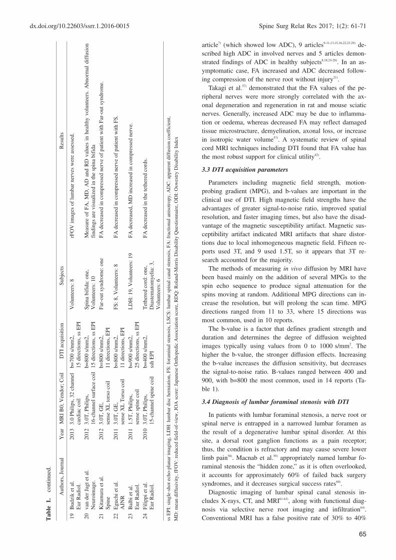
Figure 3. Coronal tractography of a lumbar nerve in a healthy volunteer (A). L3, L4, L5, and S1
indicate the third, fourth, and fifth lumbar root, and the first sacral root, respectively. Tractography
(B) and parasagittal T1-weighted image (C) of a lumbar nerve root in a 75 year old man with right
L5-S1 foraminal stenosis. Tractography showed disruption in the course through the foramen.
L3
L4
L5
S1
L3
L4
L5
S1
A B
L4
L5
C
in lumbar foraminal stenosis cases; thus, diagnosis is diffi-
cult65). Recently 3D-CT, MR myelography65,66), and 3D-MRI67)
were reported to be useful for diagnosis.
We assessed the usefulness of DTI for the diagnosis of
lumbar foraminal stenosis. Subjects in a supine position
were scanned with a 3T MR imaging scanner (Discovery
MR750; GE Healthcare, Milwaukee, WI) using a Sense XL
Torso coil. We performed DTI with echo-planar imaging at
a B value of 800 s/mm2 and with 11 directional MPGs.
Mean FA values and mean ADC values in the lumbar nerve
roots were quantified on DTI images, and the lumbar nerve
roots were visualized with tractography. In all subjects, the
lumbar nerve roots were clearly visualized (Fig. 3A), and
tractography also showed abnormalities, such as tract disrup-
tion, nerve narrowing, and indentation in their course
through the foramen (Fig. 3B). Mean FA values were sig-
nificantly lower, and mean ADC values were significantly
higher, in entrapped versus intact roots28).
We evaluated the accuracy of these parameters for the di-
agnosis of foraminal stenosis. Changes in DTI parameters
(low FA and high ADC values) were marked in nerve roots
and in the extraforaminal zone in foraminal stenosis. Addi-
tionally, the FA value was more accurate than the ADC
value in the extraforaminal zone, and a low FA value sug-
gested foraminal stenosis11).
L5 radiculopathy may occur when the nerve is com-
pressed at two levels (medial and lateral), for example, when
the L4/5 level is compressed by an intraspinal canal lesion
and the L5/S1 level is compressed by a lateral lesion. This
is called a double-crush lesion. Traditional imaging studies
do not allow a determination of whether the compressing le-
sion is inside or outside of the spinal canal, or if a double-
crush lesion is responsible, which leads to poor surgical suc-
cess rates. The distal latency of L5 has been measured elec-
trophysiologically; however, this is an invasive approach,
and noninvasive diagnostic methods are virtually nonexist-
ent68,69). Diagnosis of L5 nerve damage double-crush lesions
by DTI was studied. Low FA values and high ADC values
indicative of widespread nerve damage from the medial in-
traspinal zone to the extraforaminal zone were found for
double-crush lesions. If double-crush injury is suspected
prior to surgery, DTI assessment may help prevent failed
back surgery syndrome16)(Fig. 4).
3.5 DTI evaluation of radiculopathy in patients with lum-bar disc herniation
Mixter and Barr70) first described radicular pain of the sci-
atica as spinal root compression by a herniated intervertebral
disc; however, the underlying pathophysiology is not well
understood. In clinical practice, asymptomatic intervertebral
disc degeneration and herniation are often found, and these
discrepancies can confuse spine surgeons4,5). We previously
reported a correlation between neurological severity and DTI
parameters, such as the FA and ADC values, in patients with
radiculopathy caused by lumbar disc herniation13). Strong
correlations were found between the FA value and indica-
tions of neurological severity, including the Japanese Ortho-
pedic Association (JOA) score, the Oswestry Disability In-
dx.doi.org/10.22603/ssrr.1.2016-0015 Spine Surg Relat Res 2017; 1(2): 61-71
67
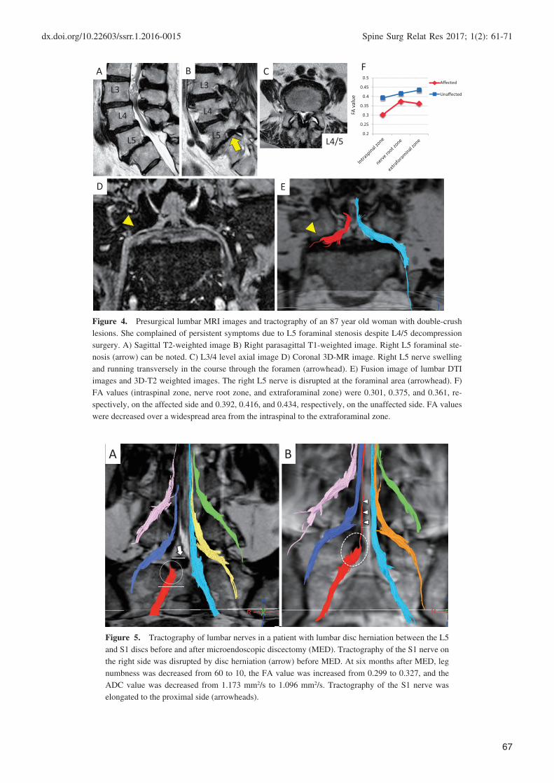
Figure 4. Presurgical lumbar MRI images and tractography of an 87 year old woman with double-crush
lesions. She complained of persistent symptoms due to L5 foraminal stenosis despite L4/5 decompression
surgery. A) Sagittal T2-weighted image B) Right parasagittal T1-weighted image. Right L5 foraminal ste-
nosis (arrow) can be noted. C) L3/4 level axial image D) Coronal 3D-MR image. Right L5 nerve swelling
and running transversely in the course through the foramen (arrowhead). E) Fusion image of lumbar DTI
images and 3D-T2 weighted images. The right L5 nerve is disrupted at the foraminal area (arrowhead). F)
FA values (intraspinal zone, nerve root zone, and extraforaminal zone) were 0.301, 0.375, and 0.361, re-
spectively, on the affected side and 0.392, 0.416, and 0.434, respectively, on the unaffected side. FA values
were decreased over a widespread area from the intraspinal to the extraforaminal zone.
L4/5
L3
L4
L5
L3
L4
L5
A B C
D E
0.2
0.25
0.3
0.35
0.4
0.45
0.5A ct
Una c
FA v
F
Figure 5. Tractography of lumbar nerves in a patient with lumbar disc herniation between the L5
and S1 discs before and after microendoscopic discectomy (MED). Tractography of the S1 nerve on
the right side was disrupted by disc herniation (arrow) before MED. At six months after MED, leg
numbness was decreased from 60 to 10, the FA value was increased from 0.299 to 0.327, and the
ADC value was decreased from 1.173 mm2/s to 1.096 mm2/s. Tractography of the S1 nerve was
elongated to the proximal side (arrowheads).
A B
Spine Surg Relat Res 2017; 1(2): 61-71 dx.doi.org/10.22603/ssrr.1.2016-0015
68
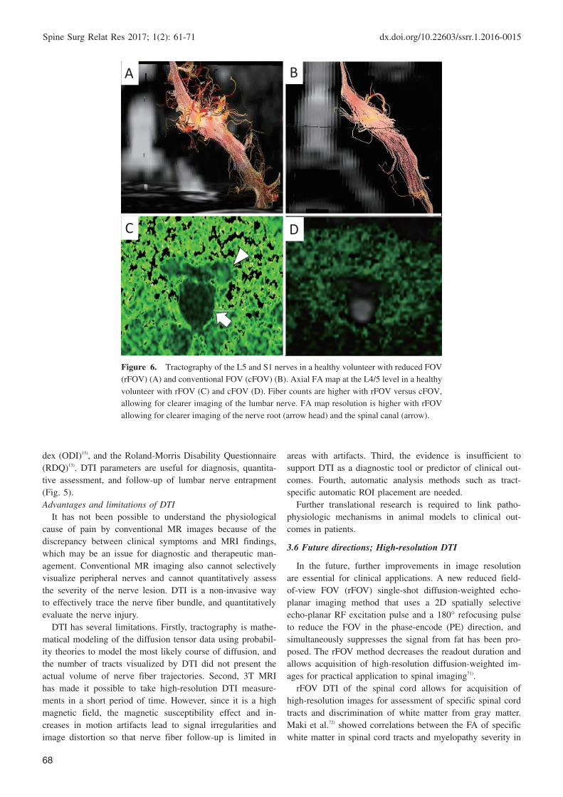
Figure 6. Tractography of the L5 and S1 nerves in a healthy volunteer with reduced FOV
(rFOV) (A) and conventional FOV (cFOV) (B). Axial FA map at the L4/5 level in a healthy
volunteer with rFOV (C) and cFOV (D). Fiber counts are higher with rFOV versus cFOV,
allowing for clearer imaging of the lumbar nerve. FA map resolution is higher with rFOV
allowing for clearer imaging of the nerve root (arrow head) and the spinal canal (arrow).
A B
DC
dex (ODI)15), and the Roland-Morris Disability Questionnaire
(RDQ)13). DTI parameters are useful for diagnosis, quantita-
tive assessment, and follow-up of lumbar nerve entrapment
(Fig. 5).
Advantages and limitations of DTIIt has not been possible to understand the physiological
cause of pain by conventional MR images because of the
discrepancy between clinical symptoms and MRI findings,
which may be an issue for diagnostic and therapeutic man-
agement. Conventional MR imaging also cannot selectively
visualize peripheral nerves and cannot quantitatively assess
the severity of the nerve lesion. DTI is a non-invasive way
to effectively trace the nerve fiber bundle, and quantitatively
evaluate the nerve injury.
DTI has several limitations. Firstly, tractography is mathe-
matical modeling of the diffusion tensor data using probabil-
ity theories to model the most likely course of diffusion, and
the number of tracts visualized by DTI did not present the
actual volume of nerve fiber trajectories. Second, 3T MRI
has made it possible to take high-resolution DTI measure-
ments in a short period of time. However, since it is a high
magnetic field, the magnetic susceptibility effect and in-
creases in motion artifacts lead to signal irregularities and
image distortion so that nerve fiber follow-up is limited in
areas with artifacts. Third, the evidence is insufficient to
support DTI as a diagnostic tool or predictor of clinical out-
comes. Fourth, automatic analysis methods such as tract-
specific automatic ROI placement are needed.
Further translational research is required to link patho-
physiologic mechanisms in animal models to clinical out-
comes in patients.
3.6 Future directions; High-resolution DTI
In the future, further improvements in image resolution
are essential for clinical applications. A new reduced field-
of-view FOV (rFOV) single-shot diffusion-weighted echo-
planar imaging method that uses a 2D spatially selective
echo-planar RF excitation pulse and a 180° refocusing pulse
to reduce the FOV in the phase-encode (PE) direction, and
simultaneously suppresses the signal from fat has been pro-
posed. The rFOV method decreases the readout duration and
allows acquisition of high-resolution diffusion-weighted im-
ages for practical application to spinal imaging71).
rFOV DTI of the spinal cord allows for acquisition of
high-resolution images for assessment of specific spinal cord
tracts and discrimination of white matter from gray matter.
Maki et al.72) showed correlations between the FA of specific
white matter in spinal cord tracts and myelopathy severity in
dx.doi.org/10.22603/ssrr.1.2016-0015 Spine Surg Relat Res 2017; 1(2): 61-71
69
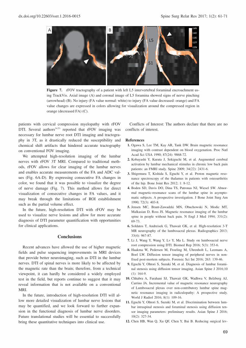
Figure 7. rFOV tractography of a patient with left L5 intervertebral foraminal encroachment us-
ing TrackVis. Axial image (A) and coronal image of L5 foramina showed signs of nerve pinching
(arrowhead) (B). No injury (FA value normal: white) to injury (FA value decreased: orange) and FA
value changes are expressed in colors allowing for visualization around the compressed region in
orange (decreased FA) (C).
L5
S1
A B
patients with cervical compression myelopathy with rFOV
DTI. Several authors24,25) reported that rFOV imaging was
necessary for lumbar nerve root DTI imaging and tractogra-
phy in 3T, as it drastically reduced the susceptibility and
chemical shift artifacts that hindered accurate tractography
on conventional FOV imaging.
We attempted high-resolution imaging of the lumbar
nerves with rFOV 3T MRI. Compared to traditional meth-
ods, rFOV allows for clear imaging of the lumbar nerves
and enables accurate measurements of the FA and ADC val-
ues (Fig. 6A-D). By expressing consecutive FA changes in
color, we found that it was possible to visualize the degree
of nerve damage (Fig. 7). This method allows for direct
visualization of consecutive changes in FA values, and it
may break through the limitations of ROI establishment
such as the partial volume effect.
In the future, high-resolution DTI with rFOV may be
used to visualize nerve lesions and allow for more accurate
diagnosis of DTI parameter quantification with opportunities
for clinical applications.
Conclusions
Recent advances have allowed the use of higher magnetic
fields and pulse sequencing improvements in MRI devices
that provide better neuroimaging, such as DTI in the lumbar
nerves. DTI of spinal nerves is more likely to be affected by
the magnetic rate than the brain; therefore, from a technical
viewpoint, it can hardly be considered a widely employed
test in the field, but reports continue to suggest that it may
reveal information that is not available on a conventional
MRI.
In the future, introduction of high-resolution DTI will al-
low more detailed visualization of lumbar nerve lesions that
may be quantified, and we look forward to further expan-
sion in the functional diagnosis of lumbar nerve disorders.
Future translational studies will be essential to successfully
bring these quantitative techniques into clinical use.
Conflicts of Interest: The authors declare that there are no
conflicts of interest.
References1. Ogawa S, Lee TM, Kay AR, Tank DW. Brain magnetic resonance
imaging with contrast dependent on blood oxygenation. Proc Natl
Acad Sci USA 1990; 87(24): 9868-72.
2. Kobayashi Y, Kurata J, Sekiguchi M, et al. Augmented cerebral
activation by lumbar mechanical stimulus in chronic low back pain
patients: an FMRI study. Spine 2009; 34(22): 2431-6.
3. Shigemura T, Kishida S, Eguchi Y, et al. Proton magnetic reso-
nance spectroscopy of the thalamus in patients with osteoarthritis
of the hip. Bone Joint Res 2012; 1: 8-12.
4. Boden SD, Davis DO, Dina TS, Patronas NJ, Wiesel SW. Abnor-
mal magnetic-resonance scans of the lumbar spine in asympto-
matic subjects. A prospective investigation. J Bone Joint Surg Am
1990; 72(3): 403-8.
5. Jensen MC, Brant-Zawadzki MN, Obuchowski N, Modic MT,
Malkasian D, Ross JS. Magnetic resonance imaging of the lumbar
spine in people without back pain. N Engl J Med 1994; 331(2):
69-73.
6. Soldatos T, Andreisek G, Thawait GK, et al. High-resolution 3-T
MR neurography of the lumbosacral plexus. Radiographics 2013;
33(4): 967-87.
7. Li J, Wang Y, Wang Y, Lv Y, Ma L. Study on lumbosacral nerve
root compression using DTI. Biomed Rep 2016; 5(3): 353-6.
8. Haakma W, Pedersen M, Froeling M, Uhrenholt L, Leemans A,
Boel LW. Diffusion tensor imaging of peripheral nerves in non-
fixed post-mortem subjects. Forensic Sci Int 2016; 263: 139-46.
9. Eguchi Y, Ohtori S, Suzuki M, et al. Diagnosis of lumbar forami-
nal stenosis using diffusion tensor imaging. Asian Spine J 2016;10
(1): 164-9.
10. Chhabra A, Farahani SJ, Thawait GK, Wadhwa V, Belzberg AJ,
Carrino JA. Incremental value of magnetic resonance neurography
of Lumbosacral plexus over non-contributory lumbar spine mag-
netic resonance imaging in radiculopathy: A prospective study.
World J Radiol 2016; 8(1): 109-16.
11. Eguchi Y, Ohtori S, Suzuki M, et al. Discrimination between lum-
bar intraspinal stenosis and foraminal stenosis using diffusion ten-
sor imaging parameters: preliminary results. Asian Spine J 2016;
10(2): 327-34.
12. Chen HB, Wan Q, Xu QF, Chen Y, Bai B. Reducing surgical lev-
Spine Surg Relat Res 2017; 1(2): 61-71 dx.doi.org/10.22603/ssrr.1.2016-0015
70
els by paraspinal mapping and diffusion tensor imaging techniques
in lumbar spinal stenosis. J Orthop Surg Res 2016; 11(1): 47.
13. Eguchi Y, Oikawa Y, Suzuki M, et al. Diffusion tensor imaging of
radiculopathy in patients with lumbar disc herniation: preliminary
results. Bone Joint J 2016; 98-B(3): 387-94.
14. Manoliu A, Ho M, Nanz D, et al. Diffusion tensor imaging of
lumbar nerve roots: comparison between fast readout-segmented
and selective-excitation acquisitions. Invest Radiol 2016; 51(8):
499-504.
15. Wu W, Liang J, Ru N, et al. Microstructural changes in com-
pressed nerve roots are consistent with clinical symptoms and
symptom duration in patients with lumbar disc herniation. Spine
2016; 41(11): E661-6.
16. Kanamoto H, Eguchi Y, Suzuki M, et al. The diagnosis of double-
crush lesion in the L5 lumbar nerve using diffusion tensor imag-
ing. Spine J 2016; 16(3): 315-21.
17. Hou ZJ, Huang Y, Fan ZW, Li XC, Cao BY. Changes in lum-
bosacral spinal nerve roots on diffusion tensor imaging in spinal
stenosis. Neural Regen Res 2015; 10(11): 1860-4.
18. Miyagi R, Sakai T, Yamabe E, Yoshioka H. Consecutive assess-
ment of FA and ADC values of normal lumbar nerve roots from
the junction of the dura mater. BMC Musculoskelet Disord 2015;
16: 156.
19. Shi Y, Zong M, Xu X, et al. Diffusion tensor imaging with quanti-
tative evaluation and fiber tractography of lumbar nerve roots in
sciatica. Eur J Radiol 2015; 84(4): 690-5.
20. Oikawa Y, Eguchi Y, Inoue G, et al. Diffusion tensor imaging of
lumbar spinal nerve in subjects with degenerative lumbar disor-
ders. Magn Reson Imaging 2015; 33(8): 956-61.
21. Sakai T, Miyagi R, Yamabe E, Fujinaga Y, N Bhatia N, Yoshioka
H. Diffusion-weighted imaging and diffusion tensor imaging of as-
ymptomatic lumbar disc herniation. J Med Invest 2014; 61(1-2):
197-203.
22. Dallaudière B, Lincot J, Hess A, et al. Clinical relevance of diffu-
sion tensor imaging parameters in lumbar disco-radicular conflict.
Diagn Interv Imaging 2014; 95(1): 63-8.
23. Chuanting L, Qingzheng W, Wenfeng X, Yiyi H, Bin Z. 3.0T MRI
tractography of lumbar nerve roots in disc herniation. Acta Radiol
2014; 55(8): 969-75.
24. Karampinos DC, Melkus G, Shepherd TM, et al. Diffusion tensor
imaging and T2 relaxometry of bilateral lumbar nerve roots: feasi-
bility of in-plane imaging. NMR Biomed 2013; 26(6): 630-7.
25. Budzik JF, Verclytte S, Lefebvre G, Monnet A, Forzy G, Cotten A.
Assessment of reduced field of view in diffusion tensor imaging of
the lumbar nerve roots at 3 T. Eur Radiol 2013; 23(5): 1361-6.
26. van der Jagt PK, Dik P, Froeling M, et al. Architectural configura-
tion and microstructural properties of the sacral plexus: a diffusion
tensor MRI and fiber tractography study. Neuroimage 2012; 62(3):
1792-9.
27. Kitamura M, Eguchi Y, Inoue G, et al. A case of symptomatic
extra-foraminal lumbosacral stenosis (“far-out syndrome”) diag-
nosed by diffusion tensor imaging. Spine 2012; 37(14): E854-7.
28. Eguchi Y, Ohtori S, Orita S, et al. Quantitative evaluation and
visualization of lumbar foraminal nerve root entrapment by using
diffusion tensor imaging: preliminary results. AJNR Am J Neuro-
radiol 2011; 32(10): 1824-9.
29. Balbi V, Budzik JF, Duhamel A, Bera-Louville A, Le Thuc V, Cot-
ten A. Tractography of lumbar nerve roots: initial results. Eur Ra-
diol 2011; 21(6): 1153-9.
30. Filippi CG, Andrews T, Gonyea JV, Linnell G, Cauley KA. Mag-
netic resonance diffusion tensor imaging and tractography of the
lower spinal cord: application to diastematomyelia and tethered
cord. Eur Radiol 2010; 20(9): 2194-9.
31. Ishikawa T, Asakura K, Mizutani Y, et al. Magnetic resonance
neurography for the evaluation of CIDP. Muscle Nerve. 2016 in
press.
32. Chhabra A, Carrino JA, Farahani SJ, et al. Whole-body MR
neurography: Prospective feasibility study in polyneuropathy and
Charcot-Marie-Tooth disease. J Magn Reson Imaging 2016 in
press.
33. Quinn JC, Fruauff K, Lebl DR, et al. Magnetic resonance
neurography of the lumbar plexus at the L4-L5 disc: Development
of a preoperative surgical planning tool for lateral lumbar
transpsoas interbody fusion (LLIF). Spine 2015; 40(12): 942-7.
34. Menezes CM, de Andrade LM, Herrero CF, et al. Diffusion-
weighted magnetic resonance (DW-MR) neurography of the lum-
bar plexus in the preoperative planning of lateral access lumbar
surgery. Eur Spine J 2015; 24(4): 817-26.
35. Kasper JM, Wadhwa V, Scott KM, Rozen S, Xi Y, Chhabra A.
SHINKEI--a novel 3D isotropic MR neurography technique: tech-
nical advantages over 3DIRTSE-based imaging. Eur Radiol 2015;
25(6): 1672-7.
36. Mürtz P, Kaschner M, Lakghomi A, et al. Diffusion-weighted MR
neurography of the brachial and lumbosacral plexus: 3.0 T versus
1.5 T imaging. Eur J Radiol 2015; 84(4): 696-702.
37. Yuh EL, Jain Palrecha S, Lagemann GM, et al. Diffusivity meas-
urements differentiate benign from malignant lesions in patients
with peripheral neuropathy or plexopathy. AJNR Am J Neurora-
diol 2015; 36(1): 202-9.
38. Yoneyama M, Takahara T, Kwee TC, Nakamura M, Tabuchi T.
Rapid high resolution MR neurography with a diffusion-weighted
pre-pulse. Magn Reson Med Sci 2013; 12(2): 111-9.
39. Eguchi Y, Ohtori S, Yamashita M, et al. Diffusion-weighted mag-
netic resonance imaging of symptomatic nerve root of patients
with lumbar disk herniation. Neuroradiology 2011; 53(9): 633-41.
40. Eguchi Y, Ohtori S, Yamashita M, et al. Clinical applications of
diffusion magnetic resonance imaging of the lumbar foraminal
nerve root entrapment. Eur Spine J 2010; 19(11): 1874-82.
41. Zhang Z, Song L, Meng Q, et al. Morphological analysis in pa-
tients with sciatica: a magnetic resonance imaging study using
three-dimensional high-resolution diffusion-weighted magnetic
resonance neurography techniques. Spine 2009; 34(7): E245-50.
42. Martin AR, Aleksanderek I, Cohen-Adad J, et al. Translating state-
of-the-art spinal cord MRI techniques to clinical use: A systematic
review of clinical studies utilizing DTI, MT, MWF, MRS, and
fMRI. Neuroimage Clin 2015; 10: 192-238.
43. Basser PJ, Jones DK. Diffusion tensor MRI: theory, experimental
design and data analysis-a technical review. NMR Biomed 2002;
15: 456-67.
44. Beaulieu C, Allen PS. Determinants of anisotropic water diffusion
in nerves. Magn Reson Med 1994; 31: 394-400.
45. Beaulieu C, Does MD, Snyder RE, et al. Changes in water diffu-
sion due to Wallerian degeneration in peripheral nerve. Magn
Reson Med 1996; 36: 627-31.
46. Basser PJ, Pierpaoli C. Microstructural and physiological features
of tissues elucidated by quantitative-diffusion-tensor MRI. J Magn
Reson B 1996; 111: 209-19.
47. Minematsu K, Fisher M, Li L, et al. Diffusion-weighted magnetic
resonance imaging: rapid and quantitative detection of focal brain
ischemia. Neurology 1992; 42: 235-40.
48. Ohgiya Y, Oka M, Hiwatashi A, et al. Diffusion tensor MR imag-
ing of the cervical spinal cord in patients with multiple sclerosis.
Eur Radiol 2007; 17: 2499-504.
49. Lin X, Tench CR, Morgan PS, Constantinescu CS. Use of com-
dx.doi.org/10.22603/ssrr.1.2016-0015 Spine Surg Relat Res 2017; 1(2): 61-71
71
bined conventional and quantitative MRI to quantify pathology re-
lated to cognitive impairment in multiple sclerosis. J Neurol Neu-
rosurg Psychiatry 2008; 79: 437-441.
50. Hiltunen J, Suortti T, Arvela S, Seppa M, Joensuu R, Hari R. Dif-
fusion tensor imaging and tractography of distal peripheral nerves
at 3 T. Clinical Neurophysiology 2005; 116: 2315-23.
51. Khalil C, Hancart C, Le Thuc V, Chantelot C, Chechin D, Cotton
A. Diffusion tensor imaging and tractography of the median nerve
in carpal tunnel syndrome: preliminary results. Eur Radiol 2008;
18: 2283-91.
52. Kabakci N, Gürses B, Firat Z, et al. Diffusion tensor imaging and
tractography of median nerve: normative diffusion values. Am J
Roentgenol 2007; 189: 923-7
53. MacDonald CL, Dikranian K, Bayly P, Holtzman D, Brody D.
Diffusion tensor imaging reliably detects experimental traumatic
axonal injury and indicates approximate time of injury. J Neurosci
2007; 27: 11869-76.
54. Fujiyoshi K, Yamada M, Nakamura M, et al. In vivo tracing of
neural tracts in the intact and injured spinal cord of marmosets by
diffusion tensor tractography. J Neurosci 2007; 27: 11991-8.
55. Yamashita T, Kwee TC, Takahara T. Whole-body magnetic reso-
nance neurography. N Engl J Med 2009; 361: 538-9.
56. Tsuchiya K, Imai M, Tateishi H, Nitatori T, Fujikawa A, Takemoto
S. Neurography of the spinal nerve roots by diffusion tensor scan-
ning applying motion-probing gradients in six directions. Magn
Reson Med Sci 2007; 6: 1-5.
57. Takagi T, Nakamura M, Yamada M, et al. Visualization of periph-
eral nerve degeneration and regeneration: monitoring with diffu-
sion tensor tractography. Neuroimage 2009; 44: 884-92.
58. Jenis LG, An HS. Spine update. Lumbar foraminal stenosis. Spine
2000; 25: 389-94.
59. MacNab I. Negative disc exploration: an analysis of the causes of
nerve root involvement in sixty-eight patients. J Bone Joint Surg
Am 1971; 53: 891-903.
60. Burton R, Kirkaldy-Willis W, Yong-Hing K, et al. Causes of fail-
ure of surgery on the lumbar spine. Clin Orthop 1981; 157: 191-7.
61. Hasegawa T, An H, Haughton V, et al. Lumbar foraminal stenosis:
critical heights of the intervertebral discs and foramina. J Bone
Joint Surg 1995; 77: 32-8.
62. Kirkaldy-Willis W, Wedge J, Yong-Hing K, et al. Lumbar spinal
nerve lateral entrapment. Clin Orthop 1982; 169: 171-8.
63. Vanderlinden RG. Subarticular entrapment of the dorsal root gan-
glion as a cause of sciatic pain. Spine 1984; 9: 19-22.
64. Herron L. Selective nerve root block in patient selection for lum-
bar surgery: surgical results. J Spinal Disord 1989; 2: 75-9.
65. Aota Y, Niwa T, Yoshikawa K, et al. Magnetic resonance imaging
and magnetic resonance myelography in the presurgical diagnosis
of lumbar foraminal stenosis. Spine 2007; 32: 896-903.
66. Krudy AG. MR myelography using heavily T2-weighted fast spin-
echo pulse sequences with fat presaturation. Am J Roentgenol
1992; 159: 1315-20.
67. Yamada H, Terada M, Iwasaki H, et al. Improved accuracy of di-
agnosis of lumbar intra and/or extra-foraminal stenosis by use of
three-dimensional MR imaging: comparison with conventional MR
imaging. J Orthop Sci 2015; 20(2): 287-94.
68. Iwasaki H, Yoshida M, Yamada H, et al. A new electrophysiologi-
cal method for the diagnosis of extraforaminal stenosis at L5-S1.
Asian Spine J 2014; 8(2): 145-9.
69. Ando M, Tamaki T, Kawakami M, et al. Electrophysiological di-
agnosis using sensory nerve action potential for the intraforaminal
and extraforaminal L5 nerve root entrapment. Eur Spine J 2013;
22: 833-9.
70. Mixter WJ, Barr JS. Rupture of the intervertebral disc with in-
volvement of the spinal canal. N Engl J Med 1934; 211: 210-5.
71. Saritas EU, Cunningham CH, Lee JH, Han ET, Nishimura DG.
DWI of the spinal cord with reduced FOV single-shot EPI. Magn
Reson Med 2008; 60(2): 468-73.
72. Maki S, Koda M, Ota M, et al. Reduced field-of-view diffusion
tensor imaging of the spinal cord shows motor dysfunction of the
lower extremities in patients with cervical compression myelopa-
thy. Spine 2015 in press.
Spine Surgery and Related Research is an Open Access article distributed under
the Creative Commons Attribution‐NonCommercial‐NoDerivatives 4.0 Inter-
national License. To view the details of this license, please visit (https://creative-
commons.org/licenses/by‐nc‐nd/4.0/).