Embed Size (px)
Citation preview

Relation of Red Cell Distribution Widht with Ascending Aortic Diameter in
Bicuspid Aortic Valve Patients Biküspit Aort Kapak Hastalarında Kırmızı Hücre Dağılım Aralığı
Ile Asendan Aort Çapı Arasındaki Ilişki
Abdullah Nabi Aslan1, Serdal Baştuğ1, Muhammed Cihad Çelik1, Hacı Ahmet Kasapkara2, Mehmet Murat Yiğitbaşı2, Yunus Emre Özbebek2,
Engin Bozkurt2 1 Ataturk Education and Research Hospital, Department of Cardiology, Ankara, Turkey
2 Yıldırım Beyazıt University Faculty of Medicine, Department of Cardiology, Ankara, Turkey
Yazışma Adresi / Correspondence: Abdullah Nabi Aslan
Angora homes, Sazyolu avenue, Number:6, Çankaya Street: Mutlukent, Postal code: 06800City/Country: Ankara/TurkeyT: +90 532 780 13 05 E-mail: [email protected]
Geliş Tarihi / Received : 04.02.2018 Kabul Tarihi / Accepted : 10.04.2018
RESEARCH ARTICLE / Araştırma Makalesi
AbstractObjective Red cell distribution widht (RDW) and neutrophil to lymphocyte ratio (NLR) plays a signifi cant role in assessing the severity and progression
of some cardiac conditions. Bicuspid aortic valve (BAV)-associated aortopathy is common and its progression for individual patients is diffi cult to predict. In this study, we aimed to investigate the association between serum levels of RDW, NLR and ascending aortic diameter (AAd) in BAV patients. Sakarya Med J, 2018, 8(2):327-335 )
Materials and Methods
This is a descriptive and methodologic study which included a total of 182 consecutive patients with a BAV. Complete blood counts were analyzed for RDW level and NLR. Patients were divided into two groups based on their AAd and patients with AAd of 3.9 and above were included in group 1, while those below 3.9 in group 2.
Results NLR was signifi cantly higher in group 1 patients than those in group 2. RDW levels were similar. In univariate correlation analysis, a positive correlation was found between AAd and RDW and NLR in group 1 patients. In multivariate logistic regression analysis, RDW (odds ratio (OR): 1.78, 95% confi dence interval (CI): 1.36–2.44, P = 0.01) remained as independent correlates of AAd in the patient population. Receiver operating characteristic (ROC) curve analysis revealed that a RDW measurement >13.0% predicted ascending aort dilatation (AAD) with a sensitivity of 58% and a specifi city of 79%.
Conclusion RDW and NLR are positively correlated with AAd in BAV patients with AAD. These markers may point out the role of infl ammation both in the pathogenesis and progression of AAD in these patients.
Keywords Bicuspid aortic valve, ascending aortic diameter, red cell distribution widht, ascending aortic dilatation
ÖzAmaç Kırmızı kan hücresi dağılım genişliği (KHDG) ve nötrofi l lenfosit oranı (NLO) bazı kardiyak durumların ciddiyetinin ve progresyonunun
değerlendirilmesinde önemli bir rol oynar. Biküspit aortik kapak (BAK)- ilişkili aortopati sık görülür ve progresyonunu bireysel hastalarda tahmin etmek zordur. Bu çalışmada, biz BAK hastalarında KHDG’nin serum düzeyleri ve NLO ile asendan aort çapı (AAÇ) arasındaki ilişkiyi araştırmayı amaçladık. ( Sakarya Tıp Dergisi, 2018, 8(2):327-335 ).
Gereç ve Yöntem
Toplamda 182 ardışık BAK hastası bu tanımlayıcı ve metodolojik çalışmaya alındı. KHDG düzeyi ve NLR için tam kan sayımı incelendi. Hastalar AAÇ’lerine göre 2 gruba ayrıldı ve AAÇ 3.9 ve üzeri olan hastalar grup 1, 3.9’ un altında olan hastalar ise grup 2’ye dahil edildi.
Bulgular NLO grup 1 hastalarında grup 2’dekilere kıyasla anlamlı olarak daha yüksekti. KHDG seviyeleri benzerdi. Tekli korelasyon analizinde, AAÇ ile KHDG ve NLO arasında anlamlı pozitif korelasyon saptandı. Çoklu regresyon analizinde RDW’nin (odds ratio (OR): 1.78, 95% confi dence interval (CI): 1.36 – 2.44, P = 0.01) asendan aort dilatasyonunun bağımsız bir belirteci olduğu belirlendi. Receiver operating characteristic (ROC) analizinde KHDG için > 13.0% sınır değerinin asendan aort dilatasyonu tanısı için %58 duyarlılık ve %79 özgüllüğe sahip olduğu bulundu.
Sonuç KHDG ve NLO asendan aort dilatasyonu olan BAK hastalarında AAÇ ile pozitif koreledir. Bu belirteçler bu hastalarda asendan aort dilatas-yonunun hem patogenezi hem de progresyonunda infl amasyonun rolü olduğunu gösterebilir.
Anahtar Kelimeler
Biküspit aortik kapak, asendan aort çapı, kırmızı kan hücresi dağılım genişliği, asendan aort dilatasyonu
Introduction
Bicuspid aortic valve (BAV) represents the most frequent congenital heart malformation and as
such it may be subject to valvular stenosis, regurgitation or infective endocarditis.1,2 Even in the
absence of echocardiographically relevant valve dysfunction, it may be associated with ascending
aorta dilatation (AAD) at different anatomical levels.3 Dilatation of the tubular ascending aorta
is the most frequent form of bicuspid aortopathy and simultaneously an issue of extensive and
controversial debate. Moreover, even patients with functionally normal BAVs have been shown to
present with larger aortic diameters both at baseline and during follow-up when compared with
their controls.4 The diversity of bicuspid aortopathy is closely related to the bicuspid valve morp-
hology as well as the type of valvular dysfunction, while the progression rates of aortic dilatation
may differ substantially.5,6
Red cell distribution widht (RDW) shows the variability in erythrocytes volume in the peripheral
blood. Neutrophil to lymphocyte ratio (NLR), as an oxidative stress and proinfl ammatory marker,
has recently emerged as a useful indicator to predict cardiovascular risk and adverse outcomes7.
Both RDW and NLR are easily available laboratory parameters and can be quickly measured in
complete blood count (CBC) test. The prognostic importance of these markers have been repor-
ted in a novel study which show an association of RDW and NLR in patients with AAD before.8 The
correlation of RDW and NLR with infl ammatory markers has showed these markers to be indica-
tors of chronic infl ammation in disease progression with high levels resulting in worse outcome.9
The relation of plasma levels of RDW and NLR with ascending aortic diameter (AAd) has not been
established yet in patients with BAV. Therefore, in this study we aimed to investigate the associa-
tion between these parameters and AAd in BAV patients.
Materials and Methods
Patient selection
At this descriptive and methodologic study we prospectively enrolled consecutive patients who
were performed transthoracic echocardiographic examination and were diagnosed to have BAV
between March 2015 and July 2017. Basic data and clinical history of these patients concerning
age, gender, hypertension, diabetes mellitus, hyperlipidemia, cardiovascular diseases and smo-
king were noted. Patients with acute and/or chronic infl ammatory disease, connective tissue di-
sease associated with AAD (Marfan syndrome, Behçet disease etc.), a malignancy, hematological
disease, anemia, alcohol abuse, a history of renal and liver disease were excluded from this study.
The study was approved by the local ethics committee and informed consents of all subjects were
obtained.
Echocardiographic examination
The patients with BAV were re-examined with transthoracic echocardiography by an independent
and experienced cardiologist who was blinded to the clinical or laboratory fi ndings of the study
population. The echocardiographic examinations were carried out using a 2.5 to 3.5 MHz trans-
ducer with the Vingmed System 7 (Vivid 7, GE, Horten, Norway). Two-dimensional (2-D) measu-
rements were performed in the parasternal long-axis view with the patient in left lateral position.
The left atrium (LA) diameter, interventricular septum (IVS) and posterior wall (PoW) thicknesses,
LV end-diastolic diameter (LVEDD) and LV end-systolic diameter (LVESD) were obtained using the
ASLAN et al.Relation of Red Cell Distribution Widht with Ascending
Aortic Diameter In Bicuspid Aortic Valve Patients
Sakarya Med J.2018;8(2):327-335
328
ASLAN et al.Relation of Red Cell Distribution Widht with Ascending Aortic Diameter In Bicuspid Aortic Valve Patients
Sakarya Med J.2018;8(2):327-335
329
M-mode echocardiographic tracings under the guide of 2-D imaging. The left ventricular ejection
fraction (LVEF) was calculated according to the biplane modifi ed Simpson’s rule. The morphology
of the aortic valve was defi ned in the parasternal short axis view. The 2-D echocardiographic
criteria for BAV included the partial or complete fusion of 2 of the aortic valve leafl ets resulting
in either a partial or complete absence of a functional commissure between the fused leafl ets.10
Aortic velocity (m/s) was measured from apical 4 chambers using continuous wave Doppler. 2-D
measurements of the aortic root were made at end-diastole in the parasternal long-axis views using
the leading edge to leading edge technique in the views showing the largest aortic diameters at
three levels as: (1) annulus, (2) sinus of Valsalva and (3) proximal ascending aorta.
Ascending aortic dilatation was determined on demonstration of aortic enlargement relative to the
expected aortic diameter based on age and body size adjusted nomograms as recommended by
the American Heart Association (AHA) guidelines.11
Laboratory measurements
Venous blood samples were drawn into EDTA-treated or plain tubes according to hospital protocol
from all participants at morning hours after a fasting period of 12 hours. Glucose, urea, creatinine
and lipid profi le were determined by standard methods. CBC testing utilized clinical laboratory
methods (Coulter LH 780 Hematology Analyzer, Beckman Coulter Ireland Inc; Mervue, Galway,
Ireland) for total white blood cell (WBC), neutrophil, lymphocyte and platelet counts, haemoglobin
level, mean platelet volume (MPV) and RDW. The reference range for RDW was between 11.0-
14.5%. Baseline NLR was measured by dividing the neutrophil count to lymphocyte count.
Statistical analysis
All statistical analyses were performed with IBM SPSS 19.0 software (IBM Corp., New York, NY,
USA). Data were tested for normal distribution using the Kolmogorov–Smirnov test. Data are exp-
ressed as mean value ± standard deviation for continuous variables, as well as numbers with per-
centage for categorical variables. Unpaired two-sided t-test was used to compare continuous vari-
ables between the study subgroups. Categorical variables were analysed by �2 test or Fisher’s exact
test as appropriate. Correlations between two continuous variables were assessed with Pearson’s
test. Binary logistic regression analysis was used to fi nd the possible independent association
between dilation of the ascending aorta and clinical parameters. P-values of 0.05 or less were
considered statistically signifi cant.
Results
A total of 182 consecutive BAV patients were enrolled into this study. The mean age of the patients
was 41.2 ± 12.4 (range 19-75) years and 68% of them was male. Among 182 patients, 45 with
AAD were included into group 1 whereas remaining patients were included into group 2. The cli-
nical and demographic characteristics of both groups are demonstrated in Table 1. There were no
signifi cant differences between the groups with respect to body mass index, smoking rate, heart
rate, systolic and diastolic blood pressure and incidence of hypertension and diabetes mellitus.
The mean age of BAV patients with AAD was 47 ± 12 (range 22-65) years which was much higher
than that of the BAV patients without AAD (p< 0.001). The rate of male patients were also signi-
fi cantly higher in group 1 compared to group 2 patients (91% vs 67%, respectively, p= 0.002).
Moreover, the incidence of hyperlipidemia was signifi cantly higher in group 1 patients when it was

ASLAN et al.Relation of Red Cell Distribution Widht with Ascending
Aortic Diameter In Bicuspid Aortic Valve Patients
Sakarya Med J.2018;8(2):327-335
330
compared to those in group 2 (51% vs 34%, respectively, p= 0.018).
Table 1. Clinical and demographic characteristics of the patient groups with bicuspid aortic valve
Variables Group 1 (n=45) Group 2 (n=137) P value
Age (years) 47 ± 12 39 ± 11 <0.001
Male, (%) 91 67 0.002
Body mass index (kg/m²) 22.1 ± 6.3 21.9 ± 4.1 0.415
Heart rate, bpm 74 ± 12 75 ± 14 0.695
Smoking rate (%) 40 35 0.234
Systolic blood pressure, mmHg 116 ± 34 114 ± 26 0.204
Diastolic blood pressure, mmHg 75 ± 16 76 ± 18 0.780
Hypertension (%) 12 11 0.902
Diabetes mellitus (%) 10 12 0.740
Hyperlipidemia (%) 51 34 0.018
Echocardiography
LVEDD (cm) 5.1 ± 0.6 4.6 ± 0.4 <0.001
LVESD (cm) 3.4 ± 0.8 2.8 ± 0.4 <0.001
IVS (cm) 1.1 ± 0.21 1.0 ± 0.18 0.058
PoW (cm) 1.0 ± 0.18 1.0 ± 0.13 0.017
LA (cm) 3.5 ± 0.4 3.4 ± 0.4 0.117
LVEF (%) 55 ± 16 63 ± 3 <0.001
Annulus diameter (cm) 2.29 ± 0.19 2.12 ± 0.13 <0.001
Sinus of Valsalva diameter (cm) 3.66 ± 0.13 3.14 ± 0.9 <0.001
Ascending aortic diameter (cm) 4.12 ± 0.27 3.31 ± 0.32 <0.001
Moderate-severe AR (%) 6 (13) 10 (14)
Moderate-severe AS (%) 1 (2) 3 (4)
Data presented as mean ± standard deviation. AR: Aortic regurgitation, AS: Aortic stenosis, IVS : interventricular septum thickness; LA : left atrium diameter; LVEDD : left ventricular end-diastolic diameter; LVEF : left ventricu-lar ejection fraction, LVESD : left ventricular end-systolic diameter; PoW: posterior wall thickness
When echocardiographic parameters were compared, it was detected that LVEDD, LVESD, PoW,
aortic annulus, ascending aorta and sinus of Valsalva diameters were signifi cantly higher whereas
LVEF was signifi cantly lower in group 1 patients than those in group 2. However, LA diameter and
IVS thickness were similar between groups. Mean AAd was 4.12 ± 0.27 cm (range 3.9-4.9 cm) in
the AAD group and 3.31 ± 0.32 cm (range 2.1-3.8 cm) in patients without AAD. Only 6 patients
in the AAD group (13%) had moderate to severe aortic regurgitation.
The laboratory parameters of the patient groups were listed in Table 2. Accordingly, WBC count,
neutrophil count and NLR (2.5 ± 1.6 vs 2.1 ± 0.7, respectively, p< 0.001) were signifi cantly
higher in patients with AAD than those without. However, there was not statistically signifi cant dif-
ferences in terms of haemoglobin, mean platelet volume, RDW levels and platelet count between
the groups. When, lipid profi les were compared, total cholesterol, HDL cholesterol and TG levels
were found to be similar whereas LDL cholesterol level was signifi cantly higher in group 1 patients
than those in group 2 (127 ± 44 vs 107 ± 35 mg/dL, respectively, p= 0.004). Receiver operating
characteristic (ROC) curve analysis revealed that a RDW measurement higher than >13.0% pre-
dicted AAD with a sensitivity of 52% and a specifi city of 79% (AUC 0.553, p= 0.027) in the
study population (Figure).
ASLAN et al.Relation of Red Cell Distribution Widht with Ascending Aortic Diameter In Bicuspid Aortic Valve Patients
Sakarya Med J.2018;8(2):327-335
331
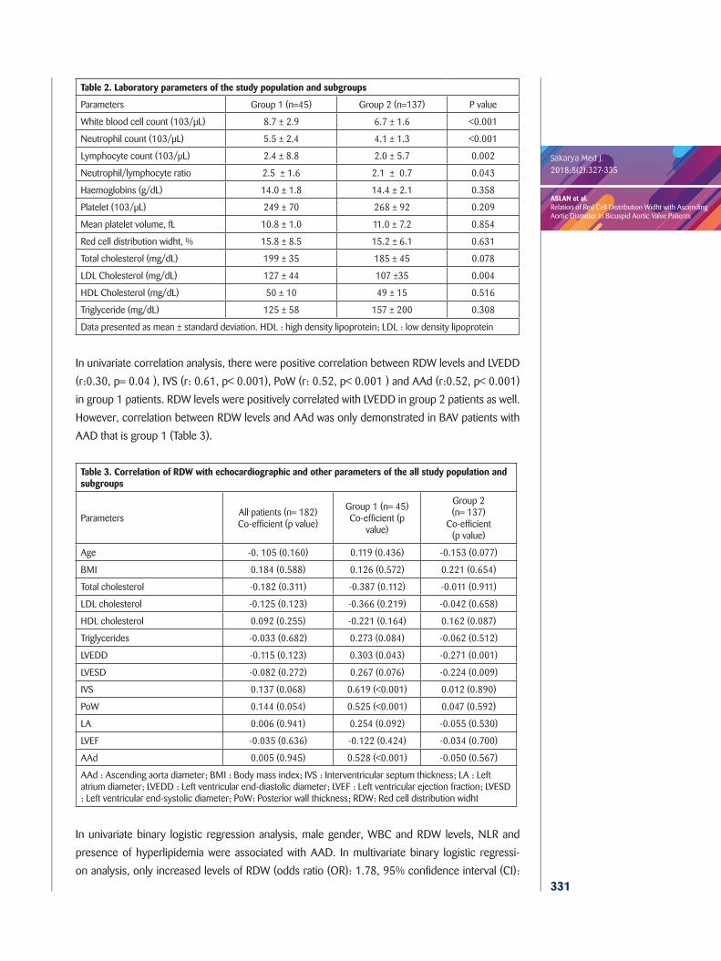
Table 2. Laboratory parameters of the study population and subgroups
Parameters Group 1 (n=45) Group 2 (n=137) P value
White blood cell count (103/µL) 8.7 ± 2.9 6.7 ± 1.6 <0.001
Neutrophil count (103/µL) 5.5 ± 2.4 4.1 ± 1.3 <0.001
Lymphocyte count (103/µL) 2.4 ± 8.8 2.0 ± 5.7 0.002
Neutrophil/lymphocyte ratio 2.5 ± 1.6 2.1 ± 0.7 0.043
Haemoglobins (g/dL) 14.0 ± 1.8 14.4 ± 2.1 0.358
Platelet (103/µL) 249 ± 70 268 ± 92 0.209
Mean platelet volume, fL 10.8 ± 1.0 11.0 ± 7.2 0.854
Red cell distribution widht, % 15.8 ± 8.5 15.2 ± 6.1 0.631
Total cholesterol (mg/dL) 199 ± 35 185 ± 45 0.078
LDL Cholesterol (mg/dL) 127 ± 44 107 ±35 0.004
HDL Cholesterol (mg/dL) 50 ± 10 49 ± 15 0.516
Triglyceride (mg/dL) 125 ± 58 157 ± 200 0.308
Data presented as mean ± standard deviation. HDL : high density lipoprotein; LDL : low density lipoprotein
In univariate correlation analysis, there were positive correlation between RDW levels and LVEDD
(r:0.30, p= 0.04 ), IVS (r: 0.61, p< 0.001), PoW (r: 0.52, p< 0.001 ) and AAd (r:0.52, p< 0.001)
in group 1 patients. RDW levels were positively correlated with LVEDD in group 2 patients as well.
However, correlation between RDW levels and AAd was only demonstrated in BAV patients with
AAD that is group 1 (Table 3).
Table 3. Correlation of RDW with echocardiographic and other parameters of the all study population and subgroups
ParametersAll patients (n= 182) Co-effi cient (p value)
Group 1 (n= 45) Co-effi cient (p
value)
Group 2 (n= 137)
Co-effi cient (p value)
Age -0. 105 (0.160) 0.119 (0.436) -0.153 (0.077)
BMI 0.184 (0.588) 0.126 (0.572) 0.221 (0.654)
Total cholesterol -0.182 (0.311) -0.387 (0.112) -0.011 (0.911)
LDL cholesterol -0.125 (0.123) -0.366 (0.219) -0.042 (0.658)
HDL cholesterol 0.092 (0.255) -0.221 (0.164) 0.162 (0.087)
Triglycerides -0.033 (0.682) 0.273 (0.084) -0.062 (0.512)
LVEDD -0.115 (0.123) 0.303 (0.043) -0.271 (0.001)
LVESD -0.082 (0.272) 0.267 (0.076) -0.224 (0.009)
IVS 0.137 (0.068) 0.619 (<0.001) 0.012 (0.890)
PoW 0.144 (0.054) 0.525 (<0.001) 0.047 (0.592)
LA 0.006 (0.941) 0.254 (0.092) -0.055 (0.530)
LVEF -0.035 (0.636) -0.122 (0.424) -0.034 (0.700)
AAd 0.005 (0.945) 0.528 (<0.001) -0.050 (0.567)
AAd : Ascending aorta diameter; BMI : Body mass index; IVS : Interventricular septum thickness; LA : Left atrium diameter; LVEDD : Left ventricular end-diastolic diameter; LVEF : Left ventricular ejection fraction; LVESD : Left ventricular end-systolic diameter; PoW: Posterior wall thickness; RDW: Red cell distribution widht
In univariate binary logistic regression analysis, male gender, WBC and RDW levels, NLR and
presence of hyperlipidemia were associated with AAD. In multivariate binary logistic regressi-
on analysis, only increased levels of RDW (odds ratio (OR): 1.78, 95% confi dence interval (CI):

ASLAN et al.Relation of Red Cell Distribution Widht with Ascending
Aortic Diameter In Bicuspid Aortic Valve Patients
Sakarya Med J.2018;8(2):327-335
332
1.36–2.44, P = 0.01) remained as the independent markers of AAD in the study subjects (Table
4). Hosmer and Lemeshow test statistic was 9.61 (df=8, p= 0.29), which shows good model fi t.
Table 4. Univariate and multivariate regression analysis of possible predictors of ascending aortic dilatation in study patients
VariablesUnadjusted OR
(95% CI) p
Adjusted OR (95% CI)�
p
Age 0.98 (0.95-1.02) 0.41
Male gender 0.19 (0.04-0.79) 0.02 1.02 (0.94-1.03) 0.54
Hyperlipidemia 3.37 (1.28-8.84) 0.01 2.46 (0.78-5.68) 0.75
LVEF 1.13 (1.04-1.23) 0.03 1.16 (0.97-1.21) 0.45
WBC 1.00 (1.00-1.01) 0.01 0.98 (0.92-1.23) 0.57
Hemoglobin 1.00 (0.69-1.45) 0.97
MPV 1.00 (0.93-1.09) 0.87
RDW 1.24 (1.09-2.18) 0.01 1.78 (1.36-2.44) 0.01
NLR 1.01 (0.58-1.72) 0.98
Adjusted for male gender, hyperlipidemia, LVEF, WBC and RDW. CI:Confi dence interval, LVEF: Left ventricular ejection fraction, MPV: Mean platelet volume, NLR: Neutrophil to lymphocyte ratio, OR: Odds ratio, RDW: Red cell distribution widht, WBC: White blood cell.
Figure: Receiver operating characteristic curve of red cell distribution widht (RDW) for predicting
ascending aortic dilatation in the study population.
Discussion
The main fi nding of our study was that both RDW and NLR are independently correlated with the
AAd in patients with BAV and AAD. In addition, RDW was found to be an independent correlate of
AAD in these patients. To our knowledge, this study is the fi rst to assess the relation of RDW values
and NLR with dilated ascending aorta in BAV patients.

ASLAN et al.Relation of Red Cell Distribution Widht with Ascending Aortic Diameter In Bicuspid Aortic Valve Patients
Sakarya Med J.2018;8(2):327-335
333
Bicuspid aortic valve is a common congenital disorder and a signifi cant proportion of such pati-
ents develop cardiovascular complications over time. The pathogenesis of BAV is unclear. Genetic
abnormalities, including neural crest abnormalities, defi ciencies in endothelial-derived nitric oxide
synthase, fi brillin-1 defi ciencies, increased matrix metalloproteinase (MMP) levels, and enhanced
hemodynamic stress on the ascending aortic wall as a result of turbulent fl ow over the malformed
valve, are implicated in the development of BAV disease and associated aortic abnormalities.12
Associated aortopathy is an important but still poorly understood problem that is frequently de-
tected in BAV patients. More than one third of patients with a BAV have AAD.13 Current guidelines
state that it is reasonable to replace the ascending aorta in patients with BAV undergoing aortic
valve replacement surgery if the AAd is larger than 5.5 cm. However, if there are additional risk
factors (family history of aortic dissection or aortic growth rate ≥ 0.5 cm per year) for dissection
and low surgical risk, surgery is recommended in AAd above 5.0 cm.14 Below these thresholds,
near follow-up with transthoracic echocardiography and if needed computed tomography is re-
commended. However, the availability of these techniques are not always possible and therefore
some new predictors may be required for follow-up of such patients with BAV and AAD. Here,
we found a positive correlation of AAd with NLR and RDW in BAV patients with AAD. In addition,
we demonstrated RDW as an independent correlates of AAD in these patients. These laboratory
parameters are easily available almost in all health centers and can be evaluated by only one tube
CBC assay. These fi ndings may at least give a small clue about AAd in BAV patients with AAD and
for that reason may be utilized as a routine assay at follow-ups of such patients.
Red blood cell distribution width is obtained from a standard CBC. It is a measure of the variability
in size of circulating erythrocytes and is indicated as the coeffi cient of variation of the erythrocyte
size.15 It is routinely analyzed in CBC and utilized as a useful marker for differential diagnosis of
anemia.16,17 Researchers attribute oxidative stress and infl ammation as the actual underlying mec-
hanism in the predictive role of RDW as a potential prognostic marker in various diseases.18,19
There have been an increasing number of studies regarding the association between RDW and
infl ammation and infl ammatory markers. Moreover, in a study increased levels of RDW levels was
reported in patients with AAD.8 Although we did not fi nd a signifi cant difference in RDW levels in
patient groups with and without AAD, we found a signifi cant correlation between RDW and AAd
in BAV patients with AAD.
Proinfl ammatory status can be measured by using various biochemical and haematological mar-
kers. Although there are novel disease-specifi c biomarkers, most of these are time consuming
and expensive. NLR is an important measure of proinfl ammatory status. It is cost-effective, readily
available, and can be calculated with a simple CBC assay. The relationship between NLR and AAD
have been demonstrated before in hypertensive patients.7 However, until now, these relationship
had not been showed in BAV patients.
Study Limitations
This is not a large scale study. Therefore, we can only observe a relationship between study pa-
rameters and AAd but can not establish a causal relationship. In addition, only hemoglobin levels
were measured in our study; the factors that may be associated with erythrocyte homeostasis
such as the levels of iron, ferrition, folat, vitamin B12 and other infl ammatory mediators were not
assessed.

ASLAN et al.Relation of Red Cell Distribution Widht with Ascending
Aortic Diameter In Bicuspid Aortic Valve Patients
Sakarya Med J.2018;8(2):327-335
334
Conclusions
There is a positive correlation between RDW, NLR and AAd in BAV patients with AAD. RDW and
NLR as a marker of chronic low-grade infl ammation may play a role in the pathogenesis of ane-
urysm of the ascending aorta in BAV patients. High levels of RDW concurrent with an increase in
AAd in BAV patients with AAD may indicate an ongoing subtle infl ammatory process that cause
progression of AAD in these patients. However, further large scale studies are needed to confi rm
our results and extrapolate whether the levels of RDW and NLR can be used as a marker of the
disease progression and prognosis in these patients.
Confl ict of Interest
The authors declare no confl icts of interest.

ASLAN et al.Relation of Red Cell Distribution Widht with Ascending Aortic Diameter In Bicuspid Aortic Valve Patients
Sakarya Med J.2018;8(2):327-335
335
1. Roberts WC. The congenitally bicuspid aortic valve. A study of 85 autopsy cases. Am J Cardiol 1970;26:72–83.
2. Sabet HY, Edwards WD, Tazelaar HD, Daly RC. Congenitally bicuspid aortic valves: a surgical pathology study of 542 cases (1991 through 1996) and a literature review of 2,715 additional cases. Mayo Clin Proc 1999;74:14–26.
3. Nistri S, Sorbo MD, Marin M, Palisi M, Scognamiglio R, Thiene G. Aortic root dilatation in young men with normally functioning bicuspid aortic valves. Heart 1999;82:19–22.
4. Beroukhim RS, Kruzick TL, Taylor AL, Gao D, Yetman AT. Progression of aortic dilation in children with a functionally normal bicuspid aortic valve. Am J Cardiol 2006;98:828–30.
5. Kang JW, Song HG, Yang DH, Baek S, Kim DH, Song JM et al. Associ-ation between bicuspid aortic valve phenotype and patterns of valvular dysfunction and bicuspid aortopathy: comprehensive evaluation using MDCT and echocardiography. JACC Cardiovasc Imaging 2013;6:150–61.
6. Della Corte A, Bancone C, Buonocore M, Dialetto G, Covino FE, Manduca Set al. Pattern of ascending aortic dimensions predicts the growth rate of the aorta in patients with bicuspid aortic valve. JACC Cardiovasc Imaging 2013;6:1301–10.
7. Cem Ö, Yilmaz S, Korkmaz A, Fahrettin T, Sahin I, Demir V. Evaluation of the neutrophil-lymphocyte ratio in newly diagnosed nondiabetic hyper-tensive patients with ascending aortic dilatation. Blood Press Monit. 2016 Aug;21(4):238-43.
8. Güngör B, Özcan KS, Özpamuk Karadeniz F, Uluganyan M, Ekmekçi A, Alper AT, et al. Red cell distribution widht is increased in patients with ascending aortic dilatation. Turk Kardiyol Dern Ars. 2014 Apr;42(3):227-35.
9. Förhécz Z, Gombos T, Borgulya G, Pozsonyi Z, Prohászka Z, Jánoskuti L. Red cell distribution width in heart failure: prediction of clinical events and relationship with markers of ineffective erythropoiesis, infl ammation, renal function, and nutritional state. Am Heart J 2009;158:659-66.
10. Roberts WC. The congenitally bicuspid aortic valve: a study of 85 autopsy studies.Am J Cardiol. 1970;26(1):72-83.
11. Hiratzka LF, Bakris GL, Beckman JA, Bersin RM, Carr VF, Casey DE Jr, et al. 2010 ACCF/AHA/AATS/ACR/ASA/SCA/SCAI/SIR/STS/SVM guide-lines for the diagnosis and management of patients with Thoracic Aortic Disease: a report of the American College of Cardiology Foundation/American Heart Association Task Force on Practice Guidelines, American Association for Thoracic Surgery, American College of Radiology, Ame-rican Stroke Association, Society of Cardiovascular Anesthesiologists, Society for Cardiovascular Angiography and Interventions, Society of Interventional Radiology, Society of Thoracic Surgeons, and Society for Vascular Medicine. Circulation 2010;121:266-69.
12. Fedak PW, Verma S, David TE, Leask RL, Weisel RD, Butany J. Clinical and pathophsiological implications of a bicuspid aortic valve.Circulation. 2002;106(8):900-04.
13. Michelena HI, Desjardins VA, Avierinos JF, Russo A, Nkomo VT, Sundt TM, et al. Natural history of asymptomatic patients with normally functi-oning or minimally dysfunctional bicuspid aortic valve in the community. Circulation 2008;21:2776-84.
14. Hiratzka LF, Creager MA, Isselbacher EM, Svensson LG, Nishimura RA, Bonow RO, et al. Surgery for Aortic Dilatation in Patients With Bicuspid Aortic Valves: a statement of clarifi cation from the American College of Cardiology/American Hear Associatiın Task force on Clinical Practice Gu-idelines. J Am Coll Cardiol. 2016 Feb 16;67(6):724-31.
15. K. Clarke, R. Sagunarthy, and S. Kansal. RDW as an additional marker in infl ammatory bowel disease/undifferentiated colitis. Digestive Diseases and Sciences 2008;53:2521-23.
16. Mawlana W, Donia A, Elamrousy D. Relation between red cell distribution width and left ventricular function in children with heart failure. ISRN Pe-diatrics 2014;2014:234835.
17. Kojima T, Yasuhara J, Kumamoto T, Shimizu H, Yoshiba S, Kobayashi T, Sumitomo N. Usefulness of the red blood cell distribution width to predict heart failure in patients with a Fontan circulation. Am J Cardiol 2015;116:965–68.
18. Polat V, Iscan S, Etli M, El Kılıc H, G� ursu O, Eker E, Ozdemir F. Red cell distribution width as a prognostic indicator in pediatric heart disease and after surgery. Biomed Res Int 2014;2014:681679.
19. Rodriguez-Carrio J, Alperi-Lopez M, Lopez P, Alonso-Castro S, Carro-Esteban SR, Ballina Garcia FJ, Suarez A. Red cell distribution width is associated with endothelial progenitor cell depletion and vascularrelated mediators in rheumatoid arthritis. Atherosclerosis 2015;240:131–36.
RE
FE
RE
NC
ES