Embed Size (px)
Citation preview

Hindawi Publishing CorporationBioMed Research InternationalVolume 2013, Article ID 760236, 9 pageshttp://dx.doi.org/10.1155/2013/760236
Research ArticleHeparanase Localization during Palatogenesis in Mice
Azumi Hirata,1 Kentaro Katayama,2 Takehito Tsuji,3 Nagato Natsume,4 Toshio Sugahara,4
Yuichi Koga,5 Kazufumi Takano,6 Yoshinori Otsuki,1 and Hiroaki Nakamura7
1 Department of Anatomy and Cell Biology, Faculty of Medicine, Osaka Medical College, Takatsuki 569-8686, Japan2 Laboratory of Veterinary Physiology, Nippon Veterinary and Life Science University, Musashino 180-8602, Japan3 Graduate School of Environmental and Life Science, Okayama University, Okayama 700-8530, Japan4Division of Research and Treatment for Oral and Maxillofacial Congenital Anomalies, School of Dentistry,Aichi Gakuin University, Nagoya 464-0821, Japan
5Departments of Material and Life Science, Graduate School of Engineering, Osaka University, Suita 565-0871, Japan6 Laboratory of Biological Chemistry, Department of Biomolecular Chemistry, Kyoto Prefectural University, Kyoto 606-8522, Japan7Department of Oral Histology, Matsumoto Dental University, Shiojiri 399-0781, Japan
Correspondence should be addressed to Azumi Hirata; [email protected]
Received 14 November 2012; Accepted 1 January 2013
Academic Editor: Erica L. Scheller
Copyright © 2013 Azumi Hirata et al. This is an open access article distributed under the Creative Commons Attribution License,which permits unrestricted use, distribution, and reproduction in any medium, provided the original work is properly cited.
Palatogenesis is directed by epithelial-mesenchymal interactions and results partly from remodeling of the extracellular matrix(ECM) of the palatal shelves. Here, we assessed heparanase distribution in developing mouse palates. No heparanase was observedin the vertically oriented palatal shelves in early stages of palate formation. As palate formation progressed, the palatal shelves werereorganized and arranged horizontally above the tongue, and heparanase localized to the epithelial cells of these shelves. Whenthe palatal bilateral shelves first made contact, the heparanase localized to epithelial cells at the tips of shelves. Later in fusingpalatal shelves, the cells of the medial epithelial seam (MES) were labeled with intense heparanase signal. In contrast, the basementmembrane heparan sulfate (HS) was scarcely observed in the palatal shelves in contact. Moreover, perlecan labeling was sparse inthe basement membrane of theMES, on which laminin and type IV collagen were observed.Moreover, we assessed the distributionof matrix metalloproteinase- (MMP-) 9, MMP-2, and MMP-3 in developing mouse palates and these MMPs were observed in theMES. Our findings indicated that heparanase was important for palate formation because it mediated degradation of the ECM ofpalatal shelves. Heparanase may, in concert with other proteases, participate in the regression of the MES.
1. Introduction
Development of the mammalian secondary palate is a dra-matic event that depends on multiple steps and a net-work of several factors. Palate formation starts with theappearance of two palatal shelves that protrude from thelateral walls of oronasal cavity. Both palatal shelves growdownward vertically along the side of the tongue. Subse-quently, the palatal shelves are raised above the tongue,which moves downward as the mandible elongates. Thebilateral palatal shelves grow towards each other until theymake contact and adhere at the midline along the medialedge epithelium (MEE). Finally, the epithelium disappearsfrom the shelves, thus allowing for complete palatal fusion[1].
The processes of the morphological changes and theaccompanying histological changes in the palate are directedby epithelial-mesenchymal interactions [1].They result partlyfrom remodeling of the extracellular matrix (ECM) of thepalatal shelves. Components of the ECM such as hyaluronanand fibronectin are expressed in the mesenchyme of thepalatal shelves [2] and may contribute to the elevation ofthe palatal shelves [3]. After adhesion of the bilateral palatalshelves in the midline, formation and subsequent disappear-ance of the medial epithelial seam (MES) are essential forcomplete palatal fusion. Changes in the distribution of ECMcomponents within the basement membrane of the MES(e.g., type IV collagen, laminin, and perlecan) during palateformation have been examined [4, 5]. However, the fate of thecells of the MES is controversial [2, 6].
2 BioMed Research International
Moreover, palatogenesis requires proteolytic degradationof the ECM, and some proteases, such as matrix metal-loproteinases (MMPs), are responsible for remodeling ofthe ECM during palatal fusion [7]. MMPs mediate changesin the basement membrane (BM) components of the MES[8]. Heparanase, an endoglucuronidase, cleaves HS chain inperlecan [9–11], and heparanase releases ECM resident, HS-bound polypeptides, and then converts them to bioactivemolecules. Heparan sulfate proteoglycan (HSPG) bindsmanyECMmolecules, growth factors, and cell surface receptors viaHS chains, and HSPG has multiple developmental and phys-iological functions [12]. Afterwards, heparanase homolog,termed heparanase 2 (Hpa2), was cloned [13]. It encodesthree proteins generated by alternative splicing (Hpa2a,Hpa2b, Hpa2c) and shares an overall identity of ∼40% withheparanase. Hpa2 tends to show tissue-specific patterns ofexpression. Additionally, Hpa2 exhibits no enzymatic activitytypical of heparanase and Hpa2c protein inhibits heparanaseenzymatic activity [14].Wehave previously reported that hep-aranase, not Hpa2, secreted by the cells of Hertwig’s epithelialroot sheath may contribute to formation of tooth roots bydegrading the dental basement membrane [15]. However, therole of heparanase during formation of the palate has notyet been reported. Mechanisms leading to the disappearanceof the cells of the MES are controversial [2, 6]. The aimof the present study was to use immunohistochemistry(IHC) to determine heparanase localization in the developingmurine palatal shelves to assess whether heparanase mightbe involved in palate formation. Moreover, we used IHC toassess the distribution of ECM components and MMPs infusing palatal shelves to determine whether expression ofthese proteins correlated with the disappearance of the MEScells during palatogenesis.
2. Materials and Methods
All animal experiments were conducted in accordance withtheGuidelines forAnimal Experiments,OkayamaUniversity,Okayama, Japan.
2.1. Tissue Preparation for Histology. C57BL/6By mice wereused in the present study. Pregnant females (𝑛 = 9) thathad mated with males were euthanized with CO
2such that
six embryos each could be harvested at different develop-mental stages, that is, embryonic day 13.5 (E13.5), E14.5,and E15.5. Twenty-seven embryos along with three newbornmice (P0) were immersed in 4% paraformaldehyde and0.1% glutaraldehyde in 0.05M phosphate buffer (pH 7.4) forheparanase, heparan sulfate, MMP-2, MMP-3, and MMP-9 IHC. The remnants of embryos and three newborn micewere immersed in an acid-alcohol fixative comprising 96%ethanol, 1% acetic acid, and 3% distilled water for perlecan,laminin, and type IV collagen IHC [15].Headswere dissected,immersed in the same fixative for 20 h at 4∘C, and decalcifiedin 5% EDTA, pH 7.4, for 2 days at 4∘C.
For light or immunofluorescent microscopy, specimenswere dehydrated in a graded ethanol series and embedded inparaffin. Sections (4𝜇m thick) were prepared and dewaxedwith xylene and graded ethanol.
2.2. Heparanase, Heparan Sulfate, and MMPs IHC. Serialsections were transferred to 5mM periodic acid for 10minto block endogenous peroxidase and then immersed inPBS containing 10% BSA for 30min. For heparanase IHC,sections were washed in PBS and then incubated withan antiheparanase polyclonal antibody (3 𝜇g/mL) [15] for12 h at 4∘C. The heparanase rabbit polyclonal antibodywas generated using a cysteine-conjugated peptide corre-sponding to residues 28–45 (DDVVDLFYTKRPLRSVS) ofmouse heparanase (GenBank accession no. AY077467) [15,16]. This antibody specifically reacts with an active formof mouse heparanase, not Hpa2 [15]. Sections were thenincubated with a secondary antibody (ChemMate ENVI-SION; Dako Cytomation, Glostrup, Denmark) for 1 h atRT. For heparan sulfate IHC, sections were washed in PBSand then incubated with an anti-heparan sulfate (NAH46epitope) monoclonal antibody (Seikagaku Biobusiness Co.,Ltd.; Tokyo, Japan) diluted 1 : 1000 for 12 h at 4∘C. Theywere reacted with Histofine MOUSESTAIN KIT (NichireiBiosciences, Inc.; Tokyo, Japan) for 1 h at RT. For MMPsIHC, sections subsequently incubated with an anti-MMP-2polyclonal antibody diluted 1 : 100 (MILLIPORE, Temecula,CA, USA) or an anti-MMP-3 polyclonal antibody diluted1 : 100 (MILLIPORE) or an anti-MMP-9 polyclonal antibodydiluted 1 : 100 (MILLIPORE) for 12 h at 4∘C. Sections werethen incubated with ChemMate ENVISION (Dako Cytoma-tion) for 1 h at RT.Control sectionswere incubatedwith rabbitIgG preimmune serum, then incubated with a secondaryantibody (ChemMate ENVISION or HISTOFINE) for 12 hat 4∘C. Immunoreactivity was visualized using diaminoben-zidine (DAB) (DAKO, Carpinteria, CA, USA). Sectionswere then counterstained with hematoxylin and examinedunder an All-in-one Type Fluorescence Microscope (BZ-9000; Keyence, Osaka, Japan) using BZ Analyzer software(BZ-9000; Keyence). These immunohistochemical stainingprocedures were performed in 120 serial sections from eachmouse.
2.3. Localization of Perlecan, Laminin, and Type IV Collagen.Serial sections were transferred to 5mM periodic acid for10min at room temperature (RT) to block endogenous perox-idase andwere then immersed in PBS containing 10%BSA for30min. For double staining of perlecan and laminin, sectionswere treated with 15,000U/mL of hyaluronidase (Sigma, St.Louis, MO, USA) in PBS for 30min at 37∘C. Sections werewashed with PBS and then incubated with an anti-perlecan(clone A7L6) monoclonal antibody (MILLIPORE) diluted1 : 200 and an anti-laminin polyclonal antibody (Sigma)diluted 1 : 100 for 12 h at 4∘C. Clone A7L6 recognizes domainIV of the core protein of perlecan. Anti-laminin antibodywas developed in rabbit using laminin purified from thebasement membrane of Englebreth Holm-Swarm (EHS)mouse sarcoma as the immunogen. For double staining ofperlecan and type IV collagen, sections were treated with15,000U/mL of hyaluronidase (Sigma) in PBS for 30minat 37∘C and 0.1% pepsin in 0.01N HCl for 15min at RT.Sectionswerewashed in PBS and then incubatedwith an anti-perlecan monoclonal antibody diluted 1 : 200 (MILLIPORE)and an anti-type IV collagen polyclonal antibody (PROGEN

BioMed Research International 3
Biotechnik; Heidelberg, Germany) diluted 1 : 100 for 12 h at4∘C. For immunofluorescence, sections were incubated withAlexa Fluor-488 goat anti-rat IgG (Molecular Probes, Eugene,OR) diluted 1 : 200 and Alexa Fluor-594 goat anti-rabbit IgG(Molecular Probes) diluted 1 : 200 for 1 h at RT. Sectionswere then observed under a microscope (Keyence). Theseimmunohistochemical staining procedures were performedin 60 serial sections from each mouse.
3. Results
3.1. Localization ofHeparanase andHeparan Sulfate. AtE13.5,the palatal shelves were positioned bilaterally along the sidesof the tongue and elongated vertically and perpendicular tothe tongue (Figure 1(a)). Heparanase signal was not observedin the palatal shelves at E13.5. Some epithelial cells andunderlyingmesenchymal cells facing the palatal shelves at thebottom of the tongue showed weak labeling (Figure 1(b)). ByE14.5, the shelves had reoriented such that they had elongatedhorizontally above and parallel with the surface of the tongue.Intense heparanase immunoreactivity was evident in thecytoplasm of epithelial cells of the palatal shelves. Heparanasewas also expressed at the mesenchyme (as shown by arrows)(Figures 1(c) and 1(d)). At E15.5, the bilateral palatal shelveshad connected, and the MES was observed at the midline(Figure 1(e)). Cells of the epithelial triangle, the MES, andepithelial island had strong heparanase labeling (Figure 1(f)).As palatogenesis was complete, heparanase localization wasevident in the basal cells of oral and nasal epithelium of thepalate at P0 (Figures 1(g) and 1(h)). Osteoblasts on the palataland maxillary bone surface also had heparanase reactivity(Figures 1(g) and 1(h)). Additionally, some epithelial cellslocated near the tip of the shelves that hadmade contact abovethe tongue had heparanase immunoreactivity (Figures 2(a)and 2(b)). In contrast, HS-labeling was faint in the basementmembrane located nearby the tip of shelves (Figure 2(c)).
No specific immunoreactivity was observed in controlsections (see Figure S1 in Supplementary Material availableonline at http://dx.doi.org/10.1155/2013/760236).
3.2. Distribution of Perlecan, Laminin, and Type IV Collagenin the MES. The MES was first observed at the midline ofthe connected palate at E15.5 (Figure 3(a)). ECM componentswere evident in the palate. Perlecan was diffusely distributedin stroma; moreover, perlecan, laminin, and type IV collagenwere evident in the basement membrane of blood vessels(Figures 3(a), 3(b), 3(d), and 3(e)). Furthermore, perlecanlabeling was largely absent on the basement membrane of theMES, which was labeled with laminin signal (Figures 3(a)–3(c)), or perlecan labeling on the basement membrane of theMES was partially absent, although type IV collagen labelingwas visible (Figures 3(d)–3(f)).
No specific immunoreactivity was detected in the controlsections (data not shown).
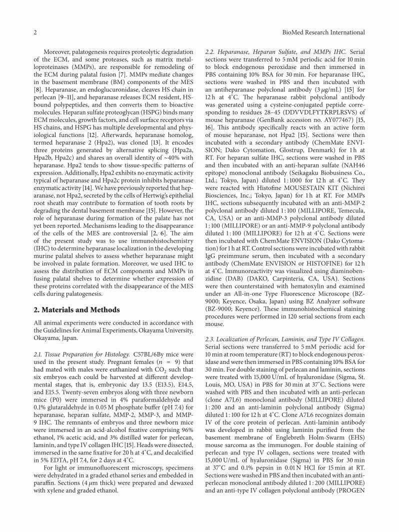
3.3. Localization of MMP-9, MMP-2, andMMP-3 in the MES.Immunolocalizations of MMP-9, MMP-2, andMMP-3 in theMES at E15.5 are shown in Figure 4. MMP-9, MMP-2, and
MMP-3 were evident in the MES. Intense MMP-9 signal wasobserved around the cells of the MES that corresponded tothe basementmembrane;moderateMMP-2 signal and strongMMP-3 signal were evident in the cells of the MES. MMP-2and MMP-3 signal were evident in the mesenchyme of thepalate.
No specific immunoreactivity was observed in the controlsections incubated without any primary antibody (Supple-mental data).
4. Discussion
Here, we assessed the distribution of heparanase duringpalate formation in mice. In the initial stage of palate forma-tion, heparanase signal was not evident in the palatal shelves.As palate formation progressed to elevation, heparanasesignal was evident in the epithelial cells of palatal shelves.Heparanase signal was evident in some nasal epithelium cellsof palate as these shelves made contact. At this same stage,the basement membrane HS was faint and largely absentfrom the epithelial cells near the tip of shelves. After bilateralpalatal shelves connected, heparanase signal in cells of theMES was very strong. These results suggest that epithelialcells of the palatal shelves mediated the degradation ofbasement membrane by secreting heparanase during palateformation. These data also suggest that heparanase activityand the disappearance of basementmembranemay have beenspatially and temporally coordinated.Heparanase secreted bythe palatal epithelial cells may participate in the formationof the palate, particularly in the fusion of palatal shelves viathe degradation of palate basement membrane. Heparanasemight also be required for the cleavage of HS on the epithelialplasma membrane during palate connection.
Moreover, heparanase labeling was evident in osteoblaststhat faced the surface of the palatal bone and the maxillarybone. Increases in HS signal were evident in the bonematrix as palate formation progressed (data not shown). Thepresence of HS in bone and its association with skeletalphysiologic and pathologic processes are well established[17–19]; osteoblasts and osteoclast lineage cells synthesizeHSPG and this HSPG localized on their plasma membraneis involved in the binding of cell-cell interaction betweenosteoblasts and osteoclast lineage cells. Moreover, HSPG inbone matrix is involved in cell-matrix attachment and is alsoa reservoir of bioactive molecules and concerned with bonemetabolism [20, 21]. Additionally, heparanase expression inosteoblasts and its biological function in osteogenesis havebeen documented [22–24]. Our data also provided evidencethat heparanase localized in osteoblasts. Perlecan expressionis associated with increased lacunocanalicular space in corti-cal bone [25]. This data is consistent with an inhibitory rolefor perlecan heparan sulfate chains in biomineralization andwith the current study which shows that heparanase signal isassociated with increased osteogenesis [25]. Heparanase mayhave an important role during the process of bone formationin palatogenesis.
Furthermore, heparanase expression is not restricted topathological conditions and the activity has been found inhair follicle [26, 27] and in the skin [28]. Heparanase was

4 BioMed Research International
E13.5
TPS
(a)
PS
(b)
T
PS PS
E14.5
(c)
PS
(d)
E15.5
N
MES O
∗∗
(e)
P
(f)
O
P0
MB N
P PB
(g)
O
N
PBOB
(h)
Figure 1: Light micrographs showing the localization of heparanase during palate formation. (a) Palatal shelf of a mouse embryo atE13.5. Heparanase signal was not evident in the palatal shelf. (b) Higher magnification of square marked in (a). Some epithelial cells andmesenchymal cells facing the palatal shelf at the bottom of the tongue had weak heparanase labeling (arrows and arrowheads). (c) Palatalshelves of a mouse at E14.5. The epithelial cells of palatal shelves had heparanase labeling. (d) Higher magnification of the square markedin (c). Intense reactivity was evident in the cytoplasm of the epithelial cells (arrowheads). Some stromal cells also had heparanase reactivity(arrows). (e) Palate of a mouse embryo at E15.5. The MES was observed in the middle of palate. Epithelial cells of palate and mesenchymalcells located at the ossification center (∗) had heparanase reactivity. (f) Higher magnification of the square marked in (e). The cells of theMES (white arrowheads), epithelial triangle (arrowheads), and epithelial island (arrow) show heparanase reactivity. (g) Palate of a mouse atP0.The palatal and maxillary bone was observed. (h) Higher magnification of the square marked in (g). Heparanase localization was evidentin osteoblasts on the palatal bone surface. Strong reactivity of heparanase was evident in the basal cells of oral and nasal epithelium of thepalate. Some stromal cells also had heparanase reactivity. PS, palatal shelf; P, palate; T:, tongue; MES, medial epithelial seam; PB, palatal bone;MB, maxillary bone; OB, osteoblasts; O, oropharynx; N, nasopharynx. Bars: (a, c, e, g, and h), 50 𝜇m; (b, d, f), 25 𝜇m.

BioMed Research International 5
E14.5
N
PS
MB
T
(a)
N
PS
HPSE
(b)
N
PS
HS
(c)
Figure 2: Lightmicrographs showing the localization of heparanaseand heparan sulfate in palatal shelves of a mouse embryo at E14.5.(a) The palatal shelves were horizontal in apposition. (b) A highermagnification of the square marked in (a). Some epithelial cellsat the tip of palatal shelves had heparanase labeling (arrowheads).Adjacent section to the section shown in (c). (c) HS labeling wasdiffuse and weak in the basement membrane at the tip of palateepithelium (arrowheads). Adjacent section to the section shown in(b). PS, palatal shelf; T, tongue;MB,maxillary bone;N; nasal septum.Bars: a, 100 𝜇m; b, c, 25 𝜇m.
detected in the outer root sheath of murine hair follicle[26], while it mainly expressed in the inner root sheath ofhuman hair follicle [27], suggesting, despite of differentialexpression between the species, that heparanase may be akey factor in differentiation of a follicular stem cell and hairhomeostasis. In human epidermis, heparanase expression hasbeen reported to be closely related to keratinocyte differen-tiation and was mainly found in the stratum granulosum
[28]. As HSPG was supposed to modulate proliferation anddifferentiation by its ability to affect growth factor signalingand binding, heparanase could play an important role inkeratinocyte differentiation by acting on heparan sulfate.Theinner root sheath of hair follicle is also keratinized and itskeratinocytes terminal differentiation process could sharesome traits with epidermal terminal differentiation process[26–28]. In our study, heparanase localization was observedin the basal cells of the oral and nasal epithelium at P0; thesefindings indicated that heparanase could have contributedto regeneration of and cell renewal in the epithelium. Inaddition, some epithelial cells and underlying mesenchymalcells facing the palatal shelves at the bottom of tongue at E13.5and the palatal mesenchyme at E14.5 showed heparanasereactivity, suggesting that heparanase could be involved inthe generation of these cells, similar to in the hair cycleand in epidermal physiology. One might speculate that hep-aranase activity might play important role in the migrationand remodeling of the palatal mesenchyme during palateformation.
The appearance of heparanase labeling in the cyto-plasm seemed to coincide with lysosomal localization ofthe enzyme. Our results are also consistent with previousfindings that heparanase localized to lysosomes in fibroblasts,osteoblasts, osteo (chondro) clasts, and tumor cells [22,23, 29, 30]. Specific localization of the latent and activeheparanase forms has been detected to perinuclear vesicles,suggesting that heparanase processing and activation occursin lysosomes [29, 30]. In other cases, heparanase appearedless localized andmore diffusely distributed in the cytoplasm,suggesting that under different biological situations, hep-aranase may be localized in different cellular compartmentsand hence may exert diverse functions [29]. The determina-tion of its exact role requires further investigation, includingelectron microscopy studies to examine its exact location.
Heparanase, an endo-𝛽-D-glucuronidase expressed in avariety of tissues and cells during normal development andin pathological conditions, can selectively cleave perlecanand syndecan, and this enzyme releases complexes of HSfragments that are bound to the core protein [31]. Thesereleased HS complexes, such as growth factors, promote cellgrowth and migration [32]. Moreover, recent studies indicatethat HSPG-growth factor complexes become available inbioactive form for binding to the cognate receptors to pro-mote growth factor-mediated signaling [33–35]. Therefore, itis possible that heparanase-labeled cells contribute to degra-dation ofHS chains present in the palate basementmembraneand that release of growth factors might accelerate the prolif-eration and differentiation ofmesenchymal cells of the palatalshelves. In addition to heparanase localization, cells of theMES had MMP-9, MMP-2, and MMP-3 reactivity. However,there were some differences in the distributions of theseproteins. For example, MMP-9 was apparently co-localizedwith the basement membrane, and moderate MMP-2 andintense MMP-3 labeling were seen in the MES cells. Addi-tionally, these MMPs were evident in mesenchyme aroundthe MES. MMPs are involved in the degradation of ECMduring normal physiological processes, such as embryonicdevelopment, reproduction, and tissue remodeling; MMPs

6 BioMed Research International
Perl
BV
P
N
O
∗
∗
(a)
Lam
(b)
Merge
∗
∗
(c)
BVP
N
O
Perl
∗∗
(d)
ColIV
(e)
Merge
∗
∗
(f)
Figure 3: Micrographs of double-immunofluorescence staining reveal the localization of perlecan and laminin ((a)–(c)) or type IV collagen((d)–(f)) in palate of a mouse embryo at E15.5. ((a), (d)) Perlecan reactivity was evident in the basement membrane of oral palate epithelium(arrows), blood vessels, and some stroma (∗) in palate. ((b), (e))Weak or discontinuous laminin (b) or type IV collagen (e) immunoreactivitywas evident in the basement membrane of the MES (arrowheads). ((c), (f)) Colocalization of perlecan and laminin (c) or type IV collagen (f)was observed in the basement membrane of oral epithelium and blood vessels. Some parts of the basement membrane of the MES had onlylaminin (c) or type IV collagen (f) reactivity (arrowheads). Adjacent sections. P, palate; O, oropharynx; N, nasal septum; BV, blood vessels;MES, medial epithelial seam. Bars: 25𝜇m.
also degrade the ECM during disease processes [36]. Thelocalization of heparanase and MMPs in this study suggeststhat they have the ability to participate in ECM remodelingduring palate growth and formation. MMP-9, MMP-2, andMMP-3 may be involved in the degradation of basementmembrane proteins, including type IV collagen, laminin,and perlecan. In our perlecan double-labeling experiments,the basement membrane of the MES had faint perlecanlabeling even though laminin was present. In addition, thebasement membrane of the MES contained type IV collagenthough perlecan was absent; however, we were unable todetect any differences between heparan sulfate and perlecanlocalization in the basement membrane of the MES. Basedon these results, it is conceivable that the degradation ofcomponents of the palatal basement membrane may occurin a regulated sequence. Thus, given the localization ofheparanase, we propose that the degradation of the MESbasement membrane results from the coordinated activity
of several proteolytic enzymes. This is supported by a pre-vious study demonstrating that heparanase knockout micedeveloped normally, were fertile, and exhibit no apparentanatomical or functional abnormalities despite the completelack of heparanase gene expression and enzymatic activity[37]. Heparanase deficiency was compensated by a tissue-specific marked elevation of MMP family members such asMMP-2, MMP-9, and MMP-14 [37]. These findings providedthe evidence for cooperation between heparanase andMMPsin spite of their different enzymatic substrate and suggestedthat a combined interdependent control mechanism betweenheparanase andMMPs regulates the ECM degrading enzymein cells and tissues.
Three processes—programmed cell death [38, 39], cellmigration to the oral and nasal side of the palate [40], andepithelial-mesenchymal transdifferentiation (EMT) [41]—have been proposed as themechanisms involved in the disap-pearance of the MES. Here, nucleus condensation, a change
BioMed Research International 7
MMP-9
N
O
(a)
MMP-2
(b)
MMP-3
(c)
Figure 4: Light micrographs showing the localization of MMP-9 (a), MMP-2 (b), and MMP-3 (c) in the MES of palate of a mouse embryo atE15.5. (a)MMP-9 reactivity was observed throughout the basementmembrane of theMES (arrows). Stroma cells had diffuseMMP-9 labeling.(b) Moderate MMP-2 reactivity was seen in cells of the MES (arrowheads). Stroma cells had weak labeling. (c) Strong labeling of MMP-3 wasseen in cells of the MES (arrowheads) and stroma cells. P, palate; O, oropharynx; N, nasopharynx. Bars: 25𝜇m.
characteristic of programmed cell death, was evident duringhistological examination in some MES cells. Additionally,MMP-3, which is known to induce EMT in mammaryepithelial cells [42], localized in theMES cells, confirming thepossibility that EMToccurred in theMES cells [42].However,based on our data, we could not determine whether theMES cells underwent, separately or concurrently, any of thesethree processes. Further studies are required to understandthe precise mechanism by which the MES cells vanish frompalate.
5. Conclusions
In conclusion, we provided evidence that heparanase local-ized in the palate epithelial cells, the MES cells, andosteoblasts during palate formation, whereas signal from thecomponents of the basement membrane was faint and weakin the palatal shelves. We also observed MMP-9, MMP-2,and MMP-3 signals in the MES, while perlecan, laminin,and collagen type IV signals disappeared from the basementmembrane of the MES. Hence, the distribution heparanaseand MMPs were consistent with the hypothesis that theseproteins had a role in palate ECM remodeling; additionally,these findings suggest that heparanase, in cooperation withother proteases, contributed to palate development. Ourresults also indicated that further analysis of heparanasemight shed light on the fate of the MES in palatogenesis.
Acknowledgments
This work was supported in part by Grants-in-Aid forScientific Research (nos. 21592571, 22592269, 24592996) fromthe Japan Society for the Promotion of Science. The authorswould like to thank Professor Takaaki Ueno, Department of
Oral Surgery, OsakaMedical College; Dr. Tomohiro Yamada,Department of Oral and Maxillofacial Surgery, Kochi Medi-cal School, Kochi University; Dr. Katsuaki Mishima, Depart-ment of Oral and Maxillofacial Surgery, Graduate Schoolof Medicine, Yamaguchi University; and Dr. Hideto Imura,Division of Research and Treatment for Oral and Max-illofacial Congenital Anomalies, School of Dentistry, AichiGakuin University; for their valuable advice. The authorswould like to send special thanks to Professor NorikazuOhno, Department of Oral Anatomy, School of Dentistry,Aichi Gakuin University, for his kind support.
References
[1] M. W. Ferguson, “Palate development,” Development, vol. 103,supplement, pp. 41S–60S, 1988.
[2] L. Meng, Z. Bian, R. Torensma, and J. W. Von Den Hoff, “Bio-logical mechanisms in palatogenesis and cleft palate,” Journal ofDental Research, vol. 88, no. 1, pp. 22–33, 2009.
[3] J. Morris-Wiman and L. Brinkley, “Rapid changes in the extra-cellular matrix accompany in vitro palatal shelf remodelling,”Anatomy and Embryology, vol. 188, no. 1, pp. 75–85, 1993.
[4] J. Morris-Wiman and L. Brinkley, “An extracellular matrixinfrastructure provides support for murine secondary palatalshelf remodelling,” Anatomical Record, vol. 234, no. 4, pp. 575–586, 1992.
[5] A. Iamaroon and V. M. Diewert, “Distribution of basementmembrane components in the mouse primary palate,” Journalof Craniofacial Genetics and Developmental Biology, vol. 16, no.1, pp. 48–51, 1996.
[6] M. Dudas, W. Y. Li, J. Kim, A. Yang, and V. Kaartinen, “Palatalfusion—where do themidline cells go?. A review on cleft palate,a major human birth defect,” Acta Histochemica, vol. 109, no. 1,pp. 1–14, 2007.

8 BioMed Research International
[7] L. Blavier, A. Lazaryev, J. Groffen, N. Heisterkamp, Y. A.DeClerck, and V. Kaartinen, “TGF-𝛽3-induced palatogenesisrequires matrix metalloproteinases,” Molecular Biology of theCell, vol. 12, no. 5, pp. 1457–1466, 2001.
[8] N. L. Brown, S. J. Yarram, J. P. Mansell, and J. R. Sandy, “Matrixmetalloproteinases have a role in palatogenesis,” Journal ofDental Research, vol. 81, no. 12, pp. 826–830, 2002.
[9] M. D. Hulett, C. Freeman, B. J. Hamdorf, R. T. Baker, M. J.Harris, and C. R. Parish, “Cloning of mammalian heparanase,an important enzyme in tumor invasion andmetastasis,”NatureMedicine, vol. 5, no. 7, pp. 803–809, 1999.
[10] M. Toyoshima and M. Nakajima, “Human heparanase. Purifi-cation, characterization, cloning, and expression,” The Journalof Biological Chemistry, vol. 274, no. 34, pp. 24153–24160, 1999.
[11] I. Vlodavsky, Y. Friedmann, M. Elkin et al., “Mammalianheparanase: gene cloning, expression and function in tumorprogression and metastasis,” Nature Medicine, vol. 5, no. 7, pp.793–802, 1999.
[12] J. Kruegel and N. Miosge, “Basement membrane componentsare key players in specialized extracellular matrices,” Cellularand Molecular Life Sciences, vol. 67, no. 17, pp. 2879–2895, 2010.
[13] E. McKenzie, K. Tyson, A. Stamps et al., “Cloning and expres-sion profiling of Hpa2, a novel mammalian heparanase familymember,” Biochemical and Biophysical Research Communica-tions, vol. 276, no. 3, pp. 1170–1177, 2000.
[14] F. Levy-Adam, S. Feld, V. Cohen-Kaplan et al., “Heparanase 2interacts with heparan sulfate with high affinity and inhibitsheparanase activity,” The Journal of Biological Chemistry, vol.285, no. 36, pp. 28010–28019, 2010.
[15] A. Hirata and H. Nakamura, “Localization of perlecan andheparanase in Hertwig’s epithelial root sheath during rootformation in mouse molars,” Journal of Histochemistry andCytochemistry, vol. 54, no. 10, pp. 1105–1113, 2006.
[16] H. Q. Miao, E. Navarro, S. Patel et al., “Cloning, expression,and purification of mouse heparanase,” Protein Expression andPurification, vol. 26, no. 3, pp. 425–431, 2002.
[17] H. Nakamura and H. Ozawa, “Immunohistochemical localiza-tion of heparan sulfate proteoglycan in rat tibiae,” Journal ofBone and Mineral Research, vol. 9, no. 8, pp. 1289–1299, 1994.
[18] W. J. Grzesik, C. R. Frazier, J. R. Shapiro, P. D. Sponseller,P. Gehron Robey, and N. S. Fedarko, “Age-related changes inhuman bone proteoglycan structure: impact of osteogenesisimperfecta,”The Journal of Biological Chemistry, vol. 277, no. 46,pp. 43638–43647, 2002.
[19] P. Newman, F. Bonello, A. S. Wierzbicki, P. Lumb, G. F.Savidge, and M. J. Shearer, “The uptake of lipoprotein-bornephylloquinone (vitamin K1) by osteoblasts and osteoblast-likecells: role of heparan sulfate proteoglycans and apolipoproteinE,” Journal of Bone and Mineral Research, vol. 17, no. 3, pp. 426–433, 2002.
[20] S. M. Cool and V. Nurcombe, “The osteoblast-heparan sulfateaxis: control of the bone cell lineage,” International Journal ofBiochemistry and Cell Biology, vol. 37, no. 9, pp. 1739–1745, 2005.
[21] S. A. Khan, M. S. Nelson, C. Pan, P. M. Gaffney, and P. Gupta,“Endogenous heparan sulfate and heparin modulate bone mor-phogenetic protein-4 signaling and activity,” American Journalof Physiology, vol. 294, no. 6, pp. C1387–C1397, 2008.
[22] M. Saijo, R. Kitazawa,M.Nakajima,M.Kurosaka, S.Maeda, andS. Kitazawa, “Heparanase mRNA expression during fracturerepair in mice,” Histochemistry and Cell Biology, vol. 120, no. 6,pp. 493–503, 2003.
[23] V. Kram, E. Zcharia, O. Yacoby-Zeevi et al., “Heparanase isexpressed in osteoblastic cells and stimulates bone formationand bone mass,” Journal of Cellular Physiology, vol. 207, no. 3,pp. 784–792, 2006.
[24] P. N. Smith, C. Freeman, D. Yu et al., “Heparanase in primaryhuman osteoblasts,” Journal of Orthopaedic Research, vol. 28, no.10, pp. 1315–1322, 2010.
[25] W. R.Thompson, S. Modla, B. J. Grindel et al., “Perlecan/Hspg2deficiency alters the pericellular space of the lacunocanalicularsystem surrounding osteocytic processes in cortical bone,”Journal of Bone and Mineral Research, vol. 26, no. 3, pp. 618–629, 2011.
[26] E. Zcharia, D. Philp, E. Edovitsky et al., “Heparanase regulatesmurine hair growth,” American Journal of Pathology, vol. 166,no. 4, pp. 999–1008, 2005.
[27] S. Malgouries, M. Donovan, S. Thibaut, and B. A. Bernard,“Heparanase 1: a key participant of inner root sheath differ-entiation program and hair follicle homeostasis,” ExperimentalDermatology, vol. 17, no. 12, pp. 1017–1023, 2008.
[28] D. Bernard, B. Mehul, C. Delattre, L. Simonetti, A. Thomas-Collignon, and R. Schmidt, “Purification and characterizationof the endoglycosidase heparanase 1 from human plantarstratum corneum: a key enzyme in epidermal physiology?”Journal of Investigative Dermatology, vol. 117, no. 5, pp. 1266–1273, 2001.
[29] O. Goldshmidt, L. Nadav, H. Aingorn et al., “Human hep-aranase is localized within lysosomes in a stable form,” Experi-mental Cell Research, vol. 281, no. 1, pp. 50–62, 2002.
[30] A. Zetser, F. Levy-Adam, V. Kaplan et al., “Processing andactivation of latent heparanase occurs in lysosomes,” Journal ofCell Science, vol. 117, no. 11, pp. 2249–2258, 2004.
[31] N. J. Nasser, “Heparanase involvement in physiology anddisease,” Cellular and Molecular Life Sciences, vol. 65, no. 11, pp.1706–1715, 2008.
[32] C. R. Parish, “The role of heparan sulphate in inflammation,”Nature Reviews Immunology, vol. 6, no. 9, pp. 633–643, 2006.
[33] C. Kirn-Safran, M. C. Farach-Carson, and D. D. Carson,“Multifunctionality of extracellular and cell surface heparansulfate proteoglycans,” Cellular and Molecular Life Sciences, vol.66, no. 21, pp. 3421–3434, 2009.
[34] A. D. Theocharis, S. S. Skandalis, G. N. Tzanakakis, and N. K.Karamanos, “Proteoglycans in health and disease: novel rolesfor proteoglycans in malignancy and their pharmacologicaltargeting,” The FEBS Journal, vol. 277, no. 19, pp. 3904–3923,2010.
[35] S. H. Kim, J. Turnbull, and S. Guimond, “Extracellular matrixand cell signalling: the dynamic cooperation of integrin, pro-teoglycan and growth factor receptor,” Journal of Endocrinology,vol. 209, no. 2, pp. 139–151, 2011.
[36] H. J. Ra and W. C. Parks, “Control of matrix metalloproteinasecatalytic activity,” Matrix Biology, vol. 26, no. 8, pp. 587–596,2007.
[37] E. Zcharia, J. Jia, X. Zhang et al., “Newly generated heparanaseknock-outmice unravel co-regulation of heparanase andmatrixmetalloproteinases,” PLoS ONE, vol. 4, no. 4, Article ID e5181,2009.
[38] C.Martınez-Alvarez, C. Tudela, J. Perez-Miguelsanz, S. O’Kane,J. Puerta, andM.W. J. Ferguson, “Medial edge epithelial cell fateduring palatal fusion,”Developmental Biology, vol. 220, no. 2, pp.343–357, 2000.
BioMed Research International 9
[39] R.Cuervo andL.Covarrubias, “Death is themajor fate ofmedialedge epithelial cells and the cause of basal lamina degradationduring palatogenesis,” Development, vol. 131, no. 1, pp. 15–24,2004.
[40] J. Z. Jin and J. Ding, “Analysis of cell migration, transdifferen-tiation and apoptosis during mouse secondary palate fusion,”Development, vol. 133, no. 17, pp. 3341–3347, 2006.
[41] F. V. Sani, K. Hallberg, B. D. Harfe, A. P. McMahon, A.Linde, and A. Gritli-Linde, “Fate-mapping of the epithelialseam during palatal fusion rules out epithelial-mesenchymaltransformation,”Developmental Biology, vol. 285, no. 2, pp. 490–495, 2005.
[42] A. Lochter, S. Galosy, J. Muschler, N. Freedman, Z. Werb, andM. J. Bissell, “Matrix metalloproteinase stromelysin-1 triggers acascade of molecular alterations that leads to stable epithelial-to-mesenchymal conversion and a premalignant phenotype inmammary epithelial cells,” Journal of Cell Biology, vol. 139, no. 7,pp. 1861–1872, 1997.

Submit your manuscripts athttp://www.hindawi.com
Hindawi Publishing Corporationhttp://www.hindawi.com Volume 2014
Anatomy Research International
PeptidesInternational Journal of
Hindawi Publishing Corporationhttp://www.hindawi.com Volume 2014
Hindawi Publishing Corporation http://www.hindawi.com
International Journal of
Volume 2014
Zoology
Hindawi Publishing Corporationhttp://www.hindawi.com Volume 2014
Molecular Biology International
GenomicsInternational Journal of
Hindawi Publishing Corporationhttp://www.hindawi.com Volume 2014
The Scientific World JournalHindawi Publishing Corporation http://www.hindawi.com Volume 2014
Hindawi Publishing Corporationhttp://www.hindawi.com Volume 2014
BioinformaticsAdvances in
Marine BiologyJournal of
Hindawi Publishing Corporationhttp://www.hindawi.com Volume 2014
Hindawi Publishing Corporationhttp://www.hindawi.com Volume 2014
Signal TransductionJournal of
Hindawi Publishing Corporationhttp://www.hindawi.com Volume 2014
BioMed Research International
Evolutionary BiologyInternational Journal of
Hindawi Publishing Corporationhttp://www.hindawi.com Volume 2014
Hindawi Publishing Corporationhttp://www.hindawi.com Volume 2014
Biochemistry Research International
ArchaeaHindawi Publishing Corporationhttp://www.hindawi.com Volume 2014
Hindawi Publishing Corporationhttp://www.hindawi.com Volume 2014
Genetics Research International
Hindawi Publishing Corporationhttp://www.hindawi.com Volume 2014
Advances in
Virolog y
Hindawi Publishing Corporationhttp://www.hindawi.com
Nucleic AcidsJournal of
Volume 2014
Stem CellsInternational
Hindawi Publishing Corporationhttp://www.hindawi.com Volume 2014
Hindawi Publishing Corporationhttp://www.hindawi.com Volume 2014
Enzyme Research
Hindawi Publishing Corporationhttp://www.hindawi.com Volume 2014
International Journal of
Microbiology