Embed Size (px)
Citation preview
Research ArticleSynthesis of Copper Nanoparticles Coated withNitrogen Ligands
Rubén Sierra-Ávila, Marissa Pérez-Alvarez, Gregorio Cadenas-Pliego,Carlos Alberto Ávila-Orta, Rebeca Betancourt-Galindo, Enrique Jiménez-Regalado,Rosa Martha Jiménez-Barrera, and Juan Guillermo Martínez-Colunga
Centro de Investigacion en Quımica Aplicada, 25294 Saltillo Coahuila, Mexico
Correspondence should be addressed to Marissa Perez-Alvarez; [email protected] Gregorio Cadenas-Pliego; [email protected]
Received 18 January 2014; Accepted 28 April 2014; Published 28 May 2014
Academic Editor: Claude Estournes
Copyright © 2014 Ruben Sierra-Avila et al. This is an open access article distributed under the Creative Commons AttributionLicense, which permits unrestricted use, distribution, and reproduction in any medium, provided the original work is properlycited.
The synthesis of copper nanoparticles was studied by wet chemical methods using copper sulfate pentahydrate (CuSO4⋅5H2O) and
nitrogen ligands allylamine (AAm) and polyallylamine (PAAm) as stabilizers.The results suggest that the use of these ligands leadsto the exclusive formation ofmetallic copper nanoparticles (Cu-NPs).Theuse of partially crosslinked polyallylamine (PAAmc) leadsto nanoparticles (NPs) with low yields and high coating content, while linear PAAm leads to NPs with high yields and low coatingcontent.The chemical composition of the particles was determined by XRD and average particle diameters were determined by theDebye-Scherrer equation. TGA analysis provided evidence of the content and thermal stability of the coating on the nanoparticlesand PAAm. The morphology, particle size distribution, and presence of PAAm coating were observed through TEM. The useof AAm in the synthesis of NPs could be a good alternative to reduce costs. By using TGA, TEM, and DSC techniques, it wasdetermined that synthesized NPs with AAm presented a coating with similar characteristics to NPs with PAAm, suggesting thatAAm underwent polymerization during the synthesis.
1. Introduction
Since 1990, scientific and technological research on thesynthesis of inorganic nanoparticles has increased markedlydue to their interesting physical properties and potentialapplications [1, 2]. This is mainly attributed because of theirsmall size, shape, composition, and high surface area tovolume ratio [3]. Particular attention has been given to metalparticles, especially copper particles due to the fact that theyexhibit unique physicochemical properties not seen in othermaterials and differ from their mass-counterparts in proper-ties such as catalytic [4], optical [5], electrical [6], electronic[7], thermal [8], magnetic [9], and antimicrobial [10, 11].Because of their attractive features nanoparticles of metalliccopper can be used in different fields at laboratory- andindustrial-scale production, for example, manufacture ofelectrical and electronic devices, preparation of thin films,high surface area catalysts in various chemical processes,
application in cancer cells treatment, additives in germicidesformulation, antibacterial materials [12, 13], additive forlubricating oils to improve antiwear properties caused byfriction, and conductive inks and coatings [14].
Several synthetic methods for the preparation of coppernanoparticles have been developed including thermal andsonochemical reduction [15], metal vapor synthesis (MVS)[16], electron beam irradiation [17], microwave irradiation[18], reversemicelles [19], pulsed laser ablation [20], chemicalreduction in aqueous solution [21], and polyol process [22].Most of the preparation methods have several factors thatlimit their use in the synthesis ofmetal particles. For instance,the method of metal evaporation in gas phase has a high costof startingmaterials and uses highly sophisticated equipment[6]; meanwhile the synthetic route by pulsed laser ablation isnot a simple process, as it is inflexible and relatively costly[20]. Among these synthetic strategies, the wet chemicalreduction is one of the most convenient methods because
Hindawi Publishing CorporationJournal of NanomaterialsVolume 2014, Article ID 361791, 8 pageshttp://dx.doi.org/10.1155/2014/361791
2 Journal of Nanomaterials
it has unique advantages over the other methods due to itsversatility, since water has the ability to solubilize a widevariety of metallic salts, wherein the nanoparticles obtainedby this method have well defined shapes and sizes [3].Compared to conventional methods, products are cheaperand have higher purity [23], simple handling equipment isused and completion time of synthesis is short, and it is easyto scale up at an industrial level [6].The reducing agents com-monly used in the wet chemical reduction synthesis includeformaldehyde [24], ascorbic acid [25], sodium hypophos-phite [26], potassium borohydride and sodium [6, 27], andhydrazine hydrate [28]. However, some reducing agentsare toxic and expensive, many of which have poor reductivecapacity and can also introduce impurities to the process [6].
Unlike noble metals such as gold (Au) and silver (Ag),copper metal particles can not be obtained from the simplereduction of precursor salts in aqueous solution, since cupricoxide (CuO) and cuprous oxide (Cu
2O) could be generated
during the process due to the presence of a large amountof water molecules. To protect copper nanoparticles againstchemical oxidation during preparation and storage, some-times the synthesis takes place in a nonaqueous mediumusing a low concentration of precursor and generally underinert atmospheres of Ar or N
2[29]. Occasionally, the above is
not sufficient to prevent the oxidation of copper nanoparticlessince their surface is very reactive. This makes necessarythe use of stabilizers or agents such as water-soluble coatingpolymers, surfactants, or coordination ligands that havefunctional groups that can form complexes with copper ionspresent in the medium [3]. Compounds which are frequentlyused to meet the above include surfactants tetraethylenepen-tamine [30], cetyltrimethylammoniumbromide (CTAB) [31],tetraoctylammoniumbromide [32], and polyelectrolytes suchas polyethyleneimine (PEI) [33], polyethylene glycols (PEG)[34], polyvinylpyrrolidone (PVP) [35], polyetheretherketone(PEEK) [36], and polyallylamine (PAAm) [37]. These mate-rials can act as surface protectors, have the function of con-trolling size, size distribution, and shape of particles, and alsoprevent agglomeration thereof.
Here, we report the synthesis of copper nanoparti-cles using hydrazine hydrate (N
2H4⋅H2O) as a reducing
agent, allylamine (AAm), linear polyallylamine (PAAm), andpartially crosslinked polyallylamine (PAAmc) as stabilizingnitrogen agents. The synthesis was performed using differentratios of nitrogen ligands and using a constant concentrationof NaOH and N
2H4⋅H2O.
2. Experimental
2.1. Materials and Reagents. Copper(II) sulfate pentahy-drate (CuSO
4⋅5H2O), hydrazine hydrate solution (50–60%),
poly(allylamine) solution (PAAm) (Mw ∼ 17 000, 20wt%aqueous solution), and sodium hydroxide (98.8%) wereobtained from Sigma-Aldrich and were used without pre-vious purification. PAAmc was obtained by PAAm’s warm-ing at 40∘C for two months. The moles of PAAm werecalculated by the molecular weight of the repeating unit[CH2–CH(CH
2NH2)] of 57 g/mol. Different molar ratios of
nitrogen ligands/CuSO4⋅5H2Owere used.The reactions with
PAAmc used higher H2O/CuSO
4⋅5H2O molar ratios due to
the lower solubility of the ligand in H2O.
2.2. Synthesis of Copper Nanoparticles. The synthesis of cop-per nanoparticles was performed using wet chemical reduc-tion in atmospheric air; the following procedure describesthe synthesis of NPs R4. In a flat bottom flask CuSO
4⋅5H2O
(0.5 g, 2.0mmol) was added, which was dissolved in 30mL ofdistilledwater undermagnetic stirring for 10minutes at 60∘C.Then 0.065 g of PAAm was dissolved in 10mL of distilledwater and then added in a dropwise manner and stirred for10 minutes. Subsequently, 33mL of NaOH (0.5M) was addeddropwise and allowed to react for 30 minutes followed by theaddition of 2.4mL (76.9mmol) of hydrazine. The reactionmixture was maintained at 60∘C with constant stirring. Thenanoparticles were recovered by centrifugation at 15,000 rpmand 25∘C; solids were washed two times with distilled waterand one time with ethanol. Final product was dried at 60∘Cfor 2 hours; the NPs R4 were obtained as a black powder(0.0952 g, 75%).
The yields were determined by the following equation:
Yield% = [Experimental yieldTheoretical yield
] × 100, (1)
theoretical yield = [(g, CuSO4⋅5H2O)/(Molecular weight,
CuSO4⋅5H2O)] × [(63.5 g Cu)/(1mol Cu)], experimental
yield = (g, Sample R1–R4)(% Cu)/100; % Cu = 100 − % lossmass at ≤600∘C (Figure 6).
2.3. Characterization Techniques. X-ray diffraction (XRD)technique was employed in order to know the oxidation stateand chemical stability of the synthesized copper nanopar-ticles on a Siemens D-5000 diffractometer with a scanninginterval in the 2𝜃 range of 30 to 80∘ with a scan speed of0.02∘/s; the radiation employed was copper K𝛼 with a wave-length of 1.54056 A; values of 25mA and 35 kV were usedfor intensity and voltage, respectively. The average particlediameter was determined from the XRD diffractograms ofeach sample based on the Debye-Scherrer equation takingthe most intense peak for calculation [38, 39]. Transmissionelectron microscopy (TEM) was used in order to observethe morphology and particle size distribution of the coppersamples on a FEI Titan high-resolution electron microscopeoperating at 300 kV.
Thermogravimetric analysis (TGA) was employed toanalyze the thermal behavior of Cu particles and the thermalstability of coating using a DuPont Instruments 951 analyzer.Operating conditions were a heating rate of 10∘C/min. andair atmosphere with a gas flow of 50mL/min.The runs of thesamples were carried out from 30∘C to 600∘C in N
2atmo-
sphere. Once 600∘Cwas reachedN2atmosphere was changed
for O2.
Differential scanning calorimetry analyses (DSC) wereperformed on a modulated TA Instrument 2920, at a heatingrate of 10∘C/min on a temperature range from −40 to 160∘C.Through a second heating cycle the thermal and mechanicalhistory of the samples was eliminated.
Journal of Nanomaterials 3
Table 1: Results of the synthesis of copper nanoparticles by wet chemical reduction method.
Sample PAAm/Cu AAm/Cu Chemical composition Average diameter (nm) Yield (%) Ligand (%)R1∗ 2.00 0.0 Cu 13.0 52.0 30.3R2 0.0 97.0 Cu 30.0 36.0 5.6R3 0.11 0.0 Cu 24.0 86.0 3.2R4 0.46 0.0 Cu 19.0 75.0 5.1Molar ratios of reagents in the synthesis of copper nanoparticles using a N2H4/Cumolar ratio of 38.0 and a NaOH/Cumolar ratio of 8.0. ∗Partially crosslinkedPAAm.
3. Results and Discussion
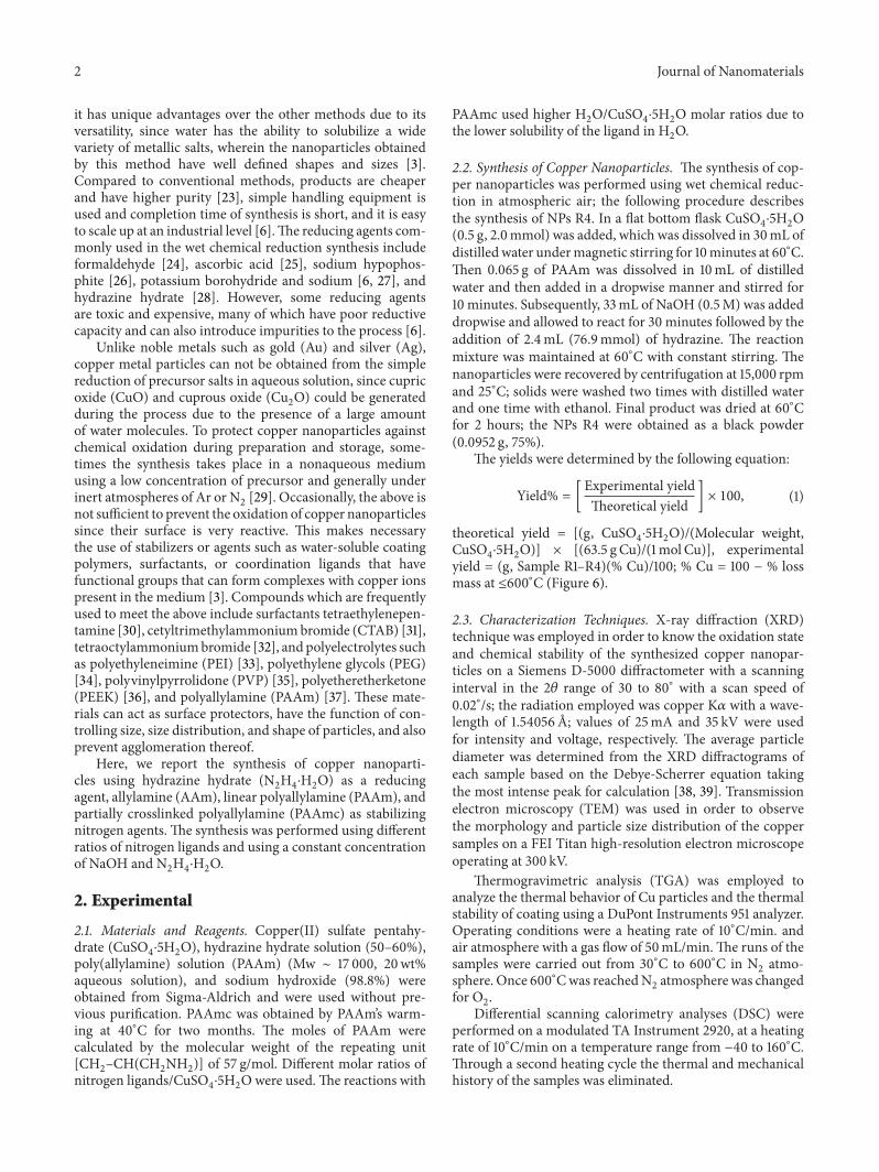
3.1. Effect of Nitrogen Ligands PAAmandAAmon the Synthesisof Copper Nanoparticles. Table 1 presents the results obtainedin the synthesis of nanoparticles obtained by wet chemicalreduction method and Figure 1 shows the XRD diffrac-tograms of copper nanoparticles identified as R1, R3, and R4and synthesized under different PAAm/Cumolar ratios: 2.00,0.11, and 0.46, respectively. Diffractogram of nanoparticlesobtained using AAm (R2) is also shown. In Figure 1, itcan be seen that all particles synthesized using nitrogenligands exhibit three reflections located at 2𝜃 = 43.4, 50.5,and 74.0∘, attributed to the (111), (200), and (220) crystalplanes, respectively, belonging to pure copper with face-centered cubic symmetry (FCC) [40, 41] and correspondingto the diffraction pattern of metallic copper (JCPDS number04-0836) [42], as shown at the bottom of this figure. In thecase of diffractogram R1, besides the signals described above,some small signs located at 2𝜃 = 35.9 and 38.6∘ (shown in theblack dashed circle on the diffractogram) are presented; thissuggests the start of an oxidative process.The yields obtainedwhen PAAm was employed as a ligand, R3 and R4, were 86%and 75%, respectively.The use of PAAmc (R1) and AAm (R2)led to lower yields of 52% and 36%, respectively. The per-centage of ligand or particle coating was higher when usingPAAmc (30.3%). The use of PAAm and AAm led to low per-centage of coating and the use of AAm ligand led to a higherpercentage of nanoparticle coating despite having a muchlower molecular weight than linear PAAm (see Table 1).
The particle diameter (𝐷) for the indicated sampleswas determined by the Debye-Scherrer equation [38, 39] asfollows:
𝐷 =
0.94𝜆
𝛽 cos 𝜃, (2)
where 𝜆 is the copper wavelength (1.54056 A), 𝜃 is theBragg diffraction angle, and 𝛽 is the half-width of the mostintense diffraction peak. The average diameter calculated forthe synthesized nanoparticles is shown in Table 1. Particlesobtained with AAm (R2) showed the larger particle diameterof 30 nm; this suggests that polymeric ligands promote theformation of small particles and is more evident when usingPAAmc (R1), as the average diameter of these particles was13 nm.
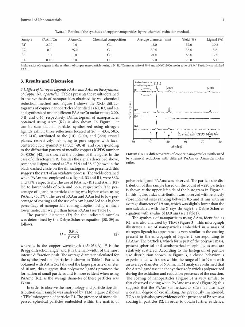
In order to observe the morphology and particle size dis-tribution each sample was analyzed by TEM. Figure 2 showsa TEMmicrograph of particles R1.The presence of monodis-persed spherical particles embedded within the matrix of
R2
Inte
nsity
(a.u
.)
R1
R4
(220)(200)
(111)
R3
Probable onset ofan oxidative process
30 40 50 60 70 80
Cu standard (JCPDS: 04-0836)
2𝜃 (deg)
Figure 1: XRD diffractograms of copper nanoparticles synthesizedby chemical reduction with different PAAm or AAm/Cu molarratios.
polymeric ligand PAAmc was observed.The particle size dis-tribution of this sample based on the count of ∼220 particlesis shown at the upper left side of the histogram in Figure 2.In this figure, a size distribution was observed with relativelyclose interval sizes ranking between 0.5 and 11 nm with anaverage diameter of 3.9 nm, which was slightly lower than theone calculated with the X-rays through the Debye-Scherrerequation with a value of 13.0 nm (see Table 1).
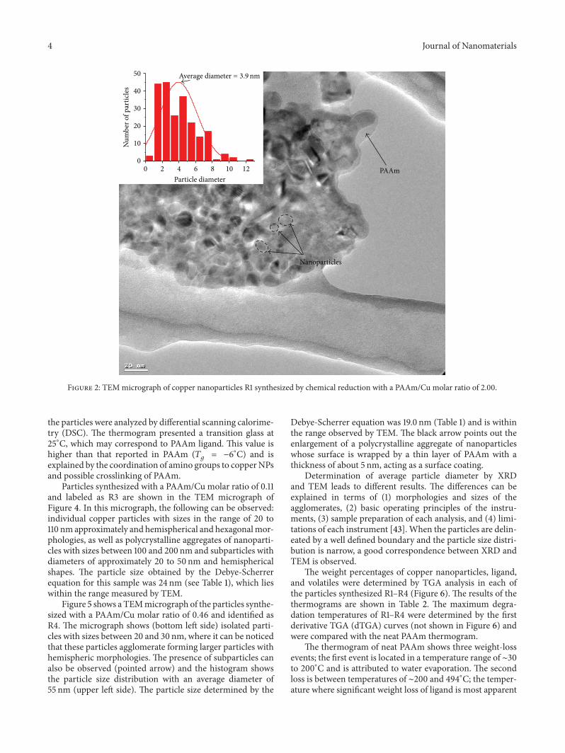
The synthesis of nanoparticles using AAm, identified asR2, was also analyzed by TEM (Figure 3). This micrographillustrates a set of nanoparticles embedded in a mass ofnitrogen ligand; its appearance is very similar to the coatingpresent in the micrograph of Figure 2, corresponding toPAAmc.The particles, which form part of the polymer mass,present spherical and semispherical morphologies and arerelatively scattered. According to the histogram of particlesize distribution shown in Figure 3, a closed behavior isexperimented with sizes within the range of 1 to 19 nm withan average diameter of 6.0 nm. TEM analysis confirmed thattheAAm ligandused in the synthesis of particles polymerizedduring the oxidation and reduction processes of the reaction.The coating of nanoparticles (Figure 3) is very similar tothat observed coating when PAAmc was used (Figure 2); thissuggests that the PAAm synthesized in situ may also havea certain degree of crosslinking. As previously mentioned,TGA analysis also gave evidence of the presence of PAAmas acoating in particles R2. In order to obtain further evidence,
4 Journal of Nanomaterials
50
40
30
20
10
0
121086420
Num
ber o
f par
ticle
s
Particle diameter
Average diameter = 3.9nm
Nanoparticles
PAAm
Figure 2: TEMmicrograph of copper nanoparticles R1 synthesized by chemical reduction with a PAAm/Cu molar ratio of 2.00.
the particles were analyzed by differential scanning calorime-try (DSC). The thermogram presented a transition glass at25∘C, which may correspond to PAAm ligand. This value ishigher than that reported in PAAm (𝑇
𝑔= −6
∘C) and isexplained by the coordination of amino groups to copperNPsand possible crosslinking of PAAm.
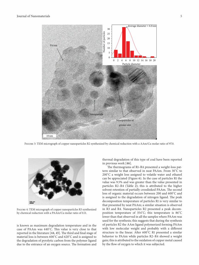
Particles synthesized with a PAAm/Cu molar ratio of 0.11and labeled as R3 are shown in the TEM micrograph ofFigure 4. In this micrograph, the following can be observed:individual copper particles with sizes in the range of 20 to110 nm approximately and hemispherical and hexagonalmor-phologies, as well as polycrystalline aggregates of nanoparti-cles with sizes between 100 and 200 nm and subparticles withdiameters of approximately 20 to 50 nm and hemisphericalshapes. The particle size obtained by the Debye-Scherrerequation for this sample was 24 nm (see Table 1), which lieswithin the range measured by TEM.
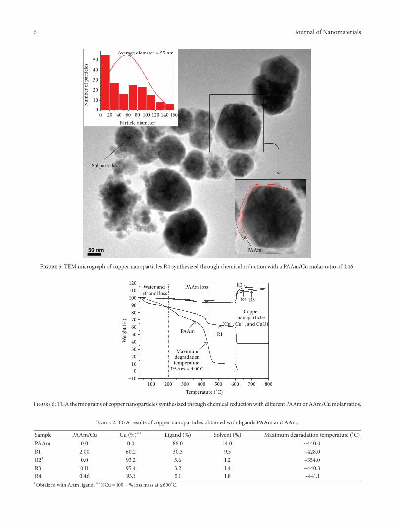
Figure 5 shows a TEMmicrograph of the particles synthe-sized with a PAAm/Cu molar ratio of 0.46 and identified asR4. The micrograph shows (bottom left side) isolated parti-cles with sizes between 20 and 30 nm, where it can be noticedthat these particles agglomerate forming larger particles withhemispheric morphologies. The presence of subparticles canalso be observed (pointed arrow) and the histogram showsthe particle size distribution with an average diameter of55 nm (upper left side). The particle size determined by the
Debye-Scherrer equation was 19.0 nm (Table 1) and is withinthe range observed by TEM. The black arrow points out theenlargement of a polycrystalline aggregate of nanoparticleswhose surface is wrapped by a thin layer of PAAm with athickness of about 5 nm, acting as a surface coating.
Determination of average particle diameter by XRDand TEM leads to different results. The differences can beexplained in terms of (1) morphologies and sizes of theagglomerates, (2) basic operating principles of the instru-ments, (3) sample preparation of each analysis, and (4) limi-tations of each instrument [43]. When the particles are delin-eated by a well defined boundary and the particle size distri-bution is narrow, a good correspondence between XRD andTEM is observed.
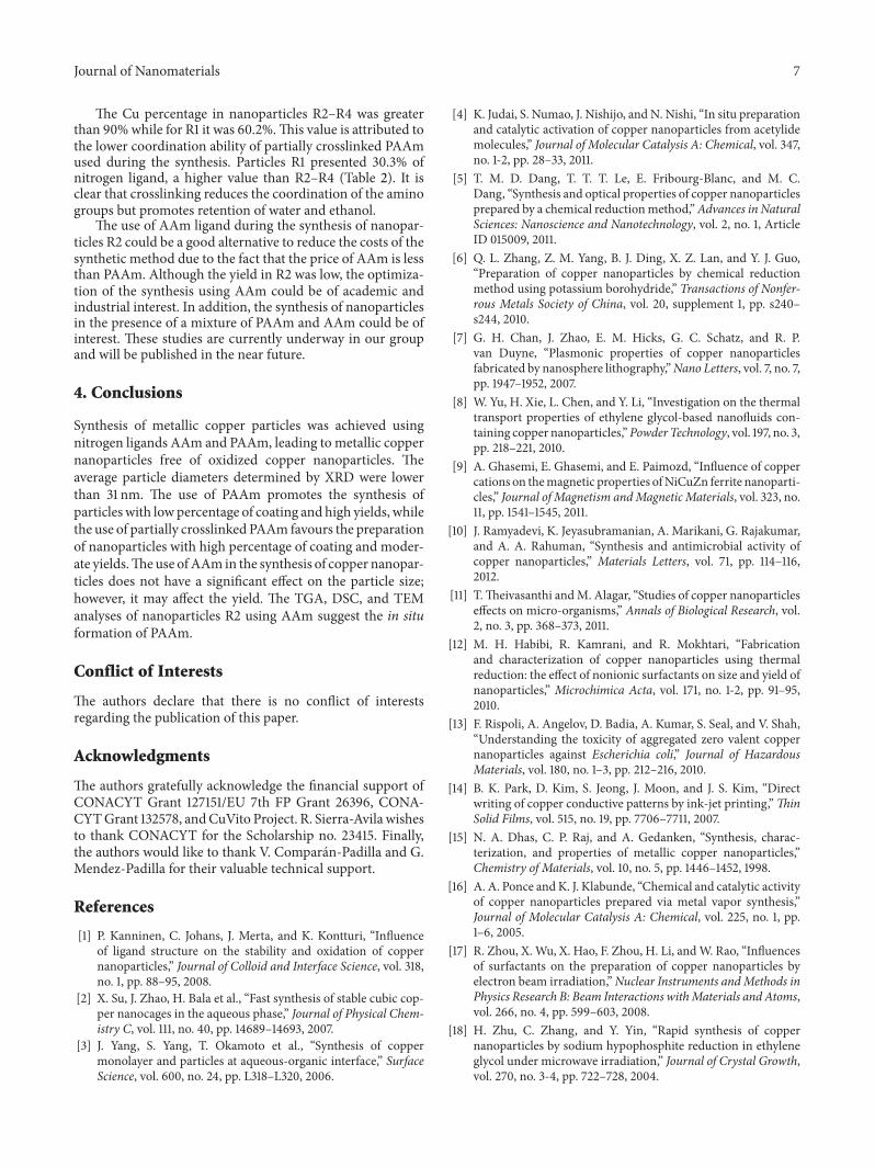
The weight percentages of copper nanoparticles, ligand,and volatiles were determined by TGA analysis in each ofthe particles synthesized R1–R4 (Figure 6). The results of thethermograms are shown in Table 2. The maximum degra-dation temperatures of R1–R4 were determined by the firstderivative TGA (dTGA) curves (not shown in Figure 6) andwere compared with the neat PAAm thermogram.
The thermogram of neat PAAm shows three weight-lossevents; the first event is located in a temperature range of ∼30to 200∘C and is attributed to water evaporation. The secondloss is between temperatures of ∼200 and 494∘C; the temper-ature where significant weight loss of ligand is most apparent
Journal of Nanomaterials 5
Average diameter = 6.0nm
10nm
Nanoparticle
Nanoparticles of
greater size
PAAm
0 2 4 6 8 10 12 14 16 18 20
0
5
10
15
20
25
30
Num
ber o
f par
ticle
s
Particle diameter
Figure 3: TEMmicrograph of copper nanoparticles R2 synthesized by chemical reduction with a AAm/Cu molar ratio of 97.0.
Figure 4: TEMmicrograph of copper nanoparticles R3 synthesizedby chemical reduction with a PAAm/Cu molar ratio of 0.11.
is known as maximum degradation temperature and in thecase of PAAm was 440∘C. This value is very close to thatreported in the literature [44, 45]. The third and final stage ofmaterial loss is between 600∘C and 620∘C and is assigned tothe degradation of pyrolytic carbon from the polymer liganddue to the entrance of an oxygen source. The formation and
thermal degradation of this type of coal have been reportedin previous work [46].
The thermograms of R1–R4 presented a weight-loss pat-tern similar to that observed in neat PAAm. From 30∘C to200∘C a weight loss assigned to volatile water and ethanolcan be appreciated (Figure 6). In the case of particles R1 thevalue was 9.5% and was greater than the value presented inparticles R2–R4 (Table 2); this is attributed to the highersolvent retention of partially crosslinked PAAm. The secondloss of organic material occurs between 200 and 600∘C andis assigned to the degradation of nitrogen ligand. The peakdecomposition temperature of particles R1 is very similar tothat presented by neat PAAm; a similar situation is observedin R3 and R4. Nanoparticles R2 presented a peak decom-position temperature of 354∘C; this temperature is 86∘Clower than that observed in all the samples where PAAmwasused for the synthesis; this suggests that during the synthesisof particles R2 the AAm ligand polymerized forming PAAmwith low molecular weight and probably with a differentstructure to the linear. After 600∘C R1 presented a similarbehavior to PAAm while particles R2–R4 showed a weightgain; this is attributed to the oxidation of coppermetal causedby the flow of oxygen to which it was subjected.
6 Journal of Nanomaterials
Subparticles
0 20 40 60 80 100 120 140 160
0
10
20
30
40
50
Num
ber o
f par
ticle
s
Particle diameter
PAAm
Average diameter = 55 nm
Figure 5: TEMmicrograph of copper nanoparticles R4 synthesized through chemical reduction with a PAAm/Cu molar ratio of 0.46.
100 200 300 400 500 600 700 800
R2
R1
PAAm loss
Wei
ght (
%)
Water and ethanol loss
PAAm
R3R4
−10
PAAm = 440∘C
(Cu0, Cu0 , and CuO)
Temperature (∘C)
degradationMaximum
temperature
Copper nanoparticles
0
10
20
30
40
50
60
70
80
90
100
110
120
Figure 6: TGA thermograms of copper nanoparticles synthesized through chemical reductionwith different PAAmorAAm/Cumolar ratios.
Table 2: TGA results of copper nanoparticles obtained with ligands PAAm and AAm.
Sample PAAm/Cu Cu (%)∗∗ Ligand (%) Solvent (%) Maximum degradation temperature (∘C)PAAm 0.0 0.0 86.0 14.0 ∼440.0R1 2.00 60.2 30.3 9.5 ∼428.0R2∗ 0.0 93.2 5.6 1.2 ∼354.0R3 0.11 95.4 3.2 1.4 ∼440.3R4 0.46 93.1 5.1 1.8 ∼441.1∗Obtained with AAm ligand, ∗∗%Cu = 100 − % loss mass at ≤600∘C.
Journal of Nanomaterials 7
The Cu percentage in nanoparticles R2–R4 was greaterthan 90%while for R1 it was 60.2%.This value is attributed tothe lower coordination ability of partially crosslinked PAAmused during the synthesis. Particles R1 presented 30.3% ofnitrogen ligand, a higher value than R2–R4 (Table 2). It isclear that crosslinking reduces the coordination of the aminogroups but promotes retention of water and ethanol.
The use of AAm ligand during the synthesis of nanopar-ticles R2 could be a good alternative to reduce the costs of thesynthetic method due to the fact that the price of AAm is lessthan PAAm. Although the yield in R2 was low, the optimiza-tion of the synthesis using AAm could be of academic andindustrial interest. In addition, the synthesis of nanoparticlesin the presence of a mixture of PAAm and AAm could be ofinterest. These studies are currently underway in our groupand will be published in the near future.
4. Conclusions
Synthesis of metallic copper particles was achieved usingnitrogen ligands AAm and PAAm, leading to metallic coppernanoparticles free of oxidized copper nanoparticles. Theaverage particle diameters determined by XRD were lowerthan 31 nm. The use of PAAm promotes the synthesis ofparticles with lowpercentage of coating and high yields, whilethe use of partially crosslinked PAAm favours the preparationof nanoparticles with high percentage of coating and moder-ate yields.Theuse ofAAm in the synthesis of copper nanopar-ticles does not have a significant effect on the particle size;however, it may affect the yield. The TGA, DSC, and TEManalyses of nanoparticles R2 using AAm suggest the in situformation of PAAm.
Conflict of Interests
The authors declare that there is no conflict of interestsregarding the publication of this paper.
Acknowledgments
The authors gratefully acknowledge the financial support ofCONACYT Grant 127151/EU 7th FP Grant 26396, CONA-CYTGrant 132578, andCuVito Project. R. Sierra-Avila wishesto thank CONACYT for the Scholarship no. 23415. Finally,the authors would like to thank V. Comparan-Padilla and G.Mendez-Padilla for their valuable technical support.
References
[1] P. Kanninen, C. Johans, J. Merta, and K. Kontturi, “Influenceof ligand structure on the stability and oxidation of coppernanoparticles,” Journal of Colloid and Interface Science, vol. 318,no. 1, pp. 88–95, 2008.
[2] X. Su, J. Zhao, H. Bala et al., “Fast synthesis of stable cubic cop-per nanocages in the aqueous phase,” Journal of Physical Chem-istry C, vol. 111, no. 40, pp. 14689–14693, 2007.
[3] J. Yang, S. Yang, T. Okamoto et al., “Synthesis of coppermonolayer and particles at aqueous-organic interface,” SurfaceScience, vol. 600, no. 24, pp. L318–L320, 2006.
[4] K. Judai, S. Numao, J. Nishijo, and N. Nishi, “In situ preparationand catalytic activation of copper nanoparticles from acetylidemolecules,” Journal of Molecular Catalysis A: Chemical, vol. 347,no. 1-2, pp. 28–33, 2011.
[5] T. M. D. Dang, T. T. T. Le, E. Fribourg-Blanc, and M. C.Dang, “Synthesis and optical properties of copper nanoparticlesprepared by a chemical reductionmethod,”Advances in NaturalSciences: Nanoscience and Nanotechnology, vol. 2, no. 1, ArticleID 015009, 2011.
[6] Q. L. Zhang, Z. M. Yang, B. J. Ding, X. Z. Lan, and Y. J. Guo,“Preparation of copper nanoparticles by chemical reductionmethod using potassium borohydride,” Transactions of Nonfer-rous Metals Society of China, vol. 20, supplement 1, pp. s240–s244, 2010.
[7] G. H. Chan, J. Zhao, E. M. Hicks, G. C. Schatz, and R. P.van Duyne, “Plasmonic properties of copper nanoparticlesfabricated by nanosphere lithography,”Nano Letters, vol. 7, no. 7,pp. 1947–1952, 2007.
[8] W. Yu, H. Xie, L. Chen, and Y. Li, “Investigation on the thermaltransport properties of ethylene glycol-based nanofluids con-taining copper nanoparticles,”Powder Technology, vol. 197, no. 3,pp. 218–221, 2010.
[9] A. Ghasemi, E. Ghasemi, and E. Paimozd, “Influence of coppercations on themagnetic properties ofNiCuZn ferrite nanoparti-cles,” Journal of Magnetism andMagnetic Materials, vol. 323, no.11, pp. 1541–1545, 2011.
[10] J. Ramyadevi, K. Jeyasubramanian, A. Marikani, G. Rajakumar,and A. A. Rahuman, “Synthesis and antimicrobial activity ofcopper nanoparticles,” Materials Letters, vol. 71, pp. 114–116,2012.
[11] T.Theivasanthi andM. Alagar, “Studies of copper nanoparticleseffects on micro-organisms,” Annals of Biological Research, vol.2, no. 3, pp. 368–373, 2011.
[12] M. H. Habibi, R. Kamrani, and R. Mokhtari, “Fabricationand characterization of copper nanoparticles using thermalreduction: the effect of nonionic surfactants on size and yield ofnanoparticles,” Microchimica Acta, vol. 171, no. 1-2, pp. 91–95,2010.
[13] F. Rispoli, A. Angelov, D. Badia, A. Kumar, S. Seal, and V. Shah,“Understanding the toxicity of aggregated zero valent coppernanoparticles against Escherichia coli,” Journal of HazardousMaterials, vol. 180, no. 1–3, pp. 212–216, 2010.
[14] B. K. Park, D. Kim, S. Jeong, J. Moon, and J. S. Kim, “Directwriting of copper conductive patterns by ink-jet printing,”ThinSolid Films, vol. 515, no. 19, pp. 7706–7711, 2007.
[15] N. A. Dhas, C. P. Raj, and A. Gedanken, “Synthesis, charac-terization, and properties of metallic copper nanoparticles,”Chemistry of Materials, vol. 10, no. 5, pp. 1446–1452, 1998.
[16] A. A. Ponce andK. J. Klabunde, “Chemical and catalytic activityof copper nanoparticles prepared via metal vapor synthesis,”Journal of Molecular Catalysis A: Chemical, vol. 225, no. 1, pp.1–6, 2005.
[17] R. Zhou, X.Wu, X. Hao, F. Zhou, H. Li, andW. Rao, “Influencesof surfactants on the preparation of copper nanoparticles byelectron beam irradiation,”Nuclear Instruments andMethods inPhysics Research B: Beam Interactions withMaterials and Atoms,vol. 266, no. 4, pp. 599–603, 2008.
[18] H. Zhu, C. Zhang, and Y. Yin, “Rapid synthesis of coppernanoparticles by sodium hypophosphite reduction in ethyleneglycol under microwave irradiation,” Journal of Crystal Growth,vol. 270, no. 3-4, pp. 722–728, 2004.
8 Journal of Nanomaterials
[19] I. Lisiecki andM. P. Pileni, “Synthesis of coppermetallic clustersusing reversemicelles asmicroreactors,” Journal of theAmericanChemical Society, vol. 115, no. 10, pp. 3887–3896, 1993.
[20] R.M. Tilaki, A. Iraji Zad, and S.M.Mahdavi, “Size, compositionand optical properties of copper nanoparticles prepared by laserablation in liquids,”Applied Physics A, vol. 88, no. 2, pp. 415–419,2007.
[21] L. Quoc, J. Hwa, C. Woo et al., “Copper nanoparticles incorpo-rated with conducting polymer: effects of copper concentrationand surfactants on the stability and conductivity,” Journal ofColloid and Interface Science, vol. 365, no. 1, pp. 103–109, 2012.
[22] B. K. Park, S. Jeong, D. Kim, J.Moon, S. Lim, and J. S. Kim, “Syn-thesis and size control ofmonodisperse copper nanoparticles bypolyol method,” Journal of Colloid and Interface Science, vol. 311,no. 2, pp. 417–424, 2007.
[23] T. M. D. Dang, T. T. T. Le, E. Fribourg-Blanc, and M. C. Dang,“The influence of solvents and surfactants on the preparationof copper nanoparticles by a chemical reduction method,”Advances in Natural Sciences: Nanoscience and Nanotechnology,vol. 2, no. 2, Article ID 025004, 2011.
[24] R. Liao, B. Sun, andH. B. Tan, “Research of preparation of ultra-fine copper powder using formaldehyde as reductive agent,”Journal of Chengdu University of Technology, vol. 30, no. 4,pp. 417–421, 2003.
[25] Q. Zhou, P. Jiang, W. Zhu, and D. Zhao, “Preparation and char-acterization of anti-oxidation copper nanopowders,”RareMetalMaterials and Engineering, vol. 33, no. 2, pp. 179–182, 2004.
[26] Z. Zhang, X. Han, and M. Sun, “Preparation of nanocopperpowder,” Fine Chemical, vol. 17, no. 2, pp. 69–71, 2000.
[27] Q.-M. Liu, D.-B. Zhou, Y. Yamamoto, R. Ichino, and M. Okido,“Preparation of Cu nanoparticles with NaBH
4by aqueous
reductionmethod,” Transactions of NonferrousMetals Society ofChina, vol. 22, no. 1, pp. 117–123, 2012.
[28] H. Chen, J. Tang, J. Xin, and W. Su, “Preparation of coppernanoparticles by reducing hydrazine,” New Chemical Materials,vol. 33, no. 11, pp. 48–50, 2005.
[29] S. Qiu, J. Dong, and J. Chen, “Preparation of Cu nanoparticlesfrom water-in-oil microemulsions,” Journal of Colloid andInterface Science, vol. 216, no. 2, pp. 230–234, 1999.
[30] C. Wu, B. Mosher, and T. Zeng, “Simple one-step synthesis ofuniform disperse copper nanoparticles,” MRS Proceedings, vol.879, 2005.
[31] B. Bozzini, L. Urzo, M. Re, and F. Riccardis, “Electrodepositionof Cu from acidic sulphate solutions containing cetyltrimethy-lammonium bromide (CTAB),” Journal of Applied Electrochem-istry, vol. 38, no. 11, pp. 1561–1569, 2008.
[32] N. Dadgostar, S. Ferdous, and D. Henneke, “Colloidal synthesisof copper nanoparticles in a two-phase liquid–liquid system,”Materials Letters, vol. 64, no. 1, pp. 45–48, 2010.
[33] P. Pulkkinen, J. Shan, K. Leppanen et al., “Poly(ethylene imine)and tetraethylenepentamine as protecting agents for metalliccopper nanoparticles,” ACS Applied Materials & Interfaces, vol.1, no. 2, pp. 519–525, 2009.
[34] X. Cheng, X. Zhang, H. Yin, A. Wang, and Y. Xu, “Modifiereffects on chemical reduction synthesis of nanostructuredcopper,” Applied Surface Science, vol. 253, no. 5, pp. 2727–2732,2006.
[35] H. Huang, F. Yan, Y. Kek et al., “Synthesis, characterization,and nonlinear optical properties of copper nanoparticles,”Langmuir, vol. 13, no. 2, pp. 172–175, 1997.
[36] F. Schwarz, G. Thorwarth, and B. Stritzker, “Synthesis of silverand copper nanoparticle containing a-C:Hby ion irradiation ofpolymers,” Solid State Sciences, vol. 11, no. 10, pp. 1819–1823,2009.
[37] Y. Wang and T. Asefa, “Poly(allylamine)-stabilized colloidalcopper nanoparticles: synthesis, morphology, and their surface-enhanced raman scattering properties,” Langmuir, vol. 26, no.10, pp. 7469–7474, 2010.
[38] J. I. Langford and A. J. C. Wilson, “Scherrer after sixty years: asurvey and some new results in the determination of crystallitesize,” Journal of Applied Crystallography, vol. 11, no. 2, pp. 102–113, 1978.
[39] A. Monshi, “Modified scherrer equation to estimate more accu-rately nano-crystallite size using XRD,” World Journal of NanoScience and Engineering, vol. 2, no. 3, pp. 154–160, 2012.
[40] O. Mondal, A. Datta, D. Chakravorty, and M. Pal, “Ultrafinenarrow dispersed copper nanoparticles synthesized by a facilechemical reduction method,”MRS Communications, vol. 3, no.2, pp. 91–95, 2013.
[41] S. V. Saikova, S. A. Vorob’ev, R. B. Nikolaeva, and Y. L. Mikhlin,“Conditions for the formation of copper nanoparticles byreduction of copper(II) ions with hydrazine hydrate solutions,”Russian Journal of General Chemistry, vol. 80, no. 6, pp. 1122–1127, 2010.
[42] P. A. Swarthmore, Card. No 04-0836, JCPDS, InternationalCenter for Powder Difraction Data, 1989.
[43] H. Taib andC.C. Sorrell, “Assessment of particle sizingmethodsapplied to agglomerated nanoscale tin oxide (SnO
2),” Journal of
the Australian Ceramic Society, vol. 44, no. 2, pp. 47–51, 2008.[44] J. M. El Khoury, D. Caruntu, C. J. O’Connor, K.-U. Jeong, S.
Z. D. Cheng, and J. Hu, “Poly(allylamine) stabilized iron oxidemagnetic nanoparticles,” Journal of Nanoparticle Research, vol.9, no. 5, pp. 959–964, 2007.
[45] S. J. Kim, S. J. Park, M.-S. Shin, Y. H. Lee, N. G. Kim,and S. I. Kim, “Thermal characteristics of IPNs composed ofpolyallylamine and chitosan,” Journal of Applied Polymer Sci-ence, vol. 85, no. 9, pp. 1956–1960, 2002.
[46] J. Rodriguez-Mirasol, T. Cordero, L. R. Radovic, and J. J.Rodriguez, “Structural and textural properties of pyrolytic car-bon formed within a microporous zeolite template,” Chemistryof Materials, vol. 10, no. 2, pp. 550–558, 1998.

Submit your manuscripts athttp://www.hindawi.com
ScientificaHindawi Publishing Corporationhttp://www.hindawi.com Volume 2014
CorrosionInternational Journal of
Hindawi Publishing Corporationhttp://www.hindawi.com Volume 2014
Polymer ScienceInternational Journal of
Hindawi Publishing Corporationhttp://www.hindawi.com Volume 2014
Hindawi Publishing Corporationhttp://www.hindawi.com Volume 2014
CeramicsJournal of
Hindawi Publishing Corporationhttp://www.hindawi.com Volume 2014
CompositesJournal of
NanoparticlesJournal of
Hindawi Publishing Corporationhttp://www.hindawi.com Volume 2014
Hindawi Publishing Corporationhttp://www.hindawi.com Volume 2014
International Journal of
Biomaterials
Hindawi Publishing Corporationhttp://www.hindawi.com Volume 2014
NanoscienceJournal of
TextilesHindawi Publishing Corporation http://www.hindawi.com Volume 2014
Journal of
NanotechnologyHindawi Publishing Corporationhttp://www.hindawi.com Volume 2014
Journal of
CrystallographyJournal of
Hindawi Publishing Corporationhttp://www.hindawi.com Volume 2014
The Scientific World JournalHindawi Publishing Corporation http://www.hindawi.com Volume 2014
Hindawi Publishing Corporationhttp://www.hindawi.com Volume 2014
CoatingsJournal of
Advances in
Materials Science and EngineeringHindawi Publishing Corporationhttp://www.hindawi.com Volume 2014
Smart Materials Research
Hindawi Publishing Corporationhttp://www.hindawi.com Volume 2014
Hindawi Publishing Corporationhttp://www.hindawi.com Volume 2014
MetallurgyJournal of
Hindawi Publishing Corporationhttp://www.hindawi.com Volume 2014
BioMed Research International
MaterialsJournal of
Hindawi Publishing Corporationhttp://www.hindawi.com Volume 2014
Nano
materials
Hindawi Publishing Corporationhttp://www.hindawi.com Volume 2014
Journal ofNanomaterials


![High-order TRAIL oligomer formation in TRAIL-coated … · sTRAIL) anchored on their surface were generated as previously described [37,42]. Briefly, after generating the lipid nanoparticles,](https://img.pdfslide.tips/doc/110x75/5b7aa5527f8b9a22238c8cec/high-order-trail-oligomer-formation-in-trail-coated-strail-anchored-on-their.jpg)