Embed Size (px)
Citation preview
Diploma Thesis
Results in the Treatment of Medial Humeral
Epicondyle Fractures in Children and Adolescents
submitted by
Martin Ortner
DOB: 01.11.1984
in order to obtain the degree of
Doktor der gesamten Heilkunde
(Dr. med. univ.) (MD)
at the
Medical University of Graz
completed at the
Department of Pediatric and Adolescent Surgery
supervised by
Dr. med. univ Elke Maria Zani-Ruttenstock
Ass. Prof. Priv.-Doz. Dr. med. univ. Georg Singer
Graz, 26.02.2014 Martin Ortner
i
Eidesstattliche Erklärung
Ich erkläre ehrenwörtlich, dass ich die vorliegende Arbeit selbstständig und ohne
fremde Hilfe verfasst habe, andere als die angegebenen Quellen nicht verwendet
habe und die den benutzten Quellen wörtlich oder inhaltlich entnommenen Stellen
als solche kenntlich gemacht habe.
Graz, am 26.02.2014 Martin Ortner
ii
Acknowledgements
First and foremost I like to thank my supervisors Dr. med. univ. Elke Maria Zani-
Ruttenstock and Ass. Prof. Priv.-Doz. Dr. med. univ. Georg Singer for their support
during the process of writing my diploma thesis. Thank you for providing me with
your knowledge and experience throughout the course of completing my first
scientific work. I sincerely hope that many more students will have the privilege
and joy to work with you on their diploma theses.
I would like to thank my unique family for giving me the opportunity to follow my
dream of studying medicine and becoming a medical doctor and for all the
encouragement during this journey. Words cannot describe my gratitude for all the
love, support and guidance you have given me, and that pushed me so much to
make my dream come true.
Last but not least I would like to thank my dear friends for their friendship, support
and for the great moments I was lucky enough to share with them throughout this
part of my life.
Thank you!
iii
Zusammenfassung
Hintergrund: Die optimale Behandlung von Frakturen des medialen
Humerusepikondyls bei Kindern und Jugendlichen ist ein kontroverses Thema in
der gegenwärtigen Literatur. Grundsätzlich kommen, in Abhängigkeit vom
Behandlungszentrum und vom Chirurgen, sowohl die konservative also auch die
operative Behandlungsmethode zur Anwendung. Obwohl hinsichtlich absoluter
Indikationen für eine operative Behandlung als auch hinsichtlich der konservativen
Behandlung von nicht verschobenen Frakturen Einigkeit herrscht, so fällt dennoch
eine beträchtliche Anzahl der Fälle in eine Art Grauzone.
Ziel der Studie war es, die an unserer Klinik erzielten Ergebnisse von Frakturen
des medialen Humerusepikondyls nach konservativer und chirurgischer Therapie
zu evaluieren, mit der diesbezüglich relevanten Literatur zu vergleichen und in
weiterer Folge Verbesserungsmöglichkeiten in der Behandlung zu identifizieren.
Patienten und Methoden: In der vorliegenden Studie wurden die Daten aller
Patienten ausgewertet, welche zwischen den Jahren 2004 und 2011 aufgrund
einer Fraktur des medialen Humerusepikondyls an unserer Klinik behandelt
wurden.
Von allen Patienten, welche die Einschlusskriterien der vorliegenden Studie erfüllt
hatten, wurden Daten hinsichtlich Verletzungsart, Diagnose, Behandlung und
Nachsorge erhoben und statistisch ausgewertet. Die Daten der verschiedenen
Behandlungsgruppen wurden anschließend gegenübergestellt, um Vor- und
Nachteile der einzelnen Behandlungsmethoden untereinander zu vergleichen.
Ergebnisse: Insgesamt erfüllten 81 Patienten die Einschlusskriterien der
vorliegenden Studie. Sechsundfünfzig Patienten (69,1%) wurden durch offene
Reposition und Osteosynthese der Fraktur behandelt. Fünfundzwanzig Patienten
(30,9%) wurden konservativ durch Ruhigstellung behandelt. In sechs dieser Fälle
(24%) wurde im Laufe der Behandlung eine Konversion zur chirurgischen
Behandlung indiziert. Komplikationen während und nach dem Heilungsprozess
waren selten und zogen wenig bis keine weiteren klinischen Konsequenzen nach
sich. Mit Ausnahme eines chirurgischen Falles, zeigte das Management von
iv
Frakturen des medialen Humerusepikondyls gute klinische Ergebnisse in allen
Behandlungsgruppen.
Schlussfolgerung: Basierend auf den Ergebnissen der vorliegenden Studie
stimmen wir mit den Empfehlungen zu den etablierten absoluten Indikationen zur
Operation auch in Zukunft überein. Beide Behandlungsmethoden, offene
Reposition mit Osteosynthese der Fraktur sowie konservative Therapie durch
Ruhigstellung, haben ihre berechtigte Anwendung. Doch anstatt die Entscheidung
für die Operation vor allem aufgrund des Dislokationsgrades des Fragments zu
fällen, empfehlen wir, klinische Stabilitätstests des Gelenks zur
Entscheidungsfindung mit einzubeziehen. Obwohl eine chirurgische Therapie eine
höhere Chance auf eine knöcherne Ausheilung der Faktur verspricht, weisen die
funktionellen Ergebnisse beider Therapieformen kaum Unterschiede auf. Deshalb,
aber auch wegen der Vermeidung zweier chirurgischer Eingriffe, sowie der
bestehenden Möglichkeit zur späteren Konversion, ist in unseren Augen in
kontroversen Fällen die konservative gegenüber der chirurgischen Therapie zu
bevorzugen.
v
Abstract
Purpose: Medial humeral epicondyle fractures (MHEF) in children and
adolescents have been the objective of various studies in the past. Both, surgical
and conservative treatments are offered, dependent on the type of MHEF fracture.
While there exists common consensus on the conservative treatment for non-
displaced MHEF and there have been established a number of absolute
indications for surgery, many cases fall into a grey zone where recommendations
for the appropriate treatment are still subject of discussion.
The aim of our study was to retrospectively analyze the pediatric and adolescent
population in our hospital treated for MHEF. The results achieved through
conservative and surgical treatment approaches were compared to the literature,
with the target of identifying possible areas for improvement.
Patients and methods: We retrospectively reviewed patients’ data concerning
injury, diagnosis, treatment and follow-up of MHEFs, treated in our clinic between
the years 2004 and 2011. The data was grouped according to method of treatment
in order to compare results and identify advantages and disadvantages of each
method.
Results: In total there were 81 patients treated for MHEF that met the inclusion
criteria for the present study. Fifty-six patients (69.1%) were treated with open
reduction and internal fixation (ORIF). Twenty-five patients (30.9%) were initially
treated conservatively with immobilization. In 6 of these cases (24%) a conversion
to ORIF was necessary at some point during treatment because of failure of the
initial conservative treatment. Complications during and after the recovery process
were rare and with little to no further clinical consequence. The treatment of MHEF
in our hospital showed good clinical results in all treatment groups. Only one
surgical case presented with complications that made a revision necessary.
Conclusion: Based on the findings of our study, we agree to adhere to the well-
established absolute indications for surgery. Both methods of treatment, ORIF and
conservative approach, are equally eligible. However, rather than basing the
decision for surgery mainly on the grade of dislocation, we recommend to
implement clinical stability tests of the elbow joint in the decision process for
vi
surgery. While a surgical approach seems to provide higher chances for bony
union, the functional outcome between surgery and conservative approach does
not differ. This fact, as well as the avoidance of two surgical interventions and the
possibility for a conversion at a later stage, leaves us with the conclusion, that
especially in controversial cases of MHEF a conservative approach may be
favorable.
vii
Table of Contents
Acknowledgements ....................................................................................................................ii
Zusammenfassung .................................................................................................................... iii
Abstract ........................................................................................................................................... v
Table of Contents ...................................................................................................................... vii
List of Figures ............................................................................................................................... ix
List of Tables ................................................................................................................................. x
Abbreviations .............................................................................................................................. xi
1. Introduction .............................................................................................................................. 1
Incidence .................................................................................................................................................. 1
Anatomy .................................................................................................................................................... 1
Mechanism of injury ............................................................................................................................. 3
Diagnosis and treatment ..................................................................................................................... 4
2. Patients and Methods ......................................................................................................... 15
3. Results ..................................................................................................................................... 18
Diagnostics ............................................................................................................................................ 21
Treatment.............................................................................................................................................. 23
Operative treatment ...................................................................................................................................... 26
Conservative treatment ............................................................................................................................... 33
Conversion ........................................................................................................................................................ 34
Follow-Up .............................................................................................................................................. 37
4. Discussion ............................................................................................................................... 45
Demographics and epidemiology ................................................................................................. 45
Outcome ................................................................................................................................................. 46
Bony union ........................................................................................................................................................ 47
Pain ....................................................................................................................................................................... 47
Ulnar nerve impairment .............................................................................................................................. 48
Range of motion .............................................................................................................................................. 48
Clinical and radiological deformities ..................................................................................................... 49
Conversion ............................................................................................................................................ 50

viii
Fragment dislocation ........................................................................................................................ 51
Conclusion ............................................................................................................................................. 53
References................................................................................................................................... 54
ix
List of Figures
Figure 1 - Six-year-old girl with non-displaced MHEF (right arm) ............................................................. 6
Figure 2 - Surgical site of MHEF with anatomical structures........................................................................ 8
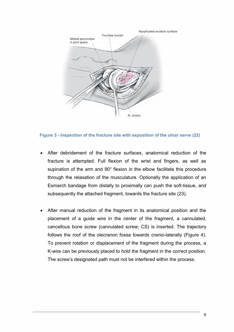
Figure 3 - Inspection of the fracture site with exposition of the ulnar nerve......................................... 9
Figure 4 - CS implant in 12-year-old boy (right arm) ................................................................................... 10
Figure 5 - Fixation of the fragment using K-wires and adjuvant suture................................................ 10
Figure 6 - Final position of K-wires after ORIF ............................................................................................... 11
Figure 7 - Two CS implants in 15-year-old boy (right arm) ....................................................................... 12
Figure 8 - Distribution of patients according to age and gender ............................................................. 18
Figure 9 - Male to female ratio and median age at time of injury ............................................................ 19
Figure 10 - Cause of injury ...................................................................................................................................... 19
Figure 11 - Ratio of elbow luxation and fragment dislocationin our patients .................................... 21
Figure 12 - Different treatment modalities in patients with MHEF ........................................................ 23
Figure 13 - Ratio of treatment groups with respect to injury ................................................................... 25
Figure 14 - Surgical patients with respect to days passed between injury and treatment ............ 27
Figure 15 - Days of hospitalization with respect to surgical method of fixation................................ 28
Figure 16 - Surgical method used with respect to injury ............................................................................ 29
Figure 17 - Days of postoperative immobilization with respect to surgical method used ............. 30
Figure 18 - Ratio of physiotherapy in surgical patients with respect to method of fixation ......... 31
Figure 19 - Time of implants in situ in surgical patients ............................................................................ 32
Figure 20 - Median number of visits to our department with respect to method of treatment ... 37
Figure 21 - Visits to our department of surgical patients with respect to method of fixation ...... 38
Figure 22 - Median number of radiologic examinations with respect to treatment ........................ 39
Figure 23 - Radiologic examinations of surgical patients with respect to method of fixation ..... 40
Figure 24 - Duration of treatment with respect to method of treatment ............................................. 41
Figure 25 - Duration of treatment in surgical patients with respect to method of fixation........... 42
Figure 26 - Influence of injury on median period of treatment ............................................................... 43
x
List of Tables
Table 1 - Classification systems of MHEF ............................................................................................................. 5
Table 2 - Information gathered for patients included in the study......................................................... 16
Table 3 - Epidemiology of treatment groups ................................................................................................... 23
Table 4 - Number, age, hospitalization and fracture characteristics of surgical patients .............. 26
Table 5 - Description of patients who had conversion ................................................................................ 34
Table 6 - Patients who needed conversion ...................................................................................................... 35
Table 7 - Criteria for the evaluation of outcome by Farsetti et al. ........................................................... 52
xi
Abbreviations
CS cannulated screw
GST gravity stress test
K-wires Kirschner wires
MCL medial collateral ligament
MHE medial humeral epicondyle
MHEF medial humeral epicondyle fracture
ROM range of motion
VST valgus stress test
1
1. Introduction
Medial humeral epicondyle fractures (MHEF) are common fractures of the elbow
region in the pediatric population and they are often associated with luxations of
the elbow. Injuries of the medial humeral epicondyle (MHE) of the elbow have
been first described by Granger as far back as 1818 and MHEFs in children and
adolescents have become the focus of various studies since the 1930s (1,2).
However, the management of MHEF still remains a controversial topic in pediatric
trauma care.
Incidence
Elbow fractures represent up to 7% of all fractures in the pediatric population and
MHEFs constitute around 8% of these fractures (3). With a reported incidence of
33% to 55%, MHEFs are the most common fractures associated with elbow
luxations (4,5). Conversely, elbow luxations are associated with MHEFs in 60% of
the cases, especially with a higher degree of fracture dislocation (6,7). MHEFs
show an increased incidence among boys, with a ratio of 2:1 (8). Most MHEFs are
reported between the ages 9 and 14, with a peak incidence between 11 to 12
years (9,10).
Anatomy
Knowledge of the anatomy of the MHE is important for the comprehension of the
injury mechanisms, the fracture’s clinical and radiographic diagnosis and the
complications associated with MHEFs.
The MHE is the second of four ossification centers responsible to form the distal
humerus. The ossification process starts at around 5 years in girls and 7.5 years in
boys. Fusion with the metaphysis of the humerus begins at around 14 years in
girls and 17 years in boys (11).
2
The MHE lies extra-articular and is considered as an apophysis. It serves as origin
for the anterior and posterior band of the medial collateral ligament (MCL) as well
as the superficial flexors of the forearm (12). During development it does not
contribute to growth in length of the humerus and therefore injuries to the MHE
during growth do not influence the final dimensions of the mature humerus (10).
The MHE’s eccentric postero-medial position and the fan-shaped extension of the
MCL’s anterior band to the medial aspect of the coronoid process of the ulna
provide medial stability throughout the whole range of motion (ROM) of the elbow
joint (12,13). In the event of an elbow luxation, the MCL is strained and the applied
force is passed on to the MHE. If the force is big enough and the ligaments do not
tear, this can subsequently result in a MHEF
The muscles originating from the MHE through the common flexor tendon are part
of the superficial flexor group including the flexor carpi radialis, the palmaris longus
and the humeral heads of the flexor carpi ulnaris and the flexor digitorum
superficialis. Also the humeral head of the pronator teres originates partially from
the proximal portion of the MHE (12,13). In 55% of the cases an accessory
humeral head of the flexor pollicis longus originates from the MHE (Gantzer's
muscle) (14).
Another important anatomical structure in this area, playing a major role in the
complications of MHEF, is the ulnar nerve. It passes through a bony groove, which
is part of the cubital tunnel located posterior to the MHE. The fascia of the flexor
carpi ulnaris and Osborne’s ligament form the roof of the cubital tunnel. The
posterior and transverse bands of the MCL form the floor. The sides are bordered
by the olecranon laterally and the MHE medially (12). Initially around 9-10% of
patients sustaining MHEFs present with ulnar nerve symptoms. Up to 16% of
patients with MHEFs develop neural symptoms throughout the course of treatment
(8,15).
3
Mechanism of injury
The mechanisms of injury responsible for MHEFs are well understood and
typically include one of the following three (5,9,10,16):
a. An avulsion mechanism, typically occurring in the event of a fall on the
outstretched arm and often accompanied by a valgus stress to the elbow
joint.
b. The injury in association with an elbow luxation.
c. The fracture as a result of a direct blow to the medial aspect of the elbow
region.
MHEFs obtained through a direct blow to the elbow are very rare and usually
result in a multi-fragmented fracture of the MHE as well as clinically notable soft-
tissue swelling and ecchymosis in the area (10).
Especially in athletes exercising arm wrestling, frequent overhead throwing or
weight bearing activities, a chronic tension stress on the MHE can subsequently
develop into an acute MHEF through isolated muscle avulsion (10,17,18). More
commonly, the acute force created in the event of a fall onto the outstretched arm,
often with the wrist and fingers hyperextended, leads to MHEF when the MHE
cannot withstand the sudden pull. In this event an additional valgus stress strains
the MCL and can increase the likelihood of MHEF (9).
4
Diagnosis and treatment
The diagnosis of MHEF in children and adolescents, and subsequently the
decision for the recommended treatment, rely on the clinical presentation together
with radiographs of the injured elbow. No commonly used classification system of
MHEF has been proposed yet, though several authors have made efforts to
describe different types of MHEF. While they can be useful for the diagnosis and
the statistical analysis, the consequences for treatment of MHEF are relatively
arguable.
Table 1 shows a range of classifications of MHEF with different levels of
complexity and different clinical and radiological factors included in the
considerations. The classifications in Table 1 are organized in chronological order
of the year of publication.
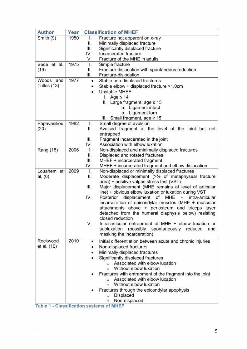
5
Author Year Classification of MHEF Smith (9) 1950 I. Fracture not apparent on x-ray
II. Minimally displaced fracture III. Significantly displaced fracture IV. Incarcerated fracture V. Fracture of the MHE in adults
Bede et al. (19)
1975 I. Simple fracture II. Fracture-dislocation with spontaneous reduction
III. Fracture-dislocation
Woods and Tullos (13)
1977 Stable non-displaced fractures
Stable elbow + displaced fracture >1.0cm
Unstable MHEF I. Age ≤ 14 II. Large fragment, age ≥ 15
a. Ligament intact b. Ligament torn
III. Small fragment, age ≥ 15
Papavasiliou (20)
1982 I. Small degree of avulsion II. Avulsed fragment at the level of the joint but not
entrapped III. Fragment incarcerated in the joint IV. Association with elbow luxation
Rang (18) 2006 I. Non-displaced and minimally displaced fractures II. Displaced and rotated fractures
III. MHEF + incarcerated fragment IV. MHEF + incarcerated fragment and elbow dislocation
Louahem et al. (6)
2009 I. Non-displaced or minimally displaced fractures II. Moderate displacement (>½ of metaphyseal fracture
area) + positive valgus stress test (VST) III. Major displacement (MHE remains at level of articular
line) + obvious elbow luxation or luxation during VST IV. Posterior displacement of MHE + intra-articular
incarceration of epicondylar muscles (MHE + muscular attachments above + periosteum and triceps layer detached from the humeral diaphysis below) resisting closed reduction
V. Intra-articular entrapment of MHE + elbow luxation or subluxation (possibly spontaneously reduced and masking the incarceration)
Rockwood et al. (10)
2010 Initial differentiation between acute and chronic injuries
Non-displaced fractures
Minimally displaced fractures
Significantly displaced fractures o Associated with elbow luxation o Without elbow luxation
Fractures with entrapment of the fragment into the joint o Associated with elbow luxation o Without elbow luxation
Fractures through the epicondylar apophysis o Displaced o Non-displaced
Table 1 - Classification systems of MHEF
6
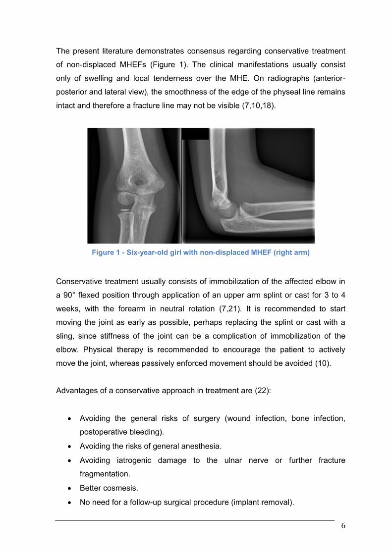
The present literature demonstrates consensus regarding conservative treatment
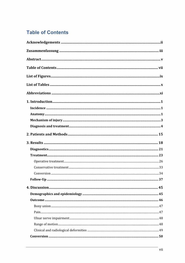
of non-displaced MHEFs (Figure 1). The clinical manifestations usually consist
only of swelling and local tenderness over the MHE. On radiographs (anterior-
posterior and lateral view), the smoothness of the edge of the physeal line remains
intact and therefore a fracture line may not be visible (7,10,18).
Figure 1 - Six-year-old girl with non-displaced MHEF (right arm)
Conservative treatment usually consists of immobilization of the affected elbow in
a 90° flexed position through application of an upper arm splint or cast for 3 to 4
weeks, with the forearm in neutral rotation (7,21). It is recommended to start
moving the joint as early as possible, perhaps replacing the splint or cast with a
sling, since stiffness of the joint can be a complication of immobilization of the
elbow. Physical therapy is recommended to encourage the patient to actively
move the joint, whereas passively enforced movement should be avoided (10).
Advantages of a conservative approach in treatment are (22):
Avoiding the general risks of surgery (wound infection, bone infection,
postoperative bleeding).
Avoiding the risks of general anesthesia.
Avoiding iatrogenic damage to the ulnar nerve or further fracture
fragmentation.
Better cosmesis.
No need for a follow-up surgical procedure (implant removal).
7
On the other hand, a number of absolute and relative indications for open
reduction and internal fixation (ORIF) of the fracture have been recognized
(10,13,22).
Absolute indications for ORIF are:
Open fractures.
Failure to reduce an incarcerated fragment from the elbow joint.
A complete lesion or displacement of the ulnar nerve into the fracture gap.
Relative indications for ORIF are:
Simple injuries to the ulnar nerve.
Valgus instability.
High-demand upper extremity-function of the patients (e.g. athletes).
The advantages of ORIF over the conservative approach are (22):
Possibility of a thorough assessment of the injury.
Visualization of the anatomical structures injured.
Anatomical refixation of the fragment.
Quick restoration of the stability of the joint.
Preparations for ORIF should include thorough assessment and documentation of
peripheral circulation, motor activity and sensibility. Adequate pain therapy,
immobilization and required radiographs should be obtained before initiation of
surgical intervention (22). When the patient is under general anesthesia and
placed in a supine position, the closed reduction of a present elbow luxation
should be attempted. This allows further clinical assessment of the injury as well
as further radiological examinations if necessary (22).
8
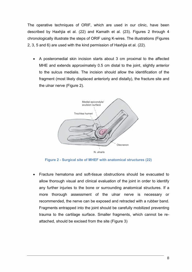
The operative techniques of ORIF, which are used in our clinic, have been
described by Haxhjia et al. (22) and Kamath et al. (23). Figures 2 through 4
chronologically illustrate the steps of ORIF using K-wires. The illustrations (Figures
2, 3, 5 and 6) are used with the kind permission of Haxhjia et al. (22).
A posteromedial skin incision starts about 3 cm proximal to the affected
MHE and extends approximately 0.5 cm distal to the joint, slightly anterior
to the sulcus medialis. The incision should allow the identification of the
fragment (most likely displaced anteriorly and distally), the fracture site and
the ulnar nerve (Figure 2).
Figure 2 - Surgical site of MHEF with anatomical structures (22)
Fracture hematoma and soft-tissue obstructions should be evacuated to
allow thorough visual and clinical evaluation of the joint in order to identify
any further injuries to the bone or surrounding anatomical structures. If a
more thorough assessment of the ulnar nerve is necessary or
recommended, the nerve can be exposed and retracted with a rubber band.
Fragments entrapped into the joint should be carefully mobilized preventing
trauma to the cartilage surface. Smaller fragments, which cannot be re-
attached, should be excised from the site (Figure 3)
9
Figure 3 - Inspection of the fracture site with exposition of the ulnar nerve (22)
After debridement of the fracture surfaces, anatomical reduction of the
fracture is attempted. Full flexion of the wrist and fingers, as well as
supination of the arm and 90° flexion in the elbow facilitate this procedure
through the relaxation of the musculature. Optionally the application of an
Esmarch bandage from distally to proximally can push the soft-tissue, and
subsequently the attached fragment, towards the fracture site (23).
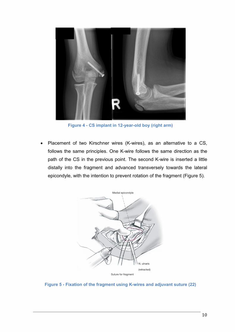
After manual reduction of the fragment in its anatomical position and the
placement of a guide wire in the center of the fragment, a cannulated,
cancellous bone screw (cannulated screw; CS) is inserted. The trajectory
follows the roof of the olecranon fossa towards cranio-laterally (Figure 4).
To prevent rotation or displacement of the fragment during the process, a
K-wire can be previously placed to hold the fragment in the correct position.
The screw’s designated path must not be interfered within the process.
10
Figure 4 - CS implant in 12-year-old boy (right arm)
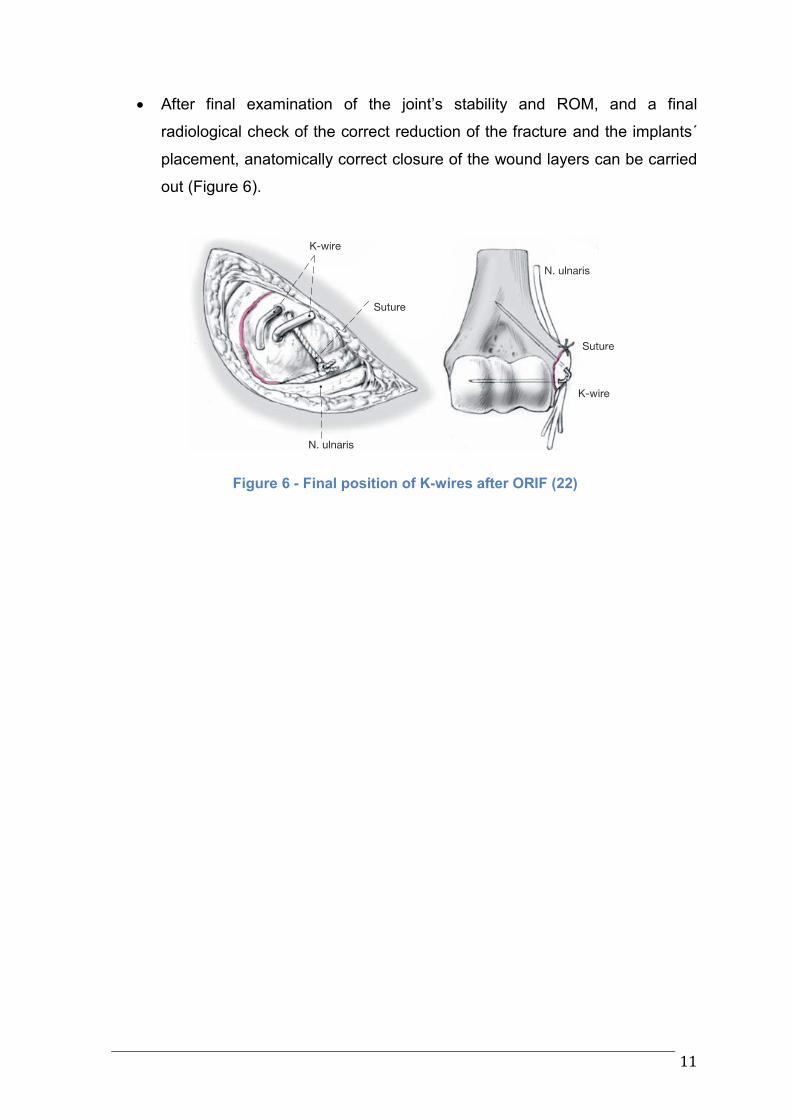
Placement of two Kirschner wires (K-wires), as an alternative to a CS,
follows the same principles. One K-wire follows the same direction as the
path of the CS in the previous point. The second K-wire is inserted a little
distally into the fragment and advanced transversely towards the lateral
epicondyle, with the intention to prevent rotation of the fragment (Figure 5).
Figure 5 - Fixation of the fragment using K-wires and adjuvant suture (22)
11
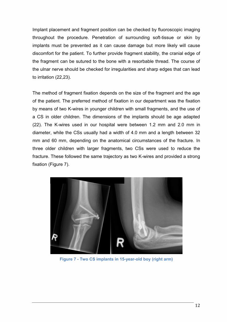
After final examination of the joint’s stability and ROM, and a final
radiological check of the correct reduction of the fracture and the implants´
placement, anatomically correct closure of the wound layers can be carried
out (Figure 6).
Figure 6 - Final position of K-wires after ORIF (22)
12
Implant placement and fragment position can be checked by fluoroscopic imaging
throughout the procedure. Penetration of surrounding soft-tissue or skin by
implants must be prevented as it can cause damage but more likely will cause
discomfort for the patient. To further provide fragment stability, the cranial edge of
the fragment can be sutured to the bone with a resorbable thread. The course of
the ulnar nerve should be checked for irregularities and sharp edges that can lead
to irritation (22,23).
The method of fragment fixation depends on the size of the fragment and the age
of the patient. The preferred method of fixation in our department was the fixation
by means of two K-wires in younger children with small fragments, and the use of
a CS in older children. The dimensions of the implants should be age adapted
(22). The K-wires used in our hospital were between 1.2 mm and 2.0 mm in
diameter, while the CSs usually had a width of 4.0 mm and a length between 32
mm and 60 mm, depending on the anatomical circumstances of the fracture. In
three older children with larger fragments, two CSs were used to reduce the
fracture. These followed the same trajectory as two K-wires and provided a strong
fixation (Figure 7).
Figure 7 - Two CS implants in 15-year-old boy (right arm)
13
While the above-mentioned indications for conservative treatment or ORIF are
commonly agreed upon, opinions differ regarding the treatment for uncomplicated
MHEF with a displaced MHE. Disagreement arises in particular over the tolerable
distance of displacement of the MHEF before ORIF is indicated, thus quantifying a
“significant” displacement of the MHE: Some authors opt for a conservative
treatment in cases of fragment displacement of up to 15 mm (21,24). Other
authors seem to have lower threshold for ORIF, recommending surgical approach
in cases of fragment displacement of as little as 2 mm (15,25).
Major complications of MHEF, resulting in a significant loss of function of the
elbow joint, are rare and typically due to missed diagnosis of an entrapped intra-
articular fragment or the development of an ulnar nerve dysfunction (10,18). Other
minor complications are only of minimal functional or cosmetic consequences and
usually require no further management (10). Nonunion between the fragment and
the distal humeral metaphysis (fibrous union or pseudoarthrosis) is especially
common in conservatively treated patients. The functional outcome is still
satisfactory in the majority of cases, though athletes may require further treatment
due to higher requirements on the joint, which can be difficult to achieve (10).
Kamath et al. (8) reported that 92,5% of patients that underwent ORIF had
reached bony union as compared to 49,2% of patients that were treated
conservatively. Loss of 5% to 10% of ROM (especially in flexion/extension) was
seen in up to 20% of patients and mostly due to longer periods of immobilization
(10).
Myositis ossificans is a rare complication, mostly inflicted iatrogenic, through the
wrong and forceful manipulation of the elbow to extract an entrapped fragment
from the joint space (10). Recurring injuries of the epicondyle and ligamentous
structures can cause asymptomatic ectopic calcifications in the medial elbow area,
creating the aspect of an increased carrying angle of the joint (10).
The most important differential diagnosis of MHEF is an injury to the medial
condylar physis with an intra-articular component (Kilfoyle II and Kilfoyle III), which
is an indication for surgery. Especially on x-rays of younger children, these
fractures can be confused with isolated MHEF, since the MHE starts the
14
ossification process earlier than the trochlea. A positive fat pad sign can hint an
intra-articular component of the injury. The clinical presentation is similar to MHEF,
with medial swelling and valgus instability. Additionally, varus instability and the
tendency of the elbow to subluxate postero-medially are symptoms associated
with medial condyle fractures because of loss of trochlear stability. Nonunion, loss
of reduction, cubitus varus and avascular necrosis of the trochlea are
complications of this injury and require further interventions. If in doubt of the
location of the fracture, additional MRI, CT scan or arthrography of the joint should
be performed (10,18,22).
In order to decide for either a conservative or a surgical treatment approach,
absolute and relative indications for surgical treatment of MHEF were taken into
consideration in our institution. MHEFs were then divided into isolated fractures
and fractures associated with elbow luxation. Furthermore, displacement of the
fragment was considered significant if the distance was 3 mm or more. The
recommendation in our department was to refrain from surgery with displacements
of a smaller distance. However, the final decision for surgery in debatable cases,
with a dislocation of the MHE between 3-6 mm, was taken in accordance with the
surgeon responsible, the patient and his/her parents, leaving room for discretion.
The aim of our present study was to analyze the results of MHEF in children and
adolescents treated in our institution and to compare the outcome after
conservative and surgical treatment.
15
2. Patients and Methods
The electronic system for patient documentation (MEDOCS®) was employed to
retrieve the data of all patients with MHEF that had received conservative or
surgical treatment at the Department for Paediatric and Adolescent Surgery of the
Medical University of Graz between the years 2004 and 2011.
Inclusion criteria were the following:
Age at the time of injury from 5 to 16 years.
The patient received treatment for MHEF in our department.
The patient was referred after initiation of MHEF treatment in a local
hospital or was directly admitted to our department after the injury.
The patient received at least 2 follow-up clinical examinations and 2
radiological examinations.
The fracture was an isolated fracture to the medial epicondyle with no other
fractures involving the affected arm’s elbow joint.
The fracture was the first MHEF of the affected arm in the patient’s medical
history.
For the purpose of the study patients were grouped according to their treatment in
three different groups. Group 1 comprised patients, who were treated with ORIF of
the fractured epicondyle (surgical group). Group 2 included all patients that were
treated by means of immobilization of the affected elbow exclusively (conservative
group). Group 3 included patients who were initially treated conservatively, but
subsequently underwent surgery (conversion group).
Detailed data were collected and organized in the following four categories (Table
2):
1. Injury
2. Diagnosis
3. Treatment
4. Follow-up
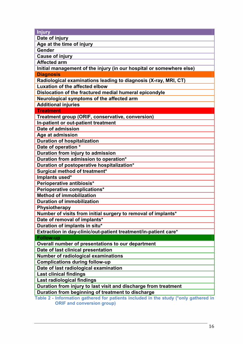
16
Injury
Date of injury
Age at the time of injury
Gender
Cause of injury
Affected arm
Initial management of the injury (in our hospital or somewhere else)
Diagnosis
Radiological examinations leading to diagnosis (X-ray, MRI, CT)
Luxation of the affected elbow
Dislocation of the fractured medial humeral epicondyle
Neurological symptoms of the affected arm
Additional injuries
Treatment
Treatment group (ORIF, conservative, conversion)
In-patient or out-patient treatment
Date of admission
Age at admission
Duration of hospitalization
Date of operation *
Duration from injury to admission
Duration from admission to operation*
Duration of postoperative hospitalization*
Surgical method of treatment*
Implants used*
Perioperative antibiosis*
Perioperative complications*
Method of immobilization
Duration of immobilization
Physiotherapy
Number of visits from initial surgery to removal of implants*
Date of removal of implants*
Duration of implants in situ*
Extraction in day-clinic/out-patient treatment/in-patient care*
Follow-up
Overall number of presentations to our department
Date of last clinical presentation
Number of radiological examinations
Complications during follow-up
Date of last radiological examination
Last clinical findings
Last radiological findings
Duration from injury to last visit and discharge from treatment
Duration from beginning of treatment to discharge Table 2 - Information gathered for patients included in the study (*only gathered in
ORIF and conversion group)
17
Microsoft Excel® was used to document the gathered information and to process
data for further statistical analysis. To provide anonymization of patient records,
clinical data were separated from patients´ personal information. Only age and
gender were included to give an epidemiologic overview of the injury discussed.
Statistical analysis was made using Graph Pad Prism for Chi-Square and Chi-
Square test for trend. Data are expressed in median and range. A P-value less
than 0.05 was considered statistical significant.
Results were compared with other studies in the literature, which thematized
surgical and non-surgical approaches for MHEF treatment. Studies related to
diagnostics of injuries to the MHE in children and adolescents were also reviewed.
The study’s design and approach was evaluated and approved by the ethics
committee of the Medical University of Graz in their ruling “24-043 ex 11/12”.
18
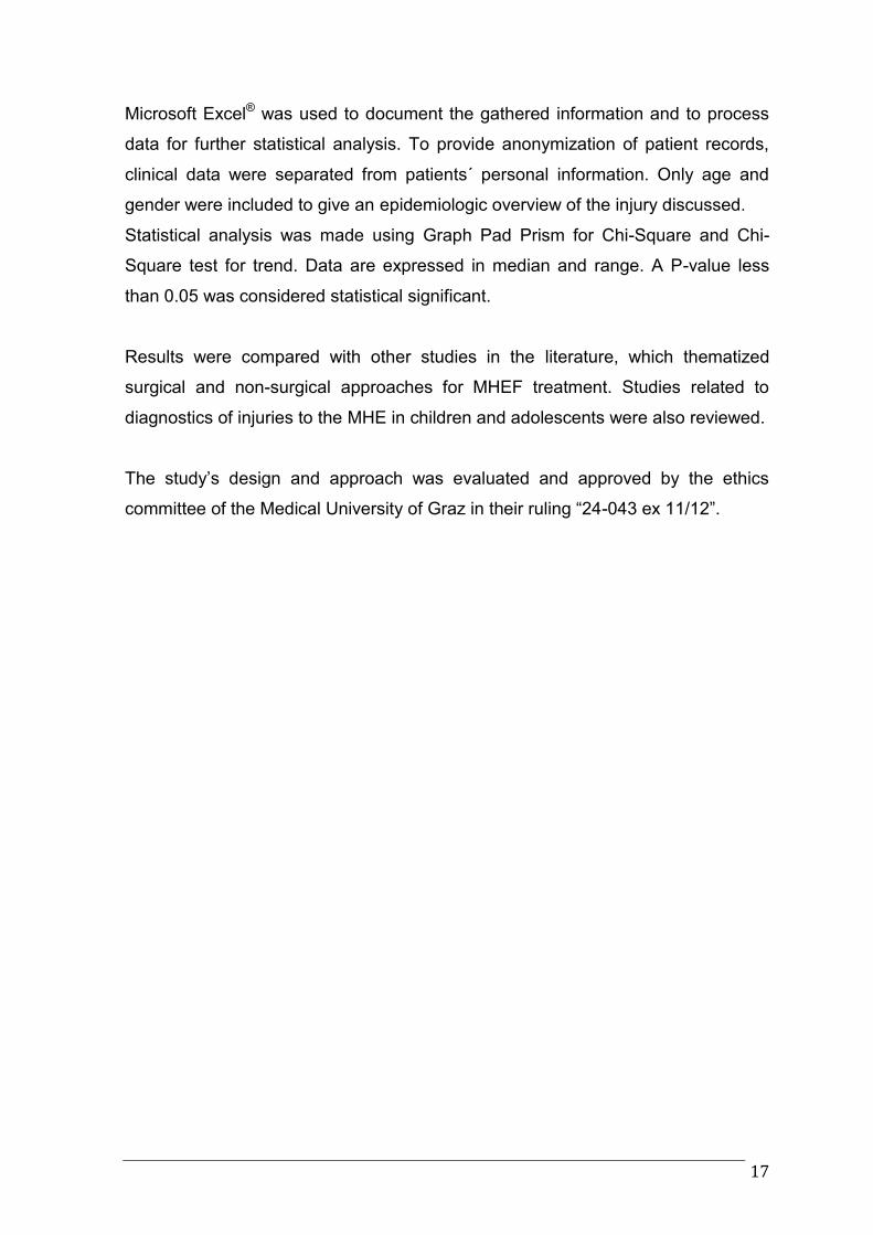
3. Results
In total, there were 81 patients with MHEF that met our study inclusion criteria.
Injuries to the MHE occurred throughout all age groups with a median age of 12
years (range 5-16). Most of the patients, however, suffered from MHEF at an age
between 12-15 years. Figure 8 demonstrates an earlier peak of injuries in female
patients, between the ages 8 and 9, compared to the male patients, with a peak
incidence between 12 and 15 years.
Figure 8 - Distribution of patients (n=81) according to age and gender
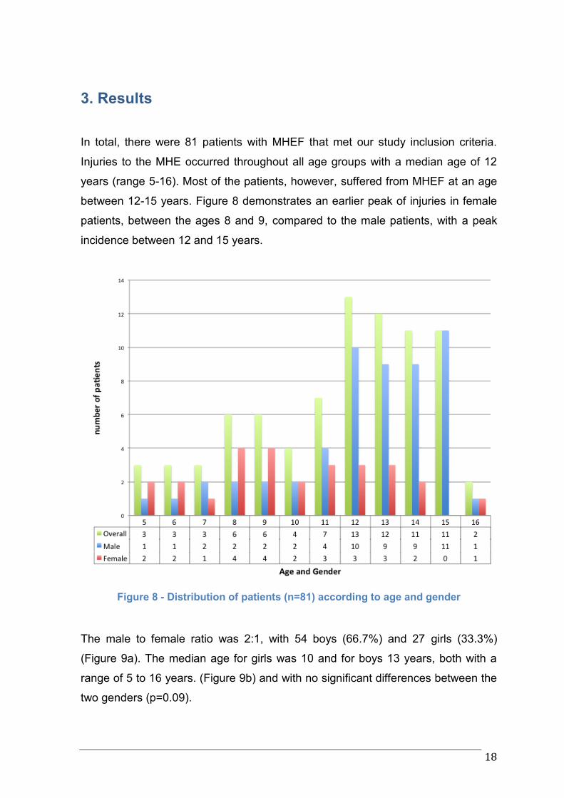
The male to female ratio was 2:1, with 54 boys (66.7%) and 27 girls (33.3%)
(Figure 9a). The median age for girls was 10 and for boys 13 years, both with a
range of 5 to 16 years. (Figure 9b) and with no significant differences between the
two genders (p=0.09).
19
Figure 9 - Male to female ratio (a) and median age at time of injury (b)
The affected extremity was the right arm in 40 cases (49.4%) and the left arm in
41 (50.6%) cases. The affected arms were also evenly distributed within the two
genders, with 27 injuries to the right arm and the same number of injuries to the
left arm in boys, and 13 injuries the right arm and 14 injuries to the left arm in girls.
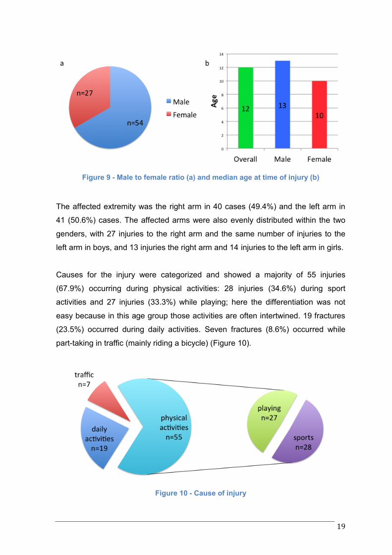
Causes for the injury were categorized and showed a majority of 55 injuries
(67.9%) occurring during physical activities: 28 injuries (34.6%) during sport
activities and 27 injuries (33.3%) while playing; here the differentiation was not
easy because in this age group those activities are often intertwined. 19 fractures
(23.5%) occurred during daily activities. Seven fractures (8.6%) occurred while
part-taking in traffic (mainly riding a bicycle) (Figure 10).
Figure 10 - Cause of injury
20
Forty patients (49.4%) were already diagnosed in their local hospital and usually
treated with provisional immobilization of the injured elbow with a cast before
referral to our Accident and Emergency department. Forty-one patients (50.6%)
were initially treated directly at our hospital.
21
Diagnostics
In the majority of our cases (n=73; 90.1%) standard x-rays (lateral and
anterior/posterior view) of the injured elbow were sufficient for diagnosis. However,
additional CT scans were required in three patients (3.7%) and additional MRI in
five patients (6.2%) in order to confirm diagnosis of MHEF.
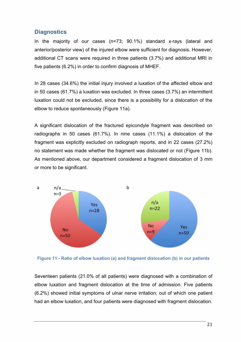
In 28 cases (34.6%) the initial injury involved a luxation of the affected elbow and
in 50 cases (61.7%) a luxation was excluded. In three cases (3.7%) an intermittent
luxation could not be excluded, since there is a possibility for a dislocation of the
elbow to reduce spontaneously (Figure 11a).
A significant dislocation of the fractured epicondyle fragment was described on
radiographs in 50 cases (61.7%). In nine cases (11.1%) a dislocation of the
fragment was explicitly excluded on radiograph reports, and in 22 cases (27.2%)
no statement was made whether the fragment was dislocated or not (Figure 11b).
As mentioned above, our department considered a fragment dislocation of 3 mm
or more to be significant.
Figure 11 - Ratio of elbow luxation (a) and fragment dislocation (b) in our patients
Seventeen patients (21.0% of all patients) were diagnosed with a combination of
elbow luxation and fragment dislocation at the time of admission. Five patients
(6.2%) showed initial symptoms of ulnar nerve irritation; out of which one patient
had an elbow luxation, and four patients were diagnosed with fragment dislocation.
22
Additional injuries, not involving the affected elbow joint per se, were present in 13
patients (16.1%). Those involved mainly contusions and excoriations of the body
surface, as well as other bone injuries of the distal upper limb. The most common
additional fractures involved the radius (n=7) with either a distal radius fracture or
a fracture of the radius head.
23
Treatment
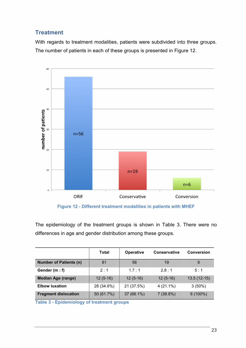
With regards to treatment modalities, patients were subdivided into three groups.
The number of patients in each of these groups is presented in Figure 12.
Figure 12 - Different treatment modalities in patients with MHEF
The epidemiology of the treatment groups is shown in Table 3. There were no
differences in age and gender distribution among these groups.
Total Operative Conservative Conversion
Number of Patients (n) 81 56 19 6
Gender (m : f) 2 : 1 1.7 : 1 2.8 : 1 5 : 1
Median Age (range) 12 (5-16) 12 (5-16) 12 (5-16) 13.5 (12-15)
Elbow luxation 28 (34.6%) 21 (37.5%) 4 (21.1%) 3 (50%)
Fragment dislocation 50 (61.7%) 37 (66.1%) 7 (36.8%) 6 (100%)
Table 3 - Epidemiology of treatment groups
24
The ORIF group consisted of 56 patients (69.1%). Surgical treatment was
performed by ORIF of the fractured MHE using either CSs or K-wires or a
combination of both. Postoperatively the affected arm was immobilized with an
upper arm splint or cast in almost all cases (Table 3).
Conservative treatment was initially attempted in 25 of our patients (30.9%).
However, while in 19 cases (overall 23.5%; 76% of initially conservatively treated
patients) conservative treatment was successful, six patients (overall 7.4%; 24% of
initially conservatively treated patients) required a conversion to an operative
management of the injury at some point during the treatment. Immobilization of the
injured arm was provided with either an upper-arm plaster cast or splint (Table 3).
An elbow luxation or a fragment dislocation was more often seen in the operative
group and conversion group when compared to the conservative group (Figure
13).
25
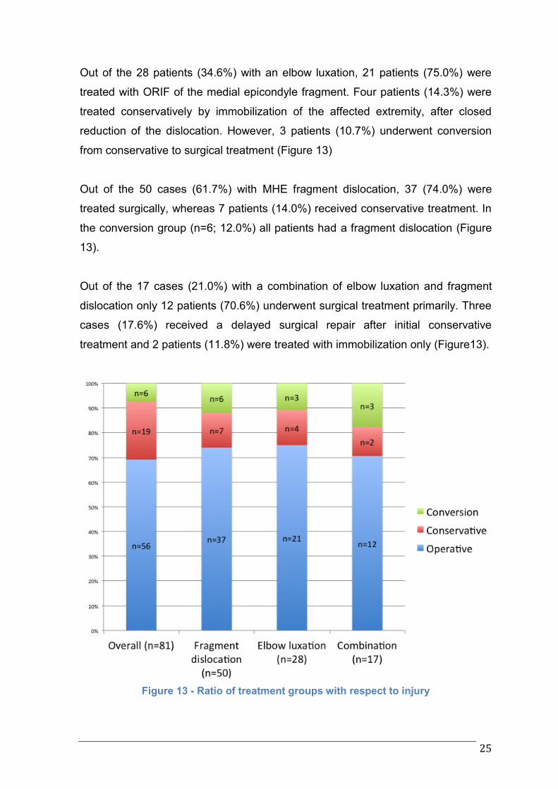
Out of the 28 patients (34.6%) with an elbow luxation, 21 patients (75.0%) were
treated with ORIF of the medial epicondyle fragment. Four patients (14.3%) were
treated conservatively by immobilization of the affected extremity, after closed
reduction of the dislocation. However, 3 patients (10.7%) underwent conversion
from conservative to surgical treatment (Figure 13)
Out of the 50 cases (61.7%) with MHE fragment dislocation, 37 (74.0%) were
treated surgically, whereas 7 patients (14.0%) received conservative treatment. In
the conversion group (n=6; 12.0%) all patients had a fragment dislocation (Figure
13).
Out of the 17 cases (21.0%) with a combination of elbow luxation and fragment
dislocation only 12 patients (70.6%) underwent surgical treatment primarily. Three
cases (17.6%) received a delayed surgical repair after initial conservative
treatment and 2 patients (11.8%) were treated with immobilization only (Figure13).
Figure 13 - Ratio of treatment groups with respect to injury
26
Operative treatment
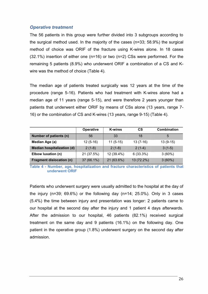
The 56 patients in this group were further divided into 3 subgroups according to
the surgical method used. In the majority of the cases (n=33; 58.9%) the surgical
method of choice was ORIF of the fracture using K-wires alone. In 18 cases
(32.1%) insertion of either one (n=16) or two (n=2) CSs were performed. For the
remaining 5 patients (8.9%) who underwent ORIF a combination of a CS and K-
wire was the method of choice (Table 4).
The median age of patients treated surgically was 12 years at the time of the
procedure (range 5-16). Patients who had treatment with K-wires alone had a
median age of 11 years (range 5-15), and were therefore 2 years younger than
patients that underwent either ORIF by means of CSs alone (13 years, range 7-
16) or the combination of CS and K-wires (13 years, range 9-15) (Table 4).
Operative K-wires CS Combination
Number of patients (n) 56 33 18 5
Median Age (a) 12 (5-16) 11 (5-15) 13 (7-16) 13 (9-15)
Median hospitalization (d) 2 (1-8) 2 (1-8) 2 (1-4) 3 (1-5)
Elbow luxation (n) 21 (37.5%) 12 (39.4%) 6 (33.3%) 3 (60%)
Fragment dislocation (n) 37 (66.1%) 21 (63.6%) 13 (72.2%) 3 (60%)
Table 4 - Number, age, hospitalization and fracture characteristics of patients that underwent ORIF
Patients who underwent surgery were usually admitted to the hospital at the day of
the injury (n=39; 69.6%) or the following day (n=14; 25.0%). Only in 3 cases
(5.4%) the time between injury and presentation was longer: 2 patients came to
our hospital at the second day after the injury and 1 patient 4 days afterwards.
After the admission to our hospital, 46 patients (82.1%) received surgical
treatment on the same day and 9 patients (16.1%) on the following day. One
patient in the operative group (1.8%) underwent surgery on the second day after
admission.
27
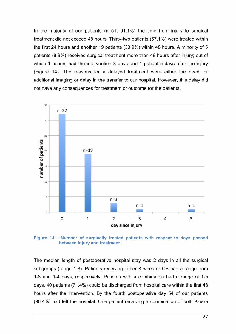
In the majority of our patients (n=51; 91.1%) the time from injury to surgical
treatment did not exceed 48 hours. Thirty-two patients (57.1%) were treated within
the first 24 hours and another 19 patients (33.9%) within 48 hours. A minority of 5
patients (8.9%) received surgical treatment more than 48 hours after injury; out of
which 1 patient had the intervention 3 days and 1 patient 5 days after the injury
(Figure 14). The reasons for a delayed treatment were either the need for
additional imaging or delay in the transfer to our hospital. However, this delay did
not have any consequences for treatment or outcome for the patients.
Figure 14 - Number of surgically treated patients with respect to days passed between injury and treatment
The median length of postoperative hospital stay was 2 days in all the surgical
subgroups (range 1-8). Patients receiving either K-wires or CS had a range from
1-8 and 1-4 days, respectively. Patients with a combination had a range of 1-5
days. 40 patients (71.4%) could be discharged from hospital care within the first 48
hours after the intervention. By the fourth postoperative day 54 of our patients
(96.4%) had left the hospital. One patient receiving a combination of both K-wire
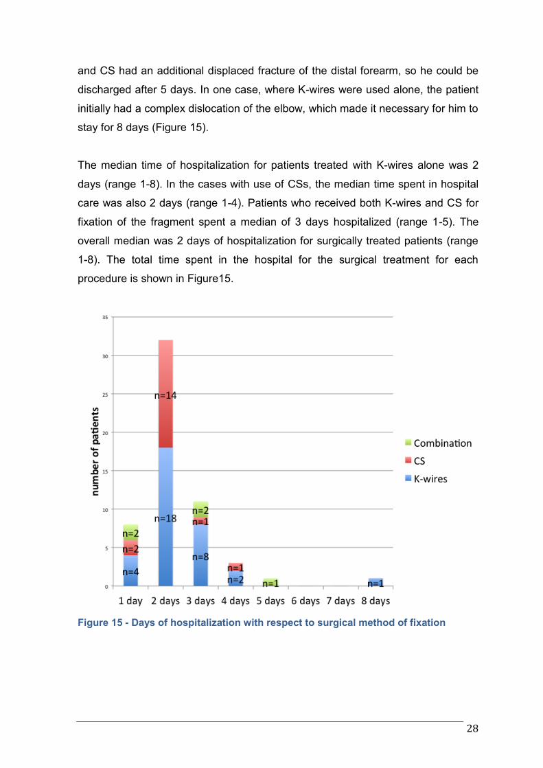
28
and CS had an additional displaced fracture of the distal forearm, so he could be
discharged after 5 days. In one case, where K-wires were used alone, the patient
initially had a complex dislocation of the elbow, which made it necessary for him to
stay for 8 days (Figure 15).
The median time of hospitalization for patients treated with K-wires alone was 2
days (range 1-8). In the cases with use of CSs, the median time spent in hospital
care was also 2 days (range 1-4). Patients who received both K-wires and CS for
fixation of the fragment spent a median of 3 days hospitalized (range 1-5). The
overall median was 2 days of hospitalization for surgically treated patients (range
1-8). The total time spent in the hospital for the surgical treatment for each
procedure is shown in Figure15.
Figure 15 - Days of hospitalization with respect to surgical method of fixation
29
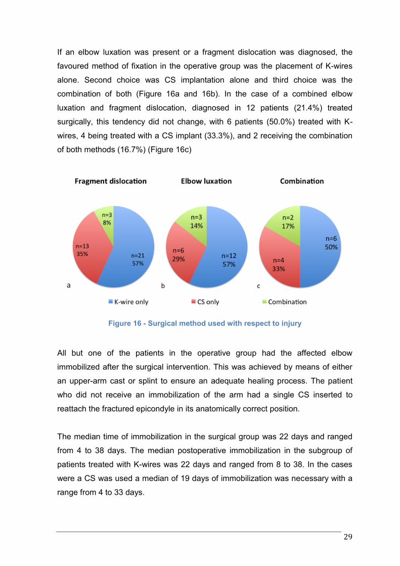
If an elbow luxation was present or a fragment dislocation was diagnosed, the
favoured method of fixation in the operative group was the placement of K-wires
alone. Second choice was CS implantation alone and third choice was the
combination of both (Figure 16a and 16b). In the case of a combined elbow
luxation and fragment dislocation, diagnosed in 12 patients (21.4%) treated
surgically, this tendency did not change, with 6 patients (50.0%) treated with K-
wires, 4 being treated with a CS implant (33.3%), and 2 receiving the combination
of both methods (16.7%) (Figure 16c)
Figure 16 - Surgical method used with respect to injury
All but one of the patients in the operative group had the affected elbow
immobilized after the surgical intervention. This was achieved by means of either
an upper-arm cast or splint to ensure an adequate healing process. The patient
who did not receive an immobilization of the arm had a single CS inserted to
reattach the fractured epicondyle in its anatomically correct position.
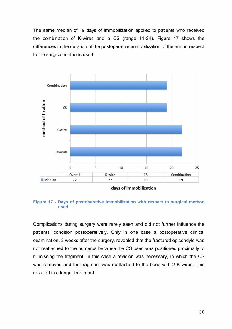
The median time of immobilization in the surgical group was 22 days and ranged
from 4 to 38 days. The median postoperative immobilization in the subgroup of
patients treated with K-wires was 22 days and ranged from 8 to 38. In the cases
were a CS was used a median of 19 days of immobilization was necessary with a
range from 4 to 33 days.
30
The same median of 19 days of immobilization applied to patients who received
the combination of K-wires and a CS (range 11-24). Figure 17 shows the
differences in the duration of the postoperative immobilization of the arm in respect
to the surgical methods used.
Figure 17 - Days of postoperative immobilization with respect to surgical method used
Complications during surgery were rarely seen and did not further influence the
patients’ condition postoperatively. Only in one case a postoperative clinical
examination, 3 weeks after the surgery, revealed that the fractured epicondyle was
not reattached to the humerus because the CS used was positioned proximally to
it, missing the fragment. In this case a revision was necessary, in which the CS
was removed and the fragment was reattached to the bone with 2 K-wires. This
resulted in a longer treatment.
31
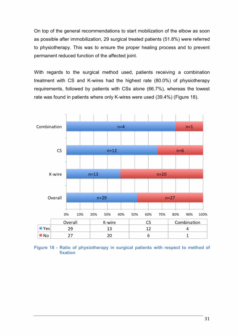
On top of the general recommendations to start mobilization of the elbow as soon
as possible after immobilization, 29 surgical treated patients (51.8%) were referred
to physiotherapy. This was to ensure the proper healing process and to prevent
permanent reduced function of the affected joint.
With regards to the surgical method used, patients receiving a combination
treatment with CS and K-wires had the highest rate (80.0%) of physiotherapy
requirements, followed by patients with CSs alone (66.7%), whereas the lowest
rate was found in patients where only K-wires were used (39.4%) (Figure 18).
Figure 18 - Ratio of physiotherapy in surgical patients with respect to method of fixation
32
All but two surgically treated patients underwent implant removal at some point. In
one case, where a combination of K-wires and CS was used, it was the patient’s
choice to refrain from removing the CS and to remove the K-wire only. In the other
case the CS used was resorbable and no extraction was planned from the
beginning. It should be mentioned that this case was one with a rather poor
outcome. The patient, after being discharged from our care, returned
approximately 1½ year later because of persisting complaints including
neurological and muscular problems. This made another intervention necessary
two years after the patient was initially released from our treatment, in which
decompression of the ulnar nerve had to be performed.
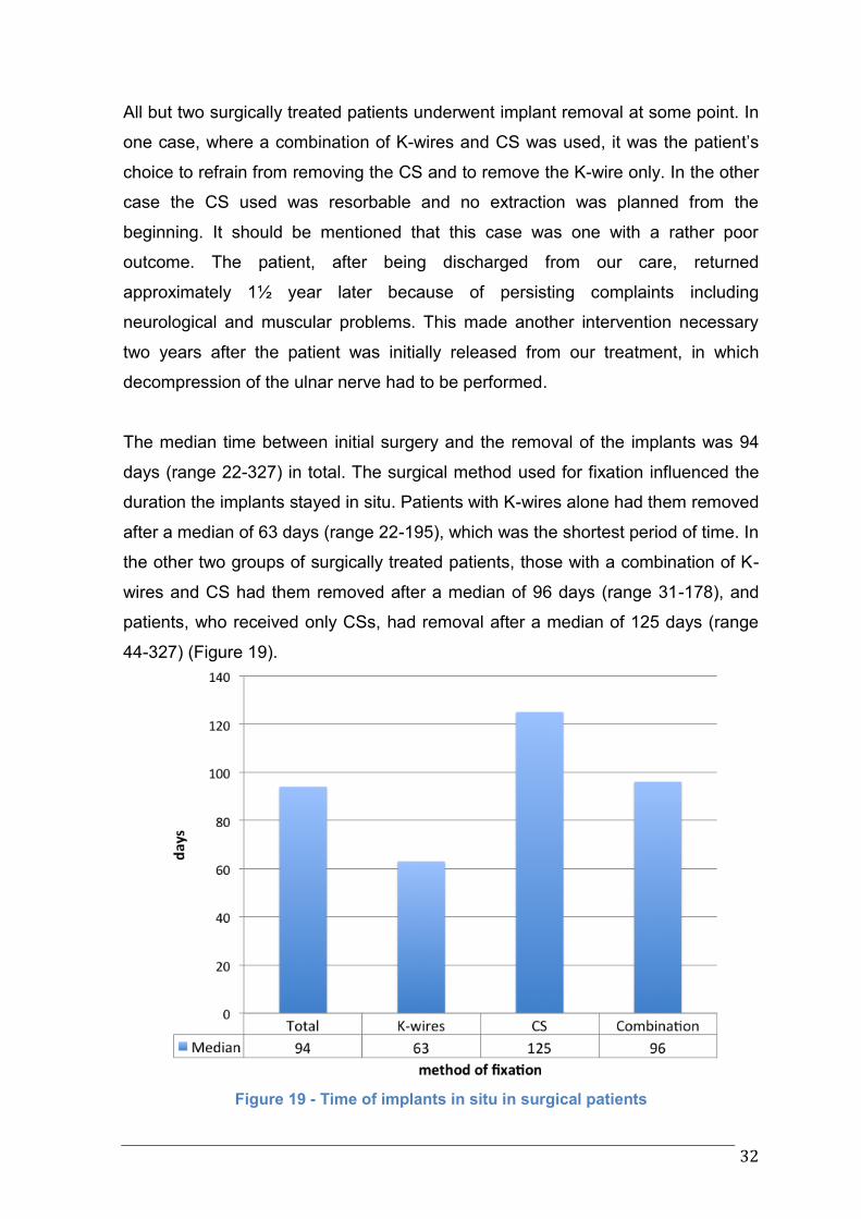
The median time between initial surgery and the removal of the implants was 94
days (range 22-327) in total. The surgical method used for fixation influenced the
duration the implants stayed in situ. Patients with K-wires alone had them removed
after a median of 63 days (range 22-195), which was the shortest period of time. In
the other two groups of surgically treated patients, those with a combination of K-
wires and CS had them removed after a median of 96 days (range 31-178), and
patients, who received only CSs, had removal after a median of 125 days (range
44-327) (Figure 19).
Figure 19 - Time of implants in situ in surgical patients
33
In 48 cases (87.3%) the removal of the implants could be performed without any
complications in our day-surgery department. In 4 cases (7.3%) a hospitalization
was necessary for the observation of the patient, but all patients were allowed to
leave on the following day. The remaining 3 removals (5.5%) were performed in
our outpatient department. Out of the 4 patients, who were hospitalized, 3 had a
CS implanted and one only K-wires as means of fixation.
Conservative treatment
The median age of the 19 patients (23.5% of the total 81 cases) treated by
immobilization of the affected arm alone, was 12 years (range 5-16). The methods
of choice were either the application of an upper-arm plaster cast or the use of an
upper-arm splint to keep the affected elbow in a 90° flexed position. Additionally,
the arm was supported by the use of a sling. Out of the 19 patients in this group,
16 patients (84.2%) could be treated in outpatient care and 3 patients (15.8%)
were treated as in-patients, but only because of late admission with no further
therapeutic consequences and all of those 3 patients, however, could be
discharged the following day. 17 of our conservative patients (89.5%) were treated
within the first two days of injury. In 2 cases (10.5%) it took up to 5 days for the
patients to be diagnosed and treated for MHEF. Both patients came to our
department because of persistent swelling and pain after they initially
underestimated the severity of their injury. The majority of patients in this
conservatively treated group did not have a luxation of the affected elbow at the
time of the first presentation. Elbow luxations were diagnosed in 4 cases (21.1%).
Also, a fragment dislocation was diagnosed in 7 cases (36.8%). The combination
of an elbow luxation and a fragment dislocation was diagnosed in 2 of our patients
(10.5%). The median period of time the injured arm was immobilized was 21 days,
with a range from 6 to 46 days. After immobilization was removed, 8 patients
(42.1%) required further physiotherapy in order to improve recovery of the affected
arm. There were three out of four patients with an elbow luxation, four out of seven
patients with fragment dislocation, and two patients with combination of fragment
dislocation and elbow luxation that were subsequently referred to physiotherapy to
improve the healing process.
34
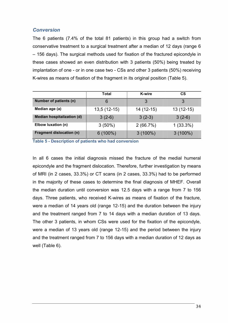
Conversion
The 6 patients (7.4% of the total 81 patients) in this group had a switch from
conservative treatment to a surgical treatment after a median of 12 days (range 6
– 156 days). The surgical methods used for fixation of the fractured epicondyle in
these cases showed an even distribution with 3 patients (50%) being treated by
implantation of one - or in one case two - CSs and other 3 patients (50%) receiving
K-wires as means of fixation of the fragment in its original position (Table 5).
Total K-wire CS
Number of patients (n) 6 3 3
Median age (a) 13,5 (12-15) 14 (12-15) 13 (12-15)
Median hospitalization (d) 3 (2-6) 3 (2-3) 3 (2-6)
Elbow luxation (n) 3 (50%) 2 (66.7%) 1 (33.3%)
Fragment dislocation (n) 6 (100%) 3 (100%) 3 (100%)
Table 5 - Description of patients who had conversion
In all 6 cases the initial diagnosis missed the fracture of the medial humeral
epicondyle and the fragment dislocation. Therefore, further investigation by means
of MRI (in 2 cases, 33.3%) or CT scans (in 2 cases, 33.3%) had to be performed
in the majority of these cases to determine the final diagnosis of MHEF. Overall
the median duration until conversion was 12.5 days with a range from 7 to 156
days. Three patients, who received K-wires as means of fixation of the fracture,
were a median of 14 years old (range 12-15) and the duration between the injury
and the treatment ranged from 7 to 14 days with a median duration of 13 days.
The other 3 patients, in whom CSs were used for the fixation of the epicondyle,
were a median of 13 years old (range 12-15) and the period between the injury
and the treatment ranged from 7 to 156 days with a median duration of 12 days as
well (Table 6).
35
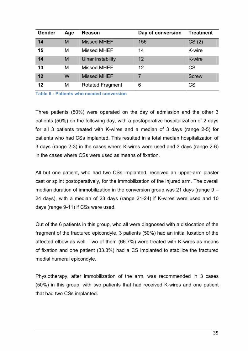
Gender Age Reason Day of conversion Treatment
14 M Missed MHEF 156 CS (2)
15 M Missed MHEF 14 K-wire
14 M Ulnar instability 12 K-wire
13 M Missed MHEF 12 CS
12 W Missed MHEF 7 Screw
12 M Rotated Fragment 6 CS
Table 6 - Patients who needed conversion
Three patients (50%) were operated on the day of admission and the other 3
patients (50%) on the following day, with a postoperative hospitalization of 2 days
for all 3 patients treated with K-wires and a median of 3 days (range 2-5) for
patients who had CSs implanted. This resulted in a total median hospitalization of
3 days (range 2-3) in the cases where K-wires were used and 3 days (range 2-6)
in the cases where CSs were used as means of fixation.
All but one patient, who had two CSs implanted, received an upper-arm plaster
cast or splint postoperatively, for the immobilization of the injured arm. The overall
median duration of immobilization in the conversion group was 21 days (range 9 –
24 days), with a median of 23 days (range 21-24) if K-wires were used and 10
days (range 9-11) if CSs were used.
Out of the 6 patients in this group, who all were diagnosed with a dislocation of the
fragment of the fractured epicondyle, 3 patients (50%) had an initial luxation of the
affected elbow as well. Two of them (66.7%) were treated with K-wires as means
of fixation and one patient (33.3%) had a CS implanted to stabilize the fractured
medial humeral epicondyle.
Physiotherapy, after immobilization of the arm, was recommended in 3 cases
(50%) in this group, with two patients that had received K-wires and one patient
that had two CSs implanted.
36
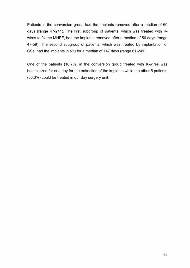
Patients in the conversion group had the implants removed after a median of 60
days (range 47-241). The first subgroup of patients, which was treated with K-
wires to fix the MHEF, had the implants removed after a median of 56 days (range
47-59). The second subgroup of patients, which was treated by implantation of
CSs, had the implants in situ for a median of 147 days (range 61-241).
One of the patients (16.7%) in the conversion group treated with K-wires was
hospitalized for one day for the extraction of the implants while the other 5 patients
(83.3%) could be treated in our day surgery unit.
37
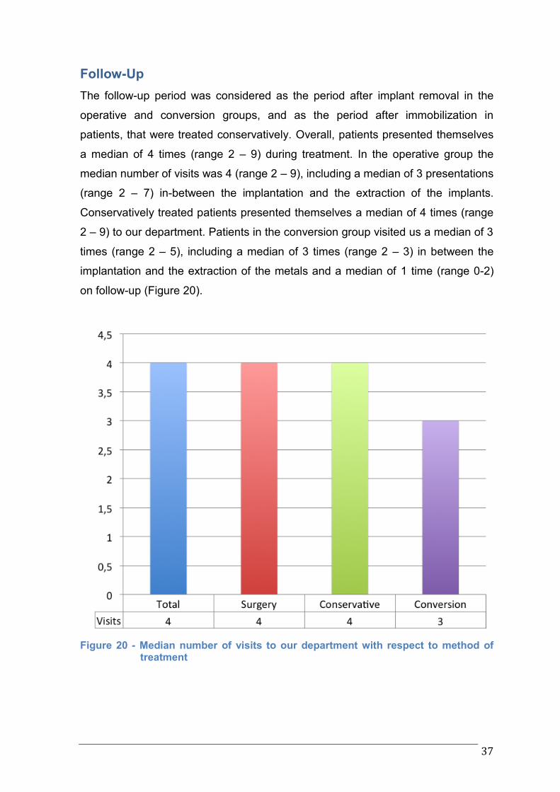
Follow-Up
The follow-up period was considered as the period after implant removal in the
operative and conversion groups, and as the period after immobilization in
patients, that were treated conservatively. Overall, patients presented themselves
a median of 4 times (range 2 – 9) during treatment. In the operative group the
median number of visits was 4 (range 2 – 9), including a median of 3 presentations
(range 2 – 7) in-between the implantation and the extraction of the implants.
Conservatively treated patients presented themselves a median of 4 times (range
2 – 9) to our department. Patients in the conversion group visited us a median of 3
times (range 2 – 5), including a median of 3 times (range 2 – 3) in between the
implantation and the extraction of the metals and a median of 1 time (range 0-2)
on follow-up (Figure 20).
Figure 20 - Median number of visits to our department with respect to method of treatment

38
Among the surgically treated patients the subgroup that had only K-wires
implanted had to visit our department a median of 3 times (range 2 – 8), including
a median of 3 times (range 1 – 5) in between the first operation and the removal of
the K-wires. In comparison, patients that were treated with implantation of CSs,
visited our department a median of 4.5 times (range 3-9) including a median of 4
times (range 2-7) before the extraction of the CSs. Combination of a CS and K-
wires made it necessary for the patients to frequent our department a median of 5
times (range 3 – 7), including a median of 3 visits (range 3-5) in between the
implantation and the extraction of the metals and a median of 1 visit (range 0-2)
after the extraction (Figure 21).
Figure 21 - Median number of visits to our department of surgical patients with respect to method of fixation
In the conversion group, all patients who had CSs implanted to fix the fractured
epicondyle presented themselves 3 times in our department with a median of 3
times (range 2 – 3) while the screws were in situ. The patients in this subgroup,
with K-wires for the fixation of the fracture, frequented our department a median of
39
4 times (range 2 – 5), including a median of 3 times (range 2 – 3) in between the
implantation and the extraction of the K-wires and a median of 1 more visit (range
0-2) afterwards.
The number of x-ray examinations necessary during the whole process of
treatment varied with the method of treatment used. Technically each examination
consisted of two x-rays, since the imaging of the affected elbow routinely had to be
done in anterior/posterior and lateral position. The following numbers indicate the
median number of examinations. Overall the patients were radiologically examined
a median of 6 times, with a range of 2 to 10 times during the whole treatment. The
most examinations were necessary in the conversion group, with a median of 8
examinations (range 5-10), as compared to a median of 6 examinations (range 3-
10) necessary in the group of primarily surgically treated patients. The fewest
examinations were necessary in the group of patients that were treated
conservatively with a median of 4 (range 2-7) (Figure 22).
Figure 22 - Median number of radiologic examinations with respect to treatment
40
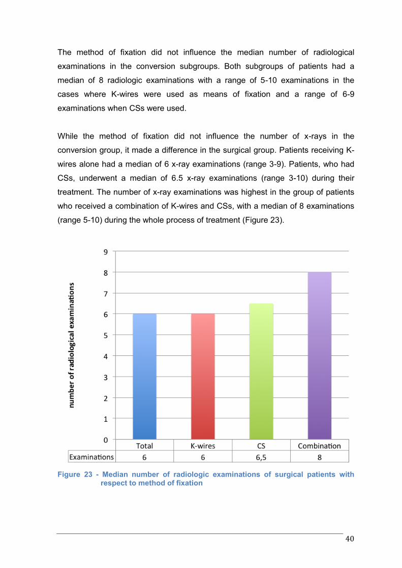
The method of fixation did not influence the median number of radiological
examinations in the conversion subgroups. Both subgroups of patients had a
median of 8 radiologic examinations with a range of 5-10 examinations in the
cases where K-wires were used as means of fixation and a range of 6-9
examinations when CSs were used.
While the method of fixation did not influence the number of x-rays in the
conversion group, it made a difference in the surgical group. Patients receiving K-
wires alone had a median of 6 x-ray examinations (range 3-9). Patients, who had
CSs, underwent a median of 6.5 x-ray examinations (range 3-10) during their
treatment. The number of x-ray examinations was highest in the group of patients
who received a combination of K-wires and CSs, with a median of 8 examinations
(range 5-10) during the whole process of treatment (Figure 23).
Figure 23 - Median number of radiologic examinations of surgical patients with respect to method of fixation
41
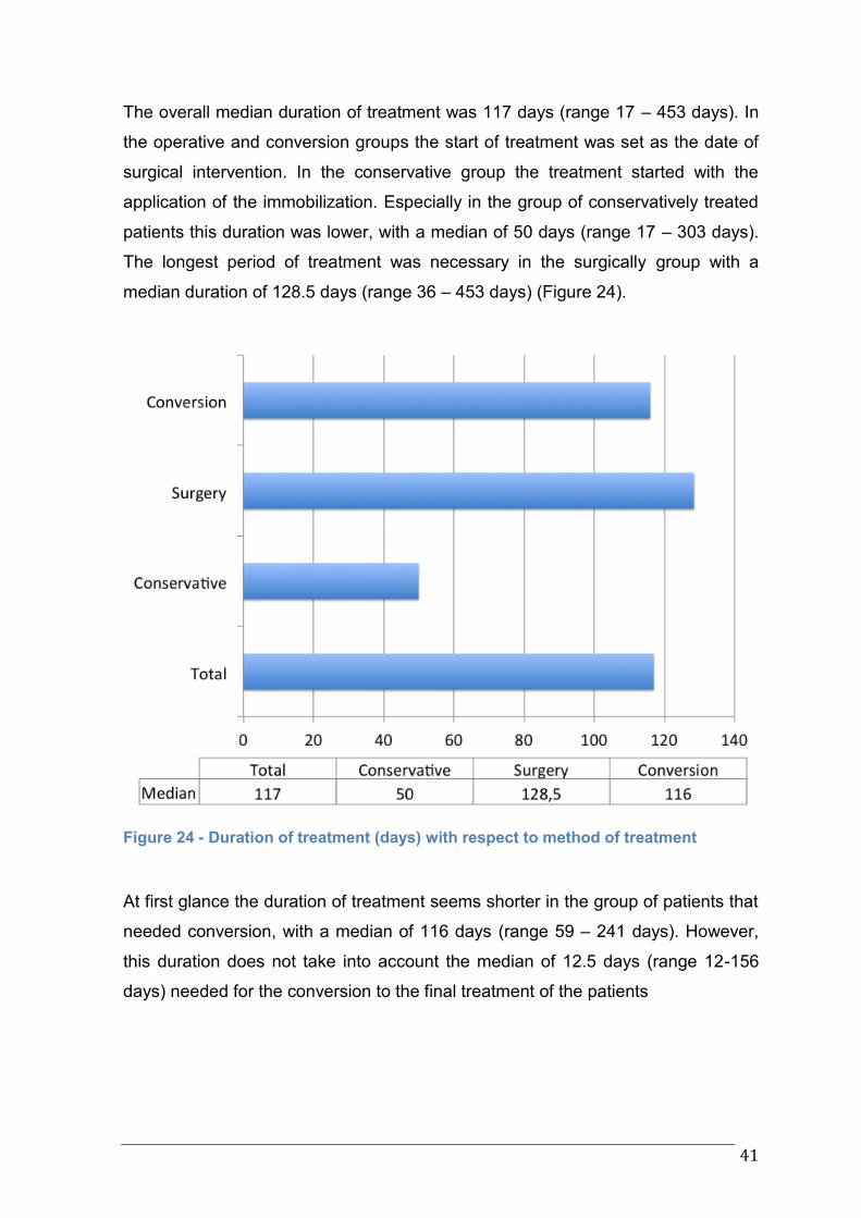
The overall median duration of treatment was 117 days (range 17 – 453 days). In
the operative and conversion groups the start of treatment was set as the date of
surgical intervention. In the conservative group the treatment started with the
application of the immobilization. Especially in the group of conservatively treated
patients this duration was lower, with a median of 50 days (range 17 – 303 days).
The longest period of treatment was necessary in the surgically group with a
median duration of 128.5 days (range 36 – 453 days) (Figure 24).
Figure 24 - Duration of treatment (days) with respect to method of treatment
At first glance the duration of treatment seems shorter in the group of patients that
needed conversion, with a median of 116 days (range 59 – 241 days). However,
this duration does not take into account the median of 12.5 days (range 12-156
days) needed for the conversion to the final treatment of the patients
42
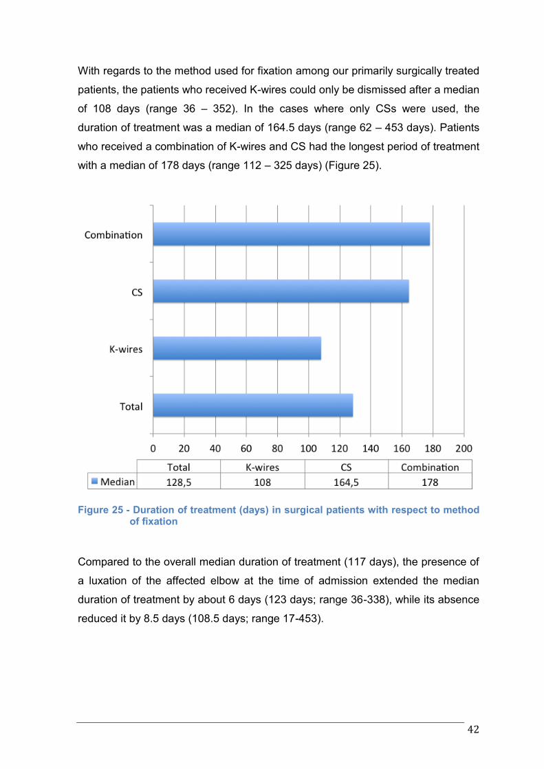
With regards to the method used for fixation among our primarily surgically treated
patients, the patients who received K-wires could only be dismissed after a median
of 108 days (range 36 – 352). In the cases where only CSs were used, the
duration of treatment was a median of 164.5 days (range 62 – 453 days). Patients
who received a combination of K-wires and CS had the longest period of treatment
with a median of 178 days (range 112 – 325 days) (Figure 25).
Figure 25 - Duration of treatment (days) in surgical patients with respect to method of fixation
Compared to the overall median duration of treatment (117 days), the presence of
a luxation of the affected elbow at the time of admission extended the median
duration of treatment by about 6 days (123 days; range 36-338), while its absence
reduced it by 8.5 days (108.5 days; range 17-453).
43
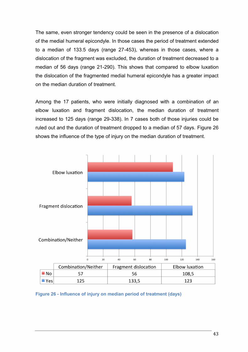
The same, even stronger tendency could be seen in the presence of a dislocation
of the medial humeral epicondyle. In those cases the period of treatment extended
to a median of 133.5 days (range 27-453), whereas in those cases, where a
dislocation of the fragment was excluded, the duration of treatment decreased to a
median of 56 days (range 21-290). This shows that compared to elbow luxation
the dislocation of the fragmented medial humeral epicondyle has a greater impact
on the median duration of treatment.
Among the 17 patients, who were initially diagnosed with a combination of an
elbow luxation and fragment dislocation, the median duration of treatment
increased to 125 days (range 29-338). In 7 cases both of those injuries could be
ruled out and the duration of treatment dropped to a median of 57 days. Figure 26
shows the influence of the type of injury on the median duration of treatment.
Figure 26 - Influence of injury on median period of treatment (days)
44
Complications during the healing process were rare and with little to no further
consequence. According to documentations at follow-up, 15 patients (18.5% of all
patients) still had reduced ROM (≤20°) of the affected elbow at the end of
treatment. Eight of those patients were in the surgical group (14.3% of all surgical
patients), 5 in the conservative group (26.3% of all conservative patients) and 2 in
the conversion group (33.3% of all conversion cases). Three patients (3.7%
overall; 5.4% of patients in operative group), who were in the surgically treated
group, still had a minor nervous deficit in the ulnar nerve region but with steady
improvement. Also, 3 operative patients (3.7% overall; 5.4% of surgical patients)
reported a slight tenderness over the operated MHE on their final clinical
examination. Seventeen patients (21% of all patients) showed persisting
anatomical irregularities on their final x-rays. Nine of those patients were treated
conservatively (47.4% of patients in conservative group), 6 surgically (10.7% of all
surgical patients), and 2 underwent a conversion (33.3% of patients on conversion
group). These irregularities included periarticular calcification, fragmented MHE,
fibrous union of the MHEF and minimal deviations of the MHE from the
anatomically correct position. Apart from one surgical patient, in whom a minimal
dorsal deviation of the MHE might have been arguably associated with a transient
ulnar nerve deficiency, none of the radiographic abnormalities were linked to any
clinically relevant findings or needed further treatment.
One of the patients with persisting complaints was the patient mentioned before,
who received a resorbable CS as a means of fixation of the fractured medial
epicondyle. Apart from the longest period of treatment, with 453 days, he was
complaining of persistent pain and muscular dysbalance in the ulnar nerve region,
as well as a reduced ROM and valgus deformity of the affected elbow in
comparison to the healthy arm. As mentioned above, he presented again 1½
years later to undergo a surgical decompression of the ulnar nerve. This was the
only case in which an additional intervention was necessary.
45
4. Discussion
Demographics and epidemiology
In a large study evaluating the epidemiology of 8,682 fractures in children, Landin
et al. (3), reported that elbow fractures make up to 7% of all fractures in the
pediatric population. Out of those fractures of the elbow region, MHEF constitute
around 8% of the fractures. In other, smaller studies these numbers are even
higher, with MHEF representing up to 11.5% of all fractures in the elbow region
and between 3.6 and 14.1% of all fractures of the distal humerus (10,26).
Fractures of the MHE have been the objective of various studies in the past. The
main objective of these studies was the establishment of a universally accepted
guideline for treatment. However, while there exists common consensus about the
conservative treatment of non-displaced MHEF and a number of absolute and
relative indications for surgery have been established, many cases fall into a grey
zone where the recommendation of the appropriate treatment procedure is still
subject of discussion among pediatric surgeons. The goal of our study was the
retrospective analysis of the pediatric population in our hospital, which was treated
for MHEF. The results, which we achieved through our conservative and surgical
treatment approaches, were compared and evaluated under the light of past and
present literature, with the target to identify possible areas for improvement in
treatment.
The epidemiological analysis of the pediatric population of our patients showed a
close resemblance to the epidemiologic aspects reported in many of the other
studies about MHEF. In some studies the gender distribution showed an increased
incidence as high as 5:1 in boys (27). In contrast, other studies reported an
increased incidence in the female pediatric population (25,26). In a systematic
review, Kamath et al. (8) reported a male to female ratio of approximately 2:1,
which corresponds with the ratio we found in our patient group. In the literature
most MHEFs were reported between the ages 9 and 14, with a peak incidence
between 11 to 12 years (9,10). Kamath et al. (8) reported an average age of 11.9
46
years at the time of injury, which shows a close resemblance to the median age of
12 years that was recorded in our patients. The question about which arm was
more likely to be injured, the dominant or the non-dominant one, is still
controversial: Lee et al. (16) reported the injury to affect the non-dominant arm in
72% of their 25 cases. Whereas other studies with larger cohorts did not address
this question and rather showed a right to left ratio of 1.4:1 (3). However, patients
in our cohort showed an almost even distribution of left-to-right arm injuries.
Although the mechanisms of injury are well understood, a clear documentation the
accident is often lacking. Especially differentiation between strictly sport activities
and mere recreational play activities is challenging in this age group. Louahem et
al. (6) related 53% of the injuries in their patients to sport activities. Haxhija et al.
(22) connected as much as 84% of the injuries in their patients to sport activities.
In the present study 67.9% of cases were related to either recreational or athletic
physical activity. An interesting additional finding of our study was that a notable
number (n=13; 16%) of MHEFs were trampoline injuries. Although MHEFs are not
explicitly mentioned in the literature reporting trampoline injuries, fractures in
general are seen more often in the upper extremities in these accidents (28–30).
Fractures of the humerus and the elbow rank second after fractures of the forearm
in these cases (31). Certainly a connection can be drawn to the most common
mechanism of injury of a fall on the outstretched arm.
Outcome
Criteria for the assessment of MHEF treatment outcome show variations in
different studies regarding MHEF. Some studies rely on subjective patient-
reported questionnaires. Two scores which are commonly used, are the Mayo
Elbow Performance Score (MEPS) (32) and the QuickDASH (Disability of Arm,
Shoulder and Hand; a shortened version of the DASH outcome measure) (33).
Other authors utilize, mostly self-developed, scoring systems with differently
weighted functional and radiological parameters. However, there are some core
elements present in most of the studies, which we compared to our results.
47
Bony union
Regarding the differences in outcome between surgical and nonsurgical treatment
of MHEFs in children and adolescents, Kamath et al. (8) conducted a systematical
review of 14 studies, published between 1975 and 2007 and including 498
patients. The most apparent and statistically significant (P<0.0001) difference
found was the inferior rate of bony union in conservative patients (60 out of 122
cases; 49.2%) compared to surgical patients (260 out of 280; 92.5%). In a long
term follow-up study of conservatively treated unreduced MHEFs in children,
Josefsson and Danielsson (24) clinically reexamined 51 patients after 35 years
and found a similar rate of fibrous union of the fracture (n=31; 60.1%).
Interestingly, the pseudoarthrosis had developed independently from the initial
degree of fracture displacement (1-15mm) in their patients. Mild to moderate
residual symptoms were reported in 21% of their patients. However, none of these
symptoms influenced daily routine or athletic activities. Apart from mild ulnar nerve
symptoms, occurring in 3 patients in their pseudoarthrotic group only, the
frequency of all other residual symptoms did not differ between patients with
fibrous union and patients with bony union of the fracture. In the present study 3
out of 56 patients (5.4%) in the surgical group, and 3 out of 19 patients (15.8%) in
the conservative group, showed a fibrous union of the MHEF at last follow-up. This
shows an overall superior rate of bony union achieved in both treatment groups. In
agreement with previous studies we still found a higher rate of bony union in the
surgical group. After conversion, bony union was present in all of our patients,
which demonstrates that bony union can still be achieved after an initial failure of
conservative treatment.
Pain
While Kamath et al. (8) found a tendency of less pain reported on follow-up in
conservative patients (10 out of 115; 8.7%) as compared to surgical patients (37
out of 246; 15%), the pooled difference was statistically not significant (P=0.140).
In a study of 139 surgically treated patients, Louahem et al. (6) reported that all of
their patients were free of pain upon follow-up. Five of the 56 conservatively
treated patients (9%), that were interviewed in the study of Josefsson and
Danielsson (24), reported mild to moderate tenderness over the ulnar part of the
48
elbow. However, they experienced no restrictions in their work or athletic activities.
Three of our surgical patients (5.4%) expressed slight tenderness in the MHE area
in their last clinical examination, though without any limitations to their daily
activities. None of the patients in the conservative group reported pain at last
follow-up.
Ulnar nerve impairment
Kamath et al. (8) did not find a statistically significant difference in ulnar nerve
symptoms between operative and conservative treatment. Also Louahem et al. (6)
did not find any neurological impairments in their patients upon follow-up. All of
initially eight patients (5.8%), who had presented themselves with neurological
symptoms, recovered completely. As mentioned before, 3 out of 56 patients (5%)
in the study of Josefsson and Danielsson (24) mentioned numbness in the ulnar
part of the hand on follow-up. All 3 patients had fibrous union of the fragment and
did not feel restricted in their daily routine. Similar to the literature, in the present
study we identified 3 of our surgical cases (5.4%) with transient ulnar nerve deficit
upon follow-up. Two of these patients did not need any further treatment for their
symptoms. The third patient, treated with the resorbable screw, eventually
returned to our department after 1.5 years due to persistent complaints. Surgical
decompression of the ulnar nerve was subsequently performed.
Range of motion
Restriction in ROM is a very influential factor for functional outcome of MHEFs.
The results we found in our patients suggest a slightly higher rate of reduced ROM
after conservative treatment but results of various other studies are still
inconclusive. Eight patients (14.3%) in our operative group showed a reduced
ROM in flexion/extension of not more than 15° on last follow-up. The rate of
reduced ROM in our conservative patients was slightly higher, with 26.3% of
patients, again with a maximum deficit of 15°. We found reduced ROM in 2 of 6
patients (33.3%) that underwent a conversion, with a maximum deficit of 20°.
Outcomes of various studies reported in the literature are very heterogenic:
Louahem et al. (13) reported a reduced ROM of less than 20° in 6 cases (4.3%).
Josefsson and Danielsson (24) observed reduction of ROM in both of their groups
49
(fibrous and bony union) equally, with an overall 2±6° loss of extension (range: 0-
25°) in 21.6% of their patients. Farsetti et al. (21) found a limitation of elbow
extension in 2 of their conservatively treated patients (10.5%) and 2 of their
surgically treated patients (11.8%). Though some authors suggest an increased
risk of joint stiffness and reduced ROM after prolonged periods of immobilization
(9,10,16,34) we did not find any correlation between the time of immobilization and
resulting reductions in ROM in our patient groups.
Clinical and radiological deformities
An abnormal valgus deviation of the injured elbow joint was only found in one of
our patients (1.2%). This patient was treated surgically with a resorbable screw.
Haxhija et al. (22) reported an average 3° valgus deviation in comparison to the
uninjured elbow in their patients available for follow-up examination. The
mechanism of the hypervalgus deformity in MHEF seems to be caused by damage
to the distal physeal plate, either by the original injury or sometimes acquired
through ORIF. This is supported also by the fact that as previously stated the
MHE, as an apophysis in its nature does not contribute to growth of the distal
humerus (7,21,35). Skak et al. (36) described different radiological deformities
deriving from MHEFs, such as ulnar sulcus deformity, double epiphysis,
hyperplasia, hypoplasia and pseudoarthrosis. However, only pseudoarthrosis and
ulnar sulcus deformity could be related to incidence of ulnar neuritis while all other
deformities did not affect the patients’ daily activities. In the present study,
radiological irregularities were found in 17 patients (21%). These radiological
findings included periarticular calcification, fragmented MHE, fibrous union of the
MHEF and minimal deviations of the MHE from the anatomically correct position.
Apart from one surgical patient, in whom a minimal dorsal deviation of the MHE
could have been responsible for transient ulnar nerve deficiency, all other patients
were free from symptoms.
As mentioned before, one patient in our surgical group, treated with a resorbable
screw implant, presented with several complications that made a surgical
decompression of the ulnar nerve necessary 1.5 years after initial treatment. The
presenting symptoms included persistent discomfort in the MHE area, reduced
50
ROM, valgus deformity and muscular dysbalance in comparison to the
contralateral arm. Although this case could have been incidental, resorbable
screws were not further used in our patients. Considering the good results we
achieved with other implants, their use can not be recommended.
Conversion
To the author’s knowledge, the topic of conversion from conservative to surgical
MHEF treatment has not been addressed thoroughly in the literature. One study,
however, evaluated the outcome of surgical treatment after an extended period of
time in the case of symptomatic nonunion following initial conservative treatment.
Smith et al. (37) reported 8 cases out of 42 (19%) that underwent ORIF after initial
conservative treatment. Similarly, our conversion rate was 24% (n=6). Indications
for conversion in the report of Smith and coworkers were radiographic evidence of
nonunion and painful palpation of the MHE. ORIF was performed after a median of
12 month following the injury, which is significantly longer than the median
duration of 12 days in our patients. While this limits the comparability to our
conversion group, it shows a similar rate of failure of the initial conservative
treatment as well as the possibility to convert to ORIF even after prolonged
periods of time. Smith et al. (37) reported a patient-reported improvement in
functional outcome, determined by Mayo Elbow Performance Score (MEPS),
Timmerman-Andrews Elbow Score and QuickDASH (Disability of Arm, Shoulder
and Hand) in all patients. Also pain, determined using a 10-point visual analog
pain scale (VAPS), decreased notably (from a mean of 6.2 to 0.5) and all patients
could return to sports activities. Bony union was achieved in all but one of their
patients, who did not show any symptoms in a prolonged follow-up period though.
Again this leads to the conclusion that, even after longer periods of time, an
initially failed conservative treatment can still yield bony union after conversion.
Smith et al. (37) reported an average ROM of 130° in their converted patients. We
found reduced ROM in 2 out of our 6 converted patients (33.3%), with a deficit of
20° in one patient and a deficit of 10° in the other patient. Irregularities on the last
radiographs were present in the same 2 patients (33.3%) and included a slight
deviation from the anatomically correct position of the MHE in the one patient and
51
an irregularity of the corticalis in the proximal area of the fracture in the other
patient. However, no further intervention was required since our patients reported
no discomfort or any other symptoms.
Fragment dislocation
As mentioned above, a number of indications for conservative treatment
approaches as well as ORIF of MHEF have been identified in previous studies.
The gray area of this injury in the literature revolves mostly around uncomplicated
injuries and the distance of fracture displacement tolerable before ORIF is
advised.
It seems that radiologic imaging is one contributing factor to this controversy.
Edmonds (38) and Pappas et al. (39) raise the question of whether the decision of
treatment should be based on the standard radiologic imaging in AP and lateral
view of the elbow. Edmonds (38) reported that control three-dimensional
computed tomography (CT) scans revealed a fragment displacement of more than
1 cm, though initial standard AP and lateral view radiographs had suggested non-
displaced or minimally displaced MHEFs (by his definition <5mm). Pappas et al.
(39) highlighted a high discrepancy of “intraobserver” and “interobserver”
measurement of displacement of MHEF on standard AP and lateral view
radiographs as well as additional oblique radiographs. In general, the correct
radiologic imaging and interpretation of x-rays of fractures of the elbow in injured
children and adolescents requires a lot of expertise and experience from both, the
radiologist and the attending surgeon (18).
Farsetti et al. (21) treated 19 patients conservatively and 17 patients through ORIF
with an average fragment displacement of 10mm in both groups (range 5-15mm
and 5-14mm respectively). Their results were rated according to their own
conducted criteria, which are shown in Table 7.
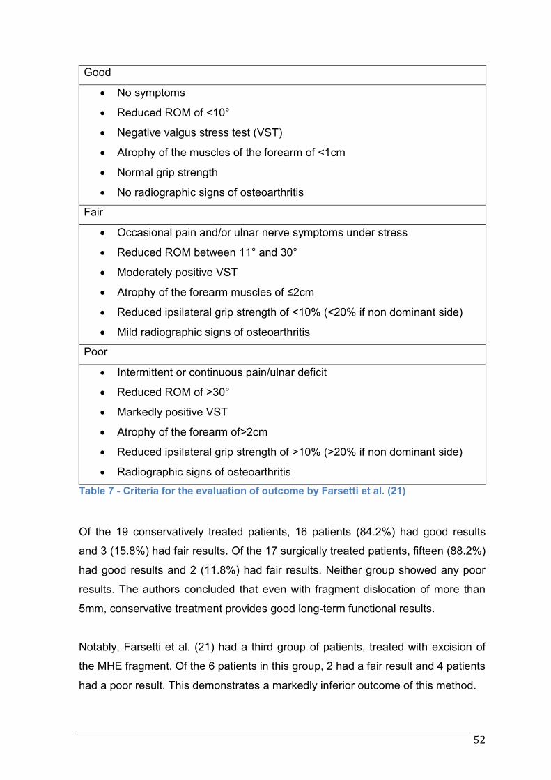
52
Good
No symptoms
Reduced ROM of <10°
Negative valgus stress test (VST)
Atrophy of the muscles of the forearm of <1cm
Normal grip strength
No radiographic signs of osteoarthritis
Fair
Occasional pain and/or ulnar nerve symptoms under stress
Reduced ROM between 11° and 30°
Moderately positive VST
Atrophy of the forearm muscles of ≤2cm
Reduced ipsilateral grip strength of <10% (<20% if non dominant side)
Mild radiographic signs of osteoarthritis
Poor
Intermittent or continuous pain/ulnar deficit
Reduced ROM of >30°
Markedly positive VST
Atrophy of the forearm of>2cm
Reduced ipsilateral grip strength of >10% (>20% if non dominant side)
Radiographic signs of osteoarthritis
Table 7 - Criteria for the evaluation of outcome by Farsetti et al. (21)
Of the 19 conservatively treated patients, 16 patients (84.2%) had good results
and 3 (15.8%) had fair results. Of the 17 surgically treated patients, fifteen (88.2%)
had good results and 2 (11.8%) had fair results. Neither group showed any poor
results. The authors concluded that even with fragment dislocation of more than
5mm, conservative treatment provides good long-term functional results.
Notably, Farsetti et al. (21) had a third group of patients, treated with excision of
the MHE fragment. Of the 6 patients in this group, 2 had a fair result and 4 patients
had a poor result. This demonstrates a markedly inferior outcome of this method.
53
Conclusion
Based on the findings of our study, we certainly agree to follow the well-
established absolute indications for surgery also in the future. However, as far as
relative indications for surgery are concerned, the following conclusions can be
drawn:
From our point of view, both, surgical as well as conservative methods of
treatment are eligible also in the future. But rather than basing the decision for
surgery mainly on the distance of fragment dislocation, we recommend to focus on
either positive valgus stress test or positive gravity stress test in the decision for
surgery. While a surgical approach seems to provide a higher chance for bony
union, the functional outcome between surgery and conservative approach does
not differ. Furthermore, a reduced duration of treatment and avoidance of two
surgical interventions (with possible complication risks) for implantation and
extraction of K-wires and/or CSs are further arguments favoring a conservative
approach. The possibility for a conversion to surgery after an initial non-
satisfactory conservative approach should also be taken into consideration in
debatable cases - especially since good functional results can still be expected
with conversions at a later stage. In patients with high requirements regarding
elbow stability, bony union is a desirable goal of treatment. A lower threshold for
an operative approach is therefore justified in these cases.
54
References
1. Granger B. On a particular fracture of the inner condyle of the humerus.
Edinb Med Surg J. 1818;14:196–397.
2. Higgs SL, Cantab BC. Fractures of the internal epicondyle of the humerus.
Br Med J. 1936;2(3952):666.
3. Landin LA, Danielsson LG. Elbow fractures in children. An epidemiological
analysis of 589 cases. Acta Orthop Scand. August 1986;57(4):309–12.
4. Rasool MN. Dislocations of the elbow in children. J Bone Joint Surg Br.
September 2004;86(7):1050–8.
5. Frongia G, Günther P, Romero P, Keßler M, Holland-Cunz S.
Ellenbogenluxationen im Kindesalter: Eine Langzeitbeobachtung. Unfallchirurg.
24. September 2010;115(2):125–33.
6. Louahem DM, Bourelle S, Buscayret F, Mazeau P, Kelly P, Dimeglio A, u.
a. Displaced medial epicondyle fractures of the humerus: surgical treatment and
results. A report of 139 cases. Arch Orthop Trauma Surg. 4. Dezember
2009;130(5):649–55.
7. Von Laer L, Kraus R, Linhart, Wolfgang E. Frakturen und Luxationen im
Wachstumsalter. 6. Aufl. Stuttgart: Thieme; 2013.
8. Kamath AF, Baldwin K, Horneff J, Hosalkar HS. Operative versus non-
operative management of pediatric medial epicondyle fractures: a systematic
review. J Child Orthop. Oktober 2009;3(5):345–57.
9. Smith FM. Medial epicondyle injuries. J Am Med Assoc. 11. Februar
1950;142(6):396.
10. Rockwood and Wilkins’ fractures in children. 7th ed. Philadelphia: Wolters
Kluwer/Lippincott, Williams & Wilkins; 2010.
11. Cheng JC, Wing-Man K, Shen WY, Yurianto H, Xia G, Lau JT, u. a. A new
look at the sequential development of elbow-ossification centers in children. J
Pediatr Orthop. April 1998;18(2):161–7.
12. Waldeyer A. Waldeyer - Anatomie des Menschen. Lehrbuch und Atlas in
einem Band. 19th ed. Streicher J, Anderhuber F, Pera F, Herausgeber. Berlin,
Boston: De Gruyter; 2012.
13. Woods GW, Tullos HS. Elbow instability and medial epicondyle fractures.
55
Am J Sports Med. Februar 1977;5(1):23–30.
14. Hemmady MV, Subramanya AV, Mehta IM. Occasional head of flexor
pollicis longus muscle: a study of its morphology and clinical significance. J
Postgrad Med. März 1993;39(1):14–6.
15. Hines RF, Herndon WA, Evans JP. Operative treatment of Medial
epicondyle fractures in children. Clin Orthop. Oktober 1987;(223):170–4.
16. Lee H-H, Shen H-C, Chang J-H, Lee C-H, Wu S-S. Operative treatment of
displaced medial epicondyle fractures in children and adolescents. J Shoulder
Elbow Surg. März 2005;14(2):178–85.
17. Lawrence JTR, Patel NM, Macknin J, Flynn JM, Cameron D, Wolfgruber
HC, u. a. Return to Competitive Sports After Medial Epicondyle Fractures in
Adolescent Athletes: Results of Operative and Nonoperative Treatment. Am J
Sports Med. 18. März 2013;41(5):1152–7.
18. Rang M. Rang’s children’s fractures. 3rd ed. Philadelphia: Lippincott
Williams & Wilkins; 2006.
19. Bede WB, Lefebvre AR, Rosman MA. Fractures of the medial humeral
epicondyle in children. Can J Surg J Can Chir. März 1975;18(2):137–42.
20. Papavasiliou VA. Fracture-separation of the medial epicondylar epiphysis of
the elbow joint. Clin Orthop. Dezember 1982;(171):172–4.
21. Farsetti P, Potenza V, Caterini R, Ippolito E. Long-term results of treatment
of fractures of the medial humeral epicondyle in children. J Bone Jt Surg.
2001;83(9):1299–305.
22. Haxhija EQ, Mayr JM, Grechenig W, Höllwarth ME. Die Behandlung der
Apophysenabrissverletzung des Epicondylus ulnaris im Kindesalter. Oper Orthop
Traumatol. Juni 2006;18(2):120–34.
23. Kamath AF, Cody SR, Hosalkar HS. Open reduction of medial epicondyle
fractures: operative tips for technical ease. J Child Orthop. 9. Juni 2009;3(4):331–
6.
24. Josefsson PO, Danielsson LG. Epicondylar elbow fracture in children: 35-
year follow-up of 56 unreduced cases. Acta Orthop. 1986;57(4):313–5.
25. Duun PS, Ravn P, Hansen LB, Buron B. Osteosynthesis of medial humeral
epicondyle fractures in children. 8-year follow-up of 33 cases. Acta Orthop Scand.
August 1994;65(4):439–41.
26. Zanella FE, Piroth P. Injuries of the elbow and forearm in children. Röntgen-
56
Bl Z Für Röntgen-Tech Med-Wiss Photogr. März 1984;37(3):111–20.
27. Fowles J V., Kassab MT, Moula T. Untreated intra-articular entrapment of
the medial humeral epicondyle. J Bone Joint Surg Br. August 1984;66(4):562–5.
28. Eberl R, Schalamon J, Singer G, Huber SS, Spitzer P, Höllwarth ME.
Trampoline-related injuries in childhood. Eur J Pediatr. 24. Dezember
2008;168(10):1171–4.
29. Smith GA, Shields BJ. Trampoline-related injuries to children. Arch Pediatr
Adolesc Med. Juli 1998;152(7):694–9.
30. Linakis JG, Mello MJ, Machan J, Amanullah S, Palmisciano LM. Emergency
Department Visits for Pediatric Trampoline-related Injuries: An Update. Acad
Emerg Med. Juni 2007;14(6):539–44.
31. Black GB, Amadeo R. Orthopedic injuries associated with backyard
trampoline use in children. Can J Surg. 2003;46(3):199.
32. Morrey BF, Sanchez-Sotelo J, Herausgeber. The elbow and its disorders.
4th ed. Philadelphia, PA: Saunders/Elsevier; 2009.
33. Beaton DE, Wright JG, Katz JN. Development of the QuickDASH:
comparison of three item-reduction approaches. J Bone Jt Surg. 2005;87(5):1038–
46.
34. Case SL, Hennrikus WL. Surgical Treatment of Displaced Medial
Epicondyle Fractures in Adolescent Athletes. Am J Sports Med. 1. September
1997;25(5):682–6.
35. Van Niekerk JL, Severijnen RS. Medial epicondyle fractures of the
humerus. Neth J Surg. Oktober 1985;37(5):141–4.
36. Skak SV, Grossmann E, Wagn P. Deformity after internal fixation of fracture
separation of the medial epicondyle of the humerus. J Bone Joint Surg Br. März
1994;76(2):297–302.
37. Smith JT, McFeely ED, Bae DS, Waters PM, Micheli LJ, Kocher MS.
Operative Fixation of Medial Humeral Epicondyle Fracture Nonunion in Children: J
Pediatr Orthop. 2010;30(7):644–8.
38. Edmonds EW. How Displaced Are “Nondisplaced” Fractures of the Medial
Humeral Epicondyle in Children? Results of a Three-Dimensional Computed
Tomography. J Bone Jt Surg Am. 1. Dezember 2010;92(17):2785.
39. Pappas N. Intraobserver and Interobserver Agreement in the Measurement
of Displaced Humeral Medial Epicondyle Fractures in Children. J Bone Jt Surg

57
Am. 1. Februar 2010;92(2):322.