Embed Size (px)
Citation preview

Reversed-engineered human alveolarlung-on-a-chip modelDi Huanga,b,1
, Tingting Liua,b,c,1, Junlong Liaoa,d,1
, Sushila Maharjana,1, Xin Xiea, Montserrat Péreza,e,
Ingrid Anayaa,e, Shiwei Wanga, Alan Tirado Mayera,f, Zhixin Kanga, Weijia Konga, Valerio Luca Mainardia,g,h,Carlos Ezio Garciamendez-Mijaresa,f, Germán García Martíneza,f, Matteo Morettig,i,j, Weijia Zhangk,l,m
,Zhongze Gud, Amir M. Ghaemmaghamin, and Yu Shrike Zhanga,2
aDivision of Engineering in Medicine, Department of Medicine, Brigham and Women’s Hospital, Harvard Medical School, Cambridge, MA 02139; bResearchCenter for Nano-Biomaterials & Regenerative Medicine, Department of Biomedical Engineering, College of Biomedical Engineering, Taiyuan University ofTechnology, Taiyuan, Shanxi 030024, People’s Republic of China; cDepartment of Laboratory Diagnosis, The 971th Hospital, Qingdao 266072, People’sRepublic of China; dState Key Laboratory of Bioelectronics, School of Biological Science and Medical Engineering, Southeast University, Nanjing 210096,People’s Republic of China; eDepartment of Biotechnological Engineering, Monterrey Institute of Technology and Higher Education, Monterrey, NuevoLeón 64849, Mexico; fDepartment of Mechatronics Engineering, Monterrey Institute of Technology and Higher Education, Monterrey, Nuevo León 64849,Mexico; gRegenerative Medicine Technologies Lab, Ente Ospedaliero Cantonale, Lugano 6900, Switzerland; hLaboratory of Biological Structures Mechanics,Department of Chemistry, Materials and Chemical Engineering Giulio Natta, Politecnico di Milano, 20133 Milano, Italy; iCell and Tissue EngineeringLaboratory, IRCCS Istituto Ortopedico Galeazzi, 20161 Milano, Italy; jFaculty of Biomedical Sciences, Università della Svizzera Italiana, Lugano 6900,Switzerland; kDepartment of Cardiac Surgery and Shanghai Institute of Cardiovascular Diseases, Zhongshan Hospital, Shanghai Medical College, FudanUniversity, Shanghai 200032, People’s Republic of China; lInstitute of Biomedical Sciences and Department of Systems Biology for Medicine, ShanghaiMedical College, Fudan University, Shanghai 200032, People’s Republic of China; mThe State Key Laboratory of Molecular Engineering of Polymers, FudanUniversity, Shanghai 200438, People’s Republic of China; and nImmunology and Immuno-Bioengineering Group, School of Life Science, Faculty of Medicineand Health Sciences, University of Nottingham, NG7 2RD Nottingham, United Kingdom
Edited by Joseph M. DeSimone, Stanford University, Stanford, CA, and approved April 2, 2021 (received for review July 30, 2020)
Here, we present a physiologically relevant model of the humanpulmonary alveoli. This alveolar lung-on-a-chip platform is com-posed of a three-dimensional porous hydrogel made of gelatinmethacryloyl with an inverse opal structure, bonded to a compart-mentalized polydimethylsiloxane chip. The inverse opal hydrogelstructure features well-defined, interconnected pores with highsimilarity to human alveolar sacs. By populating the sacs with pri-mary human alveolar epithelial cells, functional epithelial mono-layers are readily formed. Cyclic strain is integrated into the deviceto allow biomimetic breathing events of the alveolar lung, which,in addition, makes it possible to investigate pathological effectssuch as those incurred by cigarette smoking and severe acute re-spiratory syndrome coronavirus 2 pseudoviral infection. Our studydemonstrates a unique method for reconstitution of the functionalhuman pulmonary alveoli in vitro, which is anticipated to pave theway for investigating relevant physiological and pathologicalevents in the human distal lung.
alveoli | distal lung | lung-on-a-chip | inverse opal | three-dimensional
The lung, as the major organ of the human respiratory system,is responsible primarily for gas exchanges and thus, directly
exposed to the external environment. Lungs are affected by aplethora of pathologies such as asthma (1); chronic obstructivepulmonary disease (COPD) (2); and infections like influenza (3),pneumonia (4), and tuberculosis (5) as well as lung cancer (6).These pathological manifestations make lung failure one of theleading causes of death globally. More recently, this is furtherexemplified by the coronavirus disease 2019 (COVID-19) pandemic,which has so far killed more than 3 million people worldwide withtotal confirmed cases of >140 million (7).The lack of reliable and physiologically relevant animal models
for human respiratory diseases has led to a critical issue for newdrug development as more than 90% of the preclinical studiesperformed in animals do not predict the outcome of human clinicaltrials (8). This has resulted in a lack of progress in drug develop-ment in respiratory medicine, with only a handful of new drugsentering clinical use in the last 50 y (9). In addition, many of theexisting cell culture-based models do not replicate the key bio-logical aspects of the human lung and do not adequately reflect thehost responses. These models range from simple two-dimensional(2D) cultures of lung cells on polymeric or elastomeric membranesystems to the complex biomimetic lung-on-a-chip microdevices
(10). While each of these more complex models (10–14) may haveadvantages over 2D single-cell cultures, collectively they sufferfrom important limitations such as use of synthetic polymermembranes with nonphysiological stiffness to culture cells and/orlack of mechanical stimulation (inhalation/exhalation process).In fact, there is increasing acceptance that the composition
and topography of the extracellular matrix (ECM) have majorinfluences on cell functions (15) and regulate cellular responsesto various stimuli. As such, ECM features are too critical to beignored in the design and fabrication of any biologically relevanttissue and disease models. Therefore, there is a clear need foran advanced model system that not only mimics the human lungtissue structurally but also, captures its ECM physiology that is
Significance
This work reports the development of a physiologically relevanthuman alveolar lung-on-a-chip model, composed of a three-dimensional (3D) porous hydrogel made of gelatin methacryloyl(GelMA) featuring an inverse opal structure, bonded to a com-partmentalized chip device that provides air–liquid interface andcyclic breathing motions. Significantly, this GelMA structure has ahigh similarity to native human alveolar sacs in that they bothpossess sac-like pores and interconnecting windows between thesacs, in addition to a stiffness similar to the native human distallung. We showed through multiscale analyses that our 3D GelMAinverse opal structure was better able to maintain the functionsof primary human alveolar epithelial cells in a more in vivo-likemanner compared with planar models.
Author contributions: D.H., T.L., J.L., S.M., X.X., M.P., I.A., A.M.G., and Y.S.Z. designedresearch; D.H., T.L., J.L., S.M., X.X., M.P., I.A., S.W., A.T.M., Z.K., W.K., V.L.M., C.E.G.-M.,G.G.M., M.M., and Y.S.Z. performed research; D.H., T.L., J.L., S.M., W.Z., Z.G., and Y.S.Z.analyzed data; Y.S.Z. conceptualized the project; and D.H., T.L., J.L., S.M., and Y.S.Z. wrotethe paper.
The authors declare no competing interest.
This article is a PNAS Direct Submission.
This open access article is distributed under Creative Commons Attribution-NonCommercial-NoDerivatives License 4.0 (CC BY-NC-ND).1D.H., T.L., J.L., and S.M. contributed equally to this work.2To whom correspondence may be addressed. Email: [email protected].
This article contains supporting information online at https://www.pnas.org/lookup/suppl/doi:10.1073/pnas.2016146118/-/DCSupplemental.
Published May 3, 2021.
PNAS 2021 Vol. 118 No. 19 e2016146118 https://doi.org/10.1073/pnas.2016146118 | 1 of 10
ENGINEE
RING
PHARM
ACO
LOGY
Dow
nloa
ded
by g
uest
on
Feb
ruar
y 5,
202
2
essential to their functional reproduction in vitro. More impor-tantly, there are no satisfactory models of the distal lung (i.e., thealveolar space) that truly reflect the sac shape anatomy (16–18) tostudy physiology and pathophysiology. A recent work attempted todevelop such a model with bioprinting (19); however, this modelmerely reflected alveoli’s gross anatomy, albeit in much largerscales than the actual features of the human alveoli (millimetersvs. micrometers), and the material used was still incompatible tonative tissue in terms of bioactivity.To address these challenges, herein we report the development
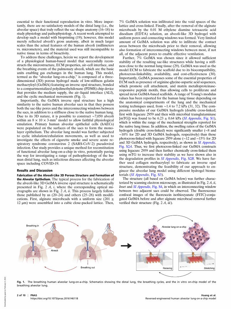
of a physiological human-based model that successfully recon-structs the microstructure, ECM properties, air–cell interface, andthe breathing events of the pulmonary alveoli, which are the basicunits enabling gas exchanges in the human lung. This model,termed as the “alveolar lung-on-a-chip,” is composed of a three-dimensional (3D) porous hydrogel made of low-stiffness gelatinmethacryloyl (GelMA) featuring an inverse opal structure, bondedto a compartmentalized polydimethylsiloxane (PDMS) chip devicethat provides the medium supply, the air–liquid interface (ALI),and the cyclic mechanical movements (Fig. 1).Importantly, the GelMA inverse opal structure has a high
similarity to the native human alveolar sacs in that they possessboth the sac-like pores and the interconnecting windows betweenthe sacs, in addition to a stiffness close to the native human lung.Due to its 3D nature, it is possible to construct ∼7,050 alveoliwithin an 8 × 10 × 3-mm3 model to allow faithful physiologicalemulation. Primary human alveolar epithelial cells (hAECs)were populated on the surfaces of the sacs to form the mono-layer epithelium. The alveolar lung model was further subjectedto cyclic inhalation/exhalation movements, as well as used toinvestigate the effects of cigarette smoke and severe acute re-spiratory syndrome coronavirus 2 (SARS-CoV-2) pseudoviralinfection. Our study provides a unique method for reconstitutionof functional alveolar lung-on-a-chip in vitro, potentially pavingthe way for investigating a range of pathophysiology of the hu-man distal lung, such as infectious diseases affecting the alveolarspace including COVID-19.
Results and DiscussionFabrication of the Alveoli-Like 3D Porous Structure and Formation ofthe Alveolar Epithelium. The typical process for the fabrication ofthe alveoli-like 3D GelMA inverse opal structure is schematicallypresented in Fig. 2 A, i, where the corresponding optical mi-crographs are shown in Fig. 2 A, ii. This process largely followsthose published by us (20–24) and others (25–28) with modifi-cations. First, alginate microbeads with a uniform size (201 ±12 μm) were assembled into a cubic close-packed lattice. Then,
7% GelMA solution was infiltrated into the void spaces of thelattice and cross-linked. Finally, after the removal of the alginatemicrobeads by the 0.01 M ethylene diamine tetraacetic aciddisodium (EDTA) solution, an alveoli-like 3D hydrogel withuniform pores and connecting windows was formed. Very limitedamount of GelMA solution was able to infiltrate the contactareas between the microbeads prior to their removal, allowingalso formation of interconnecting windows between most, if notall, of the adjacent pores to enable effective ventilation.Further, 7% GelMA was chosen since it allowed sufficient
stability of the resulting sac-like structures while having a stiff-ness close to the normal lung tissue (29). GelMA was used as themodel ECM to fabricate the scaffold due to its biocompatibility,photocross-linkability, availability, and cost-effectiveness (30).Importantly, GelMA possesses some of the essential properties ofECM such as presence of arginine-glycine-aspartic acid sequences,which promote cell attachment, and matrix metalloproteinase-responsive peptide motifs, thus allowing cells to proliferate andspread in/on GelMA-based scaffolds. A range of Young’s modulusvalues has been reported for the human lung tissue depending onthe anatomical compartments of the lung and the mechanicaltesting techniques used, from ∼1.4 to 7.2 kPa (31, 32). The com-pressive modulus of our GelMA hydrogels (double cross-linked;first with Irgacure 2959 and then with microbial transglutaminase[mTG]) was found to be 6.23 ± 0.64 kPa (SI Appendix, Fig. S1),which is within the range of the mechanical strengths reported forthe native lung tissue. In addition, the swelling ratios of the GelMAhydrogels (double cross-linked) were significantly smaller (∼8 and∼10% for 2D and 3D GelMA hydrogels, respectively) than thosephotocross-linked with Irgacure 2959 alone (∼12 and ∼15% for 2Dand 3D GelMA hydrogels, respectively), as shown in SI Appendix,Fig. S2A. Thus, we first photocross-linked our GelMA constructsusing Irgacure 2959 and then further chemically cross-linked themusing mTG to increase their stability as we have shown also inthe degradation profiles in SI Appendix, Fig. S2B. We have fur-ther used collagen methacryloyl to fabricate an inverse opalstructure, demonstrating the feasibility of our approach to en-gineer the alveolar lung model using different hydrogel bioma-terials (SI Appendix, Fig. S3).The structure (all based on GelMA below) was further charac-
terized by scanning electron microscopy, as illustrated in Fig. 2 A, ii,Inset and SI Appendix, Fig. S4, in which an interconnecting windowbetween two adjacent sacs could be observed. The fluorescenceconfocal images of the fluorescein isothiocyanate (FITC)-conju-gated GelMA before and after alginate microbead removal furtherverified their structure (Fig. 2 A, iii).
Fig. 1. The breathing human alveolar lung-on-a-chip. Schematics showing the distal lung, the breathing cycles, and the in vitro on-chip model of thebreathing alveolar lung.
2 of 10 | PNAS Huang et al.https://doi.org/10.1073/pnas.2016146118 Reversed-engineered human alveolar lung-on-a-chip model
Dow
nloa
ded
by g
uest
on
Feb
ruar
y 5,
202
2
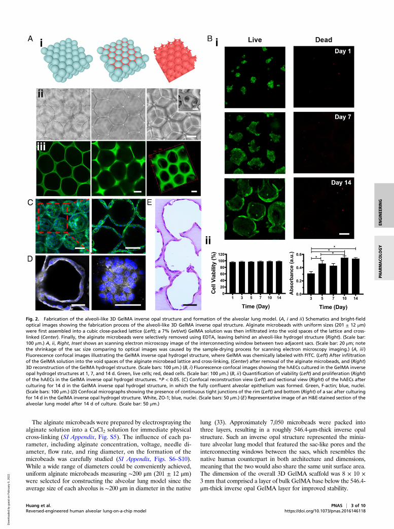
The alginate microbeads were prepared by electrospraying thealginate solution into a CaCl2 solution for immediate physicalcross-linking (SI Appendix, Fig. S5). The influence of each pa-rameter, including alginate concentration, voltage, needle di-ameter, flow rate, and ring diameter, on the formation of themicrobeads was carefully studied (SI Appendix, Figs. S6–S10).While a wide range of diameters could be conveniently achieved,uniform alginate microbeads measuring ∼200 μm (201 ± 12 μm)were selected for constructing the alveolar lung model since theaverage size of each alveolus is ∼200 μm in diameter in the native
lung (33). Approximately 7,050 microbeads were packed intothree layers, resulting in a roughly 546.4-μm-thick inverse opalstructure. Such an inverse opal structure represented the minia-ture alveolar lung model that featured the sac-like pores and theinterconnecting windows between the sacs, which resembles thenative human counterpart in both architecture and dimensions,meaning that the two would also share the same unit surface area.The dimension of the overall 3D GelMA scaffold was 8 × 10 ×3 mm that comprised a layer of bulk GelMA base below the 546.4-μm-thick inverse opal GelMA layer for improved stability.
Fig. 2. Fabrication of the alveoli-like 3D GelMA inverse opal structure and formation of the alveolar lung model. (A, i and ii) Schematics and bright-fieldoptical images showing the fabrication process of the alveoli-like 3D GelMA inverse opal structure. Alginate microbeads with uniform sizes (201 ± 12 μm)were first assembled into a cubic close-packed lattice (Left); a 7% (wt/wt) GelMA solution was then infiltrated into the void spaces of the lattice and cross-linked (Center). Finally, the alginate microbeads were selectively removed using EDTA, leaving behind an alveoli-like hydrogel structure (Right). (Scale bar:100 μm.) A, ii, Right, Inset shows an scanning electron microscopy image of the interconnecting window between two adjacent sacs. (Scale bar: 20 μm; notethe shrinkage of the sac size comparing to optical images was caused by the sample-drying process for scanning electron microscopy imaging.) (A, iii)Fluorescence confocal images illustrating the GelMA inverse opal hydrogel structure, where GelMA was chemically labeled with FITC. (Left) After infiltrationof the GelMA solution into the void spaces of the alginate microbead lattice and cross-linking, (Center) after removal of the alginate microbeads, and (Right)3D reconstruction of the GelMA hydrogel structure. (Scale bars: 100 μm.) (B, i) Fluorescence confocal images showing the hAECs cultured in the GelMA inverseopal hydrogel structures at 1, 7, and 14 d. Green, live cells; red, dead cells. (Scale bar: 100 μm.) (B, ii) Quantification of viability (Left) and proliferation (Right)of the hAECs in the GelMA inverse opal hydrogel structures. *P < 0.05. (C) Confocal reconstruction view (Left) and sectional view (Right) of the hAECs afterculturing for 14 d in the GelMA inverse opal hydrogel structure, in which the fully confluent alveolar epithelium was formed. Green, F-actin; blue, nuclei.(Scale bars: 100 μm.) (D) Confocal micrographs showing the presence of continuous tight junctions of the rim (Left) and bottom (Right) of a sac after culturingfor 14 d in the GelMA inverse opal hydrogel structure. White, ZO-1; blue, nuclei. (Scale bars: 50 μm.) (E) Representative image of an H&E-stained section of thealveolar lung model after 14 d of culture. (Scale bar: 50 μm.)
Huang et al. PNAS | 3 of 10Reversed-engineered human alveolar lung-on-a-chip model https://doi.org/10.1073/pnas.2016146118
ENGINEE
RING
PHARM
ACO
LOGY
Dow
nloa
ded
by g
uest
on
Feb
ruar
y 5,
202
2

It should be noted that, although the human alveoli are notnecessarily homogeneous in size, in our model we pursued the useof only uniform alginate microbeads as the sacrificial templates toavoid unwanted interferences caused by the heterogeneity in thealveolar sizes. Indeed, we have previously demonstrated that theuse of porous scaffolds with uniform pores is experimentally su-perior to those with random pores even if the average pore sizefalls in the same range (20, 21). In fact, we were also able tofabricate the 3D inverse opal GelMA structures with different yetuniform pore sizes using monodispersed alginate microbeads ofdifferent diameters. The microbeads with sizes of 325.9 ± 10.5,199.6 ± 1.1, and 156.3 ± 2.7 μm were used separately to fabricate3D GelMA structures with corresponding pore sizes (SI Appendix,Fig. S11). Likewise, we could also fabricate a 3D GelMA structurewith heterogeneous pore sizes by using randomly mixedmicrobeads with three diameters (∼200, ∼250, and ∼300 μm) asthe template (SI Appendix, Fig. S12). The alginate microbeadscould be rapidly removed by treating with the 0.01 M EDTA so-lution. The complete removal of the alginate microbeads from thescaffold was confirmed by both scanning electron microscopyimaging and X-ray energy dispersive spectroscopy (EDS) map-ping, shown in SI Appendix, Fig. S13. In particular, EDS mappingprofiles suggested that while 1.3% of Ca2+ was detected in thesample before removal of the microbeads, no Ca2+ (0%) wasmeasured at all after the removal. Thus, removal of both Ca2+ andthe alginate microbeads was complete.To model the alveolar epithelium, we populated hAECs on the
surfaces of the sacs within the GelMA inverse opal structures.Fluorescence confocal images revealed that the hAECs were al-most rounded and crowded at day 1 postseeding, where they wereable to gradually spread and proliferate during the following days,eventually covering the full surface areas of the alveoli-like sacs(Fig. 2 B, i and C and SI Appendix, Figs. S14 and S15). The via-bility of the hAECs on the surfaces of the pores in the porousGelMA structures was observed to exceed 95% (Fig. 2B). Theproliferation of the cells was steady during the first 10 d of cultureand slightly slowed down afterward (Fig. 2 B, ii), possibly due tothe contact inhibition upon confluency of the cells on the surfacesof the pores. The cell viability, proliferation, and spreading at thedifferent depths within a single layer of the sacs of the inverse opalstructure are shown in SI Appendix, Fig. S14. Indeed, fluorescenceconfocal images suggested that these culture conditions resulted inthe formation of a confluent alveolar epithelium (Fig. 2, Movie S1,and SI Appendix, Figs. S14 and S15). Significantly, the hAECswere linked by a continuous presence of tight junctions as indi-cated by Zonula occludens (ZO)-1 staining (Fig. 2D). Further-more, hematoxylin and eosin (H&E) staining provided anoverview of the structure of the GelMA hydrogel and the celldistributions, clearly validating the emulation of the alveoli-like,interconnected sacs and the continuous monolayer of hAECs onthe pore surfaces (Fig. 2E).
Comparisons between 2D and 3D Cultures of the Human AlveolarEpithelium. Reconstitution of the 3D structures and microenvi-ronments of the tissues is vital for realizing their functions andmodeling pathophysiology in vitro (34, 35). To investigate theadvantages of our 3D GelMA inverse opal structures in the re-capitulation of the pulmonary alveoli, the behaviors of hAECswere carefully compared with those cultured on 2D GelMAsurfaces. The morphologies of the hAECs were further studiedby capturing scanning electron microscopy images (Fig. 3A) ofthe cells cultured on both the 3D GelMA structures (Fig. 3 A,Upper) and the 2D GelMA surfaces (Fig. 3 A, Lower) at days 3, 7,and 14. Comparing with the slightly crowded and oftentimeslocally aggregated hAECs on the 2D GelMA surfaces during theculture, the hAECs in the 3D GelMA structures appeared to beflatter, where the formation of a smooth, confluent epithelium bywell-spread cells was apparent at day 14. The microscopic images
of hAECs cultured on the 2D GelMA surfaces are shown in SIAppendix, Fig. S16. Moreover, the apparent proliferation rate ofthe hAECs in the 3D GelMA structures was significantly higherthan that of the cells on the 2D surfaces (SI Appendix, Fig. S17).Nevertheless, when normalized (Fig. 3B), there were no significantdifferences between the two configurations anymore during theculture period of 10 d. The hAECs in the 3D GelMA structuresbecame higher in numbers than those on the 2D surfaces at day14, most likely due to the presence of the larger total surface areawithin the 3D GelMA structure as compared with the 2D surface,at ∼2 cm2 in the former vs. 0.8 cm2 for the latter despite that theyshare the same planar size of 8 × 10 mm. Most importantly, the3D GelMA scaffolds provided an optimal geometry and micro-architecture for cell spreading and proliferation compared withthe planar surfaces, which possibly promoted the cellular processsuch as adhesion, proliferation, and migration. These observationsindicated that the 3D culture provided an optimal geometry forcell spreading and proliferation compared with the planar culturewhen both substrate materials were the same GelMA with similarlocal mechanical properties, both close to that of the normal hu-man lung tissue (29).Apoptosis is a fundamental cellular process that is necessary for
the maintenance of tissue homeostasis in adult organisms (36). Theapoptotic activities of the cells were analyzed via caspase-3/7 stainingof hAECs and flow cytometry analyses of annexin V-FITC– andpropidium iodide (PI)-stained hAECs both on 2D GelMA surfacesand in 3D GelMA structures. Interestingly, it was found that themean fluorescence intensity of caspase-3/7 was higher for the cellsgrown on the 2D GelMA surfaces compared with those in the 3DGelMA structures (Fig. 3C and SI Appendix, Fig. S18). Similarly,the flow cytometry analyses of annexin V-FITC– and PI-stainedhAECs at day 14 and day 22 showed that the overall proportion oflate apoptotic/necrotic hAECs on the 2D GelMA surfaces wasincreased over the culture period compared with those in the 3DGelMA structures (Fig. 3D and SI Appendix, Fig. S19). While theproportions of early apoptotic cells were higher in 3D structures,the proportions of the late apoptotic/necrotic cells were higher onthe 2D GelMA surfaces, at both day 14 (Fig. 3D) and day 22 (SIAppendix, Fig. S19). It is therefore hypothesized that our unique3D GelMA inverse opal structure resembling that of the humanalveoli could possibly provide the cells with an in vivo-like mi-croenvironment, which promoted optimal cell–ECM and cell–cellinteractions.To explore the differentially expressed genes to detect biological
changes in hAECs cultured in 2D and 3D, we performed addi-tional transcriptome profiling through gene microarray analyses.Other than the 2D GelMA surfaces, we further included anothercontrol group based on the 2D PDMS surfaces (a commonly usedsubstrate in various organ-on-chip models, including those of thelung) to compare with the 3D GelMA structure-based hAEC cul-ture. Scatterplots and the Venn diagram (SI Appendix, Fig. S20 Aand B) were generated for the three comparisons (i.e., 3D GelMAvs. 2D PDMS, 3D GelMA vs. 2D GelMA, and 2D GelMA vs. 2DPDMS). We calculated the normalized expression values (frag-ments per kilobase per million mapped reads) of every gene ana-lyzed in the three groups, and those values with greater than twofoldincrease or decrease with a false discovery rate of less than 0.05 (qvalue < 0.05) were considered as the differentially expressed genes.Interestingly, the 3D GelMA vs. 2D PDMS comparison demon-
strated that there were 4,757 differentially expressed genes (2,646 up-regulated and 2,111 down-regulated); for 3D GelMA vs. 2D GelMA,there were 2,396 differentially expressed genes (1,352 up-regulatedand 1,044 down-regulated), while for 2D GelMA vs. 2D PDMS,there were 5,175 differentially expressed genes (2,553 up-regulated and 2,622 down-regulated) (SI Appendix, Fig. S20B).To elucidate the biological processes on the different culturesubstrates, functional gene ontology (GO) and Kyoto Encyclope-dia of Genes and Genomes (KEGG) pathway enrichment analyses
4 of 10 | PNAS Huang et al.https://doi.org/10.1073/pnas.2016146118 Reversed-engineered human alveolar lung-on-a-chip model
Dow
nloa
ded
by g
uest
on
Feb
ruar
y 5,
202
2
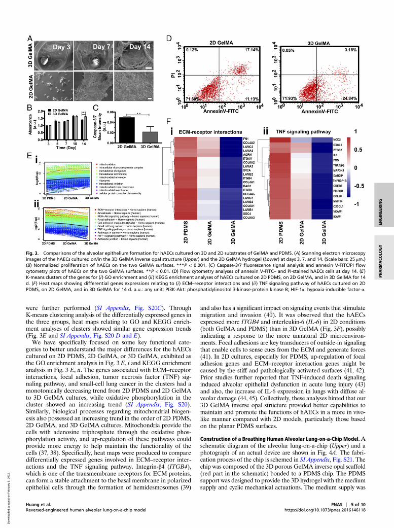
were further performed (SI Appendix, Fig. S20C). ThroughK-means clustering analysis of the differentially expressed genes inthe three groups, heat maps relating to GO and KEGG enrich-ment analyses of clusters showed similar gene expression trends(Fig. 3E and SI Appendix, Fig. S20 D and E).We have specifically focused on some key functional cate-
gories to better understand the major differences for the hAECscultured on 2D PDMS, 2D GelMA, or 3D GelMA, exhibited asthe GO enrichment analysis in Fig. 3 E, i and KEGG enrichmentanalysis in Fig. 3 E, ii. The genes associated with ECM–receptorinteractions, focal adhesion, tumor necrosis factor (TNF) sig-naling pathway, and small-cell lung cancer in the clusters had amonotonically decreasing trend from 2D PDMS and 2D GelMAto 3D GelMA cultures, while oxidative phosphorylation in thecluster showed an increasing trend (SI Appendix, Fig. S20).Similarly, biological processes regarding mitochondrial biogen-esis also possessed an increasing trend in the order of 2D PDMS,2D GelMA, and 3D GelMA cultures. Mitochondria provide thecells with adenosine triphosphate through the oxidative phos-phorylation activity, and up-regulation of these pathways couldprovide more energy to help maintain the functionality of thecells (37, 38). Specifically, heat maps were produced to comparedifferentially expressed genes involved in ECM–receptor inter-actions and the TNF signaling pathway. Integrin-β4 (ITGB4),which is one of the transmembrane receptors for ECM proteins,can form a stable attachment to the basal membrane in polarizedepithelial cells through the formation of hemidesmosomes (39)
and also has a significant impact on signaling events that stimulatemigration and invasion (40). It was observed that the hAECsexpressed more ITGB4 and interleukin-6 (IL-6) in 2D conditions(both GelMA and PDMS) than in 3D GelMA (Fig. 3F), possiblyindicating a response to the more unnatural 2D microenviron-ments. Focal adhesions are key transducers of outside-in signalingthat enable cells to sense cues from the ECM and generate forces(41). In 2D cultures, especially for PDMS, up-regulation of focaladhesion genes and ECM–receptor interaction genes might becaused by the stiff and pathologically activated surfaces (41, 42).Prior studies further reported that TNF-induced death signalinginduced alveolar epithelial dysfunction in acute lung injury (43)and also, the increase of IL-6 expression in lungs with diffuse al-veolar damage (44, 45). Collectively, these analyses hinted that our3D GelMA inverse opal structure provided better capabilities tomaintain and promote the functions of hAECs in a more in vivo-like manner compared with 2D models, particularly those basedon the planar PDMS surfaces.
Construction of a Breathing Human Alveolar Lung-on-a-Chip Model.Aschematic diagram of the alveolar lung-on-a-chip (Upper) and aphotograph of an actual device are shown in Fig. 4A. The fabri-cation process of the chip is schemed in SI Appendix, Fig. S21. Thechip was composed of the 3D porous GelMA inverse opal scaffold(red part in the schematic) bonded to a PDMS chip. The PDMSsupport was designed to provide the 3D hydrogel with the mediumsupply and cyclic mechanical actuations. The medium supply was
Fig. 3. Comparisons of the alveolar epithelium formation for hAECs cultured on 3D and 2D substrates of GelMA and PDMS. (A) Scanning electron microscopyimages of the hAECs cultured on/in the 3D GelMA inverse opal structure (Upper) and the 2D GelMA hydrogel (Lower) at days 3, 7, and 14. (Scale bars: 25 μm.)(B) Normalized proliferation of hAECs on the two GelMA surfaces. ***P < 0.001. (C) Caspase-3/7 fluorescence signal analysis and annexin V-FITC/PI flowcytometry plots of hAECs on the two GelMA surfaces. **P < 0.01. (D) Flow cytometry analyses of annexin V-FITC– and PI-stained hAECs cells at day 14. (E)K-means clusters of the genes for (i) GO enrichment and (ii) KEGG enrichment analyses of hAECs cultured on 2D PDMS, on 2D GelMA, and in 3D GelMA for 14d. (F) Heat maps showing differential genes expressions relating to (i) ECM–receptor interactions and (ii) TNF signaling pathway of hAECs cultured on 2DPDMS, on 2D GelMA, and in 3D GelMA for 14 d. a.u.: any unit; PI3K-Akt: phosphatidylinositol 3-kinase-protein kinase B; HIF-1α: hypoxia-inducible factor-α.
Huang et al. PNAS | 5 of 10Reversed-engineered human alveolar lung-on-a-chip model https://doi.org/10.1073/pnas.2016146118
ENGINEE
RING
PHARM
ACO
LOGY
Dow
nloa
ded
by g
uest
on
Feb
ruar
y 5,
202
2

realized by slowly infusing the medium through the open channelsat the bottom of the PDMS chip. The cyclic mechanical movementswere applied to the 3D hydrogel via applying the programmednegative pressures to the two side chambers at the frequency of0.2 Hz (the normal breath frequency of human).The mechanical movements of the chip allowed the airflow in
and out of the embedded 3D GelMA structure. There are somereports that have focused on building and simulating the airflowin the artificial alveoli (46, 47); however, only few successfullyreconstituted the 3D structures of the human alveolar sacs. Herein,we demonstrated the airflow in and out of the alveoli-like structurein our inverse opal alveolar lung model during a breathing cycle, asshown in the simulation results in SI Appendix, Fig. S22 A, C, and Eas well as in Movie S2. During the “breathing” of the chip, the airgradually flowed into and then out of the inverse opal sacs spon-taneously due to the volume change-induced pressure variation withvelocity values and Reynolds numbers (0.71 to 1.70), which are verysimilar to those found in the real human alveoli. In addition, directionof the airflow changed according to the structural framework withinthe inverse opal alveolar lung model, especially in correspon-dence at the “alveolus–alveolus” interfaces. On the contrary, inthe control 2D model, the airflow followed nonphysiologicalstraighter paths without showing rapid changes of directions (SIAppendix, Fig. S22 B, D, and F).After seeding the hAECs in the 3D GelMA inverse opal
structures in the chips, the cells were cultured for 14 d. Live/dead
images revealed that the cell viability remained high after 14 d ofstatic culture in the chips (Fig. 4 B, control). When the cyclicstretch was applied, the hAECs grown on the surfaces of the porescould “breathe” along with the cyclic mechanical movements thatstretched the GelMA structure, where the average size of alveoliwas observed to expand by ∼8% from the state of “breath out” to“breath in” (Fig. 4C and Movie S3). This value is within the 5 to15% physiological range of strain experienced by the alveoli in thehuman lung (10). It was, in fact, convenient for us to control thestrains or breathing frequencies on the chip as needed by varyingthe size of the two side chambers or the frequency of the negativepressure applied. As shown in Movie S4, our chip-based modelfunctioned well under a range of strain levels of 5, 10, and 15%, aswell as breathing frequencies of 0.1, 0.2, 0.4, and 0.8 Hz.An ALI was also created for the hAECs in the chip by removal
of the medium from the sacs and supplying the medium throughthe parallel channels underneath the GelMA structure at thebottom of the chip. The relatively low thickness (<3 mm) of theGelMA hydrogel and its nano-/microscale porosities were be-lieved to enable efficient medium transport to support cellularfunctions. To confirm the sufficient supply of nutrients within thetop layer of the 3D GelMA inverse opal structure, we assessedthe diffusion of both small molecules (FITC) as well as largermolecules (FITC-dextran, Mw = 10 kDa). It was found that thediffusion of both types of molecules across the entire GelMAstructure occurred within relatively short periods (SI Appendix,
Fig. 4. Construction of the alveolar lung-on-a-chip model. (A) Schematic representing the alveolar lung-on-a-chip (Upper) and photograph of a devicewithout the GelMA inverse opal structure to show the underlying fluidic channels (Lower). (Scale bar: 5 mm.) (B) Fluorescence images showing the viability ofthe hAECs cultured in the chips without and with the breathing events. Green, live cells; red, dead cells. (Scale bar: 100 μm.) (C) Micrographs showing theexpansion of the sacs under a strain of 8%. (Scale bar: 100 μm.) (D) Images of H&E-stained sections showing the epithelium formation in the chips without andwith the breathing events. (Scale bar: 50 μm.) (E) ZO-1 staining showing the tight junction formation of the epithelium in the chips without and with thebreathing events. White, ZO-1; blue, nuclei. (Scale bar: 50 μm.) (F) The quantified levels of secreted cytokines IL-8, IL-6, IL-1β, MCP-1, and GM-CSF by hAECscultured in the chips without and with the breathing events. All analyses were performed at 14 d of culture.
6 of 10 | PNAS Huang et al.https://doi.org/10.1073/pnas.2016146118 Reversed-engineered human alveolar lung-on-a-chip model
Dow
nloa
ded
by g
uest
on
Feb
ruar
y 5,
202
2
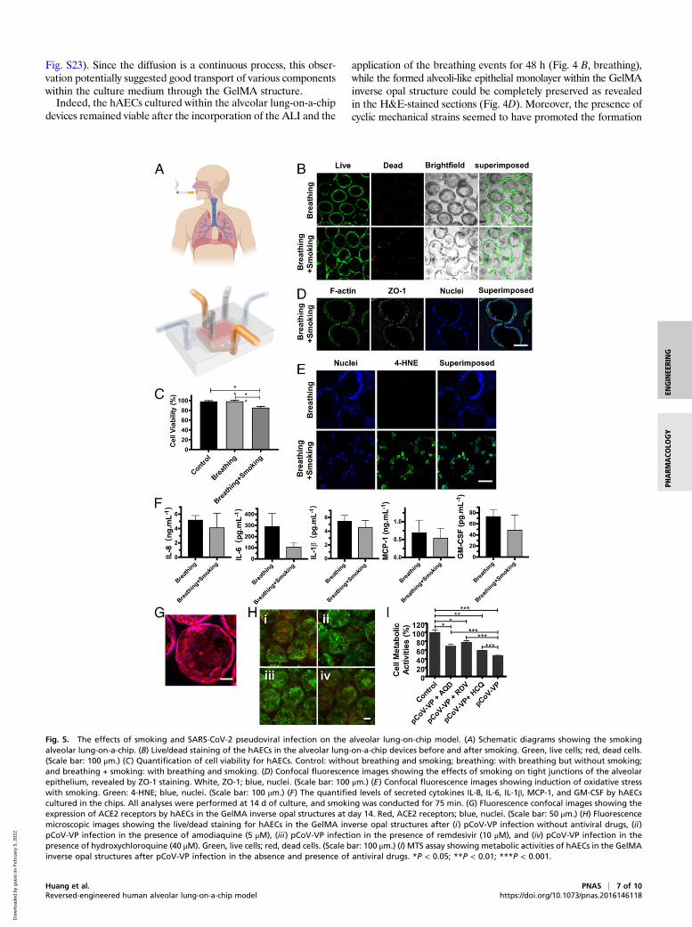
Fig. S23). Since the diffusion is a continuous process, this obser-vation potentially suggested good transport of various componentswithin the culture medium through the GelMA structure.Indeed, the hAECs cultured within the alveolar lung-on-a-chip
devices remained viable after the incorporation of the ALI and the
application of the breathing events for 48 h (Fig. 4 B, breathing),while the formed alveoli-like epithelial monolayer within the GelMAinverse opal structure could be completely preserved as revealedin the H&E-stained sections (Fig. 4D). Moreover, the presence ofcyclic mechanical strains seemed to have promoted the formation
Fig. 5. The effects of smoking and SARS-CoV-2 pseudoviral infection on the alveolar lung-on-chip model. (A) Schematic diagrams showing the smokingalveolar lung-on-a-chip. (B) Live/dead staining of the hAECs in the alveolar lung-on-a-chip devices before and after smoking. Green, live cells; red, dead cells.(Scale bar: 100 μm.) (C) Quantification of cell viability for hAECs. Control: without breathing and smoking; breathing: with breathing but without smoking;and breathing + smoking: with breathing and smoking. (D) Confocal fluorescence images showing the effects of smoking on tight junctions of the alveolarepithelium, revealed by ZO-1 staining. White, ZO-1; blue, nuclei. (Scale bar: 100 μm.) (E) Confocal fluorescence images showing induction of oxidative stresswith smoking. Green: 4-HNE; blue, nuclei. (Scale bar: 100 μm.) (F) The quantified levels of secreted cytokines IL-8, IL-6, IL-1β, MCP-1, and GM-CSF by hAECscultured in the chips. All analyses were performed at 14 d of culture, and smoking was conducted for 75 min. (G) Fluorescence confocal images showing theexpression of ACE2 receptors by hAECs in the GelMA inverse opal structures at day 14. Red, ACE2 receptors; blue, nuclei. (Scale bar: 50 μm.) (H) Fluorescencemicroscopic images showing the live/dead staining for hAECs in the GelMA inverse opal structures after (i) pCoV-VP infection without antiviral drugs, (ii)pCoV-VP infection in the presence of amodiaquine (5 μM), (iii) pCoV-VP infection in the presence of remdesivir (10 μM), and (iv) pCoV-VP infection in thepresence of hydroxychloroquine (40 μM). Green, live cells; red, dead cells. (Scale bar: 100 μm.) (I) MTS assay showing metabolic activities of hAECs in the GelMAinverse opal structures after pCoV-VP infection in the absence and presence of antiviral drugs. *P < 0.05; **P < 0.01; ***P < 0.001.
Huang et al. PNAS | 7 of 10Reversed-engineered human alveolar lung-on-a-chip model https://doi.org/10.1073/pnas.2016146118
ENGINEE
RING
PHARM
ACO
LOGY
Dow
nloa
ded
by g
uest
on
Feb
ruar
y 5,
202
2
of tight junctions, indicated by ZO-1 staining (Fig. 4E) (48). Thelevels of secreted cytokines including IL-8, IL-6, IL-1β, monocytechemoattractant protein-1 (MCP-1), and granulocyte-macrophagecolony-stimulating factor (GM-CSF) were further profiled; theywere all slightly reduced upon breathing stimulation, yet no sig-nificant differences could be found (Fig. 4F).
Analysis of Cigarette Smoking Effects on the Alveolar Lung-on-a-ChipModel. Cigarette smoking is one of the major factors leading tothe development of COPD, currently the third leading cause ofchronic morbidity and mortality worldwide (2, 49, 50), as well asresulting in lung cancer as the most common lethal neoplasm(51). Since the alveolar epithelium is the barrier between theinhaled air and the underlying components (52), the establish-ment of a smoking alveolar lung-on-a-chip model to examine thedistal lung injury and study the effects of smoking on the alveolarepithelium becomes important. While a smoking lung-on-a-chipplatform was previously demonstrated using airway epithelialcells on a synthetic polyethylene terephthalate membrane (53),no model currently exists for the distal lung (i.e., the alveoli). Oursmoking alveolar lung-on-a-chip model consisted of a smokingdevice (53, 54) and an additional compartment on top of thebreathing alveolar lung-on-a-chip platform to mimic the airway–alveoli transport route (Fig. 5A). After smoking ∼10 cigarettes andfurther culturing for 24 h, dead cells within our alveoli modelshowed an increase (Fig. 5B), and the viability of hAECs was sig-nificantly decreased compared with normal breathing and controlgroups (Fig. 5C). The apoptotic activities of the cells before andafter the application of the cigarette smoke were analyzed viacaspase-3/7 staining of hAECs on/in both 2D and 3D scaffolds (SIAppendix, Fig. S24). Consistent with the cultures without the chips(Fig. 3C and SI Appendix, Fig. S18), hAECs on 2D GelMA in thechip also showed a higher apoptotic tendency (SI Appendix, Fig.S24A) as compared with those in 3D GelMA structures (SI Ap-pendix, Fig. S23B). The smoking further resulted in the increase ofapoptosis of hAECs in both configurations (SI Appendix, Fig.S24 A–C). This observation was, in addition, reflected in cellularactivity assay (SI Appendix, Fig. S24D).In addition, the tight junctions between the hAECs were
partially damaged after smoking (Fig. 5D). The oxidants in ciga-rette smoke can induce cellular injuries (55), where 4-hydroxy-2-nonenal (4-hydroxynonenal [4-HNE]) is a major product of lipidperoxidation and a marker of oxidative stress (56, 57). 4-HNEcan react with DNAs and proteins to produce various adductsthat can lead to cell apoptosis, and the 4-HNE expression levelin the alveolar epithelium is negatively correlated with pulmo-nary functions and contributes to the pathogenesis of variouspathological manifestations such as COPD (58). Our results in-dicated an elevation in the intensity of nuclear 4-HNE stainingpostsmoking (Fig. 5E), and such a high expression of 4-HNEmay further induce downstream cellular damage. As a result,the soluble cytokine levels of IL-8, IL-6, IL-1β, MCP-1, andGM-CSF after smoking slightly declined compared with thenormal breathing group, suggesting that the functions of hAECsto inhibit proinflammatory responses were compromisedwhen they were exposed to cigarette smoke, consistent withliterature (53).
Analysis of SARS-CoV-2 Pseudoviral Infection and Treatment Efficacieson the Alveolar Lung-on-a-Chip Model. COVID-19 caused bySARS-CoV-2 has led to a global public health crisis and is asso-ciated with high mortality, mostly caused by acute respiratorydistress syndrome (59, 60). As the lungs are the first body organaffected by COVID-19, we further assessed the applicability of ouralveolar lung-on-a-chip to investigate SARS-CoV-2 pseudoviralinfection. In addition, the antiviral efficacies of two clinically ap-proved antiviral drugs, remdesivir (61) and hydroxychloroquine(62), as well as an antimalarial drug, amodiaquine (63), were
evaluated. The latter has also been given to individuals infectedwith SARS-CoV-2 (64). Since it has been well recognized thatSARS-CoV-2 uses angiotensin converting enzyme 2 (ACE2) as areceptor for cellular entry, prior to SARS-CoV-2 pseudoviralparticle (pCoV-VP) infection on the alveolar lung-on-a-chip, wefirst detected the expression of ACE2 receptors by hAECs grownon the surfaces of the alveolar sacs. The hAECs showed a higherlevel of expression of ACE2 receptors as shown in Fig. 5G and SIAppendix, Fig. S25A. The chips were then inoculated with pCoV-VPs at multiplicity of infection (MOI) of 0.5 for 48 h to identifythe virus susceptibility in vitro in the presence and absence ofantiviral drugs. At 48 h postinfection, the infection of hAECs withmcherry-labeled pCoV-VPs was observed under a fluorescencemicroscope (SI Appendix, Fig. S25B), and cytopathic effect of pCoV-VPs was assessed by live/dead assay (Fig. 5H and SI Appendix, Fig.S25C) and 3-(4,5-dimethylthiazol-2-yl)-5-(3-carboxymethoxyphenyl)-2-(4-sulfophenyl)-2H-tetrazolium (MTS) assay (Fig. 5I). The resultsshowed that the cytopathic effects of pCoV-VPs were decreasedby approximately 21.4, 30.0, and 11.3% at 5 μM amodiaquine,10 μM remdesivir, and 40 μM hydroxychloroquine, respectively(Fig. 5I). Thus, these drugs significantly inhibited the infection-induced hAEC death by the SARS-CoV-2 pseudotyped viralparticles expressing the SARS-CoV-2 spike proteins in our alveolarlung-on-a-chip.
Conclusions and PerspectivesThis study provided an example of recreating the architecturallyrelevant alveolar lung-on-a-chip model and demonstrated itsstructural and functional features. We have successfully recon-structed some of the key aspects of the human pulmonary alveoli,including the microarchitecture, the ECM microenvironment, theALI, and the mechanical breathing events. The alveolus in the lungis one of the most important structural determinants of the ar-chitecture of the respiratory parenchyma and lung functions. Ourbreathing alveolar lung-on-a-chip model could broadly reproducethe 3D anatomical structures and functions of the alveoli throughthe use of a precisely defined 3D porous GelMA hydrogel based onan inverse opal structure, while the breathing function was realizedby situating the hAEC-populated GelMA structure in PDMS chipdevice. Based upon the breathing alveolar lung-on-chip model,we subsequently built a cigarette-smoking alveolar lung-on-a-chipplatform, where the negative effects of smoking on the hAECscould be readily explored. We further demonstrated the applica-bility of our alveolar lung model for investigating SARS-CoV-2pseudoviral infection and treatment efficacies of antiviral drugssuch as hydroxychloroquine, remdesivir, and amodiaquine. Whileremdesivir and amodiaquine have been approved by the Foodand Drug Administration to treat COVID-19 (64), in vitro andin vivo studies have shown the discrepancies in antiviral activityof hydroxychloroquine against SARS-CoV-2 (62).We have also showcased the possibility of creating the alveolar–
capillary interface, which is another key microstructure in thehuman distal lung that enables the gas exchange in the alveoli byincorporating the human umbilical vein endothelial cells into thematrix of the GelMA inverse opal hydrogel to form the micro-vascular network surrounding the sacs (SI Appendix, Fig. S26). Inaddition, to investigate the capability of our strategy to reconsti-tute pathological models of the lung or in particular, lung cancer,we also built a nonsmall-cell lung cancer model with A549 cells.As shown in SI Appendix, Fig. S27, the A549 cells could form agood alveolar structure in our platform with good cell viability andmorphologies (note the multilayered growth of the A549 cells insome regions in comparison with the monolayer epithelium formedby normal hAECs).Although surfactant lining layer at the ALI is extremely im-
portant for maintaining the alveolar functions, hAECs that weused in this study were commercially sourced, where the vendorunfortunately did not provide any information on AEC types I/II.
8 of 10 | PNAS Huang et al.https://doi.org/10.1073/pnas.2016146118 Reversed-engineered human alveolar lung-on-a-chip model
Dow
nloa
ded
by g
uest
on
Feb
ruar
y 5,
202
2

It would be indeed very interesting for us in our future work tointroduce defined populations of hAECs in our model, to study theireffects on alveolar functions and pathologies such as infections.While our system has some limitations, this example of the
miniature alveolar lung-on-a-chip platform provides the founda-tion and can be readily expanded in the future to provide fullfunctionality of the alveolar units in vitro to provide a niche forlung and lung disease modeling and research.
MethodsSI Appendix, Supplementary Text has detailed experimental procedures thatdescribe the 1) synthesis of alginate microbeads; 2) synthesis of GelMA; 3)fabrication of GelMA inverse opal structures; 4) fabrication of the GelMAinverse opal structure-embedded chip device; 5) fabrication of the smokingdevice; 6) cell culture in 3D GelMA inverse opal structures; 7) cell culture on2D GelMA surfaces; 8) construction of the alveolar lung-on-a-chip; 9) cellularcharacterizations; 10) scanning electron microscopy imaging; 11) apoptosis
analyses; 12) microarray analyses; 13) analysis of chemokines and cyto-kines; 14) computational simulations; 15) SARS-CoV-2 pseudotyped virusproduction and infection of the alveolar lung-on-a-chip; and 16) statisticalanalyses.
Data Availability. All study data are included in the article and/or supportinginformation.
ACKNOWLEDGMENTS. This work was supported by the Brigham ResearchInstitute, the New England Anti-Vivisection Society (NEAVS), and the Amer-ican Fund for Alternatives to Animal Research (AFAAR). D.H. acknowledgessupport from National Natural Science Foundation of China Grants 11502158and 11632013 and Shanxi Provincial Key Research and Development Project,China Grant 201803D421060. A.M.G. acknowledges support from the Euro-pean Union’s Horizon2020 Research and Innovation Program under Grant760921 (PANBioRA: Personalised and Generalised Integrated BiomaterialRisk Assessment). We also thank Junjie Lv for sharing molecular biology toolsat the early stage of the project.
1. A. Papi, C. Brightling, S. E. Pedersen, H. K. Reddel, Asthma. Lancet 391, 783–800(2018).
2. J. L. López-Campos, W. Tan, J. B. Soriano, Global burden of COPD. Respirology 21,14–23 (2016).
3. F. Krammer et al., Influenza. Nat. Rev. Dis. Primers 4, 3 (2018).4. L. A. Mandell, M. S. Niederman, Aspiration pneumonia. N. Engl. J. Med. 380, 651–663
(2019).5. M. J. A. Reid et al., Building a tuberculosis-free world: The Lancet Commission on
tuberculosis. Lancet 393, 1331–1384 (2019).6. R. S. Herbst, D. Morgensztern, C. Boshoff, The biology and management of non-small
cell lung cancer. Nature 553, 446–454 (2018).7. World Health Organization, “Coronavirus disease (COVID-2019) situation reports"
(Rep. No. 36, World Health Organization, Geneva, Switzerland, 2020).8. P. N. Fonkwo, Pricing infectious disease. The economic and health implications of
infectious diseases. EMBO Rep. 9 (suppl. 1), S13–S17 (2008).9. A. Akhtar, The flaws and human harms of animal experimentation. Camb. Q. Healthc.
Ethics 24, 407–419 (2015).10. D. Huh et al., Reconstituting organ-level lung functions on a chip. Science 328,
1662–1668 (2010).11. D. Huh et al., Acoustically detectable cellular-level lung injury induced by fluid me-
chanical stresses in microfluidic airway systems. Proc. Natl. Acad. Sci. U.S.A. 104,18886–18891 (2007).
12. W. W. Hope et al., Pathogenesis of Aspergillus fumigatus and the kinetics of gal-actomannan in an in vitro model of early invasive pulmonary aspergillosis: Implica-tions for antifungal therapy. J. Infect. Dis. 195, 455–466 (2007).
13. D. D. Nalayanda et al., An open-access microfluidic model for lung-specific functionalstudies at an air-liquid interface. Biomed. Microdevices 11, 1081–1089 (2009).
14. L. Gregson, W. W. Hope, S. J. Howard, In vitro model of invasive pulmonary asper-gillosis in the human alveolus. Methods Mol. Biol. 845, 361–367 (2012).
15. M. M. Stevens, J. H. George, Exploring and engineering the cell surface interface.Science 310, 1135–1138 (2005).
16. B. Haefeli-Bleuer, E. R. Weibel, Morphometry of the human pulmonary acinus. Anat.Rec. 220, 401–414 (1988).
17. A. Tsuda et al., Finite element 3D reconstruction of the pulmonary acinus imaged bysynchrotron X-ray tomography. J. Appl. Physiol. 105, 964–976 (2008).
18. L. Knudsen, M. Ochs, The micromechanics of lung alveoli: Structure and func-tion of surfactant and tissue components. Histochem. Cell Biol. 150, 661–676(2018).
19. B. Grigoryan et al., Multivascular networks and functional intravascular topologieswithin biocompatible hydrogels. Science 364, 458–464 (2019).
20. S.-W. Choi, Y. Zhang, Y. Xia, Three-dimensional scaffolds for tissue engineering: Theimportance of uniformity in pore size and structure. Langmuir 26, 19001–19006(2010).
21. Y. Zhang, S.-W. Choi, Y. Xia, Modifying the pores of an inverse opal scaffold withchitosan microstructures for truly three-dimensional cell culture. Macromol. RapidCommun. 33, 296–301 (2012).
22. S. W. Choi, Y. Zhang, M. R. Macewan, Y. Xia, Neovascularization in biodegradableinverse opal scaffolds with uniform and precisely controlled pore sizes. Adv. Healthc.Mater. 2, 145–154 (2013).
23. Y. S. Zhang, K. P. Regan, Y. Xia, Controlling the pore sizes and related properties ofinverse opal scaffolds for tissue engineering applications. Macromol. Rapid Commun.34, 485–491 (2013).
24. Y. S. Zhang, C. Zhu, Y. Xia, Inverse opal scaffolds and their biomedical applications.Adv. Mater. 29, 1701115 (2017).
25. J. Lee, N. A. Kotov, Notch ligand presenting acellular 3D microenvironments forex vivo human hematopoietic stem-cell culture made by layer-by-layer assembly.Small 5, 1008–1013 (2009).
26. J. Lee, G. D. Lilly, R. C. Doty, P. Podsiadlo, N. A. Kotov, In vitro toxicity testing ofnanoparticles in 3D cell culture. Small 5, 1213–1221 (2009).
27. M. J. Cuddihy, Y. Wang, C. Machi, J. H. Bahng, N. A. Kotov, Replication of bonemarrow differentiation niche: Comparative evaluation of different three-dimensionalmatrices. Small 9, 1008–1015 (2013).
28. J. Kim, S. A. Bencherif, W. A. Li, D. J. Mooney, Cell-friendly inverse opal-like hydrogelsfor a spatially separated co-culture system. Macromol. Rapid Commun. 35, 1578–1586(2014).
29. S. S. Htwe et al., Role of Rho-associated coiled-coil forming kinase isoforms in regu-lation of stiffness-induced myofibroblast differentiation in lung fibrosis. Am. J. Respir.Cell Mol. Biol. 56, 772–783 (2017).
30. K. Yue et al., Structural analysis of photocrosslinkable methacryloyl-modified proteinderivatives. Biomaterials 139, 163–171 (2017).
31. D. Sicard et al., Aging and anatomical variations in lung tissue stiffness. Am. J. Physiol.Lung Cell. Mol. Physiol. 314, L946–L955 (2018).
32. S. R. Polio et al., Cross-platform mechanical characterization of lung tissue. PLoS One13, e0204765 (2018).
33. M. Ochs et al., The number of alveoli in the human lung. Am. J. Respir. Crit. Care Med.169, 120–124 (2004).
34. M. P. Lutolf, J. A. Hubbell, Synthetic biomaterials as instructive extracellular micro-environments for morphogenesis in tissue engineering. Nat. Biotechnol. 23, 47–55(2005).
35. B. M. Baker, C. S. Chen, Deconstructing the third dimension: How 3D culture micro-environments alter cellular cues. J. Cell Sci. 125, 3015–3024 (2012).
36. F. H. Igney, P. H. Krammer, Death and anti-death: Tumour resistance to apoptosis.Nat. Rev. Cancer 2, 277–288 (2002).
37. A. M. D’Erchia et al., Tissue-specific mtDNA abundance from exome data and itscorrelation with mitochondrial transcription, mass and respiratory activity. Mito-chondrion 20, 13–21 (2015).
38. A. A. A. Adam et al., AMC-Bio-Artificial Liver culturing enhances mitochondrial bio-genesis in human liver cell lines: The role of oxygen, medium perfusion and 3Dconfiguration. Mitochondrion 39, 30–42 (2018).
39. R. O. Hynes, Integrins: Bidirectional, allosteric signaling machines. Cell 110, 673–687(2002).
40. A. M. Mercurio, I. Rabinovitz, L. M. Shaw, The alpha 6 beta 4 integrin and epithelialcell migration. Curr. Opin. Cell Biol. 13, 541–545 (2001).
41. K. M. Mabry, S. Z. Payne, K. S. Anseth, Microarray analyses to quantify advantages of2D and 3D hydrogel culture systems in maintaining the native valvular interstitial cellphenotype. Biomaterials 74, 31–41 (2016).
42. V. Petit, J. P. Thiery, Focal adhesions: Structure and dynamics. Biol. Cell 92, 477–494(2000).
43. B. V. Patel, M. R. Wilson, K. P. O’Dea, M. Takata, TNF-induced death signaling triggersalveolar epithelial dysfunction in acute lung injury. J. Immunol. 190, 4274–4282(2013).
44. H. Schütte et al., Bronchoalveolar and systemic cytokine profiles in patients withARDS, severe pneumonia and cardiogenic pulmonary oedema. Eur. Respir. J. 9,1858–1867 (1996).
45. J. Ben-Ari et al., Cytokine response during hyperoxia: Sequential production of pul-monary tumor necrosis factor and interleukin-6 in neonatal rats. Isr. Med. Assoc. J. 2,365–369 (2000).
46. R. Fishler, P. Hofemeier, Y. Etzion, Y. Dubowski, J. Sznitman, Particle dynamics anddeposition in true-scale pulmonary acinar models. Sci. Rep. 5, 14071 (2015).
47. R. Fishler, M. K. Mulligan, J. Sznitman, Acinus-on-a-chip: A microfluidic platform forpulmonary acinar flows. J. Biomech. 46, 2817–2823 (2013).
48. K. H. Benam et al., Small airway-on-a-chip enables analysis of human lung inflam-mation and drug responses in vitro. Nat. Methods 13, 151–157 (2016).
49. B. R. Celli, J. A. Wedzicha, Update on clinical aspects of chronic obstructive pulmonarydisease. N. Engl. J. Med. 381, 1257–1266 (2019).
50. S. I. Rennard, M. B. Drummond, Early chronic obstructive pulmonary disease: Defi-nition, assessment, and prevention. Lancet 385, 1778–1788 (2015).
51. Forum of International Respiratory Societies, The Global Impact of Respiratory Dis-ease (European Respiratory Society, Sheffield, United Kingdom, ed. 2, 2017).
52. M. A. Matthay, L. Robriquet, X. Fang, Alveolar epithelium: Role in lung fluid balanceand acute lung injury. Proc. Am. Thorac. Soc. 2, 206–213 (2005).
53. K. H. Benam et al., Matched-comparative modeling of normal and diseased humanairway responses using a microengineered breathing lung chip. Cell Syst. 3,456–466.e4 (2016).
Huang et al. PNAS | 9 of 10Reversed-engineered human alveolar lung-on-a-chip model https://doi.org/10.1073/pnas.2016146118
ENGINEE
RING
PHARM
ACO
LOGY
Dow
nloa
ded
by g
uest
on
Feb
ruar
y 5,
202
2
54. K. H. Benam, R. Novak, T. C. Ferrante, Y. Choe, D. E. Ingber, Biomimetic smoking robotfor in vitro inhalation exposure compatible with microfluidic organ chips. Nat. Protoc.15, 183–206 (2020).
55. I. Rahman, W. MacNee, Role of oxidants/antioxidants in smoking-induced lung dis-eases. Free Radic. Biol. Med. 21, 669–681 (1996).
56. M. Breitzig, C. Bhimineni, R. Lockey, N. Kolliputi, 4-Hydroxy-2-nonenal: A criticaltarget in oxidative stress? Am. J. Physiol. Cell Physiol. 311, C537–C543 (2016).
57. A. Ayala, M. F. Muñoz, S. Argüelles, Lipid peroxidation: Production, metabolism, andsignaling mechanisms of malondialdehyde and 4-hydroxy-2-nonenal. Oxid. Med. Cell.Longev. 2014, 360438 (2014).
58. I. Rahman et al., 4-Hydroxy-2-nonenal, a specific lipid peroxidation product, is ele-vated in lungs of patients with chronic obstructive pulmonary disease. Am. J. Respir.Crit. Care Med. 166, 490–495 (2002).
59. I.-M. Schaefer et al., In situ detection of SARS-CoV-2 in lungs and airways of patientswith COVID-19. Mod. Pathol. 33, 2104–2114 (2020).
60. C. L. Onweni et al., ACEI/ARB therapy in COVID-19: The double-edged sword of ACE2and SARS-CoV-2 viral docking. Crit. Care 24, 475 (2020).
61. A. J. Pruijssers et al., Remdesivir inhibits SARS-CoV-2 in human lung cells and chimericSARS-CoV expressing the SARS-CoV-2 RNA polymerase in mice. Cell Rep. 32, 107940(2020).
62. P. Maisonnasse et al., Hydroxychloroquine use against SARS-CoV-2 infection in non-human primates. Nature 585, 584–587 (2020).
63. L. E. DeWald et al., In vivo activity of amodiaquine against Ebola virus infection. Sci.Rep. 9, 20199 (2019).
64. S. Weston et al., Broad anti-coronavirus activity of food and drug administration-approved drugs against SARS-CoV-2. J. Virol. 94, e01218-20 (2020).
10 of 10 | PNAS Huang et al.https://doi.org/10.1073/pnas.2016146118 Reversed-engineered human alveolar lung-on-a-chip model
Dow
nloa
ded
by g
uest
on
Feb
ruar
y 5,
202
2