Embed Size (px)
Citation preview

Spectroscopic Evidence for a Unique Bonding Interaction in Oxo-Molybdenum DithiolateComplexes: Implications for σ Electron Transfer Pathways in the Pyranopterin DithiolateCenters of Enzymes
Frank E. Inscore,† Rebecca McNaughton,† Barry L. Westcott, ‡ Matthew E. Helton,†Robert Jones,† Ish K. Dhawan,‡ John H. Enemark,*,‡ and Martin L. Kirk* ,†
Department of Chemistry, The University of New Mexico, Albuquerque, New Mexico 81731-1096, andDepartment of Chemistry, The University of Arizona, Tucson, Arizona 85721-0041
ReceiVed September 18, 1998
Solution and solid state electronic absorption, magnetic circular dichroism, and resonance Raman spectroscopieshave been used to probe in detail the excited state electronic structure of LMoO(bdt) and LMoO(tdt) (L) hydrotris-(3,5-dimethyl-1-pyrazolyl)borate; bdt) 1,2-benzenedithiolate; tdt) 3,4-toluenedithiolate). The observed energies,intensities, and MCD band patterns are found to be characteristic of LMoO(S-S) compounds, where (S-S) is adithiolate ligand which forms a five-membered chelate ring with Mo. Ab initio calculations on the 1,2-ene-dithiolate ligand fragment,-SCdCS-, show that the low-energy Sf Mo charge transfer transitions result fromone-electron promotions originating from an isolated set of four filled dithiolate orbitals that are primarily sulfurin character. Resonance Raman excitation profiles have allowed for the definitive assignment of the ene-dithiolateSin-planef Mo dxy charge transfer transition. This is a bonding-to-antibonding transition, and its intensity directlyprobes sulfur covalency contributions to the redox orbital (Mo dxy). Raman spectroscopy has identified threetotally symmetric vibrational modes at 362 cm-1 (S-Mo-S bend), 393 cm-1 (S-Mo-S stretch), and 932 cm-1
(MotO stretch), in contrast to the large number low-frequency modes observed in the resonance Raman spectrumof Rhodobacter sphaeroidesDMSO reductase. These results on LMoO(S-S) complexes are interpreted in thecontext of the mechanism of sulfite oxidase, the modulation of reduction potentials by a coordinated ene-dithiolate(dithiolene), and the orbital pathway for electron transfer regeneration of pyranopterin dithiolate Mo enzymeactive sites.
Introduction
The pterin-containing molybdenum enzymes catalyze avariety of two-electron redox reactions coupled to formal oxygenatom transfer1-10 but, unlike monooxygenases, the oxygen atominvolved in catalysis derives from water instead of dioxygen.X-ray crystallography has begun to define the salient structuralfeatures of these enzyme active sites, with the structures ofxanthine oxidase related aldehyde oxidoreductase fromDesul-foVibrio gigas,11,12 the dimethyl sulfoxide (DMSO) reductases
from Rhodobacter capsulatus13,14 and Rhodobacter sphaeroi-des,15 and chicken liver sulfite oxidase16 all having been recentlydetermined. These studies have revealed that the active sitesgenerally possess at least one MotO unit in the oxidized Mo-(VI) state, and all appear to contain at least a single pyranop-terin17 (Figure 1) which is coordinated to Mo via an ene-dithiolate (dithiolene) linkage. Although controversy still existswith respect to whether the crystallographically determinedstructures reveal the catalytically competent enzyme active site
† The University of New Mexico.‡ The University of Arizona.
(1) Hille, R. Chem. ReV. 1996, 96, 2757-2816.(2) Young, C. G.; Wedd, A. G.J. Chem. Soc., Chem. Commun.1997,
1251-1297.(3) Stiefel, E. I.J. Chem. Soc., Dalton Trans.1997, 3915-3923.(4) Enemark, J. H.; Young, C. G.AdV. Inorg. Chem.1993, 40, 1-88.(5) Pilato, R. S.; Stiefel, E. I. InBioinorganic Catalysis; Reedijik, J., Ed.;
Dekker: New York, 1993; pp 131-188.(6) Holm, R. H.Coord. Chem. ReV. 1990, 100, 183-221.(7) Rajagopalan, K. V.AdV. Enzym. Relat. Areas Mol. Biol.1991, 64,
215-290.(8) Bastian, N. R.; Kay, C. J.; Barber, M. J.; Rajagopalan, K. V.J. Biol.
Chem.1991, 266, 45-51.(9) Johnson, J. L.; Bastian, N. R.; Rajagopalan, K. V.Proc. Natl. Acad.
Sci. U.S.A.1990, 87, 3190-3194.(10) Boyington, V. C.; Gladyshev, V. N.; Khangulov, S. V.; Stadtman, T.
C.; Sun, P. D.Science1997, 275, 1305-1308.(11) Romao, M. J.; Archer, M.; Moura, I.; Moura, J. J. G.; LeGall, J.; Engh,
R.; Schneider, M.; Hof, P.; Huber, R.Science1995, 270, 1170-1176.(12) Huber, R.; Hof, P.; Duarte, R. O.; Moura, J. J. G.; Moura, I.; Liu, M.;
LeGall, J.; Hille, R.; Archer, M.; Roma˜o, M. J.Proc. Natl. Acad. Sci.USA1996, 93, 8846-8851.
(13) Schneider, F.; Lo¨we, J.; Huber, R.; Schindelin, H.; Kisker, C.;Knablein, J.J. Mol. Biol. 1996, 263, 53-69.
(14) McAlpine, A. S.; McEwan, A. G.; Shaw, A. L.; Bailey, S.J. Biol.Inorg. Chem.1997, 2, 690-701.
(15) Schindelin, H.; Kisker, C.; Hilton, J.; Rajagopalan, K. V.; Rees, D.C. Science1996, 272, 1615-1621.
(16) Kisker, C.; Schindelin, H.; Pacheco, A.; Wehbi, W. A.; Garrett, R.M.; Rajagopalan, K. V.; Enemark, J. H.; Rees, D. C.Cell 1997, 91,973-983.
(17) The generic term “pyranopterin” has been proposed by R. Hille (JBIC,J. Biol. Inorg. Chem.1997, 2, 804-809) for the tricyclic heterocycleof Figure 1. For a more detailed discussion of the nomenclature ofthis tricyclic system and its derivatives, see: Fischer, B.; Enemark, J.H.; Basu, P.J. Inorg. Biochem.1998, 72, 13-21.
Figure 1. Structure of the pyranopterin derived from protein crystal-lographic studies (shown in protonated form).10-16
1401Inorg. Chem.1999,38, 1401-1410
10.1021/ic981126o CCC: $18.00 © 1999 American Chemical SocietyPublished on Web 03/19/1999
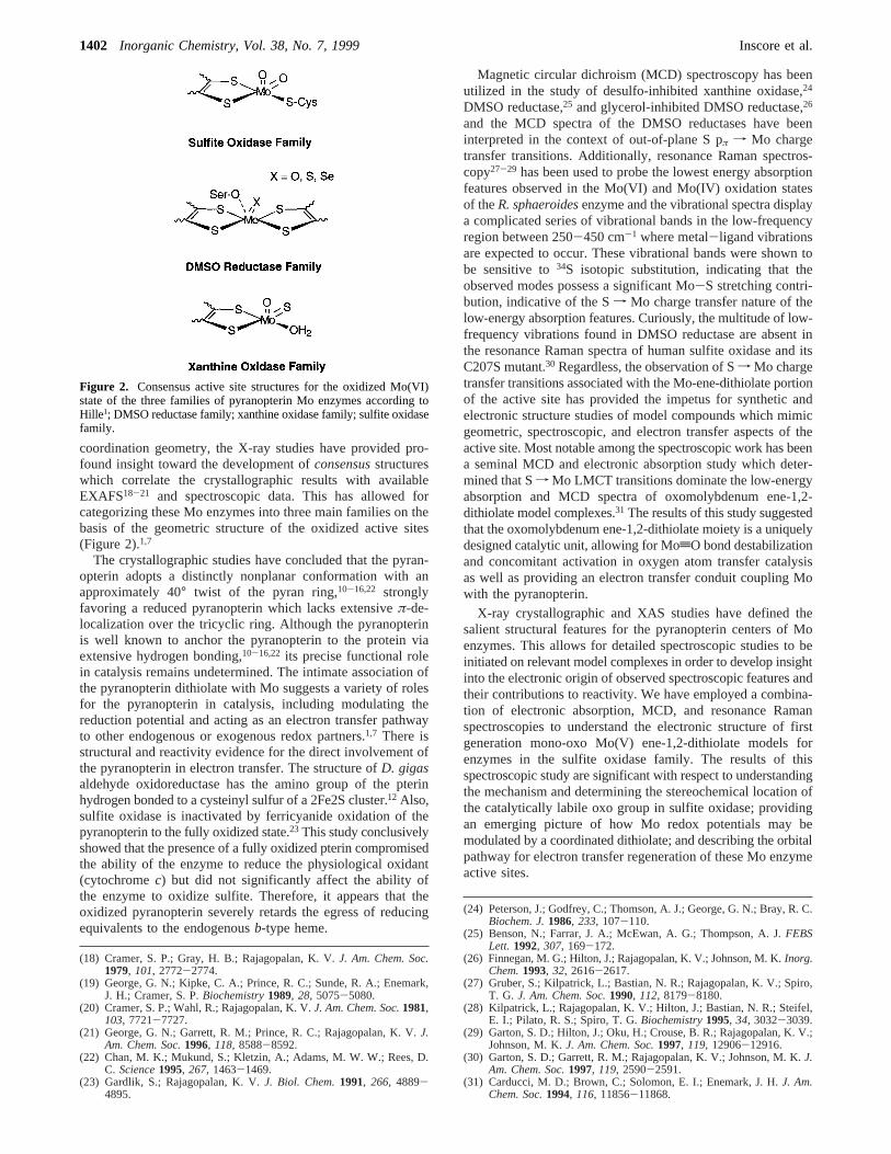
coordination geometry, the X-ray studies have provided pro-found insight toward the development ofconsensusstructureswhich correlate the crystallographic results with availableEXAFS18-21 and spectroscopic data. This has allowed forcategorizing these Mo enzymes into three main families on thebasis of the geometric structure of the oxidized active sites(Figure 2).1,7
The crystallographic studies have concluded that the pyran-opterin adopts a distinctly nonplanar conformation with anapproximately 40° twist of the pyran ring,10-16,22 stronglyfavoring a reduced pyranopterin which lacks extensiveπ-de-localization over the tricyclic ring. Although the pyranopterinis well known to anchor the pyranopterin to the protein viaextensive hydrogen bonding,10-16,22 its precise functional rolein catalysis remains undetermined. The intimate association ofthe pyranopterin dithiolate with Mo suggests a variety of rolesfor the pyranopterin in catalysis, including modulating thereduction potential and acting as an electron transfer pathwayto other endogenous or exogenous redox partners.1,7 There isstructural and reactivity evidence for the direct involvement ofthe pyranopterin in electron transfer. The structure ofD. gigasaldehyde oxidoreductase has the amino group of the pterinhydrogen bonded to a cysteinyl sulfur of a 2Fe2S cluster.12 Also,sulfite oxidase is inactivated by ferricyanide oxidation of thepyranopterin to the fully oxidized state.23 This study conclusivelyshowed that the presence of a fully oxidized pterin compromisedthe ability of the enzyme to reduce the physiological oxidant(cytochromec) but did not significantly affect the ability ofthe enzyme to oxidize sulfite. Therefore, it appears that theoxidized pyranopterin severely retards the egress of reducingequivalents to the endogenousb-type heme.
Magnetic circular dichroism (MCD) spectroscopy has beenutilized in the study of desulfo-inhibited xanthine oxidase,24
DMSO reductase,25 and glycerol-inhibited DMSO reductase,26
and the MCD spectra of the DMSO reductases have beeninterpreted in the context of out-of-plane S pπ f Mo chargetransfer transitions. Additionally, resonance Raman spectros-copy27-29 has been used to probe the lowest energy absorptionfeatures observed in the Mo(VI) and Mo(IV) oxidation statesof theR. sphaeroidesenzyme and the vibrational spectra displaya complicated series of vibrational bands in the low-frequencyregion between 250-450 cm-1 where metal-ligand vibrationsare expected to occur. These vibrational bands were shown tobe sensitive to34S isotopic substitution, indicating that theobserved modes possess a significant Mo-S stretching contri-bution, indicative of the Sf Mo charge transfer nature of thelow-energy absorption features. Curiously, the multitude of low-frequency vibrations found in DMSO reductase are absent inthe resonance Raman spectra of human sulfite oxidase and itsC207S mutant.30 Regardless, the observation of Sf Mo chargetransfer transitions associated with the Mo-ene-dithiolate portionof the active site has provided the impetus for synthetic andelectronic structure studies of model compounds which mimicgeometric, spectroscopic, and electron transfer aspects of theactive site. Most notable among the spectroscopic work has beena seminal MCD and electronic absorption study which deter-mined that Sf Mo LMCT transitions dominate the low-energyabsorption and MCD spectra of oxomolybdenum ene-1,2-dithiolate model complexes.31 The results of this study suggestedthat the oxomolybdenum ene-1,2-dithiolate moiety is a uniquelydesigned catalytic unit, allowing for MotO bond destabilizationand concomitant activation in oxygen atom transfer catalysisas well as providing an electron transfer conduit coupling Mowith the pyranopterin.
X-ray crystallographic and XAS studies have defined thesalient structural features for the pyranopterin centers of Moenzymes. This allows for detailed spectroscopic studies to beinitiated on relevant model complexes in order to develop insightinto the electronic origin of observed spectroscopic features andtheir contributions to reactivity. We have employed a combina-tion of electronic absorption, MCD, and resonance Ramanspectroscopies to understand the electronic structure of firstgeneration mono-oxo Mo(V) ene-1,2-dithiolate models forenzymes in the sulfite oxidase family. The results of thisspectroscopic study are significant with respect to understandingthe mechanism and determining the stereochemical location ofthe catalytically labile oxo group in sulfite oxidase; providingan emerging picture of how Mo redox potentials may bemodulated by a coordinated dithiolate; and describing the orbitalpathway for electron transfer regeneration of these Mo enzymeactive sites.
(18) Cramer, S. P.; Gray, H. B.; Rajagopalan, K. V.J. Am. Chem. Soc.1979, 101, 2772-2774.
(19) George, G. N.; Kipke, C. A.; Prince, R. C.; Sunde, R. A.; Enemark,J. H.; Cramer, S. P.Biochemistry1989, 28, 5075-5080.
(20) Cramer, S. P.; Wahl, R.; Rajagopalan, K. V.J. Am. Chem. Soc.1981,103, 7721-7727.
(21) George, G. N.; Garrett, R. M.; Prince, R. C.; Rajagopalan, K. V.J.Am. Chem. Soc.1996, 118, 8588-8592.
(22) Chan, M. K.; Mukund, S.; Kletzin, A.; Adams, M. W. W.; Rees, D.C. Science1995, 267, 1463-1469.
(23) Gardlik, S.; Rajagopalan, K. V.J. Biol. Chem.1991, 266, 4889-4895.
(24) Peterson, J.; Godfrey, C.; Thomson, A. J.; George, G. N.; Bray, R. C.Biochem. J.1986, 233, 107-110.
(25) Benson, N.; Farrar, J. A.; McEwan, A. G.; Thompson, A. J.FEBSLett. 1992, 307, 169-172.
(26) Finnegan, M. G.; Hilton, J.; Rajagopalan, K. V.; Johnson, M. K.Inorg.Chem.1993, 32, 2616-2617.
(27) Gruber, S.; Kilpatrick, L.; Bastian, N. R.; Rajagopalan, K. V.; Spiro,T. G. J. Am. Chem. Soc.1990, 112, 8179-8180.
(28) Kilpatrick, L.; Rajagopalan, K. V.; Hilton, J.; Bastian, N. R.; Steifel,E. I.; Pilato, R. S.; Spiro, T. G.Biochemistry1995, 34, 3032-3039.
(29) Garton, S. D.; Hilton, J.; Oku, H.; Crouse, B. R.; Rajagopalan, K. V.;Johnson, M. K.J. Am. Chem. Soc.1997, 119, 12906-12916.
(30) Garton, S. D.; Garrett, R. M.; Rajagopalan, K. V.; Johnson, M. K.J.Am. Chem. Soc.1997, 119, 2590-2591.
(31) Carducci, M. D.; Brown, C.; Solomon, E. I.; Enemark, J. H.J. Am.Chem. Soc.1994, 116, 11856-11868.
Figure 2. Consensus active site structures for the oxidized Mo(VI)state of the three families of pyranopterin Mo enzymes according toHille1; DMSO reductase family; xanthine oxidase family; sulfite oxidasefamily.
1402 Inorganic Chemistry, Vol. 38, No. 7, 1999 Inscore et al.

Experimental Section
General. Unless otherwise noted, all reactions were carried out inan inert atmosphere of nitrogen using Schlenk techniques. All solventswere dried by distillation, and deoxygenated prior to use. Purificationof solvents was accomplished using the following methodologies:pyridine and triethylamine from potassium hydroxide; toluene fromsodium benzophenone. Other solvents were used without furtherpurification. The compounds LMoOCl2,32 LMoVO(tdt),32 LMoVO-(bdt),33,34 and LMoVO(edt)32 were prepared as previously described.
Abbreviations. L, hydrotris(3,5-dimethyl-1-pyrazolyl)borate; bdt,1,2-benzenedithiolate; tdt, 3,4-toluenedithiolate; edt, 1,2-ethanedithio-late; H2qdt, quinoxaline-2,3-dithiol; qdt, 2,3-dithioquinoxaline).
Preparation of LMoO(qdt). The reagents 2,3-dihydroxyquinoxalineand phosphorus pentasulfide were purchased from Aldrich ChemicalCo. Quinoxaline-2,3-dithiol was prepared by a modified version ofMorrison.35,36To a dry toluene solution of LMoOCl2 (0.5 g, 1.05 mmol)was added a toluene solution containing 0.41 g (2.1 mmol) of H2qdtand 150µL (2.1 mmol) of triethylamine, dropwise by cannula at 70°C. This solution was allowed to react for approximately 5 h, and duringthis time the color of the solution changed from lime green to darkred. The resulting dark red solution was filtered and concentrated atreduced pressure to give a dark red powder, which was subsequentlyredissolved in a minimum amount of toluene and chromatographed onsilica gel. The compound eluted in a binary mixture of toluene/1,2-dichloroethane (9:1) as a red band. Yield) 15%. Anal. Calcd forC23H26N8OS2BMo: C, 45.93; H, 4.36. Found: C, 45.04; H, 4.33. IR(KBr, cm-1): ν(ModO) 940, ν(B-H) 2551. MS (FAB):m/z 602.1(parent ion), 507 (parent- 3,5-dimethylpyrazole), 410 (parent-dithiolate).
Physical Characterization. Elemental analysis was performed atThe University of New Mexico using a Perkin-Elmer 2400 CHNelemental analyzer equipped with a P-E AD-6 Autobalance. MassSpectra were collected at The Nebraska Center for Mass Spectrometryin the Department of Chemistry at the University of NebraskasLincoln.
Electronic Absorption Spectroscopy.Mull and solution electronicabsorption spectra were collected on a double beam Hitachi U-3501UV-vis-NIR spectrophotometer capable of scanning a wavelengthregion between 185 and 3200 nm. All absorption spectra were collectedat 2.0 nm resolution in a single-beam configuration. The instrumentwas calibrated with reference to the 656.10 nm deuterium line.Immediately following acquisition of the sample spectra, backgroundspectra were collected to correct for residual absorption due to thesolvent or mulling agent and to correct for light scattering effects.Solution samples were prepared by dissolving the compounds indegassed dichloroethane. The electronic absorption spectra weresubsequently collected in 1 cm pathlength Helma quartz cells (black-masked Suprasil II, equipped with a Teflon stopper). Mull sampleswere prepared by grinding the solid sample into a fine powder beforedispersing it into poly(dimethylsiloxane). The prepared mull wassubsequently placed between two 1 mm thick Infrasil quartz discs(ESCO) and secured in a custom designed sample holder. A JanisSTVP-100 continuous flow cryostat mounted in a custom designedcradle assembly was used for acquisition of the low-temperature (∼5K) spectra. The sample temperature was continuously monitored witha Lakeshore silicon-diode (PT-470) and regulated by a combination ofhelium flow and dual heater assemblies. Gaussian resolution of spectralbands and corrections for light scattering were accomplished withKaleidaGraph and programs incorporated within the Hitachi versionof the Grams software package.
Magnetic Circular Dichroism Spectroscopy. Low-temperatureMCD data were collected on a system consisting of a Jasco J600 CDspectropolarimeter employing Hamamatsu photomultiplier tubes of
either S-1 or S-20 response, an Oxford Instruments SM4000-7Tsuperconducting magneto-optical cryostat (0-7 Tesla and 1.4-300 K),and an Oxford Instruments ITC503 temperature controller. Thespectrometer was calibrated for CD intensity and wavelength usingcamphorsulfonic acid and a Nd-doped reference glass sample (SchottGlass). Solid-state MCD spectra were obtained by dispersing finelyground samples in poly(dimethylsiloxane) and compressing the suspen-sion between two 1 mm thick Infrasil quartz discs (ESCO). Depolar-ization of the incident radiation was checked by comparing thedifference in CD intensity of a standard Ni (+)-tartrate solutionpositioned before and then after the sample. Samples which depolarizedthe light by<5% were deemed suitable. The MCD spectra in the 250-800 nm range were obtained at 2.0 nm resolution, and data between400 and 1050 nm were collected at a fixed slit width of 150µ. AllMCD spectra were collected in an applied magnetic field of 7 Tesla.
Vibrational and Resonance Raman Spectroscopy.Infrared spectrawere recorded on a BOMEM MB-100 FT-IR spectrometer as pressedKBr disks. The infrared spectra were utilized to monitor the purity ofthe compounds, as indicated by the absence of the 962 cm-1 MotOstretch associated with the LMoOCl2 precursor complex.32
Resonance Raman spectra were collected in a 135° backscatteringgeometry. A Coherent Innova 70-5 (5W) Ar+ ion laser was the photonsource (457.9-528.7 nm, 9 discrete lines) for inducing Ramanscattering. The scattered radiation was dispersed onto a liquid N2 cooled1′′ Spex Spectrum One CCD detector using a Spex 1877E triple gratingmonochromator equipped with 600, 1200, and 1800 gr/mm holographicgratings at the spectrographic stage. The laser power at the sample waskept between 40 and 100 mW in order to prevent possible photo- andthermal degradation of the sample. Solid samples were prepared asfinely ground powders and dispersed in a NaCl(s) matrix with Na2SO4
added as an internal standard. These samples were subsequently sealedin an NMR tube and Raman spectra were obtained by spinning thesample in a modified NMR sample holder/spinner. The samples weremaintained at∼140 ( 10 K by the use of a custom designed cold N2
gas flow system. The sample temperature was periodically monitoredwith a Lakeshore silicon diode (PT-470) enclosed in a separate NMRtube. The construction of resonance Raman profiles was accomplishedby comparing the integrated intensity of a Raman band at a givenexcitation wavelength relative to that of the 992.4 cm-1 band of Na2-SO4. All data were scan averaged, and any individual data set withvibrational bands compromised by cosmic events was discarded.
Solution Raman spectra were obtained in degassed benzene and spunin a sealed NMR tube at room temperature. Depolarization ratios wereobtained by placing a rotatable polarizer before the polarizationscrambler and monochromator entrance slit. Relative Raman intensities(perpendicular and parallel to incident radiation) for a given Ramanband were measured relative to the 992 cm-1 band of benzene.
Ab Initio Calculations. Ab initio calculations were performed usingthe Gaussian 94 suite of programs.37 A 6-31G** basis set was employedin calculating the energies and wavefunctions for the model 1,2-ene-dithiolate (-SCHdCHS-).
Results
Solution Electronic Absorption Spectra.Figure 3 depictsthe room temperature electronic absorption spectrum of LMoO-(bdt) between 6000 and 35 000 cm-1 in dichloroethane. Thespectrum is very similar to that previously reported for LMoO-(tdt).31 However, the transitions observed for LMoO(tdt) aregenerally shifted to slightly lower energies relative to thecorresponding bands in LMoO(bdt). The low-energy region ofthe spectrum consists of three distinct spectral features (bands1, 2, and4) below∼20 000 cm-1. We have found these bandsto be characteristic of LMoO(S-S) compounds, where S-S is adithiolene or dithiolate ligand which forms a five-memberedchelate ring with Mo. Band3 is very weak, and only discerniblein the low-temperature MCD spectra (Vide infra). The transitionenergies and molar extinction coefficients for four LMoO(S-S)
(32) Cleland, W. E., Jr.; Barnhart, K. M.; Yamanouchi, K.; Collison, D.;Mabbs, F. E.; Ortega, R. B.; Enemark, J. H.Inorg. Chem.1987, 26,1017-1025.
(33) Dhawan, I. K.; Enemark, J. H.Inorg. Chem.1996, 35, 4873-4882.(34) Dhawan, I. K.; Pacheco, A.; Enemark, J. H.J. Am. Chem. Soc.1994,
116, 7911-7912.(35) Morrison, D. C.; Furst, A.J. Org. Chem.1956, 21, 470-471.(36) Helton, M. E.; Kirk, M. L. Submitted for publication. (37) Gaussian Incorporated, Pittsburgh, PA.
Unique Bonding in Oxo-Molybdenum Dithiolates Inorganic Chemistry, Vol. 38, No. 7, 19991403

complexes are presented in Table 1 for comparative purposes.Of particular interest is band4, which is the first absorptionfeature possessing appreciable intensity characteristic of a chargetransfer transition.
Solid-State Electronic Absorption and MCD Spectra.The5 K mull MCD/absorption overlay of LMoO(bdt) is shown inFigure 4. The electronic absorption spectrum exhibits fivedistinct features in the solid state with weak to significantintensity. The 21 500 cm-1 band is observed as a reproducibleshoulder in the mull absorption of LMoO(bdt) but is conspicu-ously absent in the corresponding absorption spectrum ofLMoO(tdt). The general similarity of the solution and mullabsorption spectra for LMoO(bdt) indicate that only minorstructural changes accompany solvation. This is true for all ofthe LMoO(S-S) compounds listed in Table 1.
The MCD spectrum of LMoO(bdt) is composed of bothC-terms and pseudo A-terms.38 MCD C-terms possess absorptivebandshapes with intensity maxima at the same energy ascorresponding absorption features, while pseudo A-terms possess
derivative-shaped dispersions which possess zero intensity atan energy corresponding to an absorption maximum. Therelationship between observed MCD and electronic absorptionbands in LMoO(bdt) is given in Table 2.
Figure 5 compares the 5 K/7 T MCD mull spectra of LMoO-(bdt) and LMoO(tdt) in the spectral region between 10 000 and40 000 cm-1. The MCD spectra are seen to be quite similar,and this band pattern is characteristic of LMoO(S-S) complexeswhere the MotO bond is oriented cis to a single dithiolateligand which forms a five-membered chelate ring with Mo.31
However, noticeable differences in MCD sign exist for thesecompounds in the 14 000-17 000 cm-1 range, where nodiscernible maxima occur in the absorption spectra (see Figure3). Close inspection of Figure 5 reveals the reason the 21 500cm-1 transition in LMoO(bdt) is not resolved in the MCD orabsorption spectra of LMoO(tdt). The positive C-term at 19 300cm-1 and the low-energy negatively signed component of the24 300 cm-1 positive pseudo A-term observed in LMoO(bdt)are energetically compressed in the MCD spectrum of LMoO-(tdt), effectively masking the 21 500 cm-1 spectral feature. TheMCD spectrum of LMoO(qdt) shows this transition as a clearlyresolved positive pseudo A-term.36
Vibrational Spectra. The IR data for LMoO(bdt) and LMoO-(tdt) display intense peaks at 932 and 926 cm-1, respectively.Strong IR bands in the 910-965 cm-1 range have been reportedfor a variety of LMoOX2 complexes,39-43 and this band has
(38) Piepho, S. B.; Schatz, P. N.Group Theory in Spectroscopy withApplications to Magnetic Circular Dichroism; Wiley-Interscience:New York, 1983.
Table 1. Summary of Electronic Absorption Data for LMoO(S-S) Complexes in Dichloroethane
Emax, cm-1 (ε, M-1 cm-1)
LMoO(bdt) LMoO(tdt) LMoO(qdt)36 LMoO(edt)
band 1 9 100 (360) 9 100 (490) 11 300 (170) 11 800 (160)band 2 13 100 (270) 13 000 (270) 13 700 (130) 15 500 (220)band 4 19 400 (sh,1220) 19 600 (sh,1320) 19 100 (1050) 20 000 (sh, 570)
Figure 3. Gaussian-resolved 293 K electronic absorption spectrum ofLMoO(bdt) in dichloroethane (2.45× 10⁻⁴ M). The dashed linesrepresent the individual Gaussians used in the fit.
Figure 4. 5 K electronic absorption (heavy line) and 4.86 K MCD(light line) spectra of LMoO(bdt) dispersed in poly(dimethylsiloxane).
Table 2. Calculated Oscillator Strengths of LMoO(bdt) and theRelationship between MCD and Electronic Absorption Bands
bandno.
E(soln)max
(cm-1)oscillatorstrength
E(mull)max
(cm-1)E(MCD)
max
(cm-1)aMCDterma
1 9 100 5.6× 10⁻³ 8 500 - - - - - - - -2 13 100 3.3× 10⁻³ 12 700 12 400 -C3 15 800 - - - - - - - - 15 700 +C4 19 400 1.6× 10⁻² 19 200 19 300 +C5 22 100 1.7× 10⁻² 21 500 21 000 +pseudo A6 25 100 9.2× 10⁻² 24 600 24 300 +pseudo A
a A positive pseudo A-term is a derivative shaped MCD feature withthe positive component at higher energy.Emax represents the point atwhich the pseudo A-term changes sign.
Figure 5. 4.86 K MCD spectra of LMoO(bdt) (solid line) and LMoO-(tdt) (dotted line) dispersed in poly(dimethylsiloxane). Note the overallsimilarity of the spectral features.
1404 Inorganic Chemistry, Vol. 38, No. 7, 1999 Inscore et al.
been assigned as the MotO stretching vibration. Vibrationalstudies on related compounds possessing the{MoVtO}3+ unitalso reveal the presence of a band in this region assignable asthe MotO stretch.44-47 The IR spectra are also useful fordetecting very small quantities of LMoOCl2 precursor complexthat may be present in the sample as a contaminant. No 961cm-1 MotO stretch characteristic of LMoOCl2 was observedin the IR spectra of the LMoO(S-S) complexes used in thisstudy.32
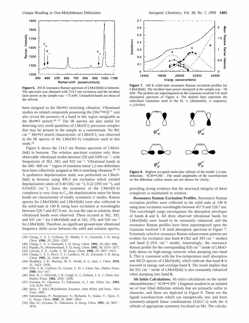
Figure 6 shows the 514.5 nm Raman spectrum of LMoO-(bdt) in benzene. The solution spectrum contains only threeobservable vibrational modes between 250 and 1000 cm-1, withfrequencies of 362, 393, and 932 cm-1. Vibrational bands inthe 300-400 cm-1 region of transition metal 1,2-ene-dithiolateshave been collectively assigned as Mo-S stretching vibrations.48-50
A qualitative depolarization study was performed on LMoO-(bdt) in benzene using 496.5 nm excitation which yieldeddepolarization ratios of 0.40 (362 cm-1), 0.22 (393 cm-1), and0.01(932 cm-1). Since the symmetry of the LMoO(S-S)complexes is very close toCs, the depolarization ratios for thesebands are characteristic of totally symmetric a′ modes. Ramanspectra for LMoO(bdt) and LMoO(tdt) were also collected inthe solid-state at 140 K using laser excitation at wavelengthsbetween 528.7 and 457.9 nm. As was the case in solution, threevibrational bands were observed. These occured at 362, 393,and 931 cm-1 for LMoO(bdt) and at 342, 376, and 926 cm-1
for LMoO(tdt). Therefore, no significant ground-state vibrationalfrequency shifts occur between the solid and solution spectra,
providing strong evidence that the structural integrity of thesecomplexes is maintained in solution.
Resonance Raman Excitation Profiles.Resonance Ramanexcitation profiles were collected in the solid state at 140 Kusing laser excitation wavelengths between 457.9 and 528.7 nm.This wavelength range encompasses the absorption envelopesof bands4 and 5. All three observed vibrational bands forLMoO(bdt) were found to be resonantly enhanced, and theresonance Raman profiles have been superimposed upon theGaussian resolved 5 K mull absorption spectrum in Figure 7.ExtremelyselectiVe resonance Raman enhancement patterns areevident for excitation into band4 (362 and 393 cm-1 modes)and band5 (931 cm-1 mode). Interestingly, the resonanceRaman profile for the corresponding 926 cm-1 mode of LMoO-(tdt) shows no high-energy turnover when pumping into band5. This is consistent with the low-temperature mull absorptionand MCD spectra of LMoO(tdt), which indicate that band6 islowered in energy and overlaps band5. The result implies thatthe 931 cm-1 mode of LMoO(bdt) is also resonantly enhancedwhen pumping into band6.
Ab Initio Calculations. Ab initio calculations on the modelethenedithiolate (-SCHdCHS-) fragment resulted in an isolatedset of four filled dithiolate orbitals that are primarily sulfur incharacter, and these are depicted in Figure 8. These are theligand wavefunctions which can energetically mix and formsymmetry-adapted linear combinations (SALC’s) with the dorbitals of appropriate symmetry localized on Mo. The calcula-
(39) Chang, C. S. J.; Collison, D.; Mabbs, F. E.; Enemark, J. H.Inorg.Chem.1990, 29, 2261-2267.
(40) Chang, C. S. J.; Enemark, J. H.Inorg. Chem.1991, 30, 683-688.(41) Nipales, N.; Westmoreland, T. D.Inorg. Chem.1995, 34, 3374-3377.(42) Lincoln, S. E.; Loehr, T. M.Inorg. Chem.1990, 29, 1907-1915.(43) Chang, C. S. J.; Pecci, T. J.; Carducci, M. D.; Enemark, J. H.Inorg.
Chem.1993, 32, 4106-4110.(44) Bradbury, J. R.; Mackay, M. F.; Wedd, A. G.Aust. J. Chem.1978,
31, 2423-2430.(45) Ellis, S. R.; Collison, D.; Garner, C. D.J. Chem. Soc. Dalton Trans.
1989, 413-417.(46) Burt, R. J.; Dilworth, J. R.; Leigh, G. J.; Zubieta, J. A.J. Chem. Soc.
Dalton Trans.1982, 2295-2298.(47) Ueyama, N.; Okamura, T.; Nakamura, A.J. Am. Chem. Soc.1992,
114, 8129-8137.(48) Spiro, T. (Ed.)Molybdenum Enzymes; John Wiley and Sons: New
York, 1985.(49) Subramanian, P.; Burgmayer, S.; Richards, S.; Szalai, V.; Spiro, T.
G. Inorg. Chem.1990, 29, 3849-3853.(50) Oku, H.; Ueyama, N.; Nakamura, A.Inorg. Chem.1995, 34, 3667-
3676.
Figure 6. 293 K resonance Raman spectrum of LMoO(bdt) in benzene.The spectrum was obtained with 514.5 nm excitation and the incidentlaser power at the sample was∼75 mW. Unmarked bands are those ofthe solvent.
Figure 7. 140 K solid-state resonance Raman excitation profiles forLMoO(bdt). The incident laser power measured at the sample was∼50mW. The profiles are superimposed on the Gaussian-resolved 5 K mullabsorption spectrum of Figure 4. The dashed lines represent theindividual Gaussians used in the fit.ν1 (diamonds),ν3 (squares),ν4 (circles).
Figure 8. Highest occupied molecular orbitals of the model 1,2-ene-dithiolate, -SCHdCHS-. The small amplitudes of the wavefunctionon the dithiolate carbon atoms are not shown for clarity.
Unique Bonding in Oxo-Molybdenum Dithiolates Inorganic Chemistry, Vol. 38, No. 7, 19991405
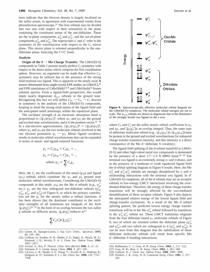
tions indicate that the electron density is largely localized onthe sulfur atoms, in agreement with experimental results fromphotoelectron spectroscopy.51 The four orbitals may be dividedinto two sets with respect to their orientation to the planecontaining the constituent atoms of the ene-dithiolate. Theseare the in-plane components,æip
a′ andæipa′′, and the out-of-plane
componentsæopa′ andæop
a′′. The superscripts a′ and a′′ refer to thesymmetry of the wavefunction with respect to theCs mirrorplane. This mirror plane is oriented perpendicular to the ene-dithiolate plane, bisecting the CdC bond.
AnalysisOrigin of the S f Mo Charge Transfer. The LMoO(S-S)
compounds in Table 1 possess nearly perfectCs symmetry withrespect to the donor atoms which comprise the first coordinationsphere. However, an argument can be made that effectiveC4Vsymmetry may be utilized due to the presence of the strongfield terminal oxo ligand. This is apparent in the nearly axialAtensors determined from single-crystal EPR studies on LMoOCl2
52
and EPR simulations of LMoO(bdt)33,34and LMoO(edt)32 frozensolution spectra. From a ligand-field perspective, this wouldimply nearly degenerate dxz,yz orbitals in the ground state.Recognizing this fact we will utilizeC4V f C2V f Cs descentin symmetry in the analysis of the LMoO(S-S) compounds,keeping in mind the strong axial nature of the ligand field andthe anticipated small anisotropy within the equatorial plane.
The oscillator strength of an electronic absorption band isproportional to|⟨ψG|r |ψE⟩|2, whereψG andψE are the groundand excited state wavefunctions, andr is the position vector.53-55
In a one-electron approximation,|⟨ψG|r |ψE⟩|2 ) |⟨ψa|r |ψb⟩|2,whereψa andψb are the two molecular orbitals involved in theone electron promotionψa f ψb. Metal-ligand covalencyresults in molecular orbital wavefunctions that can be expandedin terms of metal- and ligand-centered functions:
Here, theCi are the coefficients of the metal (æM) and ligand(æL) orbitals which constitute theψa and ψb ground statemolecular orbital wavefunctions. Considering the LMoO(S-S)compounds in this study,æM are the Mo d orbitals (e.g.,æxy
a′
etc.); æL are the four orthogonal ene-dithiolate orbitals (æipa′,
æopa′ , æip
a′′, andæopa′′) of Figure 8, to which the primary contribu-
tors are theøn for the atomic sulfur p orbital functions. Ithas been shown that the dominant contributor to the oscil-lator strengths of all transitions are integrals of the form⟨øn|r |ø′n⟩.53-55 In the limit of no overlap between the two sulfurp orbitals on different atoms,⟨øn|r |ø′n⟩ reduces to53
whereCn andC′n are the sulfur atomic orbital coefficients inψa
andψb, and⟨øn|ø′n⟩ is an overlap integral. Thus, the same typeof dithiolate molecular orbital (e.g.,⟨æip|æip⟩ or ⟨æop|æop⟩) mustbe present in the ground and excited wavefunctions for enhancedcharge transfer transition intensity, and this intensity is a directconsequence of the Mo d-dithiolate S covalency.
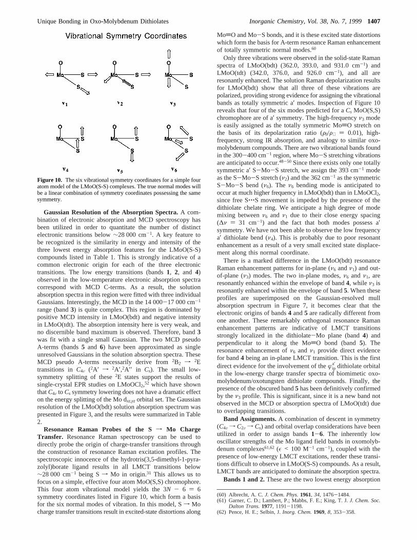
The ligand field splitting of the d-orbital manifold in LMoO-(S-S) and other high-valent metal oxo compounds is dominatedby the presence of a short 1.7-1.9 Å MtO bond.56-59 Theterminal oxo ligand is an extremely strongσ- andπ-donor, andin the presence of a moderate to weak equatorial ligand fieldthe d-orbital splitting diagram in Figure 9 results. Here, the Moψz2
a′ and ψxza′′
,ψyza′ orbitals are strongly destabilized byσ and π
antibonding interactions with the terminal oxo ligand. In d1
LMoO(S-S) complexes, all of the d orbitals may act as acceptororbitals in low-energy LMCT interactions involving the coor-dinated dithiolate. Therefore, the energy of these charge transfertransitions will be strongly affected by the oxo-mediateddestabilization of these acceptor orbitals. Figure 9 also depictsthe anticipated relative energy of the lowest ligand field andcharge-transfer excitations. As a result of the Mo d orbitalsplitting pattern, the predicted lowest energy charge transfertransitions will be to the Moψxy
a′ orbital followed by transitionsto the ψxz
a′′,ψyz
a′ orbital set. These LMCT transitions originatefrom the four dithiolate basedæL molecular orbitals of Figure8, two of which are oriented within the dithiolate plane (æip
a′
andæipa′′) and two which are orthogonal to it (æop
a′ andæopa′′). It
can be seen from this diagram that the stabilization of thesedithiolate molecular orbitals will result from specific Mod-dithiolate S bonding interactions.
(51) Gleiter, R.; Spanget-Larsen, J.Top. Curr. Chem.: Spectrosc.1979,86, 139-196.
(52) Collison, D.; Eardley, D. R.; Mabbs, F. E.; Rigby, K.; Bruck, M. A.;Enemark, J. H.; Wexler, P. A.J. Chem. Soc. Dalton Trans.1994,1003-1011.
(53) Avoird, A.; Ros, P.Theoret. Chim. Acta (Berlin)1966, 4, 13-21.(54) Solomon, E. I.Comm. Inorg. Chem.1984, 3, 227-320.(55) LaCroix, L. B.; Shadle, S. E.; Wang, Y.; Averill, B. A.; Hedman, B.;
Hodgson, K. O.; Solomon, E. I.J. Am. Chem. Soc.1996, 118, 7755-7768.
(56) Ballhausen, C. J.; Gray, H. B.Inorg. Chem.1962, 1, 111-122.(57) Gray, H. B.; Hare, C. R.Inorg. Chem.1962, 1, 363-368.(58) Collison, D.J. Chem. Soc. Dalton Trans.1990, 2999-3006.(59) Winkler, J. R.; Gray, H. B.Comments Inorg. Chem.1981, 1, 257-
263.
ψa ) C1æM + C2æL + ...
ψb ) C′1æM + C′2æL + ...
whereæL ) ∑n
Cnøn
∑n
CnC′nr ⟨øn|ø′n⟩
Figure 9. Spectroscopically effective molecular orbital diagram forthe LMoO(S-S) complexes. The molecular orbital energies are not toscale. Theψxz,yz orbitals are shown as degenerate due to the dominanceof the strongly bound oxo ligand on thez axis.
1406 Inorganic Chemistry, Vol. 38, No. 7, 1999 Inscore et al.
Gaussian Resolution of the Absorption Spectra.A com-bination of electronic absorption and MCD spectroscopy hasbeen utilized in order to quantitate the number of distinctelectronic transitions below∼28 000 cm-1. A key feature tobe recognized is the similarity in energy and intensity of thethree lowest energy absorption features for the LMoO(S-S)compounds listed in Table 1. This is strongly indicative of acommon electronic origin for each of the three electronictransitions. The low energy transitions (bands1, 2, and 4)observed in the low-temperature electronic absorption spectracorrespond with MCD C-terms. As a result, the solutionabsorption spectra in this region were fitted with three individualGaussians. Interestingly, the MCD in the 14 000-17 000 cm-1
range (band3) is quite complex. This region is dominated bypositive MCD intensity in LMoO(bdt) and negative intensityin LMoO(tdt). The absorption intensity here is very weak, andno discernible band maximum is observed. Therefore, band3was fit with a single small Gaussian. The two MCD pseudoA-terms (bands5 and 6) have been approximated as singleunresolved Gaussians in the solution absorption spectra. TheseMCD pseudo A-terms necessarily derive from2B2 f 2Etransitions in C4V (2A′ f 2A′,2A′′ in Cs). The small low-symmetry splitting of these2E states support the results ofsingle-crystal EPR studies on LMoOCl2,52 which have shownthatC4V to Cs symmetry lowering does not have a dramatic effecton the energy splitting of the Mo dxz,yz orbital set. The Gaussianresolution of the LMoO(bdt) solution absorption spectrum waspresented in Figure 3, and the results were summarized in Table2.
Resonance Raman Probes of the Sf Mo ChargeTransfer. Resonance Raman spectroscopy can be used todirectly probe the origin of charge-transfer transitions throughthe construction of resonance Raman excitation profiles. Thespectroscopic innocence of the hydrotris(3,5-dimethyl-1-pyra-zolyl)borate ligand results in all LMCT transitions below∼28 000 cm-1 being Sf Mo in origin.31 This allows us tofocus on a simple, effective four atom MoO(S,S) chromophore.This four atom vibrational model yields the 3N - 6 ) 6symmetry coordinates listed in Figure 10, which form a basisfor the six normal modes of vibration. In this model, Sf Mocharge transfer transitions result in excited-state distortions along
MotO and Mo-S bonds, and it is these excited state distortionswhich form the basis for A-term resonance Raman enhancementof totally symmetric normal modes.60
Only three vibrations were observed in the solid-state Ramanspectra of LMoO(bdt) (362.0, 393.0, and 931.0 cm-1) andLMoO(tdt) (342.0, 376.0, and 926.0 cm-1), and all areresonantly enhanced. The solution Raman depolarization resultsfor LMoO(bdt) show that all three of these vibrations arepolarized, providing strong evidence for assigning the vibrationalbands as totally symmetric a′ modes. Inspection of Figure 10reveals that four of the six modes predicted for aCs MoO(S,S)chromophore are of a′ symmetry. The high-frequencyν3 modeis easily assigned as the totally symmetric MotO stretch onthe basis of its depolarization ratio (F|/F⊥ ) 0.01), high-frequency, strong IR absorption, and analogy to similar oxo-molybdenum compounds. There are two vibrational bands foundin the 300-400 cm-1 region, where Mo-S stretching vibrationsare anticipated to occur.48-50 Since there exists only one totallysymmetric a′ S-Mo-S stretch, we assign the 393 cm-1 modeas the S-Mo-S stretch (ν2) and the 362 cm-1 as the symmetricS-Mo-S bend (ν6). The ν6 bending mode is anticipated tooccur at much higher frequency in LMoO(bdt) than in LMoOCl2,since free S‚‚‚S movement is impeded by the presence of thedithiolate chelate ring. We anticipate a high degree of modemixing betweenν6 and ν1 due to their close energy spacing(∆ν ) 31 cm-1) and the fact that both modes possess a′symmetry. We have not been able to observe the low frequencya′ dithiolate bend (ν4). This is probably due to poor resonantenhancement as a result of a very small excited state displace-ment along this normal coordinate.
There is a marked difference in the LMoO(bdt) resonanceRaman enhancement patterns for in-plane (ν6 andν1) and out-of-plane (ν3) modes. The two in-plane modes,ν6 and ν1, areresonantly enhanced within the envelope of band4, while ν3 isresonantly enhanced within the envelope of band5. When theseprofiles are superimposed on the Gaussian-resolved mullabsorption spectrum in Figure 7, it becomes clear that theelectronic origins of bands4 and5 are radically different fromone another. These remarkably orthogonal resonance Ramanenhancement patterns are indicative of LMCT transitionsstrongly localized in the dithiolate-Mo plane (band4) andperpendicular to it along the MotO bond (band5). Theresonance enhancement ofν6 and ν1 provide direct evidencefor band4 being an in-plane LMCT transition. This is the firstdirect evidence for the involvement of theæip
a′ dithiolate orbitalin the low-energy charge transfer spectra of biomimetic oxo-molybdenum/oxotungsten dithiolate compounds. Finally, thepresence of the obscured band5 has been definitively confirmedby theν3 profile. This is significant, since it is a new band notobserved in the MCD or absorption spectra of LMoO(tdt) dueto overlapping transitions.
Band Assignments.A combination of descent in symmetry(C4V f C2V f Cs) and orbital overlap considerations have beenutilized in order to assign bands1-6. The inherently lowoscillator strengths of the Mo ligand field bands in oxomolyb-denum complexes61,62 (ε < 100 M-1 cm-1), coupled with thepresence of low-energy LMCT excitations, render these transi-tions difficult to observe in LMoO(S-S) compounds. As a result,LMCT bands are anticipated to dominate the absorption spectra.
Bands 1 and 2.These are the two lowest energy absorption
(60) Albrecht, A. C.J. Chem. Phys.1961, 34, 1476-1484.(61) Garner, C. D.; Lambert, P.; Mabbs, F. E.; King, T. J.J. Chem. Soc.
Dalton Trans.1977, 1191-1198.(62) Pence, H. E.; Selbin, J.Inorg. Chem.1969, 8, 353-358.
Figure 10. The six vibrational symmetry coordinates for a simple fouratom model of the LMoO(S-S) complexes. The true normal modes willbe a linear combination of symmetry coordinates possessing the samesymmetry.
Unique Bonding in Oxo-Molybdenum Dithiolates Inorganic Chemistry, Vol. 38, No. 7, 19991407
features observed in all four of the LMoO(S-S) compounds listedin Table 1. Band1 is too low in energy to be assigned as theψxy
a′ f ψxza′′,ψyz
a′ LF transition, and thus it is assigned as a LMCTtransition. The oscillator strengths for these two low energytransitions are relatively low (ε ∼ 133-449 M-1 cm-1),allowing for their assignment asψop
a′′ f ψxya′ or ψop
a′ f ψxya′ .
Band1 corresponds to an single MCD C-term which is positivefor LMoO(tdt),31 LMoO(edt),31 and LMoO(qdt).36 However, thesign of the MCD C-term is variable for band2, being positivefor LMoO(edt) and negative for LMoO(bdt), LMoO(tdt), andLMoO(qdt). Since the sign of the C-term for these transitionsis governed by spin-orbit coupling with other excited states,38
the variation for band2 most reasonably results from differencesin out-of-state spin-orbit coupling among the four LMoO(S-S) complexes of Table 1. The definitive energy ordering of theψop
a′′ f ψxya′ andψop
a′ f ψxya′ transitions is unknown and requires
the explicit evaluation of out-of-state spin-orbit coupling matrixelements. In summary, theseψop f ψxy transitions are antici-pated to occur at low energy and possess relatively lowabsorption intensity due to poor orbital overlap.
Band 3. This band is assigned as theψxya′ f ψxz
a′′,ψyza′ LF
band, which has been observed in the 13 000-16 000 cm-1
region for a variety of oxomolybdenum(V) compounds.63 Forexample, this transition occurs at 16 000 cm-1 (ε ) 17 M-1
cm-1) in [MoOCl4]- (C4V symmetry) and at 14 180 cm-1 (ε )50 M-1 cm-1) for LMoOCl2 (Cs symmetry).31 The transition isvery weak, displaying no discernible absorption feature thatcorresponds with the positive C-term in the MCD spectrum ofLMoO(bdt), and the negative C-term for LMoO(tdt) and LMoO-(qdt). No MCD C-term was observed in this region for LMoO-(edt), presumably due to a combination of weak intensity andmasking by the more intense band4. The ψxy
a′ f ψxza′′,ψyz
a′
transition should manifest itself as a pseudo A-term in the MCDspectrum, but this is not observed experimentally. Again, thisis most likely due to complications arising from low, inequiva-lent oscillator strengths for the individualψxy
a′ f ψxza′′ and ψxy
a′
f ψyza′ components coupled with out-of-state spin-orbit cou-
pling.Band 4. This is the first intense (ε ) 1220 M-1 cm-1 for
LMoO(bdt)) LMCT transition in the LMoO(S-S) complexes andis assigned as the in-planeψip
a′ f ψxya′ LMCT transition based
on the resonance Raman enhancement of bothν6 and ν1 in-plane vibrational modes within the envelope of this band inLMoO(bdt). The intensity of this band directly probes the degreeof æxy
a′ -æipa′ orbital mixing since it is a bondingf antibonding
transition.
Theψxya′ f ψx2-y2
a′′ ligand field transition is also anticipated tooccur in this region. Where this transition has been clearlyobserved as anisolatedband, the intensity has been found tobe very low.61,62 This is partially due to the fact that thistransition is orbitally forbidden in the parentC4V symmetry31 (ε) 9 M-1 cm-1 for [MoOCl4]-), as well as being forbidden inC2V symmetry. The effects of symmetry lowering can be foundin [MoOCl3{P(NMe2)3O}2], which possessesCs symmetry withthree Cl- ligands in the equatorial plane.61 The extinctioncoefficient of theψxy
a′ f ψx2-y2a′′ transition for this compound is
only 20 M-1 cm-1. Therefore, intensity enhancement ofψxya′ f
ψx2-y2a′′ due to symmetry lowering appears to be minor. Only the
ψipa′ f ψx2-y2
a′′ and ψopa′ f ψx2-y2
a′′ LMCT excited states canconfigurationally mix withψxy
a′ f ψx2-y2a′′ to provide an inten-
sity borrowing mechanism for enhanced oscillator strength ofthe ligand field band. However, theψop
a′ f ψx2-y2a′′ transition is
not anticipated to provide an efficient intensity gaining mech-anism for theψxy
a′ f ψx2-y2a′′ LF transition since it is anψop f
ψip transition and is predicted to possess low intensity (see bands1 and 2). Furthermore, intensity borrowing is expected to beminimal due to the high energy of theψop
a′ f ψx2-y2a′′ andψip
a′ f
ψx2-y2a′′ states, which are predicted to occur at energies greater
than 30 000 and 40 000 cm-1, respectively. As a result, theψxya′
f ψx2-y2a′′ LF transition is most likely buried under the envelope
of the intenseψipa′ f ψxy
a′ LMCT transition.Band 5. The band is assigned as theψop
a′′ f ψxza′′,ψyz
a′ LMCTtransition. This is consistent with the resonance Raman en-hancement ofν3, since theψop
a′′ f ψxza′′,ψyz
a′ transition formallyresults in the promotion of an electron from an out-of-planedithiolate molecular orbital to a Mo-based orbital which isstrongly antibonding with respect to the MotO bond. Thistransition is observed as a weak shoulder in the low-temperaturemull absorption spectrum of LMoO(bdt), and is not discerniblein the absorption spectra of LMoO(tdt), LMoO(qdt), or LMoO-(edt). Gaussian resolution of the LMoO(bdt) absorption spectrumyields a molar extinction coefficient of∼1100 M-1 cm-1. Theψop
a′′ f ψxza′′,ψyz
a′ transition is observed as a poorly resolvedpseudo A-term feature in the MCD of LMoO(bdt) but is clearlyresolved in the LMoO(qdt) spectrum.36 The pseudo A-termbandshape is a direct manifestation of dominant in-state spin-orbit coupling governing the magnitude of the MCD intensity.
Band 6. This transition is easily assignable as theψopa′ f
ψxza′′,ψyz
a′ counterpart to band5, due to its pseudo A-termcharacter. This transition is higher in energy thanψop
a′′ f ψxza′′,
ψyza′ since ψop
a′ is stabilized relative toψopa′′ by virtue of its
interaction withψxya′ .
In summary, the combination of electronic absorption, MCD,and resonance Raman spectra have been interpreted in thecontext of ligand field and group theoretical arguments, allowingfor a reasonable assignment of the six lowest energy transitionsin LMoO(S-S) compounds. However, definitive assignment ofthe electronic transitions below 18,000 cm-1 is difficult due tocomplications arising from inherently low oscillator strengthand out-of-state spin-orbit coupling.
Covalency Contributions to ψxya′ . The electronic structure
of these LMoO(S-S) complexes derives from a combination oftwo factors. The first is the strong axialσ- and π-donorproperties of the terminal oxo ligand, which dominates the ligandfield and predetermines the energy of the Mo based acceptororbitals. The second is the equatorial dithiolate sulfur donors,which are the origin of the low energy LMCT features.Dithiolate covalency contributions toψxy
a′ can be directlyprobed via the relative oscillator strengths of theψop
a′ f ψxya′
andψipa′ f ψxy
a′ transitions. These three wavefunctions may beexpanded below in terms of Mo- and dithiolate-based functions:
The oscillator strength (f) of the LMoO(S-S)ψipa′ f ψxy
a′ LMCTtransition is approximately 2.6-6.0 times more intense than thelower energyψop
a′ f ψxya′ transitions. The square root of the
oscillator strength for theψopa′ f ψxy
a′ transition is proportionalto c2c′′2⟨æip
a′|æipa′⟩ + c3c′′3⟨æop
a′ |æopa′ ⟩, since⟨æip
a′|æopa′ ⟩ ) 0 due to the
(63) Lever, A. B. P.Inorganic Electronic Spectroscopy, 2nd ed.; ElsevierScience Publishing: New York, 1986; p 241.
ψxya′ ) c1æxy
a′ + c2æipa′ + c3æop
a′
ψipa′ ) c′1æxy
a′ + c′2æipa′ + c′3æop
a′
ψopa′ ) c′′1æxy
a′ + c′′2æipa′ + c′′3æop
a′
1408 Inorganic Chemistry, Vol. 38, No. 7, 1999 Inscore et al.
orthogonality of the S pz and px,y atomic orbitals. Similarly,f1/2
for the ψipa′ f ψxy
a′ transition is proportional toc2c′2⟨æipa′|æip
a′⟩ +c3c′3⟨æop
a′ |æopa′ ⟩. Clearly, the oscillator strength ratio indicates
thatc2 > c3 in the ground state, and covalency contributions toψxy
a′ are dominated byæipa′. If we make the approximation64 that
the oscillator strengths ofψipa′ f ψxy
a′ and ψopa′ f ψxy
a′ aredetermined solely by the ligand coefficients inψxy
a′ , then thec2
2/c32 ratio is∼2.6-6.0. This seems reasonable and provides
an estimate of anisotropic (in-plane vs out-of-plane) covalencycontributions to Mo-dithiolate bonding in the ground stateψxy
a′
HOMO wavefunction.
Discussion
The results of this study have provided for a reasonableassignment of the low-energy dithiolatef Mo charge transfertransitions, which are the dominant spectral features in LMoO-(S-S) compounds. In particular, resonance Raman spectroscopyhas been extremely useful in the definitive assignment of theψop
a′′ f ψxza′′,ψyz
a′ and ψipa′ f ψxy
a′ LMCT transitions by virtue oftheir radically different excitation profiles. A significant distor-tion along theν1 andν6 Mo-S modes accompanies theψip
a′ f
ψxya′ transition, indicative of a substantial change in in-plane
Mo-dithiolate bonding accompanying this one-electron promo-tion to the Mo-dithiolate antibondingψxy
a′ orbital. Theψopa′′ f
ψxza′′,ψyz
a′ transition results in an excited state distortion alongν3
and a concomitant weakening of the MotO bond. Althoughresonance Raman enhancement ofν4 may have been anticipated,it was not observed. Presumably, the distortion along this out-of-plane Mo-S symmetric bending mode is very small.
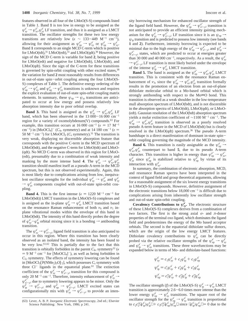
The definitive assignment of band4 as ψipa′ f ψxy
a′ , abonding-to-antibonding transition is extremely important. Figure11 depicts the bonding and antibonding combinations between
the Mo æxya′ and in-plane Sæip
a′ orbitals. The Moæxya′ orbital
derives from the t2g set inOh and is aπ-type orbital with respectto bonding interactions with simple monodentate donor ligands.However, theæip
a′ orbital of the dithiolate ligand is positionedfor good overlap with the Moæxy
a′ orbital to give a specialthree-center pseudo-σ type bonding interaction (Figure 11). Theenergy of theψip
a′ f ψxya′ LMCT transition (band4) reflects the
strength of this bonding interaction, and the intensity of the banddirectly probes the pseudo-σ mediated Mo-dithiolate cova-lency.65
The energy ofψxya′ is primarily affected by the nature of the
equatorial ligand field, and strong pseudo-σ Mo-dithiolatebonding raises the energy of this orbital. Additionally, theeffective nuclear charge (Z′eff) of the metal is a property of thedonor atoms, and appreciable changes inZ′eff for Mo may beanticipated as a result of the highly polarizable dithiolate sulfurdonor ligands.66 The relatively high intensity of theψop
a′ f ψxza′′,
ψyza′ and ψop
a′′ f ψxza′′,ψyz
a′ LMCT transitions in the LMoO(S-S)complexes reflects theπ-donor character of the dithiolates andthe concomitant reduction in Z′eff due to π-mediated chargedonation to Mo. Thus, the relativeπ-donor properties of thedithiolate ligand can modulate the reduction potential of theMo center by affecting the valence ionization energy of theψxy
a′
orbital through changes inZ′eff on Mo.The observed oscillator strength of theψip
a′ f ψxya′ transition
for the LMoO(S-S) compounds depends on the ligand backbone.The intensity of this transition for LMoO(edt), which has a C-Csingle bond, is a factor of two less than that observed for theother LMoO(S-S) compounds that possess an aromatic dithiolateC-C bond. The largeψip
a′-ψxya′ in-plane covalency for ene-
dithiolates suggests that this interaction, in concert withπ-de-localization, may play a major role in the unusual ability ofthese ligands to support multiple redox states67 and to electroni-cally buffer metal centers to large changes in charge.68 Thepossible role of in-plane covalency in electron transfer in pterin-containing molybdenum enzymes is discussed below.
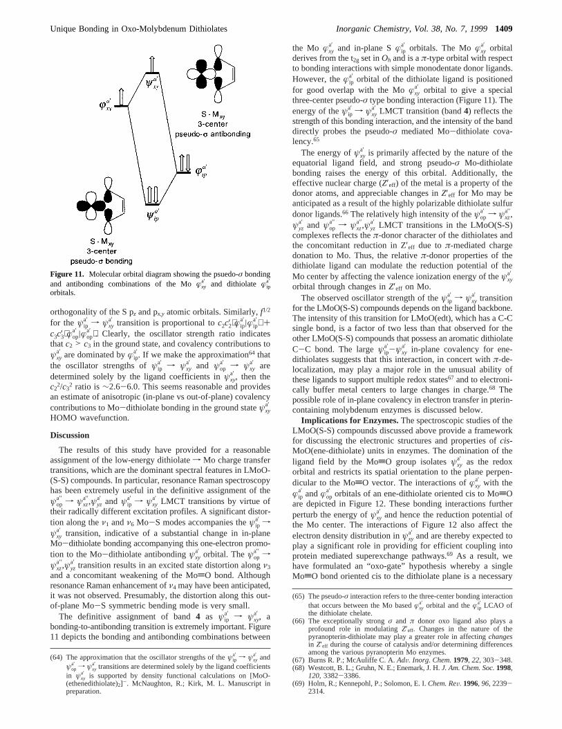
Implications for Enzymes.The spectroscopic studies of theLMoO(S-S) compounds discussed above provide a frameworkfor discussing the electronic structures and properties ofcis-MoO(ene-dithiolate) units in enzymes. The domination of theligand field by the MotO group isolatesψxy
a′ as the redoxorbital and restricts its spatial orientation to the plane perpen-dicular to the MotO vector. The interactions ofæxy
a′ with theæip
a′ andæopa′ orbitals of an ene-dithiolate oriented cis to MotO
are depicted in Figure 12. These bonding interactions furtherperturb the energy ofψxy
a′ and hence the reduction potential ofthe Mo center. The interactions of Figure 12 also affect theelectron density distribution inψxy
a′ and are thereby expected toplay a significant role in providing for efficient coupling intoprotein mediated superexchange pathways.69 As a result, wehave formulated an “oxo-gate” hypothesis whereby a singleMotO bond oriented cis to the dithiolate plane is a necessary
(64) The approximation that the oscillator strengths of theψipa′ f ψxy
a′ andψop
a′ f ψxya′ transitions are determined solely by the ligand coefficients
in ψxya′ is supported by density functional calculations on [MoO-
(ethenedithiolate)2]-. McNaughton, R.; Kirk, M. L. Manuscript inpreparation.
(65) The pseudo-σ interaction refers to the three-center bonding interactionthat occurs between the Mo basedæxy
a′ orbital and theæipa′ LCAO of
the dithiolate chelate.(66) The exceptionally strongσ and π donor oxo ligand also plays a
profound role in modulatingZ′eff. Changes in the nature of thepyranopterin-dithiolate may play a greater role in affectingchangesin Z′eff during the course of catalysis and/or determining differencesamong the various pyranopterin Mo enzymes.
(67) Burns R. P.; McAuliffe C. A.AdV. Inorg. Chem.1979, 22, 303-348.(68) Westcott, B. L.; Gruhn, N. E.; Enemark, J. H.J. Am. Chem. Soc.1998,
120, 3382-3386.(69) Holm, R.; Kennepohl, P.; Solomon, E. I.Chem. ReV. 1996, 96, 2239-
2314.
Figure 11. Molecular orbital diagram showing the psuedo-σ bondingand antibonding combinations of the Moæxy
a′ and dithiolate æipa′
orbitals.
Unique Bonding in Oxo-Molybdenum Dithiolates Inorganic Chemistry, Vol. 38, No. 7, 19991409
condition for efficient electron transfer regeneration of pyran-opterin Mo enzyme active sites following formal oxygen atomtransfer.
Facile electron transfer regeneration requires that there be ahigh degree of electronic communication between donor andacceptor sites. The magnitude of the electron transfer couplingmatrix element (HDA), between donor and acceptor sites, isaffected by the degree of metal-ligand covalency;69 anisotropyin this covalency (dictated by the nature of the ligand donorset,σ vs π bonding, in-plane vs out-of-plane bonding contribu-tions); and electron tunneling through the protein matrix.70,71
Although there has been considerable effort expended inunderstanding the contributions of electron tunneling throughproteins,70,71the effects of metal-ligand covalency are in generalmuch less understood, having been studied in detail only forblue copper proteins72 and rubredoxin.73 If the pyranopterin-dithiolate is directly involved in an effective electron transferregeneration pathway, good overlap must exist between the Moæxy
a′ orbital and at least one of the dithiolate orbitals of the samesymmetry (æip
a′ and æopa′ ). In fact, the magnitude ofHDA is a
function of this overlap and is proportional to the square of themolecular orbital coefficientsc2 andc3.74 The spectroscopicallydeterminedc2
2/c32 ratio for LMoO(bdt) is∼2.6-6.0. Thus, the
electron-transfer rate, which is proportional toHDA2, is antici-
pated to be approximately 7-36 times more efficient via thepseudo-σ æxy
a′ -æipa′ pathway than through theæxy
a′ -æopa′ pathway.
This is important because the structure of the pyranopterinfavored by current evidence (Figure 1) has a reduced (formallytetrahydro) pyrazine ring located between the Mo-dithiolatefragment and theπ-system of the pyrimidine ring of thepyranopterin. Thus, the pyranopterin structure of Figure 1presents a poorπ electron transfer pathway; efficientπ-mediatedelectron transfer for a reduced pyranopterin would requirescission of the pyran ring to give a specific dihydropterin aswell as a more covalentæxy
a′ -æopa′ interaction to coupleæxy
a′ intothe π-system.75 Therefore, we propose that the pathway ofpyranopterin mediated electron transfer most likely involves theσ-orbitals.
Efficient electron transfer rates should also be facilitated byminimimal reorganizational energy of the active site. Ourresonance Raman profiles conclusively show that one-electronpromotions into the monooxo Moψxy
a′ redox orbital (band4)result in no appreciable distortion along the high-frequencyMotO stretching mode.
The consensus active site structure of sulfite oxidase derivedfrom X-ray crystallography and EXAFS (Figure 2c) raises thequestion as to which oxo ligand is directly involved in thecatalytic oxidation of sulfite to sulfate. Crystallography hasshown that the active site is deeply buried within the Mo bindingdomain, and that substrate has access only to the oxo ligandresiding in the equatorial plane.16 The equatorial oxo ligand isalso anticipated to be the reactive oxo ligand from an electronicstructure viewpoint. Formal oxygen atom transfer results in areduced mono-oxo Mo(IV) site with the apical MotO bondoriented perpendicular to the dithiolate plane, allowing forefficient pyranopterin-dithiolate-mediated electron transferregeneration of the active site. In the reduced state, a H2Omolecule or OH- occupies this equatorial site. According toour oxo-gate hypothesis, H2O/OH- only becomes fully depro-tonated following the transfer of the second electron, resultingin the fully oxidized dioxo Mo(VI) state which is then fullycompetent for substrate oxidation. The Mo(VI) redox orbital isnow no longer oriented in a manner conducive with favorableinteractions involving the dithiolate portion of the pyranoterin.This should have the effect of lowering the Mo reductionpotential and decoupling the Mo site from the electron transferpathway, thereby preventing accidental reduction of the activesite prior to catalysis.
Summary
Detailed spectroscopic studies on LMoO(S-S) model com-plexes have been undertaken in order to develop insight intothe electronic origin of observed spectroscopic features andrelate these to electron transfer and oxo transfer reactivity inthe pyranopterin Mo enzymes. The results of this work aresignificant, detailing a highly covalent interaction between theredox active Moæxy
a′ atomic orbital and the dithiolateæipa′
SALC. This is a remarkable three-center pseudo-σ type bondinginteraction which couples theæxy
a′ orbital directly into the in-plane orbitals of the pyranopterin. This effective in-planecovalency is anticipated to play an important role in modulatingthe reduction potential of the active site by destabilizingψxy
a′ .The MotO group controls the orientation ofæxy
a′ , andæxya′ -æip
a′
overlap is maximized when the dithiolate chelate is orientedcis to the MotO bond. This has resulted in the developmentof an oxo-gate hypothesis, whereby the Mo reduction potentialand the coupling ofæxy
a′ to electron transfer pathways involvingtheσ system of the pyranopterin are dictated by the orientationof the MotO bond(s) relative to the dithiolate chelate. Thisoxo-gate hypothesis has important implications for the mech-anism of sulfite oxidase, where the catalytically labile oxo ligandresides in the equatorial plane, and the axial oxo ligand appearsto be an essential requirement for facile electron transfer toregenerate the active site of the enzyme.
Acknowledgment. The authors thank Prof. Partha Basu forsubstantive comments. We gratefully acknowledge the generousfinancial support of the The National Institutes of Health (GrantNo. GM-057378 to M.L.K. and Grant No. GM-37773 to J.H.E.).
IC981126O
(70) Beratan, D. N.; Betts, J. N.; Onuchic, J. N.Science1991, 252, 1285-1288.
(71) Beratan, D. N.; Onuchic, J. N.; Betts, J. N.; Bowler, B. E.; Gray, H.B. J. Am. Chem. Soc.1990, 112, 7915-7921.
(72) Solomon, E. I. Baldwin, M. J.; Lowery, M. D.Chem. ReV. 1992, 92,521-542.
(73) Lowery, M. D.; Guckert, J. A.; Gebhard, M. S.; Solomon, E. I.J.Am. Chem. Soc.1993, 115, 3012-3013.
(74) Balaji, V.; Ng, L.; Jordan, K. D.; Paddon-Row, M. N.; Patney, H. K.J. Am. Chem. Soc.1987, 109, 6957-6969.
(75) Enemark, J. H.; Garner, C. D.JBIC, J. Biol. Chem.1997, 2, 817-822.
Figure 12. Orbital interaction diagram detailing how theæxya′ orbital
mixes with æipa′ and æop
a′ as a function of the OtMo-S angle. Theæxy
a′ -æipa′ interaction is maximized atθ ) 90° and decreases with
increasing OtMo-S angle. Theæxya′ -æop
a′ overlap is minimized atθ )90° and increases as the OtMo-S angle increases.
1410 Inorganic Chemistry, Vol. 38, No. 7, 1999 Inscore et al.


![[Vnsharing.net][OxO Pandas][Ranma][chap 70]](https://img.pdfslide.tips/doc/110x75/568bf37e1a28ab89339a85fc/vnsharingnetoxo-pandasranmachap-70.jpg)


![[Vnsharing.net][OxO Pandas][Ranma][chap 72]](https://img.pdfslide.tips/doc/110x75/568c4d611a28ab4916a3be98/vnsharingnetoxo-pandasranmachap-72.jpg)

![[Vnsharing.net][OxO Pandas][Ranma][chap 71]](https://img.pdfslide.tips/doc/110x75/568c37861a28ab02359be53f/vnsharingnetoxo-pandasranmachap-71.jpg)