Embed Size (px)
Citation preview

Annals of Vascular Diseases Vol. 13, No. 3 (2020) 335
Ann Vasc Dis Vol. 13, No. 3; 2020; pp 335–338
Case Report
Spinal Cord Ischemia Following Endovascular Repair of Infrarenal Abdominal Aortic Aneurysm
Shingo Nakai, MD, Tetsuro Uchida, MD, PhD, Yoshinori Kuroda, MD, PhD, Atsushi Yamashita, MD, Eiichi Oba, MD, PhD, Kimihiro Kobayashi, MD, Tomonori Ochiai, MD, and Mitsuaki Sadahiro, MD, PhD
Spinal cord injury (SCI) following endovascular aortic repair (EVAR) for an abdominal aortic aneurysm (AAA) is a rare but serious complication. Case 1 presented with ruptured AAA and shock and underwent emergency EVAR. The patient developed incomplete paraplegia 2 days following EVAR. Case 2, diagnosed with impending rupture of AAA with extremely shaggy aorta, was treated with emergency EVAR. The patient was diagnosed with complete paraplegia soon after EVAR. Case 3 underwent elective EVAR and developed delayed paraplegia 2 weeks later. In EVAR, the etiology of SCI leading to paraplegia is often multifactorial. Surgeons must consider the possibility of SCI-induced paraplegia.
Keywords: spinal cord ischemia, paraplegia, EVAR
IntroductionSpinal cord injury (SCI) following infrarenal abdominal aortic aneurysm (AAA) repair is an unusual but serious complication that impairs activities of daily living. Fol-lowing endovascular aortic repair (EVAR), post-surgical paraplegia and paraparesis are less common than open surgery. Since 2010, 320 patients underwent EVAR for AAA with/without iliac arterial aneurysm in our hospital. Of these 320 patients, 3 (0.009%) experienced paraplegia. Herein, we report these 3 cases of SCI-related paraplegia following EVAR for AAA and consider the underlying
pathophysiology.
Case ReportCase 1A 79-year-old man with a 66 mm ruptured AAA was ad-mitted to our hospital in shock. Computed tomography (CT) revealed a 51 mm serial left common iliac artery (CIA) aneurysm from the AAA, retroperitoneal hemato-ma, and evidence of active bleeding from the anterior wall of the AAA (Fig. 1A). EVAR was conducted as an emer-gency surgery. Before EVAR, an intra-aortic balloon was inserted and inflated at the descending aorta for hemosta-sis. Initially, the left internal iliac artery (IIA) was occluded by using embolization coils (Tornado® Embolization Coil, Cook Inc., Bloomington, IN, USA). A bifurcated endovas-cular graft (Gore Excluder Endoprosthesis, W. L. Gore & Associates Inc., Flagstaff, AZ, USA) was deployed at the infrarenal lesion to land distally in the right CIA and ex-ternal iliac artery (EIA) with limb extension. The patient’s blood pressure increased immediately after completion of EVAR, although more than 4 h had passed since the onset of the AAA rupture. The operation time was 115 min.
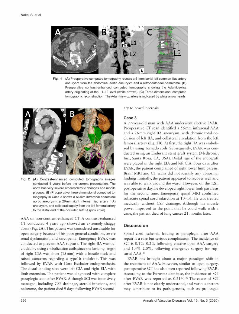
On the second postoperative day, the patient was found to have developed left lower limb incomplete paraplegia. Based on suspicion of SCI, the patient was treated with cerebrospinal fluid (CSF) drainage, high-dose steroid infu-sions, naloxone, and edaravone. Magnetic resonance im-aging (MRI) identified spinal cord infarction at T11–L1. Retrospective analysis of preoperative CT scans revealed the origin of the Adamkiewicz artery at L1–L2 level (Figs. 1B and 1C). This origin may have been unintentionally covered by the endograft. Based on patient records, the patient did not have any drop in his blood pressure below 80 mmHg in the postoperative period. Currently, the pa-tient is receiving walking rehabilitation; his bladder incon-tinence has remained unchanged 43 months post-surgery.
Case 2A 76-year-old man with AAA presenting with severe abdominal pain was found to have a 60 mm infrarenal
Online August 11, 2020doi: 10.3400/avd.cr.20-00061
Second Department of Surgery, Yamagata University Faculty of Medicine, Yamagata, Yamagata, Japan
Received: April 23, 2020; Accepted: June 14, 2020Corresponding author: Shingo Nakai, MD. Second Department of Surgery, Yamagata University Faculty of Medicine, 2-2-2 Iida-Nishi, Yamagata, Yamagata 990-9585, JapanTel: +81-23-628-5342, Fax: +81-23-628-5345E-mail: [email protected]
©2020 The Editorial Committee of Annals of Vas-cular Diseases. This article is distributed under the terms of the Creative Commons Attribution License, which permits use, distribution, and repro-duction in any medium, provided the credit of the original work, a link to the license, and indication of any change are properly given, and the origi-nal work is not used for commercial purposes. Remixed or transformed contributions must be distributed under the same license as the original.
336 Annals of Vascular Diseases Vol. 13, No. 3 (2020)
Nakai S, et al.
AAA on non-contrast-enhanced CT. A contrast-enhanced CT conducted 4 years ago showed an extremely shaggy aorta (Fig. 2A). This patient was considered unsuitable for open surgery because of his poor general condition, severe renal dysfunction, and sarcopenia. Emergency EVAR was conducted to prevent AAA rupture. The right IIA was oc-cluded by using embolization coils since the landing length of right CIA was short (15 mm) with a hostile neck and raised concerns regarding a type1b endoleak. This was followed by EVAR with Gore Excluder endoprosthesis. The distal landing sites were left CIA and right EIA with limb extension. The patient was diagnosed with complete paraplegia soon after EVAR. Although SCI was intensively managed, including CSF drainage, steroid infusions, and naloxone, the patient died 9 days following EVAR second-
ary to bowel necrosis.
Case 3A 77-year-old man with AAA underwent elective EVAR. Preoperative CT scan identified a 56 mm infrarenal AAA and a 26 mm right IIA aneurysm, with chronic total oc-clusion of left IIA, and collateral circulation from the left femoral artery (Fig. 2B). At first, the right IIA was emboli-zed by using Tornado coils. Subsequently, EVAR was con-ducted using an Endurant stent graft system (Medtronic, Inc., Santa Rosa, CA, USA). Distal legs of the endograft were placed in the right EIA and left CIA. Four days after EVAR, the patient complained of right lower limb paresis. Brain MRI and CT scans did not identify any abnormal findings. Initially, the patient appeared to recover well and was able to walk around the ward. However, on the 12th postoperative day, he developed right lower limb paralysis for the second time. Emergency spinal MRI confirmed subacute spinal cord infarction at T3–T6. He was treated medically without CSF drainage. Although his muscle power improved to the point that he could walk with a cane, the patient died of lung cancer 21 months later.
DiscussionSpinal cord ischemia leading to paraplegia after AAA repair is a rare but serious complication. The incidence of SCI is 0.1%–0.2% following elective open AAA surgery and 1.4%–2.0%, following emergency surgery for rup-tured AAA.1)
EVAR has brought about a major paradigm shift in the treatment of AAA. However, similar to open surgery, postoperative SCI has also been reported following EVAR. According to the Eurostar database, the incidence of SCI after EVAR was reported as 0.21%.2) The cause of SCI after EVAR is not clearly understood, and various factors may contribute to its pathogenesis, such as prolonged
Fig. 1 (A) Preoperative computed tomography reveals a 51 mm serial left common iliac artery aneurysm from the abdominal aortic aneurysm and a retroperitoneal hematoma. (B) Preoperative contrast-enhanced computed tomography showing the Adamkiewicz artery originating at the L1–L2 level (white arrows). (C) Three-dimensional computed tomographic reconstruction: The Adamkiewicz artery is indicated by white arrow heads.
Fig. 2 (A) Contrast-enhanced computed tomography images conducted 4 years before the current presentation. The aorta has very severe atherosclerotic changes and mobile plaques. (B) Preoperative three-dimensional computed to-mography in Case 3 shows a 56 mm infrarenal abdominal aortic aneurysm, a 26 mm right internal iliac artery (IIA) aneurysm, and collateral supply from the left femoral artery to the distal end of the occluded left IIA (pink color).

Annals of Vascular Diseases Vol. 13, No. 3 (2020) 337
EVAR-Related Spinal Cord Ischemia
aortic clamping, perioperative hypotension, atheromatous embolization, interruption of the great radicular artery (the Adamkiewicz artery), or collateral perfusion through pelvic circulation.3) Table 1 summarizes the possible causes of SCI in our cases.
In Case 1, perioperative hypotension secondary to hem-orrhagic shock was considered to be the main cause of paraplegia. In ruptured AAA, rapid completion of EVAR is required to achieve adequate arterial blood pressure. The interval between the onset of hypotension and EVAR completion could be prolonged because of IIA coiling. Peppelenbosch et al. proposed emergency EVAR with aor-to-uni-iliac stent graft system to avoid SCI by minimizing IIA ischemia.4) The Adamkiewicz artery is not occluded in most EVAR cases, because its typical origin is at the lum-bar as well as the thoracic vertebral levels.5,6) However, the Adamkiewicz artery in Case 1 originated only from the L1–L2 level. Thus, the spinal cord perfusion through the Adamkiewicz artery may have decreased because of stent graft coverage of its origin.
Case 2 was known to have a shaggy aorta with severe atherosclerotic changes based on imaging. Therefore, in-traoperative embolism, which decreased the spinal cord perfusion, was considered as pathogenesis of SCI. Fre-quent device insertion and removal from the shaggy aorta may have contributed to SCI.7) Gentle catheterization is mandatory for preventing paraplegia due to embolism.
In Case 3, the apparent cause of delayed SCI is not clear. However, bilateral occlusion of IIA may have been a possible cause of paraplegia. Collateral pelvic circula-tion, such as through IIA and lumbar artery, is known to be important for spinal cord perfusion. One report men-tions a lack of neurological deficits in cases with bilateral IIA interruption following open as well as endovascular surgery for AAA.8) Bratby et al. reported a 3% incidence of SCI in patients who underwent bilateral IIA emboliza-tion before EVAR.9) There is no reference to the timing of delayed onset of SCI in literature. Goldstein et al. reported a case of delayed SCI following the use of an aorto-uni-iliac device wherein the patient developed paraplegia 3 weeks following the procedure. The authors reported that secondary hypotension might induce further reduction in spinal cord perfusion and contribute to the development
of delayed paraplegia.10) The precise pathophysiology in Case 3 could not be determined.
ConclusionEVAR is widely accepted as a less-invasive alternative to open surgery, especially in high-risk patients. However, being aware of the possibility of postoperative SCI follow-ing EVAR is important. The main causes of SCI following EVAR are multifactorial and vary from patient to patient. Some of the factors include pre-existing complications, anatomical characteristics of spinal cord perfusion, urgen-cy of the operation, and general patient condition before surgery. Surgeons conducting EVAR should always exer-cise caution and be aware of the possibility of SCI-induced paraplegia as a procedural complication.
AcknowledgmentsWe thank Editage (www.editage.jp) for English language editing.
Ethical StatementThe ethical committee of Yamagata University approved this study (approval number: 2020-S-2).
Disclosure StatementNone of the authors have any conflict of interests to declare.
Author ContributionsStudy conception: SNData collection: SNDrafting: SNStudy supervision: TU and YKCritical review and revision: all authorsFinal approval of the article: all authorsAccountability for all aspects of the work: all authors
Table 1 Summary of three cases of paraplegia
The possible risk factors for developing paraplegia Onset and severity Outcome
Case 1 Prolonged hypotension POD 2 (subacute) AliveOcclusion of the Adamkiewicz artery incomplete Walking with a cane
Case 2 Micro embolism due to manipulation in severely shaggy aorta Immediately after EVAR (acute) Diedcomplete No improvement
Case 3 Instability of pelvic circulation POD 4 and POD 12 (delayed) Alive(occlusion of Bilateral IIA, IMA, lumbar artery, etc.) incomplete Walking with a cane
IIA: internal iliac artery; IMA: inferior mesenteric artery; POD: postoperative day

338 Annals of Vascular Diseases Vol. 13, No. 3 (2020)
Nakai S, et al.
References 1) Awad H, Ramadan ME, El Sayed HF, et al. Spinal cord
injury after thoracic endovascular aortic aneurysm repair. J Can Anesth 2017; 64: 1218-35.
2) Berg P, Kaufmann D, Van Marrewijk CJ, et al. Spinal cord ischemia after stent-graft treatment for infra-renal abdomi-nal aortic aneurysms. Analysis of the Eurostar database. Eur J Vasc Endovasc Surg 2001; 22: 342-7.
3) Bajwa A, Davis M, Moawad M, et al. Paraplegia following elective endovascular repair of abdominal aortic aneu-rysm: reversal with cerebrospinal fluid drainage. Eur J Vasc Endovasc Surg 2008; 35: 46-8.
4) Peppelenbosch N, Cuypers PW, Vahl AC, et al. Emergency endovascular treatment for ruptured abdominal aortic aneu-rysm and the risk of spinal cord ischemia. J Vasc Surg 2005; 42: 608-14.
5) Koshino T, Murakami G, Morishita K, et al. Does the Adamkiewicz artery originate from the larger segmental arteries? J Thorac Cardiovasc Surg 1999; 117: 898-905.
6) Melissano G, Bertoglio L, Civelli V, et al. Demonstration of the Adamkiewicz artery by multidetector computed tomog-raphy angiography analysed with the open-source software osiriX. Eur J Vasc Endovasc Surg 2009; 37: 395-400.
7) Morisaki K, Matsumoto T, Matsubara Y, et al. A rare com-plication of spinal cord ischemia following endovascular aneurysm repair of an infrarenal abdominal aortic aneurysm with arteriosclerosis obliterans: report of a case. Ann Vasc Dis 2016; 9: 255-7.
8) Mehta M, Veith FJ, Darling RC III, et al. Effects of bilateral hypogastric artery interruption during endovascular and open aortoiliac aneurysm repair. J Vasc Surg 2004; 40: 698-702.
9) Bratby MJ, Munneke GM, Belli AM, et al. How safe is bilateral internal iliac artery embolization prior to EVAR? Cardiovasc Intervent Radiol 2008; 31: 246-53.
10) Goldstein LJ, Rezayat C, Shrikhande GV, et al. Delayed per-manent paraplegia after endovascular repair of abdominal aortic aneurysm. J Vasc Surg 2010; 51: 725-8.