Embed Size (px)
Citation preview

Spontaneous Carotid Cavernous Fistula Presenting Only with Cranial Nerve Palsies
Akira Kurata,u Makoto Takano, 1 Kaichi Tokiwa, 1 Yoshio Miyasaka, 1 Kenzo Yada,1 and Shinichi Kan2
PURPOSE: To discuss the differences in angiographic findings between cases of spontaneous
carotid cavernous fistula with and without the classical triad of symptoms (pulsating exophthalmos,
bruit, and conjunctival chemosis). METHODS: With CT, MR, and angiography, we examined 12
cases of spontaneous carotid cavernous fistula, five of whom presented only with cranial nerve
palsies. RESULTS: In the seven cases with the triad , the main venous drainage from the cavernous
sinus was the superior ophthalmic vein. Only one or two veins drained the cavernous sinus, and
cortical venous drainage was not present in any case. In contrast, all but one case with only cranial
nerve palsies had at least three venous drainage routes from the cavernous sinus, including cortical
venous drainage. CONCLUSION: For the diagnoses of spontaneous carotid cavernous fistula, it is
important to know that some patients do not show the classical triad of symptoms. In such
patients, early diagnosis and treatment are particularly important because cortical venous drainage
and a consequent risk of hemorrhage are frequently present.
Index terms: Fistula, carotid cavernous; Arteries, carotid (internal); Angiography
AJNR 14:1097-1101 , Sep/Oct 1993
Pulsating exophthalmos, bruit, and conjunctival chemosis have long been regarded as the three classical symptoms of carotid cavernous fistula (CCF) (1, 2). However, CCF accompanied only by cranial nerve palsies without the usual triad of symptoms may be misdiagnosed.
The symptoms of patients with spontaneous CCF are commonly milder than those in traumatic CCF cases. This difference is attributable to the difference in the volume of blood flow into the cavernous sinus (3). In addition, the specific venous drainage from the cavernous sinus is important (4, 5). If the main venous drainage is via the superior ophthalmic vein, this triad will probably be present; if not, the triad is likely to be absent or incomplete. We investigated differences of arterial supply and venous drainage between spontaneous CCF cases presenting with and without the classical triad of symptoms.
Departments of 'Neurosurgery and 2Radiology, Kitasato University
School of Medicine, Sagamihara, Kanagawa Prefecture, Japan 228. 3 Address reprint requests to A . Kurata, MD, Department of Neurosur
gery, Kitasato University School of Medicine, 1-15-1 Kitasato, Sagamihara ,
Kanagawa Prefecture, Japan 228.
AJNR 14:1097-1101Sep/ Oct 1993 0195-6108/ 93/ 1405-1097
© American Society of Neuroradiology
Materials and Methods
We examined 12 patients with spontaneous CCF defined by angiography, of whom nine were hospitalized within the last 2 years, the other three in the previous 18 years (Table 1). In seven of the 12 patients, at least one component of the classical triad was recognized on admission (cases 1 through 7). In the other five (cases 8 through 12), however, no component of the triad was recognized on admission. All five of these patients had cranial nerve palsies only. In four of five cases (cases 8 , 9, 10 and 12) no component of the triad was detected at any stage of the clinical course. Three (cases 8 through 10) showed acutely complete oculomotor nerve palsy. In case 8 , ptosis developed initially and was followed by double vision. In case 9 , double vision occurred initially, followed by ptosis 3 days later. In case 10, ptosis and double vision developed simultaneously, having been preceded by 7 days of orbital pain. In cases 11 and 12, abducent nerve palsy developed suddenly, but slight tinnitus preceded it for 7 weeks in case 11 , and orbital pain preceded it for 2 weeks in case 12.
In all five cases presenting with cranial nerve palsies alone , plain and enhanced computed tomography (CT) was performed. Magnetic resonance (MR) imaging was also performed in all but one (case 8). In three cases (cases 8 through 1 0), CT and MR imaging failed to demonstrate abnormal findings. In case 11 , enhanced CT showed marked enhancement and dilatation of the left cavernous sinus, and MR imaging demonstrated an abnormal flow void in the cavernous sinus. In case 12, enhanced CT
1097
1098 KURATA
showed marked enhancement of the cortical vascular channels of the left cerebral hemisphere; MR imaging also demonstrated abnormal cortical vascular channels by increased flow void (Fig. 4 , A and B).
In all cases presenting with cranial nerve palsies alone, angiography was performed (average, 10 days after admission) to rule out aneurysms either in the cavernous sinus or elsewhere, because cranial nerve palsy is well known to be an initiator of cerebral aneurysms.
Results
Of the seven cases presenting with the classical clinical triad, two were of type B, two of type C, and three of type D; of the five cases with only cranial nerve palsies, one was of type A and four were of type D-according to the classification of inflow arteries of Barrow et al (6).
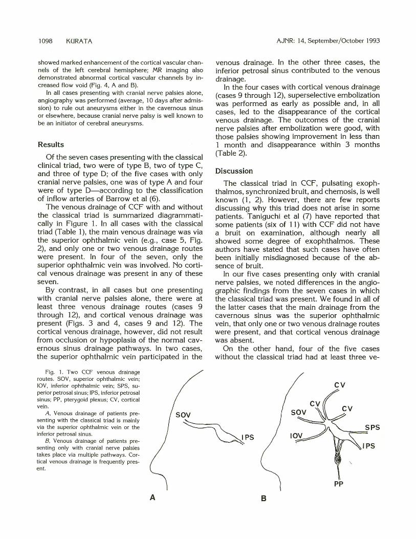
The venous drainage of CCF with and without the classical triad is summarized diagrammatically in Figure 1. In all cases with the classical triad (Table 1), the main venous drainage was via the superior ophthalmic vein (e.g., case 5, Fig. 2), and only one or two venous drainage routes were present. In four of the seven, only the superior ophthalmic vein was involved. No cortical venous drainage was present in any of these seven.
By contrast, in all cases but one presenting with cranial nerve palsies alone, there were at least three venous drainage routes (cases 9 through 12), and cortical venous drainage was present (Figs. 3 and 4, cases 9 and 12). The cortical venous drainage, however, did not result from occlusion or hypoplasia of the normal cavernous sinus drainage pathways. In two cases, the superior ophthalmic vein participated in the
Fig. 1. Two CCF venous drainage routes. SOY, superior ophthalmic vein ; lOY, inferior ophthalmic vein; SPS, superior petrosal sinus; IPS, inferior petrosal sinus; PP, pterygoid plexus; CV, cortical vein .
sov
AJNR: 14, September/October 1993
venous drainage. In the other three cases, the inferior petrosal sinus contributed to the venous drainage.
In the four cases with cortical venous drainage (cases 9 through 12), superselective embolization was performed as early as possible and, in all cases, led to the disappearance of the cortical venous drainage. The outcomes of the cranial nerve palsies after embolization were good, with those palsies showing improvement in less than 1 month and disappearance within 3 months (Table 2).
Discussion
The classical triad in CCF, pulsating exophthalmos, synchronized bruit, and chemosis, is well known (1, 2). However, there are few reports discussing why this triad does not arise in some patients. Taniguchi et al (7) have reported that some patients (six of 11) with CCF did not have a bruit on examination, although nearly all showed some degree of exophthalmos. These authors have stated that such cases have often been initially misdiagnosed because of the absence of bruit.
In our five cases presenting only with cranial nerve palsies, we noted differences in the angiegraphic findings from the seven cases in which the classical triad was present. We found in all of the latter cases that the main drainage from the cavernous sinus was the superior ophthalmic vein, that only one or two venous drainage routes were present, and that cortical venous drainage was absent.
On the other hand, four of the five cases without the classical triad had at least three ve-
A, Venous drainage of patients presenting with the classical triad is mainly via the superior ophthalmic vein or the inferior petrosal sinus.
B, Venous drainage of patients presenting only with cranial nerve palsies takes place via multiple pathways. Cortical venous drainage is frequently present.
~PS
pp
A B

AJNR: 14, September / October 1993 CAROTID CAVERNOUS FISTULA 1099
TABLE 1: Spontaneous carotid cavernous fistulas examined
Case Age,Sex Clinical Symptoms Type of Fistula
Venous Drainage (Barrow classification)
I 76,M Exophthalmos, chemosis, II , Ill , IV, V, VI D sov 2 71,F Exophthalmos, chemosis, Ill , V B sov 3 67 ,F Exophthalmos, chemosis, Ill , IV , VI c sov 4 63,F Exophthalmos, chemosis, bruit, II , Ill , IV , V, VI B sov 5 75,F Exophthalmos, chemosis, bruit, II, Ill , IV , V, VI D SOV, IPS 6 63,M Exophthalmos, chemosis, bruit, VI D SOV, IPS 7 77,F Exophthalmos, chemosis c SOV, ICS 8 72,F Ill D IPS, JCS 9 66,M Ill D IPS, PP, CCV
10 76,F 111 , 11 D IPS, IOV, CCV II 40,F VI A SOV, IOV, CCV 12 49,F VI ,V D SOV,IPS,CCV
Note. II , optic nerve palsy; Ill , oculomotor nerve palsy; IV, trochlear nerve palsy; V, trigem inal nerve palsy;
VI , abducent nerve palsy; SOV, superior ophthalmic vein; IPS, inferior petrosal sinus; ICS, intercavernous sinus; PP, pterygoid plexus; CCV, cerebral cortical vein ; IOV, inferior ophthalmic vein.
nous drainage routes. The reason for the absence of the clinical triad may have been the multiple venous drainage routes, and possibly the cortical venous drainage that was also present in four of the cases.
Halbach et al (8) reported that 11 patients with CCF showed cortical venous drainage, all of which had resulted from occlusion or hypoplasia of the normal cavernous sinus drainage pathways. In four of these 11 cases, intracerebral hemorrhage occurred. However, in our four cases with cortical venous drainage, no sinus occlusion or hypoplasia of the normal cavernous sinus drainage routes was present. Therefore, the participation of the cortical veins in our cases may have been caused by normal variation in the development of the venous pathways ( 4, 9) and by opening of the venous channels as a result of increased pressure in the cavernous sinus. The cranial nerve palsies may also have been caused by this increase in pressure because superior and
. · ... Fig. 2. Case 5 . A , Left common carotid angiogram
with CCF supplied by many meningeal branches from both internal and external carotid arteries, with venous drainage mainly to the dilated superior ophthalmic vein .
8 , Superselective angiogram of the left ascending pharyngeal artery , which is one of the feeders.
inferior divisions of the oculomotor nerve were involved in all three cases of oculomotor nerve palsy.
Turner et al ( 1 0) reported three cases of intracerebral hemorrhage associated with CCF, each of which had cortical venous drainage in the region of the hemorrhage. Therefore, patients with cortical venous drainage are at risk of intracerebral hemorrhage and should receive immediate therapy. Many authors (11-16) have reported that cortical venous drainage from dural arteriovenous malformations in locations other than the cavernous sinus is also a high risk factor for intracerebral hemorrhage. Abnormal cortical venous drainage is also a risk factor in cerebral ischemia, particularly venous ischemia, because of passive congestion secondary to retrograde increased venous pressure (17, 18).
The signs and symptoms of spontaneous CCF are usually less severe than those seen in traumatic CCF, which have greater fistulous flow. In

1100 KURATA AJNR: 14, September/October 1993
A 8 c ,-
\
./ D E
Fig. 3. Case 9. A and B, Photographs show complete right oculomotor nerve palsy. C, Photograph after embolization shows improvement of right oculomotor nerve palsy. D, Right external carotid angiogram with CCF supplied by the middle meningeal artery and distal portion of the internal maxillary
artery with venous drainage to a cortical vein (arrow). E, Right external carotid angiogram after embolization.
this series, only one (case 11) of the four cases with cortical venous drainage was classified into the high flow type of spontaneous CCF (type A).
Spontaneous CCF is not a rare disease, since we have encountered nine such cases in the last 2 years. In three of these the classical triad was not present at any time during the clinical course. Therefore, it is important not to overlook those patients with spontaneous CCF presenting only with cranial nerve palsies without the classical triad. In patients with cranial nerve palsies only, angiography, including external carotid angiog-
raphy, should be performed to rule out not only aneurysms but also CCF, as well as other possible causes.
Finally, we would emphasize that patients with spontaneous CCF should be diagnosed and, if needed, treated as early as possible in view of the high frequency of cortical venous draihage seen in our patients.
References
1. Dandy WE. Carotid-cavernous aneurysms (pulsating exophthalmos).
Zbl Neurochir 1937;2: 165-204

AJNR: 14, September/October 1993 CAROTID CAVERNOUS FISTULA 1101
B c Fig. 4. Case 12. A, Enhanced CT shows marked enhancement of the cortical vascular channels at the left cerebral hemisphere. B, Proton-weighted MR imaging (2000/60, repetition time/echo time) shows dilatation of vascular channels as increased flow voids. C, Left external carotid angiogram with CCF mainly supplied by the distal portion of the internal maxillary artery with venous
drainage to a cortical vein (arrow).
TABLE 2: Treatment and prognosis of spontaneous CCF
Case Treatment Outcome (Time until Resolution)
1 Embolization Resolution (1 month)
2 None Venous thrombosis of central
retinal vein
3 None Resolution after angiography (1 month)
4 None Resolution after angiography (1 month)
5 Embolization Resolution (2 months)
6 None Resolution after angiography (1 month)
7 None Resolution (3 months)
8 None Resolution after angiography ( 1 month)
9 Embolization Resolution (2 months)
10 Embolization Resolution (3 months)
11 Embolization• Resolution (1 day)
12 Embolization Resolution (2 months)
Note. Embolization, polyvinyl alcohol foam (300-600 11m) and gelfoam
or microfibrillary collagen were used as embolic materials. Embolization•,
two balloons were used as embolic material.
2. Walker AE, Allegre GE. Carotid-cavernous fistulas . Surgery 1956;3:411-422
3. Newton TH, Hoyt WF. Dural arteriovenous shunts in the region of
the cavernous sinus. Neuroradiology 1970;1:71-81 4. Hamby WB. Carotid-cavernous fistula. Springfield, Ill : Charles C
Thomas. 1966:3-129 5. Houser OW, Baker HL, Rhoton AL, Okazaki H. Intracranial dural
arteriovenous malformations. Radiology 1972; 105:55-64 6. Barrow DL, Spector RH, Braun I, Landman JA, Tindall SC, Tindall
GT. Classification and treatment of spontaneous carotid cavernous
fistulas. J Neurosurg 1985;62:248-256
7. Taniguchi RM, Goree JA, Odom GL. Spontaneous carotid-cavernous
shunts presenting diagnostic problems. J Neurosurg 1971 ;35:384-
391 8. Halbach VV, Hieshima GB, Higashida RT, Reicher M. Carotid cavern
ous fistulae: indication for urgent treatment. AJNR: Am J Neuroradio/
1987;8:627-633 9. Bartlow B, Penn RD. Carotid-cavernous sinus fistula presenting as a
posterior fossa mass. Case report. J Neurosurg 1975;42:585- 588
10. Turner DM, Vangilder JC, Mojtahedi S, Pierson EW. Spontaneous
intracerebral hematoma in carotid cavernous fistula. J Neurosurg
1983;59:680-686 11. Barnwell SL, Harbach VV, Dowd CF, Higashida RT, Hieshima GB,
Wilson CB. A variant of arteriovenous fistulas within the wall of dural
sinuses. J Neurosurg 1991 ;74:199-204 12. Grisoli F, Vincentelli F, Fuchs S. Surgical treatment of tentorial
arteriovenous malformations draining into the subarachnoid space.
Report of four cases. J Neurosurg 1984;60: 1059-1066 13. Ishii K, Goto K, lhara K, et al. High-risk dural arteriovenous fistulae
of the transverse and sigmoid sinuses. AJNR: Am J Neuroradio/
1987;8:1113-1120 14. Ito J , Imamura H, Kobayashi K. Dural arteriovenous malformation of
the base of the anterior cranial fossa. Neuroradiology 1983;24: 149-
154 15. Terada T , Kikuchi H, Karasawa J. Intracerebral arteriovenous malfor
mation fed by the anterior ethmoidal artery : case report. J Neurosurg
1984; 14:578-582 16. Vinuela F, Fox AJ, Pelz DM, Drake C. Unusual clinica l manifestations
of dural arteriovenous malformations. J Neurosurg 1986;64:554-558
17. Castaigne P, Bories J , Brunet P. Les fis tules arterioveineuses menin
gees pures a drainage veineux cortical. Rev Neural 1976; 132:169-
181 18. Lasjaunias P, Chiu M, Brunnge KT , Tolia A, Hurth M, Bernstein M.
Neurological manifestations of intracranial dural arteriovenous mal
formations. J Neurosurg 1986;64:724-730