Embed Size (px)
Citation preview

UNIVERSITY OF SOUTH BOHEMIA IN ČESKÉ BUDĚJOVICE
FACULTY OF AGRICULTURE
Study programme: N4101 Agriculture Engineering
Branch of study: Agricultural Biotechnology
Department: Department of Special Plant Production
Head of Department: prof. Ing. Vladislav Čurn, Ph.D.
DIPLOMA THESIS
Diversity and distribution study of viruses
in the entomopathogenic fungus Beauveria bassiana
in the Czech Republic Studium diversity a rozšíření virů entomopatogenní houby
Beauveria bassiana v České republice
Thesis Supervisor: Noemí Herrero Asensio, Ph.D.
Institute of Entomology, Biology Centre of the CAS
Co‐Supervisor: prof. Ing. Vladislav Čurn, Ph.D. Faculty of Agriculture, University of South Bohemia
Author: Bc. Petr Vaněček
České Budějovice, April 2015


Declaration of the author's diploma thesis
I hereby declare that this diploma thesis has been carried out in the Institute
of Entomology, Biology Centre of the Czech Academy of Sciences in České
Budějovice, Czech Republic under the guidance of Noemí Herrero Asensio, Ph.D.,
and in Faculty of Agriculture, University of South Bohemia, České Budějovice,
Czech Republic under the guidance of Professor Vladislav Čurn. The work is original
and has not been submitted in part or full by me for any degree or bachelor thesis at
any other University. I further declare that the material obtained from other sources
has been duly acknowledged in the thesis.
According to the § 47b law n. 111/1998 Sb., I agree to publish full version of
my diploma thesis in public electronic database STAG managed by University of
South Bohemia in České Budějovice.
Prohlášení autora DP
Student na tomto místě prohlašuje, že se jedná pouze o jeho dílo, předepsanou
formulací:
Prohlašuji, že svoji diplomovou práci jsem vypracoval samostatně pouze
s použitím pramenů a literatury uvedených v seznamu citované literatury. Prohlašuji,
že v souladu s § 47b zákona č. 111/1998 Sb. v platném znění souhlasím se
zveřejněním své diplomové práce, a to v nezkrácené podobě (v úpravě vzniklé
vypuštěním vyznačených částí archivovaných Zemědělskou fakultou JU)
elektronickou cestou ve veřejně přístupné části databáze STAG provozované
Jihočeskou univerzitou v Českých Budějovicích na jejích internetových stránkách.
Datum Podpis studenta

Thanks to
I would like to thank my thesis to my supervisor Noemí Herrero Asensio, Ph.D. for
her professional guidance in this thesis and valuable comments during the consultation
and Ing. Kateřina Šimáčková, Ph.D. for her help in the laboratory.
Poděkování
Rád bych poděkoval své vedoucí diplomové práce Noemí Herrero Asensio, Ph.D., za
její odborné vedení v této práci a cenné připomínky během konzultace, Ing. Kateřině
Šimáčkové, Ph.D. za její pomoc v laboratoři.
Abstract
Mycoviruses are viruses that infect and replicate in fungal cells, but unlike most
known viruses of plants and animals, they exceptionally produce deleterious effects
on their host. Nonetheless, the last discoveries showed that some mycoviruses can
decrease the virulence of their phytopathogenic fungal hosts, making them very
attractive for their possible use as biological control agents. Most mycoviruses have
dsRNA genomes and are widespread in all major taxa of fungi. Beauveria bassiana is
one of the most studied species of entomopathogenic fungi; it has a cosmopolitan
distribution and is used as biocontroller against invertebrates in agriculture.
In the present work, a collection of 137 isolates of B. bassiana obtained
at different locations and from different habitats in the Czech Republic was analysed.
These isolates were analysed for the presence of dsRNA elements indicative of viral
infections. The results revealed a high prevalence of viral infections in Czech
B. bassiana isolates, with 22.6% of the isolates containing dsRNA elements with viral
characteristics. Obtained dsRNA electropherotypes showed that virus diversity
in infected isolates was high and that mixed virus infections occurred among them.
Based on the characteristics of the electrophoretic band patterns, it could be
hypothesized that B. bassiana isolates collected in the Czech Republic could harbour
members of the viral families Totiviridae, Partitiviridae, Chrysoviridae
and Hypoviridae.
Keywords: Beauveria bassiana, entomopathogenic fungi, mycoviruses, dsRNA
Abstrakt
Mykoviry jsou viry, které infikují houbové buňky, kde se i replikují, ale na rozdíl
od většiny známých virů rostlin a živočichů mají jen výjimečně škodlivý vliv na jejich
hostitele. Nicméně, poslední objevy ukazují, že některé mykoviry mohou snížit
virulenci jejich fytopatogenních hostitelů, což dělá mykoviry velmi atraktivními
z důvodu jejich možného využití v biologické ochraně. Většina mykovirů má dsRNA
genomy a jsou rozšířené ve všech hlavních taxonech hub. Beauveria bassiana je
jednou z nejvíce studovaných entomopatogenních hub. Má kosmopolitní rozšíření
a v zemědělství je využívána v rámci biologické ochrany rostlin proti bezobratlým
škůdcům.
V této práci byla analyzována sbírka 137 vzorků B. bassiana získaných
z odlišných lokalit a přírodních stanovišť v České republice. Tyto izoláty byly
testovány na přítomnost dsRNA fragmentů indikujících virovou infekci. Výsledky
odhalily vysoký výskyt virové infekce mezi českými izoláty B. bassiana, bylo
detekováno 22,6% vzorků obsahujících dsRNA fragmenty s virovými
charakteristikami. Získané elektroforeotypy dsRNA ukazují, že virová rozmanitost
u infikovaných izolátů byla vysoká, a že se mezi izoláty objevily smíšené virové
infekce. Na základě charakteristik elektroforetických profilů může být předpokládáno,
že izoláty B. bassiana nasbírané v České republice mohly být infikovány zástupci
virových čeledí Totiviridae, Partitiviridae, Chrysoviridae a Hypoviridae.
Klíčová slova: Beauveria bassiana, entomopatogenní houby, mykoviry, dsRNA

Table of contents
1. Introduction ............................................................................................................................ 9
2. Review of literature …………………………………....……................................................ 11
2.1 Entomopathogenic fungi …………………………………….......................................... 11
2.1.1 Life cycle .............................................................................................................. 13
2.1.2 Lifestyles of entomopathogenic fungi ................................................................ 15
2.1.3 The entomopathogenic fungi as biocontrol agents in agriculture ........................ 15
2.2 Beauveria bassiana .................................................................................................. ......... 16
2.3 Mycoviruses ..................................................................................................................... 18
2.3.1 Taxonomy and genomic organization ................................................................. 19
2.3.2 Transmission of mycoviruses .............................................................................. 23
2.3.3 Viral effect on the fungal host .............................................................................. 24
2.3.4 Origins of mycoviruses ........................................................................................ 26
2.3.5 Mycoviruses infecting entomopathogenic fungi ................................................. 27
2.3.6 Applications and possible usages of mycoviruses .............................................. 28
3. 3. Objectives and aims ...................................................................................................... ............. 30
4. Materials and methods .............................................................................................................. 31
4.1 Fungal isolates ......................................................................................................... ......... 31
4.2 Analyses of the presence of dsRNA ..................................................................... ............. 35
4.3 Enzymatic treatment with DNase I and S1 nuclease ........................................................ 36
5. Results .......................................................................................................................................... 38
6. Discussion ................................................................................................................................... 42
7. Conclusions ......................................................................................................... ........................ 46
8. References ............................................................................................................... .................... 47
9. Supplementary materials ........................................................................................................... 55

9
1. Introduction
Social development is imminently connected with changes in nature which are
perceived at global scale. The world is modernized and changed in many ways every
day. These changes sometimes lead to side effects that are materialized in the increase
of environmental pollution. Therefore, nowadays more and more efforts are spent
in keeping pollution at acceptable levels. These efforts are extended to many different
sectors, including agriculture. According to this, there is a tendency in modern
agriculture to limit the usage of chemical pesticides replacing them by integrate pest
management practices, which include the usage of biological control agents. Actually,
entomopathogenic fungi are becoming in the recent years a very attractive method as
an alternative to the usage of chemical pesticides, since they do not have hazard effects
on human health and environment, meeting challenges of globalisation, climate
change and plant protection policies. They cause lethal infections and regulate insect
and mite populations in nature by causing epizootics. Beauveria bassiana is one
of the most studied entomopathogenic fungal species and is commercialized under
many different formulations as a biological control agent against different invertebrate
pest species.
Mycoviruses, are viruses that infect and replicate in fungal cells. They have been
detected in many fungal species, covering all four phyla of true fungi: Zygomycota,
Chytridiomycota, Ascomycota and Basidiomycota. Mycovirology is a quite young
field within general virology, being only active since 1962 when Hollings described
the first virus particles infecting mushrooms. Hence, only few studies exist until
the date reporting viruses infecting entomopathogenic fungi. Mycoviruses does not
produce obvious symptoms on their hosts, but some of them can decrease the virulence
of their phytopathogenic fungal hosts, or produce malformations in mushrooms.
Nonetheless, the cases in which mycoviruses are detrimental to their hosts could be
the exception to the rule. Indeed, the high prevalence and persistence of mycoviruses
among the major groups of fungi could indicate that their presence could be beneficial
to their fungal hosts, as is known to occur with a number of non-fungal viruses that are
able to establish mutualistic or neutral relationships with their hosts.
In this thesis a collection of 137 isolates of the entomopathogenic fungus
B. bassiana will be analysed for the presence of dsRNA molecules indicative
of mycovirus infections. New advances in the study of the entomopathogenic fungus

10
B. bassiana and their associated mycoviruses will help to better comprehend
the biology and ecology of this attractive entomopathogenic fungal species, which is
crucial for understanding its role in managed and natural ecosystems, and hence,
for their successful development as a biological control agent.
11
2. REVIEW OF LITERATURE
2.1 Entomopathogenic fungi
It is known that fungi and insects can establish relationships that can go from
mutualistic or commensal to obligate pathogenic. Fungi which cause lethal infections
and regulate insect and mite populations in nature by causing epizootics are
called entomopathogenic fungi (CARRUTHERS & SOPER, 1987; MCCOY et al.,
1988, CHARNLEY & COLLINS, 2007).
The earliest studies in entomopathogenic fungi were carried out in the 1800s,
and they were focused on the development of mechanisms to avoid the pest that was
devastating the silkworm industry of that time (STEINHAUS, 1975).
Entomopathogenic fungi group includes about 700 species belonging
to approximately 100 orders, nevertheless, only a few of these species have been
subjected to deep study. Most of these entomopathogenic species belong to orders
Hypocreales (phylum Ascomycota) and Entomophthorales (phylum
Entomophthoromycota) (HIBBETT et al., 2007; SUNG et al., 2007, HUMBER,
2012).
The occurrence of entomopathogenic fungi is quite wide and they can be found
almost all over the world (e. g., tropical rainforest, Antarctica or Arctic), so they are
a worldwide spread group of fungi (BRIDGE et al., 2005; EILENBERG et al., 2007;
AUNG et al., 2008; AUGUSTYNIUK-KRAM & KRAM, 2012).
The species diversity of entomopathogenic fungi in nature is influenced
by several factors, like the altitude, the latitude, the ecosystem type or the temperature.
Sun and Liu (2008) found that entomopathogenic fungi inhabit wide range of altitudes
(even more 5200m) and latitudes. It is important to mention that not all
entomopathogenic fungi inhabit the same range of altitudes and latitudes, or at least
differences arise at the level of frequency of occurrence of individual species.
For example, Metarhizium anisopliae occurs more frequently in northern latitudes
than Beauveria bassiana (QUESADA-MORAGA et al., 2007). In the same way,
ecosystem types significantly affects species diversity of entomopathogenic fungi,
for example, in temperate forests it is found higher diversity than in agriculture lands,
but lower than in the tropical forests. As it was mentioned earlier, temperature is
another factor which affects the occurrence of entomopathogenic fungi, because

12
species have different optimum growth temperatures. For example, the optimum
growth temperature of Hyphomycetes is generally between 20-30°C
while for Entomophthorales, it is considered as 15-25°C. Additionally, it was found
that entomopathogenic fungi occurring in forests are more tolerant to lower
temperatures (about 8°C), and those growing in agriculture lands are more tolerant
to higher temperatures (about 37°C). (BIDOCHKA et al., 2002; SOSNOWSKA et al.,
2004; AUGUSTYNIUK-KRAM & KRAM, 2012; GUL et al., 2014).
An important part of the entomopathogenic capability of these fungi lies in their
ability for the production of different enzymes and secondary metabolites which help
with the insect infection process and also play an important insecticidal role. As insect
cuticle comprises up to 70% of protein, the penetration process is facilitated
by extracellular fungal proteases like subtilisins, chymotrypsins, trypsins
and metalloproteases, usually with multiple isoforms of each. Toxic metabolites
with insecticidal effect include substances as destruxins (produced by Metarhizium
species), beauvericins, isarolides, bassianolides, beauverolides, oosporein (produced
by Beauveria species or Isaria fumosorosea), efrapeptins (produced by Tolypocladium
species) and hirsutellin (from Hirsutella thompsonii) (MAZET & VEY, 1995;
WEISER & MATHA, 1988; CHARNLEY, 2003; GUL et al., 2014). These substances
enable to kill many different species of insects and arthropods thanks to their high
variability, their ability to facilitate penetration of insect cuticle and because they are
able to overcome the insect immune system. For example, destruxins mostly cause
an initial tetanic paralysis leading to insect's death. The tetanic paralysis is attributed
to muscle depolarization by direct opening of the Ca2+ channels in membrane
(SAMUELS et al., 1988; GABARTY, 2014).
The production of the secondary metabolites is not only important for infecting
and killing insects, this substances are also crucial for the competition for the niche
of entomopathogenic fungi with other fungi. This strategy is known as antibiosis.
During this fight for the space, fungi actively compete with each other for carbon,
nitrogen, and various microelements. The site of competition is often the rhizosphere,
phyllosphere, or the intercellular space of a plant. Successful competition is often
a matter of timing, as resources are likely to go to the initial colonizer (FRAVEL,
1988). Nonetheless, there are other known mechanisms that entomopathogenic fungi
use in their competition for the space. Mycoparasitism is one of these mechanisms
and consists in the direct parasitism of one fungus by another. This strategy normally

13
starts with the production of extracellular exochitinases, which diffuse and catalyse
the release of cell-wall oligomers from the target host fungus (HARMON et al., 2004).
2.1.1 Life cycle
Life cycles of entomopathogenic fungi are synchronized with insect host stages
and environmental conditions (SHAHID et al., 2012). This way, the infection process
of fungal entomopathogens to their hosts could be summarized in six main steps:
adhesion, germination, differentiation, penetration, development, colonization,
sporulation and dispersion.
The degree of pathogenicity of an entomopathogenic fungus depends on several
factors which influence each of the cited steps. During the adhesion, the spore has
to adhere or attach to the insect cuticle, which is facilitated by a layer of mucus
composed of proteins and glucans that cover the whole spore. After the adhesion,
the spore begins to germinate and then it occurs the differentiation. At this moment,
the germinating spore produces specialized structures called appressoria, which fix the
spore to the epicuticular surface. Next step of the infection is penetration of the insect
cuticle or entrance through natural openings of the insect host such mouth or anus.
The effects of mechanical pressure and enzymatic activity of the germ tube help to
the penetration. A crucial point in this step is the secretion of several proteases, lipases
and chitinases. As soon as entomopathogenic fungi penetrate the insect body, they
grow and spread through the hemocoel as yeast-like propagules (blastospores), hyphal
bodies or protoplast lacking a cell wall (Fig. 1) (ROBERTS, 1981; HAJEK & ST.
LEGER, 1994; BOUCIAS et al., 1998; MACIÁ-VICENTE et al., 2011).
14
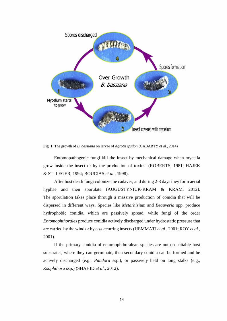
Fig. 1. The growth of B. bassiana on larvae of Agrotis ipsilon (GABARTY et al., 2014)
Entomopathogenic fungi kill the insect by mechanical damage when mycelia
grow inside the insect or by the production of toxins. (ROBERTS, 1981; HAJEK
& ST. LEGER, 1994; BOUCIAS et al., 1998).
After host death fungi colonize the cadaver, and during 2-3 days they form aerial
hyphae and then sporulate (AUGUSTYNIUK-KRAM & KRAM, 2012).
The sporulation takes place through a massive production of conidia that will be
dispersed in different ways. Species like Metarhizium and Beauveria spp. produce
hydrophobic conidia, which are passively spread, while fungi of the order
Entomophthorales produce conidia actively discharged under hydrostatic pressure that
are carried by the wind or by co-occurring insects (HEMMATI et al., 2001; ROY et al.,
2001).
If the primary conidia of entomophthoralean species are not on suitable host
substrates, where they can germinate, then secondary conidia can be formed and be
actively discharged (e.g., Pandora ssp.), or passively held on long stalks (e.g.,
Zoophthora ssp.) (SHAHID et al., 2012).

15
2.1.2 Lifestyles of entomopathogenic fungi
Entomopathogenic fungi are known as saprotrophic soil-borne fungi that can
infect insects and arthropods, but it is important to remark that they are not obligate
saprotrophs. There are many ways in which entomopathogenic fungi can receive
nutrients from their host. These ways include biotrophy (nutrition derived only
from living cells, which ceases once the cell has died), necrotrophy (killing
and utilization of dead tissues), and hemibiotrophy (initially biotrophic
and then becoming necrotrophic) (VEGA et al., 2009).
Recently, it has been discovered that many entomopathogenic fungi play
additional roles in nature besides infecting arthropods or being saprotrophs. Actually,
they can colonize plants as endophytes, act as plant pathogen antagonists,
as rhizosphere colonizers or even as plant growth promoters (VEGA et al., 2009).
Fungal endophytes are fungi which live within a plant for at least part of their
life cycle without causing apparent disease to the plant host. Endophytes can infect
either above or below ground internal plant tissues. Genera as Beauveria, Isaria
and several others, traditionally known as insect pathogens, have been naturally
isolated as endophytes (VEGA, 2008; VEGA et al., 2008).
Fungal endophytes can protect host plants against pathogens and herbivores
(ARNOLD et al., 2003; SCHULZ & BOYLE, 2005; ARNOLD & LEWIS, 2005;
RUDGERS et al., 2007; VEGA et al., 2009). They use different strategies for this
purpose, as production of metabolites, space competition, parasitism, hypovirulence,
induced systemic resistance or the increase of plant growth response (OWNLEY
& WINDHAM, 2007).
2.1.3 The entomopathogenic fungi as biocontrol agents in agriculture
Entomopathogenic fungi effectively infect a wide range of insect and arthropod
species, which make them very interesting for their usage as biological control agents
(AUGUSTYNIUK-KRAM & KRAM, 2012).
Eilenberg et al. (2001) defined biological control as the use of living organisms
to suppress the population density or the impact of a specific pest organism, making it
less abundant or less damaging than it would otherwise be. The most important goal
is to reduce the population of the pest below the economic threshold of harmfulness.
16
The use of entomopathogenic fungi as biological control agents constitute
an important component of what is known as integrated pest management (IPM).
These fungi have great potential in the management of insect pests infecting different
crop systems, since they have a big impact on the population dynamics of many
economically important species of insect, arthropods, spiders and mites. Although
entomopathogenic fungi cannot be massively produced as easily and efficiently
as chemical insecticides, and it is usually hard to meet optimal environmental and host
conditions for their application in the field, investing more in entomopathogenic fungi
research will place them in a better position at the IPM than they are now. This way,
new paradigms in the development, utilization and regulation of these insecticides will
be established (LIU, 2012).
Biological control is more and more necessary mainly because of the protection
of the environment. Chemical pesticides unlike biopesticides cannot be perceived only
as pest control agents, it is necessary to realize that they are also contaminants
of the environment. Although chemical pesticides are used mostly in a direct control
of pests on above-ground plant parts, a large amount of these pesticides can reach
the soil, groundwater or the atmosphere. They can also cause long-term negative
effects and move over long distances (ocean fog, arctic snow) (ELMOHOLT et al.,
1991; VAN DER WERF, 1996).
Another reason why chemical pesticides need to be replaced by biological
pesticides, is the increasing amount of resistances that insects are developing to various
chemical substances contained in plant protection products (MOTTA-SANCHEZ
et al., 2002). Additionally, since many invasive species have reached new countries
or continents where they do not have natural pathogens or predators, the use
of biological control agents could help to solve this problem (AUGUSTYNIUK-
KRAM & KRAM, 2012).
2.2 Beauveria bassiana
The fungi subject of this study Beauveria bassiana received this name in honour
of the Italian scientist Agostino Bassi who in 1835 formulated the germ theory
of disease after using the white muscardine fungus (B. bassiana) to treat diseased
silkworms (AUGUSTYNIUK-KRAM & KRAM, 2012). Bassi found
that the infectious agent of this worms was related to a white structures developed
on dead silkworms. He proved that the transference of a small amount of this “white

17
material” caused the death of healthy silkworms, after sprouting, growing
and mummifying them (PORTER, 1973). This discovery constituted the first step
on the utilization of fungi as biological control agents against insect pests (GILBERT
& GILL, 2010).
The genus Beauveria belongs to phylum Ascomycota, more precisely to the order
Hypocreales and family Cordycipitaceae. Beauveria is a cosmopolitan anamorphic
genus of soil-borne entomopathogenic fungi, which is facultative necrotroph
(ROBERTS & HAJEK, 1992; GOETTEL et al., 2005), but that can also occur
as saprotroph or as a plant endophyte (VEGA et al., 2008).
This genus is characterized morphologically by globose to flask-shaped
conidiogenous cells (Fig. 2a and 2b) from which one-celled terminal holoblastic
conidia are produced in sympodial succession on an indeterminate denticulate rachis.
Beauveria species are distinguished principally by the morphology of their conidia,
which are typically smooth-walled, hyaline, 1.5–5.5 μm and globose to cylindrical
or vermiform (REHNER et al., 2011).
Fig. 2. a) The growth of B. bassiana on potato dextrose agar (Source: author). b) Conidiogenous cells
and conidia of Beauveria sp. (REHNER et al., 2011).
B. bassiana can be reproduced sexually or asexually. The major reproductive
form is the asexual since the sexual stage of B. bassiana is rarely observed. The sexual
stage (the telemorph) of B. bassiana has been identified as Cordyceps bassiana (LI
et al., 2001).
B. bassiana is one of the most widespread entomopathogenic fungi. It is
a cosmopolitan fungi but it is important to mention that B. bassiana seems to be very
sensitive to the disturbance effects of cultivation (GABARTY et al., 2014). It was

18
found that B. bassiana inhabits also the Arctic Circle in Norway and Finland
(VÄNNINEN, 1995; KLINGEN et al., 2002).
Fungi of the genus Beauveria are also well-known for their production
of biologically active secondary metabolites as polyketides (e.g., oosporein, bassianin
and tennelin), non-ribosomally synthesized peptide antibiotics (e.g., beauvericin,
bassianolides and beauverilides), non-peptide pigments and other metabolites (e.g.,
oxalic acid). These secondary metabolites are mainly implicated in insect pathogenesis
and virulence and can be applied in agriculture. Actually, Beauveria bassiana is
commercialized under many different formulations as a biological control agent
against different invertebrate pest species. Nevertheless, some of the metabolites
produced by B. bassiana can have also medical applications and they are exploited by
chemical and pharmaceutical industries (GRIFFITH et al., 1993; VEY et al., 2001;
XU et al., 2009).
2.3 Mycoviruses
Mycoviruses are viruses that infect and replicate in fungal cells, but unlike
viruses of plants and animals, most known mycoviruses lack an extracellular phase
in their replication cycle (NUSS, 2011). This viruses have been detected in many
fungal species, covering all four phyla of true fungi: Zygomycota, Chytridiomycota,
Ascomycota and Basidiomycota. Typical representatives of fungal viruses are
isometric particles of 25±50 nm in diameter and with dsRNA genomes (GHABRIAL,
1998; PEARSON et al., 2009). Until the date more than 250 mycovirus species have
been sequenced and registered in the NCBI database.
The first presence of mycovirus was found in 1962 by Hollings in diseased
mushroom Agaricus bisporus, and later they were detected in the fungus Penicillium
spp., which it is well known by its capacity of stimulating the production of interferon
in mammals (BOZARTH, 1972). In 1978, Sanderlin and Ghabrial discovered
mycoviruses in plant filamentous fungal pathogens, more precisely, they were found
in the cereal fungal pathogen Helminthosporium victoriae. These discoveries
constituted the establishment of a new branch of research within general virology:
mycovirology.

19
2.3.1 Taxonomy and genomic organization
Although most mycoviruses have double stranded RNA (dsRNA) or single
stranded RNA (ssRNA) genomes, a DNA virus was recently described infecting
the fungal plant pathogen Sclerotinia sclerotiorum, it means that fungi may harbour
both, RNA and DNA viruses (XIE & JIANG, 2014). Most mycoviruses have quite
simple genomes and they can range from 2 to 14 kb in size. These genomes usually
encode for two genes, one encodes the capsid protein and the other a RNA-dependent
RNA polymerase (RdRp), but some mycoviruses encode only for a single RdRp gene.
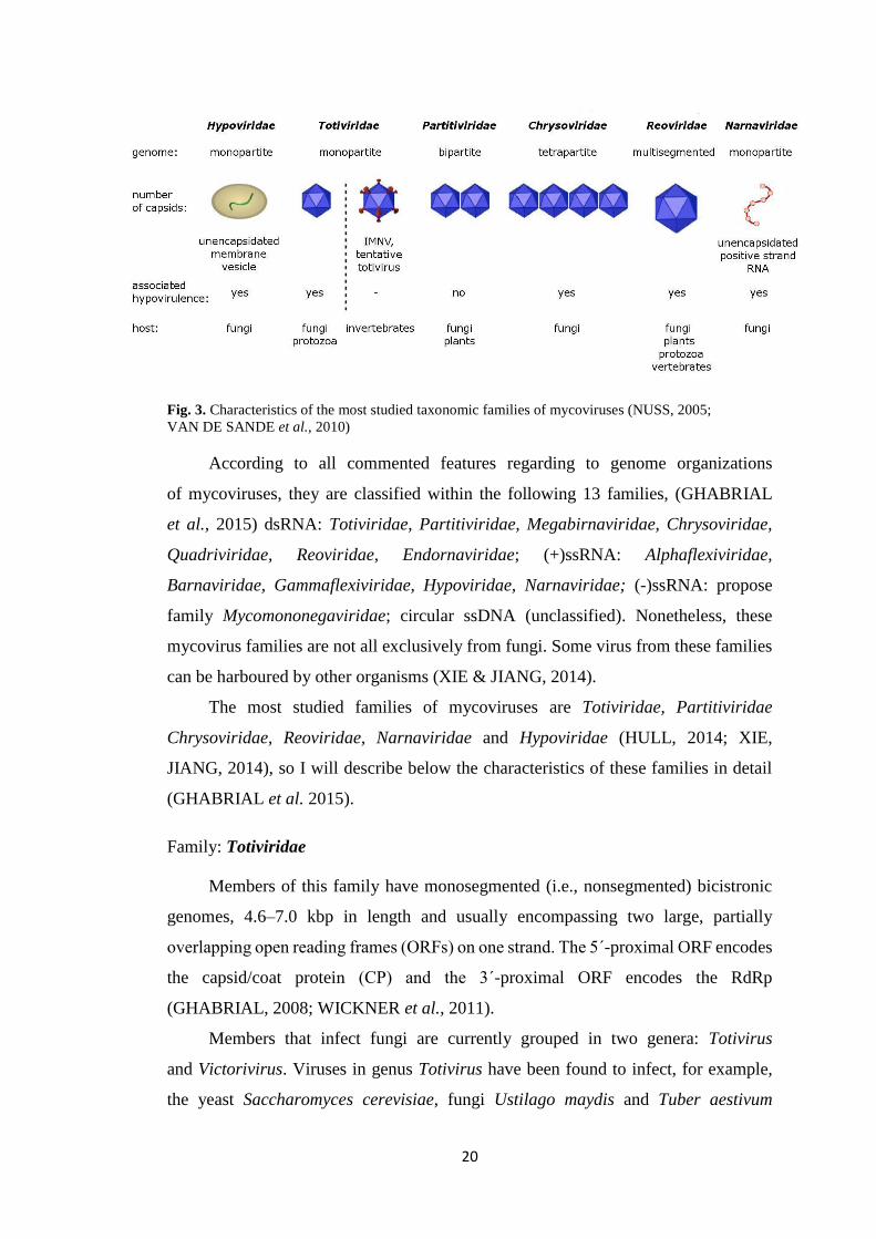
Regarding to genome organization, mycoviruses can have non-segmented
or segmented genomes. According to this, members of Reoviridae family can have
genomes made up of 10 to 12 segments, while Totiviridae members have non-
segmented genomes. Another important feature of mycoviruses is that not all of them
have their genomes encapsidated within a protein capsid (Fig. 3). For example,
members of the Hypoviridae family present their genomes within unencapsidated
membrane vesicles, and Narnaviridae family is characterized because its members
have naked (+) ssRNA genomes (GHABRIAL et al., 2015). This absence of capsids,
which protect viral genetic information, is possible because most mycoviruses lack
an extracellular phase on their replication cycle (NUSS, 2011).
Even if the genomes of these viruses seem very unique, Liu et al. (2010) found
that the capsid protein and RdRp genes from totiviruses and partitiviruses have
widespread homologs in the nuclear genomes of eukaryotes like plants, arthropods,
fungi, nematodes, and protozoa. These transfers could have been possibly mediated
by retrotransposons. Horizontally transfer of the viral genes of fungal partitiviruses
between viruses and plants was also found by Chiba et al. (2011). Liu et al. (2010)
also discovered that the amino acid sequence of the coat protein of Sclerotinia
sclerotiorum partitivirus-S (SsPV-S) has the highest amino acid sequence similarity to
the IAA-leucine-resistant protein 2 (ILR2) of Arabidopsis thaliana.
20
Fig. 3. Characteristics of the most studied taxonomic families of mycoviruses (NUSS, 2005;
VAN DE SANDE et al., 2010)
According to all commented features regarding to genome organizations
of mycoviruses, they are classified within the following 13 families, (GHABRIAL
et al., 2015) dsRNA: Totiviridae, Partitiviridae, Megabirnaviridae, Chrysoviridae,
Quadriviridae, Reoviridae, Endornaviridae; (+)ssRNA: Alphaflexiviridae,
Barnaviridae, Gammaflexiviridae, Hypoviridae, Narnaviridae; (-)ssRNA: propose
family Mycomononegaviridae; circular ssDNA (unclassified). Nonetheless, these
mycovirus families are not all exclusively from fungi. Some virus from these families
can be harboured by other organisms (XIE & JIANG, 2014).
The most studied families of mycoviruses are Totiviridae, Partitiviridae
Chrysoviridae, Reoviridae, Narnaviridae and Hypoviridae (HULL, 2014; XIE,
JIANG, 2014), so I will describe below the characteristics of these families in detail
(GHABRIAL et al. 2015).
Family: Totiviridae
Members of this family have monosegmented (i.e., nonsegmented) bicistronic
genomes, 4.6–7.0 kbp in length and usually encompassing two large, partially
overlapping open reading frames (ORFs) on one strand. The 5´-proximal ORF encodes
the capsid/coat protein (CP) and the 3´-proximal ORF encodes the RdRp
(GHABRIAL, 2008; WICKNER et al., 2011).
Members that infect fungi are currently grouped in two genera: Totivirus
and Victorivirus. Viruses in genus Totivirus have been found to infect, for example,
the yeast Saccharomyces cerevisiae, fungi Ustilago maydis and Tuber aestivum

21
representing the first evidence for mycoviruses in ectomycorrhizal fungi (ectophytes
of plant roots). Mycoviruses belonging to genus Victorivirus, in contrast, have been
found to infect only filamentous fungi (GHABRIAL & NIBERT, 2009; STIELOW
& MENZEL, 2010; GHABRIAL et al., 2015).
Family: Partitiviridae
Members of this family have bisegmented genomes, 1.4–2.4 kbp in length
and encompassing one large ORF per segment. Generally the smaller segment
(dsRNA2) encodes the CP and the larger segment (dsRNA1) encodes the RdRp. These
two genome segments are packaged into separate virus particles. Following are centre
organization of this family to reflect phylogenetic relationships, members that infect
fungi are now grouped in three genera: Alphapartitivirus, Betapartitivirus
and Gammapartitivirus (NIBERT et al., 2014). Alphapartitiviruses
and betapartitiviruses infect not only filamentous fungi but also plants,
whereas gammapartitiviruses infect only filamentous fungi. In general, partitivirus
infections are largely symptomless (GHABRIAL et al., 2015).
Family: Chrysoviridae
Penicillium chrysogenum virus (PcV) is the prototype of genus Chrysovirus,
the only current genus in this family (GHABRIAL & CASTÓN, 2011). It has four
monocistronic genome segments, 2.4–3.6 kbp in length and separately encapsidated
in virus particles. dsRNA1 encodes the RdRp, and dsRNA2 encodes the major CP
(GHABRIAL et al., 2015).
Family: Reoviridae
Genus Mycoreovirus was created to accommodate three species, Mycoreovirus1
(MyRV1) to Mycoreovirus3 (MyRV3). MyRV1 and MyRV2 were isolated
from Cryphonectria parasitica, and MyRV3 from Rosellinia necatrix (HILLMAN
& SUZUKI, 2004; WEI et al., 2004).
All mycoreoviruses confer hypovirulence to their respective natural hosts.
Mycoreovirus genome segments are monocistronic with 5´ caps on their positive
strands. MyRV1 and MyRV2 have 11 genome segments (S1–S11) whereas MyRV3
has 12 segments, 0.7–4.1 kbp in length for each virus. (KANEMATSU et al., 2004;
GHABRIAL et al., 2015).

22
Family: Narnaviridae
Members of this family contain the simplest genomes of any autonomous RNA
virus, each a single linear molecule of (+)ssRNA, 2.3–3.6 kbp in length
and encompassing a single ORF that encodes the RdRp (HILLMAN & CAI, 2013;
WICKNER et al., 2013).
The family comprises two genera based on subcellular location. Members
of genus Narnavirus have been found in the yeast S. cerevisiae as well as
in the protistan water mold Phytophthora infestans, and are confined to the cytosol.
Members of genus Mitovirus, in contrast, have been reported only in filamentous fungi
to date and are localized to the mitochondria. Lacking a CP, their genomes are confined
within intracellular lipid vesicles, as in the case of several other RNA viruses
of “lower” eukaryotes including hypoviruses (GHABRIAL et al., 2015).
Family: Hypoviridae
Cryphonectria hypoviruses1 to 4 (CHV1 to CHV4) are currently grouped in sole
genus Hypovirus within this family (NUSS & HILLMAN, 2011). They infect
the chestnut blight fungus C. parasitica, where they cause hypovirulence. Infection
of fungal mycelium is known to occur only through hyphal contact. Transmission rate
through conidiospores varies greatly but could be as high as 100% in some cases,
whereas transmission through ascospores is not known to occur. No true virions areas
sociated with members of this family. Instead, pleomorphic vesicles containing viral
RNAs and replication-associated proteins can be isolated from infected mycelia
(GHABRIAL et al., 2015).
The hypovirus genomes were originally thought to be dsRNA but are now
considered to be (+)ssRNA, ~ 9–13 kbp in length excluding the 3´ poly(A) tail
and encompassing two minimally overlapping ORFs in CHV1 and CHV2 but only one
ORF in CHV3 and CHV4. CHV3 and CHV4 are also distinct in encoding a putative
glycosyltransferase domain. Based on these differences as well as phylogenetic
analyses, it seems proper to divide the Cryphonectria hypoviruses into two new genera:
Alphahypovirus containing CHV1 and CHV2, and Betahypovirus containing CHV3
and CHV4 (GHABRIAL et al., 2015).

23
2.3.2 Transmission of mycoviruses
Mycoviruses can be vertically transmitted via host spores. This transmission is
mainly through asexual spores, but they can be also transmitted by sexual spores
(ascospores and basidiospores). However, the sexual reproduction of some species
(specially, of the phylum Ascomycota) constitute a barrier for the mycoviral
transmission (ROMO et al., 2007). Actually, some studies have shown that
mycoviruses are more frequently transmitted through basidiospores than ascospores.
(MCFADDEN et al., 1983; CHUN & LEE, 1997; PEARSON et al., 2009;
TUOMIVIRTA et al., 2009). Additionally, mycoviruses can be transmitted
horizontally via hyphal anastomosis (Fig. 4). When a virus-infected strain contacts
a virus-free strain, either hyphal anastomosis or an incompatibility response occurs
(Fig. 3), and the incompatibility response often leads to programmed cell death (PCD),
which limits the transmission of mycoviruses (CHOI et al., 2012).
Although it was believed that mycoviruses are limited to individuals with
the same or closely related vegetative compatibility groups, it was found that
a vegetative-incompatibility between two fungal strains, a virus-infected strain
and a virus-free strain, is not likely to be insurmountable barrier for mycovirus
transmission in nature. Therefore, they were described few cases in which the host
vegetative-incompatibility is supressed, but this case of mycovirus transmission is less
frequent than transmission between vegetative-compatible individuals (GHABRIAL,
1998; XIE & JIANG, 2014). The degree of suppression depends on individual
mycoviruses and their host. Most likely because of the attenuation of the vegetative-
incompatibility reaction of host fungi, the co-infection of mycoviruses is a common
phenomenon (XIE & JIANG, 2014).

24
Fig. 4. Horizontal transmission of mycoviruses (VAN DE SANDE et al., 2010)
The vegetative incompatibility could be considered as an antiviral defence
mechanisms in fungi, but not only at the individual level, a defence mechanism can
also occur at the population level, as well as at the cellular level (RNA silencing)
(NUSS, 2011).RNA silencing probably arose as an ancestral surveillance system to
protect against invading nucleic acids, including viruses of plants, insects, mammals
and fungi. It is important to mention that some fungi, for example Ustilago maydis,
seem to have completely lost the genes for the RNA silencing machinery, but these
fungi do not seem to suffer unusually high incidence of virus-induced symptoms
(NAKAYASHIKI & NGUYEN, 2008; NUSS, 2011).
2.3.3 Viral effect on the fungal host
Mycoviruses can change phenotypes of their hosts providing some advantages
or deleterious effects to them, but normally mycovirus does not produce obvious
symptoms on their hosts. Because of the lack of an infectivity assay and the frequent
occurrence of mixed infections, there is a problem with assigning any fungal
phenotypic change to a particular mycovirus (ROMAINE & SCHLAGNHAUFFER,
1995; MCCABE et al., 1999; HOWITT et al., 2006).
Symptomless or cryptic infections
Many investigators reject the possibility of the effect on fungal biology caused
by mycoviruses, because they observe only symptomless infections, but it can happen
that the virus might induce symptoms under some unexplored environmental

25
conditions. These symptoms that appear after altering some growth conditions of
the fungi, for example the abundance of available nutrients, are termed cryptic. It is
reasonable to assume that many mycovirus infections will have some slightly effect
on growth, although many mycoviruses produce no obvious phenotypic changes on
their hosts (BUCK, 1998; VAN DIEPENINGEN et al., 2006; PEARSON et al., 2009).
Detrimental infections
Some mycoviruses are able to negatively affect their hosts, this is the case of
the La France disease of the commercial mushroom Agaricus bisporus (HOLLINGS,
1962; RO et al., 2006) caused by La France isometric virus (LIV) (ROMAINE
& SCHLAGNHAUFFER, 1995) which affects to the basidiocarp formation. Another
example are oyster´s diseases (with similar symptoms the La France disease) caused
by the Oyster mushroom spherical virus (OMSV) (YU et al., 2003) and Oyster
mushroom isometric virus (RO et al., 2006). These diseases constitute one of the most
economically important diseases caused by mycoviruses.
Hypovirulence
It was found that some mycoviruses reduce the ability of their fungal
phytopathogenic hosts to cause disease in plants. This ability is referred
as hypovirulence. This phenomenon is very attractive due to the importance of fungal
diseases in agriculture and the limited strategies that are available to control them. This
property is very original, since one “pathogen” can be used to control another one
(NUSS, 2005). Hypovirulence of the chestnut blight fungus Cryphonectria parasitica
caused by hypoviruses is the best known case of hypovirulence used in biological
control (ANAGNOSTAKIS, 1982; NUSS, 2011).
Beneficial infections
Some mycoviruses have probably coevolved in parallel with their hosts to
limited detrimental effects or to mutual benefit. According to this, some mycoviruses
are linked with killer phenotypes in yeast and smuts that confer a strong advantage to
their hosts in interference competition by encoding toxins to which they are immune,
but which are lethal to sensitive cells. This killer phenomenon in the yeast
(Saccharomyces cerevisiae) and in the smut fungus (Ustilago maydis) was discovered
in 1960´s. Genetic and biochemical studies in yeast have conclusively shown that toxin

26
production and immunity are cytoplasmically inherited, and that dsRNAs of viral
origin comprise the cytoplasmic determinants (GHABRIAL, 1998). And finally, one
of the most remarkable and fascinating case within this category of viral effects is
the case of the virus infecting the root endophytic fungus Curvularia protuberata,
which confer heat tolerance to the panic grass Dichanthelium lanuginosum that hosts
the fungal endophyte (MARQUÉZ et al., 2007).
2.3.4 Origins of mycoviruses
Two main hypotheses exist which can explain origins and evolutionary history
of mycoviruses: the ancient coevolution hypothesis and the plant virus hypothesis.
The ancient coevolution hypothesis proposes that these infections are ancient,
coming from an unknown source, and have coevolved along with their hosts. It means
that mycoviruses infected ancestors of theirs present fungal hosts and have evolved
with them to give rise to present-day diversity (VOTH et al., 2006). This long time
of co-evolution could also explain the existence of asymptomatic phenotypes in many
of the infections caused by mycoviruses. The case of symptomatic infections found
in the phytopathogenic fungus Cryphonectria parasitica are hard to explain by this
hypothesis, in fact, from this example arises another hypothesis for the explanation
of mycovirus origin, the plant virus hypothesis.
The plant virus hypothesis proposes that the viruses have moved relatively
recently from the host plant to the fungus, what means that evolution of mycoviruses
and their fungal hosts is not probably congruent.
This theory is supported by a study of sequence comparisons between
mycoviruses and plant viruses which revealed the relatedness of hypoviruses to several
species of the ssRNA plant virus genus Potyvirus (FAUGUET et al., 2005; LINDER-
BASSO et al., 2005). Actually, these studies show that a higher similarity exist
between hypoviruses and plant viruses, than between hypoviruses and other
asymptomatic fungal viruses. According to this, the hypothesis proposes that
a common ancestral between hypoviruses and plant potyviruses existed, in the way
that saprophytic or pathogenic fungi acquired ssRNA viruses from plants, and after
they lost their protein capsid and evolved to dsRNA forms due to different evolution
pressures (PEARSON et al., 2009).

27
Both theories (‘the ancient coevolution’ and ‘the plant virus’ hypotheses) are
needed to explain the full range of mycovirus diversity that is being revealed. It is also
important to realize that not only these theories have to be true, but it is possible that
mycoviruses have moved from their fungal host into the plant host acting like vectors
(PEARSON et al., 2009).
2.3.5 Mycoviruses infecting entomopathogenic fungi
In the last 20 years, it is gradually increasing the interest in the detection
of mycoviruses in entomopathogenic fungi, due to the use of entomopathogenic fungi
as part of the IPM, as mentioned earlier. The more information we will have about
entomopathogenic fungi, the more effectively we will be able to use them.
Till the present time, only few studies have been developed in mycovirus
of entomopathogenic fungi research. Beauveria bassiana and Metarhizium anisopliae
are among the most tested entomopathogenic fungi for the presence of virus-like
dsRNA molecules (LEAL et al., 1994; BOGO et al., 1996; MELZER & BIDOCHKA,
1998; CASTRILLO et al., 2004; DALZOTO et al., 2006; HERRERO et al., 2009;
HERRERO et al., 2012; YIE et al., 2014).
The first research on the presence of dsRNA in B. bassiana was reported
by Melzer and Bidochka in 1998. They found dsRNA elements in 2 out of 12 Canadian
soil isolates. The presence of viral dsRNAs in B. bassiana were also detected in 7
out of 34 North American insect isolates (CASTRILLO et al., 2004), and Dalzoto
and collaborators detected dsRNA elements in 2 out of 13 Brazilian insect isolates
(DALZOTO et al., 2006). The most extensive research on the presence of dsRNA
virus-like molecules in B. bassiana was made in Spain and Portugal were 40 out of 73
soil and endophytic isolates resulted positive for the presence of viral dsRNA
(HERRERO et al., 2012). The latest research was conducted by Yie and collaborators
in 2014, in this study 8 out of 10 isolates from New Zealand were found harbouring
dsRNA elements. Till the date, only three mycoviruses have been completely
characterized infecting B. bassiana, two victorivirus and an unclassified virus related
to Partitiviridae family (HERRERO et al., 2012, YIE et al., 2014 and KOTTA-
LOIZOU et al., 2015)
The presence of mycovirus and dsRNA virus-like molecules was also found
in other species of entomopathogenic fungi as Tolypocladium cylindrosporus
(HERRERO & ZABALGOGEAZCOA, 2011).

28
2.3.6 Applications and possible usages of mycoviruses
It is important to notice that mycovirology is a relatively new scientific field.
That is the reason why only a small percentage of the possibilities of mycoviruses are
known. Moreover, most of mycovirus research is focused in those who infect
economically important fungi, such as yeasts, cultivated mushrooms and pathogens
of plants and animals (PEARSON et al., 2009).
Using mycoviruses to control crop disease
The usage of mycoviruses to control crop diseases started more than 50 years
ago, when mycoviruses were first employed to control chestnut blight disease
in Europe. Nevertheless, the use of mycoviruses against phytopathogenic fungi
depends a lot on the crop or disease to be treated. In fact, the pathosystem that can be
found among forests, orchards or agricultural differs a lot. In agricultural lands
for example, the high crop density, low species diversity and unique environmental
conditions, gives to the pathogen excellent conditions for its development,
but at the same time, these conditions can also help mycoviruses to establish
prevalence in the host populations in fields (XIE & JIANG, 2014).
Controlling crop fungal diseases with mycoviruses has also some disadvantages
compared to the use of chemical insecticides. Whereas insecticides act relatively fast,
mycoviruses need sufficient time for their establishment among the fungal population
in the field, which is vital for a successful control of the fungal pest. Viral transmission
between vegetative-incompatible individuals would make easier this establishment
of mycovirus among the fungal population, but this needs more time and the efficiency
of the proccess is very low. It should be pointed that the transmission between
vegetative-incompatible individuals is very often impossible and it leads to PCD. (XIE
& JIANG, 2014). However, Ikeda et al. (2013) recently found that it may be situations,
where vegetative-incompatibility reaction is attenuated by amending chemical
compounds. He found that zinc compounds could attenuate the heterogenic
incompatibility of Rosellinia necatrix. Other ways to overcome fungal vegetative
incompatibility could be to find mycoviruses with strong infectivity abilities that could
be used as a universal donor, creating a kind of “vector” for mycoviruses (XIE
& JIANG, 2014).

29
Future usage of mycoviruses as therapeutic agents in medicine
A thought to use viruses in medicine as biological control agents of human
diseases is not so new, actually, since the early 20th century, it has been known that
some viruses (bacteriophages) can specifically and uniquely search and destroy
bacteria (SULAKVELIDZE et al., 2001). Until the date, they have been conducted
several studies on viruses for their development as therapeutic agents in medicine, e.g.,
a bacteriophage of vancomycin resistant Enterococcus faecium (BISWAS et al.,
2002).
Because of ongoing research based on the study of bacteriophages as therapeutic
agents for bacterial infections in humans, it has been derived the idea that even
mycoviruses would be able to suppress some medically important fungi (VAN DE
SANDE et al., 2010).
Invasive fungal infections are relatively common opportunistic infections
in immunocompromised patients and are still associated with a high mortality rates.
These infections are often accompanied by complications like resistance
or refractoriness to current antimicrobial agents, so it is very important to find new
therapeutic strategies based on the identification of new microbial targets and novel
antimicrobial agents (VAN DE SANDE et al., 2010).
One of these hypothetical therapeutic strategies may involve the use
of mycoviruses that are able to selectively infect fungi, but such mycoviruses are not
still identified (VAN DE SANDE et al., 2010).
30
3. Objectives and aims
As it was showed in the introduction, viruses have been discovered in numerous
fungal species, but unlike most known animal or plant viruses, they are rarely
associated with deleterious effects on their hosts. Beauveria bassiana is one
of the most studied species of entomopathogenic fungi; it has a cosmopolitan
distribution and is used as a biological control agent against invertebrates
in agriculture.
According to this, the main objectives of this work are to study the prevalence,
variability, and patterns of distribution of virus like-dsRNA elements in a collection
of soil isolates of the entomopathogenic fungus B. bassiana obtained at different
locations and habitats in the Czech Republic.
31
4. Materials and methods
4.1 Fungal isolates
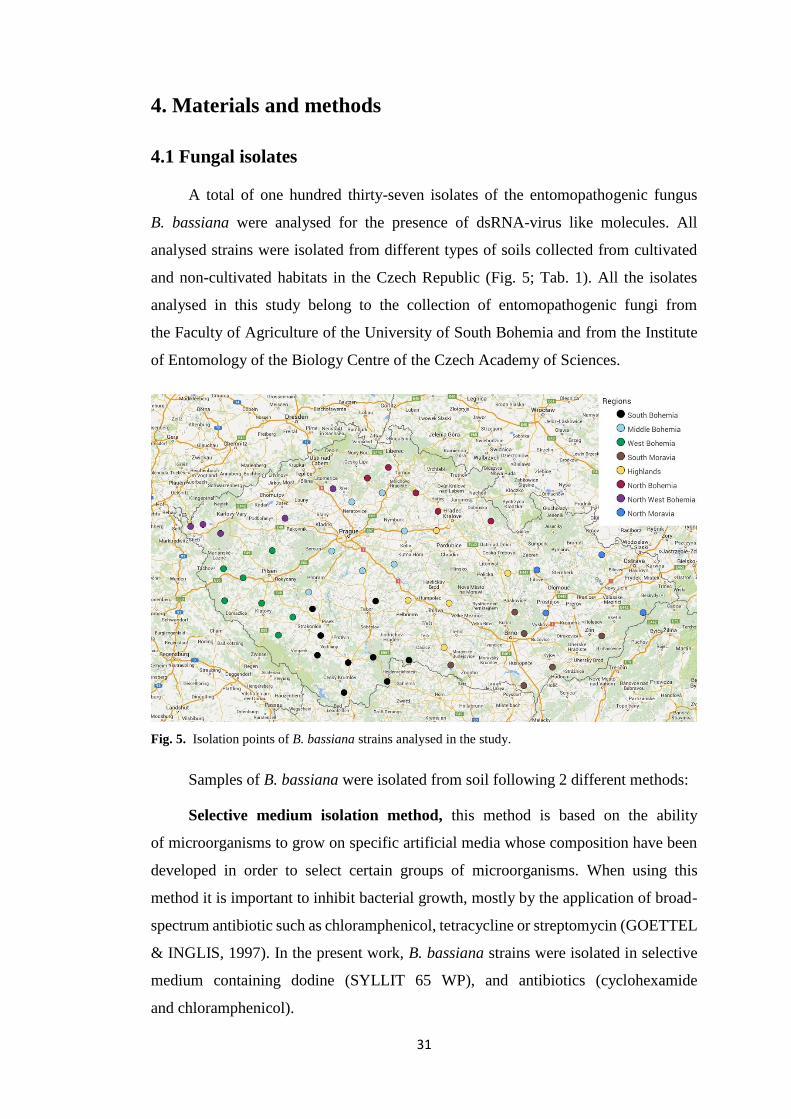
A total of one hundred thirty-seven isolates of the entomopathogenic fungus
B. bassiana were analysed for the presence of dsRNA-virus like molecules. All
analysed strains were isolated from different types of soils collected from cultivated
and non-cultivated habitats in the Czech Republic (Fig. 5; Tab. 1). All the isolates
analysed in this study belong to the collection of entomopathogenic fungi from
the Faculty of Agriculture of the University of South Bohemia and from the Institute
of Entomology of the Biology Centre of the Czech Academy of Sciences.
Fig. 5. Isolation points of B. bassiana strains analysed in the study.
Samples of B. bassiana were isolated from soil following 2 different methods:
Selective medium isolation method, this method is based on the ability
of microorganisms to grow on specific artificial media whose composition have been
developed in order to select certain groups of microorganisms. When using this
method it is important to inhibit bacterial growth, mostly by the application of broad-
spectrum antibiotic such as chloramphenicol, tetracycline or streptomycin (GOETTEL
& INGLIS, 1997). In the present work, B. bassiana strains were isolated in selective
medium containing dodine (SYLLIT 65 WP), and antibiotics (cyclohexamide
and chloramphenicol).

32
Tab. 1. Soil isolates of B. bassiana collected in the Czech Republic and analysed for the presence of dsRNA virus-like molecules
Sample name IMFS Region Locality Habitat Subhabitat Sample name IMFS Region Locality Habitat Subhabitat
NBBBA-1 SM North Bohemia Doksy N Mixed forest SMBBA-15 TBM 15 South Moravia Vranovská Ves N Conifers forest
NBBBA-2 SM North Bohemia Jičín C Meadow SMBBA-16 TBM 15 South Moravia Hlohovec C Plum crop
NBBBA-3 SM North Bohemia Jičín N Fallow land SMBBA-17 TBM 15 South Moravia Vacenovice N Hedgerow
NBBBA-4 SM North Bohemia Jičín N Hedgerow SMBBA-19 GBM 15 South Moravia Hostěnice N Mixed forest
NBBBA-5 SM North Bohemia Knežničky N Mixed forest HBBA-1 SM Highlands Kasalice C Meadow
NBBBA-6 SM North Bohemia Nechanice N Mixed forest HBBA-2 SM Highlands Kasalice C Cereal field
NBBBA-7 SM North Bohemia Jaroslav N Hedgerow HBBA-3 SM Highlands Rohovládova Bělá N Mixed forest
NBBBA-8 TBM 15 North Bohemia Doksy N Mixed forest HBBA-4 SM Highlands Kasalice N Hedgerow
NBBBA-9 TBM 15 North Bohemia Doksy N Mixed forest HBBA-5 SM Highlands Koclířov C Cereal field
NBBBA-10 TBM 15 North Bohemia Doksy N Mixed forest HBBA-6 SM Highlands Koclířov N Hedgerow
SMBBA-1 SM South Moravia Vranovská Ves N Conifers forest HBBA-7 SM Highlands Poděšín N Mixed forest
SMBBA-2 SM South Moravia Vacenovice N Hedgerow HBBA-8 SM Highlands Poděšín N Hedgerow
SMBBA-3 SM South Moravia Hostěnice N Mixed forest HBBA-9 SM Highlands Želetava C Cereal field
SMBBA-4 SM South Moravia Hostěnice N Mixed forest HBBA-11 SM Highlands Želiv N Hedgerow
SMBBA-5 SM South Moravia Hostěnice N Mixed forest HBBA-12 TBM 15 Highlands Želiv N Hedgerow
SMBBA-7 SM South Moravia Hostěnice N Hedgerow MBBBA-1 SM Middle Bohemia Skruhrov N Riverbank
SMBBA-8 SM South Moravia Hlohovec C Plum crop MBBBA-2 SM Middle Bohemia Skruhrov N Riverbank
SMBBA-9 SM South Moravia Vizovice N Hedgerow MBBBA-3 SM Middle Bohemia Čistá C Cereal field
SMBBA-10 SM South Moravia Vizovice N Meadow MBBBA-4 SM Middle Bohemia Čistá N Hedgerow
SMBBA-11 SM South Moravia Vizovice N Hedgerow MBBBA-5 SM Middle Bohemia Mělník N Mixed forest
SMBBA-12 SM South Moravia Rajec Jestřebí C Cereal field MBBBA-7 SM Middle Bohemia Vlková N Mixed forest
SMBBA-13 TBM 15 South Moravia Vranovská Ves N Conifers forest MBBBA-8 TBM 25 Middle Bohemia Mořina C Corn field
SMBBA-14 TBM 15 South Moravia Vranovská Ves N Conifers forest MBBBA-9 GBM 15 Middle Bohemia Čistá C Cereal field

33
Sample name IMFS Region Locality Habitat Subhabitat Sample name IMFS Region Locality Habitat Subhabitat
MBBBA-10 GBM 25 Middle Bohemia Vatěkov C Meadow NWBBBA-7 SM North West Bohemia Sádek N Mixed forest
MBBBA-11 SM Middle Bohemia Velký Osek N Meadow NWBBBA-8 TBM 15 North West Bohemia Krásno N Fallow land
MBBBA-12 SM Middle Bohemia Velký Osek N Mixed forest NWBBBA-9 TBM 15 North West Bohemia Libotenice C Hop field
MBBBA-13 SM Middle Bohemia Velký Osek N Mixed forest NWBBBA-10 TBM 25 North West Bohemia Horka C Rapeseed field
MBBBA-14 GBM 25 Middle Bohemia Velký Osek N Mixed forest WBBBA-1 SM West Bohemia Janov C Meadow
NMBBA-1 SM North Moravia Bílá N Conifers forest WBBBA-2 SM West Bohemia Janov C Cereal field
NMBBA-2 SM North Moravia Staré Hamry N Conifers forest WBBBA-3 SM West Bohemia Janov N Fallow land
NMBBA-3 SM North Moravia Slavkov C Cereal field WBBBA-4 SM West Bohemia Vřeskovice N Mixed forest
NMBBA-4 SM North Moravia Mohelnice C Corn field WBBBA-5 SM West Bohemia Drahotín N Mixed forest
NMBBA-5 SM North Moravia Stavenice N River edge WBBBA-6 SM West Bohemia Bezděkov N Mixed forest
NMBBA-6 SM North Moravia Mohelnice N Hedgerow WBBBA-7 SM West Bohemia Janov N Hedgerow
NMBBA-8 SM North Moravia Dětkovice N Hedgerow WBBBA-8 SM West Bohemia Plasy N Mixed forest
NMBBA-9 TBM 15 North Moravia Stavenice N Hedgerow WBBBA-9 SM West Bohemia Plasy N Hedgerow
NMBBA-10 GBM 15 North Moravia Stavenice N River edge WBBBA-10 SM West Bohemia Sušice N Mixed forest
NMBBA-11 GBM 15 North Moravia Mohelnice N Hedgerow WBBBA-11 SM West Bohemia Velký Bor N Hedgerow
NMBBA-12 GBM 25 North Moravia Stavenice N River edge WBBBA-12 SM West Bohemia Drahotín N Hedgerow
NMBBA-13 GBM 25 North Moravia Mohelnice N Hedgerow WBBBA-13 SM West Bohemia Sušice N River side
NMBBA-14 GBM 15 North Moravia Stavenice N Hedgerow WBBBA-14 SM West Bohemia Sušice N Hedgerow
NWBBBA-1 SM North West Bohemia Františkovy Lázně C Meadow WBBBA-15 SM West Bohemia Velký Bor N Conifer forest
NWBBBA-2 SM North West Bohemia Krásno N Fallow land WBBBA-16 GBM 25 West Bohemia Vřeskovice N Mixed forest
NWBBBA-3 SM North West Bohemia Krásno N Fallow land WBBBA-17 GBM 25 West Bohemia Drahotín N Hedgerow
NWBBBA-4 SM North West Bohemia Krásno N Hedgerow WBBBA-18 GBM 25 West Bohemia Lhota u Stříbra N Conifer forest
NWBBBA-5 SM North West Bohemia Želkovice C Apple tree field WBBBA-19 GBM 15 West Bohemia Drahotín N Hedgerow
NWBBBA-6 SM North West Bohemia Horka N Hedgerow WBBBA-20 TBM 15 West Bohemia Drahotín N Hedgerow

34
Sample name IMFS Region Locality Habitat Subhabitat Sample name IMFS Region Locality Habitat Subhabitat
WBBBA-21 TBM 25 West Bohemia Vřeskovice N Mixed forest SBBBA-11 GBM 15 South Bohemia Ostrov C Meadow
WBBBA-22 TBM 25 West Bohemia Plasy N Hedgerow SBBBA-12 GBM 15 South Bohemia Ostrov C Corn field
WBBBA-23 GBM 25 West Bohemia Švihov N Hedgerow SBBBA-13 SM South Bohemia Prachatice N Conifer forest
WBBBA-24 TBM 15 West Bohemia Vřeskovice N Mixed forest SBBBA-14 SM South Bohemia Prachatice N Hedgerow
WBBBA-25 TBM 15 West Bohemia Janov N Hedgerow SBBBA-15 SM South Bohemia Mladá Vožice C Cereal field
WBBBA-26 TBM 15 West Bohemia Sušice N Hedgerow SBBBA-16 SM South Bohemia Šindlovy Dvory N Fallow land
WBBBA-27 TBM 15 West Bohemia Velký Bor N Fallow land SBBBA-17 GBM 15 South Bohemia Sedliště N Hedgerow
WBBBA-28 TBM 25 West Bohemia Janov N Hedgerow SBBBA-18 GBM 25 South Bohemia Ostrov N Mixed forest
WBBBA-29 TBM 15 West Bohemia Velký Bor N Conifer forest SBBBA-19 TBM 25 South Bohemia Staré město pod L. N Hedgerow
WBBBA-30 TBM 25 West Bohemia Velký Bor N Hedgerow SBBBA-20 TBM 15 South Bohemia Vahlovice N Hedgerow
WBBBA-31 GBM 25 West Bohemia Janov C Cereal field SBBBA-21 TBM 15 South Bohemia Halámky N Mixed forest
WBBBA-32 GBM 25 West Bohemia Plasy N Mixed forest SBBBA-22 GBM 15 South Bohemia Prachatice N Conifer forest
SBBBA-1 SM South Bohemia Sedliště N Hedgerow SBBBA-23 TBM 15 South Bohemia Ostrov N Mixed forest
SBBBA-2 SM South Bohemia Staré město pod L. N Conifer forest SBBBA-24 TBM 15 South Bohemia Prachatice N Conifer forest
SBBBA-3 SM South Bohemia Ostrov N Mixed forest SBBBA-25 TBM 15 South Bohemia Čeřín N Conifer forest
SBBBA-4 SM South Bohemia Sedliště N Mixed forest SBBBA-26 TBM 25 South Bohemia Ostrov N Mixed forest
SBBBA-5 SM South Bohemia Vahlovice N Hedgerow SBBBA-27 TBM 25 South Bohemia Čeřín N Conifer forest
SBBBA-6 SM South Bohemia Mladá Vožice N Hedgerow SBBBA-28 GBM 15 South Bohemia Halámky N Mixed forest
SBBBA-7 GBM 15 South Bohemia Třeboň C Meadow SBBBA-29 TBM 15 South Bohemia Halámky N Mixed forest
SBBBA-8 GBM 25 South Bohemia Sedliště N Hedgerow SBBBA-30 GBM 15 South Bohemia Dobrotín N Edge of river
SBBBA-9 SM South Bohemia Vahlovice N Mixed forest SBBBA-31 GBM 15 South Bohemia Halámky N Hedgerow
SBBBA-10 SM South Bohemia Halámky N Hedgerow
NOTES: IMFS – isolation method from soil; SM – selective medium; TBM 15 and TBM 25 – Tenebrio bait method (isolation performed at 15 or 25°C),
GBM 15 and GBM 25 – Galleria bait method (isolation performed at 15 or 25°C); N – non-cultivated; C – cultivated

35
Insect bait method, it is based on the ability of entomopathogenic fungi to infect
an insect host. Galeria melonella and Tenebrio molitor are the most commonly used
bait species (ZIMMERMANN, 1986; KLINGEN et al., 2002). Both insect species
were used for the isolation of B. bassiana in this work.
Obtained soil isolates of B. bassiana were cultured for 3 weeks over cellophane
disks layered on top of potato dextrose agar (Sigma Life Science) Petri dishes (6 Petri
dishes per isolate). Cultures were grown in incubators at 23°C for 3 weeks. After that,
isolates were harvested and stored at -20°C.
4.2 Analyses of the presence of dsRNA
The presence of dsRNA molecules of sizes ranging from 1 to 14 kbp in fungal
isolates was used as an indicator of virus infection. This type of nucleic acid can
represent the genomes of dsRNA mycoviruses, as well as replicative forms of viruses
with ssRNA genomes (MORRIS & DODDS, 1979). However, DNA viruses recently
discovered in fungi, will not be detected by this technique (YU et al., 2010).
This viral dsRNA purification procedure relies on the property of dsRNA to bind
to cellulose (cellulose chromatography) when the concentration of ethanol in the buffer
solution is 15%. DNA and ssRNA have lower affinity for cellulose at this ethanol
concentration, allowing the selective purification of dsRNA. dsRNA is eluted from
the cellulose with ethanol free STE 1X buffer. Isolated dsRNAs were subjected to
agarose gel electrophoresis and visualized after staining with ethidium bromide.
Electrophoresis is based on the different movement of charged molecules in an electric
field. This way, negatively charged molecules, as DNA, RNA or proteins, migrate
through the agarose matrix towards positively charged pole (anode) (YILMAZ et al.,
2012).
All positive dsRNA extractions were independently repeated twice.
Steps of the procedure
About 1 g of each analysed fungal isolate was pulverized with liquid nitrogen
using mortar and pestle. After that, 4 ml of extraction buffer (compositions of all
solutions can be find at Supplementary materials section), 0.1 ml of 60 mg/ml
bentonite aqueous suspension, 2 ml of phenol (phenol/TRIS saturated solution
for molecular biology, Acros Organics) and 2 ml of chloroform (Fisher Scientific)
were added to the pulverized mycelium. The mix was homogenized with pestle,

36
transferred to a 12 ml plastic tube and centrifuged at 8000 rpm (Hettich Universal
320R) for 20 minutes at 4°C (all centrifugations were made at 4°C).
The upper aqueous phase was recovered in a new 12 ml tube, 1.5 ml of absolute
ethanol were added and the volume was adjusted to 10 ml using STE 1X buffer. It
means that the final ethanol concentration of the solution was 15%. After this, 0.2 g
of cellulose (fibres (medium), Sigma Life Science) were added and the suspension was
shaken for 5 minutes in a rotator (Multi Bio RS-24; BioSan).
After shaking, the suspension was centrifuged at 4000 rpm for 5 minutes. Then
the supernatant was discarded and the cellulose was resuspended in 10 ml of STE 15
buffer, shook for 5 minutes, and centrifuged as before. After that, the supernatant was
discarded again. This washing process with STE 15 buffer was repeated three times.
To elute the dsRNA from the cellulose, all remaining STE 15 buffer was
removed with a pipette and the cellulose was resuspended in 2 ml of STE 1X (without
ethanol). It was shaken for 5 minutes and centrifuged at 5000 rpm during 8 minutes.
Then the supernatant containing dsRNA was transferred to a new 12 ml tube.
The extracted dsRNA was precipitated with 2.5 volumes of cold ethanol at -20°C
for at least 30 minutes.
To pellet the dsRNA, the tube was centrifuged at 8000 rpm for 20 minutes.
The supernatant was discarded and the tube was dried. Then the pellet was resuspended
in 20 µl of distilled and sterile water.
Finally, the extracted dsRNA was loaded and ran in a 1% agarose (Sigma Life
Science) gel prepared with TAE buffer and stained with ethidium bromide to visualize
the dsRNA. The agarose gel electrophoresis was carried out at a voltage of 200 V,
600 mA and 300 W (MS 500V Power Supply; Major Science). Most mycoviruses have
genomes smaller than 10 kbp, so it was used a molecular weight ladder appropriate
for this range (e.g. 1-10 kbp DNA ladder; GeneRulerTM 1 kb DNA Ladder). dsRNA
elements in gel were visualized in UV light transilluminator Slite 200 SW (Life
Science Avegene).
4.3 Enzymatic treatment with DNase I and S1 nuclease
In order to prove the double stranded nature of the extracted RNA,
and to eliminate possible traces of ssRNA or DNA that could have co-purified
with the dsRNA, enzymatic treatments with Dnase I and S1 nuclease were performed.
37
DNase I degrades DNA and S1 nuclease degrades all single-stranded nucleic acids
(DNA, RNA) (YU & MANLEY, 1986; KISHI et al., 2001).
Steps of the procedure of the DNase I treatment of dsRNA
DNase I treatment of dsRNA was performed in 20 µl of reaction mixture. 5 µl
of dsRNA, 2 µl of DNase I Buffer 10X, 5 µl of DNase (1u/ µl) and 8 µl sterile
and distilled water were mixed in a 1.5 ml tube. The reaction was incubated at 37°C
for 30 minutes. After that, a phenol-chloroform extraction was performed in order to
remove enzyme rests. 180 µl of sterile and distilled water were added to the reaction
and the extraction was carried out by adding 200 µl of phenol, gentle vortexing
and centrifugation at maximum speed (MiniSpin®, Eppendorf) for 15 minutes.
After that, the supernatant was removed and transferred into a new 1.5 ml tube. 200 µl
of chloroform were added to the obtained supernatant from the previous step, vortexed
gently and centrifuged at maximum speed for 5 minutes. The supernatant was carefully
removed and transferred into a new 1.5 ml tube. The sample was precipitated
with 2.5 volumes of absolute ethanol at -20°C overnight.
Steps of the procedure of the S1 nuclease treatment of dsRNA
Precipitated dsRNA from DNase I treatment was pelleted at maximum speed
(Hettich Universal 320R) for 25 minutes and then the supernatant was discarded.
The tube was dried and the pellet was resuspended in 10 µl of sterile and distilled
water. Then the reaction mixture was prepared in a total volume of 15 µl containing
10 µl of dsRNA, 1.5 µl of S1 N Buffer 10X, 1 µl of S1 N (1u/ µl) and 2.5 µl of sterile
and distilled water. The reaction was incubated at 37°C for 15 minutes. After that,
a phenol-chloroform extraction followed by ethanol precipitation was performed
as explained before. Precipitated dsRNA was centrifuged at maximum speed
for 25 minutes, pellet was dried and resuspended in 10 µl of sterile and distilled water.
Enzymatically treated dsRNA was loaded and ran in a 1% agarose gel prepared
with TAE buffer (as it was explained before).

38
5. Results
A total of 137 B. bassiana isolates collected from soils of the Czech Republic
were analysed for the presence of dsRNA virus-like molecules, 31 isolates harboured
dsRNA elements (22.6%). It was proved after the enzymatic treatments that all
the nucleic acids observed in the gels were indeed dsRNA (Fig. 6a and 6b)
(Electrophoretic patterns obtained before enzymatic treatments were placed in
Supplementary Material section as Fig. 9a and 9b).
Regarding to the habitat where the fungal cultures were obtained, 5 (20%)
of the 25 isolates from cultivated soils and 26 (23.2%) from the 112 isolates collected
in non-cultivated soils contained dsRNA elements. Almost 55% (17 out of 31)
of the isolates harbouring dsRNA elements were isolated from forests. It should be
noticed that none dsRNA elements were detected in fungal strains isolated from fallow
lands or river edges.
The majority of fungal isolates harbouring dsRNA elements came
from the region of South Bohemia, 10 out of 31 (32.3%). Nonetheless, the region
with the highest percentage of occurrence of isolates harbouring dsRNA elements was
North Bohemia, since 50% (5 out of 10) of the isolates collected in this region were
infected by dsRNA elements. On the other hand, only 1 of the 32 analysed fungal
isolates from West Bohemia region harboured dsRNA molecules. None dsRNAs were
found in any of the 10 isolates from North West Bohemia region (Fig.7).
Regarding to the method used for the isolation of the B. bassiana cultures tested
in this study, it was found that 9 (15.3%) of the 59 fungal strains isolated by bait
methods harboured dsRNA molecules; and 22 (28.2%) from the 78 strains isolated
by selective media method contained dsRNA elements.

39
a)
b)
Fig. 6. a) Electrophoretic profiles of dsRNA elements present in several isolates of B. bassiana
after enzymatic treatments. Lanes: 1, NBBBA-4; 2, HBBA-5; 3, HBBA-9; 4, MBBBA-11; 5,
SBBBA-7; 6, NBBBA-5; 7, NBBBA-6; 8, NBBBA-8; 9, NMBBA-1; 10, MBBBA-4; 11, MBBBA-5;
12, SBBBA-1; 13, SBBBA-4; 14, SBBBA-5; 15, SBBBA-6; 16, SMBBA-3; 17, WBBBA-5; 18,
SBBBA-2. b) Electrophoretic profiles of dsRNA elements present in several isolates of B. bassiana
after enzymatic treatments. Lanes: 19, isolate NBBBA-1; 20, NBBBA-9; 21, MBBBA-13; 22,
SMBBA-2; 23, SBBBA-25; 24, SBBBA-10; 25, NMBBA-4; 26, SMBBA-19; 27, NMBBA-9; 28,
SBBBA-27. Lane Kbp contains molecular size markers, and the values on the left are sizes in kilobase
pairs.
40
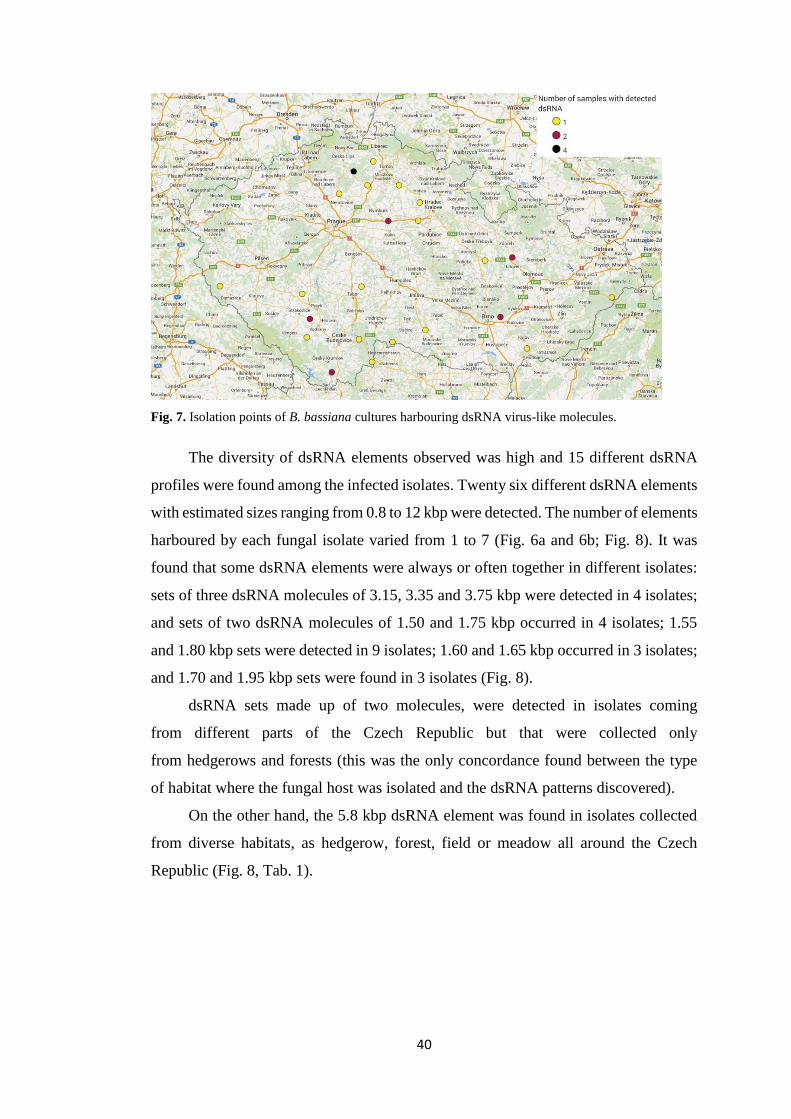
Fig. 7. Isolation points of B. bassiana cultures harbouring dsRNA virus-like molecules.
The diversity of dsRNA elements observed was high and 15 different dsRNA
profiles were found among the infected isolates. Twenty six different dsRNA elements
with estimated sizes ranging from 0.8 to 12 kbp were detected. The number of elements
harboured by each fungal isolate varied from 1 to 7 (Fig. 6a and 6b; Fig. 8). It was
found that some dsRNA elements were always or often together in different isolates:
sets of three dsRNA molecules of 3.15, 3.35 and 3.75 kbp were detected in 4 isolates;
and sets of two dsRNA molecules of 1.50 and 1.75 kbp occurred in 4 isolates; 1.55
and 1.80 kbp sets were detected in 9 isolates; 1.60 and 1.65 kbp occurred in 3 isolates;
and 1.70 and 1.95 kbp sets were found in 3 isolates (Fig. 8).
dsRNA sets made up of two molecules, were detected in isolates coming
from different parts of the Czech Republic but that were collected only
from hedgerows and forests (this was the only concordance found between the type
of habitat where the fungal host was isolated and the dsRNA patterns discovered).
On the other hand, the 5.8 kbp dsRNA element was found in isolates collected
from diverse habitats, as hedgerow, forest, field or meadow all around the Czech
Republic (Fig. 8, Tab. 1).

41
Isolate dsRNA elements detected (Kbp)
0.80 1.15 1.25 1.45 1.50 1.55 1.60 1.65 1.70 1.75 1.80 1.90 1.95 2.00 2.15 2.40 2.50 2.65 2.90 3.00 3.05 3.15 3.35 3.75 5.80 12.00
SBBBA-27 ● ● ● SBBBA-25 ● ● ●
SMBBA-3 ● ●
HBBA-1 ● ● SBBBA-6 ● ●
NBBBA-9 ● ●
MBBBA-13 ● ● SMBBA-2 ● ●
SBBBA-4 ● ●
NBBBA-1 ● ● NBBBA-5 ● ●
NBBBA-6 ● ●
NBBBA-8 ● ● MBBBA-4 ● ●
MBBBA-5 ● ●
NMBBA-1 ● ● SBBBA-5 ● ●
WBBBA-5 ● ●
SBBBA-10 ● ● ● ● ● ● ● NBBBA-4 ●
HBBA-5 ●
MBBBA-11 ● SBBBA-7 ●
HBBA-9 ● ●
NBBBA-10 ● ● ● SMBBA-19 ● ● ●
NMBBA-4 ● ● ● ● ● ●
SBBBA-2 ● ● ● ● SBBBA-1 ● ● ● ● ●
NMBBA-9 ● ● ● ● ●
SBBBA-26 ● ●
Fig. 8. dsRNA electropherotypes observed in soil isolates of B. bassiana. The circles indicate the presence in an isolate of a dsRNA molecule of the size shown at the top of each
column. Similar sets of two or three dsRNA elements observed in different isolates are indicated by identical colours.

42
6. Discussion
The incidence of dsRNA virus-like molecules found in B. bassiana isolates
collected in different parts of the Czech Republic indicates that mycovirus infections
are common among B. bassiana soil isolates from this country. dsRNA elements were
detected in 22.6% of the 137 analyzed isolates. Higher incidences were found
in previous works, for example, in B. bassiana isolates from Spain and Portugal
incidences of 54.8% were found (40 out of 73 isolates) (HERRERO et al., 2012)
and even higher in isolates from New Zealand 77.8% (7 out of 9 isolates) (YIE et al.,
2014). Nevertheless, other studies achieved lower incidences of dsRNA elements
among B. bassiana isolates, Melzer and Bidochka (1998) found dsRNA presence only
in 2 isolates of the 12 analysed in Canada (16.7 %); similar results were found in Brazil
with 2 out of 13 isolates infected (DALZOTO et al., 2006) and a 20.6% incidence was
found by Castrillo et al. (2004) in isolates of B. bassiana from North America.
Differences between the percentages of infected isolates could be explained
by differences in the number of tested isolates, the habitat, or natural conditions
of countries and regions from which isolates were collected.
Infected isolates were found in B.bassiana isolates coming from all around the
Czech Republic, except from the North West Bohemia region where not infected
isolates were found, or West Bohemia region where only one isolate harbouring
dsRNA was detected. This finding could indicate the direction of propagation of the
virus-like dsRNA molecules in the Czech Republic could have been from east to the
west.
The prevalence of virus-like dsRNA molecules in B. bassiana isolates
from cultivated soils was found slightly lower (20%) than in those from non-cultivated
ones (23.2%), which shows that this feature does not seem to be related
with the incidence of dsRNA elements in B. bassiana. Nevertheless, the majority
(54.8%) of infected isolates coming from the non-cultivated areas were isolated
from forests, which could be explained by the larger number of isolates analysed
from this type of habitat. Herrero et al. (2012) evaluated the prevalence of dsRNA
in B. bassiana strains isolated from soil or plants as endophytes and it was shown that
the prevalence was lower (51.7%) in soil isolates than in the endophytic ones (66.7%).
According to this, the niche from which the fungal host was isolated (soil, plant,
insect…) could influence the incidence of dsRNA elements. Additionally, it was also

43
found a difference in the occurrence of dsRNA elements between B. bassiana strains
isolated with bait methods (15.3%) and strains isolated with selective media (28.2%).
It could indicate that some mycoviruses adversely affect the ability
of entomopathogenic fungi to infect insects.
In this work, 15 different dsRNA profiles were observed among B. bassiana
isolates, slightly less than in similar studies carried out in Spain and Portugal
(19 different profiles observed), but significantly more than in studies by Melzer
and Bidochka (1998), Dalzoto et al. (2006) and Yie et al. (2014), nonetheless, in these
studies lower number of isolates were analysed. There were also detected twenty six
different dsRNA elements in Czech B. bassiana isolates, this is the same number
of different dsRNA elements detected in isolates from Spain and Portugal (HERRERO
et al., 2012). However, they detected more infected isolates, which could explain
the greater variability of dsRNA elements obtained in that study. All the variability
of dsRNA profiles obtained in the present work as well as in similar previous studies,
could be probably influenced by the virus way of transmission among different isolates
from different or the same compatibility groups. Additionally, by hyphal anastomoses,
different rates of transmission of the different viruses could occur.
The estimated sizes of dsRNA elements detected in this work range from 0.8
to 12 kbp. This is the largest range detected among similar published studies
in B. bassiana. Nonetheless, this difference comes mainly from the detected 12 kbp
dsRNA element. This could correspond to the replicative form of a (+) ssRNA member
of family Hypoviridae (monopartite genomes of 9-13 kbp) (ZHANG & NUSS, 2008).
The most common dsRNA profile detected in Beauveria isolates from the Czech
Republic is a set of two dsRNA molecules of 1.55 and 1.80 kbp detected in 9 of the 137
analysed isolates, which roughly corresponds the sizes of 2 dsRNA elements
(1.6 and 1.80 kbp respectively) detected also in 9 B. bassiana isolates from Spain
by Herrero et al., (2012). Similar profiles to this one were found infecting other
8 isolates but with slightly different sizes of the dsRNA elements making up the sets:
1.60 and 1.65 kbp, 1.65 and 1.90 kbp, 1.70 and 1.90; 1.90 and 2.15 kbp. These profiles
could correspond to members of the family Partitiviridae (bipartite genomes
of 1.4-2.4 kbp) (NIBERT et al., 2014). Another dsRNA element of approximately
5.8 kbp was detected in 6 of the analysed isolates, it could correspond to a member
of the Totiviridae family (monopartite genomes 4.6-7 kbp) (GHABRIAL et al., 2015).
This 5.8 dsRNA element could be similar to any of the victorivirus infecting
44
B. bassiana that were recently sequenced and characterized (HERRERO et al., 2012;
YIE et al., 2014). Four additional infected isolates showed a profile of a set of three
dsRNA elements of 3.15, 3.35 and 3.75 kbp respectively. They could correspond to
members of the Chrysoviridae family (genomes composed of 4 segments
of 2.4 to 3.6 kbp) (GHABRIAL et al., 2015). It could happen that the fourth segment
was not able to be distinguished in the electrophoresis gel, since the sizes of the four
segments of chrysovirus genomes are very close. According to this, thanks to
the characteristics of the electrophoretic band patterns, like estimated size and number
of the dsRNA elements, it can be hypothesized that B. bassiana isolates collected in
the Czech Republic could harbor members of the viral families Totiviridae,
Partitiviridae, Chrysoviridae and Hypoviridae. Nevertheless, complete or partial
sequences of all the dsRNAs detected in this study would be necessary for their correct
classification. Additionally, it is important to mention that dsRNA elements similar in
size do not always have the same nucleotide sequence. It means that virus species
diversity among Czech B. bassiana isolates could be higher than that estimated by
dsRNA electropherotypes in this work.
The presence of mixed infections could be also inferred from the dsRNA patterns
observed in this study; in some isolates, one or several dsRNA elements similar in size
occurred, but in other isolates, those elements were accompanied by different dsRNAs.
For example, the dsRNA element of 5.8 kbp which is alone in 4 of the infected isolates,
it is found also accompanied by other 6 different dsRNA elements in SBBBA-10
isolate or by one different element in HBBA-9 isolate (Fig. 8). Similar mixed virus
infections were also detected in B. bassiana isolates from Spain and New Zealand
(HERRERO et al. 2012; YIE et al., 2014) as well as in some isolates
of entomopathogenic species Tolypocladium cylindrosporum and in other fungal
species (HERRERO & ZABALGOGEAZCOA, 2011; GHABRIAL & SUZUKI,
2010).
A clear geographical or habitat distribution of isolates harbouring similar viral
infections was not found, except for some sets of two dsRNA molecules that were
hypothesised as possible members of family Partitiviridae (1.50 and 1.75 kbp,
1.55 and 1.80 kbp, 1.70 and 1.95 kbp) which were detected in isolates coming only
from hedgerows and forests.
In the conditions that all B. bassiana isolates were cultured in the laboratory,
not obvious phenotype associated with virus-infected isolates was observed, but as was
45
commented in the introduction, lack of obvious symptoms is common among virus-
infected fungi. Nonetheless, it can happen that these viruses might induce symptoms
under some unexplored environmental conditions. These effects that dsRNA virus-like
molecules could produce in their Beauveria host were not tested in this study, but it
could be really interesting for the future to assess how these dsRNAs affect B. bassiana
biology in order to improve its use as biological control agent.
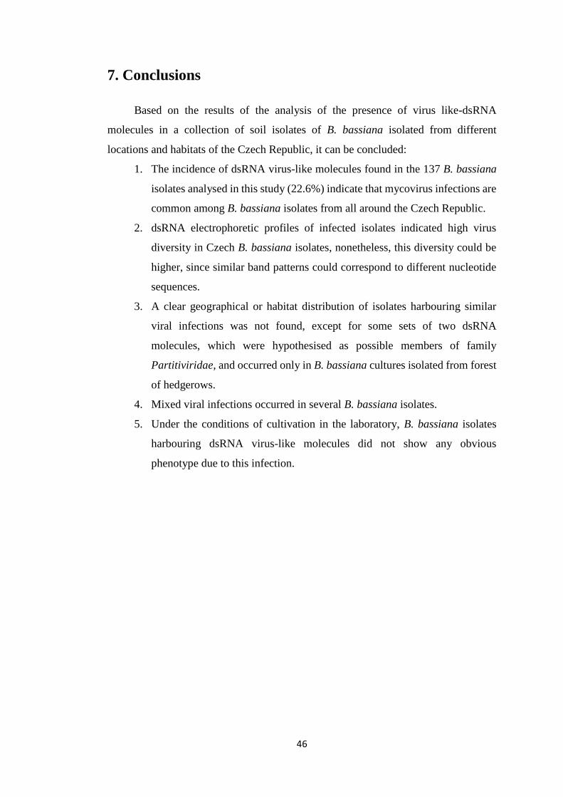
46
7. Conclusions
Based on the results of the analysis of the presence of virus like-dsRNA
molecules in a collection of soil isolates of B. bassiana isolated from different
locations and habitats of the Czech Republic, it can be concluded:
1. The incidence of dsRNA virus-like molecules found in the 137 B. bassiana
isolates analysed in this study (22.6%) indicate that mycovirus infections are
common among B. bassiana isolates from all around the Czech Republic.
2. dsRNA electrophoretic profiles of infected isolates indicated high virus
diversity in Czech B. bassiana isolates, nonetheless, this diversity could be
higher, since similar band patterns could correspond to different nucleotide
sequences.
3. A clear geographical or habitat distribution of isolates harbouring similar
viral infections was not found, except for some sets of two dsRNA
molecules, which were hypothesised as possible members of family
Partitiviridae, and occurred only in B. bassiana cultures isolated from forest
of hedgerows.
4. Mixed viral infections occurred in several B. bassiana isolates.
5. Under the conditions of cultivation in the laboratory, B. bassiana isolates
harbouring dsRNA virus-like molecules did not show any obvious
phenotype due to this infection.

47
8. References
ANAGNOSTAKIS, S. L. (1982). ANAGNOSTAKIS BIOLOGICAL CONTROL OF CHESTNUT BLIGHT. SCIENCE.
VOL. 215, P. 466–471.
ARNOLD, A. E. AND LEWIS, L. C. (2005). ECOLOGY AND EVOLUTION OF FUNGAL ENDOPHYTES, AND
THEIR ROLES AGAINST INSECTS. IN: INSECT-FUNGAL ASSOCIATIONS: ECOLOGY AND EVOLUTION, F. E.
VEGA AND M. BLACKWELL (EDS.). OXFORD UNIVERSITY PRESS, NEW YORK, P. 74-96.
ARNOLD, A. E., MEJIA, L. C., KYLLO, D., ROJAS, E. I., MAYNARD, Z., ROBBINS, N. AND HERRE, E.
A. (2003). FUNGAL ENDOPHYTES LIMIT PATHOGEN DAMAGE IN A TROPICAL TREE. PROCEEDINGS OF THE
NATIONAL ACADEMY OF SCIENCES. VOL. 100, P. 15649-15654.
AUGUSTYNIUK-KRAM, A. AND KRAM, K. J. (2012). ENTOMOPATHOGENIC FUNGI AS AN IMPORTANT
NATURAL REGULATOR OF INSECT OUTBREAKS IN FORESTS (REVIEW). IN: FOREST ECOSYSTEMS - MORE
THAN JUST TREES, DR. JUAN A. BLANCO (ED.), ISBN: 978-953-51-0202-1.
AUNG, O. M., SOYTONG, K. AND HYDE, K. D. (2008). DIVERSITY OF ENTOMOPATHOGENIC FUNGI IN
RAINFORESTS OF CHIANG MAI PROVINCE, THAILAND. FUNGAL DIVERSITY. VOL. 30, P. 15-22.
BIDOCHKA, M. J., MENZIES, F. V. AND KAMP, A. M (2002). GENETIC GROUPS OF THE INSECT-
PATHOGENIC FUNGUS BEAUVERIA BASSIANA ARE ASSOCIATED WITH HABITAT AND THERMAL GROWTH
PREFERENCES. ARCHIVES OF MICROBIOLOGY. VOL. 178, P. 531-537.
BISWAS, B., ADHYA, S., WASHART, P., PAUL, B., TROSTEL, A. N., POWELL, B., CARLTON, R. AND
MERRIL, C. R. (2002). BACTERIOPHAGE THERAPY RESCUES MICE BACTEREMIC FROM A CLINICAL
ISOLATE OF VANCOMYCINRESISTANT ENTEROCOCCUS FAECIUM. INFECTION AND IMMUNITY. VOL. 70, P.
204–210.
BOGO, M. R., QUEIROZ, M. V., SILVA, D. M., GIMENÉZ, M. P., AZEVEDO, J. L. AND SCHRANK,
A. (1996). DOUBLE-STRANDED RNA AND ISOMETRIC VIRUS-LIKE PARTICLES IN THE
ENTOMOPATHOGENIC FUNGUS METARHIZIUM ANISOPLIAE. MYCOLOGICAL RESEARCH. VOL. 100, P. 1468-
1472.
BOUCIAS, D. G., FARMERIE, W. G. AND PENDLAND, J. C. (1998). CLONING AND SEQUENCING THE
CDNA OF THE INSECTICIDAL TOXIN. JOURNAL OF INVERTEBRATE PATHOLOGY. VOL. 72, P. 258-261.
BOZARTH, R. F. (1972). MYCOVIRUSES: A NEW DIMENSION IN MICROBIOLOGY. ENVIRONMENTAL
HEALTH PERSPECTIVES. VOL. 2, P. 23 – 39.
BRIDGE, P. D., CLARK, M. S. AND PEARCE, D. A. (2005). A NEW SPECIES OF PAECILOMYCES ISOLATED
FROM THE ANTARCTIC SPRINGTAIL CRYPTOPYGUS ANTARCTICUS. MYCOTAXON. VOL. 92, P. 213-222,
ISSN 0093-4666.
BUCK, K. (1998). MOLECULAR VARIABILITY OF VIRUSES OF FUNGI. IN: MOLECULAR VARIABILITY OF
FUNGAL PATHOGENS, P. D., BRIDGE, Y. COUTEAUDIER AND J. M. CLACKSON (EDS). CAB
INTERNATIONAL. WALLINGFORD, UK, P. 53-72.
CARRUTHERS, R. I. AND SOPER, R. S. (1987). FUNGAL DISEASES. IN: EPIZOOTIOLOGY OF INSECT
DISEASES, J. R. FUXA AND Y. TANADA. JOHN WILEY AND SONS, NEW YORK, P. 357-416.
CASTRILLO, L. A, GRIGGS, M. H. AND VANDENBERG, J. D. (2004). VEGETATIVE COMPATIBILITY
GROUPS IN INDIGENOUS AND MASS-RELEASED STRAINS OF THE ENTOMOPATHOGENIC FUNGUS BEAUVERIA
BASSIANA: LIKELIHOOD OF RECOMBINATION IN THE FIELD. JOURNAL OF INVERTEBRATE PATHOLOGY. VOL.
86, P. 26–37.
CHARNLEY, A. K. (2003). FUNGAL PATHOGENS OF INSECTS: CUTICLE DEGRADING ENZYMES AND
TOXINS. ADVANCES IN BOTANICAL RESEARCH. VOL. 40, P. 241–321.
CHARNLEY, A. K. AND COLLINS, S. A. (2007). ENTOMOPATHOGENIC FUNGI AND THEIR ROLE IN PEST
CONTROL. IN: THE MYCOTA. ENVIRONMENTAL AND MICROBIAL RELATIONSHIP IV, K. ESSER, ET AL.
SPRINGER, 2ND EDITION, P. 159-187.

48
CHIBA, S., KONDO, H., TANI, A., SAISHO, D. SAKAMOTO, W.,KANEMATSU, S. AND SUZUKI, N. (2011).
WIDESPREAD ENDOGENIZATION OF GENOME SEQUENCES OF NON-RETROVIRAL RNA VIRUSES INTO
PLANT GENOMES. PLOS PATHOGENS. VOL. 7(7)
CHOI, G. H., DAWE, A. L., CHURBANOV, A., SMITH, M. L., MILGROOM, M. G. AND NUSS, D. L.
(2012). MOLECULAR CHARACTERIZATION OF VEGETATIVE INCOMPATIBILITY GENES THAT RESTRICT
HYPOVIRUS TRANSMISSION IN THE CHESTNUT BLIGHT FUNGUS CRYPHONECTRIA PARASITICA. GENETICS.
VOL. 190, P. 113–127.
CHUN, S. J. AND LEE, Y. H. (1997). INHERITANCE OF DSRNAS IN THE RICE BLAST FUNGUS,
MAGNAPORTHE GRISEA. FEMS MICROBIOLOGY LETTERS. VOL. 148, P. 159–162.
DALZOTO, P. R., GLIENKE-BLANCO, CH., KAVA-CORDEIRO, V. AND RIBEIRO, J. Z. (2006).
HORIZONTAL TRANSFER AND HYPOVIRULENCE ASSOCIATED WITH DOUBLE-STRANDED RNA IN
BEAUVERIA BASSIANA. MYCOLOGICAL RESEARCH. VOL. 110, P. 1475–1481.
EILENBERG, J., HAJEK, A. AND LOMER, C. (2001). SUGGESTION FOR UNIFYING THE TERMINOLOGY IN
BIOLOGICAL CONTROL. BIOCONTROL. VOL. 46, P. 387-400, ISSN 1573-8248.
EILENBERG, J., SCHMIDT, N. M., MEYLING, N. AND WOLSTED, C. (2007). PRELIMINARY SURVEY FOR
INSECT PATHOGENIC FUNGI IN ARCTIC GREENLAND. IOBC/WPRS BULLETIN. INSECT PATHOGENS AND
INSECT PARASITIC NEMATODES. VOL. 30, P. 117-118, ISSN 1027-3115.
ELMHOLT, S., FRISVAD, J. C. AND THRANE, U. (1991). THE INFLUENCE OF FUNGICIDES ON SOIL
MYCOFLORA WITH SPECIAL ATTENTION TO TESTS OF FUNGICIDE EFFECTS ON SOIL-BORNE PATHOGENS. IN:
PESTICIDE INTERACTIONS IN CROP PRODUCTION: BENEFICIAL AND DELETERIOUS EFFECTS, J. ALTMAN
(ED.). CRC PRESS, BOCA RATON, FL, P. 227–243
FAUQUET, C. M., MAYO, M. A., MANILOFF, J., DESSELBERGER, U. AND BALL, L. A. (EDS.) (2005).
VIRUS TAXONOMY: EIGHTH REPORT OF THE INTERNATIONAL COMMITTEE ON TAXONOMY OF VIRUSES.
ELSEVIER ACADEMIC PRESS. SAN DIEGO. ISBN 978-0-12-249951-7.
FRAVEL, D. R. (1988). ROLE OF ANTIBIOSIS IN THE BIOCONTROL OF PLANT DISEASES. ANNUAL REVIEW
OF PHYTOPATHOLOGY. VOL. 26, P. 75–91.
GABARTY, A., SALEM, H. M., FOUDA, M. A., ABAS, A. A. AND IBRAHIM, A. A (2014): PATHOGENCITY
INDUCED BY THE ENTOMOPATHOGENIC FUNGI BEAUVERIA BASSIANA AND METARHIZIUM ANISOPLIAE IN
AGROTIS IPSILON (HUFN.). JOURNAL OF RADIATION RESEARCH AND APPLIED SCIENCES. VOL. 7, P. 95-100.
GHABRIAL, S. A. (1998). ORIGIN, ADAPTATION AND EVOLUTIONARY PATHWAYS OF FUNGAL VIRUSES.
VIRUS GENES. VOL. 16, P. 119–131.
GHABRIAL, S. A. (2008). TOTIVIRUSES. IN: ENCYCLOPEDIA OF VIROLOGY, 3RD EDITION, B. W. J. MAHY
AND M. H. V.,VAN REGENMORTEL (EDS.). ELSEVIER, P. 163–174.
GHABRIAL, S. A. AND CASTÓN, J. R. (2011). FAMILY CHRYSOVIRIDAE. IN: VIRUS TAXONOMY: NINTH
REPORT OF THE INTERNATIONAL COMMITTEE ON TAXONOMY OF VIRUSES, A. M. Q. KING, M. J. ADAMS,
E. B. CARSTENS, AND E. J. LEFKOWITZ (EDS.). ELSEVIER ACADEMIC PRESS, P. 509-513, ISBN 978-0-12-
384684-6.
GHABRIAL, S. A. AND NIBERT, M. L. (2009). VICTORIVIRUS, A NEW GENUS OF FUNGAL VIRUSES IN THE
FAMILY TOTIVIRIDAE. ARCHIVES OF VIROLOGY. VOL. 154, P. 373–379.
GHABRIAL, S. A. AND SUZUKI, N. (2010). FUNGAL VIRUSES. IN: DESK ENCYCLOPEDIA OF PLANT AND
FUNGAL VIROLOGY, B. W. J MAHY AND M. H. V. VAN REGENMORTEL (EDS). ELSEVIER, P 517-524.
GHABRIAL, S. A., CASTÓN, J. R., JIANG, D., NIBERT, M. L. AND SUZUKI, N. (2015). 50-PLUS YEARS OF
FUNGAL VIRUSES. VIROLOGY [ONLINE]. FEBRUARY 2015, AVAILABLE AT: HTTP://WWW.SCIENCEDIRECT.
COM/SCIENCE/ARTICLE/PII/S004268221500080X [CIT. 2015-03-30].
GILBERT, L. I. AND GILL, S. S. (2010). INSECT CONTROL: BIOLOGICAL AND SYNTHETIC AGENTS.
ACADEMIC PRESS, P. 490.

49
GOETTEL, M. S., EILENBERG, J. AND GLARE, T. R. (2005). ENTOMOPATHOGENIC FUNGI AND THEIR
ROLE IN REGULATION OF INSECT POPULATIONS. IN: COMPREHENSIVE MOLECULAR INSECT SCIENCE, L. I.
GILBERT, K. IATROU AND S. S. GILL (EDS). ELSEVIER. VOL. 6, P. 361– 406.
GOETTEL, M. S. AND INGLIS, G. D. (1997). FUNGI: HYPHOMYCETES. IN: MANUAL OF TECHNIQUES IN
INSECT PATHOLOGY, L. A. LACEY (ED.). ACADEMIC PRESS, P. 213-249.
GRIFFITHS, D. A., BROWN, D. E. AND JEZEQUEL, S. G. (1993). METABOLISM OF XENOBIOTICS BY
BEAUVERIA BASSIANA. XENOBIOTICA. VOL. 23, P. 1085–1100.
GUL, H. T., SAEED, S. AND KHAN, F. Z. A. (2014). ENTOMOPATHOGENIC FUNGI AS EFFECTIVE INSECT
PEST MANAGEMENT TACTIC: A REVIEW. APPLIED SCIENCES AND BUSINESS ECONOMICS. VOL. 1, P. 10-18.
HAJEK, A. E. AND ST. LEGER, R. J. (1994). INTERACTIONS BETWEEN FUNGAL PATHOGENS AND INSECT
HOST. ANNUAL REVIEW OF ENTOMOLOGY. VOL. 39, P. 293-322.
HARMON G. E., HOWELL, C. R., VITERBO, A., CHET, I. AND LORITO, M. (2004). TRICHODERMA
SPECIES–OPPORTUNISTIC, AVIRULENT PLANT SYMBIONTS. NATURE REVIEW OF MICROBIOLOGY. VOL. 2,
P.43–56.
HEMMATI, F., PELL, J. K., MCCARTNEY, H. A. AND DEADMAN, M. L. (2001). AIRBORNE
CONCENTRATIONS OF CONIDIA OF ERYNIA NEOAPHIDIS ABOVE CEREAL FIELDS. MYCOLOGICAL RESEARCH.
VOL. 105, P. 485–489.
HERRERO, N., DUEÑAS, E., QUESADA-MORAGA, E. AND ZABALGOGEAZCOA, I. (2012). PREVALENCE
AND DIVERSITY OF VIRUSES IN THE ENTOMOPATHOGENIC FUNGUS BEAUVERIA BASSIANA. APPLIED AND
ENVIRONMENTAL MICROBIOLOGY. VOL. 78, P. 8523-8530.
HERRERO, N., SÁNCHEZ MÁRQUEZ, S. AND ZABALGOGEAZCOA, I. (2009). MYCOVIRUSES ARE
COMMON AMONG DIFFERENT SPECIES OF ENDOPHYTIC FUNGI OF GRASSES. ARCHIVES OF VIROLOGY. VOL.
154, P. 327–330.
HERRERO, N. AND ZABALGOGEAZCOA, I. (2011). MYCOVIRUSES INFECTING THE ENTOMOPATHOGENIC
FUNGUS
TOLYPOCLADIUM CYLINDROSPORUM. VIRUS RESEARCH. VOL. 160, P. 409–413.
HIBBETT, D. S., BINDER, M., BISCHOFF, J. F., BLACKWELL, M., CANNON, P. F., ERIKSSON, O.E.,
HUHNDORF, S., JAMES, T., KIRK, P. M., LÜCKING, R., LUMBSCH, H. T., LUTZONI, F., MATHENY, P.
B., MCLAUGHLIN, D. J., POWELL, M. J., REDHEAD, S., SCHOCH, C. L., SPATAFORA, J. W.,
STALPERS, J. A., VILGALYS, R., AIME, M. C., APTROOT, A., BAUER, R., BEGEROW, D., BENNY, G.
L.,CASTLEBURY, L. A., CROUS, P. W., DAI, Y.-C., GAMS, W., GEISER, D. M., GRIFFITH, G.
W.,GUEIDAN, C., HAWKSWORTH, D. L., HESTMARK, G., HOSAKA, K., HUMBER, R. A., HYDE, K. D.,
IRONSIDE, J. E., KŐLJALG, U., KURTZMAN, C. P., LARSSON, K.-H., LICHTWARDT, R., LONGCORE,
J., MIĄDLIKOWSKA, J., MILLER, A., MONCALVO, J.-M., MOZLEY- STANDRIDGE, S., OBERWINKLER,
F., PARMASTO, E., REEB, V., ROGERS, J. D., ROUX, C., RYVARDEN, L., SAMPAIO, J. P., SCHÜßLER,
A., SUGIYAMA, J., THORN, R. G., TIBELL, L., UNTEREINER, W. A., WALKER, C., WANG, Z., WEIR,
A., WEISS, M., WHITE, M. M., WINKA, K., YAO, Y.-J. AND ZHANG, N. (2007). A HIGHER-LEVEL
PHYLOGENETIC CLASSIFICATION OF THE FUNGI. MYCOLOGICAL RESEARCH, VOL. 111, P. 509-547, ISSN
1469-8102.
HILLMAN, B. I. AND CAI, G. (2013). THE FAMILY NARNAVIRIDAE: SIMPLEST OF RNA VIRUSES.
ADVANCES OF VIRUS RESEARCH. VOL. 86, P. 149–176.
HILLMAN, B. I. AND SUZUKI, N. (2004). VIRUSES OF THE CHESTNUT BLIGHT FUNGUS, CRYPHONECTRIA
PARASITICA. ADVANCES OF VIRUS RESEARCH. VOL. 63, P. 423–472.
HOLLINGS, M. (1962). VIRUSES ASSOCIATED WITH DIEBACK DISEASE OF CULTIVATED MUSHROOMS.
NATURE. VOL. 196, P. 962–965.

50
HOWITT, R. L. J., BEEVER, R. E., PEARSON, M. N. AND FORSTER, R. L. S. (2006). GENOME
CHARACTERIZATION OF A FLEXUOUS ROD-SHAPED MYCOVIRUS, BOTRYTIS VIRUS X, REVEALS HIGH
AMINO ACID IDENTITY TO GENES FROM PLANT POTEX-LIKE VIRUSES. ARCHIVES OF VIROLOGY. VOL. 151,
P. 563–579.
HULL, R. (2014). PLANT VIROLOGY. ELSEVIER ACADEMIC PRESS, 5TH EDITION, P. 1104, ISBN 978-0-12-
384871-0.
HUMBER, R. A. (2012). ENTOMOPHTHOROMYCOTA: A NEW PHYLUM AND RECLASSIFICATION FOR
ENTOMOPHTHOROID FUNGI. MYCOTAXON. VOL. 120, P. 477-492.
IKEDA, K., INOUE, K., KIDA, C., UWAMORI, T., SASAKI, A., KANEMATSU, S. AND PARK, P. (2013).
POTENTIATION OF MYCOVIRUS TRANSMISSION BY ZINC COMPOUNDS VIA ATTENUATION OF HETEROGENIC
INCOMPATIBILITY IN ROSELLINIA NECATRIX. APPLIED AND ENVIRONMENTAL MICROBIOLOGY. VOL. 79, P.
3684–3691.
KANEMATSU, S., ARAKAWA, M., OIKAWA, Y., ONOUE, M., OSAKI, H., NAKAMURA, H., IKEDA, K.,
KUGA-UETAKE, Y., NITTA, H., SASAKI, A., SUZAKI, K., YOSHIDA, K. AND MATSUMOTO, N. (2004).
A REOVIRUS CAUSES HYPOVIRULENCE OF ROSELLINIA NECATRIX. PHYTOPATHOLOGY. VOL. 94, P. 561–
568.
KISHI, K., YASUDA, T. AND TAKESHITA, H. (2001). DNASE I: STRUCTURE, FUNCTION, AND USE IN
MEDICINE AND FORENSIC SCIENCE. LEGAL MEDICINE (TOKYO, JAPAN). VOL. 3, P. 69-83.
KLINGEN, I., EILENBERG, J. AND MEADOW, R. (2002). EFFECTS OF FARMING SYSTEM, FIELD MARGINS
AND BAIT INSECT ON THE OCCURRENCE OF INSECT PATHOGENIC FUNGI IN SOILS. AGRICULTURE,
ECOSYSTEMS & ENVIRONMENT. VOL. 91 (2-3), P. 191-198, ISSN 0167-8809.
KOTTA-LOIZOU, I, SIPKOVA, J AND COUTTS, R. H. (2015). IDENTIFICATION AND SEQUENCE
DETERMINATION OF A NOVEL DOUBLE-STRANDED RNA MYCOVIRUS FROM THE ENTOMOPATHOGENIC
FUNGUS BEAUVERIA BASSIANA. ARCHIVES OF VIROLOGY. VOL. 160, P. 873-875.
LEAL, S. C. M., BERTIOLI, D. J., BALL, B. V. AND BUTT, T. M. (1994). PRESENCE OF DOUBLE‐STRANDED RNAS AND VIRUS‐LIKE PARTICLES IN THE ENTOMOPATHOGENIC FUNGUS METARHIZIUM
ANISOPLIAE. BIOCONTROL SCIENCE AND TECHNOLOGY. VOL. 4, P. 89-94.
LI, Z., LI, C., HUANG, B. AND FAN, M. (2001). DISCOVERY AND DEMONSTRATION OF THE TELEOMORPH
OF BEAUVERIA BASSIANA (BALS.) VUILL., AN IMPORTANT ENTOMOGENOUS FUNGUS. CHINESE SCIENCE
BULLETIN. VOL. 46, P. 751–753.
LINDER-BASSO, D., DYNEK, J. N. AND HILLMAN B. I. (2005). GENOME ANALYSIS OF CHRYPHONECTRIA
HYPOVIRUS 4, THE MOST COMMON HYPOVIRUS SPECIES IN NORTH AMERICA. VIROLOGY. VOL. 337, P.
192–203.
LIU, H. (2012). MICROBIAL CONTROL OF CROP PESTS USING ENTOMOPATHOGENIC FUNGI. IN:
INTEGRATED PEST MANAGEMENT: PRINCIPLES AND PRACTICE. D. P. ABROL AND U. SHANKAR (EDS.). CPI
GROUP, P. 237-280, ISBN 978-1-84593-808-6.
LIU, H., FU, Y., JIANG, D., LI, G., XIE J, CHENG, J., PENG, Y., GHABRIAL, S. A. AND YI, X. (2010).
WIDESPREAD HORIZONTAL GENE TRANSFER FROM DOUBLE-STRANDED RNA VIRUSES TO EUKARYOTIC
NUCLEAR GENOMES. JOURNAL OF VIROLOGY. VOL. 84, P. 11876–87
MACIÁ-VICENTE, J. G., PALMA-GUERRERO, J., GÓMEZ-VIDAL, S. AND LOPEZ-LLORCA, L. V.
(2011). NEW INSIGHTS ON THE MODE OF ACTION OF FUNGAL PATHOGENS OF INVERTEBRATES FOR
IMPROVING THEIR BIOCONTROL PERFORMANCE. IN: BIOLOGICAL CONTROL OF PLANT-PARASITIC
NEMATODES: BUILDING COHERENCE BETWEEN MICROBIAL ECOLOGY AND MOLECULAR MECHANISMS. K.
DAVIES AND Y. SPIEGEL (EDS.), SPRINGER, LONDON, P. 203-227, ISBN 978-1-4020-9647-1
MARQUÉZ, L. M., REDMAN, R. S., RODRIGUEZ, R. J. AND ROOSSINCK M. J. (2007). A VIRUS IN A
FUNGUS IN A PLANT: THREE-WAY SYMBIOSIS REQUIRED FOR THERMAL TOLERANCE. SCIENCE. VOL. 315,
P. 513–515.

51
MAZET, I. AND VEY, A. (1995). HIRSUTELLIN A, A TOXIC PROTEIN PRODUCED IN VITRO BY HIRSUTELLA
THOMPSONII. MICROBIOLOGY. VOL. 141, P. 1343-1348, ISSN 1350-0872.
MCCABE, P. M., PFEIFFER, P. AND VAN ALFEN, N. K. (1999). THE INFLUENCE OF DSRNA VIRUSES ON
THE BIOLOGY OF PLANT PATHOGENIC FUNGI. TRENDS IN MICROBIOLOGY. VOL. 7, P. 377-381.
MCCOY, C. W., SAMSON, R. A. AND BOUCIAS, D. G. (1988). ENTOMOGENOUS FUNGI. IN: HANDBOOK
OF NATURAL PESTICIDES, BOCA, RATON, FLA: MR IC PRESS. VOL. 5, MICROBIAL INSECTICIDES, PART A,
ENTOMOGENOUS PROTOZOA AND FUNGI, C. M. IGNOFFO AND N. B. MANDAVA. ISBN 9780849336607
MCFADDEN, J. J. P., BUCK, K. W. AND RAWLINSON, C. J. (1983). INFREQUENT TRANSMISSION OF
DOUBLE-STRANDED RNA VIRUS PARTICLES BUT ABSENCE OF DNA PROVIRUSES IN SINGLE ASCOSPORE
CULTURES OF GAEUMANNOMYCES GRAMINIS. JOURNAL OF GENERAL VIROLOGY. VOL. 64, P. 927–37.
MELZER, M. J. AND BIDOCHKA, M. J. (1998). DIVERSITY OF DOUBLE-STRANDED RNA VIRUSES WITHIN
POPULATIONS OF ENTOMOPATHOGENIC FUNGI AND POTENTIAL IMPLICATIONS FOR FUNGAL GROWTH AND
VIRULENCE. MYCOLOGIA. VOL. 90, P. 586 –594.
MORRIS, T. J. AND DODDS, J. A. (1979). ISOLATION AND ANALYSIS OF DOUBLE-STRANDED RNA FROM
VIRUS-INFECTED PLANT AND FUNGAL TISSUE. PHYTOPATHOLOGY. VOL. 69, P. 854–858.
MOTA-SANCHEZ, D., BILLS, P.S. AND WHALON, M.E. (2002). ARTHROPOD RESISTANCE TO
PESTICIDES: STATUS AND OVERVIEW. IN: PESTICIDES IN AGRICULTURE AND THE ENVIRONMENT. W. B.
WHEELER (ED.), CRC PRESS, NEW YORK, P. 241-272, ISBN 9780824708092
NAKAYASHIKI, H. AND NGUYEN, Q. B. (2008). RNA INTERFERENCE: ROLES IN FUNGAL BIOLOGY.
CURRENT OPINION IN MICROBIOLOGY. ELSEVIER. VOL. 11, P. 494–502.
NIBERT, M. L., GHABRIAL, S. A., MAISS, E., LESKER, T., VAINIO, E. J., JIANG, D. AND SUZUKI, N.
(2014). TAXONOMIC REORGANIZATION OF FAMILY PARTITIVIRIDAE AND OTHER RECENT PROGRESS IN
PARTITIVIRUS RESEARCH. VIRUS RESEARCH. VOL. 188, P. 128–141.
NUSS, D. L. (2005). HYPOVIRULENCE: MYCOVIRUSES AT THE FUNGAL-PLANT INTERFACE. NATURE
REVIEWS MICROBIOLOGY. VOL. 3, P. 632-642.
NUSS, D. L. (2011). MYCOVIRUSES, RNA SILENCING, AND VIRAL RNA RECOMBINATION. ADVANCES IN
VIRUS RESEARCH. VOL. 80, P. 25–48.
NUSS, D. L. AND HILLMAN, B. I. (2011). FAMILY HYPOVIRIDAE. IN: VIRUS TAXONOMY: NINTH REPORT
OF THE INTERNATIONAL COMMITTEE ON TAXONOMY OF VIRUSES, A. M. Q. KING, M. J. ADAMS, E. B.
CARSTENS, AND E. J. LEFKOWITZ (EDS.). ELSEVIER, P. 1029-1033, ISBN 978-0-12-384684-6.
OWNLEY B. H. AND WINDHAM, M. T. (2007). BIOLOGICAL CONTROL OF PLANT PATHOGENS. IN: PLANT
PATHOLOGY: CONCEPTS AND LABORATORY EXERCISES. R. N. TRIGIANO , M. T. WINDHAM AND A. S.
WINDHAM (EDS.), 2ND EDITION. CRC PRESS, BOCA RATON, FLORIDA, P. 423-436.
PEARSON, M. N., BEEVER, R. E., BOINE, B. AND ARTHUR, K. (2009). REVIEW: MYCOVIRUSES OF
FILAMENTOUS FUNGI AND THEIR RELEVANCE TO PLANT PATOLOGY. MOLECULAR PLANT PATHOLOGY.
VOL. 10, P. 115-128.
PORTER, J. R. (1973). AGOSTINO BASSI BICENTENNIAL (1773-1973). BACTERIOLOGICAL REVIEWS.
VOL. 37, P. 284-288.
QUESADA-MORAGA, E., NAVAS-CORTÉS, J. A., MARANHAO, E. A. A., ORTIZ-URQUIZA, A. AND
SANTIAGO-ÁLVAREZ, C. (2007). FACTORS AFFECTING THE OCCURRENCE AND DISTRIBUTION OF
ENTOMOPATHOGENIC FUNGI IN NATURAL AND CULTIVATED SOILS. MYCOLOGICAL RESEARCH. VOL. 111,
P. 947–966.
REHNER, S. A., MINNIS, A. S., SUNG, G.-H., LUANGSA-ARD, J. J., DEVOTTO, L. AND HUMBER, R. A.
(2011). PHYLOGENY AND SYSTEMATICS OF THE ANAMORPHIC, ENTOMOPATHOGENIC GENUS BEAUVERIA.
MYCOLOGIA. VOL. 103, P. 1055–1073.

52
RO, H. S., LEE, N. J., LEE, C.-W. AND LEE, H.-S. (2006). ISOLATION OF A NOVEL MYCOVIRUS OMIV
IN PLEUROTUS OSTREATUS AND ITS DETECTION USING A TRIPLE ANTIBODY SANDWICH-ELISA. JOURNAL
OF VIROLOGICAL METHODS. VOL. 138, P. 24–29.
ROBERTS, D. W. (1981). TOXINS OF ENTOMOPATHOGENIC FUNGI. IN: MICROBIAL CONTROL OF PESTS
AND PLANT DISEASES 1970-1980. H. D. BURGES (ED.), ACADEMIC PRESS, LONDON AND NEW YORK, P.
441-464, ISBN 0121433609.
ROBERTS, D. W. AND HAJEK, A. E. (1992). ENTOMOPATHOGENIC FUNGI AS BIOINSECTICIDES. IN:
FRONTIERS IN INDUSTRIAL MYCOLOGY, G. F. LEATHAM (ED). CHAPMAN & HALL, NEW YORK, P. 144–
159.
ROMAINE, C. P. AND SCHLAGNHAUFFER, B. (1995). PCR ANALYSIS OF THE VIRAL COMPLEX
ASSOCIATED WITH LA FRANCE DISEASE OF AGARICUS BISPORUS. APPLIED AND ENVIRONMENTAL
MICROBIOLOGY. VOL. 61, P. 2322–2325.
ROMO, M., LEUCHTMANN, A., GARCÍA, B. AND ZABALGOGEAZCOA, I. (2007). A TOTIVIRUS
INFECTING THE MUTUALISTIC FUNGAL ENDOPHYTE EPICHLOË FESTUCAE. VIRUS RESEARCH. VOL. 124 , P.
38-43.
ROY, H. E., PELL, J. K. AND ALDERSON, P. G. (2001). TARGETED DISPERSAL OF THE APHID
PATHOGENIC FUNGUS ERYNIA NEOAPHIDIS BY THE APHID PREDATOR COCCINELLA SEPTEMPUNCTATA.
BIOCONTROL SCIENCE AND TECHNOLOGY. VOL. 11, P. 99–110.
RUDGERS, J. A., HOLAH, J., ORR, S. P. AND CLAY, K. (2007). FOREST SUCCESSION SUPPRESSED BY AN
INTRODUCED PLANT-FUNGAL SYMBIOSIS. ECOLOGY. VOL. 88, P. 18-25.
SAMUELS, R. I., REYNOLDS, S. E. AND CHARNLEY, A. K. (1988). CALCIUM CHANNEL ACTIVATION OF
INSECT MUSCLE BY DESTRUXINS, INSECTICIDAL COMPOUNDS PRODUCED BY THE ENTOMOPATHOGENIC
FUNGUS METARRHIZIUM ANISOPLIAE. COMPARATIVE BIOCHEMISTRY AND PHYSIOLOGY. VOL. 90, P. 403–
412.
SANDERLIN, R. S. AND GHABRIAL, S. A. (1978). PHYSICOCHEMICAL PROPERTIES OF TWO DISTINCT
TYPES OF VIRUS-LIKE PARTICLES FROM HELMINTHOSPORIUM VICTORIAE. VIROLOGY. VOL. 87, P. 142 –
151.
SCHULZ, B. AND BOYLE, C. (2005). THE ENDOPHYTIC CONTINUUM. MYCOLOGICAL RESEARCH. VOL.
109, P. 661-686.
SHAHID, A. A., RAO, A. Q., BAKHSH, A. AND HUSNAIN, T. (2012). ENTOMOPATHOGENIC FUNGI AS
BIOLOGICAL CONTROLLERS: NEW INSIGHTS INTO THEIR VIRULENCE AND PATHOGENICITY. ARCHIVES OF
BIOLOGICAL SCIENCE, BELGRADE. VOL. 64, P. 21-42.
SOSNOWSKA, D., BAŁAZY, S., PRISHCHEPA, L. AND MIKULSKAYA, N. (2004). BIODIVERSITY OF
ARTHROPOD PATHOGENS IN THE BIAŁOWIEŹA FOREST. JOURNAL OF PLANT PROTECTION RESEARCH. VOL.
44, P. 313-321, ISSN 1427-4345.
STEINHAUS, E. A. (1975). DISEASE IN A MINOR CHORD. COLUMBUS: OHIO STATE UNIVERSITY PRESS.
STIELOW, B. AND MENZEL, W. (2010). COMPLETE NUCLEOTIDE SEQUENCE OF TAV1,A NOVEL
TOTIVIRUS ISOLATED FROM A BLACK TRUFFLE ASCOCARP (TUBER AESTIVUM VITTAD.). ARCHIVES OF
VIROLOGY. VOL. 155, P. 2075–2078.
SULAKVELIDZE, A., ALAVIDZE, Z. AND MORRIS, J. G. JR (2001). BACTERIOPHAGE THERAPY.
ANTIMICROBIAL AGENTS AND CHEMOTHERAPY. VOL. 45, P. 649–659.
SUN, B.-D. AND LIU X.-Z. (2008). OCCURRENCE AND DIVERSITY OF INSECT-ASSOCIATED FUNGI IN
NATURAL SOILS IN CHINA. APPLIED SOIL ECOLOGY. VOL. 39 (1), (MAY 2008), P. 100-108, ISSN 1873-
0272.

53
SUNG, G.-H., HYWEL-JONES, N. L., SUNG, J.-M., LUANGSA-ARD, J. J., SHRESTHA, B. AND
SPATAFORA, J. W. (2007). PHYLOGENETIC CLASSIFICATION OF CORDYCEPS AND THE CLAVICIPITACEOUS
FUNGI. STUDIES IN MYCOLOGY. VOL. 57, P. 5–59, ISSN 1872-9797.
TUOMIVIRTA, T. T., KAITERA, T. AND HANTULA, J. (2009). A NOVEL PUTATIVE VIRUS OF
GREMMENIELLA ABIETINA TYPE B (ASCOMYCOTA: HELOTIACEAE) HAS A COMPOSITE GENOME WITH
ENDORNAVIRUS AFFINITIES. JOURNAL OF GENERAL VIROLOGY. VOL. 90, P. 2299–2305.
VAN DE SANDE, W. W. J., LO-TEN-FOE, J. R., VAN BELKUM, A., NETEA, M. G., KULLBERG, B. J. AND
VONK, A. G. (2010). MYCOVIRUSES: FUTURE THERAPEUTIC AGENTS OF INVASIVE FUNGAL INFECTIONS
IN HUMANS?. EUROPEAN JOURNAL OF CLINICAL MICROBIOLOGY & INFECTIOUS DISEASES. VOL. 29, P.
755-763.
VAN DER WERF, H. M. G (1996). ASSESSING THE IMPACT OF PESTICIDES ON THE ENVIRONMENT.
AGRICULTURE, ECOSYSTEMS & ENVIRONMENT, VOL. 60, P. 81–96.
VAN DIEPENINGEN, A. D., DEBETS, A. J. M. AND HOEKSTRA, R. F. (2006). DYNAMICS OF DSRNA
MYCOVIRUSES IN BLACK ASPERGILLUS POPULATIONS. FUNGAL GENETICS AND BIOLOGY. VOL. 43, P. 446–
452.
VÄNNINEN, I. (1995). DISTRIBUTION AND OCCURRENCE OF FOUR ENTOMOPATHOGENIC FUNGI IN
FINLAND: EFFECT OF GEOGRAPHICAL LOCATION, HABITAT TYPE AND SOIL TYPE. MYCOLOGICAL
RESEARCH, VOL. 100, P. 93–101, ISSN 1469-8102.
VEGA, F. E. (2008). INSECT PATHOLOGY AND FUNGAL ENDOPHYTES. JOURNAL OF INVERTEBRATE
PATHOLOGY. VOL. 98, P. 277-279.
VEGA, F. E., GOETTEL, M. S., BLACKWELL, M., CHANDLER, D., JACKSON, M. A., KELLER, S.,
KOIKE, M., MANIANIA, N. K., MONZON, A., OWNLEY, B. H., PELL, J. K., RANGEL, D. E. N. AND
ROY, H. E. (2009). FUNGAL ENTOMOPATHOGENS: NEW INSIGHTS ON THEIR ECOLOGY. FUNGAL ECOLOGY
2, P. 149–159.
VEGA, F. E., POSADA, F., AIME, M. C., PAVA-RIPOLL, M., INFANTE, F. AND REHNER, S. A. (2008).
ENTOMOPATHOGENIC FUNGAL ENDOPHYTES. BIOLOGICAL CONTROL. VOL. 46, P. 72-82.
VEY, A., HOAGLAND, R. E. AND BUTT, T. M. (2001). TOXIC METABOLITES OF FUNGAL BIOCONTROL
AGENTS. IN: FUNGI AS BIOIOCONTROL AGENTS: PROGRESS, PROBLEMS AND POTENTIAL, T. M. BUTT, C.
W. JACKSON AND N. MAGAN (EDS.). CAB INTERNATIONAL, WALLINGFORD, UK. P. 311–346.
VOTH, P. D., MAIRURA, L., LOCKHART, B. E. AND MAY, G. (2006). PHYLOGEOGRAPHY OF USTILAGO
MAYDIS VIRUS H1 IN THE USA AND MEXICO. THE JOURNAL OF GENERAL VIROLOGY. VOL. 87, P. 3433–
3441.
WEI, C. Z., OSAKI, H., IWANAMI, T., MATSUMOTO, N. AND OHTSU, Y. (2004). COMPLETE NUCLEOTIDE
SEQUENCES OF GENOME SEGMENTS 1 AND 3 OF ROSELLINIA ANTI-ROTVIRUS IN THE FAMILY REOVIRIDAE.
ARCHIVES OF VIROLOGY. VOL. 149, P. 773–777.
WEISER, J. AND MATHA, V. (1988). TOLYPIN: A NEW INSECTICIDAL METABOLITE OF FUNGI OF THE
GENUS TOLYPOCLADIUM. JOURNAL OF INVERTEBRATE PATHOLOGY. VOL. 51, P. 94-96, ISSN 0022-2011.
WICKNER, R. B., FUJIMURA, T. AND ESTEBAN, R. (2013). VIRUSES AND PRIONS OF SACCHAROMYCES
CEREVISIAE. ADVANCES OF VIRUS RESEARCH. VOL. 86, P. 1–36.
WICKNER, R. B., GHABRIAL, S. A., NIBERT, M. L., PATTERSON, J. L. AND WANG, C. C. (2011).
FAMILY TOTIVIRIDAE. IN: VIRUS TAXONOMY: NINTH REPORT OF THE INTERNATIONAL COMMITTEE ON
TAXONOMY OF VIRUSES, A. M. Q. KING, M. J. ADAMS, E. B. CARSTENS, AND E. J. LEFKOWITZ (EDS.).
ELSEVIER ACADEMIC PRESS, P. 639-650, ISBN 978-0-12-384684-6.
XIE, J. AND JIANG, D. (2014). NEW INSIGHTS INTO MYCOVIRUSES AND EXPLORATION FOR THE
BIOLOGICAL CONTROL OF CROP FUNGAL DISEASES. ANNUAL REVIEW OF PHYTOPATHOLOGY. VOL. 52, P.
45-68.
54
XU, Y., OROZCO, R., WIJERATNE, K. E. M., ESPINOSA-ARTILES, P., GUNATILAKA, L. A. A, STOCK,
P. S. AND MOLNÁR, I. (2009). BIOSYNTHESIS OF THE CYCLOOLIGOMER DEPSIPEPTIDE BASSIANOLIDE, AN
INSECTICIDAL VIRULENCE FACTOR OF BEAUVERIA BASSIANA. FUNGAL GENETICS AND BIOLOGY. VOL. 46,
P. 353–364.
YIE, S. W., KHALIFA, M. E., HAHN, T. AND PEARSON, M. N. (2014). MOLECULAR CHARACTERIZATION
OF A NOVEL VICTORIVIRUS FROM THE ENTOMOPATHOGENIC FUNGUS BEAUVERIA BASSIANA. ARCHIVES OF
VIROLOGY. VOL. 159, P. 1321–1327.
YILMAZ, M., OZIC, C. AND GOK, I. (2012). PRINCIPLES OF NUCLEIC ACID SEPARATION BY AGAROSE GEL
ELECTROPHORESIS. IN: GEL ELECTROPHORESIS - PRINCIPLES AND BASICS, DR. SAMEH MAGDELDIN (ED.).
INTECH. P. 33-41, ISBN: 978-953-51-0458-2.
YU, H. J., LIM, D. AND LEE, H.-S. (2003). CHARACTERIZATION OF A NOVEL SINGLE STRANDED RNA
MYCOVIRUS IN PLEUROTUS OSTREATUS. VIROLOGY. VOL. 314, P. 9–15.
YU, X., LI, B., FU, Y., JIANG, D., GHABRIAL, S. A., LI, G., PENG, Y., XIE, J., CHENG, J., HUANG, J.
AND YI, X. (2010). A GEMINIVIRUS-RELATED DNA MYCOVIRUS THAT CONFERS HYPOVIRULENCE TO A
PLANT PATHOGENIC FUNGUS. PROCEEDINGS OF THE NATIONAL ACADEMY OF SCIENCES OF THE UNITED
STATES OF AMERICA. VOL. 107, P. 8387– 8392.
YU, Y. T. AND MANLEY, J. L. (1986). STRUCTURE AND FUNCTION OF THE S1 NUCLEASE-SENSITIVE SITE
IN THE ADENOVIRUS LATE PROMOTER. CELL. VOL. 45, P. 743-751.
ZHANG, X. AND NUSS, D. L. (2008). A HOST DICER IS REQUIRED FOR DEFECTIVE VIRAL RNA
PRODUCTION AND RECOMBINANT VIRUS VECTOR RNA INSTABILITY FOR A POSITIVE SENSE RNA VIRUS.
PROCEEDINGS OF THE NATIONAL ACADEMY OF SCIENCES OF THE UNITED STATES OF AMERICA. VOL. 105,
P. 16749-16754.
ZIMMERMANN, G. (1986). THE GALLERIA BAIT METHOD FOR DETECTION OF ENTOMOPATHOGENIC
FUNGI IN SOIL. JOURNAL OF APPLIED ENTOMOLOGY. VOL. 102, P. 213-215.

55
9. Supplementary materials
Solutions
STE 10X (400 ml):
40 ml Tris-HCl 1M (tris base for molecular biology, Fisher Science),
80 ml NaCl 5M (sodium chloride, Fisher Science),
4 ml EDTA (edetic acid G. R., Lach-Ner),
276 ml sterile water
STE 1X: from STE 10X
BENTONITE (60 mg/ml) (80 ml):
4.8 g bentonite (bentonite, Acros Organics),
80 ml sterile water
SDS 10 % (80 ml):
8g SDS (sodium dodecyl sulphate, Fisher Science),
80 ml sterile water
Extraction buffer (50 ml):
10 ml STE 10X,
5 ml SDS 10%,
50 µl 2-mercaptoethanol (for electrophoresis, Fisher Science),
Sterile water to a final volume of 50 ml
STE 15 (200 ml):
30 ml absolute ethanol (HPLC grade absolute ethanol, Fisher Science),
20 ml STE 10X,
Sterile water to a final volume of 200 ml

56
Pictures
a)
b)
Fig. 9. a) Electrophoretic profiles of dsRNA elements present in isolates of B. bassiana
before enzymatic treatment. Lanes: 1, isolate NBBBA-1; 2, NBBBA-5; 3, NBBBA-6; 4, NBBBA-8; 5,
NBBBA-9; 6, MBBBA-4; 7, MBBBA-5; 8, MBBBA-13; 9, NMBBA-1; 10, SMBBA-2; 11, SMBBA-3;
12, HBBA-1; 13, SBBBA-4; 14, SBBBA-5; 15, SBBBA-6; 16, SBBBA-25; 17, WBBBA-5. b)
Electrophoretic profiles of dsRNA elements present in isolates of B. bassiana before enzymatic
treatment. Lanes: 18, isolate NBBBA-4; 19, HBBA-5; 20, MBBBA-11; 21, SBBBA-7; 22, SBBBA-10;
23, HBBA-9; 24, NBBBA-10; 25, SBBBA-1; 26, NMBBA-4; 27, SBBBA-2; 28, SMBBA-19; 29,
NMBBA-9; 30, SBBBA-27; 31, SBBBA-26. Lane Kbp contains molecular size markers, and the values
on the left are sizes in kilobase pairs.