Embed Size (px)
Citation preview
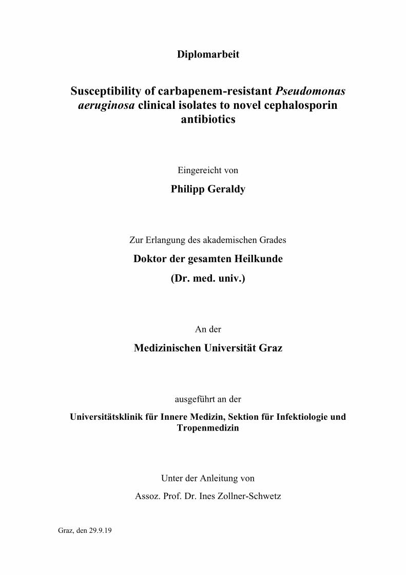
Diplomarbeit
Susceptibility of carbapenem-resistant Pseudomonas
aeruginosa clinical isolates to novel cephalosporin
antibiotics
Eingereicht von
Philipp Geraldy
Zur Erlangung des akademischen Grades
Doktor der gesamten Heilkunde
(Dr. med. univ.)
An der
Medizinischen Universität Graz
ausgeführt an der
Universitätsklinik für Innere Medizin, Sektion für Infektiologie und
Tropenmedizin
Unter der Anleitung von
Assoz. Prof. Dr. Ines Zollner-Schwetz
Graz, den 29.9.19

1
Eidesstattliche Erklärung
Ich erkläre ehrenwörtlich, dass ich die vorliegende Arbeit selbstständig und ohne fremde
Hilfe verfasst habe, andere als die angegebenen Quellen nicht verwendet habe und die den
benutzten Quellen wörtlich oder inhaltlich entnommenen Stellen als solche kenntlich
gemacht habe.
Graz, am 29.9.2019 Philipp Geraldy eh

2
Danksagung
Mein Dank gilt Frau Assoz. Prof. Zollner-Schwetz für steten Rat und Geduld.
3
Inhaltsverzeichnis
Eidesstattliche Erklärung.................................................................................................... 1
Danksagung ....................................................................................................................... 2
Inhaltsverzeichnis .............................................................................................................. 3
Abkürzungen ..................................................................................................................... 5
Abbildungsverzeichnis ....................................................................................................... 7
Tabellenverzeichnis ........................................................................................................... 8
Zusammenfassung ............................................................................................................. 9
Abstract ........................................................................................................................... 10
1. Introduction .............................................................................................................. 11
1.1 Clinical relevance of P. aeruginosa ........................................................................ 13
1.2 Antimicrobial resistance in P. aeruginosa ............................................................... 14
1.3 Biofilm ................................................................................................................... 17
1.4 Quorum sensing ...................................................................................................... 18
1.5 Virulence and Pathogenicity ................................................................................... 20
1.6 Treatment options for P. aeruginosa ....................................................................... 21
Novel antimicrobial substances ................................................................................. 22
Ceftazidime/Avibactam ............................................................................................ 22
Ceftolozane/Tazobactam ........................................................................................... 22
Ceftobiprole .............................................................................................................. 23
1.7 Emerging strategies against P. aeruginosa .............................................................. 24
Vaccinations ............................................................................................................. 24
Phage Therapy .......................................................................................................... 24
Murepavadin ............................................................................................................. 25
1.8 KRINKO classification ........................................................................................... 26
1.9 Aim of this paper .................................................................................................... 26
4
2. Methods .................................................................................................................... 27
Susceptibility and MBL testing ..................................................................................... 27
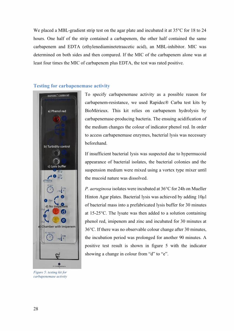
Testing for carbapenemase activity ............................................................................... 28
3. Results ...................................................................................................................... 29
Origin of isolates .......................................................................................................... 29
Susceptibility of P. aeruginosa isolates to novel cephalosporin antibiotics ................... 30
Distribution of MICs .................................................................................................... 31
Phenotypic testing of Carbapenemase and MBL production ......................................... 36
4. Discussion ................................................................................................................ 37
Bibliography .................................................................................................................... 40
5
Abkürzungen
AHL Acylated homoserine lactone
BHL N-butyrylhomoserine lactone
BSI blood stream infection
CF cystic fibrosis
EUCAST The European Committee on Antimicrobial Susceptibility Testing
HHQ 2-heptyl-4-quinolone
ICU intensive care unit
IQS integrated quorum sensing
KPC Klebsiella pneumoniae-based carbapenemase
MRGN multidrug-resistant Gram-negative
MRSA methicillin-resistant Staphylococcus aureus
OdDHL N-(3-oxododecanoyl)-homoserine lactone
OMPTA outer membrane protein targeting antibiotic
P. aeruginosa Pseudomonas aeruginosa
PBP penicillin binding protein
PK/PD pharmacokinetic/pharmacodynamic
PMN polymorphonuclear neutrophil
PQS Pseudomonas quinolone signal
QS quorum sensing
ROS reactive oxygen species
T2SS type 2 secretion system

6
T3SS type 3 secretion system
VAP ventilation associated pneumonia
XDR extensive drug-resistant
7
Abbildungsverzeichnis
Figure 1: illustration of the bacterial structure of P. aeruginosa ........................................ 12
Figure 2: hierarchic structure of QS organisation.............................................................. 20
Figure 3: susceptibility testing for ceftazidime and ceftobiprole ....................................... 27
Figure 4: testing for MBL-activity and susceptibility to colistin ....................................... 27
Figure 5: testing kit for carbapenemase activity ................................................................ 28
8
Tabellenverzeichnis
Table 1: percentage of isolates susceptible to each antibiotic and isolates that showed
MBL-activity ................................................................................................................... 30
Table 2: frequency of each MIC for meropenem .............................................................. 31
Table 3: frequency of each MIC for imipenem ................................................................. 31
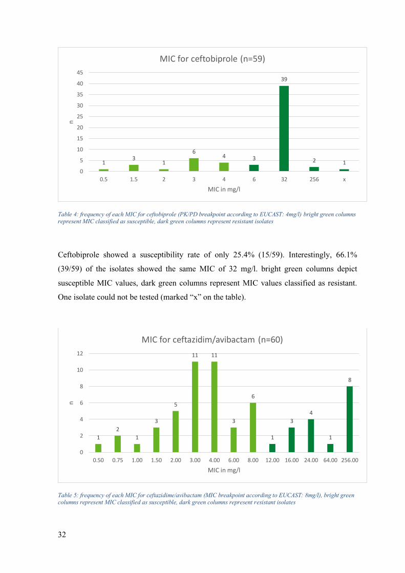
Table 4: frequency of each MIC for ceftobiprole .............................................................. 32
Table 5: frequency of each MIC for ceftazidime/avibactam .............................................. 32
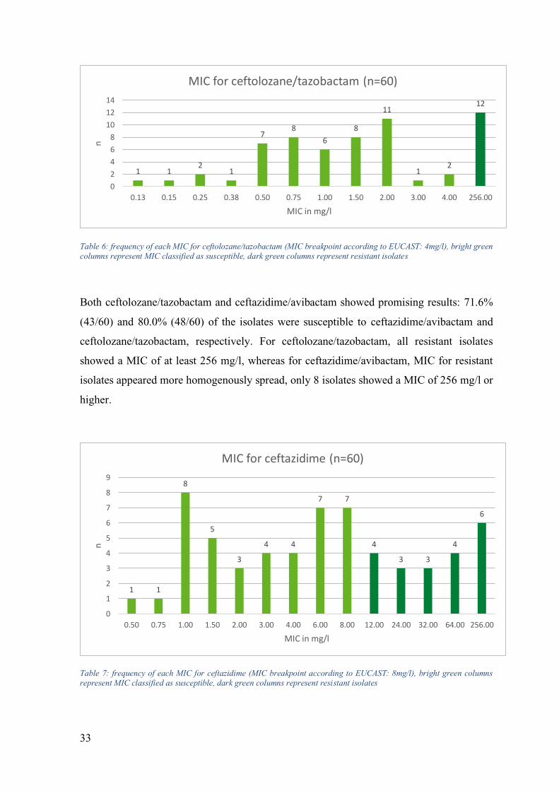
Table 6: frequency of each MIC for ceftolozane/tazobactam ............................................ 33
Table 7: frequency of each MIC for ceftazidime............................................................... 33
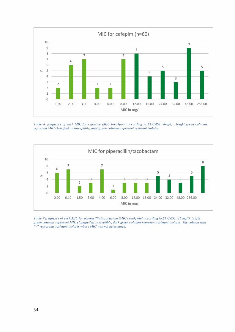
Table 8: frequency of each MIC for cefepime .................................................................. 34
Table 9: frequency of each MIC for piperacillin/tazobactam ............................................ 34
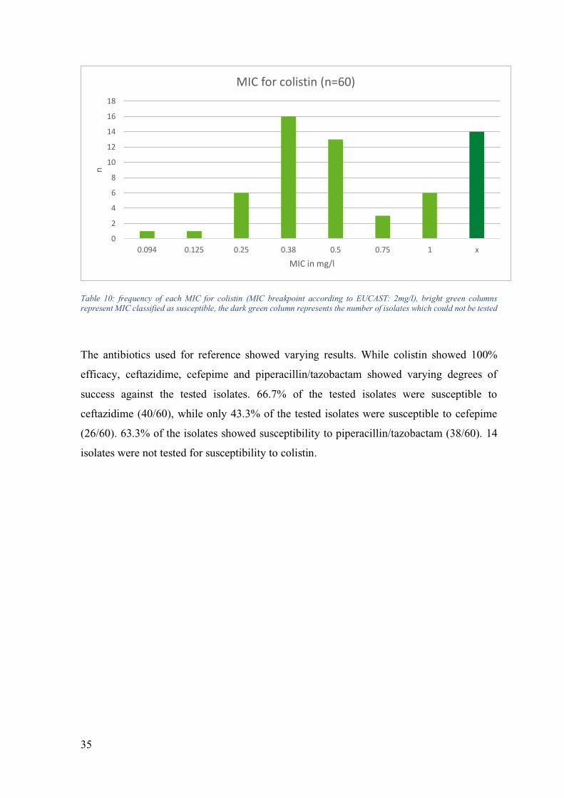
Table 10: frequency of each MIC for colistin ................................................................... 35
Table 11: comparison of MBL- and carbapenemase-activity ............................................ 36
9
Zusammenfassung
Hintergrund: Carbapeneme sind wichtige Antibiotika in der Therapie von Infekten, welche
durch multiresistente Pseudomonas aeruginosa-Stämme ausgelöst werden. Sowohl weltweit
als auch regional im mikrobiologischen Labor der Universitätsklinik für Innere Medizin an
der Medizinischen Universität Graz, werden besorgniserregende Zahlen Carbapenem-
resistenter P. aeruginosa Isolate vermerkt. Vor kurzem wurden drei weitere Cephalosporine
für die Therapie von P. aeruginosa-Infektionen zugelassen (Ceftolozan/Tazobactam,
Ceftazidim/Avibactam und Ceftobiprol). Wir testeten die Empfindlichkeit Carbapenem-
resistenter P. aeruginosa Isolate gegenüber diesen neuen Cephalosporine.
Methoden: 60 Carbapenem-resistente P. aeruginosa-Isolate wurden verwendet. Wir
bestimmten die minimale Hemmkonzentration (MHK) mittels Gradientstreifentests
(BioMérieux) entsprechend dem von EUCAST empfohlenen Standardprocedere.
Potenzielle Carbapenemase-Aktivität wurde mittels Rapidec® Carba Testkits bestimmt, zur
Evaluierung der MBL-Aktivität wurden E-Teststreifen (BioMérieux) verwendet.
Ergebnisse: 80,0% (48/60) der Carbapenem-resistenten P. aeruginosa-Isolate waren
empfindlich auf Ceftolozan/Tazobactam (MHK50 = 4 mg/l). 71,7% (43/60) zeigten
Empfindlichkeit auf Ceftazidim/Avibactam (MHK50 = 4 mg/l). Im Vergleich dazu waren
66,7% (40/60) der Isolate empfindlich auf die Therapie mit Ceftazidim ohne β-Lactamase-
Inhibitor. 25,0% (15/60) der Keime waren empfindlich auf Ceftobiprol. Für diese
Resistenzbestimmung von Ceftobiprol verwendeten wir PK/PD (pharmakakokinetisch-
pharmakodynamische, speziesunabhängige) Grenzwerte (MHK = 32mg/l). 22,0% (13/59)
der 59 getesteten P. aeruginosa-Isolate zeigten MBL-Aktivität.
Zusammenfassung: Die neuen Cephalosporine Ceftolozan/Tazobactam und
Ceftazidim/Avibactam erscheinen vielversprechende Therapieoptionen in der Therapie
Carbapenem-resistenter P. aeruginosa-Infekte darzustellen.
10
Abstract
Background: Carbapenems are important antimicrobials for the treatment of infections due
to multidrug-resistant Pseudomonas aeruginosa. Alarming numbers of carbapenem-resistant
Pseudomonas isolates are found worldwide but also locally at the Microbiology Laboratory,
Dept. of Internal Medicine, Medical University of Graz. Three novel antimicrobial
cephalosporin agents (ceftolozane/tazobactam, ceftazidime/avibactam and ceftobiprole)
have been introduced for the treatment of Pseudomonas infections. We tested the
susceptibility of carbapenem-resistant P. aeruginosa clinical isolates to these antimicrobial
agents.
Materials/Methods: 60 carbapenem-resistant P. aeruginosa clinical isolates were identified.
MIC (minimal inhibitory concentration) was determined using gradient strip tests
(BioMérieux) using standard procedures according to EUCAST, carbapenemase-activity
was detected by using Rapidec® Carba testing kits and MBL activity was detected by using
E-test strips.
Results: 80.0% (48/60) of carbapenem-resistant P. aeruginosa isolates were susceptible to
ceftolozane/tazobactam (MIC50 = 4 mg/l). 71.7% (43/60) were susceptible to
ceftazidime/avibactam (MIC50 = 4 mg/l). In comparison, 66.7% (40/60) were susceptible to
ceftazidime alone (MIC50 = 4 mg/l). 25.0% (15/60) were susceptible to ceftobiprole using
PK/PD breakpoints (MIC50 = 32 mg/l). 22.0% (13/59) of the tested P. aeruginosa isolates
expressed MBL-activity.
Conclusions: The novel cephalosporin combinations ceftolozane/tazobactam and
ceftazidime/avibactam appear to be a promising treatment option in carbapenem-resistant P.
aeruginosa.
11
1. Introduction
Pseudomonas aeruginosa is a bacterium, which is often acquired in a clinical setting and can
be extraordinarily hard to treat. It has various possibilities of expressing virulence or
resistance to antimicrobial substances and multiple ways of adapting to environmental
stresses (Moradali, Ghods et al. 2017). This diversity is reflected by its large genome. The
strain PAO1 has been sequenced in 2000 by Stover, Pham et al., counting 6.3 million base
pairs (Stover, Pham et al. 2000).
P. aeruginosa is Gram negative and aerobic. It thrives in soil and water, as well as on animal
and plant tissue (Green, Schroth et al. 1974, Loveday, Wilson et al. 2014). It is often found
in wet environments, such as sinks, toilets, washing machines, bathtubs as well as sewage
pipes. Consequentially, P. aeruginosa can cause so-called hot tub folliculitis and otitis
externa in swimmers (Reid, Porter 1981, Yu, Cheng et al. 2007). Due to its extraordinary
perseverance, P. aeruginosa may even be found in certain disinfectants, if those contain
traces of biological components (Burdon, Whitby 1967).
In a clinical setting, P. aeruginosa is a highly relevant pathogen due to its tendency to cause
nosocomial infections such as wound infections, infections in the genito-urinary system and
severe, nearly ineradicable chronic lung infections in patients suffering from Cystic Fibrosis
(Bjarnsholt, Jensen et al. 2009). In Austria, an incidence of 8.0 infections per 100,000
healthy individuals was estimated in 2017 (BMGF 2017). Its ability to form biofilms, the
innate resistance to a variety of antibiotics and its ability to rapidly develop resistance to
antimicrobial chemotherapy provide notable therapeutic difficulties (Mulcahy, Isabella et al.
2014, Breidenstein, de la Fuente-Núñez et al. 2011). Morphologically, P. aeruginosa is a
rod-shaped bacterium, expressing multiple pili and a single flagellum, which provides P.
aeruginosa with unidirectional motility. Depending on multiple factors, such as the density
12
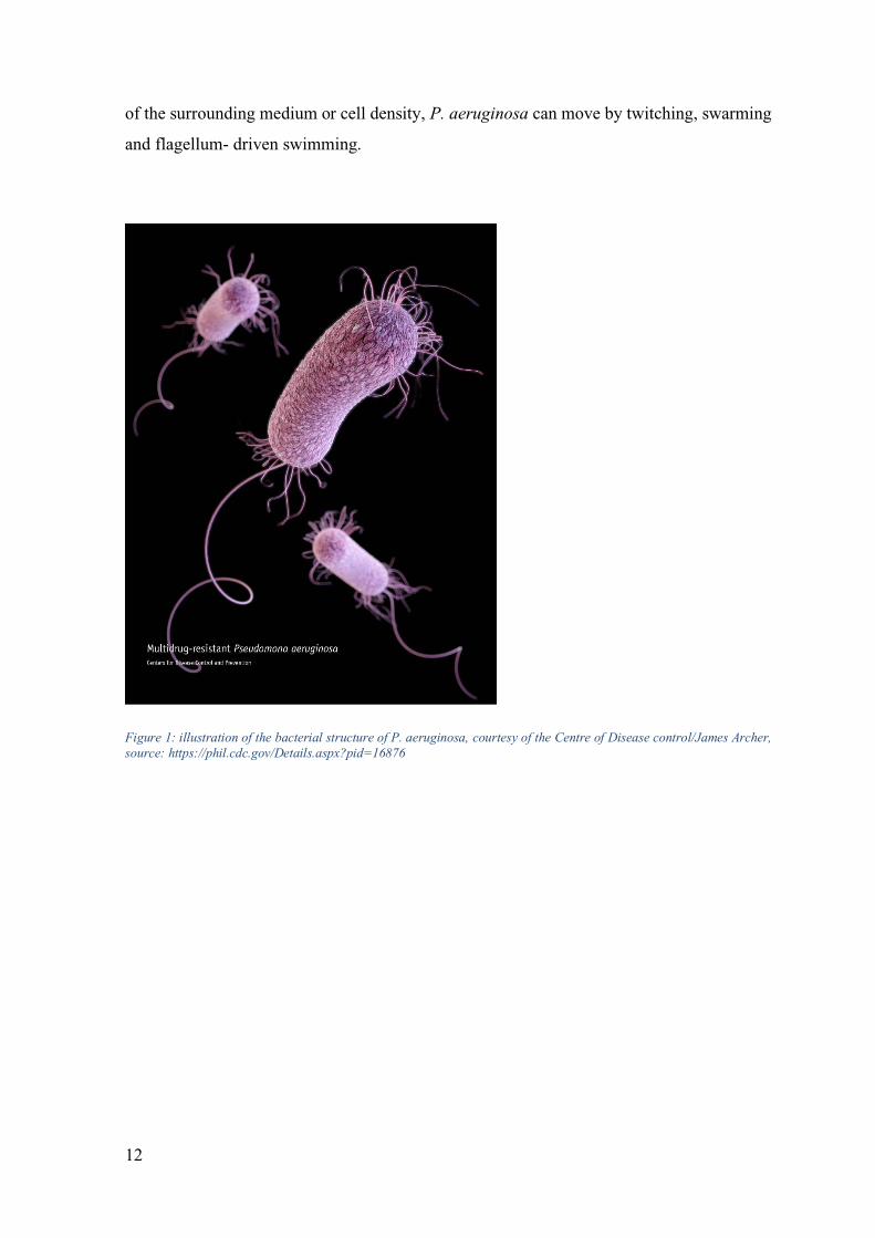
of the surrounding medium or cell density, P. aeruginosa can move by twitching, swarming
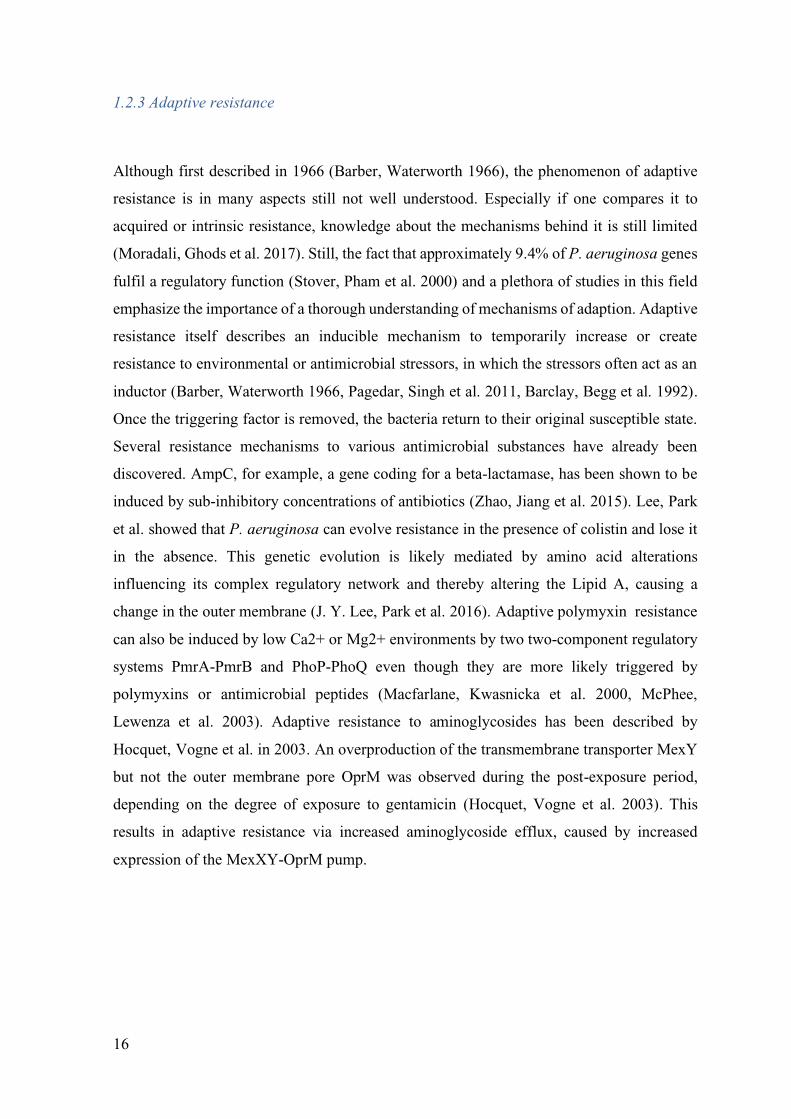
and flagellum- driven swimming.
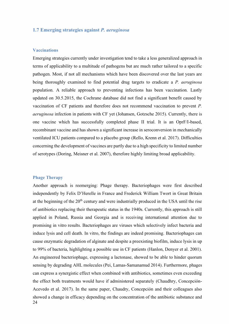
Figure 1: illustration of the bacterial structure of P. aeruginosa, courtesy of the Centre of Disease control/James Archer,
source: https://phil.cdc.gov/Details.aspx?pid=16876
13
1.1 Clinical relevance of P. aeruginosa
The AURES, a yearly report provided by the Austrian ministry of health and women
(Bundesministerium für Gesundheit und Frauen) shows data on the development and
prevalence of different human pathogens. In 2016, there were 697 newly reported isolates
of P. aeruginosa derived from a blood sample, indicating an incidence of P. aeruginosa
bloodstream infections (BSI) of 8.0 per 100,000 healthy individuals per year (BMGF 2017).
At the university hospital in Graz (LKH Universitätsklinikum Graz), Department of Internal
Medicine , P. aeruginosa was detected in blood samples (n=46), urine samples derived from
midstream urine or from a catheter (n=69 and n=77, respectively), from sputum or tracheal
secret, derived from bronchioalveolar lavage (n=55 and n=30, respectively), as well as from
material obtained via aspiration (n=4), from the tip of a central venous line (n=1) and from
wounds by swabs (n=142) in 2017 (local report on antimicrobial susceptibility). This
demonstrates the diversity of infections caused by P. aeruginosa as well as its ability to
colonize patients. Unfortunately, these infections are often associated with high mortality
and morbidity, as Thaden, Park et al. have shown for bloodstream infections: BSI caused by
P. aeruginosa are linked to the highest mortality, showing a hazard ratio of 1.435 compared
to BSI caused by Gram-negative bacteria and Staphylococcus aureus, regardless of
comorbidities (Thaden, Park et al. 2017). In the USA (United States of America), an overall
crude mortality of 29.5% for P. aeruginosa septicaemia between 1999 and 2008 has been
reported by Ani, Farshidpanah et al. in 2015 by examining data of over 5,000,000 severe
sepsis hospitalisations (Ani, Farshidpanah et al. 2015).
14
1.2 Antimicrobial resistance in P. aeruginosa
Antimicrobial resistance is a significant obstacle in the treatment of infections caused by P.
aeruginosa, as the yearly surveillance report published by the European Centre for Disease
Prevention and Control undoubtedly shows (European Centre for Disease Prevention and
Control 2017). Approximately one third of P. aeruginosa isolates found in cerebrospinal
fluid or blood in Europe showed resistance to at least one antibiotic substance commonly
used in treatment of such infections (carbapenems, fluoroquinolones, piperacillin with or
without tazobactam, aminoglycosides and ceftazidime), 4.9% of which showing resistance
to three of the tested antimicrobial groups and 4.3% showing resistance to four tested
antimicrobial groups and 4.4% being resistant to five different antimicrobial groups
(European Centre for Disease Prevention and Control 2018).
1.2.1 Intrinsic resistance
An intrinsic resistance of a bacterial pathogen describes its innate ability and its genetic
reservoir to develop or express resistance to certain antibiotics without acquiring foreign
DNA (Alvarez-Ortega, Wiegand et al. 2011). The intrinsic resistance of P. aeruginosa is
based on multiple different principles which will be described in the following. First of all,
it possesses a very low outer membrane permeability (Hancock 1998) which acts as a
selective barrier for beta-lactam antibiotics (Nicas, Hancock 1983). Although most Gram-
negative bacteria share that common trait of low permeability of the outer membrane, this
trait is especially pronounced in P. aeruginosa, probably best illustrated by the fact that its
overall permeability is ~12-100 times lower than that of E. coli (Nicas, Hancock 1983).
Though this may already count as old news, P. aeruginosa’s membrane permeability is still
as much of an issue as it was in 1983. In addition to the fact that only a fraction of the
antibiotic substance available in the immediate vicinity passes through the outer membrane,
its resistance increases even more through an upregulation of efflux pump systems, the most
common being MexAB-OprM. This pump system is responsible for the export of
quinolones, penicillins and cephalosporins. Its intrinsic resistance to aminoglycosides, if
given, is often mediated by another efflux-pump system, in this case MexXY-OprM
(Hocquet, Muller et al. 2008). On the other hand, an induction of AmpC beta-Lactamase and
15
therefore a decreased efficiency of beta-lactam antibiotics completes the feature of P.
aeruginosa’s impressive intrinsic resistance (Alvarez-Ortega, Wiegand et al. 2011). The
importance in a clinical environment is obvious with P. aeruginosa showing intrinsic
resistance against penicillin, ampicillin, amoxicillin/clavulanic acid, ampicillin/sulbactam,
cefazolin, cefalexin, cefotaxime, ceftriaxone, ertapenem, tetracyclines, tigecycline and
moxifloxacin.
1.2.2 Acquired resistance
In addition to the ways of resistance P. aeruginosa features innately, there are two pathways
to acquire additional resistance mechanisms: horizontal transfer of genetic components and
via mutation (Breidenstein, de la Fuente-Núñez et al. 2011). Although horizontal transfer
can also be accomplished via resistance islands, transposons, integrons and prophages, in the
case of P. aeruginosa, it is often plasmid-mediated, with each gene cassette possibly carrying
multiple resistance genes (Tanimoto, Tomita et al. 2008). This mechanism mostly mediates
aminoglycoside resistance, even though it has been recorded for beta-lactam antibiotics as
well.
Mutational resistance describes a phenomenon of antimicrobial resistance, which is caused
by spontaneous mutation. Therefore, this can possibly affect every part of the multiple steps
involved in developing a reduced susceptibility to antibiotics. It has been shown that P.
aeruginosa spontaneous mutation frequency varies from 1:106 to 1:109, but increases if
exposed to DNA-damaging substances, for example fluoroquinolones (Breidenstein, de la
Fuente-Núñez et al. 2011), and with it the chance of reducing antibacterial susceptibility.
Genetic mutation can cause an overexpression of AmpC beta-lactamases, therefore
decreasing susceptibility to most beta-lactams (Berrazeg, Jeannot et al. 2015). Another
consequence of genetic mutation relevant to antimicrobial resistance are alterations in the
oprD-Protein, a porin channel for basic amino acids, small peptides and carbapenems, which,
if changed in structure or reduced in number, result in a reduced susceptibility (Kao, Chen
et al. 2016). Another mutational feature often observed in clinical resistant P. aeruginosa
isolates is fluoroquinolone resistance, either mediated by structural changes in GyrA and
GyrB (J. K. Lee, Lee et al. 2005), or by greater production of active or inducible efflux
pumps (Sun, Deng et al. 2014).
16
1.2.3 Adaptive resistance
Although first described in 1966 (Barber, Waterworth 1966), the phenomenon of adaptive
resistance is in many aspects still not well understood. Especially if one compares it to
acquired or intrinsic resistance, knowledge about the mechanisms behind it is still limited
(Moradali, Ghods et al. 2017). Still, the fact that approximately 9.4% of P. aeruginosa genes
fulfil a regulatory function (Stover, Pham et al. 2000) and a plethora of studies in this field
emphasize the importance of a thorough understanding of mechanisms of adaption. Adaptive
resistance itself describes an inducible mechanism to temporarily increase or create
resistance to environmental or antimicrobial stressors, in which the stressors often act as an
inductor (Barber, Waterworth 1966, Pagedar, Singh et al. 2011, Barclay, Begg et al. 1992).
Once the triggering factor is removed, the bacteria return to their original susceptible state.
Several resistance mechanisms to various antimicrobial substances have already been
discovered. AmpC, for example, a gene coding for a beta-lactamase, has been shown to be
induced by sub-inhibitory concentrations of antibiotics (Zhao, Jiang et al. 2015). Lee, Park
et al. showed that P. aeruginosa can evolve resistance in the presence of colistin and lose it
in the absence. This genetic evolution is likely mediated by amino acid alterations
influencing its complex regulatory network and thereby altering the Lipid A, causing a
change in the outer membrane (J. Y. Lee, Park et al. 2016). Adaptive polymyxin resistance
can also be induced by low Ca2+ or Mg2+ environments by two two-component regulatory
systems PmrA-PmrB and PhoP-PhoQ even though they are more likely triggered by
polymyxins or antimicrobial peptides (Macfarlane, Kwasnicka et al. 2000, McPhee,
Lewenza et al. 2003). Adaptive resistance to aminoglycosides has been described by
Hocquet, Vogne et al. in 2003. An overproduction of the transmembrane transporter MexY
but not the outer membrane pore OprM was observed during the post-exposure period,
depending on the degree of exposure to gentamicin (Hocquet, Vogne et al. 2003). This
results in adaptive resistance via increased aminoglycoside efflux, caused by increased
expression of the MexXY-OprM pump.
17
1.3 Biofilm
Another feature contributing to virulence, antibiotic resistance and persistence of P.
aeruginosa colonisation and infection is the formation of biofilms. It plays a critical role in
clinical settings, forming on abiotic surfaces, such as medical equipment, orthopaedic
implants or central venous access devices, or biotic surfaces. Especially patients suffering
from cystic fibrosis are affected by chronic infections and biofilm formation, which poses
significant difficulties in treatment. The development and maturation of P. aeruginosa
biofilm is a highly sophisticated process, often mediated by cell-to-cell-signalling, e.g.
quorum sensing and involving regulatory changes in ~1% of its genes (Whiteley, Bangera
et al. 2001). A key component of biofilm formation is the secretion of extracellular
polymeric matrix, which can consist of exopolysaccharides (mostly pel, psl and Alginate)
(Friedman, Kolter 2004a, Friedman, Kolter 2004b), extracellular DNA, lipids, biosurfactants
and proteins (Friedman, Kolter 2004a, Barken, Pamp Sünje J. et al. 2008, Mikkel, Arne et
al. 2003). Due to different premises provided by different environments, e.g. a gradient in
nutrient availability, being higher at the outer parts and significantly lower at the interior part
of a biofilm, bacteria form heterogeneous subpopulations in a mature biofilm. For example,
cells at the interior part mature and divide slower than cells at the outer part (Taylor, Yeung
et al. 2014). Just like its counterpart in a planktonic state, bacteria in biofilm matrix feature
ways of showing resistance, either acquired, adaptive or intrinsic, of comparable complexity.
Still, biofilms are considerably more difficult to eradicate than bacteria in a planktonic form,
as P. aeruginosa in a biofilm possesses inherent properties, impeding successful treatment
(de la Fuente-Nunez, Reffuveille et al. 2013).This has been attributed to the broad array of
adaptive gene expression changes. Some of these changes influence antibiotic susceptibility
by increasing the number of enzymes in the extracellular matrix which are capable of
degrading antibiotics. Other adaptive mechanisms attributed to biofilm formation is the slow
metabolic state of cells in the basal part of the biofilm, commonly attributed to very limited
availability of nutrients (Taylor, Yeung et al. 2014). This creates an anaerobic environment
at the inner part of the biofilm, which, in consequence, greatly limits the therapeutic
efficiency of aminoglycosides (Kindrachuk, Fernandez et al. 2011, Borriello, Werner et al.
2004). Cells surviving antimicrobial treatment due to non-mutational mechanisms are called
“persister” cells and are assumed to be a major contributing factor to persistence of foreign
body infections (Lewis 2010). The persistence of biofilm infections is not yet fundamentally
18
understood, but it is believed that low translational, transcriptional and metabolic activity in
some cells are a key component in its recalcitrance (Taylor, Yeung et al. 2014). Furthermore,
recent research suggests adaptive mechanisms such as stringent response, a cell’s response
to a limited supply to nutrients, may influence the development of “persister” cells as well
(Maisonneuve, Castro-Camargo et al. 2013).
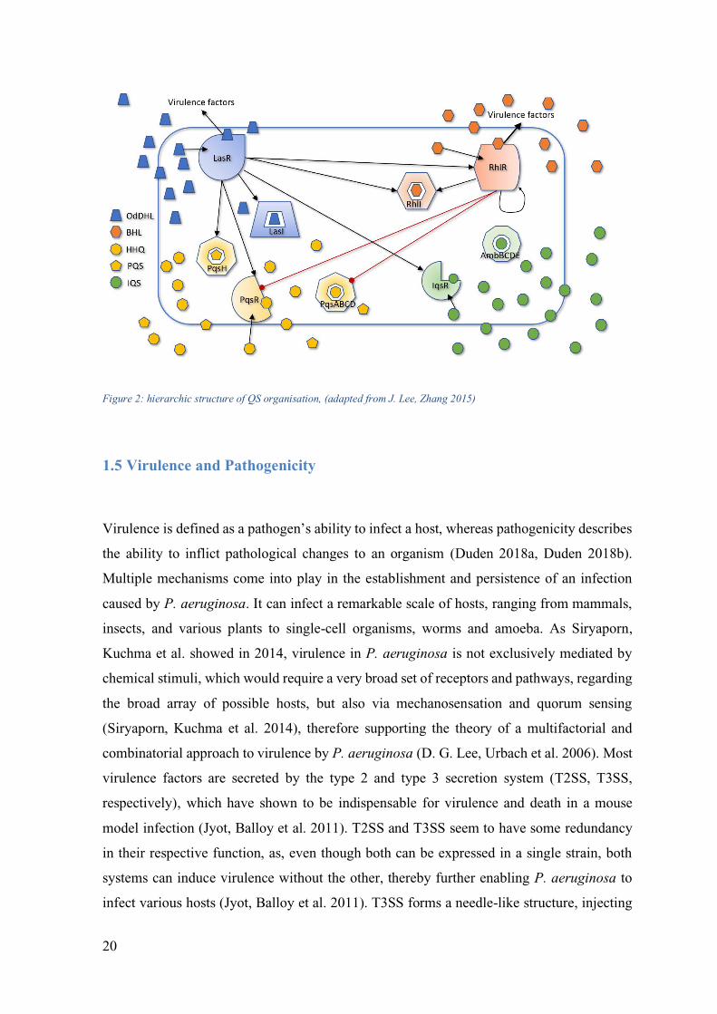
1.4 Quorum sensing
P. aeruginosa is a complicated and complex organism, capable of performing tasks which
require a meticulously orchestrated approach, such as the formation of biofilms or the
development from an acute to a chronic infection. The term quorum sensing (QS) was
suggested first by a team of three microbiologists in 1994, when they described population
density-based regulatory mechanisms, taking place in P. aeruginosa amongst others (Fuqua,
Winans et al. 1994), and named the process quorum sensing. Different bacteria use different
molecules for quorum sensing, nevertheless, the underlying principle is very similar: bacteria
produce small, specific molecules, called autoinducers, which diffuse freely through the cell
membranes. Accumulation of autoinducers is detected by cognate receptors of the same
species of microbes. As soon as a certain threshold is reached, usually representing a certain
concentration of bacteria in the immediate vicinity, the bacteria adapt their behaviour in
multifarious ways. Most of these autoinducers emanate from three different subgroups of
autoinducers; even though the molecular structure is different from genus to genus, they
share structural similarities. The subgroups known so far are acylated homoserine lactones
(AHL), used by Gram negative bacteria, peptide signals, exclusively used by Gram positive
bacteria and a group called autoinducer-2, used by Gram-negative and Gram-positive
bacteria (LaSarre, Federle 2013).
For P. aeruginosa, quorum sensing is responsible for a vast array of regulatory mechanisms.
Up to 10% of the bacterium’s entire genome can be affected by means of quorum sensing or
adaptive mechanisms (Schuster, Peter Greenberg 2006). There are four different regulatory
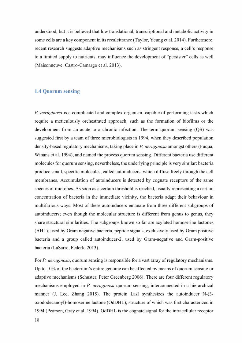
mechanisms employed in P. aeruginosa quorum sensing, interconnected in a hierarchical
manner (J. Lee, Zhang 2015). The protein LasI synthesizes the autoinducer N-(3-
oxododecanoyl)-homoserine lactone (OdDHL), structure of which was first characterized in
1994 (Pearson, Gray et al. 1994). OdDHL is the cognate signal for the intracellular receptor
19
LasR. The LasR-OdDHL complex enhances or enables the transcription of multiple
virulence factor genes (Schuster, Peter Greenberg 2006, Schuster, Lostroh et al. 2003),
stimulating the expression of LasI, thereby establishing a direct positive feedback loop
(Kiratisin, Tucker et al. 2002). LasR-OdDHL also activates the expression of pqsH, pqsR,
pqsABCD, rhlI, rhlR and iqsR (Latifi, Foglino et al. 1996, Déziel, Lépine et al. 2004,
Hentzer, Wu et al. 2003, J. Lee, Wu et al. 2013). For a better visualisation of the
organisational structure, please refer to figure 2.
RhlI synthesizes the autotransmitter N-butyrylhomoserine lactone (BHL), which forms a
complex with RhlR. The compound RhlR-BHL is responsible for the transcription of a vast
array of virulence factor genes (Nouwens, Beatson et al. 2003), as well as inducing rhlI and
its own operon (Winson, Camara et al. 1995) and inhibiting pqsABCDE and pqsH (Wade,
Calfee et al. 2005).
Pseudomonas quinolone signal (PQS) and its precursor 2-heptyl-4-quinolone (HHQ) both
bind to PqsR. Both complexes, PqsR-PQS as well as PqsR-HHQ, induce the transcription of
the enzymes pqsABCDE, catalysing the reactions leading to HHQ, pqsH, catalysing the
reaction from HHQ to PQS (Déziel, Lépine et al. 2004). PqsR-PQS enhances the
transcription of RhlI, thereby forming an extended negative feedback loop (McKnight,
Iglewski et al. 2000).
A fourth quorum sensing system has only recently been discovered: the so called IQS-system
– to represent its role in integrating quorum sensing and detection of extracellular stress cues
(J. Lee, Wu et al. 2013). The synthesis of IQS is regulated by the operons ambABCDE. This
system has shown to influence the LasI/R and the RhlI/R systems, even playing a vital role
in regulating synthesis of BHL, though the exact mechanisms remain unclear (J. Lee, Wu et
al. 2013). IQS-synthesis appears to be regulated by the Las-system in a rich medium but also
via phosphate depletion stress (J. Lee, Wu et al. 2013).
To this point, several super regulators, controlling various steps of the quorum sensing
process, are known and knowledge about genes being expressed by means of QS is
constantly growing. Unfortunately, due to the extensive nature of quorum sensing, a more
profound approach to this subject would exceed the limitations of this paper.
20
Figure 2: hierarchic structure of QS organisation, (adapted from J. Lee, Zhang 2015)
1.5 Virulence and Pathogenicity
Virulence is defined as a pathogen’s ability to infect a host, whereas pathogenicity describes
the ability to inflict pathological changes to an organism (Duden 2018a, Duden 2018b).
Multiple mechanisms come into play in the establishment and persistence of an infection
caused by P. aeruginosa. It can infect a remarkable scale of hosts, ranging from mammals,
insects, and various plants to single-cell organisms, worms and amoeba. As Siryaporn,
Kuchma et al. showed in 2014, virulence in P. aeruginosa is not exclusively mediated by
chemical stimuli, which would require a very broad set of receptors and pathways, regarding
the broad array of possible hosts, but also via mechanosensation and quorum sensing
(Siryaporn, Kuchma et al. 2014), therefore supporting the theory of a multifactorial and
combinatorial approach to virulence by P. aeruginosa (D. G. Lee, Urbach et al. 2006). Most
virulence factors are secreted by the type 2 and type 3 secretion system (T2SS, T3SS,
respectively), which have shown to be indispensable for virulence and death in a mouse
model infection (Jyot, Balloy et al. 2011). T2SS and T3SS seem to have some redundancy
in their respective function, as, even though both can be expressed in a single strain, both
systems can induce virulence without the other, thereby further enabling P. aeruginosa to
infect various hosts (Jyot, Balloy et al. 2011). T3SS forms a needle-like structure, injecting
21
cytotoxins Exo-S, Exo -U, Exo-T and Exo-Y in the cytoplasm of a host cell (Feltman,
Schulert et al. 2001).
To avoid clearance by the host’s immune system, P. aeruginosa employs different strategies.
A loss of its flagellum and therefore its swimming motility provides protection in a
planktonic state from phagocytosis by macrophages and polymorphonuclear neutrophils
(PMN) (Lovewell, Hayes et al. 2014, Floyd, Winn et al. 2016). On the other hand, flagella
play an important role in establishing an infection, especially in cystic fibrosis (CF) patients
(Rada 2017). Additionally, biofilm formation not only increases antimicrobial resistance, it
also reduces immune response: bacteria in a biofilm show a lower activation of the host’s
complement system and a reduced burst of reactive oxygen species (ROS) when compared
to planktonic cells. Furthermore, P. aeruginosa in a QS-proficient biofilm formation produce
Rhamnolipid B, a QS-dependent metabolite capable of inducing necrosis in PMN (Jensen,
Bjarnsholt et al. 2007). Another component of P. aeruginosa biofilm is alginate, providing
protection various host defence mechanisms, such as uptake by macrophages (Simpson,
Smith et al. 1988).
1.6 Treatment options for P. aeruginosa
Antibiotics tested in case of a P. aeruginosa infection, and therefore representing possible
first line therapies, at the LKH Graz are piperacillin/tazobactam, ceftazidime, cefepime,
aztreonam, imipenem, meropenem, amikacin, gentamicin, tobramycin. ciprofloxacin and
levofloxacin (local report on antimicrobial susceptibility). The EUCAST guidelines
(European Committee on Antimicrobial Susceptibility Testing) provide additional MICs for
ticarcillin +/- clavulanic acid, ceftazidime/avibactam, ceftobiprole, ceftolozane/tazobactam,
doripenem, colistin, amikacin and netilmicin (EUCAST 2018). Especially for patients
suffering from cystic fibrosis, a combination therapy of multiple, possibly effective
antibiotics may be applied.
22
Novel antimicrobial substances
In the past decade three new antimicrobial substances or combinations have been introduced
which are useful for the treatment of P. aeruginosa infections. These are
ceftazidime/avibactam, ceftolozane/tazobactam and ceftobiprole.
Ceftazidime/Avibactam
Ceftazidime/Avibactam was approved as Zavicefta® in the European Union in 2016.
Indications for the use of Zavicefta® are complicated intra-abdominal infections,
complicated urinary tract infections and hospital-acquired pneumonia, including ventilator-
associated pneumonia (VAP). Additionally, Zavicefta may be used for treatment of aerobic
Gram-negative pathogens for adults if therapeutic options are limited. In vitro, it has shown
efficacy especially against Gram-negative microorganisms, amongst others P. aeruginosa
(European Medicines Agency 2018a). Ceftazidime is a third-generation cephalosporin,
whose bactericidal effect is caused by inhibition of the synthesis of the peptidoglycan layer
of bacteria currently being in the process of mitosis. Avibactam functions as a non-beta-
lactam, beta-lactamase inhibitor, inhibiting Ambler class A, C beta lactamases. In addition,
avibactam inhibits some Ambler class D lactamases, including extended spectrum
lactamases (ESBL), OXA48-carbapenemase and Klebsiella pneumoniae-carbapenemase
(KPC), which belongs to the group of serine-based carbapenemases (European Medicines
Agency 2018a).
Ceftolozane/Tazobactam
Ceftolozane/Tazobactam was approved for use in the European Union in 2015 under the
name Zerbaxa®. Indications for the use of Zerbaxa® are complicated intraabdominal
infections and complicated urinary tract infections, including pyelonephritis. In vitro, it has
shown to be effective against multiple Gram-negative bacteria, including Pseudomonas
aeruginosa, as well as certain Streptococcus subspecies (European Medicines Agency
2018b). Ceftolozane is a fifth-generation cephalosporin. Its bactericidal effect is – as is
generally the case for all cephalosporins - based on binding to certain penicillin binding
proteins (PBP), in consequence inhibiting cell wall synthesis, thus causing cell death.
Tazobactam is a beta-lactamase-inhibitor, showing efficacy against many, but not all
molecular class A beta-lactamases. It shows no effect against MBL, AMP-C enzymes
23
produced by Enterobacteriaceae, serine-based lactamases and Ambler class D lactamases
(OXA-carbapenemases) (European Medicines Agency 2018b).
Ceftobiprole
Ceftobiprole is a fifth-generation cephalosporin, which is sold in Austria under the name
Zeftera®. Like other cephalosporins, ceftobiprole unfolds its antimicrobial effect by binding
to PBP and in consequence inhibits cell wall synthesis, causing cell death in bacteria.
Approval for ceftobiprole under the name Zevtera® was not granted by the EU in 2010, due
to the conducted studies provided in the application not holding up to the European
Medicines Agency’s standards (European Medicines Agency 2010). Ceftobiprole has been
nationally approved for use in Austria in 2014 under the name Zeftera®, just differing in one
letter from the European version Zevtera®. Its intended use is in treatment of pneumonia,
acquired in either in a hospital environment or community acquired. Its intended use does
not include ventilation acquired pneumonia. Ceftobiprole has shown clinical efficiency
against Staphylococcus aureus, including methicillin-resistant S. aureus (MRSA),
Streptococcus pneumoniae, and Enterobacteriaceae. In vitro, ceftobiprole has been efficient
against even more bacteria, including Pseudomonas spp. assumed that there are no resistance
mechanisms present. The following beta-lactamases bind and degrade ceftobiprole: KPC,
ESBL, as well as enzymes belonging to the Ambler A, Ambler B and Ambler D subclass.
Ceftobiprole also seems stable to certain AmpC enzymes, although AmpC-expressing P.
aeruginosa isolates provide high minimal inhibitory concentrations (MIC) (European
Medicines Agency 2010).
24
1.7 Emerging strategies against P. aeruginosa
Vaccinations
Emerging strategies currently under investigation tend to take a less generalized approach in
terms of applicability to a multitude of pathogens but are much rather tailored to a specific
pathogen. Most, if not all mechanisms which have been discovered over the last years are
being thoroughly examined to find potential drug targets to eradicate a P. aeruginosa
population. A reliable approach to preventing infections has been vaccination. Lastly
updated on 30.5.2015, the Cochrane database did not find a significant benefit caused by
vaccination of CF patients and therefore does not recommend vaccination to prevent P.
aeruginosa infection in patients with CF yet (Johansen, Gotzsche 2015). Currently, there is
one vaccine which has successfully completed phase II trial. It is an OprF/I-based,
recombinant vaccine and has shown a significant increase in seroconversion in mechanically
ventilated ICU patients compared to a placebo group (Rello, Krenn et al. 2017). Difficulties
concerning the development of vaccines are partly due to a high specificity to limited number
of serotypes (Doring, Meisner et al. 2007), therefore highly limiting broad applicability.
Phage Therapy
Another approach is reemerging: Phage therapy. Bacteriophages were first described
independently by Felix D’Herelle in France and Frederick William Twort in Great Britain
at the beginning of the 20th century and were industrially produced in the USA until the rise
of antibiotics replacing their therapeutic status in the 1940s. Currently, this approach is still
applied in Poland, Russia and Georgia and is receiving international attention due to
promising in vitro results. Bacteriophages are viruses which selectively infect bacteria and
induce lysis and cell death. In vitro, the findings are indeed promising. Bacteriophages can
cause enzymatic degradation of alginate and despite a preexisting biofilm, induce lysis in up
to 99% of bacteria, highlighting a possible use in CF patients (Hanlon, Denyer et al. 2001).
An engineered bacteriophage, expressing a lactonase, showed to be able to hinder quorum
sensing by degrading AHL molecules (Pei, Lamas-Samanamud 2014). Furthermore, phages
can express a synergistic effect when combined with antibiotics, sometimes even exceeding
the effect both treatments would have if administered separately (Chaudhry, Concepción-
Acevedo et al. 2017). In the same paper, Chaudry, Concepción and their colleagues also
showed a change in efficacy depending on the concentration of the antibiotic substance and
25
the time interval between administration of phage and antibiotic influencing the ability to
kill P. aeruginosa.
Unfortunately, trials in mouse models and in vitro data are not necessarily replicable in a
clinical setting. It is difficult to mirror chronic infection models and cystic fibrosis patients
in a mouse model, let alone in vitro. Therefore, clinical trials to evaluate usability are very
important. Currently, there is one phase I/II study in progress, called Phagoburn
(NCT02116010). The aim of this trial is to assess the tolerance and efficacy of local
bacteriophage treatment for wound infections caused by P. aeruginosa or E. coli with a
control group treated with the current standard of care, silver (NCT02116010).
Murepavadin
There is one especially promising therapeutic option currently undergoing phase III trials.
Murepavadin belongs to a new group of antibiotics called OMPTA (short for: outer
membrane protein targeting antibiotics). It inhibits LptD-mediated transport of LPS to the
outer membrane (Werneburg, Zerbe et al. 2012), showcasing promising results against XDR
(extensive drug-resistant) P. aeruginosa in vitro (Sader, Flamm et al. 2018). Murepavadin
has proven to be well tolerated even in doses exceeding the expected efficacious levels in
phase I and phase II clinical trials and is currently undergoing two phase III clinical trials,
investigating the efficacy in hospital acquired pneumonia (NCT03409679, NCT03582007).
26
1.8 KRINKO classification
For easier clinical classification of Gram negative, multi-resistant bacteria, the commission
for hospital hygiene and prevention of infectious diseases (the German translation
abbreviated as KRINKO – Komission für Krankenhaushygiene und Infektionsprävention)
has developed a simple scheme, called MRGN. MRGN is short for multi drug-resistant,
Gram negative, rod shaped bacteria. Consequentially, 3MRGN bacteria show resistance to
three of the four tested groups of antimicrobial substances, whereas 4MRGN describes
bacterial resistance to all four of the four tested antimicrobials. For each group of
antimicrobials, the KRINKO designated one representative antimicrobial substance. The
four tested groups are acylureidopenicillins (Piperacillin), third and fourth generation
cephalosporins (Cefotaxime and or ceftazidime), carbapenems (imipenem and or
meropenem) and fluoroquinolones (Ciprofloxacin). In the case of P. aeruginosa, a sample
is classified as 3MRGN if it shows susceptibility to only one of the four tested substances
and as 4MRGN if testing shows resistance to all four tested groups of antimicrobials. The
Robert-Koch-Institute suggests a variety of measures, including isolation and increased
standards of hygiene, a hospital ward should take if a case of colonization or infection by a
3MRGN of 4MRGN strain occurs. At the LKH Graz, a modified version of these measures
is employed (Institut für Krankenhaushygiene und Mikrobiologie 2017). In other countries,
other classifications are in use, such as MDR, XDR and PDR, short for multidrug-resistant,
extensively drug-resistant and pandrug-resistant, respectively (Magiorakos, Srinivasan et al.
2012).
1.9 Aim of this paper
The aim of this paper is to evaluate the susceptibility of carbapenem-resistant P. aeruginosa
isolates against ceftobiprole, ceftazidime/avibactam and ceftolozane/tazobactam in vitro.
In addition, we evaluated the prevalence of MBL-production and carbapenemase-activity in
carbapenem-resistant P. aeruginosa isolates.
27
2. Methods
Clinical Pseudomonas aeruginosa isolates after routine microbiological testing were
collected. Each sample of P. aeruginosa was stored at – 20 °C until testing. To obtain a
bacterial mass sufficient for testing, we bred the isolates on an agar plate for 24 hours at
36°C. To verify the classification “Pseudomonas aeruginosa”, mass spectrometric analytical
methods were applied, using MALDI-TOF MS. Since no patient data was used in this study,
a vote by the ethics committee was not necessary.
Susceptibility and MBL testing
The EUCAST-guidelines 2017 were consulted to determine the MIC of the respective
antibiotics. The antibiotics used for testing were ceftazidime/avibactam,
ceftolozane/tazobactam, ceftobiprole, cefepime,
ceftolozane, piperacillin/tazobactam, meropenem,
imipenem and colistin.
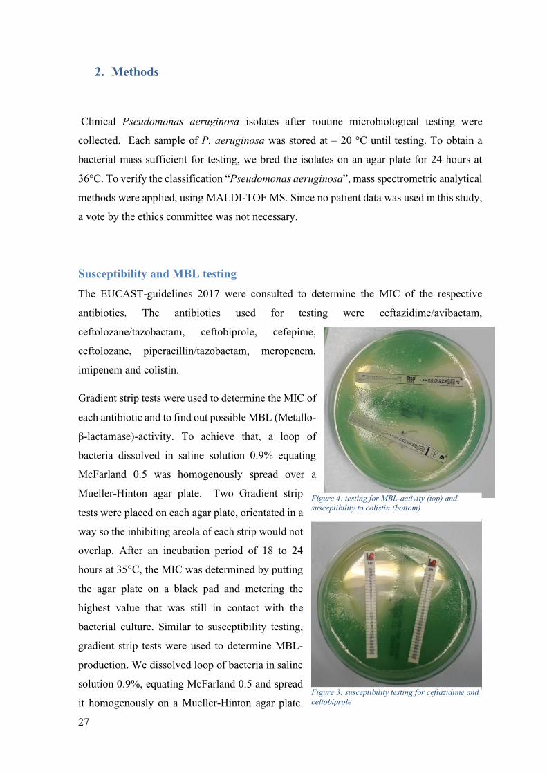
Gradient strip tests were used to determine the MIC of
each antibiotic and to find out possible MBL (Metallo-
β-lactamase)-activity. To achieve that, a loop of
bacteria dissolved in saline solution 0.9% equating
McFarland 0.5 was homogenously spread over a
Mueller-Hinton agar plate. Two Gradient strip
tests were placed on each agar plate, orientated in a
way so the inhibiting areola of each strip would not
overlap. After an incubation period of 18 to 24
hours at 35°C, the MIC was determined by putting
the agar plate on a black pad and metering the
highest value that was still in contact with the
bacterial culture. Similar to susceptibility testing,
gradient strip tests were used to determine MBL-
production. We dissolved loop of bacteria in saline
solution 0.9%, equating McFarland 0.5 and spread
it homogenously on a Mueller-Hinton agar plate.
Figure 4: testing for MBL-activity (top) and susceptibility to colistin (bottom)
Figure 3: susceptibility testing for ceftazidime and ceftobiprole
28
We placed a MBL-gradient strip test on the agar plate and incubated it at 35°C for 18 to 24
hours. One half of the strip contained a carbapenem, the other half contained the same
carbapenem and EDTA (ethylenediaminetetraacetic acid), an MBL-inhibitor. MIC was
determined on both sides and then compared. If the MIC of the carbapenem alone was at
least four times the MIC of carbapenem plus EDTA, the test was rated positive.
Testing for carbapenemase activity
To specify carbapenemase activity as a possible reason for
carbapenem-resistance, we used Rapidec® Carba test kits by
BioMérieux. This kit relies on carbapenem hydrolysis by
carbapenemase-producing bacteria. The ensuing acidification of
the medium changes the colour of indicator phenol red. In order
to access carbapenemase enzymes, bacterial lysis was necessary
beforehand.
If insufficient bacterial lysis was suspected due to hypermucoid
appearance of bacterial isolates, the bacterial colonies and the
suspension medium were mixed using a vortex type mixer until
the mucoid nature was dissolved.
P. aeruginosa isolates were incubated at 36°C for 24h on Mueller
Hinton Agar plates. Bacterial lysis was achieved by adding 10µl
of bacterial mass into a prefabricated lysis buffer for 30 minutes
at 15-25°C. The lysate was then added to a solution containing
phenol red, imipenem and zinc and incubated for 30 minutes at
36°C. If there was no observable colour change after 30 minutes,
the incubation period was prolonged for another 90 minutes. A
positive test result is shown in figure 5 with the indicator
showing a change in colour from “d” to “e”.
Figure 5: testing kit for
carbapenemase activity
29
3. Results
Origin of isolates
We identified a total of 76 bacterial isolates, using MALDITOF-MS. Amongst those, 92.1%
were classified as Pseudomonas aeruginosa isolates (70 out of 76). The remaining 6 isolates
consisted of 4 Stenotrophomonas maltophilia isolates (5%), one Pseudomonas fluorenscens
and one Pseudomonas monteilii.
85.7% (60/70) P. aeruginosa isolates tested intermediate or resistant, according to EUCAST
guidelines (EUCAST 2018), to imipenem, meropenem or both (86%). In the end, a total of
60 isolates matched the inclusion criteria of mass spectrometric identification as
Pseudomonas aeruginosa and non-susceptibility to carbapenems.
The 60 included isolates were isolated from the following sites of infection: skin swabs
(n=22), respiratory tract (n=17), urinary tract (n=9), bloodstream (n=7), aspirate (n=2),
wound swabs (n=2) and one isolate was found on a swab from a PEG (percutaneous
endoscopic gastrostomy) feeding tube.
30
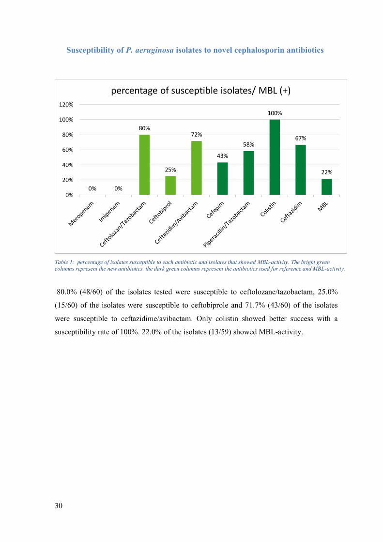
Susceptibility of P. aeruginosa isolates to novel cephalosporin antibiotics
80.0% (48/60) of the isolates tested were susceptible to ceftolozane/tazobactam, 25.0%
(15/60) of the isolates were susceptible to ceftobiprole and 71.7% (43/60) of the isolates
were susceptible to ceftazidime/avibactam. Only colistin showed better success with a
susceptibility rate of 100%. 22.0% of the isolates (13/59) showed MBL-activity.
0% 0%
80%
25%
72%
43%
58%
100%
67%
22%
0%
20%
40%
60%
80%
100%
120%
percentage of susceptible isolates/ MBL (+)
Table 1: percentage of isolates susceptible to each antibiotic and isolates that showed MBL-activity. The bright green columns represent the new antibiotics, the dark green columns represent the antibiotics used for reference and MBL-activity.
31
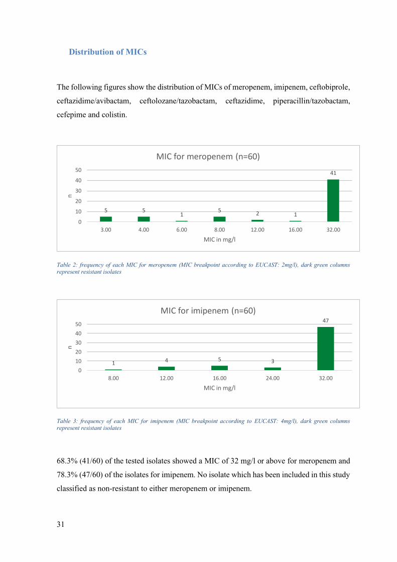
Distribution of MICs
The following figures show the distribution of MICs of meropenem, imipenem, ceftobiprole,
ceftazidime/avibactam, ceftolozane/tazobactam, ceftazidime, piperacillin/tazobactam,
cefepime and colistin.
Table 2: frequency of each MIC for meropenem (MIC breakpoint according to EUCAST: 2mg/l), dark green columns represent resistant isolates
Table 3: frequency of each MIC for imipenem (MIC breakpoint according to EUCAST: 4mg/l), dark green columns represent resistant isolates
68.3% (41/60) of the tested isolates showed a MIC of 32 mg/l or above for meropenem and
78.3% (47/60) of the isolates for imipenem. No isolate which has been included in this study
classified as non-resistant to either meropenem or imipenem.
5 51
52 1
41
0
10
20
30
40
50
3.00 4.00 6.00 8.00 12.00 16.00 32.00
n
MIC in mg/l
MIC for meropenem (n=60)
1 4 5 3
47
0
10
20
30
40
50
8.00 12.00 16.00 24.00 32.00
n
MIC in mg/l
MIC for imipenem (n=60)
32
Table 4: frequency of each MIC for ceftobiprole (PK/PD breakpoint according to EUCAST: 4mg/l) bright green columns represent MIC classified as susceptible, dark green columns represent resistant isolates
Ceftobiprole showed a susceptibility rate of only 25.4% (15/59). Interestingly, 66.1%
(39/59) of the isolates showed the same MIC of 32 mg/l. bright green columns depict
susceptible MIC values, dark green columns represent MIC values classified as resistant.
One isolate could not be tested (marked “x” on the table).
Table 5: frequency of each MIC for ceftazidime/avibactam (MIC breakpoint according to EUCAST: 8mg/l), bright green columns represent MIC classified as susceptible, dark green columns represent resistant isolates
13
1
64 3
39
2 1
0
5
10
15
20
25
30
35
40
45
0.5 1.5 2 3 4 6 32 256 x
n
MIC in mg/l
MIC for ceftobiprole (n=59)
1
2
1
3
5
11 11
3
6
1
3
4
1
8
0
2
4
6
8
10
12
0.50 0.75 1.00 1.50 2.00 3.00 4.00 6.00 8.00 12.00 16.00 24.00 64.00 256.00
n
MIC in mg/l
MIC for ceftazidim/avibactam (n=60)
33
Table 6: frequency of each MIC for ceftolozane/tazobactam (MIC breakpoint according to EUCAST: 4mg/l), bright green columns represent MIC classified as susceptible, dark green columns represent resistant isolates
Both ceftolozane/tazobactam and ceftazidime/avibactam showed promising results: 71.6%
(43/60) and 80.0% (48/60) of the isolates were susceptible to ceftazidime/avibactam and
ceftolozane/tazobactam, respectively. For ceftolozane/tazobactam, all resistant isolates
showed a MIC of at least 256 mg/l, whereas for ceftazidime/avibactam, MIC for resistant
isolates appeared more homogenously spread, only 8 isolates showed a MIC of 256 mg/l or
higher.
Table 7: frequency of each MIC for ceftazidime (MIC breakpoint according to EUCAST: 8mg/l), bright green columns represent MIC classified as susceptible, dark green columns represent resistant isolates
1 12
1
78
6
8
11
12
12
0
2
4
6
8
10
12
14
0.13 0.15 0.25 0.38 0.50 0.75 1.00 1.50 2.00 3.00 4.00 256.00
n
MIC in mg/l
MIC for ceftolozane/tazobactam (n=60)
1 1
8
5
3
4 4
7 7
4
3 3
4
6
0
1
2
3
4
5
6
7
8
9
0.50 0.75 1.00 1.50 2.00 3.00 4.00 6.00 8.00 12.00 24.00 32.00 64.00 256.00
n
MIC in mg/l
MIC for ceftazidime (n=60)
34
Table 8: frequency of each MIC for cefepime (MIC breakpoint according to EUCAST: 8mg/l) , bright green columns represent MIC classified as susceptible, dark green columns represent resistant isolates
Table 9 frequency of each MIC for piperacillin/tazobactam (MIC breakpoint according to EUCAST: 16 mg/l), bright green columns represent MIC classified as susceptible, dark green columns represent resistant isolates. The column with “-“ represents resistant isolates whose MIC was not determined.
2
6
7
2 2
7
8
4
5
3
9
5
0
1
2
3
4
5
6
7
8
9
10
1.50 2.00 3.00 4.00 6.00 8.00 12.00 16.00 24.00 32.00 48.00 256.00
n
MIC in mg/l
MIC for cefepim (n=60)
67
23
7
1
3 3 3
54
3
5
8
0
2
4
6
8
10
0.00 0.10 1.50 3.00 4.00 6.00 8.00 12.00 16.00 24.00 32.00 48.00 256.00 -
n
MIC in mg/l
MIC for piperacillin/tazobactam
35
Table 10: frequency of each MIC for colistin (MIC breakpoint according to EUCAST: 2mg/l), bright green columns represent MIC classified as susceptible, the dark green column represents the number of isolates which could not be tested
The antibiotics used for reference showed varying results. While colistin showed 100%
efficacy, ceftazidime, cefepime and piperacillin/tazobactam showed varying degrees of
success against the tested isolates. 66.7% of the tested isolates were susceptible to
ceftazidime (40/60), while only 43.3% of the tested isolates were susceptible to cefepime
(26/60). 63.3% of the isolates showed susceptibility to piperacillin/tazobactam (38/60). 14
isolates were not tested for susceptibility to colistin.
0
2
4
6
8
10
12
14
16
18
0.094 0.125 0.25 0.38 0.5 0.75 1 x
n
MIC in mg/l
MIC for colistin (n=60)
36
Phenotypic testing of Carbapenemase and MBL production
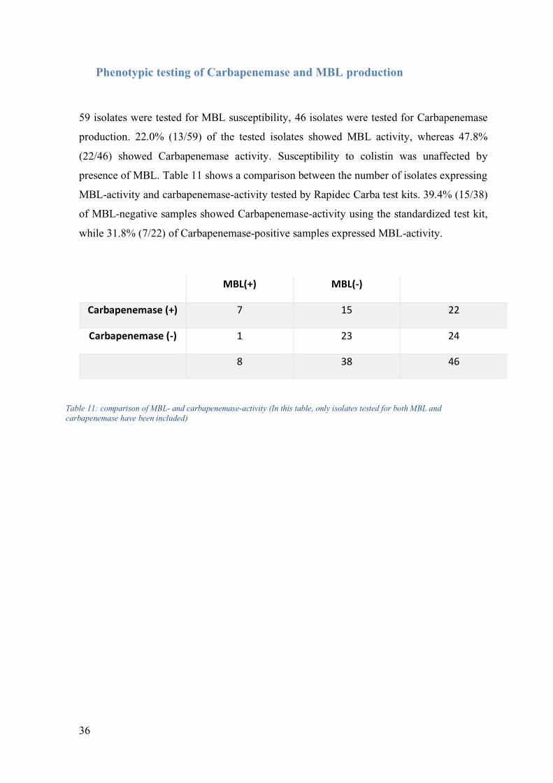
59 isolates were tested for MBL susceptibility, 46 isolates were tested for Carbapenemase
production. 22.0% (13/59) of the tested isolates showed MBL activity, whereas 47.8%
(22/46) showed Carbapenemase activity. Susceptibility to colistin was unaffected by
presence of MBL. Table 11 shows a comparison between the number of isolates expressing
MBL-activity and carbapenemase-activity tested by Rapidec Carba test kits. 39.4% (15/38)
of MBL-negative samples showed Carbapenemase-activity using the standardized test kit,
while 31.8% (7/22) of Carbapenemase-positive samples expressed MBL-activity.
MBL(+) MBL(-)
Carbapenemase (+) 7 15 22
Carbapenemase (-) 1 23 24
8 38 46
Table 11: comparison of MBL- and carbapenemase-activity (In this table, only isolates tested for both MBL and
carbapenemase have been included)
37
4. Discussion
The 2017 AURES report impressively shows the state of antimicrobial susceptibility of P.
aeruginosa in Austria in comparison to the rest of the European Union. In 2017, 8.7% of P.
aeruginosa isolates showed resistance to ceftazidime. Additionally, 13.9% were resistant to
carbapenems. While resistance to ceftazidime was the lowest in 2017 compared to resistance
levels since 2013, resistance to carbapenems has increased steadily up to its peak in 2017
since 2013 (BMASGK 2018). This alone highlights the importance of antibiotic alternatives
in treatment of P. aeruginosa infections as well as information about the reliability of current
and new therapeutic options in case of resistance to carbapenems or other antimicrobial
groups. Comparison of antimicrobial resistance between European countries highlights this
even more: for the prevalence of resistance to carbapenems as well as resistance to
ceftazidime, more than the half of European countries reported worse results than Austria
(BMASGK 2018). Therefore, we tested the recently admitted antibiotics
ceftolozane/tazobactam, ceftazidime/avibactam and ceftobiprole for efficacy in clinical
carbapenem-resistant P. aeruginosa isolates.
Our findings show that ceftolozane/tazobactam and ceftazidime/avibactam deliver
promising results in vitro against carbapenem-resistant P. aeruginosa-isolates.
Ceftolozane/Tazobactam as well as ceftazidime/avibactam showed second and third highest
susceptibility rates of all antibiotics included in this study. This indicates valuable utility in
treatment of increasingly more resistant P. aeruginosa-isolates, as two more antibiotics may
be added to standard antimicrobial susceptibility testing. Additionally, direct comparison
between a susceptibility rate of 71.6% (43/60) and 66.7% (40/60) for ceftazidime/avibactam
and ceftazidime, respectively, suggest a possible benefit in combination of current antibiotics
with beta lactamase-inhibitors. In contrast, ceftobiprole only proved to be effective in 25%
(15/60) of all isolates. It appears that ceftobiprole does not present a reliable alternative if
there is a pre-existing resistance to carbapenems. As we did not conduct molecular or genetic
testing, we could not determine molecular mechanisms of antimicrobial resistance.
Unfortunately, the precise cause of resistance to ceftobiprole in this study remains unclear,
especially as in 2014, Farrell, Flamm et al. found a susceptibility of 64.6% to ceftobiprole,
using the same MIC (Farrell, Flamm et al. 2014). It is noteworthy though, that Farrell, Flamm
and colleagues did not select P. aeruginosa isolates for carbapenem resistance. In 2007,
Queenan, Shang et al. found increased lability of ceftobiprole to class A, C and D extended
38
spectrum beta-lactamases when compared to ceftazidime (Queenan, Shang et al. 2007). This
finding may partially explain the subpar results of ceftobiprole in this study.
Between 2009 and 2011, Castanheira, Deshpande et al. collected, compared and screened
529 P. aeruginosa isolates which were non-susceptible to doripenem, meropenem and
imipenem for carbapenem resistance mechanisms from Mediterranean and European
countries. Using EUCAST guidelines, susceptibility for meropenem, imipenem and
cefepime was 48.5%, 67.6% and 36.0%, respectively with cefepime being the most effective
β-lactam antibiotic included in this study. Using CLSI breakpoints, they found colistin to be
effective against 99.3% (Castanheira, Deshpande et al. 2014).
In Brazil, Dias, Diniz et al. screened 28 carbapenem-resistant P. aeruginosa isolates for
susceptibility to various antimicrobials and phenotypic and genotypic characteristics. They
found 100% percent of the isolates to be resistant to imipenem, meropenem, ceftazidime and
cefepime. Similar to our findings, 100% of their tested isolates were susceptible to
polymyxin B, whereas 100% of our tested isolates were susceptible to colistin (Dias, Diniz
et al. 2016).
Wi, Greenwood-Quaintance et al. tested 42 non-carbapenemase producing, carbapenem-
resistant P. aeruginosa isolates for susceptibility to ceftolozane/tazobactam and referenced
their findings by comparing them to susceptibility rates to other antimicrobials. They found
ceftolozane/tazobactam to be effective in 95.2%, whereas ceftazidime/avibactam,
ceftazidime, cefepime, piperacillin/tazobactam and meropenem showed susceptibility rates
of 71.4%, 42.9%, 21.4%, 23.8% and 2.4%, respectively (Wi, Greenwood-Quaintance et al.
2017). The abovementioned findings demonstrate the local variability of P. aeruginosa
susceptibility to various antibiotics.
In addition, we tested the carbapenem-resistant isolates for carbapenemase- and MBL-
production. We found 22.0% (13/59) of our tested isolates to express MBL-activity. In
comparison, a recent study conducted by Matzkies, Leitner et al. conducted in south-east
Austria found MBL activity in 44.0% (22/50) of the included isolates by retrospectively
screening 4MRGN P. aeruginosa isolates for MBL activity (Matzkies, Leitner et al. 2019).
The test kits we employed to detect carbapenemase activity showed difficulties in practical
use. Interpretation of results was difficult, as it was hard to distinguish a positive result from
a negative one. A negative result showed no change in colour, a positive result was supposed
to show a change in colour from red to orange or various shades of yellow. Regrettably, the
39
interpretation of the results was only rarely unquestionable. Most results remained unclear,
as shades of red and orange could be influenced by lighting and could only rarely be
classified unequivocally. Therefore, we decided to discontinue testing for carbapenemase-
activity after 46 samples. Of those tested, 47.8% (22/46) showed carbapenemase activity.
In conclusion, ceftazidime/avibactam and ceftolozane/tazobactam show high efficiency
against carbapenem-resistant P. aeruginosa in vitro. Further research is needed to evaluate
actual clinical benefit by conducting studies in a hospital setting.
40
Bibliography
ALVAREZ-ORTEGA, C., WIEGAND, I., OLIVARES, J., HANCOCK, R.E. and
MARTINEZ, J.L., 2011. The intrinsic resistome of Pseudomonas aeruginosa to beta-
lactams. Virulence, 2(2), pp. 144-146.
ANI, C., FARSHIDPANAH, S., BELLINGHAUSEN STEWART, A. and NGUYEN,
H.B., 2015. Variations in organism-specific severe sepsis mortality in the United States:
1999-2008. Critical Care Medicine, 43(1), pp. 65-77.
BARBER, M. and WATERWORTH, P.M., 1966. Activity of gentamicin against
Pseudomonas and hospital Staphylococci. British medical journal, 1(5481), pp. 203-205.
BARCLAY, M.L., BEGG, E.J. and CHAMBERS, S.T., 1992. Adaptive resistance
following single doses of gentamicin in a dynamic in vitro model. Antimicrobial Agents
and Chemotherapy, 36(9), pp. 1951-1957.
BARKEN, K.B., PAMP SÜNJE J., LIANG, Y., MORTEN, G., BERTRAND, J.J.,
MIKKEL, K., MICHAEL, G., WHITCHURCH, C.B., ENGEL, J.N. and TOLKER‐NIELSEN TIM, 2008. Roles of type IV pili, flagellum‐mediated motility and extracellular
DNA in the formation of mature multicellular structures in Pseudomonas aeruginosa
biofilms. Environmental microbiology, 10(9), pp. 2331-2343.
BERRAZEG, M., JEANNOT, K., NTSOGO ENGUENE, V.Y., BROUTIN, I.,
LOEFFERT, S., FOURNIER, D. and PLESIAT, P., 2015. Mutations in beta-Lactamase
AmpC Increase Resistance of Pseudomonas aeruginosa Isolates to Antipseudomonal
Cephalosporins. Antimicrobial Agents and Chemotherapy, 59(10), pp. 6248-6255.
BJARNSHOLT, T., JENSEN, P.O., FIANDACA, M.J., PEDERSEN, J., HANSEN, C.R.,
ANDERSEN, C.B., PRESSLER, T., GIVSKOV, M. and HOIBY, N., 2009. Pseudomonas
aeruginosa biofilms in the respiratory tract of cystic fibrosis patients. Pediatric
pulmonology, 44(6), pp. 547-558.
BMASGK, 2018. Resistenzbericht Österreich AURES 2017. November 2018 edn. Wien:
Bundesministerium für Arbeit, Soziales, Gesundheit und Konsumentenschutz.
BMGF, 2017. Resistenzbericht Österreich AURES 2016. November 2017 edn. Wien:
Bundesministerium für Gesundheit und Frauen.
BORRIELLO, G., WERNER, E., ROE, F., KIM, A.M., EHRLICH, G.D. and STEWART,
P.S., 2004. Oxygen limitation contributes to antibiotic tolerance of Pseudomonas
aeruginosa in biofilms. Antimicrobial Agents and Chemotherapy, 48(7), pp. 2659-2664.
BREIDENSTEIN, E.B.M., DE LA FUENTE-NÚÑEZ, C. and HANCOCK, R.E.W., 2011.
Pseudomonas aeruginosa: all roads lead to resistance.
BURDON, D.W. and WHITBY, J.L., 1967. Contamination of hospital disinfectants with
Pseudomonas species. British medical journal, 2(5545), pp. 153-155.
41
CASTANHEIRA, M., DESHPANDE, L.M., COSTELLO, A., DAVIES, T.A. and JONES,
R.N., 2014. Epidemiology and carbapenem resistance mechanisms of carbapenem-non-
susceptible Pseudomonas aeruginosa collected during 2009-11 in 14 European and
Mediterranean countries. The Journal of antimicrobial chemotherapy, 69(7), pp. 1804-
1814.
CHAUDHRY, W.N., CONCEPCIÓN-ACEVEDO, J., PARK, T., ANDLEEB, S., BULL,
J.J. and LEVIN, B.R., 2017. Synergy and Order Effects of Antibiotics and Phages in
Killing Pseudomonas aeruginosa Biofilms. PLOS ONE, 12(1), pp. e0168615.
DE LA FUENTE-NUNEZ, C., REFFUVEILLE, F., FERNANDEZ, L. and HANCOCK,
R.E., 2013. Bacterial biofilm development as a multicellular adaptation: antibiotic
resistance and new therapeutic strategies. Current opinion in microbiology, 16(5), pp. 580-
589.
DÉZIEL, E., LÉPINE, F., MILOT, S., HE, J., MINDRINOS, M.N., TOMPKINS, R.G. and
RAHME, L.G., 2004. Analysis of Pseudomonas aeruginosa 4-hydroxy-2-alkylquinolines
(HAQs) reveals a role for 4-hydroxy-2-heptylquinoline in cell-to-cell communication.
Proceedings of the National Academy of Sciences of the United States of America, 101(5),
pp. 1339-1344.
DIAS, V.C., DINIZ, C.G., PETER, A.C., BASTOS, A.N., BASTOS, V.Q., BASTOS, L.Q.
and DA SILVA, V.L., 2016. Epidemiological characteristics and antimicrobial
susceptibility among carbapenem-resistant non-fermenting bacteria in Brazil. Journal of
infection in developing countries, 10(6), pp. 544-553.
DORING, G., MEISNER, C., STERN, M. and FLAGELLA VACCINE TRIAL STUDY
GROUP, 2007. A double-blind randomized placebo-controlled phase III study of a
Pseudomonas aeruginosa flagella vaccine in cystic fibrosis patients. Proceedings of the
National Academy of Sciences of the United States of America, 104(26), pp. 11020-11025.
DUDEN, 2018a-last update, Pathogenität [Homepage of Bibliographisches Institut
GmbH], [Online]. Available: https://www.duden.de/rechtschreibung/Pathogenitaet [10/21,
2018].
DUDEN, 2018b-last update, Virulenz [Homepage of Bibliographisches Institut GmbH],
[Online]. Available: https://www.duden.de/rechtschreibung/Virulenz [10/21, 2018].
EUCAST, 16.5.2018, 2018-last update, Breakpoint tables for interpretation of MICs and
zone diameters, version 8.0, 2018 [Homepage of The European Committee on
Antimicrobial Susceptibility Testing], [Online]. Available:
http://www.eucast.org/clinical_breakpoints/ [20.7.2018, 2018].
EUROPEAN CENTRE FOR DISEASE PREVENTION AND CONTROL, 2018.
Antimicrobial consumption. ECDC. Annual epidemiological report for 2016, .
EUROPEAN CENTRE FOR DISEASE PREVENTION AND CONTROL, ed, 2017.
Surveillance of antimicrobial resistance in Europe 2016. Annual Report of the
European Antimicrobial Resistance Surveillance Network (EARS-Net). Stockholm: ECDC.
42
EUROPEAN MEDICINES AGENCY, 05.03, 2018a-last update, Zavicefta: EPAR -
Product information. Available:
http://www.ema.europa.eu/docs/en_GB/document_library/EPAR_-
_Product_Information/human/004027/WC500210234.pdf [29.08, 2018].
EUROPEAN MEDICINES AGENCY, 12.06, 2018b-last update, Zerbaxa: EPAR -
Product Information. Available:
http://www.ema.europa.eu/docs/en_GB/document_library/EPAR_-
_Product_Information/human/003772/WC500194595.pdf [30.08, 2018].
EUROPEAN MEDICINES AGENCY, 21.09, 2010-last update, Zeftera: Public assessment
report. Available: http://www.ema.europa.eu/docs/en_GB/document_library/EPAR_-
_Public_assessment_report/human/000883/WC500096883.pdf [31.08.2018, 2018].
FARRELL, D.J., FLAMM, R.K., SADER, H.S. and JONES, R.N., 2014. Ceftobiprole
activity against over 60,000 clinical bacterial pathogens isolated in Europe, Turkey, and
Israel from 2005 to 2010. Antimicrobial Agents and Chemotherapy, 58(7), pp. 3882-3888.
FELTMAN, H., SCHULERT, G., KHAN, S., JAIN, M., PETERSON, L. and HAUSER,
A.R., 2001. Prevalence of type III secretion genes in clinical and environmental isolates of
Pseudomonas aeruginosa. Microbiology (Reading, England), 147(Pt 10), pp. 2659-2669.
FLOYD, M., WINN, M., CULLEN, C., SIL, P., CHASSAING, B., YOO, D., GEWIRTZ,
A.T., GOLDBERG, J.B., MCCARTER, L.L. and RADA, B., 2016. Swimming Motility
Mediates the Formation of Neutrophil Extracellular Traps Induced by Flagellated
Pseudomonas aeruginosa. PLoS Pathogens, 12(11), pp. e1005987.
doi:10.1371/journal.ppat.1005987.
FRIEDMAN, L. and KOLTER, R., 2004a. Genes involved in matrix formation in
Pseudomonas aeruginosa PA14 biofilms. Molecular microbiology, 51(3), pp. 675-690.
FRIEDMAN, L. and KOLTER, R., 2004b. Two genetic loci produce distinct carbohydrate-
rich structural components of the Pseudomonas aeruginosa biofilm matrix. Journal of
Bacteriology, 186(14), pp. 4457-4465.
FUQUA, W.C., WINANS, S.C. and GREENBERG, E.P., 1994. Quorum sensing in
bacteria: the LuxR-LuxI family of cell density-responsive transcriptional regulators.
Journal of Bacteriology, 176(2), pp. 269-275.
GREEN, S.K., SCHROTH, M.N., CHO, J.J., KOMINOS, S.D. and VITANZA-JACK,
V.B., 1974. Agricultural Plants and Soil as a Reservoir for Pseudomonas aeruginosa.
Applied Microbiology, 28(6), pp. 987-991.
HANCOCK, R.E.,W., 1998. Resistance Mechanisms in Pseudomonas aeruginosa and
Other Nonfermentative Gram-Negative Bacteria. Clinical Infectious Diseases,
27(Supplement_1), pp. S93-S99.
HANLON, G.W., DENYER, S.P., OLLIFF, C.J. and IBRAHIM, L.J., 2001. Reduction in
exopolysaccharide viscosity as an aid to bacteriophage penetration through Pseudomonas
aeruginosa biofilms. Applied and Environmental Microbiology, 67(6), pp. 2746-2753.
43
HENTZER, M., WU, H., ANDERSEN, J.B., RIEDEL, K., RASMUSSEN, T.B., BAGGE,
N., KUMAR, N., SCHEMBRI, M.A., SONG, Z., KRISTOFFERSEN, P., MANEFIELD,
M., COSTERTON, J.W., MOLIN, S., EBERL, L., STEINBERG, P., KJELLEBERG, S.,
HøIBY, N. and GIVSKOV, M., 2003. Attenuation of Pseudomonas aeruginosa virulence
by quorum sensing inhibitors. The EMBO journal, 22(15), pp. 3803-3815.
HOCQUET, D., MULLER, A., BLANC, K., PLESIAT, P., TALON, D., MONNET, D.L.
and BERTRAND, X., 2008. Relationship between antibiotic use and incidence of MexXY-
OprM overproducers among clinical isolates of Pseudomonas aeruginosa. Antimicrobial
Agents and Chemotherapy, 52(3), pp. 1173-1175.
HOCQUET, D., VOGNE, C., EL GARCH, F., VEJUX, A., GOTOH, N., LEE, A.,
LOMOVSKAYA, O. and PLESIAT, P., 2003. MexXY-OprM efflux pump is necessary for
a adaptive resistance of Pseudomonas aeruginosa to aminoglycosides. Antimicrobial
Agents and Chemotherapy, 47(4), pp. 1371-1375.
INSTITUT FÜR KRANKENHAUSHYGIENE UND MIKROBIOLOGIE, 2017.
Hygienemaßnahmen beim Nachweis von multiresistenten gramnegativen Bakterien
(MRGN). Graz: .
JENSEN, P.O., BJARNSHOLT, T., PHIPPS, R., RASMUSSEN, T.B., CALUM, H.,
CHRISTOFFERSEN, L., MOSER, C., WILLIAMS, P., PRESSLER, T., GIVSKOV, M.
and HOIBY, N., 2007. Rapid necrotic killing of polymorphonuclear leukocytes is caused
by quorum-sensing-controlled production of rhamnolipid by Pseudomonas aeruginosa.
Microbiology (Reading, England), 153(Pt 5), pp. 1329-1338.
JOHANSEN, H.K. and GOTZSCHE, P.C., 2015. Vaccines for preventing infection with
Pseudomonas aeruginosa in cystic fibrosis. The Cochrane database of systematic reviews,
(8):CD001399. doi(8), pp. CD001399.
JYOT,Jeevan, BALLOY,Viviane, JOUVION,Gregory, VERMA,Amrisha,
TOUQUI,Lhousseine, HUERRE,Michel, CHIGNARD,Michel and RAMPHAL,Reuben,
2011. Type II Secretion System of Pseudomonas aeruginosa: In Vivo Evidence of a
Significant Role in Death Due to Lung Infection. The Journal of Infectious Diseases,
203(10), pp. 1369-1377.
KAO, C.Y., CHEN, S.S., HUNG, K.H., WU, H.M., HSUEH, P.R., YAN, J.J. and WU,
J.J., 2016. Overproduction of active efflux pump and variations of OprD dominate in
imipenem-resistant Pseudomonas aeruginosa isolated from patients with bloodstream
infections in Taiwan. BMC microbiology, 16(1), pp. 107-016-0719-2.
KINDRACHUK, K.N., FERNANDEZ, L., BAINS, M. and HANCOCK, R.E., 2011.
Involvement of an ATP-dependent protease, PA0779/AsrA, in inducing heat shock in
response to tobramycin in Pseudomonas aeruginosa. Antimicrobial Agents and
Chemotherapy, 55(5), pp. 1874-1882.
KIRATISIN, P., TUCKER, K.D. and PASSADOR, L., 2002. LasR, a Transcriptional
Activator of Pseudomonas aeruginosa Virulence Genes, Functions as a Multimer. Journal
of Bacteriology, 184(17), pp. 4912-4919.
44
LASARRE, B. and FEDERLE, M.J., 2013. Exploiting quorum sensing to confuse bacterial
pathogens. Microbiology and molecular biology reviews : MMBR, 77(1), pp. 73-111.
LATIFI, A., FOGLINO, M., TANAKA, K., WILLIAMS, P. and LAZDUNSKI, A., 1996.
A hierarchical quorum-sensing cascade in Pseudomonas aeruginosa links the
transcriptional activators LasR and RhIR (VsmR) to expression of the stationary-phase
sigma factor RpoS. Molecular microbiology, 21(6), pp. 1137-1146.
LEE, D.G., URBACH, J.M., WU, G., LIBERATI, N.T., FEINBAUM, R.L., MIYATA, S.,
DIGGINS, L.T., HE, J., SAUCIER, M., DEZIEL, E., FRIEDMAN, L., LI, L., GRILLS,
G., MONTGOMERY, K., KUCHERLAPATI, R., RAHME, L.G. and AUSUBEL, F.M.,
2006. Genomic analysis reveals that Pseudomonas aeruginosa virulence is combinatorial.
Genome biology, 7(10), pp. R90-2006-7-10-r90. Epub 2006 Oct 12.
LEE, J., WU, J., DENG, Y., WANG, J., WANG, C., WANG, J., CHANG, C., DONG, Y.,
WILLIAMS, P. and ZHANG, L.H., 2013. A cell-cell communication signal integrates
quorum sensing and stress response. Nature chemical biology, 9(5), pp. 339-343.
LEE, J. and ZHANG, L., 2015. The hierarchy quorum sensing network in Pseudomonas
aeruginosa. Protein & cell, 6(1), pp. 26-41.
LEE, J.Y., PARK, Y.K., CHUNG, E.S., NA, I.Y. and KO, K.S., 2016. Evolved resistance
to colistin and its loss due to genetic reversion in Pseudomonas aeruginosa. Scientific
reports, 6, pp. 25543.
LEE, J.K., LEE, Y.S., PARK, Y.K. and KIM, B.S., 2005. Alterations in the GyrA and
GyrB subunits of topoisomerase II and the ParC and ParE subunits of topoisomerase IV in
ciprofloxacin-resistant clinical isolates of Pseudomonas aeruginosa.
LEWIS, K., 2010. Persister cells. Annual Review of Microbiology, 64, pp. 357-372.
LOVEDAY, H.P., WILSON, J.A., KERR, K., PITCHERS, R., WALKER, J.T. and
BROWNE, J., 2014. Association between healthcare water systems and Pseudomonas
aeruginosa infections: a rapid systematic review.
LOVEWELL, R.R., HAYES, S.M., O'TOOLE, G.A. and BERWIN, B., 2014.
Pseudomonas aeruginosa flagellar motility activates the phagocyte PI3K/Akt pathway to
induce phagocytic engulfment. American Journal of Physiology - Lung Cellular and
Molecular Physiology, 306(7), pp. L698-707.
MACFARLANE, E.L., KWASNICKA, A. and HANCOCK, R.E., 2000. Role of
Pseudomonas aeruginosa PhoP-phoQ in resistance to antimicrobial cationic peptides and
aminoglycosides. Microbiology (Reading, England), 146 ( Pt 10)(Pt 10), pp. 2543-2554.
MAGIORAKOS, A.-., SRINIVASAN, A., CAREY, R.B., CARMELI, Y., FALAGAS,
M.E., GISKE, C.G., HARBARTH, S., HINDLER, J.F., KAHLMETER, G., OLSSON-
LILJEQUIST, B., PATERSON, D.L., RICE, L.B., STELLING, J., STRUELENS, M.J.,
VATOPOULOS, A., WEBER, J.T. and MONNET, D.L., 2012. Multidrug-resistant,
extensively drug-resistant and pandrug-resistant bacteria: an international expert proposal
for interim standard definitions for acquired resistance. Clinical Microbiology and
Infection, 18(3), pp. 268-281.
45
MAISONNEUVE, E., CASTRO-CAMARGO, M. and GERDES, K., 2013. (p)ppGpp
controls bacterial persistence by stochastic induction of toxin-antitoxin activity. Cell,
154(5), pp. 1140-1150.
MATZKIES, L., LEITNER, E., BADURA, A., MASOUD-LANDGRAF, L.,
MERTSCHNIGG, T., WAGNER-EIBL, U. and FEIERL, G., 2019. Resistance-rates of
antibiotics of last resort in 4MRGN Pseudomonas aeruginosa in 2018 in South-east
Austria. Graz, Austria: Diagnostic & Research Institute of Hygiene, Microbiology and
Environmental Medicine.
MCKNIGHT, S.L., IGLEWSKI, B.H. and PESCI, E.C., 2000. The Pseudomonas
quinolone signal regulates rhl quorum sensing in Pseudomonas aeruginosa. Journal of
Bacteriology, 182(10), pp. 2702-2708.
MCPHEE, J.B., LEWENZA, S. and HANCOCK, R.E., 2003. Cationic antimicrobial
peptides activate a two-component regulatory system, PmrA-PmrB, that regulates
resistance to polymyxin B and cationic antimicrobial peptides in Pseudomonas aeruginosa.
Molecular microbiology, 50(1), pp. 205-217.
MIKKEL, K., ARNE, H., PAULA, R., LAMBERTSEN LOTTE, AAES‐JØRGENSEN
ANDERS, MOLIN SØREN and TOLKER‐NIELSEN TIM, 2003. Biofilm formation by
Pseudomonas aeruginosa wild type, flagella and type IV pili mutants. Molecular
microbiology, 48(6), pp. 1511-1524.
MORADALI, M.F., GHODS, S. and REHM, B.H., 2017. Pseudomonas aeruginosa
Lifestyle: A Paradigm for Adaptation, Survival, and Persistence. Frontiers in cellular and
infection microbiology, 7, pp. 39.
MULCAHY, L.R., ISABELLA, V.M. and LEWIS, K., 2014. Pseudomonas aeruginosa
biofilms in disease. Microbial ecology, 68(1), pp. 1-12.
NICAS, T.I. and HANCOCK, R.E., 1983. Pseudomonas aeruginosa outer membrane
permeability: isolation of a porin protein F-deficient mutant. Journal of Bacteriology,
153(1), pp. 281-285.
NOUWENS, A.S., BEATSON, S.A., WHITCHURCH, C.B., WALSH, B.J.,
SCHWEIZER, H.P., MATTICK, J.S. and CORDWELL, S.J., 2003. Proteome analysis of
extracellular proteins regulated by the las and rhl quorum sensing systems in Pseudomonas
aeruginosa PAO1. Microbiology (Reading, England), 149(Pt 5), pp. 1311-1322.
PAGEDAR, A., SINGH, J. and BATISH, V.K., 2011. Efflux mediated adaptive and cross
resistance to ciprofloxacin and benzalkonium chloride in Pseudomonas aeruginosa of dairy
origin. Journal of Basic Microbiology, 51(3), pp. 289-295.
PEARSON, J.P., GRAY, K.M., PASSADOR, L., TUCKER, K.D., EBERHARD, A.,
IGLEWSKI, B.H. and GREENBERG, E.P., 1994. Structure of the autoinducer required for
expression of Pseudomonas aeruginosa virulence genes. Proceedings of the National
Academy of Sciences of the United States of America, 91(1), pp. 197-201.
46
PEI, R. and LAMAS-SAMANAMUD, G.R., 2014. Inhibition of biofilm formation by T7
bacteriophages producing quorum-quenching enzymes. Applied and Environmental
Microbiology, 80(17), pp. 5340-5348.
QUEENAN, A.M., SHANG, W., KANIA, M., PAGE, M.G. and BUSH, K., 2007.
Interactions of ceftobiprole with beta-lactamases from molecular classes A to D.
Antimicrobial Agents and Chemotherapy, 51(9), pp. 3089-3095.
RADA, B., 2017. Interactions between Neutrophils and Pseudomonas aeruginosa in Cystic
Fibrosis. Pathogens (Basel, Switzerland), 6(1), pp. 10.3390/pathogens6010010.
REID, T.M. and PORTER, I.A., 1981. An outbreak of otitis externa in competitive
swimmers due to Pseudomonas aeruginosa. The Journal of hygiene, 86(3), pp. 357-362.
RELLO, J., KRENN, C.G., LOCKER, G., PILGER, E., MADL, C., BALICA, L.,
DUGERNIER, T., LATERRE, P.F., SPAPEN, H., DEPUYDT, P., VINCENT, J.L.,
BOGAR, L., SZABO, Z., VOLGYES, B., MANEZ, R., CAKAR, N., RAMAZANOGLU,
A., TOPELI, A., MASTRUZZO, M.A., JASOVICH, A., REMOLIF, C.G., DEL
CARMEN SORIA, L., ANDRESEN HERNANDEZ, M.A., RUIZ BALART, C.,
KREMER, I., MOLNAR, Z., VON SONNENBURG, F., LYONS, A., JOANNIDIS, M.,
BURGMANN, H., WELTE, T., KLINGLER, A., HOCHREITER, R. and
WESTRITSCHNIG, K., 2017. A randomized placebo-controlled phase II study of a
Pseudomonas vaccine in ventilated ICU patients. Critical Care (London, England), 21(1),
pp. 22-017-1601-9.
SADER, H.S., FLAMM, R.K., DALE, G.E., RHOMBERG, P.R. and CASTANHEIRA,
M., 2018. Murepavadin activity tested against contemporary (2016-17) clinical isolates of
XDR Pseudomonas aeruginosa. The Journal of antimicrobial chemotherapy, .
SCHUSTER, M., LOSTROH, C.P., OGI, T. and GREENBERG, E.P., 2003. Identification,
Timing, and Signal Specificity of Pseudomonas aeruginosa Quorum-Controlled Genes: a
Transcriptome Analysis. Journal of Bacteriology, 185(7), pp. 2066-2079.
SCHUSTER, M. and PETER GREENBERG, E., 2006. A network of networks: Quorum-
sensing gene regulation in Pseudomonas aeruginosa.
SIMPSON, J.A., SMITH, S.E. and DEAN, R.T., 1988. Alginate inhibition of the uptake of
Pseudomonas aeruginosa by macrophages. Journal of general microbiology, 134(1), pp.
29-36.
SIRYAPORN, A., KUCHMA, S.L., O'TOOLE, G.A. and GITAI, Z., 2014. Surface
attachment induces Pseudomonas aeruginosa virulence. Proceedings of the National
Academy of Sciences of the United States of America, 111(47), pp. 16860-16865.
STOVER, C.K., PHAM, X.Q., ERWIN, A.L., MIZOGUCHI, S.D., WARRENER, P.,
HICKEY, M.J., BRINKMAN, F.S., HUFNAGLE, W.O., KOWALIK, D.J., LAGROU, M.,
GARBER, R.L., GOLTRY, L., TOLENTINO, E., WESTBROCK-WADMAN, S., YUAN,
Y., BRODY, L.L., COULTER, S.N., FOLGER, K.R., KAS, A., LARBIG, K., LIM, R.,
SMITH, K., SPENCER, D., WONG, G.K., WU, Z., PAULSEN, I.T., REIZER, J., SAIER,
M.H., HANCOCK, R.E., LORY, S. and OLSON, M.V., 2000. Complete genome sequence
47
of Pseudomonas aeruginosa PAO1, an opportunistic pathogen. Nature, 406(6799), pp. 959-
964.
SUN, J., DENG, Z. and YAN, A., 2014. Bacterial multidrug efflux pumps: Mechanisms,
physiology and pharmacological exploitations.
TANIMOTO, K., TOMITA, H., FUJIMOTO, S., OKUZUMI, K. and IKE, Y., 2008.
Fluoroquinolone enhances the mutation frequency for meropenem-selected carbapenem
resistance in Pseudomonas aeruginosa, but use of the high-potency drug doripenem inhibits
mutant formation. Antimicrobial Agents and Chemotherapy, 52(10), pp. 3795-3800.
TAYLOR, P.K., YEUNG, A.T. and HANCOCK, R.E., 2014. Antibiotic resistance in
Pseudomonas aeruginosa biofilms: towards the development of novel anti-biofilm
therapies. Journal of Biotechnology, 191, pp. 121-130.
THADEN, J.T., PARK, L.P., MASKARINEC, S.A., RUFFIN, F., FOWLER, V.G.,Jr and
VAN DUIN, D., 2017. Results from a 13-Year Prospective Cohort Study Show Increased
Mortality Associated with Bloodstream Infections Caused by Pseudomonas aeruginosa
Compared to Other Bacteria. Antimicrobial Agents and Chemotherapy, 61(6), pp.
10.1128/AAC.02671-16. Print 2017 Jun.
WADE, D.S., CALFEE, M.W., ROCHA, E.R., LING, E.A., ENGSTROM, E.,
COLEMAN, J.P. and PESCI, E.C., 2005. Regulation of Pseudomonas quinolone signal
synthesis in Pseudomonas aeruginosa. Journal of Bacteriology, 187(13), pp. 4372-4380.
WERNEBURG, M., ZERBE, K., JUHAS, M., BIGLER, L., STALDER, U., KAECH, A.,
ZIEGLER, U., OBRECHT, D., EBERL, L. and ROBINSON, J.A., 2012. Inhibition of
lipopolysaccharide transport to the outer membrane in Pseudomonas aeruginosa by
peptidomimetic antibiotics. Chembiochem : a European journal of chemical biology,
13(12), pp. 1767-1775.
WHITELEY, M., BANGERA, M.G., BUMGARNER, R.E., PARSEK, M.R., TEITZEL,
G.M., LORY, S. and GREENBERG, E.P., 2001. Gene expression in Pseudomonas
aeruginosa biofilms. Nature, 413(6858), pp. 860-864.
WI, Y.M., GREENWOOD-QUAINTANCE, K.E., SCHUETZ, A.N., KO, K.S., PECK,
K.R., SONG, J.H. and PATEL, R., 2017. Activity of Ceftolozane-Tazobactam against
Carbapenem-Resistant, Non-Carbapenemase-Producing Pseudomonas aeruginosa and
Associated Resistance Mechanisms. Antimicrobial Agents and Chemotherapy, 62(1), pp.
10.1128/AAC.01970-17. Print 2018 Jan.
WINSON, M.K., CAMARA, M., LATIFI, A., FOGLINO, M., CHHABRA, S.R.,
DAYKIN, M., BALLY, M., CHAPON, V., SALMOND, G.P. and BYCROFT, B.W.,
1995. Multiple N-acyl-L-homoserine lactone signal molecules regulate production of
virulence determinants and secondary metabolites in Pseudomonas aeruginosa.
Proceedings of the National Academy of Sciences of the United States of America, 92(20),
pp. 9427-9431.
YU, Y., CHENG, A.S., WANG, L., DUNNE, W.M. and BAYLISS, S.J., 2007. Hot tub
folliculitis or hot hand-foot syndrome caused by Pseudomonas aeruginosa. Journal of the
American Academy of Dermatology, 57(4), pp. 596-600.
48
ZHAO, J., JIANG, H., CHENG, W., WU, J., ZHAO, J., WANG, J. and DONG, L., 2015.
The role of quorum sensing system in antimicrobial induced ampC expression in
Pseudomonas aeruginosa biofilm. Journal of Basic Microbiology, 55(5), pp. 671-678.

![Differential susceptibility to insecticides by ... · Differential susceptibility to insecticides by Leptinotarsa decemlineata [Coleoptera: Chrysomelîdae] populations from western](https://img.pdfslide.tips/doc/110x75/5f2b0df3d23fa0242b352a1a/differential-susceptibility-to-insecticides-by-differential-susceptibility-to.jpg)