Embed Size (px)
Citation preview
Book of Abstracts
5th Workshop
The Center for Nanoscience & Nanotechnology
Tel Aviv University
February 22-24, 2009 Ha’Goshrim, Israel
הסדנה החמישיתהמרכז לננו–מדעים וננו–טכנולוגיה
אוניברסיטת תל–אביב
הגושרים, 24-22 בפברואר, 2009
The Student Poster Contest is sposored by:
5TH Workshop The Center for Nanoscience &
Nanotechnology
Tel Aviv University
February 22-24, 2009
Ha'Goshrim, Israel
Book of Abstracts
הסדנה החמישיתטכנולוגיה-מדעים וננו-המרכז לננו
אביב-אוניברסיטת תל
2009, בפברואר22-24, הגושרים
The Student Poster Contest is sponsored by:
� .ק.א.ש � � � � מ � � � � � � � � מ"�
S.E.C. SCIENTIFIC EQUIPMENT CO. LTD.
II III
Organizers
Workshop Co-chairsRon Lifshitz
Shachar Richter
Head of the Center for Nanoscience & NanotechnologyOri Cheshnovsky
Scientific CommitteeItai BenharNoam Eliaz
Ronit Satchi-FainaroRon Lifshitz
Shachar Richter
Administrative OrganizersMoshe EvenorLauren Itzhak
Ruth Peretz
The Center for Nanoscience & Nanotechnology

II III
Table of Contents
Greetings / IV
Organizers / V
Scientific Program / VI
Abstracts
Oral Presentations / 1
Poster Presentations / 19
IV V
Dear Colleagues,
Welcome to the 5th Workshop of the Tel Aviv University Center for Nanoscience and Nanotechnology.
We gather once again to discuss the latest developments in nanoscience and nanotechnology on campus. As you flip through the abstracts in this book you will see a broad spectrum of lectures by speakers from all the participating disciplines in the university, as well as a number of leading guest speakers from abroad. In addition, you will find an exciting list of poster abstracts, attesting to the vigorous research performed on campus by our students. We are glad to announce that a prize will be awarded to the best of these student presentations.
We would like to encourage all of you to take an active part in the Workshop and contribute to its success, and we ask that you make a special effort to be understood outside your discipline. We invite you to join us in a critical examination of our scientific activities, through your personal contributions as speakers and poster presenters, and through formal and informal exchange of ideas and views. We have set aside ample time for informal interaction, during the beer & poster session on Sunday and the banquet on Monday, during the afternoon breaks, as well as during the long bus trips to and from the North. Please make good use of this time.
We hope that the workshop will foster new cross-discipline friendships and collaborations so that the impact of our Center will become greater than the sum of its parts.
Lastly, we would like to take this opportunity and thank our co-members on the Scientific Committee, Itai Benhar, Noam Eliaz, and Ronit Satchi-Fainaro, as well as the Head of the Center, Ori Cheshnovsky, for their help in setting up the program. We also wish to thank Moshe Evenor, Lauren Itzhak, and Ruth Peretz for doing all the real work in organizing the Workshop.
We wish you a pleasant and stimulating stay in the North,
Ron Lifshitz and Shachar Richter,Workshop Co-Chairs
IV V
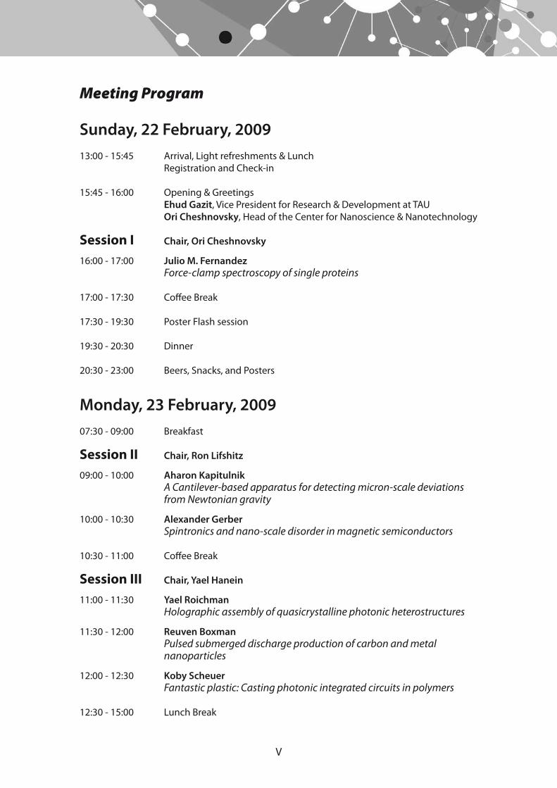
Meeting Program
Sunday, 22 February, 200913:00 - 15:45 Arrival, Light refreshments & Lunch Registration and Check-in
15:45 - 16:00 Opening & Greetings Ehud Gazit, Vice President for Research & Development at TAU Ori Cheshnovsky, Head of the Center for Nanoscience & Nanotechnology
Session I Chair, Ori Cheshnovsky
16:00 - 17:00 Julio M. Fernandez Force-clamp spectroscopy of single proteins
17:00 - 17:30 Coffee Break
17:30 - 19:30 Poster Flash session
19:30 - 20:30 Dinner
20:30 - 23:00 Beers, Snacks, and Posters
Monday, 23 February, 200907:30 - 09:00 Breakfast Session II Chair, Ron Lifshitz
09:00 - 10:00 Aharon Kapitulnik A Cantilever-based apparatus for detecting micron-scale deviations from Newtonian gravity
10:00 - 10:30 Alexander Gerber Spintronics and nano-scale disorder in magnetic semiconductors
10:30 - 11:00 Coffee Break
Session III Chair, Yael Hanein
11:00 - 11:30 Yael Roichman Holographic assembly of quasicrystalline photonic heterostructures
11:30 - 12:00 Reuven Boxman Pulsed submerged discharge production of carbon and metal
nanoparticles
12:00 - 12:30 Koby Scheuer Fantastic plastic: Casting photonic integrated circuits in polymers
12:30 - 15:00 Lunch Break
VI
Session IV Chair, Shachar Richter
15:00 - 15:30 Yoram Selzer Laser light as a tool to analyze and control processes in molecular
junctions
15:30 - 16:00 Tamar Seideman Electron- and photon-controlled dynamics in nano-junctions
16:00 - 16:30 Oded Hod Electromechanical properties of suspended graphene nanoribbons
16:30 - 17:00 Coffee Break
Session V Chair, Ronit Satchi-Fainaro
17:00 - 17:30 Noam Shomron MicroRNAs involved in nerve blockade using drug-containing
microparticles
17:30 - 18:00 Ella Sklan Host factors required for hepatitis C virus replication
18:00 - 18:30 Slava Krylov Experimental and numerical investigation of spatial vibrational
modes of nano cantilevers
19:30 - 21:30 Conference Banquet
Tuesday, 24 February, 200907:30 - 09:00 Breakfast
Session VI Chair, Itai Benhar
09:00 - 10:00 Andreas Herrmann Nanoparticles from nucleic acid/synthetic polymer-hybrids
10:00 - 10:30 Yoel Kashman & Drorit Neumann New bioactive marine sponge-derived natural compounds as
inhibitors of proliferation and inducers of cell death
10:30 - 11:00 Coffee Break
Session VII Chair, Rimona Margalit
11:00 - 11:30 Abdussalam Azem Variations on the function of type I chaperonins
11:30 - 12:00 Dan Peer Harnessing nanotechnology and RNA interference for therapy of
leukocyte-implicated diseases
12:00 - 12:30 Closing
12:30 - 14:00 Lunch and Departure

VI
Oral PresentationsAbstracts

3
O1: Force-clamp spectroscopy of single proteins
Julio M. Fernandez
Columbia University, New York, USA
We have developed single molecule AFM techniques to study how mechanical forces affect the dynamics and chemistry of proteins. Using molecular biological techniques, we engineer tandem modular proteins that are made of identical repeats of a protein of interest. These polyproteins act as handles for atomic force microscopes, without the need for linkers or special attachment chemistry. When such polyproteins are extended by an AFM, their force properties are unique mechanical fingerprints that unambiguously distinguish them from the more frequent non-specific events that plague single molecule studies. We combine polyprotein engineering together with active forceclamp AFM techniques. With this approach, the length of an extending polyprotein is measured while the pulling force is actively kept constant by negative feedback control. The force-clamp technique combined with polyprotein engineering has become a powerful approach to study proteins. We have investigated the force-dependency of protein folding, unfolding and of chemical reactions. From the force-dependence, we extract features of the transition state of these reactions that reveal underlying molecular mechanisms. Our data will help guide the development of new theories on areas such as the statistical dynamics of a folding polymer and ab-initio studies of a chemical reaction while placed under a stretching force.
References
1. Fernandez, J. M. and Li, H. B. Force-clamp spectroscopy monitors the folding trajectory of a single protein (2004), Science, 303: 1674-1678.
2. Wiita et al, (2007) Probing the chemistry of thioredoxin catalysis with force. Nature, 450: 124-7.
4
O2: A Cantilever-based apparatus for detecting micron-scale deviations from Newtonian gravity
Aharon Kapitulnik
Stanford University, California, USA
To test new theories of physics beyond the Standard Model, we have built a low temperature probe to measure forces as small as 10-18N between masses separated by distances on the order of 20 microns. Our experiment is fundamentally a Cavendish-type experiment in the sense that its purpose is to directly measure the force between two masses [1]. A cryogenic helium gas bearing is used to rotate a disc containing a drive mass pattern of alternating density under a small test mass mounted on a micromachined cantilever. Any mass-dependent force between the two will produce a time-varying force on the test mass, and consequently a time-varying displacement of the cantilever. This displacement is read out with a laser interferometer, and the position of the drive mass is simultaneously recorded using an optical encoder. The displacement is then averaged over many cycles and converted to a force using measured properties of the cantilever. This AC ``lock-in'' type measurement enables significant noise rejection and allows us to operate on resonance to take advantage of the cantilever's high quality factor. A novel feature of the apparatus is the utilization of feedback regulation of the response of the microcantilever using the radiation pressure of a laser. Our approach does not require a high-finesse cavity, and the feedback force is due solely to the momentum of the photons in the second laser.
Reference
1. D. M. Weld, J. Xia, B. Cabrera and A. Kapitulnik, Phys. Rev. D 77, 062006 (2008).
5
O3: Spintronics and nano-scale disorder in magnetic semiconductors
Alexander (Sasha) Gerber
The Raymond and Beverly Sackler School of Physics and Astronomy
Spintronics, which combines the key advantages of microelectronics and micromagnetics, represents the new frontier in device physics for future integrated circuit technology. A crucial step in developing the semiconductor spintronics is generation of semiconducting materials that possess the ferromagnetic properties at room temperature. Despite huge efforts this task has not yet been achieved because of a low solubility of magnetic dopants in a semiconductor host. Magnetic dopants like Mn or Fe tend to coagulate and form a separate disordered system of nanoscale magnetic clusters embedded in a conducting non-magnetic matrix. Remarkably, the presence of these precipitates and clusters is essential to trigger a new class of physical phenomena that includes the metal – insulator transition driven by applied magnetic field and huge magnetoresistance of thousands of percent.
6
O4: Holographic assembly of quasicrystalline photonic heterostructures
Yael Roichman
School of Chemistry, Raymond and Beverly Sackler Faculty of Exact Sciences
This talk focuses on a qualitatively new approach to fabricating complex three dimensional photonic heterostructures. Recently, icosahedral quasicrystals have been shown to exhibit prominent gaps in the photonic density of states at an effective Brillouin zone boundary. Because the triacontahedral Brillouin zone is proven to come closest to spherical symmetry, a dielectric icosahedral quasicrystal should be the best possible candidate for a full three-dimensional photonic bandgap material in the visible. This structure cannot be realized through conventional lithography, or even through holographic projection lithography.
We have demonstrated that large quasicrystalline domains can be rapidly assembled using holographic optical traps to directly place micrometer-scale dielectric colloidal spheres into the necessary three-dimensional configuration, including structures with specifically engineered defects. Holographic micromanipulation is uniquely useful for creating research-grade photonic materials and devices in laboratory-scale quantities. It also provides the infrastructure needed to probe and control mechanisms of self assembly in specifically engineered microparticles. Understanding both the kinetics and dynamics of how such particles associate may provide a new framework for self-organizing photonic systems on a large scale.
7
O5: Pulsed Submerged Discharge Production of Carbon and Metal Nanoparticles
N. Parkanskya, R. L. Boxmana, B. Alterkopa, Z. Barkayb, Yu. Rosenbergb, G. Leitusc, N. Eliazd, V. Fillere
aElectrical Discharge and Plasma LaboratorybThe Wolfson Applied Materials Research Center
cDepartment of Chemical Research Support, Weizmann Institute of Science, RehovotdSchool of Mechanical Engineering
eMetal Tech Ltd., Beer Sheva
Nanoparticles were produced using two types of pulsed submerged discharges: the contact mode (CM), where electrodes were periodically contacted and separated, and the breakdown mode (BM), where a high voltage pulse broke down the inter-electrode gap, which was maintained at constant distance. 99.5% pure Ni - Ni, W – W, W – C, Zn –Zn, and 99.99% pure C - C electrode pairs were used. Pulsed arcs were ignited between electrodes submerged in analytical (99.8%) ethanol or deionized water. Structure, composition and size distribution of the produced nanoparticles were examined by XRD, HRSEM, HRTEM and XPS. The particles’ magnetic properties were studied using a Quantum Design SQUID MPMS2 field shielded magnetometer.
Particles produced included those with a metal-rich core surrounded by a carbon-rich shell, magnetic particles, and electrically charged particles. Carbon particles with peak binding energy of ~285.5 eV, which is approximately equal to that of diamond, were produced using the CM with Ni electrodes submerged in ethanol.
Some magnetic carbon particles were produced using the BM in ethanol by arcing C electrodes. The magnetization curve of “raw” powder, i.e. containing all of the particles produced, whether magnetic or not, at 20 and 300 K showed hysteresis, with saturation magnetization Ms ~ 0.90-0.93 emu/g, residual magnetization Mr =0.022 and 0.018 emu/g, and coercive force Hc = 11 and 8 Oe , respectively. Magnetic nanoparticles were separated from the raw powder by passing a suspension of the raw powder in ethanol through a bio-ferrograph. SEM examination of the separated magnetic particles showed that they were agglomerates of ~30-50 nm diam spheres, and nanotubes and nano-rods with length of 50-250 nm and diameters of 20-30 nm.
8
O6: Fantastic Plastic: Casting Photonic Integrated Circuits in Polymers
Jacob (Koby) Scheuer
School of Electrical Engineering, The Iby and Aladar Fleischman Faculty of Engineering
The transition from the contemporary low scale integration of optical devices to future highly-integrated photonic processors necessitates the development of new high-quality materials on one hand and inexpensive mass-production fabrication methods on the other. Polymeric materials have interesting optical and mechanical properties, making them an attractive choice for future photonic systems. In addition to low optical losses and material dispersion, polymers are simple to manipulate and to cast using a wide variety of fabrication methods such as soft-lithography techniques. Moreover, the ability to dope polymeric materials with molecules that exhibit a large electro-optic coefficient, nonlinear response or optical gain, paves the way to all-polymer integrated optical circuits that include on-chip sources, processors and detectors.
In this talk, I will present polymer-based active and passive tunable optical devices such as lasers, switches and Add/Drop filters, all fabricated using a simple and inexpensive molding method.
9
O7: Laser light as a tool to analyze and control processes in molecular junctions
Yoram Selzer
School of Chemistry, Raymond and Beverly Sackler Faculty of Exact Sciences
This talk will describe two studies involving irradiation of laser light on molecular junctions:
In the first study I will demonstrate using a novel molecular junction a systematic investigation that facilitates understanding at the molecular level of how heat is generated in current carrying molecular junctions and what is the resulting (non-equilibrium) effective temperature of some of their vibrational modes. Inelastic Electron Tunneling Spectroscopy (IETS) is used to identify the vibrational modes that interact with the tunneling electrons. The resulting temperature of the junctions is determined by measuring their Raman spectra both in the Stokes (S) and anti-Stokes (AS) regimes. The AS/S ratio of each Raman active vibrational mode, ν, is a direct probe of its steady state nonequilibrium population in the presence of inelastic tunneling current, that can be translated into an effective temperature, Teff(ν), for that mode. Comparison of the results to existing theories will be presented, as well as some discussion on the possibility to initiate and detect cooling of vibrational modes by tunneling electrons. Future directions for further research will be mentioned as well.
In the second study the effect of laser light on the conductance of metal quantum point contacts (MQPCs) will be described. These contacts, with dimensions comparable to the de Broglie wavelength of conducting electrons, reveal ballistic transport of electrons and quantized conductance in units of G0=2e2/h. We measure the transport properties of 1G0 Au contacts under laser irradiation. The observed enhancement of conductance appears to be wavelength-dependent, while thermal effects on conductance are determined to be negligible. The results are consistent with a photoasisted transport mechanism in which conductance depends both on the electronic structure of the leads, and on the interaction of the transporting electrons with oscillating electric fields originating from excitation of local plasmons. The results are important for future interpretation of light effects on the conductance of molecular junctions.
10
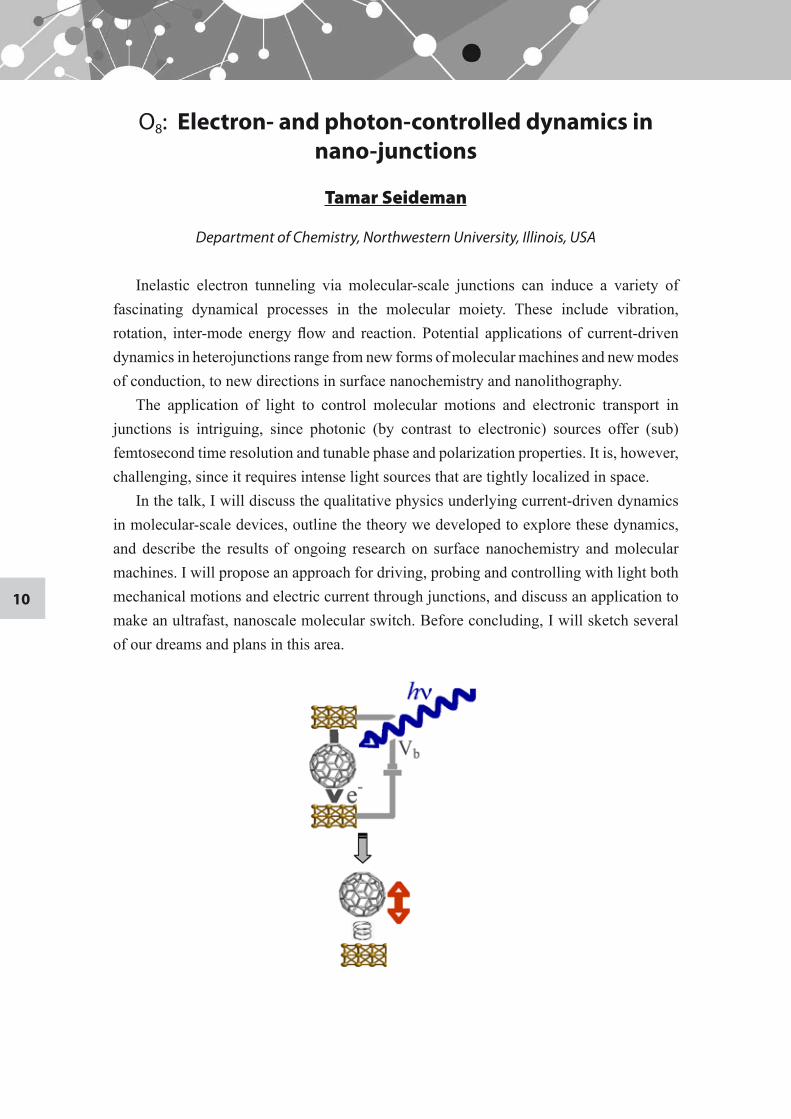
O8: Electron- and photon-controlled dynamics in nano-junctions
Tamar Seideman
Department of Chemistry, Northwestern University, Illinois, USA
Inelastic electron tunneling via molecular-scale junctions can induce a variety of fascinating dynamical processes in the molecular moiety. These include vibration, rotation, inter-mode energy flow and reaction. Potential applications of current-driven dynamics in heterojunctions range from new forms of molecular machines and new modes of conduction, to new directions in surface nanochemistry and nanolithography.
The application of light to control molecular motions and electronic transport in junctions is intriguing, since photonic (by contrast to electronic) sources offer (sub)femtosecond time resolution and tunable phase and polarization properties. It is, however, challenging, since it requires intense light sources that are tightly localized in space.
In the talk, I will discuss the qualitative physics underlying current-driven dynamics in molecular-scale devices, outline the theory we developed to explore these dynamics, and describe the results of ongoing research on surface nanochemistry and molecular machines. I will propose an approach for driving, probing and controlling with light both mechanical motions and electric current through junctions, and discuss an application to make an ultrafast, nanoscale molecular switch. Before concluding, I will sketch several of our dreams and plans in this area.
11
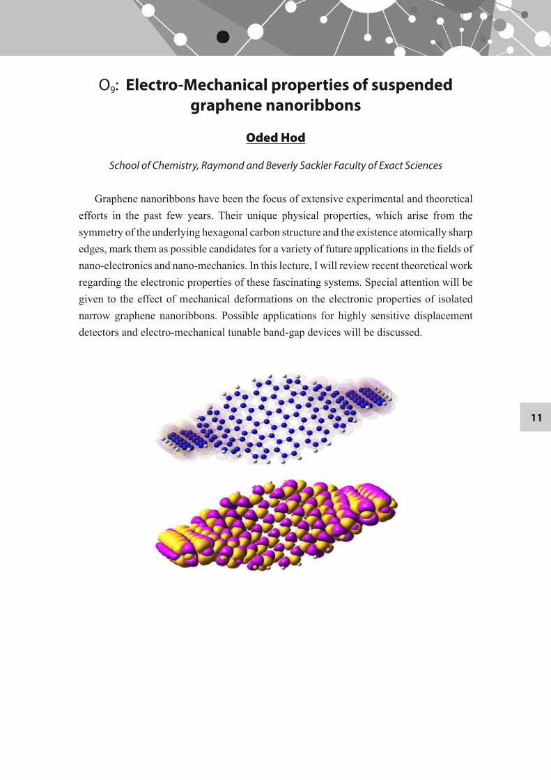
O9: Electro-Mechanical properties of suspended graphene nanoribbons
Oded Hod
School of Chemistry, Raymond and Beverly Sackler Faculty of Exact Sciences
Graphene nanoribbons have been the focus of extensive experimental and theoretical efforts in the past few years. Their unique physical properties, which arise from the symmetry of the underlying hexagonal carbon structure and the existence atomically sharp edges, mark them as possible candidates for a variety of future applications in the fields of nano-electronics and nano-mechanics. In this lecture, I will review recent theoretical work regarding the electronic properties of these fascinating systems. Special attention will be given to the effect of mechanical deformations on the electronic properties of isolated narrow graphene nanoribbons. Possible applications for highly sensitive displacement detectors and electro-mechanical tunable band-gap devices will be discussed.
12
O10: MicroRNAs involved in nerve blockade using drug-containing microparticles
Noam Shomron
Department of Cell and Developmental Biology, School of Medicine, Sackler Faculty of Medicine
There has long been strong interest in developing means of providing prolonged duration local anesthesia for chronic or prolonged acute pain, particularly by using sustained-release technology. Apart from the technical difficulties in developing such systems, questions have been raised as to the safety of that approach, partly because local anesthetics are known to be both myotoxic and neurotoxic. Here we study the effects of prolonged nerve blockade, using drug-containing microparticles, on neurobehavior, nerve morphology, and gene and microRNA expression in the dorsal root ganglia. Our data demonstrate that some treatments showed very little neurotoxicity thus offering alternative safer modes of anesthesia.
13
O11: Host factors required for hepatitis C virus replication
Elazar M1, Staschke, K.2, Myers, T.2, Glenn, J.S1. Sklan, E. H3
Division of Gastroenterology and Hepatology, Stanford University School of Medicine1 Veterans Administration Medical Center, Palo Alto, California2
Lilly Research Laboratories3
School of Medicine, Sackler Faculty of Medicine
One hundered and seventy million people worldwide are estimated to be infected with hepatitis C virus (HCV), a major cause of liver disease including liver cirrhosis, and hepatocellular carcinoma. There is no vaccine for HCV and the current thearpy has limited efficacy and serious side effects. HCV is an enveloped virus and its nanoparticle sized virion contains a positive, single-stranded RNA genome. This genome encodes a single ~3000 amino acid polyprotein which is processed into structural proteins composing the mature viral particle and non-structural (NS) proteins involved in replicating the viral genome. Host factors participate in most, if not all, steps of positive-strand RNA virus’s life cycle. Our goal is to identify these host factors and their role in the viral life cycle, aiming to translate these findings into novel antiviral strategies. We have recently discovered such an interaction between HCV NS protein 5A (NS5A) and TBC1D20 a protein involved in intracellular transport. TBC1D20 depletion impaired HCV replication, with no effect on cell viability. Currently, we are developing high throughput nanoparticle-based FRET assays, such as quantum dots or gold nanoparticles, in order to screen for small molecule inhibitors of this interaction. Viral particles are thought to contain all the essential proteins needed for initiation of replication in the target cell. To further understand the interaction of HCV with its host, we aim to isolate tissue culture grown viral particles using sucrose density gradients and identify host factors that are integral parts of the mature viral particle using a comprehensive proteomic analysis. Identification of the components of these virions has important implications for understanding the HCV biology and can serve as leads for the development of improved antiviral therapies.
14
O12: Optical Excitation of Spatial Vibrational Modes of Nano Oscillators
S. Krylov1, B. Ilic2, and H. G. Craighead2
1School of Mechanical Engineering, The Iby and Aladar Fleischman Faculty of Engineering2School of Applied and Engineering Physics, Nanobiotechnology Center and Cornell
Nanofabrication Facility, Cornell University, New York, USA
Excitation of micro and nanomechanical structures using optical fields is an emerging arena of research that couples the fields of optics, fluidics, electronics and mechanics with potential of generating novel chemical and biological sensors. Widely studied NEMS devices are surface micromachined mechanical cantilever-type oscillators that generally comprise of thin film layers patterned into various shapes. Conventional driving and motion transduction methods typically activate and detect only bending motion in the "out-of-plane", transverse direction perpendicular to the plane of the thin film. In this work we describe an approach that can activate and detect both out-of-plane and in-plane motions of nano oscillators. Because the frequency ranges and mechanical loss mechanisms can vary for the motion in the two directions, access to the different modes of oscillation can be useful in exploring anisotropic material properties or utilizing alternate directions of motion in device applications. As a model system, we used 200 nm and 250 nm thick single crystal silicon cantilevers with dissimilar lengths and widths ranging from 6 to 12 μm and 45 nm to 1 μm, respectively. The 1st and 2nd in-plane vibrational modes and up to 3rd transverse modes for slender cantilevers with lengths exceeding 8.5 μm were observed in experiments. The experimental quality factor of a particular in-plane harmonic was consistently higher than that of the transverse mode. Due to the high deformations at the clamped edge arising from the undercut of the sacrificial silicon dioxide (SiO2) layer, measured higher order out-of-plane modes showed deviation from the ideally clamped Euler-Bernoulli beam calculations. A full three dimensional finite element analysis, considering dimensions of the cantilever derived from scanning electron microscopy, enabled us to identify various observed flexural and torsional modes and to analyze the influence of the clamping compliances. We show that the influence of the silicon overhang on the out-of-plane vibrations is considerably more pronounced when compared with the in-plane mode. Finally, we demonstrate vibrational detachment of sub-micron polystyrene spheres on the oscillator surface using in-plane mode excitation.
15
O13: Nanoparticles from Nucleic Acid/Synthetic Polymer-Hybrids
Andreas Herrmann
Zernike Institute for Advanced Materials, University of Groningen, the Netherlands
In recent years, so called “hybrids” consisting of biomacromolecules and organic polymers have attracted considerable attention. We have prepared bioorganic hybrids consisting of nucleic acids and organic polymers. Different synthetic strategies for the generation of linear, single stranded (ss) and double stranded (ds) DNA block copolymers have been elaborated some of them relying on fully automated processes using a DNA synthesizer [1], on hybridization [3] or on molecular biology techniques like the polymerase chain reaction [2]. The formation of nanoscopic objects which are adopted by amphiphilic DNA block copolymers will be discussed [4].
In the context of applications, a novel triblock architecture for DNA detection is introduced [5]. Furthermore, sequence specific organic reactions in nanometer-sized spherical DNA block copolymer micelles will be presented [1]. Finally, the uptake of DNA block copolymer aggregates with different shapes into various cell lines was studied [6] and their use as combinatorial platform for drug delivery was successfully demonstrated [7].
References
Alemdaroglu FE, Ding K, Berger R, Herrmann1. A: Angew. Chem. Int. Ed. 2006, 45: 4206-4210. Safak M, Alemdaroglu FE, Li Y, Ergen E, Herrmann2. A: Adv. Mat. 2007, 19: 1499-1505.Alemdaroglu FE, Safak M, Berger R, Herrmann3. A: Chem. Commun. 2007, 1358-1359.Ding K, Alemdaroglu FE, Börsch M, Berger R, Herrmann4. A: Angew. Chem. Int. Ed. 2007, 46: 1172-1175.Ergen 5. E, Weber M, Jacob J, Herrmann A, Müllen K: Chem. Eur. J. 2006, 12: 3707-3713.Alemdaroglu FE, Alemdaroglu CN, Langguth P, Herrmann6. A: Macromol. Rapid Commun. 2008, 29: 326-329.Alemdaroglu FE, Alemdaroglu CN, Langguth P, Herrmann7. A: Adv. Mat. 2008, 20: 899-902.
16
O14: New bioactive marine sponge-derived natural compounds as inhibitors of proliferation and
inducers of cell death
Drorit Neumann1, Nathalie Ben-Califa1, Ashgan Bashira2 and Yoel Kashman2
1 Department of Cell and Developmental Biology, School of Medicine, Sackler Faculty of Medicine
2School of Chemistry, Raymond and Beverly Sackler Faculty of Exact Sciences
Natural products have attracted extensive research towards their possible application as potential clinically-bioactive compounds. A battery of new natural compounds (salarins A, B and C, and tulearin A) that we have recently isolated from marine sponges now appear promising as candidates for novel clinical applications. Our current study concerns the evaluation of these compounds and their derivatives, by employing a multidisciplinary approach that combines both chemistry and biology. We have found that these compounds inhibit cell proliferation, and modulate the cycle of cultured cell lines from both mouse and human origins. The novelty regarding these compounds stems from the fact that the "effect size" on proliferation and on cell cycle parameters that they manifest is different for each one of the tested cell lines. These features may be reflected in differential effects/potency of the compounds on transformed cells versus normal cell lineages, thus placing these new compounds as attractive candidates for further development as “custom-designed” anti-cancer agents. Our research plans are thus focused on resolving the molecular mechanisms underlying the activity of these compounds and pinpointing functional groups that are responsible for their differential bio-activity.
References
Bishara A, A. Rudi, M. Aknin, D. Neumann, N. Ben-Califa, Y. Kashman. Salarins A and B 1. and tulearin a: new cytotoxic sponge-derived macrolides. Org Lett. 10(2):153-6 (2008).Bishara, A., A. Rudi, M. Aknin, D. Neumann, N. Ben-Califa, and Y. Kashman. Taumycins A 2. and B, two bioactive lipodepsipeptides from the Madagascar sponge Fascaplysinopsis sp. Org Lett 10:4307, (2008).Bishara A., A. Rudi, M. Aknin, D. Neumann, N. Ben-Califa and Y. Kashman Salarin C, a new 3. cytotoxic sponge-derived nitrogenous macrolide Tetrahedron Lett. 49: 4355-4358 (2008)
17
O15: Variations in the structure and function of chaperonins
Avital Parnas, Shahar Nisemblat, Abdussalam Azem
Department of Biochemistry, The George S. Wise Faculty of Life Sciences
Type I chaperonins are protein folding nanomachines that play an essential role in the folding of newly translated and stress-denatured proteins in eubacteria, mitochondria and chloroplasts. Since their discovery, the bacterial chaperonins have provided an excellent model system for investigating the mechanism by which chaperonins mediate protein folding. Due to the high conservation of the primary sequence among type I chaperonins, it is generally accepted that organellar chaperonins function similar to the bacterial ones. However, mounting evidence indicate that the mitochondrial chaperonins possess unique structural and functional properties that distinguish them from their bacterial homologs. In this talk, we will present results of studies carried out in our laboratory, which shed some light on the interesting specificity of the mitochondrial chaperonin for its homologous co-chaperonins. Such structure-function studies will lead us toward an understanding of the mechanism of function of human mitochondrial chaperonins.
O16: Harnessing nanotechnology and RNA interference for therapy of leukocyte-implicated
diseases
Dan Peer
Department of Cell Research and Immunology, George S. Wise Faculty of life Sciences & the Center for Nanoscience and Nanotechnology
RNA interference (RNAi) is a ubiquitous and highly specific, endogenous, evolutionarily conserved mechanism of gene silencing. Since the discovery that RNAi occurs in mammalian cells, RNAi has emerged as a powerful tool for elucidating gene function and identifying potential drug targets. Harnessing RNAi holds enormous promise for therapeutic use for diseases that have proven difficult to treat with conventional drugs. RNAi can also be exogenously activated either by transducing cells with vectors to express small hairpin RNAs (shRNA) or by introducing already processed short double-stranded RNAs (siRNAs) into the cytoplasm of cells.
To realize the potential of siRNAs for in vivo drug discovery and therapy there is a need to overcome the considerable hurdle of intracellular delivery across the plasma membrane. siRNAs are not taken up into most cells in vitro in the absence of a transfection reagent. For many cells, mixing siRNAs at nanomolar concentrations with a lipid transfection can efficiently induce gene silencing. However, some important cells, such as leukocytes (immune cells), remain highly resistant to lipid transfection schemes.
To overcome these limitations, we have developed a platform for robust in vivo siRNA delivery selectively to leukocytes.
The platform can be used for siRNAs delivery for therapeutic purposes as well as for validating in vivo the role of selected genes in different pathologies.
For example, cyclin D1, a pivotal cell-cycle-regulatory molecule and a well-studied therapeutic target for cancer, is upregulated at sites of inflammation; however, its roles in this context remain uncharacterized. To address this, we generated targeted stabilized nanoparticles (tsNP) that were loaded with siRNAs and, then via antibodies to β7 integrin (β7 I), were directed to the specific leukocyte subsets involved in gut inflammation. Systemic application of β7 I-tsNP silenced cyclin D1 in leukocytes and reversed experimentally induce colitis. The tsNP strategy reveals cyclin D1 to be a novel anti-inflammation target for inflammatory bowel diseases.
Since the tsNP is a platform, by replacing the directing agent and the specific siRNA, it is possible to target different diseases. For example, in a preclinical model of HIV, we have shown that it is possible to prevent the infection of HIV by silencing HIV’s co-receptor CCR5. In addition, recently, a clinical trial was launch to test this system in an aggressive blood cancer hoping to silence important genes related to this type of cancer.

Poster PresentationsAbstracts

21
P1: Novel applications of self-assembled aromatic dipeptides nanostructures
Lihi Adler-Abramovich and Ehud Gazit
Department of Molecular Microbiology and Biotechnology, George S. Wise Faculty of Life Sciences
Organic and inorganic self-assembled tubular nanostructures were suggested to have key potential in nanotechnological devices and applications. Several studies have shown the possible use of bionanometric material for applications ranging from molecular electronic to drug delivery. The diphenylalanine peptide, the core recognition motif of the Alzheimer's Beta-amyloid polypeptide, efficiently self-assembles into discrete, well-ordered peptide nanotubes.
In the current research, using different microscopy and spectroscopy tools we describe a remarkable thermal stability of the aromatic dipeptide nanotubes both in aqueous solution and under dry conditions. In addition, the ADNT exhibit substantial chemical stability in various organic solvents. Furthermore, we studied the peptide nanotubes mechanical properties, which were directly measured by indentation type experiments, using an atomic force microscopy. We found that the peptide nanotubes maintain high averaged point stiffness of 160 N/m, and correspondingly high Young’s modulus of approximately 19 GPa, which places these peptide nanotubes among the stiffest bio-inspired materials presently known.
A limiting factor in the utilization of the ADNT system was the ability to temporally control the assembly process. This was resolved by the use of a self-immolative dendritic system as a platform for the controlled assembly of peptide nanotubes that was enzymatically activated. Self-immolative dendrimers are a novel class of molecules that can amplify a single cleavage event, which is received at a focal point, into multiple releases of tail groups at the periphery. The extremely short length of the peptide building blocks and their ability to self-assemble enable the controlled assembly applications.
Various methodologies were also developed for the horizontal and vertical alignment of the ADNT and for their patterning. The alignment of the ADNT using an external strong magnetic field of 12 Tesla was demonstrated. The alignment was attributed to the effect of the magnetic torque associated with the diamagnetic anisotropy of the aromatic rings of phenylalanine.
Additionally, we used the inkjet technology for the application of peptide nanostructures on non-biological surfaces. The ADNT which self-assemble readily in solution were used as an "ink" and patterned on transparency foil and ITO plastic surfaces using a commercial inkjet printer.
In summary, the remarkable thermal, chemical and mechanical durability and the ability to pattern align and control the assembly of the peptide nanotubes suggests their application in conventional microelectronic and microelectromechanics processes, as well as fabrication into functional nanotechnological devices.
22
P2: Quantum Confinement in Self Assembled Peptide Nanostructures
N. Amdursky1, E. Gazit1, and G. Rosenman2
1Department of Molecular Microbiology and BiotechnologyGeorge S. Wise Faculty of Life Sciences
2School of Electrical Engineering, The Iby and Aladar Fleischman Faculty of Engineering
Quantum confinement (QC) has been intensively investigated in semiconductors and applied in optical devices. The most common example is a QC-structures of GaAs/AlGaAs, where a thin layer of GaAs (dozens to hundreds of angstroms thick) is surrounded by a bulk of AlGaAs possessing wider energy gap. Another QC that has been firstly revealed by Canham (1990) is in mesoporous Si layers. Recently such a space QC has been found in inorganic nanotubes, mainly in arrays of ZnO nanowires. QC structures produce remarkable changes in the optical and electrical properties of semiconductors.
In this study, we report for the first time the direct observation of QC and photoluminescence (PL) in organic self-assembled peptide nanostructures. We studied QC effect in several quite different bio-nanostructures: hydrogels made from peptide nanotube network scaffold, peptide nanospheres and surfaces of normally aligned high diameter peptide nanotubes. All the structures are comprise from small monomers containing the diphenylalanine (FF) element. In all sorts of bio-nanotubes we found a pronounced step-like optical absorption behavior which is a distinguished feature of 2D-quantum wells (QW) characterized by the similar behavior of the electron density of states. The absorbance steps are located in the UV spectral range. The found absorption peaks are ascribed to Frenkel exciton in these organic structures. We have also found a distinguish PL peaks in the range of the QWs for both sorts of nanostructures. Our estimations show that found QC regions in the bio-nanotubes are extremely small in the order of 1 nm width and represent building-blocks of these self-assembly nanostructures. We suggest that our observation of QC effects is related to these sub-nano-crystalline areas embedded in the nanotube structures along its Z-axis. Understanding this physical phenomenon could help to understand the self-assembly processes responsible for neurodegenerative diseases associated with growth of nanostructural amyloid fibrils. On the other hand, the observed photoluminescence allows developing a new generation of photonic bio-devices, such as light emitting diodes and bio-lasers.
23
P3: Modeling drug diffusion across cell membranes by nano- and micro-sized liposomes.
Mirit Argov and Rimona Margalit
Department of Biochemistry, Department of Biochemistry, The George S. Wise Faculty of Life Science
Multidrug resistance (MDR), a major impediment in cancer treatment, is operated by ATP-dependant transporters that pump the drug out of the cells. In principle, this situation can be remedied by pump inhibition, using modulators with an acceptable safety profile. In this study, we tested two progesterone-like new modulators designed against the dominant MDR pump P-glycoprotein (Pgp). Drug efflux in MDR cancer cells is by at least two pathways: extrusion through the MDR pumps and simple diffusion through the lipid regions of the cell membrane. To assess the mechanism by which the new modulators exert their inhibitory effect, we made use of the cell line HCT-15 (human colon cancer) that over-expresses Pgp and the MDR drug paclitaxel (Taxol). Paclitaxel efflux from these cells fit the case of a single rate-limiting pathway with a rate constant of 0.4 (±0.1) hours-1. Adding a bench-mark chemosensitizer (verapamil) as well as each of the two new modulators (code-named SB4723 and SB4769), did not change the mechanism in terms of a single rate-limiting pathway for paclitaxel efflux, but reduced the rate constant 4 fold, to 0.1(±0.004) hours-1. One possibility for the reduced rate could be transition of the single efflux pathway from the pump to the lipid regions of the cell membrane. To pursue this option, we used liposomes to model the latter. Two liposome types can be selected for the task, micro-sized (MLV) and nano-sized (ULV). The curvature of MLV’s membranes is closer (compared to ULV) to that of cell membranes. ULV – like cell membranes – has a single lipid bilayer, where as MLV has multiple bilayers. Thus both liposomes have advantages and drawbacks for the task. We therefore decided to study paclitaxel efflux from both liposome types, also comparing fluid membranes made from natural phosphatidylcholine (PC) alone, to solid membranes made from natural PC and cholesterol. Data analysis was according to the three following options: (a) a single pathway (b) two parallel pathways with one rate limiting step and (c) similar to (b) but with two rate-limiting steps. The data fit option (a), with negligible sensitivity to liposome type or membrane fluidity, yielding a rate constant of 0.003 (±0.0003) hours-1 for paclitaxel diffusion across lipid bilayers, which is 30-fold slower than the rate constant in the pump-inhibited cells. Based on these findings, we propose that the inhibitory effect of the modulators was reduction in the rate constant of drug efflux through the pump rather than a shift from the pump to the lipid bilayer. We offer in conclusion, that despite their drawbacks liposomes can be a valid tool for investigations into mechanisms by which inhibitors of pathology-related transporters operate, as well as into the design of new inhibitors.
24
P4: Ultracapacitors based on peptide nanotubes-engineered electrodes
P. Beker, D. Aronov, G. RosenmanSchool of Electrical Engineering, The Iby and Aladar Fleischman Faculty of Engineering
L. Abramovitch, E. GazitDepartment of Molecular Microbiology and Biotechnology, George S. Wise
Faculty of Life Sciences
Supercapacitors are promising energy storage devices due to their unique combination of high power density and relatively large energy density. We report on environmentally clean bio-supercapacitors based on peptide nanotubes (ADNT)-modified electrodes.
Short aromatic dipeptides can self-assemble into ordered structures at the nano-scale. These assemblies include nanotubes, nanospheres, nano-plates and hydrogels with nano-scale order (Reches and Gazit, Science, 2003; Nature Nanotechology, 2006, 2007). Peptide nanotubes represent a novel class of nanotubes of biological origin as an appealing alternative to carbon nanotubes. It has been observed that these biological nanostructures possess paramount properties of different origin allowing finding new advanced nanotechnological applications, at the intersection Biology-Physics-Engineering, using the ADNT building blocks.
The basis for the new nanotechnology presented in this report is recently developed new biomolecules deposition method (E. Gazit, L. Abramovitch, D. Aronov, G. Rosenman, "Biomolecules Vapor Deposition" Provisional patent, September 13, 60960,066/, (2007)) which allows to drastically changing the previous ADNT deposition technology that based on peptide evaporation from aqueous or organic solutions. The method may be applied to ADNT coatings on unlimited area with high density and homogeneity, controllable thickness as well for fabricating patterned ADNT structures. A new ADNT-based technology has been applied to development of "green" energy storage devices-Supercapacitors. Deposition of ADMT arrays on carbon electrodes strongly increases efficiency of these electrochemical units due to high density ADNT coating that leads to surface-area increscent. In the developed electrostatic supercapacitors, aromatic vertically oriented ADNT have been used for modification of carbon electrodes. The conducted studies show that ADNT-modified electrodes demonstrate pronounced rectangular shape voltammograms and possess a high double-layer capacitance. When we made electrochemically deposition of Au on ADNT it improves the capacitance which means improving of conductivity in the ADNT electrode. We strongly believe that the Au deposition occurs not only on the outer surface of the ADNT, but on the inner surface of the tube as well, due to our recently conducted studies of ADNT wettability.
In these wettability measurements we discover that the electrolyte can either penetrate the nanotube inner hole, or to cover the outer surface of the tube. As we know, the wettability of the electrodes plays a key role in the formation of electric double layer at the surface-electrolyte interface, therefore ADNT possess great advantage, in comparison to well-known carbon nanotubes structures, in their unique wettability properties for electrochemical devices.
25
P5: A novel microfluidic whole cell biosensor based on electrochemical detection for water toxicity
analysis
Hadar Ben-Yoav1, Alva Biran2, Rami Pedahzur2, Shimshon Belkin2, Sebastian Buchinger3, Georg Reifferscheid3,
Yosi Shacham-Diamand1
1Department of Physical Electronics, School of Electrical Engineering2Institute of Life Sciences, the Hebrew University of Jerusalem
3Division of Qualitative Hydrology, Federal Institute of Hydrology (BfG), Germany
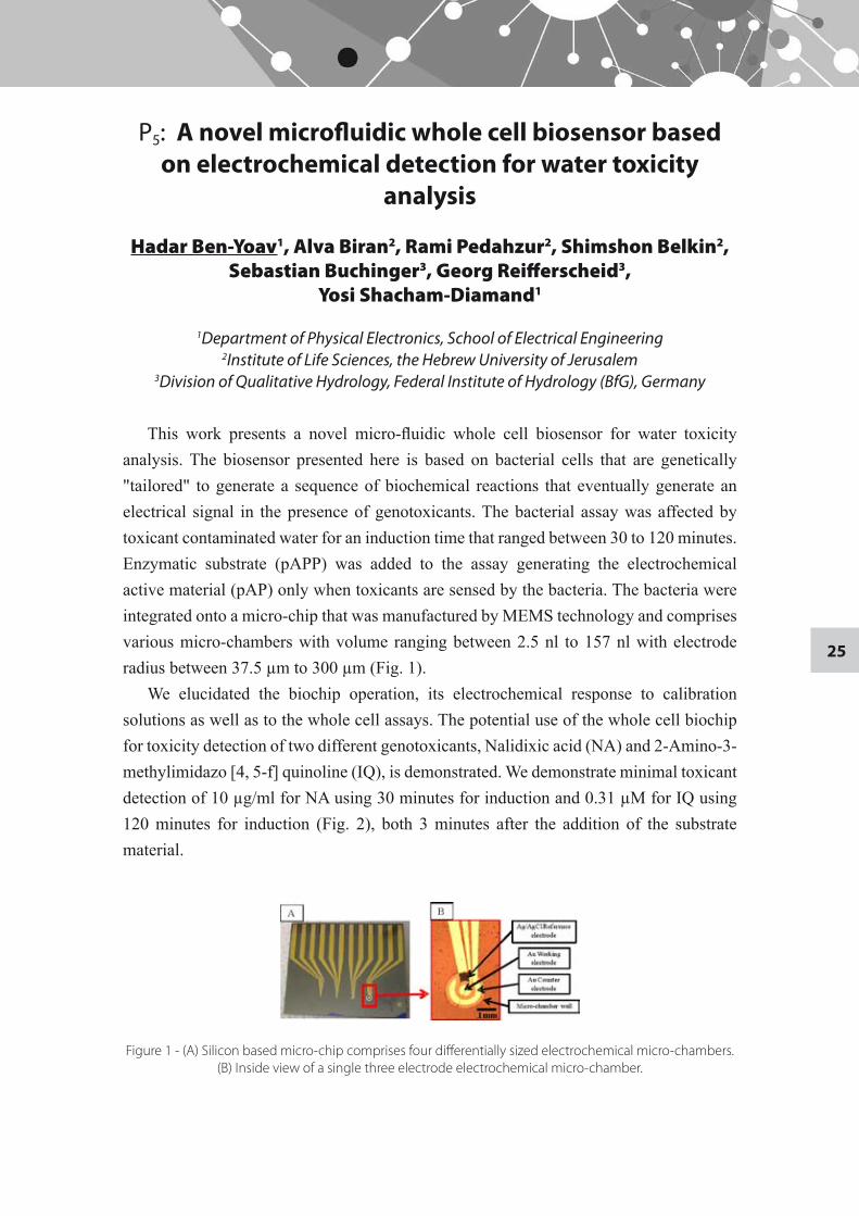
This work presents a novel micro-fluidic whole cell biosensor for water toxicity analysis. The biosensor presented here is based on bacterial cells that are genetically "tailored" to generate a sequence of biochemical reactions that eventually generate an electrical signal in the presence of genotoxicants. The bacterial assay was affected by toxicant contaminated water for an induction time that ranged between 30 to 120 minutes. Enzymatic substrate (pAPP) was added to the assay generating the electrochemical active material (pAP) only when toxicants are sensed by the bacteria. The bacteria were integrated onto a micro-chip that was manufactured by MEMS technology and comprises various micro-chambers with volume ranging between 2.5 nl to 157 nl with electrode radius between 37.5 µm to 300 µm (Fig. 1).
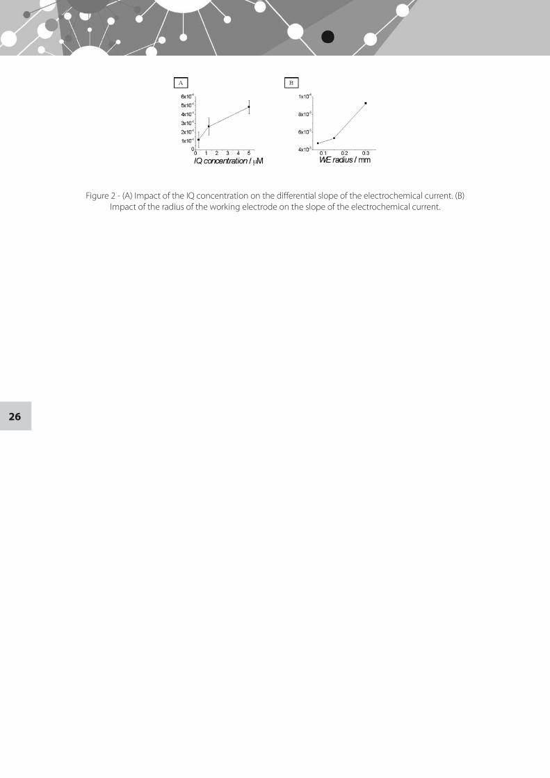
We elucidated the biochip operation, its electrochemical response to calibration solutions as well as to the whole cell assays. The potential use of the whole cell biochip for toxicity detection of two different genotoxicants, Nalidixic acid (NA) and 2-Amino-3-methylimidazo [4, 5-f] quinoline (IQ), is demonstrated. We demonstrate minimal toxicant detection of 10 µg/ml for NA using 30 minutes for induction and 0.31 µM for IQ using 120 minutes for induction (Fig. 2), both 3 minutes after the addition of the substrate material.
Figure 1 - (A) Silicon based micro-chip comprises four differentially sized electrochemical micro-chambers. (B) Inside view of a single three electrode electrochemical micro-chamber.
26
Figure 2 - (A) Impact of the IQ concentration on the differential slope of the electrochemical current. (B) Impact of the radius of the working electrode on the slope of the electrochemical current.
27
P6: Molecular Electronics with Squeezable Junction
Ben Zion Matan Yah, Yoram Selzer
School of Chemistry, Raymond and Beverly Sackler Faculty of Exact Sciences
Squeezable Electron Tunneling Junction (SET) is a mechanically controlled electrical junction allowing sub-nanometric approach of two surfaces. Though a very simple system, previous work already showed its ability to measure the superconducting energy gap1 and IETS induced molecular light emission2. In our work we wish to farther investigate the properties and abilities of this technique (translational resolution, mechanical sensitivity and stability, junction geometry). With these in hand we wish to investigate application of SET to electronic and thermal measurements of self assembled monolayer.
References
1 Squeezable Electron Tunneling Junction, J. Moreland et al Appl. Phys. Lett. 43, 387 (1983).2 Molecular Light Emission Induced by Inelastic Electron Tunneling, E. Flaxer, O. Sneh and O.
Cheshnovsky Science 262. 5142 (1993).
28
P7: Constructing Spin Interference Devices from Nanometric Rings
Guy Cohen
School of Chemistry, Raymond and Beverly Sackler Faculty of Exact Sciences
The study of nanospintronic devices utilizing coherent transport through molecular scale multiply connected geometries in the presence of moderate magnetic fields is presented. It is shown how two types of simple devices, spin filters and spin splitters, may be constructed from molecular nanometric rings utilizing the Aharonov-Bohm effect. The current is calculated within a single-electron approximation and within a many-body master equation approach where charging effects are accounted for in the Coulomb blockade regime. We provide rules and tools to develop and analyze efficient spintronic devices based on nanometric interferometers.
29
P8: Uptake of PEGylated hyperbranched polyesters in intestinal cell model
Shmuel Cohen1, Zili Sideratou2, Constantinos M. Paleos 2 and Rafi Korenstein1
1Department of Physiology and Pharmacology, Sackler Faculty of Medicine2Institute of Physical Chemistry, NCSR "Demokritos", Attiki, Greece
Background and Objectives: Following the development of various nanoparticles linked to or encapsulating proteins /peptides for oral administration, there is a growing need for evaluation of their uptake properties and intracellular fate. We assume that the structure and physiochemical properties of nanoparticles affect their pharmacodynamic and pharmacokinetic behavior. In this in-vitro study we are trying to: (i) evaluate a biodegradable PEGylated hyperbranched aliphatic polyester as a nanocarrier of proteins or peptides in endothelial and epithelial cells; (ii) explore the factors responsible for improved adsorption and transport of these nanoparticles.
Methods: A commercially available hyperbranched aliphatic polyester (Boltron BH40), was functionalized with approximately 18 poly(ethylene glycol) chains, (MW=2000), affording water soluble BH40-PEG. This polymer was also labeled with FITC. The fluorescence labelling enables tracking the nanoparticles in the various subcellular compartments. Their toxicity, adsorption and cellular uptake were determined by FACS and confocal microscopy in human-intestinal epithelial Caco-2/TC7 cells and in Bovine aorta endothelial (BAE) cells.
Results: Toxicity assays on Caco-2/TC7 and BAE cells showed no toxicity for BH40-PEG at concentrations up to 50 μM. Uptake time-course experiments revealed that BH40-PEG was efficiently internalized by both cell lines even at concentrations as low as 2 μM. The uptake was found to increase with incubation time, reaching saturation at 2.5 h at BH40-PEG concentration equal or higher than 15 μM . The uptake at 37ºC was significantly higher at 4ºC. The pathways by which BH40-PEG is internalized and directed to different subcellular compartments (endosomes, lysosomes and intracellular inclusion bodies), their transport rate across the intestinal monolayer of Caco-2/TC7 cells are now under investigation.
Conclusions: The uptake of PEGylated hyperbranched aliphatic polyester BH40-PEG is temperature-dependent, a function of concentration up to 15 μM where it reaches a plateau and also a function of time up to 2.5 h of incubation when saturation is reached. Moreover BH40-PEG was found to be non-toxic to both cell lines at concentrations up to 50 μM. These results indicate that BH40-PEG could potentially be applicable as a nanocarrier for oral delivery of proteins and peptides.
30
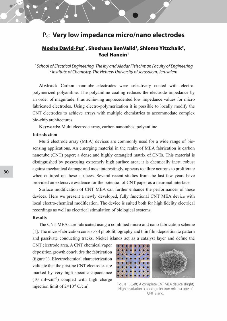
P9: Very low impedance micro/nano electrodes
Moshe David-Pur1, Shoshana BenValid2, Shlomo Yitzchaik2, Yael Hanein1
1 School of Electrical Engineering, The Iby and Aladar Fleischman Faculty of Engineering 2 Institute of Chemistry, The Hebrew University of Jerusalem, Jerusalem
Abstract: Carbon nanotube electrodes were selectively coated with electro-polymerized polyaniline. The polyaniline coating reduces the electrode impedance by an order of magnitude, thus achieving unprecedented low impedance values for micro fabricated electrodes. Using electro-polymerization it is possible to locally modify the CNT electrodes to achieve arrays with multiple chemistries to accommodate complex bio-chip architectures.
Keywords: Multi electrode array, carbon nanotubes, polyaniline
IntroductionMulti electrode array (MEA) devices are commonly used for a wide range of bio-
sensing applications. An emerging material in the realm of MEA fabrication is carbon nanotube (CNT) paper; a dense and highly entangled matrix of CNTs. This material is distinguished by possessing extremely high surface area; it is chemically inert, robust against mechanical damage and most interestingly, appears to allure neurons to proliferate when cultured on these surfaces. Several recent studies from the last few years have provided an extensive evidence for the potential of CNT paper as a neuronal interface.
Surface modification of CNT MEA can further enhance the performances of these devices. Here we present a newly developed, fully functional CNT MEA device with local electro-chemical modification. The device is suited both for high fidelity electrical recordings as well as electrical stimulation of biological systems.
ResultsThe CNT MEAs are fabricated using a combined micro and nano fabrication scheme
[1]. The micro-fabrication consists of photolithography and thin film deposition to pattern and passivate conducting tracks. Nickel islands act as a catalyst layer and define the CNT electrode area. A CNT chemical vapor deposition growth concludes the fabrication (figure 1). Electrochemical characterization validate that the pristine CNT electrodes are marked by very high specific capacitance (10 mF•cm−2) coupled with high charge injection limit of 2×10⁻³ C/cm2. Figure 1. (Left) A complete CNT MEA device. (Right)
High resolution scanning electron microscope of CNT island.
31
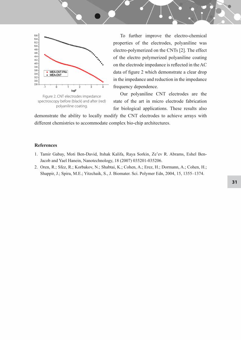
To further improve the electro-chemical properties of the electrodes, polyaniline was electro-polymerized on the CNTs [2]. The effect of the electro polymerized polyaniline coating on the electrode impedance is reflected in the AC data of figure 2 which demonstrate a clear drop in the impedance and reduction in the impedance frequency dependence.
Our polyaniline CNT electrodes are the state of the art in micro electrode fabrication for biological applications. These results also
demonstrate the ability to locally modify the CNT electrodes to achieve arrays with different chemistries to accommodate complex bio-chip architectures.
References
1. Tamir Gabay, Moti Ben-David, Itshak Kalifa, Raya Sorkin, Ze’ev R. Abrams, Eshel Ben-Jacob and Yael Hanein, Nanotechnology, 18 (2007) 035201-035206.
2. Oren, R.; Sfez, R.; Korbakov, N.; Shabtai, K.; Cohen, A.; Erez, H.; Dormann, A.; Cohen, H.; Shappir, J.; Spira, M.E.; Yitzchaik, S., J. Biomater. Sci. Polymer Edn, 2004, 15, 1355–1374.
Figure 2. CNT electrodes impedance spectroscopy before (black) and after (red)
polyaniline coating.
32
P10: Novel insulin formulations for oral administration: system characterization and in vivo
studies in diabetic mice
Yaron Dekel and Rimona Margalit
Department of Biochemistry, Department of Biochemistry, The George S. Wise Faculty of Life Science
The goals driving development of novel insulin formulations are to alleviate adverse effects, provide less-frequent and less-invasive methods of administration, improve patient’s compliance and quality of life. Among current efforts, the oral route is considered advantageous over other options (such as intranasal or pulmonary). Moreover, specific to insulin, the oral route is device-free and will reproduce the delivery route of endogenous insulin from the pancreas and on. Yet, free insulin (as any bioactive-protein for oral administration) does not survive the hostile enzymatic proteolytic environments along the GI tract. Formulating insulin in a carrier could, in principle, overcome these obstacles. Provided the carrier formulation protected its insulin load from the GI-tract, retained insulin activity and performed as a mucoadhesive slow-release depot. To that end we designed and developed novel formulations in which insulin, in the form of nano fibrils, is loaded inside carriers made from biomaterials. These novel insulin formulations were found to be non-toxic and bio-degradable, capable of high-efficiency loading over the insulin dose range of 1-10 mg/ml. The majority of loaded insulin (≥80%) was protected from hostile gastric/intestinal simulated environments and retained insulin activity (tested in an in vitro assay). These attributes justified progressing to the next step –in vivo studies in diabetic animal models.. We selected the frequently-used world-wide Streptozotocin (STZ)-induced IDDM model in ICR mice. We found this model in need of further modifications of both model and protocols, prior to testing our novel systems. The focus was on key parameters such as: STZ doses and their influence on the weight and blood glucose levels (BGL) of diabetic mice; setting proper BGL limits for fasting/non-fasting diabetic and healthy mice; calibrating age and weight of mice prior to an experiment and studying the correlation between animal responses to STZ and their initial age and weight; fasting/no fasting regimens as well as flipping light/dark cycles. Based on the animal’s responses to these parameters we developed and implemented guidelines for stabilizing BGL of untreated diabetic mice, and for testing therapeutic responses under conditions realistic for diabetic patients. A single dose of an orally-administered novel formulation was found sufficient to reduce BGL values of diabetic mice close up those of normal non-diabetic mice, a reduction that was sustained for several hours. The in vivo results support the potential of our novel insulin formulations for BGL control, in a patient-friendly mode. We also suggest that the approach applied in the present study – using the nano fibrillar form of insulin – has merit for oral delivery of additional therapeutic proteins.
33
P11: Regulation of endogenous amyloid beta release by spatiotemporal pattern of neuronal activity
I. Dolev1, E. Abramov1, and I. Slutsky1
1 Department of Physiology and Pharmacology, School of Medicine, Sackler Faculty of Medicine
Accumulation of cerebral amyloid-β peptide (Aβ) is critical for developing synaptic and cognitive deficits in (AD). However, the mechanisms regulating extracellular Aβ concentration and, consequently, synaptic function, remain elusive. Recent studies demonstrate that neural and synaptic activity rapidly and directly regulate Aβ release. Given that the spatiotemporal pattern of ongoing spikes may have a profound influence on synaptic plasticity and memory encoding, we explored the reciprocal relationships between the pattern of neuronal activity and endogenous release of Aβ isoforms. Aβ release was determined in hippocampal culture for single, low-frequency vs. temporally-correlated inputs, preserving the mean rate constant. Endogenously-released extracellular [Aβ40] and [Aβ42] was measured by ELISA for different patterns of stimulation. We found that [Aβ40] was sensitive to the input pattern: bursts triggered higher Aβ40 release than single inputs. In contrast, Aβ42 release exhibited very weak pattern dependency. As a result, [Aβ40]/[Aβ42] ratio was higher during periods of temporally-correlated activity. Next, we tested the effects of endogenously-released Aβ on synaptic transmission triggered by simple and complex spikes bursts. In order to rapidly increase endogenous [Aβ] in the synaptic cleft we used thiorphan, a specific inhibitor of the presynaptic rate-limiting Aβ degrading enzyme, neprilysin. Thiorphan-induced increase in the [Aβ] enhanced basal synaptic transmission in CA3-CA1 connections of acute hippocampal slices and synaptic vesicle release in hippocampal cultures. As a result, short-term synaptic facilitation was reduced through temporal redistribution of vesicle release during burst. Understanding of the mechanisms regulating Aβ release and its effects on synaptic transmission should contribute to the elucidation of physiological Aβ functions and to identify endogenous mechanisms that trigger primary synaptic deficits at very early stages of AD.
34
P12: Cyclooxygenase inhibition by diclofenac formulated in bioadhesive carriers
Inbar Elron-Gross, Yifat Glucksam, Dina Melikhov, and Rimona Margalit
Department of Biochemistry, Department of Biochemistry, The George S. Wise Faculty of Life Science
Adverse effects and risks of gastrointestinal toxicity are limitations to the use of the COX inhibitor Diclofenac, a frequently-used NSAID in treatments of rheumatic disorders and other chronic inflammatory diseases. Formulating Diclofenac in a carrier such as liposomes may alleviate the adverse effects, increase efficacy and allow local administration. We report here our first step, focused on biophysical and biochemical investigations of Diclofenac formulated in our previously-developed bioadhesive liposomes carrying hyaluronan (HA-BAL) or collagen (COL-BAL) on their surface. Diclofenac was encapsulated at high efficiency in both types of liposomes, achieving encapsulated doses up to 13 mg drug/ml. These systems performed as sustained-release Diclofenac depots, half-lives of drug release (under fastest conditions) ranging from 1 to 3 days. Therapeutic activity of liposome-formulated Diclofenac was evaluated in CT-26 cells, a bench-mark for intracellular COX enzymes. This cell line, possessing the CD44 hyaluronan receptors and integrins, bound HA-BAL and COL-BAL with high affinity that was 40 fold and 6 fold over that of regular liposomes. Encapsulated Diclofenac retained activity on a par with free drug. For example: 3nM Diclofenac given to intact cells generated COX-inhibition levels of: 62%, 42%, 73% and 71% for free drug and for encapsulated drug in COL-BAL, HA-BAL and RL, respectively. We propose these novel Diclofenac formulations possess key physicochemical and biochemical attributes for task performance, meriting the next step into in vivo studies.
35
P13: Diphenylalanine Peptide Nanotubes Coatings as Antibacterial Surfaces
Even Nitzan1, Adler-Abramovich Lihi1, Aronov Daniel2, Rosenman Gil2, Gazit Ehud1
1 Department of Molecular Microbiology and Biotechnology, George S. Wise Faculty of Life Sciences
2 School of Electrical Engineering, The Iby and Aladar Fleischman Faculty of Engineering
Many bacteria are found as surface associated communities know as bacterial biofilm. Biofilm formation as indwelling of bacteria on medical devices is major health problems due to increased resistance to biocides and antibiotics that can lead to severe infections.
Recent studies investigated the antibacterial properties of several nano materials such as carbon nanotubes, a nanometric tub shaped structures. The nanometric tubes expressed antimicrobial activity due to severe membrane damage and subsequent cell inactivation.
In the current study the antibacterial effect of peptide nanotubes made of diphenylalanine nanotubes is investigated. Well-ordered arrays of nanotubes are formed by vapour deposition technique (VD) on various surfaces. These arrays are being used as a surface for the inhbition of biofilm formation by a Curli producing strain of E. coli K-12 Ymel and its Curli deficient Ymel-1 strain at various growth conditions. Curli are required for the formation of a three-dimensional mature biofilm. Scanning electron microscopy is being used to visualize morphology of E. coli K-12 biofilm on the peptide nanotubes VD surfaces.
Further studies will probe whether the presence of diphenylalanine peptide nanotubes interfering with the Curli formation by bacteria and therefore preventing the biofilm formation. Coatings made of these peptide nanotubes can be in use for medical devices such as catheters.
36
P14: Remotely-controlled optical gas sensor for environmental and biological applications based on
nano-structured porous silicon
Tanya Favstov and Shlomo Ruschin
School of Electrical Engineering, The Iby and Aladar Fleischman Faculty of Engineering
In this project we develop optical sensors for biomedical and environmental applications. The sensors are based on nano-structured porous silicon (PSi) coated by an activation layer for specific analyte detection. PSi mesh-like structures were formed by electrochemical etching and micro-machining etching of bulk crystalline silicon. The interference pattern of the pSi layer is observed and measured. The phase shift depends on the thickness of the film and on the refractive index of the film itself. Upon adsorption of the analyte molecules, the optical reflectance spectrum changes, yielding a signal proportional to the analyte concentration.
We report the use of chemical pH indicator for ammonia detection in gaseous phase. In many applications of such a gas sensor, water vapor (humidity) is present and condensates inside the pores causing also changes in sensors signal. Data-analysis methods were applied in order to enable the discrimination between the two signal types.
37
P15: Short time Dynamics in Quasi-One-Dimensional (Q1D) Colloidal Suspension
Derek Frydel
School of Chemistry, Raymond and Beverly Sackler Faculty of Exact Sciences
Because of strong confinement of channel walls, in a Q1D colloidal suspension the vorticity, which determines hydrodynamic interactions in an unbounded fluid, fails to develop beyond the length comparable to the channel height. On the other hand, the contribution of propagating sound to the hydrodynamic interaction, which is essentially unimportnant for an unbounded fluid, due to confinement of channel walls changes to a diffusive bahavior. With help of lattice-Boltzmann simulation results, we investigate the role of the diffusive sound in the collective short dynamics of a Q1D suspension.
38
P16: Nanoliposome surface properties are instrumental in targeting drugs to their intracellular
or cell-membrane sites of action
Yifat Glucksam, Tsaffrir Zor and Rimona Margalit
Department of Biochemistry, Department of Biochemistry, The George S. Wise Faculty of Life Science
Chronic and acute inflammations, world-wide major clinical problems, are under intensive investigations pursuing ways and means to improve the currently-poor clinical outcomes. Macrophages (MΦ) phagocytic cells that are major constituents of defense mechanisms stimulate the immune system to secrete both pro- and anti-inflammatory cytokines. A major goal of inflammation treatment is to skew - MΦ - In the balance of secreted pro/anti inflammatory cytokines in favor of the latter. As in any drug-therapy, treatment with a targeted drug-carrier formulation can increase therapeutic efficacy and at the same time reduce adverse effects and risks of toxicity. Drug targeting has several steps in which the final is directing the drug to its molecular site of action, be it inside the cell or at the cell membrane. Selecting liposomes as the drug-carriers, we prepared two different nano (250 nm diameter) liposome species: (1) regular liposomes (RL) in which the liposomal surface was left un-modified and (2) bioadhesive liposomes (HA-BAL) in which the liposomal surface was modified to carry covalently-bound hyaluronan – the natural ligand of the CD44 receptors expressed on the MΦ’s cell membrane. The working hypothesis was that the RL would be internalized by the MΦ (exhibiting their normal phagocytic activity) while the HA-BAL will remain bound at the cell membrane. Selecting a particular liposome species would therefore direct a given drug to the specific location where its site of action resides. Employing imaging techniques we first evaluated CD44 expression in MΦ, finding extensive expression of CD44 not only on membranes of an immortalized murine MΦ cell line (RAW264.7) serving as a research tool, but also on the real targets – murine activated and non-activated peritoneal MΦ. These cells bound both liposome species with high affinity, the corresponding Kd values of 10 and 50 µM lipid for RL and HA-BAL, respectively. To determine the cellular distribution of each liposome species, double-labeled imaging was employed. The results showed that the RL were internalized, while the HA-BAL remained “glued” to the cell membrane. In conclusion, these studies strongly-confirm the working hypothesis and we now have at our hands the ability to deliver a given drug to its molecular target.
39
P17: Capacitance-Voltage Measurements of Biologically Functionalized Silicon Surfaces
E. Halpern1, B. Khmaisi1, A. Doron2, G, Shalev2, I. Levy2, and Y. Rosenwaks1
1 School of Electrical Engineering, The Iby and Aladar Fleischman Faculty of Engineering2 Intel Research Israel, Intel Electronics, Jerusalem
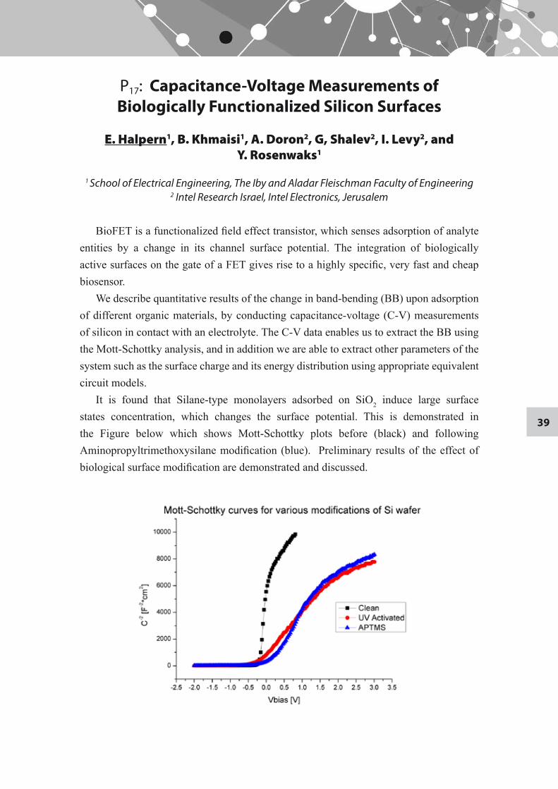
BioFET is a functionalized field effect transistor, which senses adsorption of analyte entities by a change in its channel surface potential. The integration of biologically active surfaces on the gate of a FET gives rise to a highly specific, very fast and cheap biosensor.
We describe quantitative results of the change in band-bending (BB) upon adsorption of different organic materials, by conducting capacitance-voltage (C-V) measurements of silicon in contact with an electrolyte. The C-V data enables us to extract the BB using the Mott-Schottky analysis, and in addition we are able to extract other parameters of the system such as the surface charge and its energy distribution using appropriate equivalent circuit models.
It is found that Silane-type monolayers adsorbed on SiO2 induce large surface states concentration, which changes the surface potential. This is demonstrated in the Figure below which shows Mott-Schottky plots before (black) and following Aminopropyltrimethoxysilane modification (blue). Preliminary results of the effect of biological surface modification are demonstrated and discussed.
40
P18: Bio inspired nanometric scale separation in aqua media of hydrophobic nanomaterials using bovine
submaxillary mucin
N. Hendler, E. D. Mentovich, B. Belgorodsky, S. Richter
The Center for Nanoscience and Nanotechnology and School of Chemistry, Raymond and Beverly Sackler Faculty of Exact sciences
Fullerenes are considered a major allotrope of carbon ever since discovered in 1985. Isolated fullerenes and their derivatives have shown remarkable electronic and mechanical properties and are suggested as major components in various complex materials.
However, due to their hydrophobicity and their tendency to form aggregates, their use in biological
applications is limited.
This work demonstrates the impressive capabilities of the protein bovine submaxillary mucin for binding and solubilizing water-insoluble materials such as Fullerene and Carbon Nanotubes in physiological solution. Different modes of binding are observed and measured as a function of the fullerenes concentration inside the protein.
This new type of material can be exploited in future to entry of hydrophobic materials and various nano-sized materials into living organisms.
41
P19: FMCI as means for high resolution mapping of intra cluster signal propagation in clustered
patterned rat cortical neural networks
Nitzan Herzog, Mark Shein, Yael Hanein
School of Electrical Engineering, The Iby and Aladar Fleischman Faculty of Engineering
Activity of cultured un-patterned neuronal networks often display bursting type of activity. Understanding this bursting dynamics may be fundamental to unraveling of information encoded in the network. However, previously utilized technique of multi electrode recording (MEA) in conjunction with spike sorting is unable to delineate individual cell activity in complex networks due to low spatial resolution and overwhelming complexity.
It therefore desired to devise experimental methods suited for better investigation of the dynamics in neuronal systems. Patterned neural networks can be regarded as a promising approach for the investigation of the computational functionalities of such systems, owing to their reduced complexity. Specifically, cluster-like patterning of neural cultures may be viewed as a model appropriate for the clustered organization of neural tissue seen in all animals.
Previous studies in our group presented a novel technology suited for patterning neural clusters in culture using carbon nanotubes (CNT) patterned multi electrode arrays (MEAs).
This work aims to utilize the high spatial resolution of multi-neuronal calcium imaging (FMCI) (an optical technique which acquires intra-cellular calcium signal which are correlated with electrical activity) to map intra-cluster signal propagation and bursting dynamics. Additionally, information extracted in this technique will be coupled to MEA analysis for important verification of its robustness.
42
P20: Building nanostructures and nanodevices based on DNA-Polyfluorene Block Copolymer micelles
Amir Holtzman
School of Chemistry, Raymond and Beverly Sackler Faculty of Exact Sciences
Using polymer chemistry a block copolymer can be synthesized so that its chains will contain a block of DNA and a block of polyfluorene. When these chains are dissolved in water they self-assemble into micelles, in which the hydrophobic polyfluorene turns into the core and the hydrophilic DNA turns out. Utilizing the self-recognition characteristics of DNA molecules these micelles can self assemble into complex 3D structures by using additional DNA chains that can hybridize with two micelles. The current work is focusing on utilizing the semi conducting behavior of polyfluorene for building transistors and sensors based on these micelles and their 3D structures.

43
P21: Raman Spectroscopy of Electrically Conductive Nanoscale Structures
Z. Ioffe, E. Pri-Gal, D. Dermer, O. Cheshnovsky
School of Chemistry, Raymond and Beverly Sackler Faculty of Exact Sciences
We use a Confocal Raman Microscope (CRM) to create Raman scattering maps of molecular conductive structures (SWCNTs and tunneling junctions). The resulting spatial spectral analysis allows chemical examination on molecular scale. Spatially-resolved Raman spectroscopy may provide useful information about the conduction structures, the molecules within and their vibrational excitation processes. Moreover, it allows us to monitor the enhancement effects on Raman scattering. Application of Squeezable Electron Tunneling Junction (SETJ) will provide for continuously variable and repeatable nanoscale gap geometry.
44
P22: Measurement of Electronic Transport through 1G0 Gold Contacts under Laser Irradiation.
Naomi Ittah, Ilan Yutsis, Gilad Noy and Yoram Selzer
School of Chemistry, Raymond and Beverly Sackler Faculty of Exact Sciences
Metal quantum point contacts (MQPCs) with dimensions comparable to the de Broglie wavelength of conducting electrons, reveal ballistic transport of electrons and quantized conductance in units of G0=2e2/h. Here we report on a new method to form MQPCs with quantized conductance values in the range of 1-4G0. The contacts appear to be stable at room temperature for hours and can be deterministically switched between conductance values, or reform in case they break, using voltage pulses. Also, measurements of the transport properties of 1G0 Au contacts under laser irradiation were carried. The observed enhancement of conductance appears to be wavelength-dependent. For wavelengths that are not absorbed by Au, the results are consistent with a photoasisted transport mechanism. For wavelengths absorbed by Au, photo-induced mechanism is suggested to be the dominant transport mechanism. The results are important for future interpretation of light effects on the conductance of molecular junctions.
45
P23: Carbon nanotubes integration process into NEMS devices
Gabriel A. Karp1, Asaf Yaakobovitz2, Slava Krylov2, and Yael Hanein1
1School of Electrical Engineering, The Iby and Aladar Fleischman Faculty of Engineering 2School of Mechanical Engineering, The Iby and Aladar Fleischman Faculty of Engineering
Several recent studies have demonstrated significant carbon nanotubes (CNT)-resistance changes in response to nanometer scale motion. Therefore, the integration of CNTs into MEMS devices offer exciting advantages in the scope of nano scale motion detection.
To achieve CNT based NEMS, fabrication methods are needed to allow seamless integration between standard silicon fabrication methods and CNT production and assembly. This integration must ensure proper growth, placement and electrical interfacing between the silicon and the CNTs. In this work we propose a simple approach to achieve such integration through the use of standard MEMS and CNT growth procedures combined with a sacrificial layer etching procedure.
A standard DRIE micro-fabrication procedure is utilized to construct isolated oxidized Islands over highly doped silicon pillar-like electrodes. As single wall CNTs do not readily grow directly onto silicon, SiO2 is used to ensure proper support for the growth step. However, this layer hinders the contact between the tubes and the conducting silicon therefore additional fabrication steps are needed to ensure direct electrical contacts. Using a simple chemical vapor deposition, SWCNTs are grown on the top surface of the pillars. Due to dynamical processes that are taking place during the CVD process, the resulting tubes are taut and bridge neighboring pillars. When the pillar geometry is replaced with finger geometry, CNT bridge the fingers, so their position can be controlled. The removal of the oxide is performed using a brief HF etch which does not attack the CNTs. The obtained structure includes silicon fingers bridged with single or very small bundles of CNT.
References
1. Ze’ev R. Abrams, Zvi Ioffe, Alexander Tsukernik, Ori Cheshnovsky and Yael Hanein, Nano Letters, 7 (2007) 2666 -2671.
2. Ze’ev Abrams and Yael Hanein, Journal of Physical Chemistry B, 110 (2006) 21419-21423, 2006.
46
P24: Pattern selection in parametrically-driven arrays of nonlinear resonators
Eyal Kenig1, Ron Lifshitz1, and M. C. Cross2
1The Raymond and Beverly Sackler School of Physics and Astronomy 2California Institute of Technology, California, USA
Motivated by recent experimental studies of microelectromechanical and nanoelectromechanical systems (MEMS & NEMS) [1] we have been studying the nonlinear dynamics of arrays of coupled nonlinear micromechanical and nanomechanical resonators [2, 3]. Here we invetigate the problem of pattern selection in an array of parametrically-driven nonlinear resonators using a novel amplitude equation [4]. We describe the transitions between standing-wave patterns of different wave numbers as the drive amplitude is varied either quasistatically, abruptly, or as a linear ramp in time. We find novel hysteretic effects, which are confirmed by numerical integration of the original equations of motion of the interacting nonlinear resonators, suggesting new possibilities for future experiments [5].
References
Buks & Roukes, J. MEMS 11 (2002) 802-807.1. Lifshitz & Cross, PRB 67 (2003) 134302.2. Lifshitz & Cross, Review of Nonlinear Dynamics & Complexity 1 (2008) 1-52.3. Bromberg, Cross, & Lifshitz, PRE 73 (2006) 016214.4. Kenig, Lifshitz, & Cross,Preprint (arXiv:0808.3589), accepted to PRE.5.
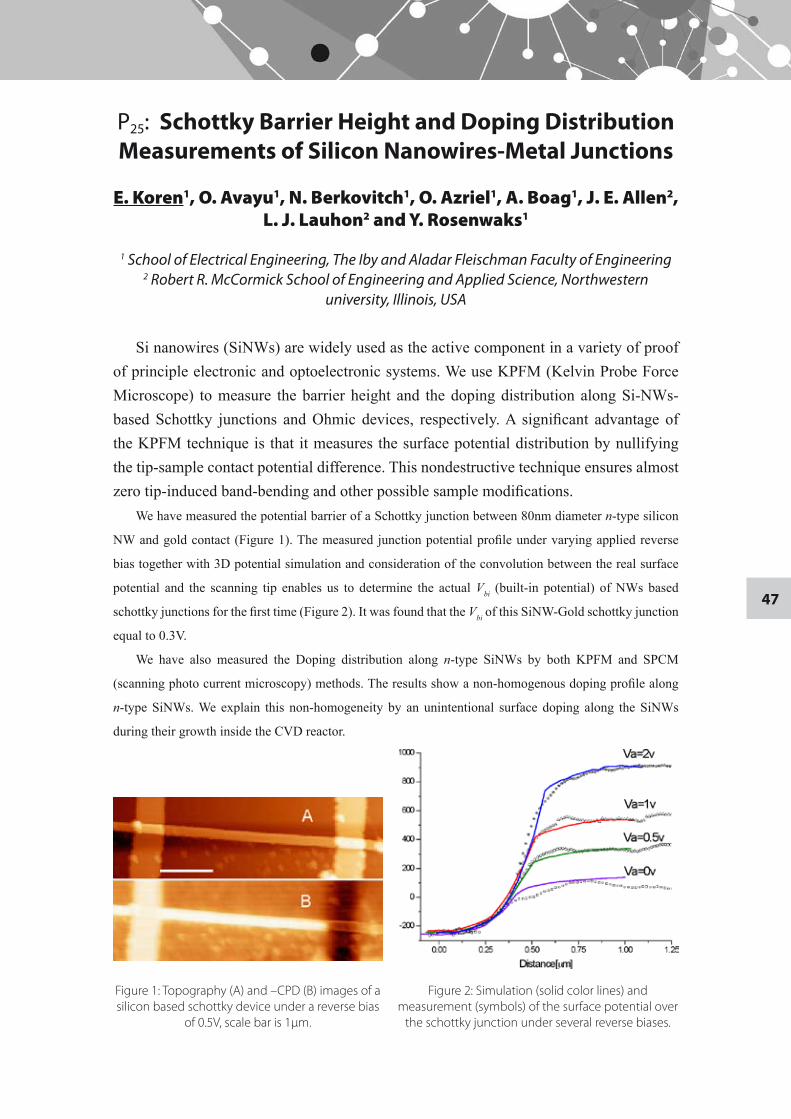
47
P25: Schottky Barrier Height and Doping Distribution Measurements of Silicon Nanowires-Metal Junctions
E. Koren1, O. Avayu1, N. Berkovitch1, O. Azriel1, A. Boag1, J. E. Allen2, L. J. Lauhon2 and Y. Rosenwaks1
1 School of Electrical Engineering, The Iby and Aladar Fleischman Faculty of Engineering
2 Robert R. McCormick School of Engineering and Applied Science, Northwestern university, Illinois, USA
Si nanowires (SiNWs) are widely used as the active component in a variety of proof of principle electronic and optoelectronic systems. We use KPFM (Kelvin Probe Force Microscope) to measure the barrier height and the doping distribution along Si-NWs-based Schottky junctions and Ohmic devices, respectively. A significant advantage of the KPFM technique is that it measures the surface potential distribution by nullifying the tip-sample contact potential difference. This nondestructive technique ensures almost zero tip-induced band-bending and other possible sample modifications.
We have measured the potential barrier of a Schottky junction between 80nm diameter n-type silicon
NW and gold contact (Figure 1). The measured junction potential profile under varying applied reverse
bias together with 3D potential simulation and consideration of the convolution between the real surface
potential and the scanning tip enables us to determine the actual Vbi (built-in potential) of NWs based
schottky junctions for the first time (Figure 2). It was found that the Vbi of this SiNW-Gold schottky junction
equal to 0.3V.
We have also measured the Doping distribution along n-type SiNWs by both KPFM and SPCM
(scanning photo current microscopy) methods. The results show a non-homogenous doping profile along
n-type SiNWs. We explain this non-homogeneity by an unintentional surface doping along the SiNWs
during their growth inside the CVD reactor.
Figure 1: Topography (A) and –CPD (B) images of a silicon based schottky device under a reverse bias
of 0.5V, scale bar is 1μm.
Figure 2: Simulation (solid color lines) and measurement (symbols) of the surface potential over
the schottky junction under several reverse biases.
48
P26: Endomorphin-1 and endomorphin-2 as a promising endogenous beta-amyloid aggregation
inhibitors
Ayala Lampel, Anat Marom-Frydman and Ehud Gazit
Department of Molecular Microbiology and Biotechnology, George S. Wise Faculty of Life Sciences
It is widely accepted that the assembly of amyloid fibrils is a fundamental factor in the generation of various diseases, among them is Alzheimer's disease (AD). AD is becoming a great concern due to the constant increasing in life expectancy. The early study of AD as amyloid-associated disease focused on the mature fibrils of β-amyloid (Aβ) polypeptide as the pathological agents. However, contemporary studies provide strong evidence that the Aβ soluble oligomers are the main toxic factor which causes apoptotic death of neural cells. According to these novel studies, the therapeutic target should be aimed toward the formation of soluble oligomers. Previous work in the lab has demonstrated the importance of small peptide containing aromatic amino acid and β-breaking element as a potential inhibitor for the amyloid fibril formation.
The endogenous tetrapeptides endomorphin-1 (Tyr-Pro-Trp-Phe) and endomorphin-2 (Tyr-Pro-Phe-Phe) appear to be appropriate potential inhibitors agents. Endomorphins are found in the central and peripheral nervous system and have an important role in pain modulation. Both endomorphins possess Pro that has the β-breaking element, and aromatic amino acids that can interact with the aromatic core of the Aβ peptide. Based on the properties of the endomorphins structural units and on the important fact that they are endogenous tetrapeptides found in the CNS, our hypothesis is that endomorphin-1 and endomorphin-2 can act as Aβ oligomers inhibitors and fibrils formation blockers. As shown in previous studies by our group, α-aminoisobutyricacid (Aib) is a better potent β-breaker than Pro. Therefore, we designed two new peptides (Aib1 and Aib2) which contain the Aib instead of the Pro.
We assume that these peptides will serve as even more effective inhibitors than the endomorphins.
In this study we demonstrated using spectroscopic, microscopic and cells methods that the different peptides can inhibit amyloid aggregation. We believe that the peptides interact with Aβ in the early stage of aggregation and inhibit the formation of the toxic species.
49
P27: A simple model for pairing in ultra-small nano-particles
Ze'ev Lindenfeld, Eli Eisenberg, Ron Lifshitz
The Raymond and Beverly Sackler School of Physics and Astronomy
We explore the possibility of a BCS-like pairing interaction in ultra-small isolated nano-particles within the framework of a simplified model. We start with the wave functions of free electrons in an infinite spherical well potential, while the phonons of the nano-particle are considered as the quantized normal modes of vibration of a stress-free elastic sphere. The interaction between the electrons and the phonons is evaluated explicitly, and the phonon mediated electron-electron interaction is estimated by means of a canonical unitary transformation. A reduced pairing Hamiltonian is obtained and its effects on the electron spectrum are evaluated and discussed.
50
P28: STM Measurements of Bismuth Nanoparticles
Debora Marchak, Eyal Shapira, Einat Tirosh, Gil Markovich, and Yoram Selzer
School of Chemistry, Raymond and Beverly Sackler Faculty of Exact Sciences
Bismuth Nanoparticles (Bi NP) are prepared in a one-step synthesis at room temperature under inert atmosphere. During the reaction, a self-assembled-monolayer of a mercaptoalkylic acid is formed on the surface of the particles, serving as a stabilizing capping agent dictating the final dimension of the nanocrystals. Through TEM observation, the morphology, crystallinity and size distribution of the Bi NP are determined. The electronic properties of individual NP are studied by UHV-STM at 90K. The measurements results are examined and discussed.
51
P29: Large-scale fabrication of 4-nm-channel vertical - protein -transistor
Elad D. Mentovich1, 2, Bogdan Belgorodsky1, Itsik Kalifa2, Michael Gozin1, 2 and Shachar Richter1, 2
1 School of Chemistry, Raymond and Beverly Sackler Faculty of Exact Sciences2 The Center for Nanoscience and Nanotechnology
The increasing demand for smaller and faster complementary transistors (in which
both n- and p-type devices exist on one wafer) arranged in dense arrays requires the development of new methods for the parallel fabrication of nanometer-sized transistors. However, due to limitations in the existing technology, achievement of these goals is very challenging. Although some isolated examples of such devices and architectures have been demonstrated, they exhibit only moderate or limited performance or are constructed via sophisticated multistep methodologies.
In this work, we suggest and demonstrate a universal method in which a new type of nanometer-sized, complementary vertical molecular transistor is fabricated in parallel fashion. This Central-Gate Molecular Vertical Transistor (C-Gate MolVeT) is fabricated using a combination of conventional micro-lithography techniques and self-assembly methods..
We further show that these ambipolar C-Gate MolVeTs can be operated at extremely low voltages with high gate sensitivity. Our findings indicate that the C-Gate MolVeT can be used as a powerful tool to investigate transport in various molecular systems and can be adopted as a building block for future ultra-fast logic devices.
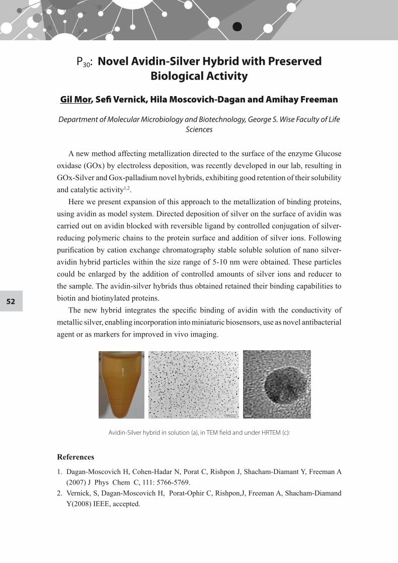
52
P30: Novel Avidin-Silver Hybrid with Preserved Biological Activity
Gil Mor, Sefi Vernick, Hila Moscovich-Dagan and Amihay Freeman
Department of Molecular Microbiology and Biotechnology, George S. Wise Faculty of Life Sciences
A new method affecting metallization directed to the surface of the enzyme Glucose oxidase (GOx) by electroless deposition, was recently developed in our lab, resulting in GOx-Silver and Gox-palladium novel hybrids, exhibiting good retention of their solubility and catalytic activity1,2.
Here we present expansion of this approach to the metallization of binding proteins, using avidin as model system. Directed deposition of silver on the surface of avidin was carried out on avidin blocked with reversible ligand by controlled conjugation of silver-reducing polymeric chains to the protein surface and addition of silver ions. Following purification by cation exchange chromatography stable soluble solution of nano silver-avidin hybrid particles within the size range of 5-10 nm were obtained. These particles could be enlarged by the addition of controlled amounts of silver ions and reducer to the sample. The avidin-silver hybrids thus obtained retained their binding capabilities to biotin and biotinylated proteins.
The new hybrid integrates the specific binding of avidin with the conductivity of metallic silver, enabling incorporation into miniaturic biosensors, use as novel antibacterial agent or as markers for improved in vivo imaging.
Avidin-Silver hybrid in solution (a), in TEM field and under HRTEM (c):
References
1. Dagan-Moscovich H, Cohen-Hadar N, Porat C, Rishpon J, Shacham-Diamant Y, Freeman A (2007) J Phys Chem C, 111: 5766-5769.
2. Vernick, S, Dagan-Moscovich H, Porat-Ophir C, Rishpon,J, Freeman A, Shacham-Diamand Y(2008) IEEE, accepted.
53
P31: A Novel Fabrication Method of Graphene Nanoribbons
Ayelet Ophir, Michal Vadai, and Yoram Selzer
School of Chemistry, School of Chemistry, Raymond and Beverly Sackler Faculty of Exact Sciences
The recent discovery of Graphene, a single layer of Graphite, has ignited intense research activities to elucidate the electronic properties of this novel two-dimensional (2D) electronic system. While 2D Graphene exhibits fascinating properties such as a relativistic massless-dispersion for its charge carriers and ballistic transport on large distances, its gapless spectrum makes it unsuitable for direct application as a channel in field-effect transistors (FET) and other semiconducting devices. This problem might be overcome by designing Graphene nanoribbons (GNRs) in which lateral quantum confinement opens an energy gap, which is a function of the ribbon's width. We note however, that all current calculations regarding this structural effect and others are for ribbons that are only few nanometers in width and few nanometers in length. Currently there is no experimental system that enables direct corroboration of the theoretical calculations.
Here we demonstrate a new method to fabricate GNRs with an area as small as 3x5nm2, using a new Top-Down approach. These small GNRs would allow us to verify various theoretical predictions. In addition they may enable us to explore vibrational effects on conductance by new methods.
54
P32: Pair diffusion of membrane proteins and their effect on membrane viscosity
Naomi Oppenheimer and Haim Diamant
School of Chemistry, Raymond and Beverly Sackler Faculty of Exact Sciences Biological membranes contain a high concentration of embedded mobile proteins.
While the Brownian motion of single membrane proteins has been studied in detail, the dynamic correlations between proteins, as mediated by flows within the membrane and in the surrounding liquid, have received little attention. We account for the correlated motion of membrane proteins, along with the effect of protein concentration on that correlation and on the response of the membrane to stresses. Expressions for the coupling diffusion coefficients of protein pairs and their concentration dependence are derived in the limit of small protein size relative to the inter-protein separation. Membrane viscosity has an additional role as determining the characteristic length scale for membrane response. This dependence leads to unusual concentration effects at large separation – the transverse coupling increases with protein concentration, whereas the longitudinal one becomes concentration-independent.
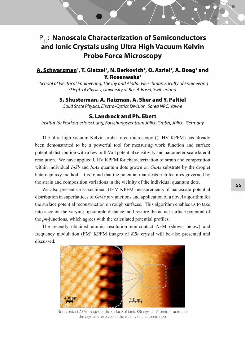
55
P33: Nanoscale Characterization of Semiconductors and Ionic Crystals using Ultra High Vacuum Kelvin
Probe Force Microscopy
A. Schwarzman1, T. Glatzel2, N. Berkovich1, O. Azriel1, A. Boag1 and Y. Rosenwaks1
1 School of Electrical Engineering, The Iby and Aladar Fleischman Faculty of Engineering 2Dept. of Physics, University of Basel, Basel, Switzerland
S. Shusterman, A. Raizman, A. Sher and Y. PaltielSolid State Physics, Electro-Optics Division, Soreq NRC, Yavne
S. Landrock and Ph. EbertInstitut für Festkörperforschung, Forschungszentrum Jülich GmbH, Jülich, Germany
The ultra high vacuum Kelvin probe force microscopy ((UHV KPFM) has already been demonstrated to be a powerful tool for measuring work function and surface potential distribution with a few milliVolt potential sensitivity and nanometer-scale lateral resolution. We have applied UHV KPFM for characterization of strain and composition within individual InSb and InAs quantum dots grown on GaAs substrate by the droplet heteroepitaxy method. It is found that the potential manifests rich features governed by the strain and composition variations in the vicinity of the individual quantum dots.
We also present cross-sectional UHV KPFM measurements of nanoscale potential distribution in superlattices of GaAs pn-junctions and application of a novel algorithm for the surface potential reconstruction on rough surfaces. This algorithm enables us to take into account the varying tip-sample distance, and restore the actual surface potential of the pn-junctions, which agrees with the calculated potential profiles.
The recently obtained atomic resolution non-contact AFM (shown below) and frequency modulation (FM) KPFM images of KBr crystal will be also presented and discussed.
Non-contact AFM images of the surface of ionic KBr crystal. Atomic structure of the crystal is resolved in the vicinity of an atomic step.
56
P34: Targeting Angiogenesis-Dependent Calcified Neoplasms Using Nanoconjugates
Ehud Segal1, Huaizhong Pan2, Taturo Udagawa3, Pavla Kopeckova2, Jindrich Kopecek2 and Ronit Satchi-Fainaro1
1 Department of Physiology and Pharmacology, Sackler School of Medicine
2 Department of Pharmaceutics and Pharmaceutical Chemistry, University of Utah, Utah, USA.
3 Vascular Biology Program, Children's Hospital Boston and Harvard Medical School, MA, USA
Angiogenesis is a major regulator of tumor progression and bone metastases. Angiogenesis inhibitors, such as TNP-470, its non-toxic N-(2-hydroxypropyl)methacrylamide (HPMA) copolymer-conjugated form, caplostatin, and Avastin are emerging as a new modality of anticancer drugs. Recently, aminobisphosphonates, such as zoledronic acid, have been shown to inhibit angiogenesis and entered clinical practice in the treatment of bone metastases of several neoplasms.
We developed a new therapeutic strategy to target bone metastases and calcified neoplasms using combined nanoconjugates of polymer-bound angiogenesis inhibitors. Using a novel “living polymerization” technique, the reversible addition-fragmentation chain transfer (RAFT), we conjugated the aminobisphosphonate alendronate (ALN), and the potent anti-angiogenic agent TNP-470 with N-(2-hydroxypropyl)methacrylamide (HPMA) copolymer through a Glycine-Glycine-Proline-Norleucine linker, cleaved by cathepsin K, a cysteine protease overexpressed at resorption sites in bone tissues. We characterized our nanoconjugate using size exclusion chromatography (SEC) and the Nanosight LM-20 particle analyzer. HPMA copolymer-ALN-TNP-470 nanoconjugate was well-dispersed exhibiting a low and narrow PDI of ~1.2 with a mean diameter size of 100 nm. We show that free and conjugated ALN-TNP-470 have synergistic anti-angiogenic and antitumor activity by inhibiting proliferation, migration and capillary-like tube formation of endothelial and human osteosarcoma cells. Our bi-specific nanoconjugate reduced vascular hyperpermeability and remarkably inhibited osteosarcoma growth in mice by 96%. This is the first report to describe a new concept of a narrowly-dispersed combined polymer therapeutic designed to target both tumor and endothelial compartments of bone metastases and calcified neoplasms at a single administration.
57
P35: Thermopower measurements of individual 30nm Bismuth nanowires
Eyal Shapira, Debora Marchak, and Yoram Selzer
School of Chemistry, Raymond and Beverly Sackler Faculty of Exact Sciences
Bismuth nanowires are predicted to undergo a semi-metal to semiconductor transition below a size of 50 nm which should increase their thermopower and consequently their figure of merit ZT. To obtain ZT experimentally both the Seebeck coefficient, S, as well as the electrical resistivity, ρ, must be determined, in addition to the thermal conductivity. In this work, the electrical resistance and the Seebeck coefficient of individual, 30nm in diameter, bismuth nanowires (NWs) have been measured in the temperature range 77K–300 K. Bismuth NWs show a negative temperature coefficient of resistance. The Seebeck coefficient is negative and its magnitude is found to increase with temperature.
58
P36: Tracing the mechanism of molecular gated transistors
O. Shaya1, M. Shaked1, Y. Usherenko1, E. Halpern1, G. Shalev2, A. Doron2, I. Levy2, and Y. Rosenwaks1
1 School of Electrical Engineering, The Iby and Aladar Fleischman Faculty of Engineering2 Intel Research Israel, Intel Electronics, Jerusalem
A good method to sense trace amounts of biological molecules is to use biosensors based on biochemical field-effect transistor (BioFETs) that are robust, reliable, inexpensive, and compact. The BioFET has an exposed gate electrode covered with a thin film of biochemical sensitive material that reacts with the molecule to be detected. Upon a biorecognition event, the transistor channel band bending is modified, resulting in a change of the source-drain current and consequently the transistor threshold (turn on) voltage thV .
Despite the wide interest and importance of these field effect biosensors, it is still unclear how they actually work. This is primarily due to the lack of suitable experimental data and modeling. In this work we combine in-situ nanoscale potential measurements using Kelvin probe force microscopy (KPFM) of BioFET channels with macroscopic electrical measurements and appropriate device modeling and simulations. We focus on self-assembled monolayers of various siloxanes.
Very large changes in both work function (~1.5eV) and back threshold voltage (~20V) were measured following (3-aminopropyl)-trimethoxysilane (APTMS), and 11-aminoundecyl-triethoxysilane (AUTES) self assembly. A detailed analysis indicates that these changes are due to negative induced charges on the top dielectric layer, and an effective dipole due to the polar monolayer. The results were compared with metal gated transistors fabricated on the same die and a factor converting the molecular charge to the metal gate voltage is extracted.
59
P37: Carbon Nanotube Based MEA for Retinal Interfacing Applications
Shoval A1, David-Pur M1, Adams C2, Sernagor E2, Shein M1, and Hanein Y1
1 School of Electrical Engineering, The Iby and Aladar Fleischman Faculty of Engineering
2 Institute of Neuroscience, Medical School, Newcastle University, Newcastle upon Tyne, UK
Vision loss due to retinal degeneration is a major impairment in the ageing population with devastating consequences, often leading to complete blindness. A possible solution to this impairment is the use of a retinal implant; an array of micro electrodes suited for electrical stimulation of individual neurons in the retina. Contemporary devices provide only limited efficacy due to poor cell-electrode coupling and a need for better technology is evident.
A newly developed, fully functional carbon nanotube (CNT) based multi electrode array (MEA) device for neuronal interfacing applications is presented. The suitability of this new device for retinal interfacing application was specifically investigated. Foremost, the CNT coated electrodes have very large surface area which contributes to high specific capacitance and good electrode performances, thus high efficacy stimulation can be achieved. Additionally, the roughness of the coated electrodes facilitates effective cell attachment and proliferation (as tested with dissociated cells), thus also contributing to better stimulation selectivity.
Recordings with CNT MEA from mice retinal whole mounts are showing high signal to noise ratio.
Moreover, the recorded signals are typified by a gradual increase of the amplitude over time. This amplitude
increase in the recorded signals implies that cell-electrode coupling improves over time, probably due to
process (neurites) regeneration and entanglement with the CNT surface.
60
P38: Propagation of synchronized activity in clustered networks
M Shein1, E Ben-Jacob2 and Y Hanein1
1School of Electrical Engineering, The Iby and Aladar Fleischman Faculty of Engineering 2 The Raymond and Beverly Sackler School of Physics and Astronomy
Large neuronal networks are hierarchically organized in neuronal sub-groups. These sets of neuronal clusters exhibit strong spatial and functional intra cluster connectivity with weaker connections between clusters. Patterned neuronal cultures in-vitro are a valuable model system for studying the organization and functionality of such clustered neuronal populations. We have previously shown that carbon nanotube-based multi electrode arrays (CNT-MEA), can be utilized to monitor the electrical activity of neuronal clusters as they can play the double role of anchoring neurons directly and only onto the carbon nanotube electrodes while facilitating high fidelity electrical interfacing–recording and stimulation. Here we utilize CNT-MEA to study the synchronized activity patterns within neuronal clusters and the propagation of this activity to neighboring clusters.
61
P39: Self-Assembled FtsZ Filaments as a Novel Nano-Molecular Scaffold
Anna Shusterovitch, Nili Ostrov and Ehud Gazit
Dept. of Molecular Microbiology and Biotechnology George S. Wise Faculty of Life Science
The prokaryotic homologue of tubulin, Filamentous temperature-sensitive protein Z (FtsZ), plays an essential role in the prokaryotic cell division machinery. It assembles into membrane-associated z-ring to provide the framework for cytokinesis. By a GTP dependent self-assembly process, FtsZ was shown to generate ordered sheets, tubules, helices and mini-rings in vitro. In our study, FtsZ is utilized as a molecular scaffold for novel design of protein-based nanostructures. By applying molecular scale manipulations, we introduce modified FtsZ structures, which are capable of specifically binding various materials based on chemical and molecular recognition. Such custom-made nanostructures may be used in nanotechnological applications and devices. For instance, we have successfully confirmed the potential use of altered FtsZ protein as a feasible building block in the self-assembly of metal-binding structures.
Furthermore, by enabling the specific attachment of different molecular recognition agents, such as biotin, we have demonstrated the use of FtsZ characteristic structures as a versatile mold for the design of numerous nanoscale complexes. Such a bottom up approach for the spontaneous tailored fabrication of novel nanoscale materials can be incorporated as a key component in more elaborated systems in the field of nanoelectronics, highly sensitive biosensors, diagnostic tools, and even as a scaffold matrix for tissue-engineering.
62
P40: Study of peptide self assembly for the design of nanostructures and inhibitors using quantitative
structure activity relationship models
Shiri Stempler, Lihi Adler-Abramovich, Michal Levy, Yaron Bram and Ehud Gazit
Department of Molecular Microbiology and Biotechnology, The George S. Wise Faculty of Life Sciences
The design of molecular building blocks that can self assemble into well defined structures is a challenging task in nanobiology. Short peptides are easy to design and synthesize, and therefore, comprise an excellent model system for studying biological self-assembly processes. Recent studies characterized nanostructures which self assembled from various short peptides. An example for these nanostructures is the discrete nanotubes and nanospheres formed by the diphenylalanine peptide and its analogues. These nanostructures can be potentially used in a number of applications, including tissue regeneration, biosensors and drug delivery.
The current research aim is to understand the relations between the structural and chemical properties of these short peptides and their ability to form nanostructures. For this purpose a library of short peptides, experimentally tested for their capability of forming nanostructures is being built. Calculating their physical and chemical characteristics will be used for constructing a quantitative structure activity relationships (QSAR) model.
One example of naturally occurring nanostructures is the amyloid fibrils which self assemble by different peptides. These fibrils are a main characteristic of several diseases, such as Alzheimer’s disease and Type II diabetes. As part of the ongoing effort to find inhibitors for the formation of the amyloid fibrils, we have constructed a library of small molecules classified by their ability to prevent amyloid fibrilization. Developing a QSAR model based on this data, will help to identify the chemical features most important for the inhibition of amyloid self assembly process and to rationally design new potent inhibitors.
63
P41: Studying the quality of self-assembled monolayers
Y. Usherenko, E. Halpern, and Y. Rosenwaks
School of Electrical Engineering, The Iby and Aladar Fleischman Faculty of Engineering
The adsorption of organic molecules on inorganic semiconductors substrates has attracted increasing interest recently due to their ability to control and tune the electronic properties of semiconductor and metal surfaces. Self-assembled monolayers (SAMs) are highly ordered two-dimensional structures that form spontaneously on a variety of surfaces. One of the most important applications of SAMs is in field effect biosensors; these are devices that can detect and quantify molecules of interest, when an interaction between the sensing molecule and an analyte molecule bonded to the SAM takes place.
The most common adsorbate/substrate combinations are alkylsilanes on oxide surfaces, and sulfur-containing molecules on gold. The covalent nature of the assembly process posses superior stability, which allows extensive handling and further modification steps without deterioration of the monolayer. However the ability to quantify SAM’s coverage and defectiveness is still very limited.
In this work we present capacitance-voltage (CV) and electrochemical impedance spectroscopy (EIS) measurements of SAM silicon oxide surfaces. We show that the correlation between these electrical measurements and AFM topography measurements enable us to estimate the SAMs coverage. Additional ellipsometry, contact angle and angle-resolved XPS measurements of the same layers are presented and discussed.
64
P42: Targeted drug carrying filamentous phage nanoparticles: a novel anti-bacterial drug carrier
approach
Lilach Vax, Iftach Yacoby and Itai Benhar
Department of Molecular Microbiology and Biotechnology, The George S. Wise Faculty of Life Sciences
Targeted drug-carrying bacteriophages are a novel targeted drug-delivery platform consisting of genetically and chemically modified filamentous bacteriophage nanoparticles. The phages are genetically modified to display a targeting moiety on their surface and are used to deliver a large payload of a cytotoxic drug to the target bacteria. The drug is linked to the phages by means of chemical conjugation through a labile linker subject to controlled release. Thus, in the phage-conjugated state, the drug can be considered as a prodrug devoid of cytotoxic activity which is activated upon its dissociation from the phage at the target site in a temporally and spatially controlled manner. We demonstrated the potential of using such engineered filamentous phage nanoparticles as universal drug carriers for pathogenic bacteria.
The presented study involved evaluating phages that were genetically-modified to display the one of two alternative targeting moieties on the phage minor coat protein p3: (1) the antibody Fc-binding ZZ domain of protein A, which enables the phage to from a stable complex with target-specific IgGs or (2) peptide AviTag p3 fusion that undergoes efficient biotinylation and enables targeting the biotinylated issues via biotin-avidin bridge. Results obtained targeting the Staphylococcus aureus bacteria are shown here.
The drug (chloramphenicol as a model) is linked to the phage through a solubility-enhancing aminoglycoside linker by EDC chemistry that targets carboxyl groups on the phage coat.
Toxicity and immunogenicity of the platform were tested on BALB/c mice. Our results suggest that drug-carrying bacteriophages are non-toxic and less immunogenic than native phages. Finally, we demonstrated that mice injected intravenously with a single dose of targeted drug-carrying phages showed partial protection from a lethal systemic infection of Staphylococcus aureus bacteria in comparison to the control.
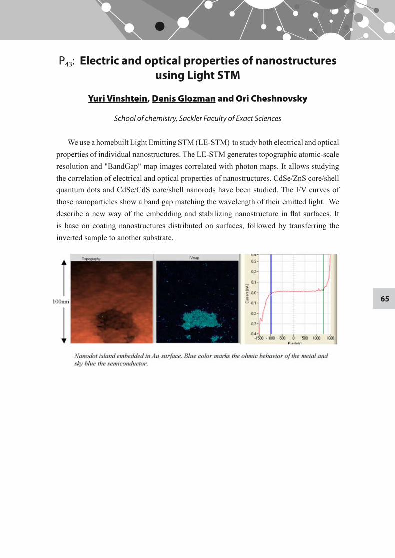
65
P43: Electric and optical properties of nanostructures using Light STM
Yuri Vinshtein, Denis Glozman and Ori Cheshnovsky
School of chemistry, Sackler Faculty of Exact Sciences
We use a homebuilt Light Emitting STM (LE-STM) to study both electrical and optical properties of individual nanostructures. The LE-STM generates topographic atomic-scale resolution and "BandGap" map images correlated with photon maps. It allows studying the correlation of electrical and optical properties of nanostructures. CdSe/ZnS core/shell quantum dots and CdSe/CdS core/shell nanorods have been studied. The I/V curves of those nanoparticles show a band gap matching the wavelength of their emitted light. We describe a new way of the embedding and stabilizing nanostructure in flat surfaces. It is base on coating nanostructures distributed on surfaces, followed by transferring the inverted sample to another substrate.
66
P44: Development of new technique for early diagnosis of neurodegenerative diseases
Inbal Yanai and Ehud Gazit
Department of Molecular Microbiology and Biotechnology, George S. Wise Faculty of Life Sciences
Lose of cerebral neural cells is observed in several age-associated disorders such as Alzheimer's disease, Parkinson's disease and other neurodegenerative disorders. Currently, the medical procedures for the diagnosis of those diseases are not very conclusive. The need for new methods is relevant now more than ever with the extended life expectancy and the increase of the elderly population.
The study of self-assembly mechanism, in our laboratory, has led to the discovery of aromatic dipeptides that self-assemble into various structures such as nanotubes, nanospheres, nano-plates and others. These are well-ordered and discrete structures.
This study is directed towards the development of novel contrast agents to produce magnetic resonance imaging (MRI) based on peptide nanostructures loaded with ferromagnetic nano-particles. The experimental model includes short peptides which self assemble into closed-caged nanospheres under certain conditions and encapsulate the magnetite nano-particles during the self-assembling process. In order to develop efficient diagnostic tool, the nanospheres were decorated with recognition motifs that could specifically bind antigens that are associated with various degenerative diseases. The formation and stability of the nanospheres was investigated using various tools at different conditions. The tertbutoxycarbonyl-diphenylalanine (BocFF) nanospheres have been successfully decorated with amyloid beta recognition motif. In Addition, the BocFF nanospheres structures were stable and uniform at the examined conditions. Hence, the BocFF nanospheres are an appropriate candidate for fabrication of a new diagnostic agent.
67
P45: Detection of heating in current carrying graphene by Raman Spectroscopy
Ilan Yutsis, Tamar Shamai, Denis Glozman, Zvika Ioffe, Oded Hod, Ori Cheshnovsky and Yoram Selzer
School of Chemistry, Raymond and Beverly Sackler Faculty of Exact Sciences
We use Raman spectroscopy to detect heating processes in current carrying single layer graphene flakes at room temperature. The effective temperature under potential bias of the long wavelength optical phonons (G-band), and the zone boundary phonons (2D band) is determined both by the ratio of their Stokes and anti-Stokes components, as well as by the change in their Raman shift in the Stokes regime. A detailed comparison between the two methods is given. The apparent mode specific temperatures are subsequently used to determine characteristic length scales of the transporting charge carriers, using numerical simulations.
68
P46: Secondary Electron Doping Contrast: Theory based on SEM and KPFM Measurements
I. Zhebova1, M. Molotskii1, Z. Barkay2, E. Grunbaum1, and Y. Rosenwaks1
1 School of Electrical Engineering, The Iby and Aladar Fleischman Faculty of Engineering2 The Wolfson Applied Materials Research Center
The secondary electron (SE) emission flux in a high-resolution scanning electron microscope (HRSEM) is a powerful tool for delineation of electrically active dopant concentration, built-in potentials and surface electric fields in semiconductor junctions. In all the measured SE images p-doped regions appear brighter than n-doped regions, but to the best of our knowledge this phenomena was never quantitatively explained.
A quantitative theory of SE doping contrast in the p- and n-sides of p-n junctions was developed and compared to measurements on various samples. In our model the contrast is a function of the secondary electron escape probability and their escape depth. The escape probability depends on the energy distribution of the SE as well as on the electron affinity. The affinity is affected by changes in the built-in potential as well as the potentials due to band bending. The escape depth depends on the surface electric field induced by surface states.
The contrast theory is based on surface band bending extracted from Kelvin probe force microscopy (KPFM) measurements. It is found that the escape depth contribution to the SE contrast is much larger than the escape probability contribution. In addition, the logarithmic dependence of measured contrast for different p-doped areas was found to be in a good agreement with the developed theory.