Embed Size (px)
Citation preview

81
DOI: 10.4046/trd.2010.69.2.81ISSN: 1738-3536(Print)/2005-6184(Online)Tuberc Respir Dis 2010;69:81-94CopyrightⒸ2010. The Korean Academy of Tuberculosis and Respiratory Diseases. All rights reserved.
영양분이 결핍된 H460 세포주에서 자가포식이 세포사멸에 미치는 영향원광대학교 의과대학 1내과학교실, 2병리학교실, 3방사선종양학교실, 4흉부외과학교실, 5핵의학교실, 6생명과학부
장혜연1, 조향정2, 황기은1, 김소영1, 이강규3, 문성록3, 신정현1, 조경화1, 이미경4, 이삼윤4, 박순아5, 박종군6, 김휘정1, 양세훈1
The Effect of Autophagy to Cell Death in Nutrient-Deprived H460 CellsHye Yeon Jang, B.S.1, Hyang Jeong Jo, M.D.2, Ki Eun Hwhang, M.D.1, So Young Kim, M.D.1, Kang Kyoo Lee, M.D.3, Sun Rock Moon, M.D.3, Jeong Hyun Shin, M.D.1, Kyung Hwa Cho, M.D.1, Mi Kung Lee, M.D.4, Sam Youn Lee, M.D.4, Soon Ah Park, M.D.5, Jong Kun Park, Ph.D.6, Hui Jung Kim, M.D.1, Sei Hoon Yang, M.D.1
Departments of 1Internal Medicine, 2Pathology, 3Radiation Oncology, 4Thoracic Surgery, 5Nuclear Medicine, 6Biological Science, Wonkwang University College of Medicine, Iksan, Korea
Background: Autophagy is an important adaptive mechanism in normal development and in response to changing environmental stimuli in cancer. Previous papers have reported that different types of cancer underwent autophagy to obtain amino acids as energy source of dying cells in nutrient-deprived conditions. However, whether or not autophagy in the process of lung cancer causes death or survival is controversial. Therefore in this study, we investigated whether nutrient deprivation induces autophagy in human H460 lung cancer cells.Methods: H460, lung cancer cells were incubated in RPMI 1640 medium, and the starved media, which are BME and RPMI media without serum, including 2-deoxyl-D-glucose according to time dependence. To evaluate the viability and find out the mechanism of cell death under nutrient-deprived conditions, the MTT assay and flow cytometry were done and analyzed the apoptotic and autophagic related proteins. It is also measured the development of acidic vascular organelles by acridine orange.Results: The nutrient-deprived cancer cell is relatively sensitive to cell death rather than normal nutrition. Massive cytoplasmic vacuolization was seen under nutrient-deprived conditions. Autophagic vacuoles were visible at approximately 12 h and as time ran out, vacuoles became larger and denser with the increasing number of vacuoles. In addition, the proportion of acridine orange stain-positive cells increased according to time dependence. Localization of GFP-LC3 in cytoplasm and expression of LC-3II and Beclin 1 were increased according to time dependence on nutrient-deprived cells.Conclusion: Nutrient deprivation induces cell death through autophagy in H460 lung cancer cells.
Key Words: Autophagy; Malnutrition; Lung Neoplasms; Cell Death
Address for correspondence: Sei Hoon Yang, M.D.Department of Internal Medicine, Wonkwang University Hospital, 344-2, Shinyong-dong, Iksan 570-711, KoreaPhone: 82-63-859-2582, Fax: 82-63-855-2025E-mail: [email protected]
Received: May. 8, 2010Accepted: Jun. 29, 2010
서 론
지난 20∼30년 동안 폐암 발생률은 전 세계적으로 꾸
준히 증가하는 추세를 보이고 있으나, 완치율은 15% 전후
로 큰 변화가 없어 최근에는 폐암이 가장 흔한 암 사망
원인으로 대두되고 있다1. 국내에서도 폐암 발생률은 지
속적으로 증가하고 있으며, 현재 암으로 인한 사망률 1위
를 차지하고 있다2. 많은 연구가 진행되었음에도 불구하
Original Article

HY Jang et al: Autophagic cell death in lung cancer
82
고 전체 폐암 75%를 차지하는 비소세포폐암(non small
cell lung cancer)은 여전히 사망률이 높고 치료하기 어려
운 질병으로 분류되고 있다. 비소세포폐암 환자에서 항암
치료성적은 상대적으로 적은 지속률과 완치율로 인해 낮
은 치료 효과를 보이고 있다3,4. 이에 비소세포폐암을 치료
하기 위해서는 보다 새롭고 강력한 치료 방법의 개발이
필요한 실정이다.
세포사멸 또는 세포예정사(programed cell death, PCD)
는 세포의 다양한 신호전달기전을 조절하고, 그로 인해
세포사멸을 유도하여 항상성을 유지하는 세포 내 기전이
다. 세포예정사는 형태학적인 기준으로 세포고사(apopto-
sis, PCD I)와 자가포식(autophagy, PCD II) 두 가지로 구
분한다5. 클락 역시 자가포식은 세포고사와 다르다고 주
장하였다6,7. 자가포식은 진핵세포에서 세포 내 오래된 단
백질, 손상된 소기관(organelles) 그리고 세포질이 탐식
및 소화되고 재사용되는 기전을 가지며, 이는 기본적인
세포질 구성요소들의 turnover에 관여하거나 외부적인 환
경의 변화, 즉 영양분 고갈, 병원균 침윤, 산소 결핍 등으로
유도되기도 한다8,9
. 최근 발표되는 연구들은 암세포에서
자가포식을 유발하여 항암 효과를 볼 수 있다고 보고하고
있다. 자가포식은 여러 종양 세포주에서 종양 치료제에 의
해 활성화된다고 보고되고 있지만, 암의 형성이나 억제와
의 연관성에 대해서는 확실히 규명된 바가 없다10-14.
암세포는 일반적으로 높은 당 분해(glycolysis) 활동이
일어나는 것으로 알려져 있다15
. 암세포는 자라면서 많은
산소와 영양분을 필요로 하는데, 이것은 끊임없이 새로운
신생혈관들의 형성을 통해서 얻어진다. 혈관에서 산소,
포도당, 성장 인자, 기타 영양소 등은 제한된 범위 내에서
확산에 의하여 공급되기 때문에 신생혈관이 없는 암 조직
의 종양세포들은 저산소증과 영양분 결핍에 빠지게 되므
로 반드시 신생혈관의 형성이 필요하다15-17
.
본 연구는 비소세포폐암 세포주인 NCI-H460 세포주에
서 신생 혈관이 생성되기 전 성장하는 암 조직에서 발생될
수 있는 영양분이 결핍된 환경, 영양분과 산소 모두 결핍
된 환경을 만든 후, 이들 환경이 암세포의 세포사멸에 어
떠한 영향을 미치는지를 알아보고자 하였다.
대상 및 방법
1. 시약
RPMI 1640, basal medium eagle (BME), 우태아 혈청
(fetal bovine serum), HEPES, 항생제(antibiotics) 및 트립
신(trypsin) 등 세포배양에 필요한 재료는 GIBCO BRL
(Grand Island, NY, USA) 제품을 구입하였으며, 배양용기
(24-well plate, 48-well plate, 6 cm dish, 10 cm dish)는
Nunc (Thermo Fisher Scientific, Roskilde, Denmark)에
서 구입하여 사용하였다. 2-deoxyl-D-glucose (2DG), ac-
ridin orange (AO), bicinchoninic acid (BCA), methyl-
thiazol-2-yl-2,5-diphenyl-tetrazolium bromide (MTT), Ho-
echst 33342, propidium iodide (PI), dimethyl sulfoxide
(DMSO)는 Sigma (St. Louis, MO, USA)에서, 세포사멸 검
출 키트인 Annexin V-FITC는 Assay Designs (Ann Arbor,
MI, USA)에서 구입하였다. PARP, Caspase-3, HMGB1 항
체는 Cell Signaling (Boston, MA, USA)에서 구입했으며,
MAP-LC3, Actin, GAPDH 항체는 Santa Cruz Biotechnol-
ogy (Santa Cruz, CA, USA)에서 구입하여 사용하였다.
Beclin 1 항체는 Assay Designs (Ann Arbor, MI, USA)에
서 구입하였다. 2차 항체는 Amersham (Buckinghamshire,
UK)제품을 구입하였다. Polyvinylidene fluoride (PVDF)
막, Nitrocellulose (NC) 막과 enhanced chemilumines-
cent kit (ECL)은 Milipore Co. (Billerica, MA, USA)에서
구입하였다.
2. 세포주의 정상 성장 배양과 영양결핍 하의 배양
NCI-H460 폐암 세포주는 American Type Culture
Collection (Rockville, MD, USA)로부터 구입하여 계대 배
양하면서 실험을 실시하였다. NCI-H460 세포는 CO2 세포
배양기(37oC, 5% CO2)에서 1% 항생제와 1% HEPES 그리
고 10% 우태아 혈청이 포함된 RPMI 1640으로 배양하였으
며, 24시간 간격으로 배양액을 교체하였다. 영양결핍 환
경은 세포를 phosphate buffered saline (PBS)으로 두 번
세척한 후, 영양분이 결핍된 배양액으로 교체하였다. 아
미노산(amino acid) 결핍을 위해서는 혈청이 포함되지 않
은 BME (Table 1)를 사용하였고, 포도당(glucose)과 산소
결핍을 유도하기 위해서는 10 mM 2DG를 BME에 첨가하
여 세포를 배양하였다. 모든 실험은 세포사멸 현상과 이
에 연관된 생화학적 변화를 확인하기 위해 2시간, 6시간,
12시간, 24시간, 48시간 동안 진행하였다.
3. 세포 생존율 측정
NCI-H460 폐암 세포(1×10⁵ cells/mL)는 48-well plate
세포 배양판에 분주하여 12시간 이상 CO2 세포 배양기
안에서 안정시킨 후, 먼저 혈청이 포함된 군과 포함되지
않은 군으로 나누고, 이들을 다시 정상 배양액인 RPMI

Tuberculosis and Respiratory Diseases Vol. 69. No. 2, Aug. 2010
83
Table 1. Media formulation
RPMI Basal medium 1640 medium eagle
Componentsconcentration, concentration,
mg/mL mg/mL
Amino acids Glycine 10 - L-Arginine 200 - L-Asparagine 50 - L-Cystine 2HCl 65 16 L-Glutamic acid 20 - L-Glutamine 300 - L-Histidine 15 8 L-Hydroxyproline 20 - L-Isoleucine 50 26 L-Leucine 50 26 L-Methionine 15 7.5 L-Proline 20 - L-Serine 30 -Vitamins Bioth 0.2 1 Choline chloride 3 1 D-Calcium pantothenate 0.25 1 Pyridoxine hydrochloride 1 - Riboflavin 0.2 0.1 Vitamin B12 0.005 - i-Inositol 35 2Inorganic salts Calcium nitrate 100 200 Magnesium sulfate 48.84 97.7 Sodium phosphate dibasic 800 140Other components D-Glucose (dextrose) 2,000 1,000 Glutathione (reduced) 1 -
1640, 10 mM 2DG가 포함된 RPMI 1640, BME, 10 mM
2DG가 포함된 BME을 첨가하여 각각 0시간, 2시간, 6시
간, 12시간, 24시간, 48시간 동안 관찰하였다. 세포 생존
을 확인하기 위해 MTT 용액(5 mg/mL, PBS, pH 7.4)의
최종 농도를 1.2 mM이 되도록 첨가하여 4시간 동안 반응
시켰다. 4시간 후 배양액을 제거하고 0.5 mL DMSO를 첨
가하여 살아있는 세포에 의해 생성된 보라색 formazan을
용해시킨 다음, 96 well용 분광광도계(THERMOmax; Mo-
lecular Devices Co., Sunnyvale, CA, USA)를 이용하여
590 nm 파장에서 흡광도를 측정하였다. 세포 생존율은
정상 대조군과 비교하여 백분율(%)로 표시하였다.
4. 유세포분석기(Flow cytometry)로 세포 주기 및 세포
고사 분석
정상 영양상태와 영양결핍상태에서의 세포주기를 관
찰하기 위해 H460 세포는 대조군인 정상 성장 RPMI
1640과 영양분이 결핍된 BME, 10 mM 2DG가 포함된
BME에서 12시간 동안 배양된 세포를 포집하여 원심 분
리한 후, 각 세포를 70% 에탄올로 4oC에서 12시간 이상
고정시킨 후 PBS로 두 번 세척하였다. 세포는 1 mL PI
용액(PI, 50 μg/mL; RNase A, 0.1 μg/mL, 0.05% NP40)
을 넣어 30분 동안 암실에서 보관한 후, 유세포분석기
(FACS Vantage flow cytometry; Becton Dickinson Im-
munocytometry System, San Jose, CA, USA)를 이용하여
세포 주기를 분석하였다. 유세포분석에서 얻은 정보 분석
은 Cell Quest software (Becton Dickinson, Franklin
Lakes, NJ, USA)를 이용하였다. 영양상태에 따른 세포고
사를 관찰하기 위하여 H460 세포를 대조군으로 혈청이
포함된 RPMI 1640에서 배양하였고, 세포고사 양성 대조
군으로는 RPMI 1640에 20 uM cisplatin을 36시간 처리하
였다. 실험군인 영양결핍군으로 BME, 10 mM 2DG가 포
함된 BME에서 12시간 처리하였다. 세포는 트립신을 사용
하여 배양판에서 떼어낸 후, 포집하여 PBS로 세척하였다.
세척된 세포의 세포고사를 분석하기 위해 검출 키트
(Assay Designs)를 사용하였으며, 방법은 제시된 방법을
따라 시행하였다. 96 μL binding buffer (PBS containing
1 mM calcium chloride)에 FITC가 결합 된 annexin V (0.5
μg/mL final concentration)로 염색을 시행하였다. 실온에
서 10분 동안 반응 후 250 μL binding buffer를 더 첨가하
여 유세포분석기로 분석하였다.
5. 영양 상태에 따른 세포의 형태적 변화 관찰
세포의 형태적 변화를 조사하기 위하여 H460 세포
(1×10⁵ cells/mL)를 24-well plate 세포 배양판에 분주하
였다. 대조군과 영양 결핍된 배양액으로 각각 처리한 실
험군은 12시간과 24시간 배양 후 배양액을 모두 제거하
고, PBS (pH 7.4)로 2회 세척하였다. 세포에 300 μL crys-
tal violet 용액(0.05% crystal violet, 3.7% paraformalde-
hyde)을 처리하여 실온에서 5분 동안 반응 시킨 후, 1 mL
PBS로 두 번 세척한 다음 염색된 세포질에서 소체(va-
cuole)를 관찰하기 위해 광학 현미경(Olympus, Tokyo,
Japan)으로 관찰하였다. 세포를 대조군과 영양 결핍된 배
양액으로 각각 처리한 실험군은 12시간과 24시간 반응시

HY Jang et al: Autophagic cell death in lung cancer
84
킨 다음 3.7% formaldehyde 용액으로 실온에서 30분 동
안 고정시킨 후, PBS로 세척하고 세포 핵산 염색제인 Ho-
echst 33342을 이용하여 30분간 염색, 다시 PBS로 세척하
여 염색사 응축과 DNA의 분절현상을 관찰하였다. 영양
상태에 따라 자가포식의 현상인 산성소포체(acidic vesic-
ular organelle)가 세포질에 발생하는지를 확인하기 위하
여 대조군과 영양 결핍된 배양액으로 각각 처리한 실험군
은 12시간과 24시간 반응시킨 다음 배양액을 모두 제거하
였다. 세포는 PBS로 두 번 세척하였고 1 μg/mL acridine
orange를 첨가하여 37oC에서 15분 동안 반응시켰다. 반응
시킨 후 PBS로 세척하고 산성소포체를 관찰하였다.
6. H460 세포에 Plasmid GFP-LC3 형질도입
GFP-LC3 재조합 DNA는 서울여자대학교 이도희 교수
로부터 분주 받아 실험을 진행하였다. H460 세포(5×10⁴/
mL)는 48-well plate 세포 배양판에 형질도입(transfec-
tion)하기 1일 전에 항생제가 들어가지 않은 배양액을 사
용하여 분주한 후 안정화를 시켰다. 1 μg plasmid DNA
와 형질도입 용액인 Lipofectamin 2000 (40 μL/mL)을 넣
은 후 사용방법에 따라 실험을 진행하였다. GFP-LC3가
세포질 내에 발현되는지를 형광현미경을 통해서 관찰하
였다.
7. Western blot assay
세포 고사와 자가 포식의 기능적 역할을 확인하기 위하
여 배양된 H460 세포를 포집하여 차가운 PBS로 2회 세척
하였다. 얻어진 세포는 파쇄용액(50 mM HEPES, pH 7.4,
150 mM NaCl, 1% deoxycholate, 1 mM EDTA, 1 mM
PMSF, 1 μg/mL aprotinin)과 4oC에서 30분 동안 반응시
켰다. 동량의 세포파쇄액과 5X sample buffer를 혼합하여
100oC에서 5분간 가열하여 단백질 변성을 유도한 후에
10%, 12% 및 15% SDS-polyacrylamide gel electrophore-
sis (SDS-PAGE)를 시행하였다. 전기영동이 끝난 gel 단백
질은 Mini Trans-Blot (Bio-Rad, Hercules, CA, USA)을 이
용(0.8 mA/cm)하여 PVDF membrane과 NC membrane
으로 이동시키고, blocking buffer (5% skim milk)와 상온
에서 1시간 반응하였다. PARP, Casease-3, HMGB1,
MA-LC3, Becline 1, Actin 그리고 GAPDH 에 대한 항체는
0.05% (v/v) tween-20이 함유된 1% skim milk에 1 : 1,000
으로 희석하여 PVDF membrane과 NC membrane에 4oC
에서 12시간 이상 반응시켰으며, 일차항체에 대한 이차항
체 anti-rabbit IgG conjugated horse radish peroxidase
(HRP), anti-goat IgG conjugated HRP와 anti-mouse IgG
conjugated HRP를 1 : 5,000으로 희석하여 상온에서 1시
간 반응시킨 후, ECL kit을 이용하여 현상하였다.
8. 통계 처리
표시된 결과는 3번 이상의 독립적인 실험결과이며,
One-way ANOVA test를 이용하였다. 이들의 평균(mean)
과 표준편차(standard deviation, SD)를 산출하여 결과를
표시하였다. 실험결과의 통계처리는 Student’s t-test에 준
하여 처리하였으며, p-value가 p<0.05 경우를 유의한 것
으로 인정하였다.
결 과
1. 영양결핍에 의한 NCI-H460 세포주 생존율
세포가 성장하기 위해서 반드시 필요한 요소인 성장인
자(growth factor), 영양소(nutrient)와 산소(oxygen)의 결
핍이 세포사멸에 어떠한 영향을 줄 것인지를 알아보기 위
해 실험을 진행하였다. 영양결핍 실험은 크게 성장인자를
다량 포함하고 있는 혈청(serum)이 포함된 배양액에서 자
란 그룹(Figure 1A)과 혈청이 포함되지 않은 배양액에서
자란 그룹(Figure 1B)으로 구분하였다. 또한 포도당과 유
사한 구조로 포도당 대사를 방해하여 당 분해 과정을 억제
하며, 세포에 영양결핍 스트레스를 야기하는 2DG를 세포
에 처리하여 영양분이 결핍되는 것과 유사한 환경을 만들
었다18,19. 정상 성장 배양액 RPMI 1640에서 자란 군을 대
조군으로, RPMI 1640에 10 mM 2DG를 첨가한 배양액에
서 자란 군, 아미노산과 포도당의 양이 1/3 이하로 들어있
는 BME (Table 1) 배양액에서 자란 군, 마지막으로 BME
배양액에 10 mM 2DG가 첨가 된 배양액에서 자란 군으로
각각을 구분하고 이 세 가지 군을 실험군으로 정한 후,
세포의 생존율을 MTT 방법으로 확인하였다. 실험은 6시
간, 12시간, 24시간, 48시간 동안 진행하였고 생존율 측정
의 기준은 배양액을 교체한 0시간을 100%로 하였다. 10%
우태아 혈청이 포함되어 있는 군(Figure 1A)에서는 대조
군인 RPMI 1640 배양액에서 자란 H460 세포가 48시간
동안 300%로 성장이 증가했음을 확인하였고, BME 배양
액과 10 mM 2DG가 첨가 된 RPMI 1640 배양액에서 자란
군은 각각 31%와 10%의 성장을 보였으나, 10 mM 2DG가
첨가된 BME 배양액에서 자란 군은 38%의 생존감소를 확
인하였다. 혈청이 포함되지 않은 군 중에서 48시간 동안
RPMI 1640 배양액에서 자란 군은 81%의 성장을 보였으

Tuberculosis and Respiratory Diseases Vol. 69. No. 2, Aug. 2010
85
Figure 1. Effect of nutrient deprivation on cell survival in H460 cell line. Cells were seeded at a density of 1x10⁵ cells/mLin 48 well plates and incubated in completely fresh medium for 20 hours. The cells were washed with PBS and themedium was then changed to a medium without the nutrient factors. Basal medium eagle (BME) was deprived of eachnutrient and prepared by adding 10 mM 2-deoxyl-D-glucose (2DG) or 10% of dialyzed fetal bovine serum to the basalmedium. Cell survival in the presence (A) and absence (B) of serum were shown. Cell numbers at the start of starvationwere considered to be 100%. The viability of cell was measured by MTT assay. Means±SD (n=4). *p≤0.05, †p≤0.01.
Table 2. Induction of S-phase arrest in Nutrient-deprived
condition
Cell cycle (%)
Sub-G1 G1 S G2/M
RPMI 1640 1.17 44.35 29.26 8.74BME 1.25 38.17 38.18 7.3510 mM 2DG+BME 1.74 35.67 39.6 6.23
BME: basal medium eagle; 2DG: 2-deoxyl-D-glucose.
며, 이는 혈청이 포함된 군의 RPMI 1640에서 자란군보다
약 220%의 성장률이 억제되었음을 확인하였다(Figure
1B). 또한 아미노산과 포도당 양이 적은 BME 배양액에서
자란 실험군에서는 24시간과 48시간 동안에 각각 25%와
60%의 생존율 감소를 확인하였다. 또한 10 mM 2DG를
포함한 BME 배양액에서 자란 군은 12시간에 34%, 24시
간에 52%, 48시간에 78%의 유의적인 생존율 감소를 확인
하였다.
2. 영양결핍 조건 하에서 H460 세포의 세포주기 변화
와 세포 고사 관찰
H460 세포에서 영양결핍 조건이 세포의 성장과 분화에
어떠한 영향을 주는지 알아보기 위해서 대조군인 RPMI
1640, BME, 10 mM 2DG가 포함된 BME에서 자란 각 각
의 세포를 포집하여 PI로 염색한 후, 유세포분석기로 세포
주기를 분석하였다. 12시간 동안 배양시킨 후, 세포고사를
의미하는 sub G1기는 대조군 1.17%, BME에서 자란 군
1.25%, 10 mM 2DG가 포함된 BME에서 자란 군 1.74%로
유의적 증가는 관찰할 수 없었다. 하지만 S기는 대조군
29.26%인 것에 비해 BME와 10 mM 2DG를 포함한 BME
에서 자란 실험군은 각각 38.18%, 39.6%로 세포의 응집도
가 증가하였다. 또한 G1기와 G2/M기는 대조군에 비해 실
험군의 감소를 확인하였다(Table 2).
H460 세포에서 영양결핍 조건에 의해 유도되는 세포사
멸 기전을 확인하고자 RPMI 1640, BME 그리고 10 mM
2DG가 포함된 BME에서 12시간 동안 배양된 세포를 포집
하여 세포고사 검출 방법인 Annexin-V kit로 염색한 후,
유세포 분석기로 분석하였다. 세포고사의 정확한 비교를
하기 위해 20 μM cisplatin을 36시간동안 처리한 군을 세
포고사 양성 대조군으로 사용하였다. Cisplatin을 처리한
양성 대조군에서 세포고사는 91.18%의 높은 수치를 나타
내지만, 대조군과 영양결핍 조건에서 자란 두 실험군 모두
에서는 세포고사를 확인할 수 없었다. 따라서 영양결핍에
의한 세포사멸은 세포고사와는 다른 죽음이라는 것을 추
측할 수 있다(Figure 2).
영양결핍에 의한 NCI-H460 세포사멸의 특성을 알아보

HY Jang et al: Autophagic cell death in lung cancer
86
Figure 2. Apoptosis is not related with cell death induced by nutrient deprivation in NCI-H460 cells. Cells were incubatedin RPMI 1640 medium, basal medium eagle (BME) medium or 10 mM 2-deoxyl-D-glucose (2DG) with BME medium(12 hours) assayed for apoptosis by FACS following annexin V-FITC staining. Control cells were contained RPMI 1640Medium. 20 μM cisplatin was used as a positive control. The data shown are representative of three independentexperiment.
기 위하여 핵산 염색인 Hoechst을 이용하여 세포핵의 형
태를 관찰하였다. RPMI 1640 배양액에서 배양한 정상 대
조군과 BME군에서는 시간이 경과하여도 세포의 핵은 타
원 형태의 핵이 균질하게 염색되어 관찰되었다. 그러나
10 mM 2DG가 포함된 BME에서 자란 세포군은 12시간
배양했을 때까지는 정상 대조군과 유사한 핵 모양이 관찰
되었으나, 24시간 배양했을 때의 세포핵은 세포고사 형태
학적 특징으로 알려진 염색사 응축현상(chromatin con-
densation)이 관찰되었다(Figure 3).
H460 세포에서 영양결핍에 의해 유도되는 세포사멸 경
로의 단백질 확인하기 위해 실험군인 BME와 10 mM 2DG
가 포함된 BME에서 자란 세포군을 각각 2시간, 6시간,
12시간, 24시간 동안 배양시키고, 시간에 따라 세포를 포
집하여 Caspase 3, PARP, HMGB1 발현과 분절을 확인하
였다20,21
. BME 배양액에서 자란 세포군은 시간이 지나도
Caspase 3와 Caspase 3의 기질인 PARP 단백질의 분절을
확인하지 못하였으나, 10 mM 2DG가 포함된 BME에서 자
란 세포군은 24시간 후 Caspase 3와 PARP 분절이 증가함
을 확인하였다. 또한 세포사멸의 다른 기전인 세포괴사
(necrosis)와의 연관성을 확인하기 위해서 HMGB1 단백
질 발현 양을 측정하였다22
. 그러나 HMGB1의 발현 양은
모든 실험군에서 유의한 차이를 확인할 수 없었다(Figure
4). 그러므로 영양결핍 하의 세포사멸은 세포괴사와는 연
관이 없는 것으로 보이며, 세포고사는 2DG가 포함된 BME
에서 24시간 동안 배양된 실험군에서만 유도되는 것을 확
인할 수 있었다.
3. 영양결핍 하에서 H460 세포의 자가포식 관찰
영양결핍에 의한 H460 세포의 형태학적 변화를 알아보
기 위하여 BME 와 10 mM 2DG를 포함한 BME으로 구분
하여 각각 12시간과 24시간 동안 배양시켜 세포를 염색하
여 세포의 변화를 관찰하였다. 두 그룹 모두 시간이 지나

Tuberculosis and Respiratory Diseases Vol. 69. No. 2, Aug. 2010
87
Figure 3. No apoptotic nuclear change occurred in nutrient-deprived H460 cells. H460 cells were plated on glass coverslips. Cells were incubated in RPMI 1640 medium, basal medium eagle (BME) medium (A) or 10 mM 2-deoxyl-D-glucose (2DG) with BME medium (B) for various periods. Hoechst staining was used to identify morphological features of cell death such as nuclear condensation and fragmentation. Scale bar, ×200. Samples were examined under a fluorescence microscope.
면서 자가포식의 형태학적 특징 중 하나인 세포질에서의
소체(vacuole) 형성이 발생되었다. 이러한 소체들은 두 실
험군 모두 초기부터 발견되기 시작하여 시간 의존적으로
더 크고 많이 증가하는 것을 확인하였다(Figure 5).
자가포식은 자가포식소체(autophagosome)라 불리는
이중막으로 둘러싸인 소포를 형성한다. 이 소체는 lyso-
some과 융합하여 자가 용해소체(autolyso-some)를 형성
하고 이 소체는 산성화된 내용물을 함유하고 있어 산성소
포체(acidic vesicular organelle, AVOs)라고 불린다23
. 영
양결핍 조건 하의 세포에서 AO 염색을 사용하여 산성소
포체를 확인하였다. 시간이 지나면서 대조군을 제외한
BME와 10 mM 2DG가 포함된 BME 배양액에서 자란 실
험군에서는 산성소포체들이 관찰되었고, 시간 의존적으
로 수가 증가함을 확인할 수 있었다(Figure 6). 또한 H460
세포에서 산성소포체를 정량적으로 분석하기 위해서 AO
로 염색 한 후 유세포분석기로 분석한 결과 BME에서 자
란 군은 정상 대조군에 비해 12시간 약 30%, 24시간 약
45%의 증가율을 보였으며(Figure 7A), 10 mM 2DG가 포
함된 BME에서 자란 군은 12시간 배양 후 약 45%, 24시간
배양 후 약 55%로 유의적으로 증가하는 것을 확인할 수
있었다(Figure 7B). 이들 두 실험군 사이에서는 2DG를 포
함한 BME에서 자란 군이 BME에서 자란 군보다 산성소포
체가 12시간에 약 16%, 24시간에 약 11% 더 많이 발현되
는 것을 확인할 수 있었다(Figure 7).
LC3는 자가포식의 발생에 있어 중요한 구성요소 중 하
나로 자가포식소체 이중막을 형성하는 데 관여하여 자가

HY Jang et al: Autophagic cell death in lung cancer
88
Figure 4. No apoptosis and necrosis is not associated with the nutrient-deprived H460 cell death. Cells probed withWestern immunoblots were incubated in RPMI 1640 medium (CTR), basal medium eagle (BME) medium (A) or 10 mM2-deoxyl-D-glucose (2DG) with BME medium (B) for various periods. The equal amounts of protein from cell lysatewere subjected on 12% and 15% SDS-polyacrylamide gel electrophoresis (SDS-PAGE), transferred onto polyvinylidenefluoride (PVDF) membrane and immunoblotted with antibodies against Caspase 3 and cleaved caspase 3, PARP, cleavedPARP, HMGB1 and Actin. The immunoreactive signals were visualized by enhanced chemiluminescent kit (ECL). Actinwas used as a roading control.
Figure 5. Cytoplasmic Va-cuoles were increased ac-cording to time depend-ence in nutrient-deprived H460 cells. H460 cells were incubated in RPMI 1640 medium (CTR), bas-al medium eagle (BME) medium or 10 mM 2-de-oxyl-D-glucose (2DG) with BME medium for 12 hoursand 24 hours. Scale bar, ×200. Samples were ex-amined under a light mi-croscope.

Tuberculosis and Respiratory Diseases Vol. 69. No. 2, Aug. 2010
89
Figure 6. Red acidic autophagic vacuoles were detected in cytoplasm of nutrient-deprived H460 cells according totime dependence. H460 cells were incubated in RPMI 1640 medium (CTR), basal medium eagle (BME) medium or 10 mM 2-deoxyl-D-glucose (2DG) with BME medium for 12 hours and 24 hours as shown. And then stained by 1μg/mL acridine orange in medium for 15 minutes. The acidic autolysosomes induced by nutrition deprivation were stained orange-red.
Figure 7. Development of acidic vesicular organelles (AVO) in nutrient-deprived H460 cells and its quantification usingFACS analysis. H460 cells were incubated in RPMI 1640 medium, basal medium eagle (BME) medium or 10 mM 2-deox-yl-D-glucose (2DG) with BME medidum for 6 hours, 12 hours, and 24 hours. AVOs were counted in cytoplasm ofH460 cells. *p≤0.05, †p≤0.01.

HY Jang et al: Autophagic cell death in lung cancer
90
Figure 8. Fluorescence staining of GFP-LC3 in response to nutrient deprivation. H460 cells were plated on glass coverslips. Following transfection with GFP-LC3 plasmid, cells were incubated in RPMI 1640 medium, basal mediumeagle (BME) medium, 10 mM 2-deoxyl-D-glucose (2DG) with BME medium for 12 hours and 24 hours as indicated.The transfected cells were observed using a single-photon fluorescence microscope.
포식의 특이적 마커로 사용되고 있다24
. 본 실험에서는
LC3가 영양결핍에 의해서 유발되는 자가포식과 관련이
있는지를 알아보기 위해 H460 세포에 plasmid GFP-LC3
의 발현 분포를 확인하였다. 대조군 RPMI 1640에서 자란
세포는 plasmid GFP-LC3가 발현되지 않았지만, 영양결핍
BME에서 자란 세포군과 10 mM 2DG가 포함된 BME에서
자란 세포군의 세포질에서 점 형태의 plasmid GFP-LC3의
발현이 확인되었다. 이는 12시간 배양 후부터 명확하게
확인할 수 있으며 24시간 때는 12시간 보다 더 많은 발현
을 관찰할 수 있었다(Figure 8).
LC3는 세포 내에서 2가지 형태로 존재한다. LC3-I은 세
포질 내에서 선형으로 존재하다가 세포 내부나 외부에서
스트레스를 받으면 LC3-II로 변형되며, 이는 자가포식소
체 이중막을 형성에 관여하게 된다25. 따라서 LC-3II가 많
이 발현되면 자가용해소체 역시 많이 만들어지는 상호관
계에 있다. Beclin 1은 자가포식 현상에서 중요한 작용을
하는 Class III phosphatidylinositol-3kinase (PI3K) 복합
체부분으로 ATG(autophagy related gene) 단백질 발현을
조절하는 작용을 한다26,27
. 따라서 Beclin 1의 발현량의 증
가 역시 자가포식이 발생하는 증거로 사용할 수 있다. 영
양결핍 조건에서 자가포식현상의 발현을 확인하기 위해
서 LC3, beclin 1에 대한 western blot을 시행하였다.
H460 세포에서 LC3 단백질은 두 실험군 모두 초기 시간
부터 LC3-I에서 LC3-II로 변화하는 것을 확인할 수 있었다
(Figure 9). BME에서 자란 세포군은 대조군과 비교하였을
때 6시간부터 뚜렷하게 LC3-I가 LC3-II로 바뀌는 것을 확
인할 수 있었으며 이는 시간 의존적으로 증가하였다. 10
mM 2DG가 들어있는 BME에서 자란 세포군은 배양액 교
체 후 2시간부터 LC3-I가 LC3-II로 바뀌고, 12시간까지
LC3-II의 발현량은 유의적으로 증가하였으나, 24시간이
Tuberculosis and Respiratory Diseases Vol. 69. No. 2, Aug. 2010
91
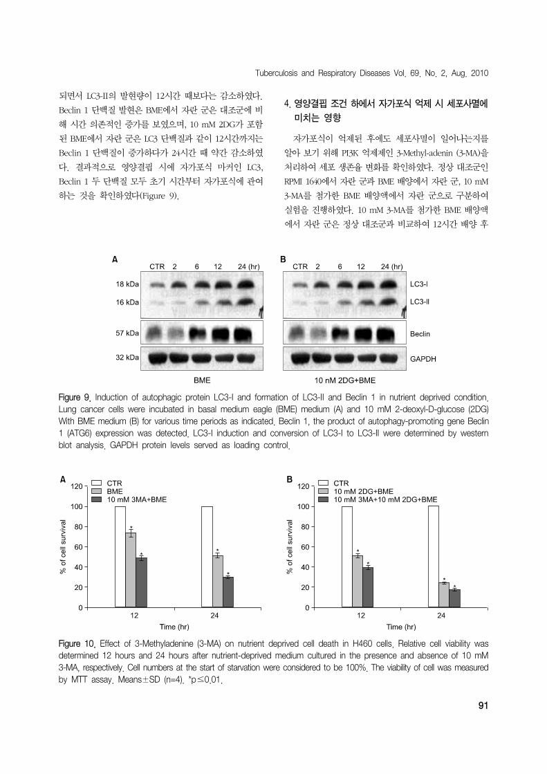
Figure 9. Induction of autophagic protein LC3-I and formation of LC3-II and Beclin 1 in nutrient deprived condition.Lung cancer cells were incubated in basal medium eagle (BME) medium (A) and 10 mM 2-deoxyl-D-glucose (2DG)With BME medium (B) for various time periods as indicated. Beclin 1, the product of autophagy-promoting gene Beclin1 (ATG6) expression was detected. LC3-I induction and conversion of LC3-I to LC3-II were determined by western blot analysis. GAPDH protein levels served as loading control.
Figure 10. Effect of 3-Methyladenine (3-MA) on nutrient deprived cell death in H460 cells. Relative cell viability wasdetermined 12 hours and 24 hours after nutrient-deprived medium cultured in the presence and absence of 10 mM3-MA, respectively. Cell numbers at the start of starvation were considered to be 100%. The viability of cell was measuredby MTT assay. Means±SD (n=4). *p≤0.01.
되면서 LC3-II의 발현량이 12시간 때보다는 감소하였다.
Beclin 1 단백질 발현은 BME에서 자란 군은 대조군에 비
해 시간 의존적인 증가를 보였으며, 10 mM 2DG가 포함
된 BME에서 자란 군은 LC3 단백질과 같이 12시간까지는
Beclin 1 단백질이 증가하다가 24시간 때 약간 감소하였
다. 결과적으로 영양결핍 시에 자가포식 마커인 LC3,
Beclin 1 두 단백질 모두 초기 시간부터 자가포식에 관여
하는 것을 확인하였다(Figure 9).
4. 영양결핍 조건 하에서 자가포식 억제 시 세포사멸에
미치는 영향
자가포식이 억제된 후에도 세포사멸이 일어나는지를
알아 보기 위해 PI3K 억제제인 3-Methyl-adenin (3-MA)을
처리하여 세포 생존율 변화를 확인하였다. 정상 대조군인
RPMI 1640에서 자란 군과 BME 배양에서 자란 군, 10 mM
3-MA를 첨가한 BME 배양액에서 자란 군으로 구분하여
실험을 진행하였다. 10 mM 3-MA를 첨가한 BME 배양액
에서 자란 군은 정상 대조군과 비교하여 12시간 배양 후

HY Jang et al: Autophagic cell death in lung cancer
92
51.7%, 24시간 배양 후 70.4%의 생존율 감소하는 것을
확인하였고, BME 배양액 단독에서 처리한 군에 비해 자
가포식 억제제가 들어있는 군에서 12시간에 21%, 24시간
에 30% 세포가 더 죽는 것을 확인할 수 있었다(Figure
10A). 또 다른 실험군에서는 정상 대조군과 10 mM 2DG
가 포함 된 BME 배양에서 자란 군, 10 mM 2DG가 포함
된 BME 배양액에 10 mM 3-MA를 첨가한 배양액에서 자
란 군으로 구분하여 실험을 진행하였다. 10 mM 3-MA를
첨가한 배양액에서 자란 군은 정상 대조군과 비교하여 12
시간에 60.5%, 24시간에 81.8%의 생존율 감소를 확인하
였고, 10 mM 2DG가 포함 된 BME 배양액 단독에서 처리
한 군 보다는 12시간, 24시간에 각각 23%와 26%의 세포
증식이 감소되는 것을 확인하였다(Figure 10B).
고 찰
대부분 고형암은 종양이 성장하면서 종양 주변부로 신
생혈관이 형성되며 중앙부위는 신생혈관이 소멸되면서
영양분 고갈과 산소 결핍이 발생된다. 이러한 극한 환경
은 세포 생존과 관련된 기전 중 하나인 자가포식을 유발하
는 것으로 알려져 있다28,29
.
본 연구에서는 종양 성장과정에서 위와 같이 발생할 수
있는 환경을 in vitro에서 만들어, 자가포식이 암세포의 사
멸이나 생존에 미치는 역할과 기전을 알아보고자 하였다.
실험에 이용한 영양분 결핍조건으로 BME와 2DG를 사
용하여, 영양결핍 실험군이 대조군에 비해 시간 의존적으
로 세포사멸이 증가함을 확인하였다. 이 세포사멸이 세포
고사와 관련이 있는지를 확인하기 위하여 영양결핍 실험
군에서 핵의 형태학적 변화, 세포 주기, 세포고사의 특징
적인 단백질 발현을 관찰하였다. 형태학적으로도 세포고
사를 의미하는 염색사 응축현상만 2DG가 포함된 BME에
서만 미약하게 관찰되고, 유세포 분석에서도 세포고사에
해당하는 sub G1이 24시간에도 거의 관찰되지 않았으며,
세포고사의 마지막 경로 단백질인 Caspase 3, PARP 단백
질의 분절은 24시간 때 2DG를 사용한 실험군을 제외한
모든 실험군에서는 확인할 수 없었다. 이는 영양결핍에
의한 세포사멸은 세포고사 기전 외 다른 기전에 의해 발생
한다고 추측할 수 있다.
다른 기전으로 세포괴사와의 관련성을 알아보기 위하
여 HMGB1 단백질의 발현량을 측정해 보았지만 대조군과
비교하여 유의한 차이를 나타내지 않았다. 따라서 영양
결핍에 의한 세포사멸은 세포괴사와도 관련이 없음을 확
인하였다.
영양결핍 조건에서의 세포사멸이 세포고사 또는 세포
괴사가 아닌 자가포식에 의해 일어나는지를 알아보고자
하였다. 영양결핍 조건에서 세포고사에서는 볼 수 없었던
크고 많은 수의 소체(vacuole)가 세포질에서 관찰되었고,
이는 자가포식이 발생하였음을 예상할 수 있다. 또한 acri-
dine orange 염색을 통해서 산성소포체의 발현을 형태적,
수치적으로 확인할 수 있었고, 시간 의존적으로 유의하게
산성소포체의 증가를 확인하였다. 형태학적인 자가포식
의 확인뿐만 아니라 자가포식의 기전에 중요한 역할을 하
는 LC-3의 세포 내의 변화도 분자생물학적으로 관찰하였
다. GFP-LC3를 세포 안으로 형질도입 시켰고, 세포질 내
에서 LC3의 발현 양상을 확인하였다. 자가포식 마커인
LC-3와 Beclin 1 단백질의 발현 정도를 확인하였다. Wes-
tern blots에서 LC-3 II, Beclin 1 단백질은 영양 결핍 조건
의 초기 시간에서부터 증가하여 24시간까지 발현이 증가
하는 것은 스트레스의 초기 시간부터 자가포식이 일어남
을 의미한다.
이상의 결과로 영양분이 결핍된 상태에서 폐암 세포주
H460 세포는 초기 단계 세포사멸에 자가포식이 주도적인
역할을 하며, 이는 시간 의존적으로 세포고사와 공유되는
부분이 있음을 추측할 수 있다. 즉, 자가포식을 억제하는
실험을 한다면 세포사멸을 세포고사를 통해서 이루어질
것으로 사료되며, 향후 자가포식 억제제를 이용한 연구가
필요하리라 사료된다. 또한 자가포식과 세포고사의 상호
간의 조절기전에 대한 연구가 필요할 것으로 사료되며,
자가포식의 연구가 항암치료에 있어서 새로운 패러다임
을 제시해 줄 것으로 생각한다.
감사의 글
This work was supported by Wonkwang Clinical
Medical Research Center Grant in 2006 (WCMRC-2006-
01).
참 고 문 헌
1. Jassem J. Combined chemotherapy and radiation in lo-
cally advanced non-small-cell lung cancer. Lancet
Oncol 2001;2:335-42.
2. Bae JM, Won YJ, Jung KW, Suh KA, Ahn DH, Park
JG. Annual report of the Central Cancer Registry in
Korea-1999: based on registered data from 128 hos-

Tuberculosis and Respiratory Diseases Vol. 69. No. 2, Aug. 2010
93
pitals. Cancer Res Treat 2001;33:367-72.
3. Ferreira CG, Span SW, Peters GJ, Kruyt FA, Giaccone
G. Chemotherapy triggers apoptosis in a caspase-8-de-
pendent and mitochondria-controlled manner in the
non-small cell lung cancer cell line NCI-H460. Cancer
Res 2000;60:7133-41.
4. Fukuoka M, Yano S, Giaccone G, Tamura T, Nakagawa
K, Douillard JY, et al. Multi-institutional randomized
phase II trial of gefitinib for previously treated patients
with advanced non-small-cell lung cancer (The IDEAL
1 Trial). J Clin Oncol 2003;21:2237-46.
5. Abraham MC, Shaham S. Death without caspases, cas-
pases without death. Trends Cell Biol 2004;14:184-93.
6. Bowen ID, Mullarkey K, Morgan SM. Programmed cell
death during metamorphosis in the blow-fly Calliphora
vomitoria. Microsc Res Tech 1996;34:202-17.
7. Zakeri ZF, Ahuja HS. Apoptotic cell death in the limb
and its relationship to pattern formation. Biochem Cell
Biol 1994;72:603-13.
8. Bursch W, Grasl-Kraupp B, Ellinger A, Török L, Kienzl
H, Müllauer L, et al. Active cell death: role in hep-
atocarcinogenesis and subtypes. Biochem Cell Biol
1994;72:669-75.
9. Dunn WA Jr. Studies on the mechanisms of autophagy:
formation of the autophagic vacuole. J Cell Biol 1990;
110:1923-33.
10. Bursch W, Ellinger A, Kienzl H, Török L, Pandey S,
Sikorska M, et al. Active cell death induced by the an-
ti-estrogens tamoxifen and ICI 164 384 in human mam-
mary carcinoma cells (MCF-7) in culture: the role of
autophagy. Carcinogenesis 1996;17:1595-607.
11. Kanzawa T, Kondo Y, Ito H, Kondo S, Germano I.
Induction of autophagic cell death in malignant glioma
cells by arsenic trioxide. Cancer Res 2003;63:2103-8.
12. Paglin S, Hollister T, Delohery T, Hackett N, McMahill
M, Sphicas E, et al. A novel response of cancer cells
to radiation involves autophagy and formation of acidic
vesicles. Cancer Res 2001;61:439-44.
13. Yao KC, Komata T, Kondo Y, Kanzawa T, Kondo S,
Germano IM. Molecular response of human glio-
blastoma multiforme cells to ionizing radiation: cell cy-
cle arrest, modulation of the expression of cyclin-de-
pendent kinase inhibitors, and autophagy. J Neurosurg
2003;98:378-84.
14. Butler R, Mitchell SH, Tindall DJ, Young CY. Nonapop-
totic cell death associated with S-phase arrest of pros-
tate cancer cells via the peroxisome proliferator-ac-
tivated receptor gamma ligand, 15-deoxy-delta12,14-
prostaglandin J2. Cell Growth Differ 2000;11:49-61.
15. Dang CV, Semenza GL. Oncogenic alterations of meta-
bolism. Trends Biochem Sci 1999;24:68-72.
16. Sutherland RM. Cell and environment interactions in tu-
mor microregions: the multicell spheroid model. Sci-
ence 1988;240:177-84.
17. Helmlinger G, Yuan F, Dellian M, Jain RK. Interstitial
pH and pO2 gradients in solid tumors in vivo: high-res-
olution measurements reveal a lack of correlation. Nat
Med 1997;3:177-82.
18. Zhong D, Liu X, Schafer-Hales K, Marcus AI, Khuri FR,
Sun SY, et al. 2-Deoxyglucose induces Akt phosphor-
ylation via a mechanism independent of LKB1/AMP-ac-
tivated protein kinase signaling activation or glycolysis
inhibition. Mol Cancer Ther 2008;7:809-17.
19. Lum JJ, Bauer DE, Kong M, Harris MH, Li C, Lindsten
T, et al. Growth factor regulation of autophagy and cell
survival in the absence of apoptosis. Cell 2005;120:
237-48.
20. Cummings BS, Schnellmann RG. Cisplatin-induced re-
nal cell apoptosis: caspase 3-dependent and -indepen-
dent pathways. J Pharmacol Exp Ther 2002;302:8-17.
21. Kaushal GP, Kaushal V, Hong X, Shah SV. Role and
regulation of activation of caspases in cisplatin-induced
injury to renal tubular epithelial cells. Kidney Int 2001;
60:1726-36.
22. Scaffidi P, Misteli T, Bianchi ME. Release of chromatin
protein HMGB1 by necrotic cells triggers inflammation.
Nature 2002;418:191-5.
23. Kim J, Huang WP, Stromhaug PE, Klionsky DJ. Conver-
gence of multiple autophagy and cytoplasm to vacuole
targeting components to a perivacuolar membrane
compartment prior to de novo vesicle formation. J Biol
Chem 2002;277:763-73.
24. Kabeya Y, Mizushima N, Ueno T, Yamamoto A, Kirisa-
ko T, Noda T, et al. LC3, a mammalian homologue of
yeast Apg8p, is localized in autophagosome mem-
branes after processing. EMBO J 2000;19:5720-8.
25. Asanuma K, Tanida I, Shirato I, Ueno T, Takahara H,
Nishitani T, et al. MAP-LC3, a promising autophagoso-
mal marker, is processed during the differentiation and
recovery of podocytes from PAN nephrosis. FASEB J
2003;17:1165-7.
26. Pattingre S, Tassa A, Qu X, Garuti R, Liang XH, Mizu-
shima N, et al. Bcl-2 antiapoptotic proteins inhibit
Beclin 1-dependent autophagy. Cell 2005;122:927-39.
27. Erlich S, Mizrachy L, Segev O, Lindenboim L, Zmira O,
Adi-Harel S, et al. Differential interactions between

HY Jang et al: Autophagic cell death in lung cancer
94
Beclin 1 and Bcl-2 family members. Autophagy 2007;
3:561-8.
28. Bursch W, Ellinger A. Autophagy: a basic mechanism
and a potential role for neurodegeneration. Folia Neu-
ropathol 2005;43:297-310.
29. Debnath J, Baehrecke EH, Kroemer G. Does autophagy
contribute to cell death? Autophagy 2005;1:66-74.