Embed Size (px)
Citation preview
CASE REPORT Open Access
The impact of early diagnosis on theprognosis of extranodal NK/T-celllymphoma with massive lung involvement:a case reportTomohiro Yabushita1, Satoshi Yoshioka1*, Takeru Furumiya1, Momoko Nakamura1, Daisuke Yamashita2,Yukihiro Imai2 and Takayuki Ishikawa1
Abstract
Background: Pulmonary non-Hodgkin lymphoma (NHL) is rare. The most frequent subtype of pulmonary NHL islow-grade B-cell lymphoma, such as lymphoma of mucosa-associated lymphoma tissue. Extranodal natural killercell/T-cell lymphoma, nasal type (ENKL) is characterized by predominant extranodal involvement and associationwith Epstein-Barr virus (EBV). ENKL with massive lung involvement has been infrequently reported, and its prognosisis extremely poor.
Case presentation: A 20-year-old Japanese man presented with intermittent fever lasting for 2 months. Radiologicalimaging demonstrated multiple nodules of uneven shape and size in both lungs. Video-assisted thoracic surgical lungbiopsy showed abnormal lymphocyte infiltration, which was positive for CD3, CD56, and perforin. In situ hybridizationfor EBV-encoded RNA was positive. From these findings, he was diagnosed with ENKL with lung involvement. Thepatient was successfully treated with intensive combinational chemotherapy followed by allogeneic cord bloodtransplantation. He has been alive with continuous complete remission for 1 year after diagnosis.
Conclusions: Although ENKL involving the lung has been reported to have dismal outcomes, our patient showedlong-term survival after intensive chemotherapy and up-front allogeneic hematopoietic transplantation. The presentcase highlights the importance of early diagnosis as well as allogeneic transplantation.
Keywords: Lymphoma, Lung, Extranodal NK/T-cell lymphoma, Epstein-Barr virus
BackgroundPulmonary non-Hodgkin lymphoma (NHL) is a raredisease representing approximately 1% of all NHLs and0.5–1.0% of all pulmonary malignancies [1]. The major-ity of pulmonary NHLs are of mature B-cell lineage andmost are lymphomas of mucosa-associated lymphomatissue, characterized by an indolent clinical course andgood treatment response. In contrast, mature T-cell ornatural killer (NK) cell lymphomas involving the lungare less common, and usually have an aggressive clinicalcourse [2].
Extranodal NK/T-cell lymphoma, nasal type (ENKL) isa subtype associated with Epstein-Barr virus (EBV)infection and commonly involves the upper respiratorytract, such as the nasal cavity, nasopharynx, and parana-sal sinuses [3]. In previous reports, approximately 14–23% of ENKLs occurred in extranasal sites: the skin, softtissue, gastrointestinal tract, and testis [4–7]. Pulmonaryinvolvement of ENKL (pulmonary ENKL) is extremelyrare. Here, we report a young Japanese man withpulmonary ENKL who was successfully treated withintensive combination chemotherapy and cord bloodtransplantation (CBT).
* Correspondence: [email protected] of Hematology, Kobe City Medical Center General Hospital,2-1-1, Minatojima-Minamimachi, Chuo-ku, Kobe 650-0047, JapanFull list of author information is available at the end of the article
© The Author(s). 2019 Open Access This article is distributed under the terms of the Creative Commons Attribution 4.0International License (http://creativecommons.org/licenses/by/4.0/), which permits unrestricted use, distribution, andreproduction in any medium, provided you give appropriate credit to the original author(s) and the source, provide a link tothe Creative Commons license, and indicate if changes were made. The Creative Commons Public Domain Dedication waiver(http://creativecommons.org/publicdomain/zero/1.0/) applies to the data made available in this article, unless otherwise stated.
Yabushita et al. BMC Pulmonary Medicine (2019) 19:48 https://doi.org/10.1186/s12890-019-0815-9
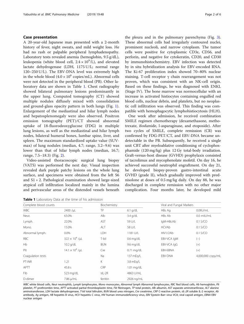
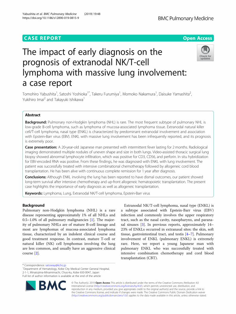
Case presentationA 20-year-old Japanese man presented with a 2-monthhistory of fever, night sweats, and mild weight loss. Hehad no rash or palpable peripheral lymphadenopathy.Laboratory tests revealed anemia (hemoglobin, 9.5 g/dL),leukopenia (white blood cell, 2.4 × 10⁹/L), and elevatedlactate dehydrogenase (LDH, 1175 U/L; normal range120–250 U/L). The EBV-DNA level was extremely highin the whole blood (4.0 × 10⁶ copies/mL). Abnormal cellswere not detected in the peripheral blood (PB). Other la-boratory data are shown in Table 1. Chest radiographyshowed bilateral pulmonary lesions predominantly inthe upper lung. Computed tomography (CT) showedmultiple nodules diffusely mixed with consolidationand ground-glass opacity pattern in both lungs (Fig. 1).Enlargement of the mediastinal and hilar lymph nodesand hepatosplenomegaly were also observed. Positronemission tomography (PET)/CT showed abnormaluptake of 18-fluorodeoxyglucose (FDG) in multiplelung lesions, as well as the mediastinal and hilar lymphnodes, bilateral humeral bones, lumbar spine, liver, andspleen. The maximum standardized uptake value (SUV-max) of lung nodules (median, 4.7; range, 3.2–9.6) waslower than that of hilar lymph nodes (median, 16.7;range, 7.5–18.3) (Fig. 2).Video-assisted thoracoscopic surgical lung biopsy
(VATS) was performed the next day. Visual inspectionrevealed dark purple patchy lesions on the whole lungsurface, and specimens were obtained from the left S6and S1 + 2. Pathological examination showed large-sizedatypical cell infiltration localized mainly in the luminaand perivascular areas of the distended vessels beneath
the pleura and in the pulmonary parenchyma (Fig. 3).These abnormal cells had irregularly contoured nuclei,prominent nucleoli, and narrow cytoplasm. The tumorcells were positive for cytoplasmic CD3ε, CD56, andperforin, and negative for cytokeratin, CD20, and CD30by immunohistochemistry. EBV infection was detectedby in situ hybridization analysis for EBV-encoded RNA.The Ki-67 proliferation index showed 70–80% nuclearstaining. T-cell receptor γ chain rearrangement was notproven, which was consistent with an NK-cell origin.Based on these findings, he was diagnosed with ENKL(Stage IV). The bone marrow was normocellular with anincrease in activated histiocytes containing engulfed redblood cells, nuclear debris, and platelets, but no neoplas-tic cell infiltration was observed. This finding was com-patible with hemophagocytic lymphohistiocytosis (HLH).One week after admission, he received combination
SMILE regimen chemotherapy (dexamethasone, metho-trexate, ifosfamide, l-asparaginase, and etoposide). Aftertwo cycles of SMILE, complete remission (CR) wasconfirmed by FDG-PET/CT, and EBV-DNA became un-detectable in the PB. Subsequently, he received a singleunit CBT after myeloablative conditioning of cyclophos-phamide (120 mg/kg) plus 12 Gy total-body irradiation.Graft-versus-host disease (GVHD) prophylaxis consistedof tacrolimus and mycophenolate mofetil. On day 16, heachieved successful neutrophil engraftment. On day 21,he developed biopsy-proven gastro-intestinal acuteGVHD (grade II), which gradually improved with pred-nisolone at doses of 0.5 mg/kg daily. On day 88, he wasdischarged in complete remission with no other majorcomplication. Four months later, he developed mild
Table 1 Laboratory Data at the time of his admission
Complete blood counts Biochemistry Viral and Fungal Markers
WBC 2400 /μL TP 6.1 g/dL HBs Ag 0.0IIU/mL
Neut. 63.0% Alb 3.4 g/dL HBs Ab 0.0 mIU/mL
Lymph. 22.0% AST 59 U/L IgM-HBcAb 0.1 S/CO
Mono. 15.0% ALT 58 U/L HCVAb 0.1 S/CO
Abnormal lymph. 0.0% LDH 1181 U/L HIV1/2Ab 0.1 S/CO
RBC 322 × 104 /μL T-bil 0.6 mg/dL EBV-VCA IgM (−)
Hb 10.2 g/dL BUN 9.6 mg/dL EBV-VCA IgG (+)
Plt 14.1 × 10⁴ /μL Cre 0.71 mg/dL EBV-EBNA (+)
Coagulation tests Na 137mEq/L EBV-DNA 4,000,000 copy/mL
PT-INR 1.21 K 3.8 mEq/L
APTT 45.8 s CRP 1.01 mg/dL
Fib 523 mg/dL sIL-2R 4863 U/mL
D-dimer 7.86 μ/mL ferritin 2926 ng/mL
WBC white blood cells, Neut neutrophils, Lymph lymphocytes, Mono monocytes, Abnormal lymph Abnormal lymphocytes, RBC Red blood cells, Hb hemoglobin, Pltplatelet, PT prothrombin time, APTT activated partial thromboplastin time, Fib fibrinogen, TP total protein, Alb albumin, AST asparate aminotranferase, ALT alanineaminotransferase, LDH lactate dehydrogenase, T-bil total bilirubin, BUN blood urea nitrogen, Cre creatinine, CRP C-reactive protein, sIL-2R soluble IL-2 receptor, Abantibody, Ag antigen, HB hepatitis B virus, HCV hepatitis C virus, HIV human immunodeficiency virus, EBV Epstein-Barr virus VCA; viral capsid antigen, EBNA EBVnuclear antigen
Yabushita et al. BMC Pulmonary Medicine (2019) 19:48 Page 2 of 6
cutaneous chronic GVHD limited to the head and neck,which could be managed with topical steroids. He hasbeen alive with continuous CR for 1 year after diagnosis.
Discussions and conclusionsENKL is a rare type of lymphoma associated with EBVinfection that most commonly involves the upper aero-digestive mucosa and skin. Although localized ENKL hasa better prognosis when treated with concurrent chemo-radiotherapy [8], advanced ENKL often displays a fulmin-ant clinical course with very poor prognosis. Therefore,especially in advanced ENKL, a delay in diagnosis couldprovoke multiple organ dysfunction and make treatmentdifficult. In our case, surgical lung biopsy contributed to
the rapid and precise diagnosis of ENKL, which resultedin maximal therapeutic efficacy.ENKL involving the lung is rare, and only 14 cases of
ENKL with massive pulmonary involvement have beenreported in the English literature [2, 9–16]. The diagno-sis of pulmonary ENKL is often difficult because of thenonspecific clinical symptoms and radiological findings.In previously published reports, the major initial symptomsof this disorder were fever (n = 12) and cough (n = 10).Moreover, the common CT findings were multiple nodulesin both lungs (n = 9), consolidation (n = 4), bronchial ormediastinal involvement (n = 3), pleural effusion (n = 3),and a single lesion in one lung (n = 2). Although laboratorydata were not available in all cases (n = 6), pancytopenia
Fig. 2 The axial view of FDG-PET/CT scan on diagnosis. (A) PET/CT scan shows intense FDG uptake in bilateral lung masses and the mediastinaland hilar lymph nodes. Each lung nodule is surrounded by a solid-line circle (1–9). Each mediastinal lymph node is surrounded by a dotted linecircle (a-e). (B) The maximum standardized uptake values (SUVmax) of the lesions are shown here
Fig. 1 Radiological imaging on admission. Chest computed tomography revealed multiple nodules in both lungs that showed diffusely mixedwith consolidation and ground-glass opacity pattern. (a; coronal section, b; horizontal section)
Yabushita et al. BMC Pulmonary Medicine (2019) 19:48 Page 3 of 6

(n = 4) and elevated LDH (median, 498 U/L; range135–1192 IU/L) were commonly observed.Lung biopsy is required for definite diagnosis of ENKL
mainly involving the lungs. Eleven of the 14 patientswere diagnosed using lung biopsy, including transbron-chial lung biopsy (TBLB; n = 2), CT-guided biopsy (n = 2),surgical biopsy (n = 1), and unspecified core biopsy (n = 6).The other 3 patients were diagnosed at autopsy despiterepeated antemortem biopsies. Specimens obtained byTBLB or CT-guided biopsy are often inappropriate be-cause of their small-sized sampling. In contrast, surgicalbiopsy enables a sufficient amount of specimen for precisediagnosis. Therefore, the surgical approach should beconsidered as a helpful diagnostic method for ENKLbefore rapid exacerbation, especially when ENKL is sus-pected on the basis of clinical features. In the present case,considering the complication of HLH and the highlyelevated level of EBV-DNA, we included ENKL in thedifferential diagnosis list, and chose surgical biopsy as adiagnostic procedure before rapid exacerbation.Some ENKL cases are complicated by lymphoma-asso-
ciated HLH, which usually presents with persistent highfever, pancytopenia, hepatosplenomegaly, or elevatedlevels of soluble interleukin-2 receptor or ferritin. Ap-proximately half of the cases of lymphoma-associated
HLH have been reported to be correlated with EBV-asso-ciated lymphoid malignancies, such as ENKL [17]. Inaddition, it is well known that almost all ENKL cases showpositive EBV-DNA in their PB, but not in the normalcontrol. EBV-DNA detection using PB samples is veryhelpful for diagnosis.The prognosis of advanced ENKL is very poor; overall
survival (OS) at 5 years for advanced ENKL has beenreported to be approximately 25% [8]. Pulmonary ENKLusually has a fatal outcome [18, 19]. Eight of the 14patients described in the literature were treated withchemotherapy. One received autologous hematopoieticstem cell transplantation followed by high-dose chemo-therapy. However, all died of disease progression regardlessof intensive treatment. The median OS in 12 evaluable pa-tients was 2months (range, 1 week to 7months). Recently,combinational chemotherapy containing L-asparaginase(the SMILE regimen) has been reported to be effective forENKL compared to the conventional anthracycline-basedregimen, such as CHOP (cyclophosphamide, doxorubicin,vincristine, and prednisone) [20]. In several guidelines,subsequent hematopoietic stem cell transplantation(HSCT) is recommended for patients with advanced ENKLachieving CR after induction chemotherapy because of itspoor prognosis [21, 22]. We determined to perform
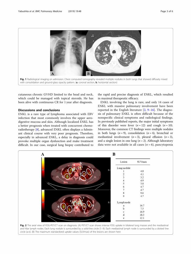
Fig. 3. Pathological findings of the lung biopsy. Atypical large-sized lymphoid cells filled several distended vessels beneath the pleura and in thepulmonary parenchyma (hematoxylin and eosin; × 400 (A)). The neoplastic cells had irregular nuclear contours, prominent nucleoli, and narrowcytoplasm (black arrow), described in a partially expanded image on the lower right field. Abnormal mitosis was prominent in the cells. Themoderate inflammatory response caused mainly by plasma cells was seen in perivascular areas, but no fibrin thrombi or necrosis was observed.The alveolar epithelial cells showed reactive nuclear enlargement. The tumor cells in pulmonary arterioles were positive for cytoplasmic CD3 (B),CD56 (C), perforin, and Ki-96 (not shown). In situ hybridization analysis for Epstein-Barr virus-encoded messenger RNA was positive (D).
Yabushita et al. BMC Pulmonary Medicine (2019) 19:48 Page 4 of 6
allogeneic (allo-) HSCT for this patient after achieving CRwith two courses of the SMILE regimen, because the ex-tremely poor prognosis of pulmonary ENKL was estimatedfrom the above literature review. Since neither related norunrelated HLA-matched donor was found, we selectedcord blood donor. He has been alive with continuous CRfor 1 year after diagnosis. However, there is no clear evi-dence to determine whether autologous (auto-) orallo-HSCT is preferred for advanced ENKL. Yim et al. re-ported the outcome of upfront auto-HSCT for ENKL. Pa-tients with advanced disease had significantly worseprognosis than those with limited disease: 3-year progres-sion free survival (PFS), 40.1% versus 64.5%, p = 0.017;OS, 52.3% versus 67.6%, p = 0.048 [23]. In contrast,allo-HSCT for ENKL has been evaluated only by smallretrospective studies: OS rates were 34–57% [24–26].In a retrospective nationwide survey of auto-HSCT andallo-SCT for Japanese patients with ENKL, the 2-yearOS in auto-HSCT group was superior compared toallo-SCT group: 69% versus 41%, p = 0.002. However,when adjusted by advanced stage, disease status, andperformance status at transplant, there was no signifi-cant difference of 2-year OS between both transplantprocedures, because the allo-HSCT group includedmore patients with advanced stage and refractory dis-ease condition [27]. Allo-SCT could be one of the cura-tive options for some patients with advanced ENKL. Ingeneral, the primary indication for allo-HSCT shouldbe cautiously considered in each individual case.In summary, we described a rare case of a pulmonary
ENKL in a 20-year-old man who was successfullydiagnosed using early surgical biopsy and treated withintensive multi-agent chemotherapy and up-front CBT,and he has survived more than 1 year with no relapse. Inthe present case, VATS was helpful for prompt diagnosisand successful treatment. Collection of sufficient speci-men with any invasive procedure is crucial for diagnosisof pulmonary aggressive lymphoma like ENKL.
AbbreviationsCBT: cord blood transplantation; CHOP: cyclophosphamide, doxorubicin,vincristine and prednisone; CR: complete response; CT: computedtomography; EBV: Epstein-Barr virus; ENKL: extranodal NK/T-cell lymphoma,nasal type; FDG: 18-fluorodeoxyglucose; HLH: hemophagocyticlymphohistiocytosis; LDH: lactate dehydrogenase; NHL: non-Hodgkinlymphoma; NK: natural killer; OS: overall survival; PB: peripheral blood;PET: positron emission tomography; SMILE: dexamethasone, methotrexate,ifosfamide, l-asparaginase, and etoposide; TBLB: transbronchial lung biopsy;VATS: video-assisted thoracoscopic surgical lung biopsy
AcknowledgementsWe thank Mr. Hayato Maruoka, Mr. Kaoru Sueoka, Mr. Masahiro Yoshida, Ms.Yukiko Yano, Ms. Eiko Yamashita, Mr. Kazuyuki Ueno, and Ms. Kyoko Tanakafrom the Department of Clinical Laboratory, Kobe City Medical CenterGeneral Hospital for their technical assistance.
FundingWe have not received any source of support in the form of grants,equipment, or drugs.
Availability of data and materialsThe data used and analyzed during the current study are available from thecorresponding author on reasonable request.
Authors’ contributionsT.Y., S.Y., T.F., and M.N. treated the patient as the attending physician; D.Y.and Y.I. performed the histological examination of the lung; T.Y., S.Y., and T.I.wrote the manuscript; and all authors revised and approved the manuscript.
Ethics approval and consent to participateThis case report complied with the Declaration of Helsinki and was approvedby the Ethical Committee of Kobe City Medical Center General Hospital.(Approval No. zn171210).
Consent for publicationWritten informed consent for publication was obtained from the patient,according to the governance that the Ethical Committee of Kobe CityMedical Center General Hospital approved.
Competing interestsThe authors declare that they have no competing interests.
Publisher’s NoteSpringer Nature remains neutral with regard to jurisdictional claims inpublished maps and institutional affiliations.
Author details1Department of Hematology, Kobe City Medical Center General Hospital,2-1-1, Minatojima-Minamimachi, Chuo-ku, Kobe 650-0047, Japan.2Department of Pathology, Kobe City Medical Center General Hospital, 2-1-1,Minatojima-Minamimachi, Chuo-ku, Kobe 650-0047, Japan.
Received: 20 December 2017 Accepted: 14 February 2019
References1. Cadranel J, Wislez M, Antoine M. Primary pulmonary lymphoma. Eur Respir
J. 2002;20:750–62.2. Laohaburanakit P, Hardin KA. NK/T cell lymphoma of the lung: a case report
and review of literature. Thorax. 2006;61:267–70.3. Campo E, Swerdlow SH, Harris NL, Pileri S, Stein H, Jaffe ES. The 2008 WHO
classification of lymphoid neoplasms and beyond: evolving concepts andpractical applications. Blood. 2011;117:5019–32.
4. Shet T, Suryawanshi P, Epari S, Sengar M, Rangarajan V, Menon H, et al.Extranodal natural killer/T cell lymphomas with extranasal disease in non-endemic regions are disseminated or have nasal primary: a study of 84cases from India. Leuk Lymphoma. 2014;55:2748–53.
5. Li S, Feng X, Li T, Zhang S, Zuo Z, Lin P, et al. Extranodal NK/T-celllymphoma, nasal type: a report of 73 cases at MD Anderson Cancer Center.Am J Surg Pathol. 2013;37:14–23.
6. Gualco G, Domeny-Duarte P, Chioato L, Barber G, Natkunam Y, Bacchi CE.Clinicopathologic and molecular features of 122 Brazilian cases of nodal andextranodal NK/T-cell lymphoma, nasal type, with EBV subtyping analysis. AmJ Surg Pathol. 2011;35:1195–203.
7. Chan JK, Sin VC, Wong KF, Ng CS, Tsang WY, Chan CH, et al. Nonnasallymphoma expressing the natural killer cell marker CD56: aclinicopathologic study of 49 cases of an uncommon aggressive neoplasm.Blood. 1997;89:4501–13.
8. Yamaguchi M, Suzuki R, Oguchi M, Asano N, Amaki J, Akiba T, et al.Treatments and outcomes of patients with Extranodal natural killer/T-celllymphoma diagnosed between 2000 and 2013: A cooperative study inJapan. J Clin Oncol. 2017;35:32–9.
9. Oshima K, Tanino Y, Sato S, Inokoshi Y, Saito J, Ishida T, et al. Primarypulmonary extranodal natural killer/T-cell lymphoma: nasal type withmultiple nodules. Eur Respir J. 2012;40:795–8.
10. Morovic A, Aurer I, Dotlic S, Weisenburger DD, Nola M. NK celllymphoma, nasal type, with massive lung involvement: a case report. JHematop. 2010;3:19–22.
11. Liu CH, Wang HH, Perng CL, Peng CK, Chian CF, Shen CH. Primaryextranodal NK/T-cell lymphoma of the lung: mimicking bronchogeniccarcinoma. Thorac Cancer. 2014;5:93–6.
Yabushita et al. BMC Pulmonary Medicine (2019) 19:48 Page 5 of 6
12. Li Y, Damjanov I. Extranodal NK/T cell lymphoma causing cardiorespiratoryfailure. Case Rep Hematol. 2016;2016:2394809.
13. Lee S, Shin B, Yoon H, Lee JY, Chon GR. A case of primary pulmonary NK/T celllymphoma presenting as pneumonia. Respir Med Case Rep. 2015;17:1–4.
14. Lee BH, Kim SY, Kim MY, Hwang YJ, Han YH, Seo JW, et al. CT of nasal-typeT/NK cell lymphoma in the lung. J Thorac Imaging. 2006;21:37–9.
15. Ding W, Wang J, Zhao S, Yang Q, Sun H, Yan J, et al. Clinicopathologicalstudy of pulmonary extranodal nature killer/T-cell lymphoma, nasal typeand literature review. Pathol Res Pract. 2015;211:544–9.
16. Chien CC, Lee HS, Lin MH, Hsieh PP. Primary extranodal natural killer/T-celllymphoma of bronchus and lung: A case report and review of literature.Thorac Cancer. 2016;7:140–4.
17. Kawa K. Epstein-Barr virus--associated diseases in humans. Int J Hematol.2000;71:108–17.
18. Suzuki R, Suzumiya J, Yamaguchi M, Nakamura S, Kameoka J, Kojima H, et al.Prognostic factors for mature natural killer (NK) cell neoplasms: aggressiveNK cell leukemia and extranodal NK cell lymphoma, nasal type. Ann Oncol.2010;21:1032–40.
19. Au WY, Weisenburger DD, Intragumtornchai T, Nakamura S, Kim WS, Sng I,et al. Clinical differences between nasal and extranasal natural killer/T-celllymphoma: a study of 136 cases from the international peripheral T-celllymphoma project. Blood. 2009;113:3931–7.
20. Yamaguchi M, Kwong Y-L, Kim WS, Maeda Y, Hashimoto C, Suh C, et al.Phase II study of SMILE chemotherapy for newly diagnosed stage IV,relapsed, or refractory extranodal natural killer (NK)/T-cell lymphoma, nasaltype: the NK-cell tumor study group study. J Clin Oncol. 2011;29:4410–6.
21. National Comprehensive Cancer Network. NCCN Clinical Practice Guidelinesin Oncology T-cell Lymphomas. version 4. 2018. (https://www.nccn.org/professionals/physician_gls/pdf/t-cell.pdf). Accessed 2018 May 14.
22. Kharfan-Dabaja MA, Kumar A, Ayala E, Hamadani M, Reimer P, GisselbrechtC, et al. Clinical practice recommendations on indication and timing ofhematopoietic cell transplantation in mature T cell and NK/T celllymphomas: an international collaborative effort on behalf of the guidelinesCommittee of the American Society for blood and marrow transplantation.Biol Blood Marrow Transplant. 2017;23:1826–38.
23. Yhim HY, Kim JS, Mun YC, Moon JH, Chae YS, Park Y, et al. And theconsortium for improving survival of lymphoma study. Clinical outcomesand prognostic factors of up-front autologous stem cell transplantation inpatients with Extranodal natural killer/T cell lymphoma. Biol Blood MarrowTransplant. 2015;21:1597–604.
24. Murashige N, Kami M, Kishi Y, Kim SW, Takeuchi M, Matsue K, et al.Allogeneic haematopoietic stem cell transplantation as a promisingtreatment for natural killer-cell neoplasms. Br J Haematol. 2005;130:561–7.
25. Tse E, Chan TSY, Koh LP, Chung WJ, Kim WS, Tang T, et al. Allogeneichaematopoietic SCT for natural killer/T-cell lymphoma: a multicentreanalysis from the Asia lymphoma study group. Bone Marrow Transplant.2014;49:902–6.
26. Kanate AS, DiGilio A, Ahn KW, AL Malki M, Jacobsen E, Steinberg A, et al.Allogeneic haematopoietic cell transplantation for extranodal natural killer/T-cell lymphoma, nasal type: a CIBMTR analysis. Br J Haematol. 2017. https://doi.org/10.1111/bjh.14879.
27. Suzuki R, Kako S, Hyo R, Izutsu K, Ito T, Shinagawa K, et al. Comparison ofAutologous and Allogeneic Hematopoietic Stem Cell Transplantation forExtranodal NK/T-Cell Lymphoma, Nasal Type: Analysis of the Japan Societyfor Hematopoietic Cell Transplantation (JSHCT) Lymphoma Working Group.Blood 2011;118:Abstract 503.
Yabushita et al. BMC Pulmonary Medicine (2019) 19:48 Page 6 of 6


![Apresentação do PowerPoint - Repositório Comum: Página ... · embriogenesis are being discussed and the prognosis is not well known due to the small number of reported cases[7]](https://img.pdfslide.tips/doc/110x75/5ca1f44d88c9932f098d2c2b/apresentacao-do-powerpoint-repositorio-comum-pagina-embriogenesis.jpg)