Embed Size (px)
Citation preview

1
THE MUSCULOSKELETAL
SYSTEM

Musculoskeletal system
The short
term effects
on MUSCLES/
JOINTS and
BONES
Musculoskeletal response: increased blood
supply; increase in muscle pliability; increased
range of movement; muscle fibre micro tears

REMEMBER...
•“Diseases of the musculoskeletal system rank first among disease conditions that alter the quality of life…”

• Skeleton: 206 bones
• Long: femur, humerus, radius
• Short: carpals, tarsals
• Irregular: vertebrae
• Bones protect, support, allow for locomotion and mineral storage (Ca,Mg)

• Joints: Range from joints that don’t move to joints that freely move.
• Ligaments and tendons: stabilize joints
• Ligaments: attached from bone to bone
• Tendons: attached from muscle to bone
• Cartilage: ends on bones

• Muscles: controlled by nervous system
• Fascia: surrounds muscles, divides muscles, main blood vessels and nerves.
• Bursae: cushions moving parts
• Muscle tone: ability to resist force; graded 0-5
• Atrophy: decrease size

• Joint pain:
• SLIDA:
Severity,
Location,
Intensity,
Duration,
Aggravating factors (alleviating factors, associated symptoms)
• Stiffness
• Limited movement
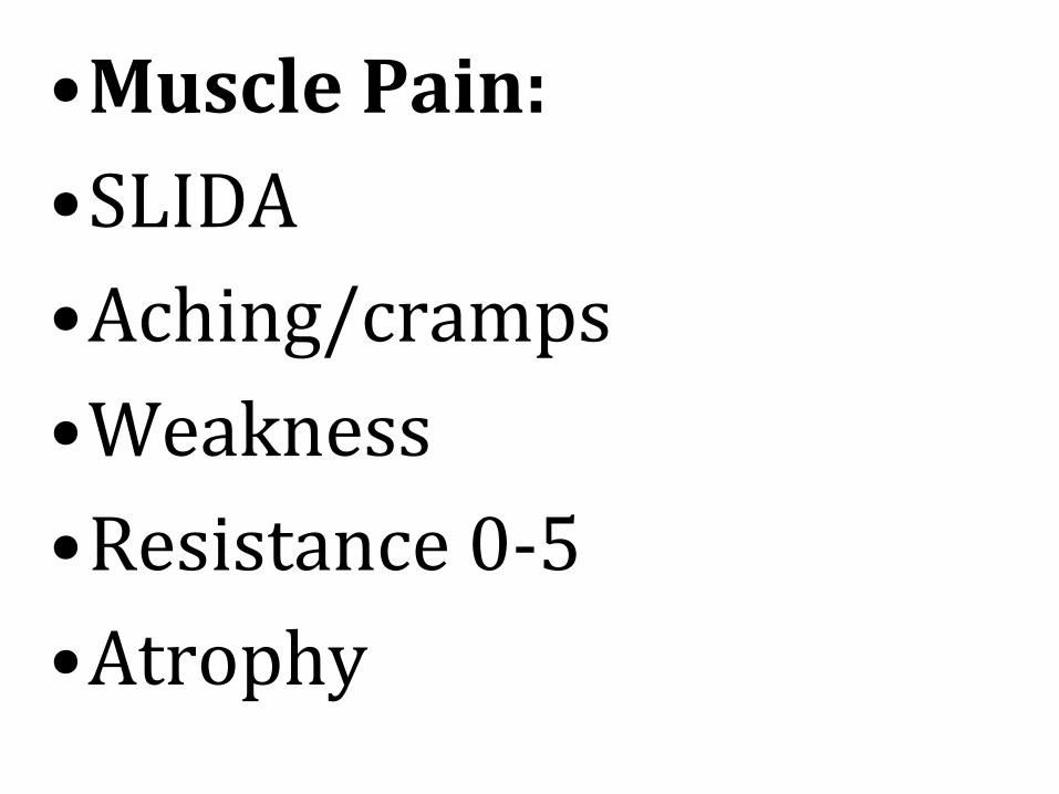
•Muscle Pain:
•SLIDA
•Aching/cramps
•Weakness
•Resistance 0-5
•Atrophy

•Bone pain:
•SLIDA
•Hx
•Deformity
•Trauma- limitations as a result of trauma

The musculoskeletal system comprises
bones, muscles and joints, and makes up most of the body’s mass. It performs a number of essential functions including:
• Maintenance of body shape.
• Support and protection of soft tissues structures such as the brain, heart and lungs.

• Movement.
• Breathing.
• Storage of calcium and phosphate in bone.
• The manufacture of red blood cells, white
blood cells and platelets in the bone marrow (haematopoiesis)

BONES •Bone is a rigid structure
ideally suited for its supportive and protective function. Bones contain sites for muscle attachment, the mechanical basis for movement.

MUSCLES
• Muscle tissue is made up of contractile cells which have the ability to shorten in length or contract. It is this characteristic that is responsible for movement, maintenance of posture and heat production

Joints A joint is the site at which two or more bones are united, providing the allows movement. Fibrous joints unite bones by fibrous connective tissue and allow very little movement.


16
Structure and Function
Forms the body framework
Enables the body to move
Protects and supports internal organs
17
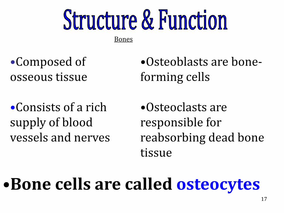
Bones Bones
•Composed of osseous tissue
•Consists of a rich supply of blood vessels and nerves
•Osteoblasts are bone-forming cells
•Osteoclasts are responsible for reabsorbing dead bone tissue
•Bone cells are called osteocytes

18
Ossification BONES
The development of osteocytes and the hardening process is called ossification.
calcium
phosphorus
vitamin D
OSSIFICATION DEPENDS ON:

19

20
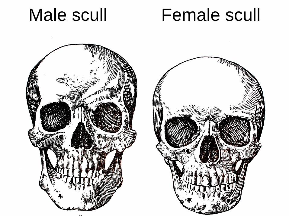
Male skeleton Female skeleton.
21
Male scull Female scull
22
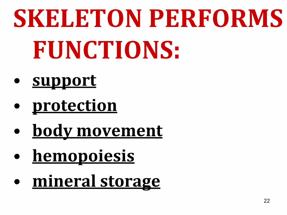
SKELETON PERFORMS FUNCTIONS:
• support
• protection
• body movement
• hemopoiesis
• mineral storage

• Bones are body organs with blood supply, nerves, and lymphatic vessels
• Bones are connected to each other to form skeleton
–Framework for the body
–206 bones

•Red bone marrow within bones produces blood cells
•Bones also:
–Protect vital organs
–Store minerals

•Joint
–Place where two bones meet
–Held together by ligaments
–Gives flexibility to skeleton

Bones
• Also called osseous tissue
• One of hardest materials in body
• Formed from gradual process before birth called ossification
• Fetal skeleton is formed from a cartilage model

Bones • Flexible tissue is gradually replaced
by osteoblasts (immature bone cells)
• In adult bones osteoblasts mature into osteocytes
• Formation of strong bones dependant on adequate supply of minerals

Four Shapes of Bones
Long bones
Short bones
Flat bones
Irregular bones
Longer than wide
Example:
femur
humerus
Roughly as long as wide
Example:
carpals
tarsals
Plate- shaped
Example:
sternum
scapula
pelvis
Shape very irregular
Example:
vertebrae

Classification of bones by shape.
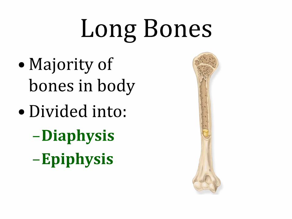
Long Bones • Majority of
bones in body
• Divided into:
–Diaphysis
–Epiphysis

Diaphysis • Central shaft
• Medullary cavity
– Open canal within diaphysis
– Contains yellow bone marrow
• Mostly fat

Epiphysis • Wide ends of long bone
– Distal epiphysis
– Proximal epiphysis
• Articular cartilage
– Covers epiphysis
– Prevents bone rubbing on bone

Periosteum
• Covers surface of bone not covered by articular cartilage
• Thin connective tissue membrane
• Contains numerous nerve and lymphatic vessels

Compact Bone
• Also called cortical bone
• Very dense and hard
• Outer layer of bone
• Found in both epiphysis and diaphysis

Cancellous Bone • Also called spongy bone
• Found inside bone
• Has spaces containing red bone marrow – Manufactures blood cells

Components of a long bone.

Bony Processes
• Projection from the surface of a bone
• Rough processes provide place for muscle attachment
• Smooth rounded processes articulate with another bone in a joint
• Named for shape and location

Common Bony Processes
Head Large smooth ball-shaped end of a long bone
Condyle Smooth rounded portion at end of bone
Epicondyle Projection above or on a condyle
Trochanter Large rough process
Tubercle Small rough process
Tuberosity Large rough process

Bony processes found on the femur

Bony Depressions • Sinus
– Hollow cavity within bone
• Foramen
– Smooth opening for nerves and blood vessels
• Fossa
– Shallow cavity or depression within a bone
• Fissure
– Deep groove or slit-like opening

The Skeleton
•Skeleton has two divisions
–Axial skeleton
–Appendicular skeleton

Axial Skeleton
• Includes bones in:
–Head
–Neck
–Spine
–Chest
–Trunk

Bones of the axial skeleton.

The Skull
• Is divided into two parts
– Cranium
– Facial bones
• Protects brain, eyes, ears, nasal cavity, and oral cavity
• Attachment for muscles of chewing and turning the head

Cranium • Frontal – 1
– Forehead
• Parietal – 2
– Upper sides and roof of skull
• Temporal – 2
– Sides & base of skull

Cranium
• Ethmoid – 1
– Part of eye orbit, nose, & floor of skull
• Sphenoid – 1
– Part of floor of skull
• Occipital – 1
– Back & base of skull

Bones of the skull.

Facial Bones
• Mandible – 1
– Lower jawbone
• Maxilla – 1
– Upper jawbone
• Zygomatic – 2
– Cheek bones
• Vomer – 1
– Part of nasal septum

Facial Bones
• Palatine – 1
– Hard palate and floor of nose
• Nasal – 2
– Part of nasal septum and bridge of nose
• Lacrimal – 2
– Inner corner of eye

Bones of the skull.

Hyoid Bone
• Single U-shaped bone
• In neck between mandible and larynx
• Attachment point for swallowing and speech muscles

The Trunk
• Vertebral column
• Sternum
• Rib cage

The Vertebral Column
• Divided into five sections
– Cervical
– Thoracic
– Lumbar
– Sacrum
– Coccyx

The Vertebral Column
• Cervical
– 7 vertebrae of neck
• Thoracic
– 12 vertebrae of chest
• Lumbar
– 5 vertebrae of low back
• Sacrum
– 5 fused vertebrae at base of spine
• Coccyx
– 3–5 small vertebrae attached to sacrum

Divisions of the vertebral column.

The Rib Cage
• 12 pairs of ribs
• Attached to vertebral column at back
• Provides support for organs, such as heart and lungs

The Rib Cage
• True ribs
– 10 pairs attached to sternum in front
• Floating ribs
– Inferior 2 pairs
– No attachment in front

The structure of the rib cage.

Appendicular Skeleton
• Includes bones of:
– Pectoral girdle
– Upper extremity
– Pelvic girdle
– Lower extremity
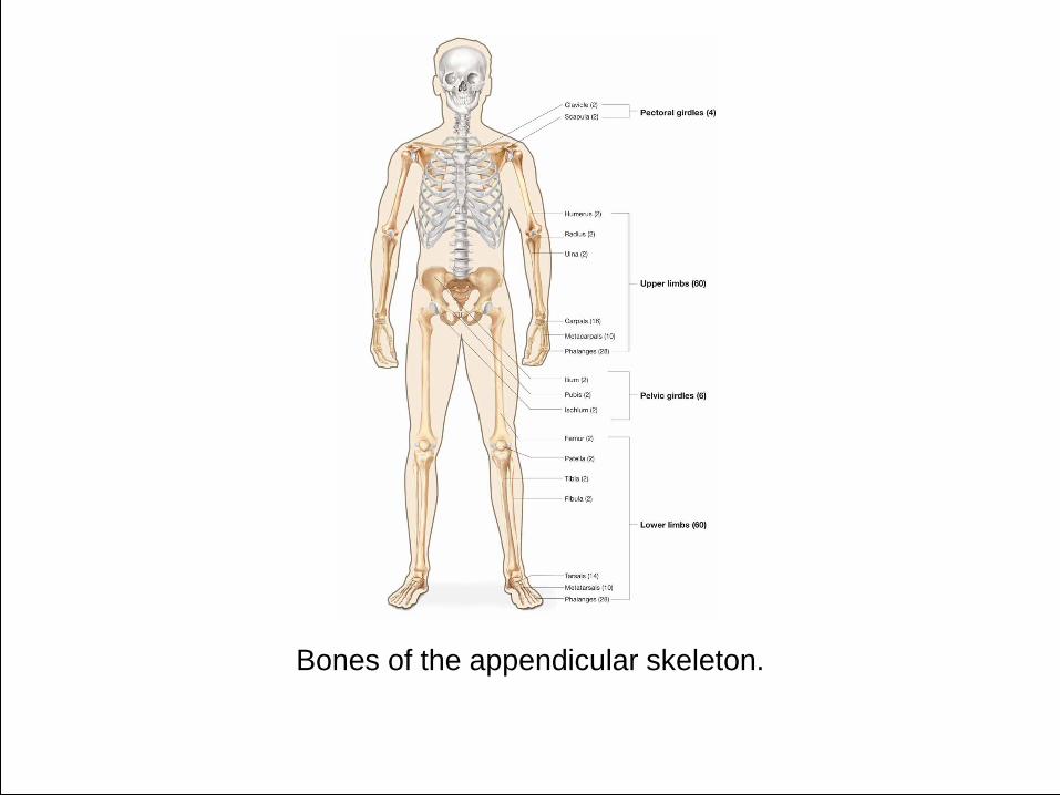
Bones of the appendicular skeleton.

Pectoral Girdle • Attaches upper extremity to axial
skeleton
• Articulates with:
– Sternum anteriorly
– Vertebral column posteriorly
• Consists of:
– Clavicle – collar bone
– Scapula – shoulder blade

Upper Extremity • Arm
• Consists of:
– Humerus – upper arm
– Ulna – part of forearm
– Radius – part of forearm
– Carpals – wrist bones
– Metacarpals – hand bones
– Phalanges – finger bones

Anatomical and common names for the pectoral girdle and upper extremity.

Pelvic Girdle • Also called os coxae, innominate bone,
or hipbone
• Attaches lower extremity to axial skeleton
• Articulates with sacrum posteriorly
• Consists of:
– Ilium
– Ischium
– Pubis

Lower Extremity • Leg
• Consists of:
– Femur – thigh bone
– Patella – knee cap
– Tibia – shin bone
– Fibula – lower leg bone
– Tarsals – ankle bones
– Metatarsals – foot bones
– Phalanges – toe bones

Anatomical and common names for the pelvic girdle and lower extremity.

Joints
• Formed where two bones meet
• Also called an articulation
• Three types based on movement allowed
between the 2 bones:
– Synovial
– Cartilaginous
– Fibrous

Synovial Joints
• Freely moving joints
• Most common type of joint
• Example is ball-and-socket joint
• Bones held together by ligaments
– Strong bands of connective tissue
• Some contain a bursa
– Sac-like structure lined with synovial
membrane

Synovial Joints
• Enclosed in an
elastic joint
capsule
• Contains synovial
fluid
– Lubricant secreted
by synovial
membrane
• Ends of bones are
covered with
articular
cartilage

Structure of a synovial joint.

Cartilaginous Joints
• Allow slight
movement
• Hold bones firmly in
place by solid piece of
cartilage
• Example
– Pubic symphysis

Fibrous Joints
• Allow almost no
movement
• Joined by thick
fibrous tissue
• Example
– Sutures of the skull

Examples of three types of joints found in the body.

Abnormal spinal curvatures: kyphosis, lordosis, and scoliosis.


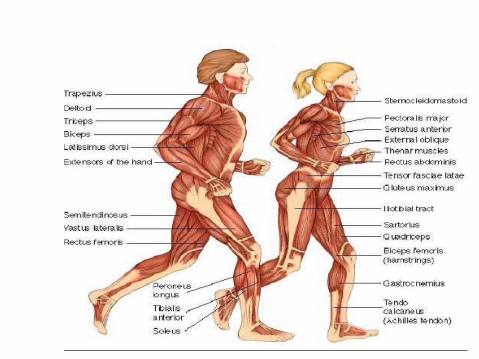
Muscular System at a Glance
• Function of Muscular System
– Individual cells are able to contract or shorten
in length
– Shortening produces movement

Muscular System at a Glance
• Organs of Muscular System
– Muscles


• Bundles of parallel muscle tissue fibers
• Fibers contract
– Shorten in length
– Produce movement
– Move bones closer together
– Push food through digestive system
– Pump blood through blood vessels


Types of Muscles
• Skeletal muscle
• Smooth muscle
• Cardiac muscle
• Voluntary muscles
– Consciously choose to contract the muscle
– Skeletal muscles
• Involuntary muscles
– Under control of subconscious brain
– Smooth muscles and cardiac muscle

The three types of muscles: skeletal, smooth, and cardiac.

Skeletal Muscles
• Attached to bones
• Produce voluntary movement of skeleton
• Also referred to as striated muscle
– Looks striped under microscope

Skeletal Muscles
• Muscle is wrapped in layers of connective
tissue
– Called fascia
– Tapers at the end to form tendon
– Inserts into periosteum to attach muscle to
bone
• Are stimulated by motor neurons
– Point of contact with muscle fiber is called
myoneural junction

Characteristics of the three types of muscles.

Smooth Muscles
• Associated with internal organs
– Also called visceral muscle
– Stomach
– Respiratory airways
– Blood vessels
• Called smooth because has no
microscopic stripes
• Produces involuntary movement of these
organs

Characteristics of the three types of muscles.

Cardiac Muscle
• Also called myocardium
• Makes up walls of heart
• Involuntary contraction of heart to pump
blood

Characteristics of the three types of muscles.

Skeletal Muscle Actions
• Skeletal muscles attach to two different
bones and overlap a joint
• When muscle contracts both bones move,
but not equally
– Origin: less moveable of 2 bones
– Insertion: more moveable of 2 bones

Skeletal Muscle Actions
• Action
– Type of movement produced by the muscle
• Antagonistic pairs
– Pair of muscles arranged around a joint
– Produce opposite actions

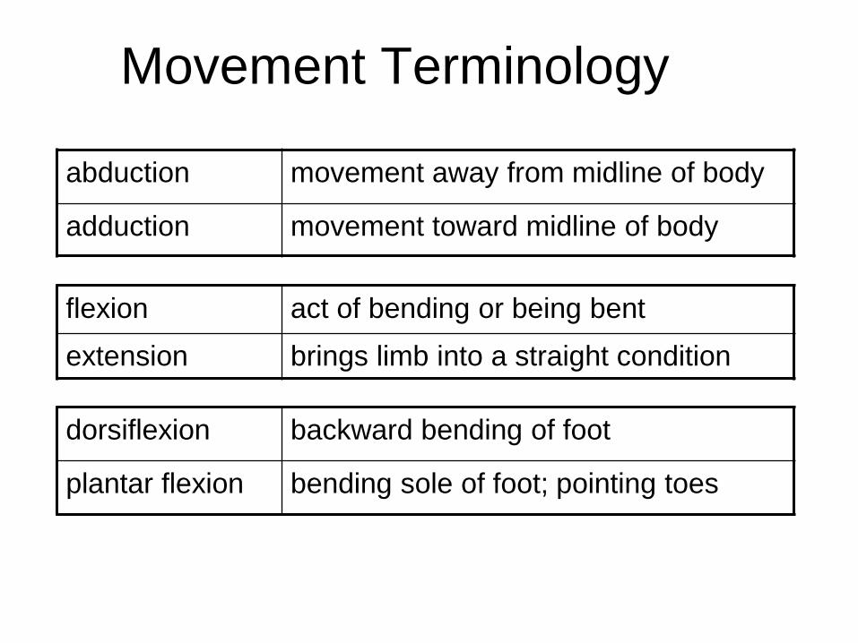
Movement Terminology
dorsiflexion backward bending of foot
plantar flexion bending sole of foot; pointing toes
flexion act of bending or being bent
extension brings limb into a straight condition
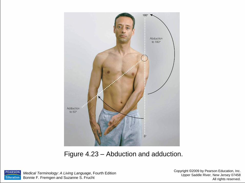
abduction movement away from midline of body
adduction movement toward midline of body

Medical Terminology: A Living Language, Fourth Edition
Bonnie F. Fremgen and Suzanne S. Frucht
Copyright ©2009 by Pearson Education, Inc.
Upper Saddle River, New Jersey 07458
All rights reserved.
Figure 4.23 – Abduction and adduction.

Click here to view an animation on humerus adduction and abduction.
Humerus Adduction/Abduction
Animation
Back to Directory

Medical Terminology: A Living Language, Fourth Edition
Bonnie F. Fremgen and Suzanne S. Frucht
Copyright ©2009 by Pearson Education, Inc.
Upper Saddle River, New Jersey 07458
All rights reserved.
Figure 4.24 – Flexion and extension.

Click here to view an animation on elbow flexion and extension.
Elbow Flexion/Extension
Animation
Back to Directory

Dorsiflexion and plantar flexion.

Click here to view an animation on ankle dorsiflexion and plantar flexion.
Ankle Dorsiflexion and Plantar
Flexion Animation
Back to Directory

Movement Terminology
elevation to raise
depression to drop down
pronation turning palm downward
supination turning palm upward
eversion turning outward
inversion turning inward

Eversion and inversion.
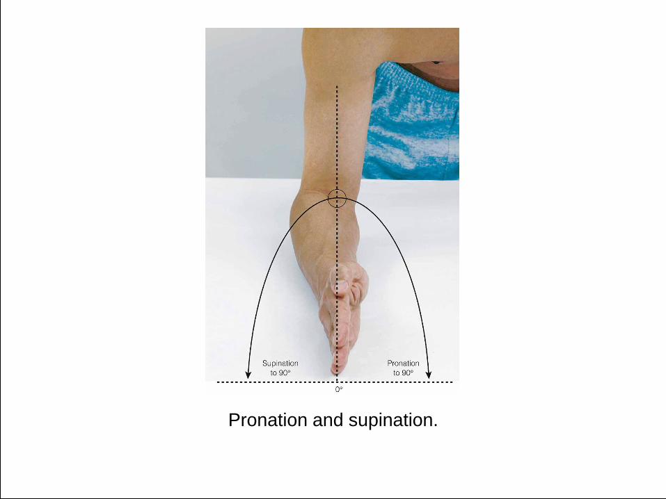
Pronation and supination.

Different Circular Movements
• Circumduction
– Movement in circular direction from a central
point
• Opposition
– Moving thumb away from palm to contact tip
of other fingers
• Rotation
– Moving around a central axis


106
All the bones of an
organism united in
bone system (skeleton
or systema skeletale),
which is usually called
skeleton.

107

108
Stages of the development
of the skeleton: 1.connective-tissue (membranouse)
2.cartilaginous
3.bony

109

110
The types of ossification
(osteogenesis) are
distinguished:
• Intramembranous or endesmal
• Perichondral
• Enchondral

111
The bones, which go through these three developmental stages, called secondary bone.

112
The bones, which are formed
directly from connective tissue
without going through the
stage of the cartilage, called
primary bone.

113
The skeleton is
devided into axial and
appendicular portion.

114
The components of the axial
skeleton are as follow:
1.Skull
2.Auditory ossicle
3.Hyoid bone
4.Vertebral column
5.Thoracic cage

115
The appendicular skeleton is
composed of the:
1.Pectoral girgle
2.Upper limb
3.Pelvic girgle
4.Lower limb

116
The skeleton is composed of more
then 200 bones.
Bone classification according
the shape:
1. Long bones
2. Flat bones
3. Short bones
4. Mixed bones

117 The types of bones

118
As a organ bone has:
1. bone tissue: compact tissue
spongy tissue
2. bone marrow
3. periosteum
4. epiphyseal cartilage
5. vessels and nerve

119

120 The part of the tubulare bone

121

122
The structure of bone

123
Osteon – is a structuring
unit compact tissue of the
bone. This is a system of bone
lamellas, which are situated
around the thin (Haversion)
canal, where vessels and
nerves are going through.

124
Group of osteons form the
trabecule.
Substantia spongiosa
(trabecularis) is formed by the
many trabecules, which are
situated crumbly. There are a lot
of lacunaes.

125
Substantia compacta consist of
trabecules, which are situated
tightly.

126
On the surface of the bone the
compact bone tissue is situated.
Spongy tissue is situated
under the compact tissue.
In the wall of diaphysis of
tubular bones and flat bones the
compact tissue is the main.

127 .

128

129
130
The bone lacunars of the
spongy tissue and bones canal
of tubular bones contain bone
marrow. These lacunars of the
spongy bones is called bone-
medullary cavity. This canal of
bones is called bone-
medullary canal.
131
The bone marrow are divided on red bone marrow and yellow bone marrow.
The function of red bone marrow is hemopoesis.
The yellow bone marrow consist of fat cells.

132

133

134

Different types of muscles • Each of the muscles reacts differently to exercise
• Skeletal – Demands Oxygen and Glycogen
• At rest 20% of our blood goes to our muscles.
• In a warm up 50% of our blood goes to our working muscles
• In intense exercise 80% of our blood goes to our working muscles
– Works harder
– Warms up
• Cardiac – Works harder (beats more often and with larger amount of blood in
each beat) to provide the Oxygen and Nutrients to the skeletal muscle via the blood, and get rid of the Waste products of exercise (Carbon Dioxide, Water and Heat).
• Involuntary
Blood is shunted away from the parts of the body that don’t need it
Eg the stomach gets 25% of our blood during rest. This can reduce to 1% during
exercise

Thank You