Embed Size (px)
Citation preview

Archeometriai Műhely 2016/XIII./1. 19
HU ISSN 1786-271X; urn: nbn: hu-4106 © by the author(s)
THE PALAEOPATHOLOGY OF WILD MAMMALS IN ARCHAEOLOGY
VADON ÉLŐ EMLŐSÁLLATOK BETEGSÉGEI A RÉGÉSZETBENLÁSZLÓ BARTOSIEWICZ
Osteoarchaeological Research Laboratory, Stockholm University Lilla Frescativägen 7, 106 91 Stockholm (Sweden)
E-mail: [email protected]
‘Hey babe, take a walk on the wild side…’
(Lou Reed)
AbstractDomestication is known to have increased animal morbidity. Wild animals, however, should not be looked upon romantically like Jean-Jacques Rousseau’s “noble sauvage”, untainted by civilisation. Rare pathological lesions found on the bones of wild animals in archaeozoological assemblages, they offer valuable information both from a zoological and a archaeological point of view. In addition to discussing problems of sampling, this paper is a re-view of major factors such as taphonomy, environment, and heritability that determine the manifestation of disease in wild animals in archaeological assemblages. A simple classification, specifically developed for wild animals, is presented that helps better understand these conditions. Numerous examples from both the author’s own work and the broad base of international literature (especially on Europe and the Southwest Asia) are cited to help illustrate how disease is manifested on the bones of wild animals recovered from a variety of archaeological periods. The re-sults of this paper show that although domestication undoubtedly brought about an increase in animal morbidity, depending on the chances of survival of a game species and the functional importance of the body part affected, a variety of pathological lesions regularly occur on the remains of wild animals as well.
KivonatA háziasítás köztudottan csökkentette az állatok betegségekkel szembeni ellenálló képességét. Azonban a vadál-latokat sem szabad a Rousseau romantikus szellemében a „nemes vadság” állapotában megrekedt, a civilizáció ártalmai által érintetlen teremtményeknek tekintenünk. Noha a régészeti állattani leletegyüttesek vadállatcsontjain csak ritkán figyelhetünk meg betegségre utaló elváltozásokat, ezek a tünetek fontos állattani és régészeti ismere-tekkel szolgálnak. A mintavételi kérdések áttekintése mellett ez a dolgozat a tafonómiai tényezők, a környezet és az örökletesség hatásaival foglalkozik a kóros csonttani elváltozások régészeti megfigyelhetőségében.
A betegségek egyszerű, vadállatok maradványaira kidolgozott osztályozása segít az ilyen esetek pontosabb meg-értésében. Az egyes betegségek tüneteinek megjelenését a vadállatok csontjain a szerző saját munkásságából és a szakirodalom áttekintéséből vett, elsősorban európai és közel-keleti példák szemléltetik a különböző régészeti ko-rokban. Az összefoglalás eredményeiből az látszik, hogy az állatbetegségek számának növekedése ugyan részben a háziasítás következménye, de az adott faj túlélési esélyeitől, illetve az érintett testrész működésbeli fontosságától függően különböző tünetek a vadállatok maradványain is rendre megjelennek.
Keywords: archaeozoology, palaeopathology, wild mammals, taphonomy
KulcsszavaK: régészeti állattan, paleopatológia. vad emlősállatoK, tafonómia
IntroductionFor a variety of reasons – that have actually inspired the writing of this brief summary – our recent syn-thesis of animal disease in archaeology (Bartosie-wicz 2013; Gál 2013) was dominated by the discus-sion of domestic animals whose relationships with humans are far more demonstrable archaeologically those of their wild brethren. It is time to look at the
other side of the coin. What can be said about wild animal morbidity in the past? According to a widely held view natural selection would eradicate inherited disease in game popula-tions, as wild animals “afflicted with disease or in-jury … soon succumbed to the hostile acts of preda-tory animals or man. Few survived sufficiently long

Archeometriai Műhely 2016/XIII./1. 20
HU ISSN 1786-271X; urn: nbn: hu-4106 © by the author(s)
for osseous changes to develop” (Moodie 1923: 141-142). In other words, the paucity of pathological finds from game is to a great extent a consequence of the fact that a host of factors prevent the develop-ment of grave pathological conditions in the major-ity of functionally important skeletal parts of most wild animals. Of these factors, natural selection is unquestionably the strongest. In human palaeopa-thology, healing is considered the most reliable evi-dence of pre-mortem trauma (Aufderheide & Rod-riguez-Martin 1998: 23). However, for a fracture to show signs of recovery at least two weeks of surviv-al are required in humans (Mann & Murphy 1990). In the case of wild animals, even for species of sizes and metabolic rates comparable to that of humans, the chances of survival that long would indeed have been limited to special cases.
In this paper an attempt is made to systematically review the gross types of pathological lesions vis-ible on wild animal bone finds, looking at some spe-cial cases within the context of the relevant litera-ture. This work is based on the macromorphological identification of osteological symptoms, sometimes with limited diagnosis as to the concrete disease that caused the lesion. The actual occurrence of patho-logically modified animal bones in archaeozoologi-cal assemblages is a product of at least three general factors: taphonomy, environment, and inheritance. These three interact with each other in determin-ing what would be available for palaeopathological studies. They also conspire to reduce the incidence of pathological specimens from wild animals rela-tive to those from domesticates in most find assem-blages.
In a borad sense, taphonomy (with special emphasis on the anthropogenic element in biostratinomy), en-vironment, and heritability determine the presence or absence of pathological specimens in archaeozoo-logical assemblages. The classification of disease in palaeopathology, however, is made difficult by the fact that usually only osseous materials are available for study. In addition, living bone has but a limited repertoire of reactions to a variety of disease. Indi-vidual symptoms may thus frequently have multiple and/or complex aetiologies. Classifying pathologi-cal conditions manifested on bones, therefore, is a complex task far beyond the focus of the narrowly defined topic of this study. Consequently, for purely practical purposes, lesions on the excavated bones of wild animals will be reviewed following a simplified scheme, rather than the complex rules of detailed veterinary nosology. This classification is reduced to the study of pathological specimens as archaeozoo-logical finds, products of the factors reviewed previ-ously (Fig. 1).
Sampling and TaphonomyArchaeozoological assemblages, in general, tend to contain relatively few bone remains showing path-ological lesions. From the Neolithic onwards, as farming societies began increasingly relying on do-mestic animals in meat provisioning, an overwhelm-ing majority of even the few pathologically modi-fied animal bones tend to originate from domestic mammals. The manifestation of these phenomena, to a great extent, depends on the size and richness of the assemblage itself. This may be most easily demonstrated by reviewing the example of bone fractures in the literature that have been reported most consistently. Siegel (1976) reported 19 such cases (0.04%) in her review article based on 47,300 excavated animal bones. In what has become a clas-sic on the topic, Animal diseases in archaeology, Baker and Brothwell (1980: 91) arrived at the same percentage based on the review of 34,926 Holocene animal remains. Statistically speaking, in individual assemblages, the remains of the best represented an-imal species have the greatest likelihood to display pathological lesions. Fig. 2 shows the occurrence of some individual pathological cases (Bökönyi & Bartosiewicz 1999; During 1986; Graf 1967; König 1993; Krauß 1975; Missel 1987: 81; Noddle et al. 1977; Noe-Nygaard 1989; Prummel 1987; Rauh 1981; Reichstein 1991; Swegat 1976; Teichert 1979)
Fig. 1.: Summary of factors influencing the manifes-tation of pathological lesions on the bones of wild animals in archaeozoological assemblages
1. ábra: A régészeti leletegyüttesek vadállatcsont-jain megfigyelt tünetek megjelenését befolyásoló tényezők

Archeometriai Műhely 2016/XIII./1. 21
HU ISSN 1786-271X; urn: nbn: hu-4106 © by the author(s)
in light of assemblage size (NISP) and the percent-age of bones from the particular wild animal (spe-cies or animal group) within that assemblage. The results suggest that fewer than a thousand bones are unlikely to yield pathologically modified remains from game animals. The chances of such finds in-crease along with assemblage size, but especially with the percentual contribution of the wild animal in question even in relatively small collections. This trend is best shown by the remains of red deer (Cer-vus elaphus Linnaeus, 1758), rather commonly oc-curring at sites across Eurasia.
Representation, however, is influenced by a host of taphonomic factors. While natural environment and heritability impact on the phenotypic manifestation of pathological lesions on the skeleton intra-vitam, the taphonomic process will act as a series of post mortem filters that further reduce the chances of re-covering the odd cases of diseased bones from wild animals. The generally rare occurrence of patho-logical remains from wild animals is thus, to some extent, a form of ancient “sampling” bias: hunters’ decisions had a primary impact on how animal dis-ease is represented in find assemblages. The hunt-er’s choice of its prey is the first step in determining
whether a pathological lesion will be present at the archaeological site or not. Diseased game typically fall victim to animal predation. Even traditional hunters, however, could easily take individuals in prime condition using sophisticated hunting tech-niques, and would not need to kill only feeble indi-viduals (Kay 1994). Wild animal remains in archae-ological assemblages therefore are biased by human behaviour, possibly presenting a lower percentage of remains from diseased prey items in comparison with, for example, wolf kills.
Loss of disease-related information continues with the possibility of off-site primary butchering, a rou-tine procedure in the hunting and processing of large game, where large bones (including those poten-tially affected by disease) are left behind. Although the ‘schlepp-effect’, the selective representation of skeletal parts brought back to the settlement, does not hold as a general law in zooarchaeology, Perkins and Daly (1968, 103) directed attention to the car-casses of large game which were treated differently from those of domesticates of comparable body size. While large game which have fewer predators, have a better chance to survive and develop osteological deformations, it is exactly this group whose patho-logically altered bones may be decimated by selec-tive human transport.
At the settlement, the preservation of diseased bone depends rather on the type of pathological condition than on the wild or domestic status of the animal ex-ploited. Pathological changes in bone density deter-mine the rates of bone loss both before deposition (biostratinomy) and thereafter (fossil diagenesis; Bartosiewicz 2008). A curious example of pre-dep-ositional interference was described on the collum area of a Mesolithic elk (Alces alces Linnaeus, 1758) scapula from Star Carr, which had partially dissolved in a major inflammation. In addition to de-fleshing marks, a concentration of ‘exploratory’ cuts around the pathological lesion suggests that the dis-eased tissue was carefully investigated (Legge and Rowley-Conwy 1988, 97).
Inherited anomalies and analogous lesionsDecapacitating hereditary disorders of the skeleton can indeed be discounted in wild animals, since natural selection would have prevented their pass-ing from one generation to the other. Among wild herbivores, present day elk (Alces alces Linnaeus, 1758) remains from Kenai Peninsula (Alaska) and Isle Royale (Lake Superior, USA) have exhibited ample evidence of skeletal lesions not only related to old age and poor nutritional status but genetic causes as well (Peterson et al. 1982). These animals would have had less chance to survive in mainland popula-tions which are regularly predated upon. A curious
Fig. 2.: The occurrence of some pathological speci-mens as a function of assemblage size (NISP) and the percentage of bones from the particular animal within that assemblage
2. ábra: A betegségek tüneteit viselő egyes csontok előfordulása a meghatározható állatcsontleletek szá-mának (x) és ezen belül az adott faj százalékos ará-nyának (y) tükrében

Archeometriai Műhely 2016/XIII./1. 22
HU ISSN 1786-271X; urn: nbn: hu-4106 © by the author(s)
case of multiple antlers was documented in a cervid (putatively fallow deer) by Marsigli (1726, Fig. 3), although no comparable archaeological evidence is known to me.
Articular disordersEven the manifestations of lesser genetic anoma-lies may be limited by the degree of selection pres-sures on wild populations. A group of 252 present-day foxes (Vulpes vulpes Linnaeus, 1758) from the London area are worth referring to here, 34.5% of whom were affected by spondylosis in spite of their young ages (Harris 1977, 185). These urban foxes were, however, commensal animals, not hunted game, scavenging in neighbourhoods densely inhab-ited by humans. Pathological lesions on the bones of fox from archaeological sites tend to be unusually rare. Two lesions of non-hereditary nature have been published from the tell site of Demircihüyük (north-western Anatolia, Turkey; Rauh 1981, 134). The neurocranium fragment of an adult fox from an Ear-ly Bronze Age (c. 2700-2400 BC) context showed a deformation of the right orbita – the maxilla and lachrymal bone were porous and remodelled. The upper P1 tooth was lost in vivo as a result of trauma. The trochanter major of a Middle Bronze Age (c. 2000-1500 BC) right femur developed into a nose-like point as a result of muscle strain.
In the wild, bony bridges (osteophytes) may occur between the vertebrae of the lower neck and the sacrum in bear, while in Hyaenidae, they may also develop chiefly in the thoracic and lumbar sections (Fox 1939). An exostosis was observed on the great articular surface of a cervical vertebra from otter (Lutra lutra Linnaeus, 1758) at the Neolithic site of Hekelingen III in the Netherlands (Prummel 1987, 218). Aside from age, such disorders seem to be re-lated to posture and species specific patterns of loco-motion in large-bodied mammals, the most general form of genetic ‘predisposition’. These, however, are minor lesions compared to the dramatic cases of presumably inherited vertebral fusion in domes-tic animals, especially horses (e.g. Bartosiewicz & Bartosiewicz 2002).
It must be emphasised that by no means are all ar-ticular disorders inherited. Arthritis may result, for example, from overworking, i.e. repetitive strain syndrome (RSI) in domesticates, although this tends to rarely affect wild animals. The chronic inflamma-tion of the hock joint (periarthritis et osteoarthritis tarsi circumscripta; Tamás 1987, 377) culminat-ing in the fusion between the metatarsal and tarsal bones, may be observed in many cases of unknown aetiology. This condition, also known as spavin (os-titis rarefaciens et condensans), is caused by a com-plex of inherited and functional disorders. In horse
and cattle, it has been – in a rather simplistic man-ner – attributed to draught exploitation (Blumenfeld 1909; Stillfried 1926, 151; Wamberg & McPhearson 1968; Wells 1972). Interspecific comparisons of this disease are difficult due to differences in size, loco-motion, longevity and selection pressures on inher-ited foot conformation. This is clearly shown by the example of Cervids. The static loading on the hind legs of deer is significantly smaller than in cattle (Berg & Butterfield 1976, 142), although this dif-ference somewhat decreases with age (Bartosiewicz 1987, 445). More importantly, dynamic strain on the hock joint of a leaping deer is greater than in a run-ning cow (Kreutzer 1992, 274). Spavin, however, is still rare in deer. The author observed a modern case of spavin in a fallow deer (Dama dama Lin-naeus, 1758) from the game park in Gent, Belgium (Laboratorium voor Paleontologie, Geologisch In-stituut Gent, Inv. No: P.2422). This is less surpris-ing in a game species artificially introduced to game parks in Western Europe (Clutton-Brock 1987, 182) where the lack of selection pressure gave rise to a variety of skeletal disorders (Chaplin 1971, 118, Fig. 17). It must be noted, however, that natural popu-lations of cervids may also show minor pathologi-cal lesions. Marín-Arroyo et al. (2015: 49) reported
Fig 3.: The development of multiple antlers in a cer-vid from the Lower Danube region (Marsigli 1726)
3. ábra: Több agancsú szarvas az Al-Duna térségé-ből (Marsigli 1726)

Archeometriai Műhely 2016/XIII./1. 23
HU ISSN 1786-271X; urn: nbn: hu-4106 © by the author(s)
bone resorption on the cortex of the axial surface of the proximal phalanges of Palaeolithic Mesopotami-an fallow deer (Dama mesopotamica Brooke, 1875) from Level B in Tabun Cave (Israel). They attributed this lesion to an infection originating on the hoof, a possible case of footrot caused by Fusobacterium necrophorum or Dichelobacter nodusus. Even a typ-ical case of spavin, i.e. the full fusion between the right centrotarsal bone and the proximal end of the metatarsus, was also reported from red deer, found at the seventh–eighth century AD Slavic settlement of Wiesenau (Kreis Eisenhüttenstadt, former GDR; Teichert 1979, Abb. 2/6).
In modern-day draught oxen, the advancement of spavin is in positive correlation with both age and body weight (Bartosiewicz et al. 1997). It is less surprising, therefore, that this condition, involving the left os tarsale secundum et tertium, occurred in a third – fifth century AD aurochs (Bos primigenius Bojanus, 1827) from Hildesheim-Bavenstedt (Mis-sel 1987, 81). The few natural predators and result-ing longevity of aurochs make the manifestation of minor lesions possible. For example, extensive ex-ostoses also developed on the dorsal surface of the right metacarpus from a mature aurochs, found in Layer 6 at the site of Sahr-i Sokhta, Sistan (Iran; Bökönyi & Bartosiewicz 2000).
Joints may also be infected as a side effect of com-pound fractures. Such arthritic deformations have nothing to do with heritability. Healed compound fractures, however, tend to be very rare in wild ani-mals. Interestingly, the first ever description of a palaeopathological lesion by Johann Friederich Es-per (1774, 74, Plate XIV/2) was observed on the di-aphysis of a cave bear (Ursus spelaeus Rosenmüller et Heinroth, 1794) femur. Carl Mayer (1854, 673) identified this as a healed fracture with callus for-mation and some necrosis, although contradictory diagnoses have appeared ever since partly due to in-accurate quoting (Waldron 2015). Tasnádi Kubacska (1960, 95, Fig. 141) identified a broken cave bear
tibia from Szeleta Cave, Hungary, whose splinters fused into a solid block. As a result, the bone shrunk nearly to its half. The development of such grave de-formations is attributable to the status of cave bears as top predators, but still subsisting on a vegetarian diet: even when feeble, these large animals would have been threatened by only a few other animals, while they did not need the agility indispensable for carnivores during the hunt. It seems also unlikely that human predation would have exerted a particu-lar pressure on cave bear populations. In later peri-ods game habitats shrunk due to expanding human occupation, the chances of developing such grave symptoms also radically declined. Lesions, there-fore, tend to be a lot less pronounced. Lipping and slight exostoses appeared around the proximal artic-ular surface of the right radius from an adult brown bear (Ursus arctos Linnaeus, 1758), recovered from a Bronze Age deposit at Arslantepe, Central Anatolia (Turkey; Bartosiewicz 2002). The left radius of an-other adult brown bear, from the sixth–fifth century BC Celtic settlement of Hundersingen an der Donau, showed callus formation on the middle of its diaphy-sis resulting from a healed fissure (Graf 1967, Taf. 3/4). The animal must have survived this accident by at least three months.
Dental anomaliesIn contrast to some arthritic lesions, dental anoma-lies are primarily considered congenital phenomena posing relatively little or no disadvantage to the individual animal. While oligodonty is frequently treated a sign of domestication, it is also known to various degrees in wild animals. The statistical anal-ysis of badger skulls (Meles meles Linnaeus, 1758) from Twann (Lake Biel, Switzerland; Grundbacher et al. 1990, 103) shows no statistically significant difference (P1: Chi2=2.643, p≤0.100, P1: Chi2=0.046, p≤0.527) in the rate of missing first premolar (P1) teeth between the assemblages of 93 Neolithic and 98 present-day badgers (Table 1).
Table 1.: Comparison of oligodontia between Neolithic and modern badgers
1. táblázat: Az első előzápfogak előfordulása újkőkori és mai borzok esetében
Form Upper P1 Lower P1
present absent Total present Absent TotalNeolithic 29 21 50 84 9 93Modern 43 55 98 91 7 98Total 72 76 148 175 16 191
These small teeth are of no vital importance in the dentition of badgers. These large, omnivorous mustelids can survive even greater forms of tooth loss. The left mandible of an adult badger from the
Middle Neolithic assemblage from Alvästra (Swe-den) showed the in vivo loss of P4-M2 teeth, possibly as a consequence of inflammation resulting from ad-vanced caries (During 1986, 142, Fig. 23). Although
Archeometriai Műhely 2016/XIII./1. 24
HU ISSN 1786-271X; urn: nbn: hu-4106 © by the author(s)
this lesion had little to do with inherited traits, it il-lustrates why the lesser degree of congenital oligo-dontia poses no risk to the species in evolutionary terms.
Comparative studies on a present-day reference col-lection of 291 wild and 264 domestic silver foxes (Liehn 1952, 8-9), on the other hand, showed a dia-chronic shortening of the mandible in silver foxes
that resulted in the disappearance of the M3 tooth (of minor size and no functional significance in canids), while the rest of the cheek tooth row retained its size. My statistical analysis of these data (Table 2) showed a highly significant difference (Chi2=107.2, p≤0.000) between the distributions of oligodonty in the two forms: in spite of comparable sample sizes, the incidence of oligodonty was 3-4 times higher in domestic silver foxes.
Table 2.: Comparison of oligodontia between wild (red) and domestic (silver) foxes
2. táblázat: Az alsó 3. zápfog előfordulása vad (vörös) és háziasított (ezüst) rókákban
Form both M3 present Both M3 missing one M3 missing Totalred fox 267 14 10 291silver fox 172 58 34 264Total 439 72 20 555
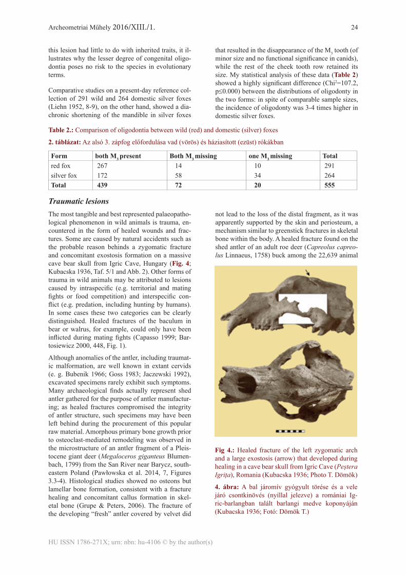
Traumatic lesionsThe most tangible and best represented palaeopatho-logical phenomenon in wild animals is trauma, en-countered in the form of healed wounds and frac-tures. Some are caused by natural accidents such as the probable reason behinds a zygomatic fracture and concomitant exostosis formation on a massive cave bear skull from Igric Cave, Hungary (Fig. 4; Kubacska 1936, Taf. 5/1 and Abb. 2). Other forms of trauma in wild animals may be attributed to lesions caused by intraspecific (e.g. territorial and mating fights or food competition) and interspecific con-flict (e.g. predation, including hunting by humans). In some cases these two categories can be clearly distinguished. Healed fractures of the baculum in bear or walrus, for example, could only have been inflicted during mating fights (Capasso 1999; Bar-tosiewicz 2000, 448, Fig. 1).
Although anomalies of the antler, including traumat-ic malformation, are well known in extant cervids (e. g. Bubenik 1966; Goss 1983; Jaczewski 1992), excavated specimens rarely exhibit such symptoms. Many archaeological finds actually represent shed antler gathered for the purpose of antler manufactur-ing; as healed fractures compromised the integrity of antler structure, such specimens may have been left behind during the procurement of this popular raw material. Amorphous primary bone growth prior to osteoclast-mediated remodeling was observed in the microstructure of an antler fragment of a Pleis-tocene giant deer (Megaloceros giganteus Blumen-bach, 1799) from the San River near Barycz, south-eastern Poland (Pawłowska et al. 2014, 7, Figures 3.3-4). Histological studies showed no osteons but lamellar bone formation, consistent with a fracture healing and concomitant callus formation in skel-etal bone (Grupe & Peters, 2006). The fracture of the developing “fresh” antler covered by velvet did
not lead to the loss of the distal fragment, as it was apparently supported by the skin and periosteum, a mechanism similar to greenstick fractures in skeletal bone within the body. A healed fracture found on the shed antler of an adult roe deer (Capreolus capreo-lus Linnaeus, 1758) buck among the 22,639 animal
Fig 4.: Healed fracture of the left zygomatic arch and a large exostosis (arrow) that developed during healing in a cave bear skull from Igric Cave (Peștera Igrița), Romania (Kubacska 1936; Photo T. Dömök)
4. ábra: A bal járomív gyógyult törése és a vele járó csontkinövés (nyíllal jelezve) a romániai Ig-ric-barlangban talált barlangi medve koponyáján (Kubacska 1936; Fotó: Dömök T.)
Archeometriai Műhely 2016/XIII./1. 25
HU ISSN 1786-271X; urn: nbn: hu-4106 © by the author(s)
bones from the Late Neolithic causewayed enclosure site of Diconche, France, may fall within the same category (Bökönyi & Bartosiewicz 1999).
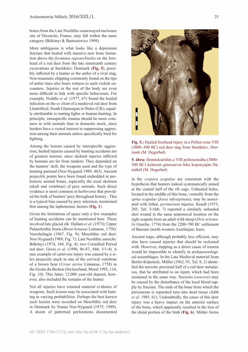
More ambiguous is what looks like a depression fracture that healed with massive new bone forma-tion above the foramina supraorbitalia on the fore-head of a red deer from the late nineteenth century excavations at Snoldelev, Denmark (Fig. 5), possi-bly inflicted by a hunter or the antler of a rival stag. Non-traumatic chipping commonly found on the tips of antler tines also bears witness to such violent en-counters. Injuries in the rest of the body are even more difficult to link with specific behaviours. For example, Noddle et al. (1977, 67) found the healed infection on the os ilium of a medieval red deer from Llantrithyd, South Glamorgan in Wales (UK), equal-ly attributable to mating fights or human hunting. In principle, intraspecific trauma should be more com-mon in wild animals than in domestic stock, since herders have a vested interest in suppressing aggres-sion among their animals unless specifically bred for fighting.
Among the lesions caused by interspecific aggres-sion, healed injuries caused by hunting accidents are of greatest interest, since skeletal injuries inflicted by humans are far from random. They depended on the hunters’ skill, the weapons used and the type of hunting pursued (Noe-Nygaard 1989, 463). Ancient projectile points have been found embedded in pre-historic animal bones, especially the axial skeleton (skull and vertebrae) of prey animals. Such direct evidence is most common in herbivores that provid-ed the bulk of hunters’ prey throughout history . This is a typical bias caused by prey selection, mentioned first among the taphonomic factors (Fig. 1).
Given the limitations of space only a few examples of hunting accidents can be mentioned here. These involved late glacial elk: Hallam et al. (1973); Upper Palaeoloithic bison (Bison bonasus Linnaeus, 1758): Vereshchagin (1967, Fig. 9); Mesolithic red deer: Noe-Nygaard (1989, Fig. 7); Late Neolithic aurochs: Bökönyi (1974, 104, Fig. 4); two Cortaillod Period red deer: Gross et al. (1990, 86-87, Abb. 3/1-4). A rare example of carnivore injury was caused by a si-lex projectile stuck in one of the cervical vertebrae of a brown bear (Ursus arctos Linnaeus, 1758) in the Grotte du Bichon (Switzerland; Morel 1993, 114, Fig. 10). This latter, 12,000 year-old deposit, how-ever, also included the remains of the hunter.
Not all injuries have retained material evidence of weapons. Such lesions may be associated with hunt-ing in varying probabilities. Perhaps the best known such lesions were recorded on Mesolithic red deer in Denmark by Nanna Noe-Nygaard (1975; 1989). A dozen of patterned perforations documented
in the corpora scapulae are consistent with the hypothesis that hunters indeed systematically aimed at the cranial half of the rib cage. Unhealed holes, located in the middle of this bone, ventrally from the spina scapulae (fossa infraspinata), may be associ-ated with lethal, perimortem injuries. Krauß (1975, 205, Taf. 3/Abb. 7) reported a similarly unhealed shot wound in the same anatomical location on the right scapula from an adult wild sheep (Ovis orienta-lis Gmelin, 1774) from the 2200-700 BC settlement of Bassam (north-western Azerbaijan, Iran).
Ancient traps, although probably less efficient, may also have caused injuries that should be reckoned with. However, trapping as a direct cause of trauma would be impossible to identify in archaeozoologi-cal assemblages. In the Late Medieval material from Berlin-Köpenick, Müller (1962, 93, Taf. 8, 2) identi-fied the necrotic proximal half of a red deer metatar-sus, that he attributed to an injury which had been sustained in the same way. Necrosis (osteosis) may be caused by the disturbance of the local blood sup-ply by fracture. The ends of the bone from which the periosteum is separated turn into dead tissue (Jubb et al. 1985, 61). Undoubtedly, the cause of this deer injury was a heavy impact on the anterior surface of the bone, which apparently resulted in the loss of the distal portion of the limb (Fig. 6). Milder forms
Fig. 5.: Healed forehead injury in a Pollen zone VIII (3000–500 BC) red deer stag from Snoldelev, Den-mark (M. Degerbøl)
5. ábra: Homloksérülés a VIII pollenzónába (3000–500 BC) keltezett gímszarvas bika koponyáján Dá-niából (M. Degerbøl)

Archeometriai Műhely 2016/XIII./1. 26
HU ISSN 1786-271X; urn: nbn: hu-4106 © by the author(s)
of trauma were observed on other deer metapodia as well. For example, a metatarsus diaphysis frag-ment from the Roman provincial site of Künzing-Quintana (Bavaria), showed periostitis ossificans covering most of the plantar side (c. 100 mm), in the form of exostoses (Swegat 1976, 83). This le-sion may have equally resulted from chronic tend-ovaginitis or minor trauma. An even less debilitating fracture showed incomplete healing on a second or fifth rudimentary metacarpus (the so-called ‘stilleto’ bone) of a Middle Neolithic adult elk from Alvästra (During 1986, 112, fig. 20). The formation of pseu-do-arthrosis and callus at the proximal end probably did not put this mighty deer at a particular disadvan-tage, except for eventual human predation.
Bone deformations caused by parasitesMacroscopic bone deformations caused by parasites have been relatively often noted in wild animals. A cave bear skull from Doubs (France) had multiple perforations on the frontal sinus interpreted as symp-toms of nematode and trematode parasitosis (Koby 1953). These parasites cause conspicuous lesions in the skulls of modern mustelids as well. An ar-chaeological example on the skull of an European polecat was found in a cistern at the Late Antique site of Vranje, Slovenia (Fig. 7). Superficially, the numerous small holes are reminiscent of buckshot wounds. Their edges, however, show bulbous new bone formation characteristic of nematode activ-ity. They look like the diagnostic exit holes left by Troglotrema acutum Leuckart 1842 (Wetzel & Rick 1962, 78). Another nematode, sinus worm (Skrjabin-gylus nasicola Leuckart, 1842), also attacks mus-telids. However, it tends to affect the post-orbital region making it swollen and thinned, often perfo-rated with holes that may grow into large openings, which are visible in clean skulls (Lewis 1967, 562; King & Moody 1982; King 1989). According to van Soest et al. (1972), 56% of the viscerocrania in over 200 stoat and weasel skulls showed perforation at-tributable to this nematode. Similar perforations are caused by several genera in the families of parasite flies (Tachinidae) and blow-flies (Calliphoridae), whose maggots live in the cranial cavities of mam-mals including the paranasal sinuses in some ursids (Capasso 1998, 114).
Fig. 6.: Three views of the proximal half of a necrotic Late Medieval red deer metatarsus from Berlin-Köpenick (Müller 1962, Taf. 8, 2)
6. ábra: Gímszarvas elhalt végű lábközépcsontjá-nak proximális fele Berlin-Köpenick késő középkori lelőhelyéről (Müller 1962, Taf. 8, 2)
Fig. 7.: Exit holes left by Troglotrema acutum on the skull of a Late Antique European polecat from Vranje, Slovenia (Photo: A. M. Choyke)
7. ábra: Troglotrema acutum kimeneti nyílásai me-zei görény koponyáján a szlovéniai Vranje késő an-tik lelőhelyéről (Fotó: A. M. Choyke)

Archeometriai Műhely 2016/XIII./1. 27
HU ISSN 1786-271X; urn: nbn: hu-4106 © by the author(s)
ConclusionsIn contrast to expectations represented by the classic point of Moodie (1923) quoted in the introduction, a whole range of skeletal elements of wild animals found in archaeological deposits show pathological lesions. Natural selection does not affect all spe-cies (and all body parts) equally, although it tends to stamp out extreme inherited anomalies. Healed traumatic disorders are most frequently encountered. The larger the species, and the fewer its natural pred-ators, the better the chances of recovery.
Although handicapped carnivores will sooner or later suffer disadvantage in their ‘struggle for life’, they are still less directly at risk than disabled prey species. Among wild herbivores, large-bodied spe-cies, such as aurochs or elk, again less threatened by predators, may survive long enough to develop pathological symptoms in their skeletons. In addi-tion, killing these animals took advanced hunting skills and a degree of luck. Several finds prove that in some instances the animals were able to flee and recover from the minor injuries before they fell vic-tim to another hunting incident.
Aside from taxonomic differences, the anatomical location of lesions also determines the chances of survival. Sub-pathological anomalies, such as oli-godontia, are to some extent normal in wild animal
populations. Many pathological phenomena on the postcranial skeleton were also observed in regions less critical from the viewpoint of survival. Some unhealed scapula injuries in red deer and wild sheep, on the other hand, are seen as perimortal trauma re-lated to lethal chest wounds.
The relatively great number of lesions found on the metapodia of large wild ungulates raises the question as to whether the under-representation of other bones in palaeopathological assemblages could be related to the fact that many parts of the skeleton were left behind following off-site butchering. Catching large game would also have required cooperative efforts. It is reasonable to assume that old and feeble ani-mals were easier to hunt. On the other hand, those who survived previous attacks by hunters may have learnt to behave increasingly cautiously.
The weakness of diseased animals is more likely to have been taken advantage of by subsistence hunters in early farming societies, who still heavily relied upon the exploitation of wild animals for food. De-cisions regarding the conscious culling of disabled game must have depended upon the skills and de-mands of hunters and may have been made on an opportunistic basis.
ReferencesAUFDERHEIDE, A.C. & RODRIGUEZ-MARTIN, C. (1998): The Cambridge encyclopedia of human paleopathology. Cambridge University Press, Cam-bridge, 478 pp.
BAKER, J.R. & BROTHWELL, D. (1980): Animal diseases in archaeology. Academic Press, London, 235 pp.
BARTOSIEWICZ, L. (1987): Bone morphometry and function: a comparison between cattle and Euro-pean elk. Acta Veterinaria Hungarica 35/4: 437–48.
BARTOSIEWICZ, L. (2000): Baculum fracture in carnivores: osteological, behavioural and cultural implications. International Journal of Osteoarchae-ology 10: 447–50.
BARTOSIEWICZ, L. (2002): Pathological lesions on prehistoric animal remains from Southwest Asia. In: H. BUITENHUIS, M. MASHKOUR, A.M. CHOYKE & A.H. AL-SHIYAB (eds): .Archaeozo-ology of the Near East V. ARC Publicaties 62, Gro-ningen, 320–336.
BARTOSIEWICZ, L. (2008): Taphonomy and pa-laeopathology in archaeozoology. GeoBios 41/1: 69–77.
BARTOSIEWICZ, L. (2013): Shuffling nags, Lame ducks. The archaeology of animal disease. Oxbow Books, Oxford, 302 pp.
BARTOSIEWICZ, L., DEMEURE, R., MOTTET, I., VAN NEER, W. & LENTACKER, A. (1997): Magnetic resonance imaging in the study of spavin in recent and subfossil cattle. Anthropozoologica 25–26: 57–60.
BARTOSIEWICZ, L. & BARTOSIEWICZ, G. (2002): ‘Bamboo spine’ in a Migration Period horse from Hungary. Journal of Archaeological Science 29(8): 819–30.
BERG, R. T. & BUTTERFIELD, R.M. (1976): New concepts of cattle growth. Sydney University Press, Sydney, 240 pp.
BLUMENFELD, H. (1909): Über den Spat der Rinder. Unpublished dissertation, Leipzig, 147 pp.
BÖKÖNYI, S. (1974): History of domestic mam-mals in central and eastern Europe. Akadémiai Ki-adó, Budapest, 597 pp.
BÖKÖNYI, S. & BARTOSIEWICZ, L. (1999): IX. Analyse de la faune Diconche. In: C. BURNEZ &
Archeometriai Műhely 2016/XIII./1. 28
HU ISSN 1786-271X; urn: nbn: hu-4106 © by the author(s)
P. FOUERE (eds): Les enceintes néolithiques de Diconche à Saintes Charente-Maritime. Société Préhistorique Française, Mémoire XXV, Volume 1, Chauvigny–Paris: 147–166.
BÖKÖNYI, S. & BARTOSIEWICZ, L. (2000): A review of animal remains from Shahr-i Sokhta Eas-tern Iran. In: M. MASHKOUR, A.M. CHOYKE & H. BUITENHUIS (eds): Archaeozoology of the Near East IVB. ARC Publicaties 32, Groningen, 116–52.
BUBENIK, A.B. (1966): Das Geweih. Entwicklung, Aufbau und Ausformung der Geweihe und Gehörne und ihre Bedeutung für das Wild und für die Jagd. Verlag Paul Parey, Hamburg und Berlin, 214 pp.
CAPASSO, L. (1998): Cranial pathology of Ursus spelaeus Rosenmüller and Heinroth from Chateau Pignon, Basque Territories Spain. International Journal of Osteoarchaeology 8: 107–15.
CAPASSO, L. (1999): A healed fracture in an Odo-benus rosmarus baculum from the Holocene of Saint Lawrence Island, Alaska. International Journal of Osteoarchaeology 9(4): 260–62.
CHAPLIN, R.E. (1971): The study of animal bones from Archaeological sites. Seminar Press, London and New York, 170 pp.
CLUTTON-BROCK, J. (1987): A natural history of domesticated mammals. Cambridge University Press, Cambridge, 243 pp.
DURING, E. (1986): The fauna of Alvastra: an os-teological analysis of animal bones from a Neolithic pile dwelling. Lund: OSSA 12 (Suppl. 1).
ESPER, J.F. (1774): Ausführliche Nachricht von nev entdeckten Zoolithen unbekannter vierfüsiger Thiere, und denen sie enthaltenden, so wie verschie-denen anderen denkwürdigen Grüften der Oberge-bürgischen Lande des Marggrafthums Bayreuth. Georg Wolfgang Knorr, Nürnberg, 422 pp.
FOX, H. (1939): Chronic arthritis in wild animals. Transactions of the American Philosophical Society, New Series 31: 73–149.
GÁL, E. (2013): Pathological changes in bird bones. In: Shuffling nags, Lame ducks. The archaeology of animal disease. Oxbow Books, Oxford, 217–238.
GOSS, R.J. (1983): Deer Antlers: Regeneration, Function and Evolution. Academic Press, London.
GRAF, G. (1967): Tierknochenfunde von der Heune-burg einem frühkeltischen Herrensitz an der Donau. Nichtpaarhufer. Dissertation Universität München, Naturwiss. Unters. Vor- u. Frühgesch. Württemberg u. Hohenzollern 6, Stuttgart, 49 pp.
GROSS, E., JACOMET, S. & SCHIBLER, J. (1990): Stand und Ziele der Wirtschaftsarchäolo-
gischen Forschung an Neolithischen Ufer- und In-selsiedlungen im unteren Zürichseeraum Kt. Zürich, Schweiz.. In: J. SCHIBLER, J. SEDLMEIER & H.-P. SPYCHER (eds): Festschrift für Hans R. Stampfli. Beiträge zur Archäozoologie, Archäologie, Anthro-pologie, Geologie und Paläontologie. Helbing & Lichtenhahn, Basel, 76–100.
GRUNDBACHER, B., LÜPS, P. & NUSSBAU-MER, M.A. (1990): Osteometrische Untersuchun-gen an neolitischen Dachsen Meles meles. aus Twann Kanton Bern, Schweiz.. In: J. SCHIBLER, J. SEDLMEIER & H.-P. SPYCHER (eds): Festschrift für Hans R. Stampfli. Beiträge zur Archäozoologie, Archäologie, Anthropologie, Geologie und Paläon-tologie. Helbing & Lichtenhahn, Basel, 101–13.
GRUPE, G. & PETERS, J. (2006). Histomorpholog-ical perspectives of human and animal bone, and soft tissue. In: G. GRUPE & J. PETERS (eds.): Micro-scopicExaminations of Bioarchaeological Remains. Keeping a Close Eye on Ancient Tissues. Verlag Ma-rie Leidorf GmbH, Leidorf, 15–103.
HALLAM, J.S., EDWARDS, B.J.N., BARNES, B. & STUART, A.J. (1973): A late glacial elk with associated barbed points from High Furlong, Lan-cashire. Proceedings of the Prehistoric Society 39: 100–128.
HARRIS, S. (1977): Spinal arthritis spondylosis deformans in the red fox, Vulpes vulpes, with some methodology of relevance to zooarchaeology. Jour-nal of Archaeological Science 4: 183–195.
JACZEWSKI, Z. (1992): Poroże jeleniowatych [Deer Antlers]. Państwowe Wydawnictwo Rolnicze i Leśne, Warszawa, 342 pp.
JUBB, K.V.F., KENNEDY, P.C. & PALMER, N. (1985): Pathology of domestic animals. Volume 1 [third edition]. Academic Press, San Diego, 582 pp.
KAY, C.E. (1994): Aboriginal overkill: the role of Native Americans in structuring western ecosys-tems. Human Nature 5: 359–398.
KING, C.M. & MOODY, J.E. (1982): The biology of the stoat Mustela erminea. in the National Parks of New Zealand. New Zealand Journal of Zoology 9: 49–144.
KING, C. M. (1989): The Natural History of Wea-sels and Stoats. London, Christopher Helm.
KOBY, F.E. (1953): Lesions pathologiques aux si-nus frontaux d’un ours des cavernes. Eclogae Geo-logicae Helvetiae 46: 295–97.
KÖNIG, E. (1993): Tierknochenfunde aus ei-ner Feuchtbodensiedlung der Chamer Gruppe in Griesstetten, Ldkr. Neumarkt. Institut für Paläoana-tomie, Domestikationsforschung und Geschichte der

Archeometriai Műhely 2016/XIII./1. 29
HU ISSN 1786-271X; urn: nbn: hu-4106 © by the author(s)
Tiermedizin der Universität München, unpublished dissertation, München, 119 pp.
KRAUß, R. (1975): Tierknochenfunde aus Bastam in Nordwest-Azerbaidjan/Iran. Institut für Paläoana-tomie, Domestikationsforschung und Geschichte der Tiermedizin der Universität München, unpublished dissertation, München, 207 pp.
KREUTZER, L.A. (1992): Bison and deer bone mineral densities: comparisons and implications for the interpretation of archaeological faunas. Journal of Archaeological Science 19(3): 271–94.
KUBACSKA, A. (1936): Pathologische Untersu-chungen an ungarl ändischen Versteinerungen. VI. Verletzungen an Schädeln pläistozäner Raubtiere. Palaeontologische Zeitschrift 18/1: 95-108.
LEGGE, A.J. & ROWLEY-CONWY, P.A. (1988): Starr Carr revisited. Centre for Extra-Mural Studies, University of London, London, 112 pp.
LEWIS, J.W. (1967): Observations on the skull of Mustelidae infected with the nematode, Skrjabingy-lus nasicola. Journal of Zoology 153: 561–64.
LIEHN, H-D. (1952): Die Gebissreduktion beim Sil-berfuchs. München, Institut für Tierzucht der Uni-versität, unpublished dissertation, München, 97 pp.
MANN, R. & MURPHY, S.P. (1990): Regional atlas of bone disease. C. C. Thomas, Springfield, Il., 297 pp.
MARSIGLI, LUIGI FERNANDO (1726): Danubi-us Pannonico-Mysicus, observationibus geographi-cis, astronomicis, hydrographicis, historicis,physicis perlustratus. Vol. VI. Amsterdam–The Hague.
MARÍN-ARROYO, A. B., CANO, F. G. & MARK LEWIS, M. (2015): Late Pleistocene foot infection in Dama mesopotamica from Tabun B (Mount Car-mel, Israel) International Journal of Paleopathology 8: 48–50.
MAYER, C. (1854): Ueber krankhafte Knochen vor-weltlicher Thiere. Nova Acta Leopoldiana naturae curiosa 24, Breslau–Bonn, 673 pp.
MISSEL, M.H. (1987): Tierknochenfunde aus einer germanischen Siedlung in Hildesheim-Bavenstedt. Institut für Paläoanatomie, Domestikationsfors-chung und Geschichte der Tiermedizin der Univer-sität München, unpublished dissertation, München, 93 pp.
MOODIE, R.L. (1923): The antiquity of disease. The University of Chicago Press, Chicago
MOREL, P. (1993): Une chasse à l’ours brun il y a 12’000 ans: nouvelle découverte à la Grotte du Bi-chon La Chaux-de-Fonds. Archäologie der Schweiz 1993(3): 110–17.
MÜLLER, H.-H. (1962): Die Säugetierreste aus der Burg Berlin-Köpenick nach den Grabungen von 1955 bis 1958. In: J. HERRMANN (ed.): Ergebnisse der archäologischen Stadtkernforschung in Berlin. Teil I. Köpenick. Ein Beitrag zur Frühgeschichte gross-Berlins. Akademie-Verlag, Berlin, 81–97.
NODDLE, B.A., BRAMWELL, D. & JONES, A. (1977): Llantrithyd, a ringwork in South Glamor-gan: the animal bones. Cardiff Archaeological So-ciety 12: 63–73.
NOE-NYGAARD, N. (1975): Bone injuries caused by human weapons in Mesolithic Denmark. In: A. T. CLASON (ed.): Archaeozoological studies. North Holland and American Elsevier, Amsterdam and New York, 151–59.
NOE-NYGAARD, N. (1989): Man-made trace fos-sils on bones. Human Evolution 4(6): 461–91.
PAWŁOWSKA, K., STEFANIAK, K. & NOWA-KOWSKI, D. (2014): Healed antler fracture from a giant deer (Megaloceros giganteus) from the Pleis-tocene in Poland. 17 (1-23A), 1–9. http://palaeo-electronica.org/content/2014/732-antler-fracture-in-giant-deer
PERKINS, D. & DALY, P. (1968): A hunters’ vil-lage in Neolithic Turkey. Scientific American 219/5: 97–106.
PETERSON, R.O., SCHEIDLE, J.M. & STE-PHENS, P.W. (1982): Selected skeletal morphology and pathology of moose from the Kenai Peninsula, Alaska and Isle Royale, Michigan. Canadian Jour-nal of Zoology 60: 2812–17.
PRUMMEL, W. (1987): The faunal remains from the Neolithic site of Hekelingen III. Helinium XX-VII: 190–258.
RAUH, H. (1981): Knochenfunde von Säugetieren aus dem Demircihüyük Nordwestanatolien. Insti-tut für Paläoanatomie, Domestikationsforschung und Geschichte der Tiermedizin der Universität München, unpublished dissertation, München, 108 pp.
REICHSTEIN, H. (1991): Die Fauna des germa-nischen Dorfes Feddersen Wierde (Feddersen Wi-erde, 4). Franz Steiner Verlag, Stuttgart, 346 pp.
SIEGEL, J. (1976): Animal palaeopathology: pos-sibilities and problems. Journal of Archaeological Science 3: 349–84.
SOEST, R.W.M. VAN, J. VAN DER LAND & P.J.H. VAN BREE (1972): Skrjabingylus nasicola in skulls of Mustela erminea and Mustela nivalis from the Netherlands. Beaufortia 20: 85–97.

Archeometriai Műhely 2016/XIII./1. 30
HU ISSN 1786-271X; urn: nbn: hu-4106 © by the author(s)
STILLFRIED, M. (1926): A szarvasmarhák idült tarsitise [Chronic tarsitis in cattle]. Közlemények az Összehasonlító Élet- és Kórtan Köréből 19: 147–154.
SWEGAT, W. (1976): Die Knochenfunde aus dem römischen Kastell Künzing-Quintana. Institut für Paläoanatomie, Domestikationsforschung und Ge-schichte der Tiermedizin der Universität München, unpublished dissertation, München, 135 pp.
TAMÁS, L. (ed.) (1987): Állatorvosi sebészet [Vet-erinary surgery] 2. Mezőgazdasági Kiadó, Budapest, 564 pp.
TASNÁDI KUBACSKA, A. (1960): Az ősállatok pathologiája [The pathology of prehistoric animals]. Medicina Könyvkiadó, Budapest, 230 pp.
TEICHERT, L. (1979): Tierknochenfunde aus dem altslawischen Burgwall bei Wiesenau, Kr. Eisenhüt- tenstadt. Veröffentlichungen des Museums für Ur- u. Frühgeschichte Potsdam 12: 167–218.
WALDRON, T. (2015): Roy Lee Moodie (1880–1934) and the beginnings of palaeopathology. Jour-nal of Medical Biography 23(1): 8–13.
WAMBERG, K. & MCPHEARSON, E.A. (1968): Veterinary encyclopedia Volume 4. Medical Book Company, Copenhagen, 2509 pp.
WELLS, C. (1972): Ancient arthritis. May and Bak-er Pharmaceutical Bulletin 21: 67–70.
WETZEL, R. & RICK, W. (1962): Krankheiten des Wildes. Verlag Paul Parey, Hamburg–Berlin, 223 pp.