Embed Size (px)
Citation preview
ORIGINAL ARTICLE
The prevalence of morphological changes in the thoracolumbarspine on whole-spine computed tomographic images
Aya Nakajima & Akihito Usui & Yoshiyuki Hosokai &Yusuke Kawasumi & Kenta Abiko & Masato Funayama &
Haruo Saito
Received: 16 May 2013 /Revised: 21 August 2013 /Accepted: 26 August 2013 /Published online: 20 September 2013# The Author(s) 2013. This article is published with open access at Springerlink.com
AbstractObjectives This article reviews the prevalence of lumbarisation,sacralisation and lumbar ribs, and their morphological relevanceby evaluating multi-slice computed tomography (MSCT) im-ages. These segment variations can cause miscounting ofvertebrae at the lumbar spinal level.Methods A retrospective radiographic analysis of 226 casesscanned by MSCT prior to forensic autopsy was undertaken.MSCT scans of the entire spine were obtained. Radiologicaldata were evaluated on a three-dimensional image worksta-tion. Vertebral levels were determined by counting downwardfrom the first cervical vertebra, based on the assumption ofseven cervical, 12 thoracic and five lumbar vertebrae. Theprevalence of lumbarisation, sacralisation and lumbar ribs wasassessed.Results Lumbar ribs were observed in 13 of the 226 cases(5.8 %). Lumbarisation and sacralisation were observed in 14cases (6.2 %) and six cases (2.7 %), respectively. Lumbar ribswere present in 11 of the 14 cases with lumbarisation, and intwo of the 206 cases with normal lumbar vertebral configuration.Lumbarisation had a statistically significant association withlumbar ribs (p<0.01).
Conclusions There was a strong association between lumbarribs and lumbarisation, with a resulting miscount rate for thelumbar spine of slightly less than 10 %.Teaching points• Lumbarisation and sacralisation are observed 6.2 % and2.7 %, respectively.
• Thoracolumbar segment variations can cause a miscountrate for the lumbar spine of less than 10 %.
• Lumbar rib is significantly associated to lumbarisation.
Keywords Lumbosacral transitional vertebrae .
Lumbarisation . Sacralisation . Lumbar rib . Multi-slicecomputed tomography
Introduction
Lumbosacral transitional vertebrae (LSTV) are congenitalspinal anomalies. LSTV associated with the fifth lumbar ver-tebra may show assimilation to the sacrum (sacralisation), andthose affecting the first sacral vertebra may show transition toa lumbar configuration (lumbarisation) [1], causing misinter-pretation as four or six lumbar vertebrae, respectively. LSTVare common in the general population, with a reported prev-alence of 4–35.9 % [1–17]. Inaccurate identification of anLSTV may lead to surgical and procedural errors and poorcorrelation with clinical symptoms [18].
Identification of LSTVand accurate numeric identificationof vertebral segments on conventional lumbar radiographs ormagnetic resonance imaging (MRI) are essential to ensure thatinterventional procedures or surgery are performed at theintended level [19]. However, establishing whether anLSTV is a lumbarised S1 or a sacralised L5 on MRI alone canbe problematic. Conventional spine radiographs are often
A. Nakajima :A. Usui (*) :Y. Hosokai :K. Abiko :H. SaitoDepartment of Diagnostic Image Analysis, Tohoku UniversityGraduate School of Medicine, 2-1 Seiryo-machi, Sendai 980-8575,Japane-mail: [email protected]
Y. KawasumiDepartment of Clinical Imaging, Tohoku University Graduate Schoolof Medicine, 2-1 Seiryo-machi, Sendai 980-8575, Japan
M. FunayamaDepartment of Forensic Medicine, Tohoku University GraduateSchool of Medicine, 2-1 Seiryo-machi, Sendai 980-8575, Japan
Insights Imaging (2014) 5:77–83DOI 10.1007/s13244-013-0286-0
unavailable at the time of imaging, and cervicothoraciclocalisers may not be routinely obtained. Konin et al. [18]reported that radiographs of the entire spine allowed theradiologist not only to count from C2 inferiorly but alsoto differentiate hypoplastic ribs from lumbar transverseprocesses. This approach allows accurate counting ofthoracic segments, correct identification of the L1 verte-bral body and correct numeric assignment of the LSTV.They also reported that patients undergoing lumbar spinalMRI commonly had radiographs of the lumbar spinealone, rather than of the entire spine. Correct enumerationwas often achieved in these cases, but there remained casesin which the presence of thoracolumbar transitions as well assegmentation anomalies complicated evaluation [18].
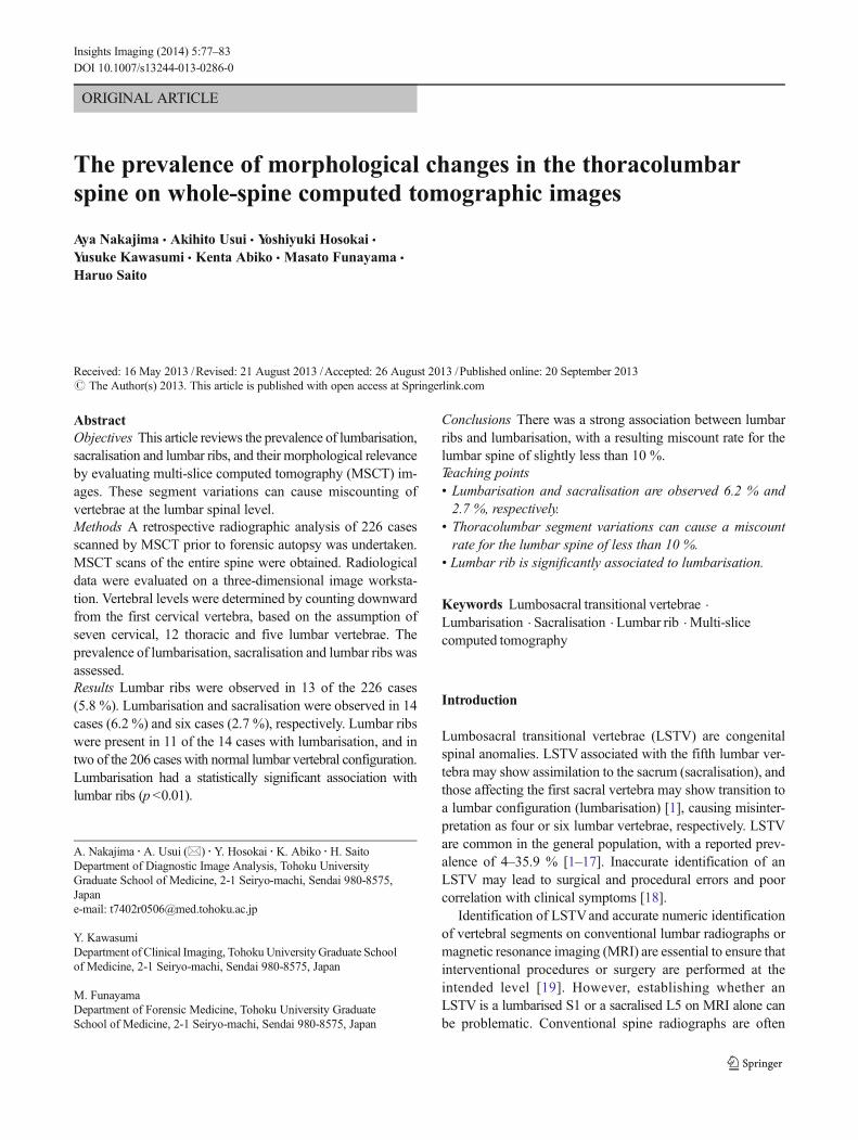
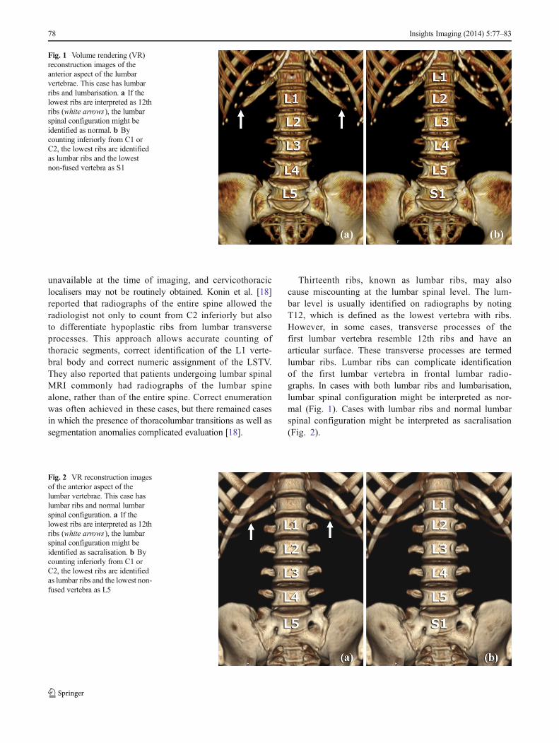
Thirteenth ribs, known as lumbar ribs, may alsocause miscounting at the lumbar spinal level. The lum-bar level is usually identified on radiographs by notingT12, which is defined as the lowest vertebra with ribs.However, in some cases, transverse processes of thefirst lumbar vertebra resemble 12th ribs and have anarticular surface. These transverse processes are termedlumbar ribs. Lumbar ribs can complicate identificationof the first lumbar vertebra in frontal lumbar radio-graphs. In cases with both lumbar ribs and lumbarisation,lumbar spinal configuration might be interpreted as nor-mal (Fig. 1). Cases with lumbar ribs and normal lumbarspinal configuration might be interpreted as sacralisation(Fig. 2).
Fig. 1 Volume rendering (VR)reconstruction images of theanterior aspect of the lumbarvertebrae. This case has lumbarribs and lumbarisation. a If thelowest ribs are interpreted as 12thribs (white arrows), the lumbarspinal configuration might beidentified as normal. b Bycounting inferiorly from C1 orC2, the lowest ribs are identifiedas lumbar ribs and the lowestnon-fused vertebra as S1
Fig. 2 VR reconstruction imagesof the anterior aspect of thelumbar vertebrae. This case haslumbar ribs and normal lumbarspinal configuration. a If thelowest ribs are interpreted as 12thribs (white arrows), the lumbarspinal configuration might beidentified as sacralisation. b Bycounting inferiorly from C1 orC2, the lowest ribs are identifiedas lumbar ribs and the lowest non-fused vertebra as L5
78 Insights Imaging (2014) 5:77–83
In the present study, we investigated the prevalence of LSTV(lumbarisation and sacralisation) and lumbar ribs and their mor-phological relevance, by evaluating computed tomography (CT)images. We highlight the importance of accurate enumeration ofLSTVand of communicationwith the referring clinician to avoidsevere complications such as wrong-level spine surgery.
Materials and methods
Materials
We performed a retrospective radiographic analysis of 226cases (148 men, 78 women; age range, 17–94 years; mean
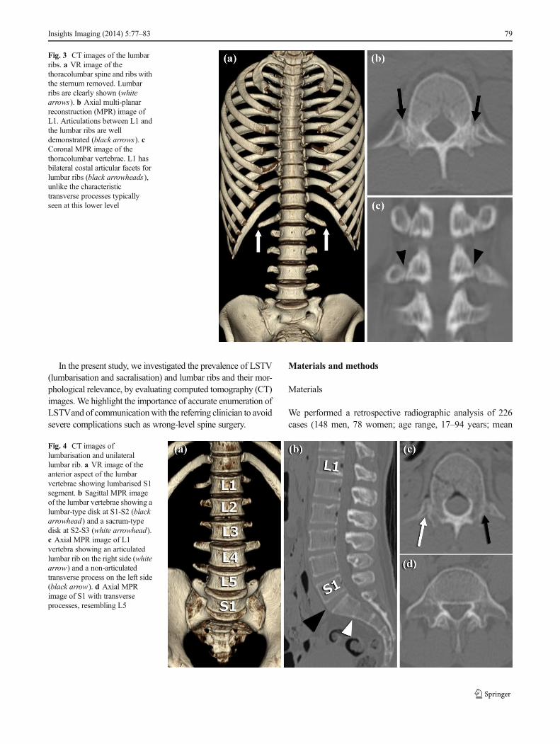
Fig. 3 CT images of the lumbarribs. a VR image of thethoracolumbar spine and ribs withthe sternum removed. Lumbarribs are clearly shown (whitearrows). b Axial multi-planarreconstruction (MPR) image ofL1. Articulations between L1 andthe lumbar ribs are welldemonstrated (black arrows). cCoronal MPR image of thethoracolumbar vertebrae. L1 hasbilateral costal articular facets forlumbar ribs (black arrowheads),unlike the characteristictransverse processes typicallyseen at this lower level
Fig. 4 CT images oflumbarisation and unilaterallumbar rib. a VR image of theanterior aspect of the lumbarvertebrae showing lumbarised S1segment. b Sagittal MPR imageof the lumbar vertebrae showing alumbar-type disk at S1-S2 (blackarrowhead) and a sacrum-typedisk at S2-S3 (white arrowhead).c Axial MPR image of L1vertebra showing an articulatedlumbar rib on the right side (whitearrow) and a non-articulatedtransverse process on the left side(black arrow). d Axial MPRimage of S1 with transverseprocesses, resembling L5
Insights Imaging (2014) 5:77–83 79
age, 59.2 years) scanned from October 2009 to June 2011 bymulti-slice computed tomography (MSCT) before forensicautopsy. Cases under 10 years of age, and cases with bodiesseverely damaged by fire, traffic accident or putrefaction wereexcluded from the study.
CT machines
MSCT scanning was performed on an eight-channel scanner(Aquilion; Toshiba Medical Systems, Tokyo, Japan).Volumetric helical scans were obtained from the head tothe proximal femur at 120 kV with variable mAs, a beampitch of 0.875 and 2.0-mm collimation. The volumetricdata allow observation of the whole spine in all directions
and differentiation of the presence of rib articular facets.Evaluation of the radiological data was based on a com-bination of axial images, multi-planar reconstructions(MPRs), and three-dimensional (3D) reconstruction usinga 3D image workstation (ziostation2 ver. 2.1.0.3; Ziosoft,Tokyo, Japan).
Vertebrae numbering
The vertebral levels were counted craniocaudally, startingfrom C1, based on the assumption of seven cervical, 12thoracic, and five lumbar vertebrae. The 20th and 25th verte-brae were defined as L1 and S1, respectively.
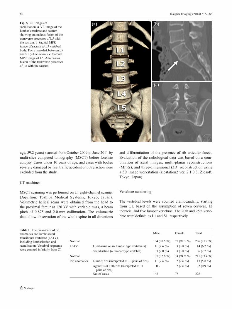
Fig. 5 CT images ofsacralisation. a VR image of thelumbar vertebrae and sacrumshowing anomalous fusion of thetransverse processes of L5 withthe sacrum. b Sagittal MPRimage of sacralised L5 vertebralbody. There is no disk between L5and S1 (white arrow). c CoronalMPR image of L5. Anomalousfusion of the transverse processesof L5 with the sacrum
Table 1 The prevalence of ribanomalies and lumbosacraltransitional vertebrae (LSTV),including lumbarisation andsacralisation. Vertebral segmentswere counted inferiorly from C1
Male Female Total
Normal 134 (90.5 %) 72 (92.3 %) 206 (91.2 %)
LSTV Lumbarisation (6 lumbar type vertebrara) 11 (7.4 %) 3 (3.8 %) 14 (6.2 %)
Sacralisation (4 lumbar type vertebra) 3 (2.0 %) 3 (3.8 %) 6 (2.7 %)
Normal 137 (92.6 %) 74 (94.9 %) 211 (93.4 %)
Rib anomalies Lumber ribs (interpreted as 13 pairs of ribs) 11 (7.4 %) 2 (2.6 %) 13 (5.8 %)
Agenesis of 12th ribs (interpreted as 11pairs of ribs)
0 - 2 (2.6 %) 2 (0.9 %)
No. of cases 148 78 226
80 Insights Imaging (2014) 5:77–83
Lumbar ribs were defined as ribs articulating with L1(Fig. 3). Lumbarisation was defined as non-fusion of S1 andS2 (26th vertebra), meaning that there was one additionalarticulated vertebra (Fig. 4). Sacralisation was defined asanomalous fusion of L5 (24th vertebra) and S1 (Fig. 5).
Statistical analysis
Fisher’s exact test was used to compare categorical variables.Differences were assessed with an alpha level of 0.05.
Results
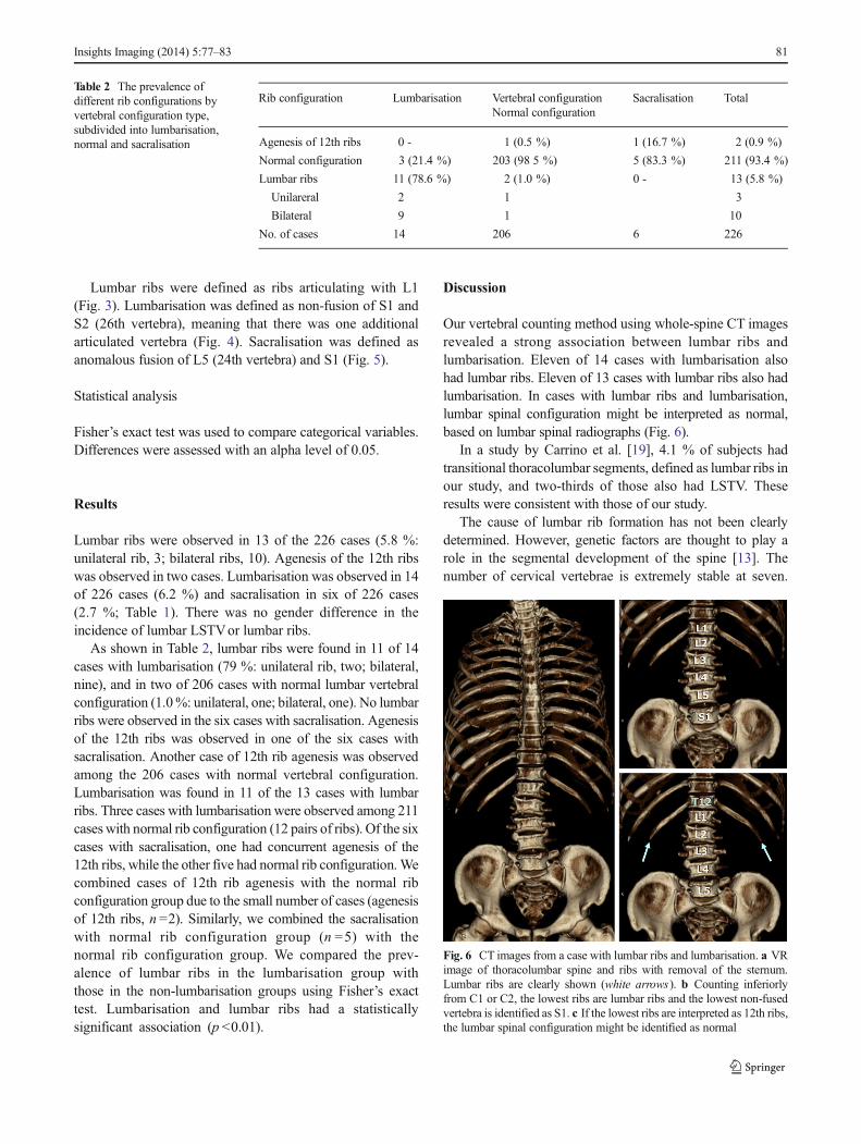
Lumbar ribs were observed in 13 of the 226 cases (5.8 %:unilateral rib, 3; bilateral ribs, 10). Agenesis of the 12th ribswas observed in two cases. Lumbarisation was observed in 14of 226 cases (6.2 %) and sacralisation in six of 226 cases(2.7 %; Table 1). There was no gender difference in theincidence of lumbar LSTVor lumbar ribs.
As shown in Table 2, lumbar ribs were found in 11 of 14cases with lumbarisation (79 %: unilateral rib, two; bilateral,nine), and in two of 206 cases with normal lumbar vertebralconfiguration (1.0%: unilateral, one; bilateral, one). No lumbarribs were observed in the six cases with sacralisation. Agenesisof the 12th ribs was observed in one of the six cases withsacralisation. Another case of 12th rib agenesis was observedamong the 206 cases with normal vertebral configuration.Lumbarisation was found in 11 of the 13 cases with lumbarribs. Three cases with lumbarisation were observed among 211caseswith normal rib configuration (12 pairs of ribs). Of the sixcases with sacralisation, one had concurrent agenesis of the12th ribs, while the other five had normal rib configuration.Wecombined cases of 12th rib agenesis with the normal ribconfiguration group due to the small number of cases (agenesisof 12th ribs, n =2). Similarly, we combined the sacralisationwith normal rib configuration group (n =5) with thenormal rib configuration group. We compared the prev-alence of lumbar ribs in the lumbarisation group withthose in the non-lumbarisation groups using Fisher’s exacttest. Lumbarisation and lumbar ribs had a statisticallysignificant association (p <0.01).
Discussion
Our vertebral counting method using whole-spine CT imagesrevealed a strong association between lumbar ribs andlumbarisation. Eleven of 14 cases with lumbarisation alsohad lumbar ribs. Eleven of 13 cases with lumbar ribs also hadlumbarisation. In cases with lumbar ribs and lumbarisation,lumbar spinal configuration might be interpreted as normal,based on lumbar spinal radiographs (Fig. 6).
In a study by Carrino et al. [19], 4.1 % of subjects hadtransitional thoracolumbar segments, defined as lumbar ribs inour study, and two-thirds of those also had LSTV. Theseresults were consistent with those of our study.
The cause of lumbar rib formation has not been clearlydetermined. However, genetic factors are thought to play arole in the segmental development of the spine [13]. Thenumber of cervical vertebrae is extremely stable at seven.
Table 2 The prevalence ofdifferent rib configurations byvertebral configuration type,subdivided into lumbarisation,normal and sacralisation
Rib configuration Lumbarisation Vertebral configurationNormal configuration
Sacralisation Total
Agenesis of 12th ribs 0 - 1 (0.5 %) 1 (16.7 %) 2 (0.9 %)
Normal configuration 3 (21.4 %) 203 (98 5 %) 5 (83.3 %) 211 (93.4 %)
Lumbar ribs 11 (78.6 %) 2 (1.0 %) 0 - 13 (5.8 %)
Unilareral 2 1 3
Bilateral 9 1 10
No. of cases 14 206 6 226
Fig. 6 CT images from a case with lumbar ribs and lumbarisation. a VRimage of thoracolumbar spine and ribs with removal of the sternum.Lumbar ribs are clearly shown (white arrows). b Counting inferiorlyfrom C1 or C2, the lowest ribs are lumbar ribs and the lowest non-fusedvertebra is identified as S1. c If the lowest ribs are interpreted as 12th ribs,the lumbar spinal configuration might be identified as normal
Insights Imaging (2014) 5:77–83 81
The number of thoracic vertebrae may be reduced to 11 orincreased to 13, and the number of lumbar vertebrae mayrange from four to six [20]. Variations in the thoracolumbarsegment have the potential to promote morphological shifts inthe lumbosacral segment, because the thoracic spine, lumbarspine and sacral spine develop craniocaudally in early fetal life[21]. If ribs form on L1 (20th vertebrae), then S1 (25thvertebra) might separate from S2, resulting in lumbarisation.
The incidence of lumbarisation was 6.2 % and ofsacralisation was 2.7 % in the present study. Widely variableincidences of LSTV have been reported in the literature, rang-ing from 4% to 35.9% [1–17]. This variationmay be explainedby differences in diagnostic criteria, imaging techniques andconfounding factors among the investigated populations.
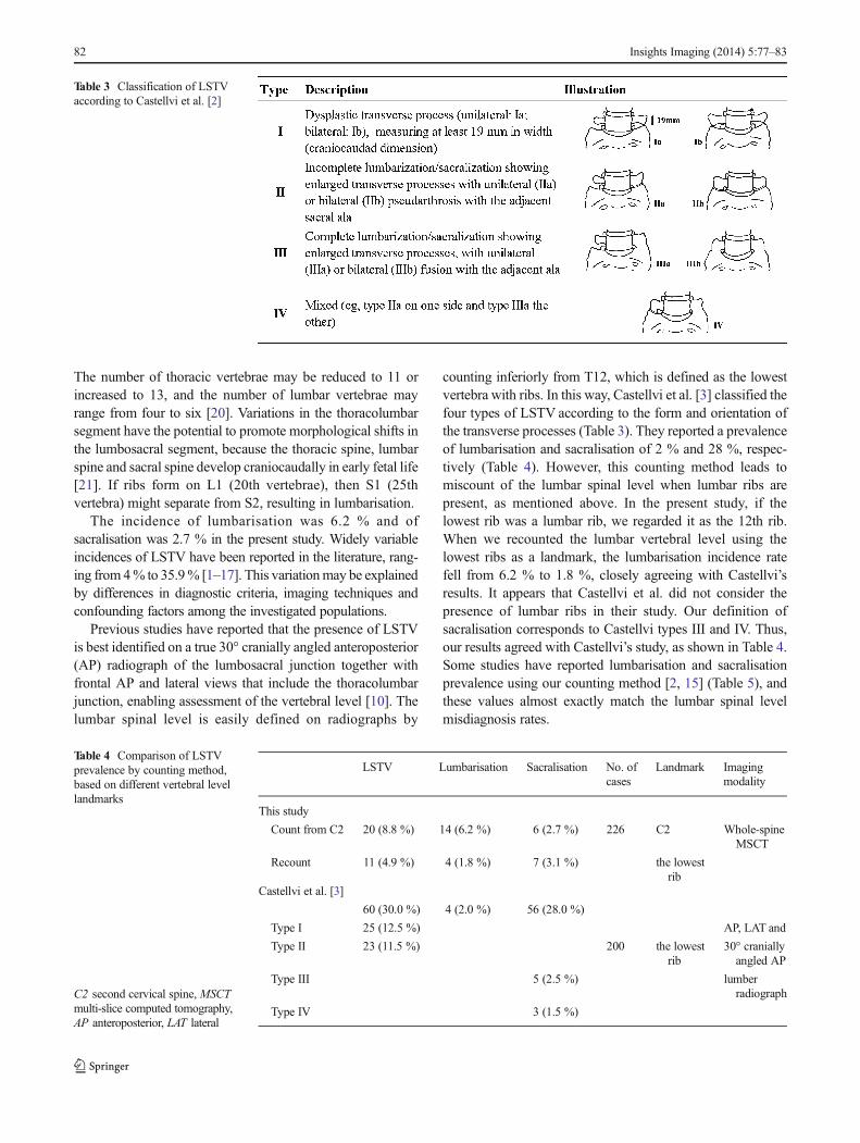
Previous studies have reported that the presence of LSTVis best identified on a true 30° cranially angled anteroposterior(AP) radiograph of the lumbosacral junction together withfrontal AP and lateral views that include the thoracolumbarjunction, enabling assessment of the vertebral level [10]. Thelumbar spinal level is easily defined on radiographs by
counting inferiorly from T12, which is defined as the lowestvertebra with ribs. In this way, Castellvi et al. [3] classified thefour types of LSTV according to the form and orientation ofthe transverse processes (Table 3). They reported a prevalenceof lumbarisation and sacralisation of 2 % and 28 %, respec-tively (Table 4). However, this counting method leads tomiscount of the lumbar spinal level when lumbar ribs arepresent, as mentioned above. In the present study, if thelowest rib was a lumbar rib, we regarded it as the 12th rib.When we recounted the lumbar vertebral level using thelowest ribs as a landmark, the lumbarisation incidence ratefell from 6.2 % to 1.8 %, closely agreeing with Castellvi’sresults. It appears that Castellvi et al. did not consider thepresence of lumbar ribs in their study. Our definition ofsacralisation corresponds to Castellvi types III and IV. Thus,our results agreed with Castellvi’s study, as shown in Table 4.Some studies have reported lumbarisation and sacralisationprevalence using our counting method [2, 15] (Table 5), andthese values almost exactly match the lumbar spinal levelmisdiagnosis rates.
Table 3 Classification of LSTVaccording to Castellvi et al. [2]
Table 4 Comparison of LSTVprevalence by counting method,based on different vertebral levellandmarks
C2 second cervical spine, MSCTmulti-slice computed tomography,AP anteroposterior, LAT lateral
LSTV Lumbarisation Sacralisation No. ofcases
Landmark Imagingmodality
This study
Count from C2 20 (8.8 %) 14 (6.2 %) 6 (2.7 %) 226 C2 Whole-spineMSCT
Recount 11 (4.9 %) 4 (1.8 %) 7 (3.1 %) the lowestrib
Castellvi et al. [3]
60 (30.0 %) 4 (2.0 %) 56 (28.0 %)
Type I 25 (12.5 %) AP, LAT and
Type II 23 (11.5 %) 200 the lowestrib
30° craniallyangled AP
Type III 5 (2.5 %) lumberradiograph
Type IV 3 (1.5 %)
82 Insights Imaging (2014) 5:77–83
Hughes et al. [22] reported another technique to correctlynumber an LSTV by locating the iliolumbar ligaments.Sacralisation was determined by the lack of an iliolumbarligament at the level above the sacrum. When an iliolumbarligament was identified above the LSTV, the vertebra with theiliolumbar ligament was considered L5 and the LSTV wastermed lumbarisation. However, this technique assumes thatthere are always seven cervical, 12 thoracic and five lumbarvertebrae. Identification of the iliolumbar ligament in caseswith various segmentation anomalies might not accuratelyidentify the L5 vertebral body [18].
LSTV is a common benign anatomical variation of thelumbosacral spine. However, the clinical significance of thecondition is still unknown and its relationship with low backpain is controversial [1]. The presence of LSTV and ribanomalies can lead to inaccurate identification of vertebrallevels. Caution in numbering the lumbosacral vertebrae inpatients with LSTV is of the utmost importance in spinalsurgery [1]. Our report suggests that evaluating AP radio-graphs alone could result in miscounting of the lower spinallevels. There is no foolproof method for accurately numberinga transitional segment without high-quality imaging of theentire spine; therefore, communication between radiologistsand referring clinicians, and correlation of intraoperative andpreoperative imaging, are very important.
Conflicts of interest The authors declare no conflicts of interest. Nofunding was received for this work. Institutional Review Board approvalwas obtained, informed patient content was waived.
Open Access This article is distributed under the terms of the CreativeCommons Attribution License which permits any use, distribution, andreproduction in any medium, provided the original author(s) and thesource are credited.
References
1. Bron JL, van Royen BJ,Wuisman PI (2007) The clinical significanceof lumbosacral transitional anomalies. Acta Orthop Belg 73:678–695
2. Hahn PY, Strobel JJ, Hahn FJ (1992) Verification of lumbosacralsegments on MR images: identification of transitional vertebrae.Radiology 182:580–581
3. Castellvi AE, Goldstein LA, Chan DPK (1984) Lumbosacral transi-tional vertebrae and their relationship with lumbar extradural defects.Spine 9:493–495
4. O’Driscoll CM, Irwin A, Saifuddin A (1996) Variations in morphol-ogy of the lumbosacral junction on sagittal MRI: correlation withplain radiography. Skeletal Radiol 25:225–230
5. Hsieh CY, Vanderford JD, Moreau SR, Prong T (2000) Lumbosacraltransitional segments: classification, prevalence, and effect on diskheight. J Manipulative Physiol Ther 23:483–489
6. Erken E, Ozer HT, Gulek B, Durgun B (2002) The associationbetween cervical rib and sacralization. Spine 27:1659–1664
7. Chang HS, Nakagawa H (2004) Altered function of lumbar nerveroots in patients with transitional lumbosacral vertebrae. Spine 29:1632–1635
8. Delport EG, Cucuzzella TR, Kim N et al (2006) Lumbosacral tran-sitional vertebrae: incidence in a consecutive patient series. PainPhysician 9:53–56
9. Elster AD (1989) Bertolotti’s syndrome revisited: transitional verte-brae of the lumbar spine. Spine 14:1373–1377
10. Hughes RJ, Saifuddin A (2004) Imaging of lumbosacral transitionalvertebrae. Clin Radiol 59:984–991
11. Luoma K, Vehmas T, Raininko R et al (2004) Lumbosacral transi-tional vertebra: relation to disc degeneration and low back pain. Spine29:200–205
12. TaskaynatanMA, Izci Y, Ozgul A et al (2005) Clinical significance ofcongenital lumbosacral malformations in young male populationwith prolonged low back pain. Spine 30:E210–E213
13. Tini PG, Wieser C, Zinn WM (1977) The transitional vertebra of thelumbosacral spine: its radiological classification, incidence, preva-lence, and clinical significance. Rheumatol Rehabil 16:180–185
14. Otani K, Konno S, Kikuchi S (2001) Lumbosacral transitional vertebraeand nerveroot symptoms. J Bone Joint Surg Br 83-B:1137–1140
15. Peh WC, Siu TH, Chan JH (1999) Determining the lumbar vertebralsegments on magnetic resonance imaging. Spine 24:1852–1855
16. Seçer M, Muradov JM, Dalgiç A (2009) Evaluation of congenitallumbosacral malformations and neurological findings in patients withlow back pain. Turk Neurosurg 19:145–148
17. Wigh RE, Anthony HF Jr (1981) Transitional lumbosacral discs:probability of herniation. Spine 6:168–171
18. Konin GP, Walz DM (2010) Lumbosacral transitional vertebrae:classification, imaging findings, and clinical relevance. AJNR Am JNeuroradiol 31:1778–1786
19. Carrino JA, Campbell PD Jr, Lin DC,MorrisonWB, Schweitzer ME,Flanders AE, Eng J, Vaccaro AR (2011) Effect of spinal segmentvariants on numbering vertebral levels at lumbar MR imaging.Radiology 259:196–202
20. Wigh RE (1980) The thoracolumbar and lumbosacral transitionaljunctions. Spine 3:215–222
21. Standring S (2008) Development of the back: development of verte-brae. In: Newell RLM (ed) Gray’s Anatomy, 40th edn. Elsevier,London, pp 768–769
22. Hughes RJ, Saifuddin A (2006) Numbering of lumbosacral transi-tional vertebrae on MRI: role of the iliolumbar ligaments. AJR Am JRoentgenol 187:59–66
Table 5 Comparison of the prevalence of lumbarisation and sacralisationin studies using the same counting method. Vertebral segments werecounted from C1 or C2 inferiorly on whole-spine image
Lumbarisation Sacralisation No. ofcases
Imagingmodality
This study 14 (6.2 %) 6 (2.7 %) 226 Whole spineMSCT
Hahn et al. [2] 9 (4.5 %) 15 (7.5 %) 200 TL and LSsagittal MRI
Peh et al. [15] 9 (7.0 %) 8 (6.2 %) 129 TL and LSsagittal MRI
MSCT multi-slice computed tomography, TL thoracolumber, LSlumbosacral
Insights Imaging (2014) 5:77–83 83