Embed Size (px)
Citation preview
International Journal of
Molecular Sciences
Article
The Stress-Inducible Protein DRR1 Exerts DistinctEffects on Actin Dynamics
Anja Kretzschmar 1, Jan-Philip Schülke 1, Mercè Masana 1,2,3 , Katharina Dürre 4,Marianne B. Müller 1,2, Andreas R. Bausch 4 and Theo Rein 1,*
1 Max Planck Institute of Psychiatry, Kraepelinstraße 2-10, 80805 München, Germany;[email protected] (A.K.); [email protected] (J.-P.S.); [email protected] (M.M.);[email protected] (M.B.M.)
2 Department of Psychiatry and Psychotherapy & Focus Program Translational Neuroscience, JohannesGutenberg Universität Medical Center, 55131 Mainz, Germany
3 Department of Biomedical Sciences, Faculty of Medicine and Health Sciences, University of Barcelona,IDIBAPS, CIBERNED, Casanova, 143, 08036 Barcelona, Spain
4 Lehrstuhl für Biophysik E27, Technische Universität München, 85748 Garching, Germany;[email protected] (K.D.); [email protected] (A.R.B.)
* Correspondence: [email protected]; Tel.: +49-(0)89-30622-531
Received: 29 October 2018; Accepted: 10 December 2018; Published: 11 December 2018�����������������
Abstract: Cytoskeletal dynamics are pivotal to memory, learning, and stress physiology, and thuspsychiatric diseases. Downregulated in renal cell carcinoma 1 (DRR1) protein was characterizedas the link between stress, actin dynamics, neuronal function, and cognition. To elucidate theunderlying molecular mechanisms, we undertook a domain analysis of DRR1 and probed theeffects on actin binding, polymerization, and bundling, as well as on actin-dependent cellularprocesses. Methods: DRR1 domains were cloned and expressed as recombinant proteins to performin vitro analysis of actin dynamics (binding, bundling, polymerization, and nucleation). Cellularactin-dependent processes were analyzed in transfected HeLa cells with fluorescence recovery afterphotobleaching (FRAP) and confocal microscopy. Results: DRR1 features an actin binding site ateach terminus, separated by a coiled coil domain. DRR1 enhances actin bundling, the cellular F-actincontent, and serum response factor (SRF)-dependent transcription, while it diminishes actin filamentelongation, cell spreading, and actin treadmilling. We also provide evidence for a nucleation effect ofDRR1. Blocking of pointed end elongation by addition of profilin indicates DRR1 as a novel barbedend capping factor. Conclusions: DRR1 impacts actin dynamics in several ways with implications forcytoskeletal dynamics in stress physiology and pathophysiology.
Keywords: stress physiology; cytoskeleton; actin dynamics; DRR1; TU3A; FAM107A
1. Introduction
Stress is a risk factor for several pathologies, including mental disorders such as psychiatricdiseases [1,2]. Underlying mental disorders are alterations in the pattern of synaptic structureand activity, which has been repeatedly shown to be impacted by stress [2,3]. Actin, as the mostprominent cytoskeletal component at the synapse, plays a major role in synaptic transmissionby regulating synaptic shape, neurotransmitter vesicle release, and post-synaptic receptordistribution [4]. Actin dynamics and rearrangements of actin filaments are crucial during structuraland functional alterations of neurons in response to stress shaping synaptic plasticity and behavior [5].More specifically, acute and chronic stress have been shown to dramatically impact on numerousprocesses, including neuronal architecture, network dynamics, synaptic efficacy, and dendritic spine
Int. J. Mol. Sci. 2018, 19, 3993; doi:10.3390/ijms19123993 www.mdpi.com/journal/ijms
Int. J. Mol. Sci. 2018, 19, 3993 2 of 30
shape [2,6]. Further, dynamics of dendritic spines have been implicated in both memory formation andthe development of psychiatric or neurological disorders [7]. Dysregulation of synaptic actin dynamicshas been proposed as a convergent mechanism of mental disorders [8]. Therefore, investigating howspecific actin binding proteins modulate actin dynamics is essential to understanding cell physiologyand disease pathophysiology. Since the actin cytoskeleton exerts a major modulatory function in aplethora of additional cellular processes such as morphogenesis, motility or endocytosis, decipheringthe processes contributing to dynamic actin cytoskeleton rearrangements is relevant to understandingseveral human pathologies [9].
The variety of actin-dependent processes is accomplished by its highly dynamic structure:globular actin (G-actin) polymerizes to filamentous actin (F-actin), while this polymerization reactionand the organization of actin filaments to higher-order actin-structures is orchestrated by numerousactin binding proteins. Adenosine triphosphate (ATP)-bound actin monomers are added at the barbed(+) end of the filament, ATP is then hydrolyzed by actin along the filament leading to its destabilizationand depolymerization at the opposite, pointed (−) end. Thereby, actin filaments undergo a constantturnover of monomers called treadmilling [10].
The rate-limiting step of filament polymerization, the formation of actin dimers and trimers,is enhanced by nucleating factors like formins [11,12]. In contrast, the Arp2/3 complex generatesnew actin filaments by nucleation from existing filaments [13]. Elongation is terminated by cappingproteins that bind to the barbed ends and inhibit the addition of further actin monomers, therebylimiting the length of the filament [14,15].
While sheet-like structures necessary for lamellipodial protrusions of the cells are createdby Arp2/3 and crosslinkers like filamin, finger-like filopodia are arranged by thick actin bundlescrosslinked, e.g., by fascin or α-actinin [16–18]. The cellular G-/F-actin equilibrium further changesthe intracellular processes, for example, the transcription factor serum response factor (SRF) [19].SRF-responsive genes, in turn, encode regulators of the actin and microtubule cytoskeleton, cell growth,and motility, adhesion, extracellular matrix synthesis and processing, and transcription [20].
Previously, we have identified a novel stress-induced protein enhancing cognition andsocial behavior, primarily localizing to actin-rich structures like stress fibers, membrane ruffles,and synapses [21–23]. This protein had initially been described as a tumor suppressor and, thus,had been termed downregulated in renal cell carcinoma gene 1 (DRR1). It is also known as TohokuUniversity cDNA clone A on chromosome 3 (TU3A) or Family with sequence similarity 107, memberA (FAM107A) [24,25]. DRR1 is downregulated in various cancer cell lines, including renal cell, ovarian,cervical, laryngeal, gastric, prostate, liver, lymph, and non-small cell lung cancer and is associatedwith the progression of neuroblastoma, meningioma and malignant glioma [26–36]. On the otherhand, DRR1 is highly expressed in outer radial glial cells [37] and in the invasive component ofglioblastoma [38,39]. Lately, DRR1 has been associated to several brain disorders. Gene expressionanalyses indicated altered expression of DRR1 in neurodegenerative diseases as well as in bipolardisorder, autism spectrum disorder, and schizophrenia, presumably indicating an aberrant adaptationto chronic stress [40–46].
DRR1 shows basal expression in several brain regions and is strongly upregulated in mousemodels of stress, as well as by dexamethasone in the hippocampus [23,47–49]. Its virus-mediatedupregulation—aiming at mimicking stress-induced DRR1 increase—in the Cornu Ammonis region 3(CA3) hippocampal region and the lateral septum increased hippocampus-dependent memory andsocial behavior, respectively [22,23]. Recently, cognitive impairment was measured 4 h after socialdefeat stress, when DRR1 protein levels were not increased yet, but not after 8 h, when DRR1 proteinlevels were found increased [50]. However, viral-mediated overexpression of DRR1 was not able toprevent the cognitive impairments 4 h after social defeat [50]. These findings suggest DRR1 to act as anadaptation factor that contributes to the molecular machinery counterbalancing aversive stress effects,but cannot act in a preventive manner. On the molecular and cellular levels, it was found to directlyinteract with β-actin and inhibit neurite outgrowth [23].
Int. J. Mol. Sci. 2018, 19, 3993 3 of 30
The link between stress and actin dynamics appears to be a critical component of the generaladaptation mechanism [5,22,23]. However, up to now, a more detailed mechanistic understanding ofDRR1’s action on actin is lacking. Given the relevance of DRR1 not only during the stress response,but also in brain disorders and tumor development and progression, we aimed at elucidating itsmolecular mechanism and its significance in actin-dependent cell function. We found that DRR1impacts actin dynamics in an intriguing multifaceted fashion by bundling, capping and nucleatingfilaments, altogether leading to stabilization of F-actin.
2. Results
2.1. DRR1 Features an Actin Binding Site at Each Terminus
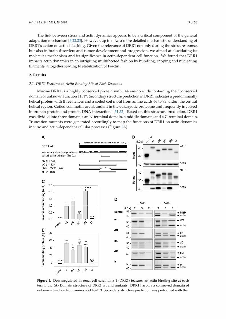
Murine DRR1 is a highly conserved protein with 144 amino acids containing the “conserveddomain of unknown function 1151”. Secondary structure prediction in DRR1 indicates a predominantlyhelical protein with three helices and a coiled coil motif from amino acids 66 to 93 within the centralhelical region. Coiled coil motifs are abundant in the eukaryotic proteome and frequently involvedin protein-protein and protein-DNA interactions [51,52]. Based on this structure prediction, DRR1was divided into three domains: an N-terminal domain, a middle domain, and a C-terminal domain.Truncation mutants were generated accordingly to map the functions of DRR1 on actin dynamicsin vitro and actin-dependent cellular processes (Figure 1A).
Int. J. Mol. Sci. 2018, 19, 3933 3 of 29
but also in brain disorders and tumor development and progression, we aimed at elucidating its
molecular mechanism and its significance in actin-dependent cell function. We found that DRR1
impacts actin dynamics in an intriguing multifaceted fashion by bundling, capping and nucleating
filaments, altogether leading to stabilization of F-actin.
2. Results
2.1. DRR1 Features an Actin Binding Site at Each Terminus
Murine DRR1 is a highly conserved protein with 144 amino acids containing the “conserved
domain of unknown function 1151”. Secondary structure prediction in DRR1 indicates a
predominantly helical protein with three helices and a coiled coil motif from amino acids 66 to 93
within the central helical region. Coiled coil motifs are abundant in the eukaryotic proteome and
frequently involved in protein-protein and protein-DNA interactions [51,52]. Based on this structure
prediction, DRR1 was divided into three domains: an N-terminal domain, a middle domain, and a
C-terminal domain. Truncation mutants were generated accordingly to map the functions of DRR1
on actin dynamics in vitro and actin-dependent cellular processes (Figure 1A).
Figure 1. Downregulated in renal cell carcinoma 1 (DRR1) features an actin binding site at each
terminus. (A) Domain structure of DRR1 wt and mutants. DRR1 harbors a conserved domain of
unknown function from amino acid 16–133. Secondary structure prediction was performed with the
“Predict Protein Server” (dark: helix, light: loop; https://www.predictprotein.org/, accessed on 31 July
Figure 1. Downregulated in renal cell carcinoma 1 (DRR1) features an actin binding site at eachterminus. (A) Domain structure of DRR1 wt and mutants. DRR1 harbors a conserved domain ofunknown function from amino acid 16–133. Secondary structure prediction was performed with the
Int. J. Mol. Sci. 2018, 19, 3993 4 of 30
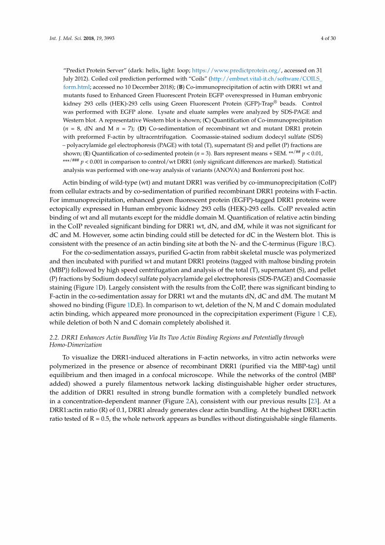
“Predict Protein Server” (dark: helix, light: loop; https://www.predictprotein.org/, accessed on 31July 2012). Coiled coil prediction performed with “Coils” (http://embnet.vital-it.ch/software/COILS_form.html; accessed no 10 December 2018); (B) Co-immunoprecipitation of actin with DRR1 wt andmutants fused to Enhanced Green Fluorescent Protein EGFP overexpressed in Human embryonickidney 293 cells (HEK)-293 cells using Green Fluorescent Protein (GFP)-Trap® beads. Controlwas performed with EGFP alone. Lysate and eluate samples were analyzed by SDS-PAGE andWestern blot. A representative Western blot is shown; (C) Quantification of Co-immunoprecipitation(n = 8, dN and M n = 7); (D) Co-sedimentation of recombinant wt and mutant DRR1 proteinwith preformed F-actin by ultracentrifugation. Coomassie-stained sodium dodecyl sulfate (SDS)– polyacrylamide gel electrophoresis (PAGE) with total (T), supernatant (S) and pellet (P) fractions areshown; (E) Quantification of co-sedimented protein (n = 3). Bars represent means + SEM. **/## p < 0.01,***/### p < 0.001 in comparison to control/wt DRR1 (only significant differences are marked). Statisticalanalysis was performed with one-way analysis of variants (ANOVA) and Bonferroni post hoc.
Actin binding of wild-type (wt) and mutant DRR1 was verified by co-immunoprecipitation (CoIP)from cellular extracts and by co-sedimentation of purified recombinant DRR1 proteins with F-actin.For immunoprecipitation, enhanced green fluorescent protein (EGFP)-tagged DRR1 proteins wereectopically expressed in Human embryonic kidney 293 cells (HEK)-293 cells. CoIP revealed actinbinding of wt and all mutants except for the middle domain M. Quantification of relative actin bindingin the CoIP revealed significant binding for DRR1 wt, dN, and dM, while it was not significant fordC and M. However, some actin binding could still be detected for dC in the Western blot. This isconsistent with the presence of an actin binding site at both the N- and the C-terminus (Figure 1B,C).
For the co-sedimentation assays, purified G-actin from rabbit skeletal muscle was polymerizedand then incubated with purified wt and mutant DRR1 proteins (tagged with maltose binding protein(MBP)) followed by high speed centrifugation and analysis of the total (T), supernatant (S), and pellet(P) fractions by Sodium dodecyl sulfate polyacrylamide gel electrophoresis (SDS-PAGE) and Coomassiestaining (Figure 1D). Largely consistent with the results from the CoIP, there was significant binding toF-actin in the co-sedimentation assay for DRR1 wt and the mutants dN, dC and dM. The mutant Mshowed no binding (Figure 1D,E). In comparison to wt, deletion of the N, M and C domain modulatedactin binding, which appeared more pronounced in the coprecipitation experiment (Figure 1 C,E),while deletion of both N and C domain completely abolished it.
2.2. DRR1 Enhances Actin Bundling Via Its Two Actin Binding Regions and Potentially throughHomo-Dimerization
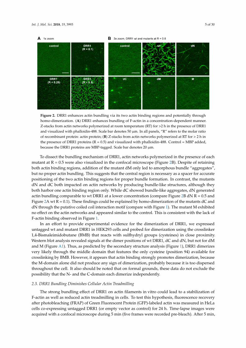
To visualize the DRR1-induced alterations in F-actin networks, in vitro actin networks werepolymerized in the presence or absence of recombinant DRR1 (purified via the MBP-tag) untilequilibrium and then imaged in a confocal microscope. While the networks of the control (MBPadded) showed a purely filamentous network lacking distinguishable higher order structures,the addition of DRR1 resulted in strong bundle formation with a completely bundled networkin a concentration-dependent manner (Figure 2A), consistent with our previous results [23]. At aDRR1:actin ratio (R) of 0.1, DRR1 already generates clear actin bundling. At the highest DRR1:actinratio tested of R = 0.5, the whole network appears as bundles without distinguishable single filaments.
Int. J. Mol. Sci. 2018, 19, 3993 5 of 30
Int. J. Mol. Sci. 2018, 19, 3933 5 of 29
Figure 2. DRR1 enhances actin bundling via its two actin binding regions and potentially through
homo-dimerization. (A) DRR1 enhances bundling of F-actin in a concentration-dependent manner.
Z-stacks from actin networks polymerized at room temperature (RT) for >2 h in the presence of DRR1
and visualized with phalloidin-488. Scale bar denotes 50 µm. In all panels, “R” refers to the molar
ratio of recombinant protein: actin protein; (B) Z-stacks from actin networks polymerized at RT for >
2 h in the presence of DRR1 proteins (R = 0.5) and visualized with phalloidin-488. Control = MBP
added, because the DRR1 proteins are MBP-tagged. Scale bar denotes 20 µm.
To dissect the bundling mechanism of DRR1, actin networks polymerized in the presence of each
mutant at R = 0.5 were also visualized in the confocal microscope (Figure 2B). Despite of retaining
both actin binding regions, addition of the mutant dM only led to amorphous bundle “aggregates”,
but no proper actin bundling. This suggests that the central region is necessary as a spacer for accurate
positioning of the two actin binding regions for proper bundle formation. In contrast, the mutants
dN and dC both impacted on actin networks by producing bundle-like structures, although they both
harbor one actin binding region only. While dC showed bundle-like aggregates, dN generated actin
bundling comparable to wt DRR1 at a lower concentration (compare Figure 2B dN R = 0.5 and Figure
2A wt R = 0.1). These findings could be explained by homo-dimerization of the mutants dC and dN
through the putative coiled coil interaction motif (compare with Figure 1). The mutant M exhibited
no effect on the actin networks and appeared similar to the control. This is consistent with the lack of
F-actin binding observed in Figure 1.
In an effort to provide experimental evidence for the dimerization of DRR1, we expressed
untagged wt and mutant DRR1 in HEK293 cells and probed for dimerization using the crosslinker
1,4-Bismaleimidobutane (BMB) that reacts with sulfhydryl groups (cysteines) in close proximity.
Western blot analysis revealed signals at the dimer positions of wt DRR1, dC and dN, but not for dM
and M (Figure A1). Thus, as predicted by the secondary structure analysis (Figure 1), DRR1 dimerizes
very likely through the middle domain that features the only cysteine (position 94) available for
crosslinking by BMB. However, it appears that actin binding strongly promotes dimerization,
because the M-domain alone did not produce any sign of dimerization, probably because it is too
dispersed throughout the cell. It also should be noted that on formal grounds, these data do not
exclude the possibility that the N- and the C-domain each dimerize independently.
2.3. DRR1 Bundling Diminishes Cellular Actin Treadmilling
The strong bundling effect of DRR1 on actin filaments in vitro could lead to a stabilization of F-
actin as well as reduced actin treadmilling in cells. To test this hypothesis, fluorescence recovery after
photobleaching (FRAP) of Green Fluorescent Protein (GFP)-labeled actin was measured in HeLa cells
co-expressing untagged DRR1 (or empty vector as control) for 24 h. Time-lapse images were acquired
with a confocal microscope during 5 min (five frames were recorded pre-bleach). After 5 min, the
recovery of fluorescence in control cells reached about 75% of the bleached fluorescence. In DRR1 wt
Figure 2. DRR1 enhances actin bundling via its two actin binding regions and potentially throughhomo-dimerization. (A) DRR1 enhances bundling of F-actin in a concentration-dependent manner.Z-stacks from actin networks polymerized at room temperature (RT) for >2 h in the presence of DRR1and visualized with phalloidin-488. Scale bar denotes 50 µm. In all panels, “R” refers to the molar ratioof recombinant protein: actin protein; (B) Z-stacks from actin networks polymerized at RT for > 2 h inthe presence of DRR1 proteins (R = 0.5) and visualized with phalloidin-488. Control = MBP added,because the DRR1 proteins are MBP-tagged. Scale bar denotes 20 µm.
To dissect the bundling mechanism of DRR1, actin networks polymerized in the presence of eachmutant at R = 0.5 were also visualized in the confocal microscope (Figure 2B). Despite of retainingboth actin binding regions, addition of the mutant dM only led to amorphous bundle “aggregates”,but no proper actin bundling. This suggests that the central region is necessary as a spacer for accuratepositioning of the two actin binding regions for proper bundle formation. In contrast, the mutantsdN and dC both impacted on actin networks by producing bundle-like structures, although theyboth harbor one actin binding region only. While dC showed bundle-like aggregates, dN generatedactin bundling comparable to wt DRR1 at a lower concentration (compare Figure 2B dN R = 0.5 andFigure 2A wt R = 0.1). These findings could be explained by homo-dimerization of the mutants dC anddN through the putative coiled coil interaction motif (compare with Figure 1). The mutant M exhibitedno effect on the actin networks and appeared similar to the control. This is consistent with the lack ofF-actin binding observed in Figure 1.
In an effort to provide experimental evidence for the dimerization of DRR1, we expresseduntagged wt and mutant DRR1 in HEK293 cells and probed for dimerization using the crosslinker1,4-Bismaleimidobutane (BMB) that reacts with sulfhydryl groups (cysteines) in close proximity.Western blot analysis revealed signals at the dimer positions of wt DRR1, dC and dN, but not for dMand M (Figure A1). Thus, as predicted by the secondary structure analysis (Figure 1), DRR1 dimerizesvery likely through the middle domain that features the only cysteine (position 94) available forcrosslinking by BMB. However, it appears that actin binding strongly promotes dimerization, becausethe M-domain alone did not produce any sign of dimerization, probably because it is too dispersedthroughout the cell. It also should be noted that on formal grounds, these data do not exclude thepossibility that the N- and the C-domain each dimerize independently.
2.3. DRR1 Bundling Diminishes Cellular Actin Treadmilling
The strong bundling effect of DRR1 on actin filaments in vitro could lead to a stabilization ofF-actin as well as reduced actin treadmilling in cells. To test this hypothesis, fluorescence recoveryafter photobleaching (FRAP) of Green Fluorescent Protein (GFP)-labeled actin was measured in HeLacells co-expressing untagged DRR1 (or empty vector as control) for 24 h. Time-lapse images wereacquired with a confocal microscope during 5 min (five frames were recorded pre-bleach). After 5 min,
Int. J. Mol. Sci. 2018, 19, 3993 6 of 30
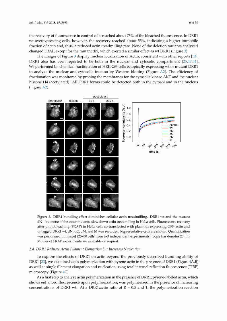
the recovery of fluorescence in control cells reached about 75% of the bleached fluorescence. In DRR1wt overexpressing cells, however, the recovery reached about 55%, indicating a higher immobilefraction of actin and, thus, a reduced actin treadmilling rate. None of the deletion mutants analyzedchanged FRAP, except for the mutant dN, which exerted a similar effect as wt DRR1 (Figure 3).
The images of Figure 3 display nuclear localization of Actin, consistent with other reports [53];DRR1 also has been reported to be both in the nuclear and cytosolic compartment [25,47,54].We performed biochemical fractionation of HEK-293 cells ectopically expressing wt or mutant DRR1to analyze the nuclear and cytosolic fraction by Western blotting (Figure A2). The efficiency offractionation was monitored by probing the membranes for the cytosolic kinase AKT and the nuclearhistone H4 (acetylated). All DRR1 forms could be detected both in the cytosol and in the nucleus(Figure A2).
Int. J. Mol. Sci. 2018, 19, 3933 6 of 29
overexpressing cells, however, the recovery reached about 55%, indicating a higher immobile fraction
of actin and, thus, a reduced actin treadmilling rate. None of the deletion mutants analyzed changed
FRAP, except for the mutant dN, which exerted a similar effect as wt DRR1 (Figure 3).
The images of Figure 3 display nuclear localization of Actin, consistent with other reports [53];
DRR1 also has been reported to be both in the nuclear and cytosolic compartment [25,47,54]. We
performed biochemical fractionation of HEK-293 cells ectopically expressing wt or mutant DRR1 to
analyze the nuclear and cytosolic fraction by Western blotting (Figure A2). The efficiency of
fractionation was monitored by probing the membranes for the cytosolic kinase AKT and the nuclear
histone H4 (acetylated). All DRR1 forms could be detected both in the cytosol and in the nucleus
(Figure A2).
Figure 3. DRR1 bundling effect diminishes cellular actin treadmilling. DRR1 wt and the mutant dN—
but none of the other mutants–slow down actin treadmilling in HeLa cells. Fluorescence recovery
after photobleaching (FRAP) in HeLa cells co-transfected with plasmids expressing GFP-actin and
untagged DRR1 wt, dN, dC, dM, and M was recorded. Representative cells are shown. Quantification
was performed in ImageJ (25–30 cells from 2–3 independent experiments). Scale bar denotes 20 µm.
Movies of FRAP experiments are available on request.
2.4. DRR1 Reduces Actin Filament Elongation but Increases Nucleation
To explore the effects of DRR1 on actin beyond the previously described bundling ability of
DRR1 [23], we examined actin polymerization with pyrene-actin in the presence of DRR1 (Figure
4A,B) as well as single filament elongation and nucleation using total internal reflection fluorescence
(TIRF) microscopy (Figure 4C).
As a first step to analyze actin polymerization in the presence of DRR1, pyrene-labeled actin,
which shows enhanced fluorescence upon polymerization, was polymerized in the presence of
increasing concentrations of DRR1 wt. At a DRR1:actin ratio of R = 0.5 and 1, the polymerization
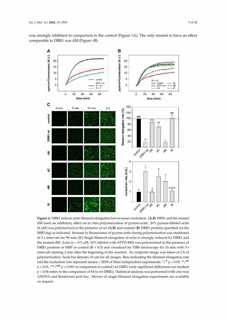
reaction was strongly inhibited in comparison to the control (Figure 4A). The only mutant to have an
effect comparable to DRR1 was dM (Figure 4B).
Figure 3. DRR1 bundling effect diminishes cellular actin treadmilling. DRR1 wt and the mutantdN—but none of the other mutants–slow down actin treadmilling in HeLa cells. Fluorescence recoveryafter photobleaching (FRAP) in HeLa cells co-transfected with plasmids expressing GFP-actin anduntagged DRR1 wt, dN, dC, dM, and M was recorded. Representative cells are shown. Quantificationwas performed in ImageJ (25–30 cells from 2–3 independent experiments). Scale bar denotes 20 µm.Movies of FRAP experiments are available on request.
2.4. DRR1 Reduces Actin Filament Elongation but Increases Nucleation
To explore the effects of DRR1 on actin beyond the previously described bundling ability ofDRR1 [23], we examined actin polymerization with pyrene-actin in the presence of DRR1 (Figure 4A,B)as well as single filament elongation and nucleation using total internal reflection fluorescence (TIRF)microscopy (Figure 4C).
As a first step to analyze actin polymerization in the presence of DRR1, pyrene-labeled actin, whichshows enhanced fluorescence upon polymerization, was polymerized in the presence of increasingconcentrations of DRR1 wt. At a DRR1:actin ratio of R = 0.5 and 1, the polymerization reaction
Int. J. Mol. Sci. 2018, 19, 3993 7 of 30
was strongly inhibited in comparison to the control (Figure 4A). The only mutant to have an effectcomparable to DRR1 was dM (Figure 4B).
Int. J. Mol. Sci. 2018, 19, 3933 7 of 29
In order to verify the slowdown of polymerization by DRR1, the polymerization reaction of
fluorescently labeled G-actin was monitored using TIRF. The overall slowdown of actin
polymerization by DRR1 observed in the pyrene-assay was well reproduced in the TIRF
polymerization. DRR1 significantly slowed down actin polymerization to less than 20% control
(Figure 4C). The mutant dM was the only mutant to retain the inhibitory effect of wt DRR1 on
filament elongation, even though it had previously not shown an effect on bundling. The mutant dC
showed a mild reduction of filament elongation, which was not significant. This finding indicates
that the reduction of single filament elongation by DRR1 is independent of actin bundling, but both
actin binding sites are necessary to affect elongation, since mutants lacking one or both actin binding
sites displayed no significant effect.
Figure 4. DRR1 reduces actin filament elongation but increases nucleation. (A,B) DRR1 and the
mutant dM exert an inhibitory effect on in vitro polymerization of pyrene-actin. 20% pyrene-labeled
actin (4 µM) was polymerized in the presence of wt (A,B) and mutant (B) DRR1 proteins (purified via
the MBP-tag) as indicated. Increase in fluorescence of pyrene-actin during polymerization was
monitored in 5 s intervals for 90 min; (C) Single filament elongation of actin is strongly reduced by
DRR1 and the mutant dM. Actin (c = 0.5 µM, 10% labeled with ATTO-488) was polymerized in the
presence of DRR1 proteins or MBP as control (R = 0.5) and visualized by TIRF microscopy for 10 min
Figure 4. DRR1 reduces actin filament elongation but increases nucleation. (A,B) DRR1 and the mutantdM exert an inhibitory effect on in vitro polymerization of pyrene-actin. 20% pyrene-labeled actin(4 µM) was polymerized in the presence of wt (A,B) and mutant (B) DRR1 proteins (purified via theMBP-tag) as indicated. Increase in fluorescence of pyrene-actin during polymerization was monitoredin 5 s intervals for 90 min; (C) Single filament elongation of actin is strongly reduced by DRR1 andthe mutant dM. Actin (c = 0.5 µM, 10% labeled with ATTO-488) was polymerized in the presence ofDRR1 proteins or MBP as control (R = 0.5) and visualized by TIRF microscopy for 10 min with 3 sintervals starting 2 min after the beginning of the reaction. An endpoint image was taken at 2 h ofpolymerization. Scale bar denotes 10 µm for all images. Bars indicating the filament elongation rateand the nucleation rate represent means + SEM of three independent experiments. */# p < 0.05, **/##
p < 0.01, ***/### p < 0.001 in comparison to control/wt DRR1 (only significant differences are marked;p = 0.06 refers to the comparison of M to wt DRR1). Statistical analysis was performed with one-wayANOVA and Bonferroni post hoc. Movies of single filament elongation experiments are availableon request.
Int. J. Mol. Sci. 2018, 19, 3993 8 of 30
In order to verify the slowdown of polymerization by DRR1, the polymerization reactionof fluorescently labeled G-actin was monitored using TIRF. The overall slowdown of actinpolymerization by DRR1 observed in the pyrene-assay was well reproduced in the TIRF polymerization.DRR1 significantly slowed down actin polymerization to less than 20% control (Figure 4C). The mutantdM was the only mutant to retain the inhibitory effect of wt DRR1 on filament elongation, even thoughit had previously not shown an effect on bundling. The mutant dC showed a mild reduction of filamentelongation, which was not significant. This finding indicates that the reduction of single filamentelongation by DRR1 is independent of actin bundling, but both actin binding sites are necessary toaffect elongation, since mutants lacking one or both actin binding sites displayed no significant effect.
Intriguingly, the visualization of single filament elongation revealed more but shorter filamentsin the presence of DRR1 versus the control. Thus, the number of new filaments per time frame wasquantified and the slope of the resulting plot was determined as read-out of the nucleation rate. DRR1moderately enhanced the filament nucleation rate up to three-fold above the control at a molar ratio ofDRR1:actin of 0.5. The mutants dC and dM both showed a trend towards increased nucleation versusthe control, although not statistically significant (Figure 4C).
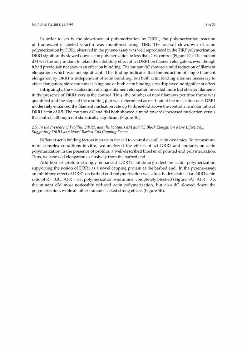
2.5. In the Presence of Profilin, DRR1, and the Mutants dM and dC Block Elongation More Effectively,Suggesting DRR1 as a Novel Barbed End Capping Factor
Different actin binding factors interact in the cell to control overall actin dynamics. To reconstitutemore complex conditions in vitro, we analyzed the effects of wt DRR1 and mutants on actinpolymerization in the presence of profilin, a well-described blocker of pointed end polymerization.Thus, we assessed elongation exclusively from the barbed end.
Addition of profilin strongly enhanced DRR1’s inhibitory effect on actin polymerizationsupporting the notion of DRR1 as a novel capping protein at the barbed end. In the pyrene-assay,an inhibitory effect of DRR1 on barbed end polymerization was already detectable at a DRR1:actinratio of R = 0.01. At R = 0.1, polymerization was almost completely blocked (Figure 5A). At R = 0.5,the mutant dM most noticeably reduced actin polymerization, but also dC slowed down thepolymerization, while all other mutants lacked strong effects (Figure 5B).
Int. J. Mol. Sci. 2018, 19, 3993 9 of 30
Int. J. Mol. Sci. 2018, 19, 3933 9 of 29
Figure 5. In the presence of profilin, DRR1 and the mutants dM and dC block elongation more
effectively, suggesting DRR1 as a novel barbed end capping factor. (A,B) Pyrene-actin polymerization
is blocked by DRR1 and the mutant dM at R = 0.5 in the presence of profilin (12 µM). 20% pyrene-
labeled actin (4 µM) was polymerized in the presence of wt (A,B) and mutant (B) DRR1 proteins
(purified via the MBP tag) as indicated. An increase in fluorescence of pyrene-actin during
polymerization was monitored in 5 s intervals for 60 min; (C) Visualization of actin in vitro
polymerization by TIRF microscopy (c = 0.5 µM, 10% labeled with ATTO-488) in the presence of
profilin (1.5 µM). Actin was polymerized in the presence of DRR1 proteins for 10 min with 3 s intervals
imaging starting 2 min after the beginning of the reaction. An endpoint image was taken at 2 h of
polymerization. Scale bar denotes 10 µm for all images. Bars indicating the filament elongation rate
represent means + SEM of three independent experiments. */# p < 0.05, ***/### p < 0.001 in comparison
to control/wt DRR1 (only significant differences are marked). Statistical analysis was performed with
one-way ANOVA and Bonferroni post hoc. Movies of single filament elongation experiments are
available on request.
Similar results were obtained with polymerization of actin and single filament analysis using
TIRF microscopy. At R = 0.5, DRR1 wt displayed a pronounced barbed end capping activity in the
presence of profilin by reducing the filament elongation rate to about 10% of the control. The same
effect was reproduced for dM. In addition, dC which had only a mild effect in the absence of profilin,
reached significant capping activity in its presence reducing the filament elongation rate to around
Figure 5. In the presence of profilin, DRR1 and the mutants dM and dC block elongation moreeffectively, suggesting DRR1 as a novel barbed end capping factor. (A,B) Pyrene-actin polymerizationis blocked by DRR1 and the mutant dM at R = 0.5 in the presence of profilin (12 µM). 20% pyrene-labeledactin (4 µM) was polymerized in the presence of wt (A,B) and mutant (B) DRR1 proteins (purifiedvia the MBP tag) as indicated. An increase in fluorescence of pyrene-actin during polymerizationwas monitored in 5 s intervals for 60 min; (C) Visualization of actin in vitro polymerization by TIRFmicroscopy (c = 0.5 µM, 10% labeled with ATTO-488) in the presence of profilin (1.5 µM). Actin waspolymerized in the presence of DRR1 proteins for 10 min with 3 s intervals imaging starting 2 minafter the beginning of the reaction. An endpoint image was taken at 2 h of polymerization. Scale bardenotes 10 µm for all images. Bars indicating the filament elongation rate represent means + SEM ofthree independent experiments. */# p < 0.05, ***/### p < 0.001 in comparison to control/wt DRR1 (onlysignificant differences are marked). Statistical analysis was performed with one-way ANOVA andBonferroni post hoc. Movies of single filament elongation experiments are available on request.
Similar results were obtained with polymerization of actin and single filament analysis using TIRFmicroscopy. At R = 0.5, DRR1 wt displayed a pronounced barbed end capping activity in the presenceof profilin by reducing the filament elongation rate to about 10% of the control. The same effect wasreproduced for dM. In addition, dC which had only a mild effect in the absence of profilin, reachedsignificant capping activity in its presence reducing the filament elongation rate to around 36% of thecontrol. Nevertheless, this capping activity of dC was less pronounced than for DRR1 wt (Figure 5C).

Int. J. Mol. Sci. 2018, 19, 3993 10 of 30
2.6. DRR1 Modulates Actin-Dependent Processes in Cells
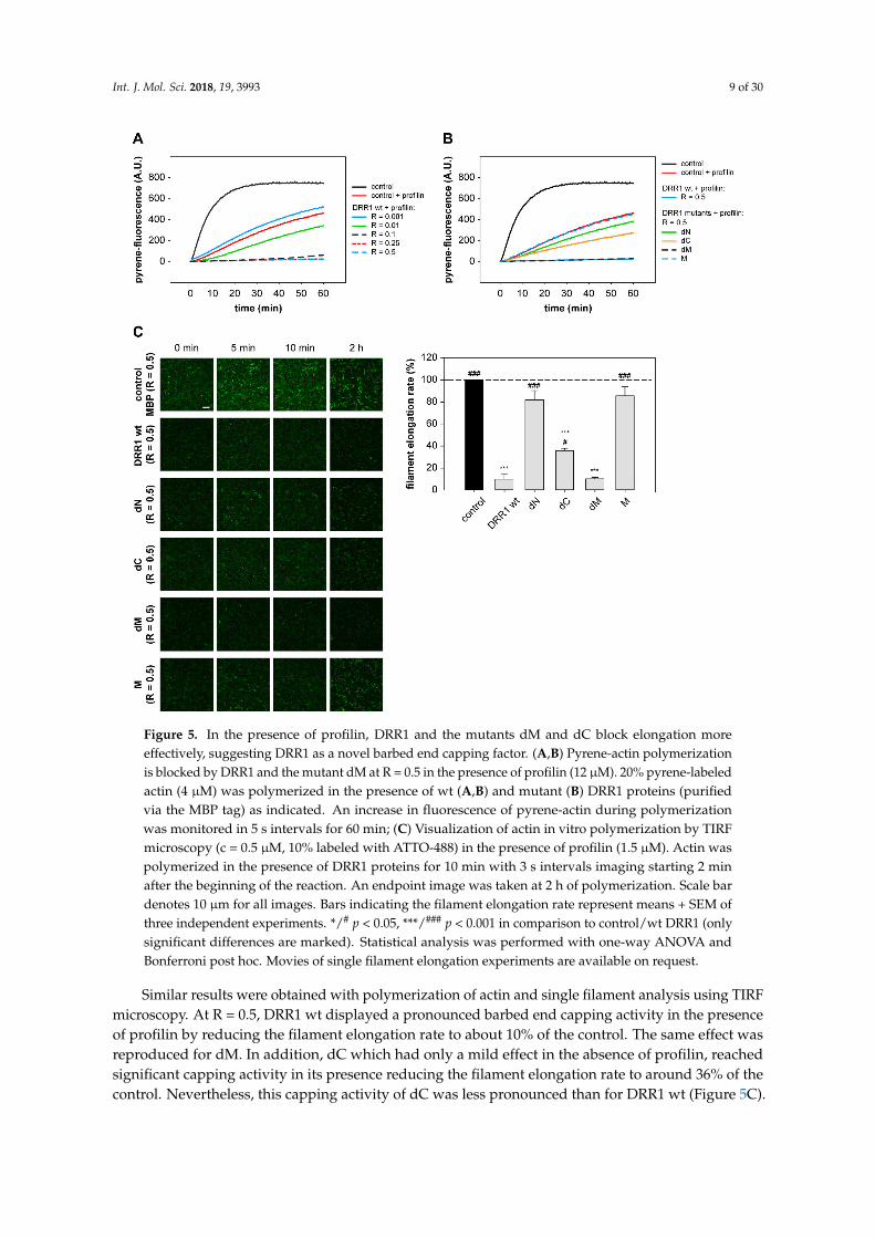
To further analyze the cellular consequences of DRR1-induced changes of actin dynamics,we evaluated known actin-dependent processes such as cell spreading and activity of the transcriptionfactor serum response factor (SRF). Cell spreading is relevant in many cellular functions, such asmigration or wound healing. Spreading of HeLa cells ectopically expressing EGFP-DRR1 wt ormutants, was analyzed by replating on a fibronectin-coated surface and fixation after 30 min ofspreading; F-actin was stained with phalloidin.
DRR1 wt strongly reduced spreading of HeLa cells: while (EGFP transfected) control cells showeda mean size of about 700 µm2, DRR1 wt expressing cells had a mean cell size below 500 µm2 (Figure 6A).In addition, control cells expressing EGFP showed extension of filopodial protrusions after 30 minof spreading, while DRR1 wt-expressing cells were still round–shaped, lacking any protrusions.In these freshly-seeded cells, DRR1 wt colocalized with F-actin at the cortex area of the cells, where thefilaments’ barbed ends are oriented [55]; this is consistent with DRR1’s capping activity at the barbedends, thereby inhibiting extension of protrusions during cell spreading. Evaluation of the deletionmutants of DRR1 revealed that all mutants except M also inhibited cell spreading. This indicates thateither capping or bundling by DRR1 is sufficient to reduce cell spreading.
Cell imaging revealed that DRR1 colocalizes with F-actin (Figure 3 and [23]). As expectedfrom the binding analyses (Figure 1), the mutants dN, dC, and dM also exhibit colocalization withactin filaments (Figure A3). Among them, dC, whose actin binding did not reach significance in theco-immunoprecipitation experiment (Figure 1C), exhibited the lowest correlation coefficient. M showedno colocalisation with F-actin (Figure A3). The overall cellular content of F-actin was increased by wtDRR1, dN, and dM, while dC and M had no significant effect (Figure A4).
The equilibrium between G- and F-actin has further repercussions for intracellular processes,for example activation of the transcription factor serum response factor (SRF). With decreasing levelsof G-actin, the SRF cofactor MAL detaches from G-actin, translocates to the nucleus and activatesSRF [19]. We employed SRF reporter gene assays as a G-actin sensor to monitor the effects of wt andmutant DRR1. The nucleator formin mDia lacking its autoinhibitory “DAD” region was used as apositive control for SRF activation [56].
DRR1 wt increased SRF activity about 10-fold in serum-stimulated cells, similar to the effect ofmDia. In the absence of serum, the stimulation was still about eight-fold above the serum-stimulatedcontrol sample and again comparable to mDia, indicating strong SRF activation by DRR1 independentlyof serum. The mutant dN also significantly enhanced SRF activity, while the mutants dC, and Mshowed no effect. While dM showed a minor increase in SRF activity, its stimulation did not reachsignificance (Figure 6B). This data indicate that DRR1 expression levels modulate SRF-dependent geneexpression through modulation of the equilibrium between G- and F-actin
Int. J. Mol. Sci. 2018, 19, 3993 11 of 30Int. J. Mol. Sci. 2018, 19, 3933 11 of 29
Figure 6. DRR1 modulates actin-dependent processes in cells. (A) DRR1 wt and the mutants dN, dC,
and dM inhibit spreading of HeLa cells. Cells were transfected with constructs expressing EGFP-
DRR1 wt or mutants (control: EGFP), cultivated for 24 h and re-plated on fibronectin-coated
coverslips. After 30 min, cells were fixed and F-actin was stained with phalloidin. Representative cells
are displayed (green: EGFP or EGFP-DRR1; red: F-actin). Scale bar denotes 20 µm. Bars represent
mean cell sizes + SEM of four independent experiments (50–200 cells in each experiment). * p < 0.05,
** p < 0.01, *** p < 0.001 in comparison to control. Statistical analysis was performed with one-way
ANOVA and Bonferroni post hoc; (B) DRR1 overexpression leads to a strong activation of the serum
response factor (SRF) independently of serum, indicating a stabilization of cellular F-actin by DRR1
bundling and capping effects. SRF reporter gene assays in HEK-293 cells show 8–10 fold enhanced
SRF activity after overexpression of DRR1 wt or dN with and without serum. Cells were transfected
with the SRF reporter 3DA.luc, the gaussia luciferase control vector and the indicated plasmids or
vector control. Serum stimulation or withdrawal was for 16–20 h. Luciferase activity is shown as the
fold-increase of serum-stimulation over control samples. Bars represent means + SEM of five
independent experiments. * p < 0.05, ** p < 0.01, n.s. = not significant in comparison to control.
Statistical analysis was performed with one-way ANOVA and Bonferroni post hoc.
3. Discussion
Actin binding proteins orchestrate the temporal and spatial remodeling of the actin cytoskeleton
in cells as the structural basis for several cellular functions [57]. This highly dynamic process also
responds to specific stimuli and, thus, conveys the ability to adapt to new environmental demands.
Here, we present a domain and functional analysis of the stress-induced protein DRR1 with respect
to its action on actin dynamics.
Our findings extend the characterization of DRR1 as actin bundler [23] and add it to the list of
actin cappers. While the contact points of DRR1 on actin filaments remain unknown, capping might
be achieved in two ways by the binding of at least one of the two actin binding domains of DRR1
described here close to the barbed end of the filament: either by inducing a conformational change at
the outmost actin unit or by sterically interfering with the addition of the next actin molecule to the
Figure 6. DRR1 modulates actin-dependent processes in cells. (A) DRR1 wt and the mutants dN, dC,and dM inhibit spreading of HeLa cells. Cells were transfected with constructs expressing EGFP-DRR1wt or mutants (control: EGFP), cultivated for 24 h and re-plated on fibronectin-coated coverslips. After30 min, cells were fixed and F-actin was stained with phalloidin. Representative cells are displayed(green: EGFP or EGFP-DRR1; red: F-actin). Scale bar denotes 20 µm. Bars represent mean cell sizes+ SEM of four independent experiments (50–200 cells in each experiment). * p < 0.05, ** p < 0.01,*** p < 0.001 in comparison to control. Statistical analysis was performed with one-way ANOVA andBonferroni post hoc; (B) DRR1 overexpression leads to a strong activation of the serum response factor(SRF) independently of serum, indicating a stabilization of cellular F-actin by DRR1 bundling andcapping effects. SRF reporter gene assays in HEK-293 cells show 8–10 fold enhanced SRF activityafter overexpression of DRR1 wt or dN with and without serum. Cells were transfected with the SRFreporter 3DA.luc, the gaussia luciferase control vector and the indicated plasmids or vector control.Serum stimulation or withdrawal was for 16–20 h. Luciferase activity is shown as the fold-increase ofserum-stimulation over control samples. Bars represent means + SEM of five independent experiments.* p < 0.05, ** p < 0.01, n.s. = not significant in comparison to control. Statistical analysis was performedwith one-way ANOVA and Bonferroni post hoc.
3. Discussion
Actin binding proteins orchestrate the temporal and spatial remodeling of the actin cytoskeletonin cells as the structural basis for several cellular functions [57]. This highly dynamic process alsoresponds to specific stimuli and, thus, conveys the ability to adapt to new environmental demands.Here, we present a domain and functional analysis of the stress-induced protein DRR1 with respect toits action on actin dynamics.
Our findings extend the characterization of DRR1 as actin bundler [23] and add it to the list ofactin cappers. While the contact points of DRR1 on actin filaments remain unknown, capping mightbe achieved in two ways by the binding of at least one of the two actin binding domains of DRR1described here close to the barbed end of the filament: either by inducing a conformational changeat the outmost actin unit or by sterically interfering with the addition of the next actin molecule to
Int. J. Mol. Sci. 2018, 19, 3993 12 of 30
the extending filament. Deletion of the N-terminal domain leads to complete loss of capping activity,while deletion of the C-terminus retains a somewhat lower capping activity. Therefore, we hypothesizethat the N-terminal binding domain of DRR1 is required for capping. In addition, the second actinbinding domain appears to contribute to capping possibly by stabilizing the interaction with actin.According to our model, the C-terminal deletion mutant dC is able to form dimers yielding two actinbinding sites and, thus, enhances binding affinity to actin. Thus, the deletion mutant dC exerts somecapping activity.
Most capping proteins appear to exert their activity at nanomolar concentrations [58], similar tothe concentrations used here for DRR1. The ratio of DRR1 to actin of 1:10 at which significant cappingwas observed is also close to the range of other capping proteins, for example gelsolin [59]. However,the dominant actin capper in the cell, called “capping protein”, displays a very high binding affinityand is effective at ratios as low as 1:1000 [60]. Thus, the other capping proteins, like DRR1 and gelsolin,may have more specialized roles. In general, both actin interaction domains and capping mechanismsare not conserved. Nevertheless, the mode of action of DRR1 in capping at the barbed end mightbe similar to the mechanism proposed for Twinfilin [61,62] and the Gelsolin protein family [63–65].These proteins feature multiple actin binding sites and contact the actin filament both at the barbedend and at the side of the filament.
We cannot exclude that the nucleation effect of DRR1 observed here in the in vitro assays could besecondary to the capping activity, i.e., due to the extended availability of non-polymerized actin whenpolymerization is diminished. For capping protein, a concentration-dependent nucleation activity hasbeen reported: it inhibits elongation of actin already at low concentration by blocking the barbed end,while at higher concentrations it enhances nucleation by mimicking a non-dissociable actin dimer [14].At this stage, it is likely that DRR1 may act as a nucleation factor by pulling together actin monomersor by stabilizing short oligomers, which appears possible in particular with dimerized DRR1 (comparealso graphical abstract).
Efficient bundling of actin by DRR1 requires both binding sites of DRR1 as deletion of one ofthe domains severely compromises actin bundling. Reduced bundling was also observed for thedeletion of the middle domain, suggesting that proper spacing of the two actin binding domains isrequired, possibly in conjunction with dimerization of the full length protein. Similarly, the residualbundling activity of each of the terminal deletion mutants might be attributed to their dimerization.Even though the middle domain does not bind to actin, our data do not allow excluding the possibilitythat it actively contributes to bundling.
Visually, with respect to bundle thickness and length, and mesh size of the bundled network,bundled actin networks with DRR1 and α-actinin, respectively, look similar. Furthermore, actin:DRR1networks at a ratio of 1:2 are comparable to α-actinin:actin networks of 1:1 ([66] and this work),suggesting DRR1’s bundling activity to be at least as strong as the respective effect of α-actinin.Other actin bundling proteins inhibit in vitro actin depolymerization similarly to DRR1 in thisstudy [67], and DRR1 itself has been shown to reduce dilution-induced actin depolymerization atratios of DRR1:actin of 0.7 [23].
It should be noted that all experiments with recombinant DRR1 in this study had to be performedwith a large (MBP) tag at the wt and mutant DRR1 proteins. Even though the cellular effects of DRR1with a smaller (GFP) tag or with no tag reflect the in vitro results, and even though we distinguishthe DRR1 effects from the effects of MBP alone, we cannot exclude the possibility that he MBP taginfluenced the experimental outcome.
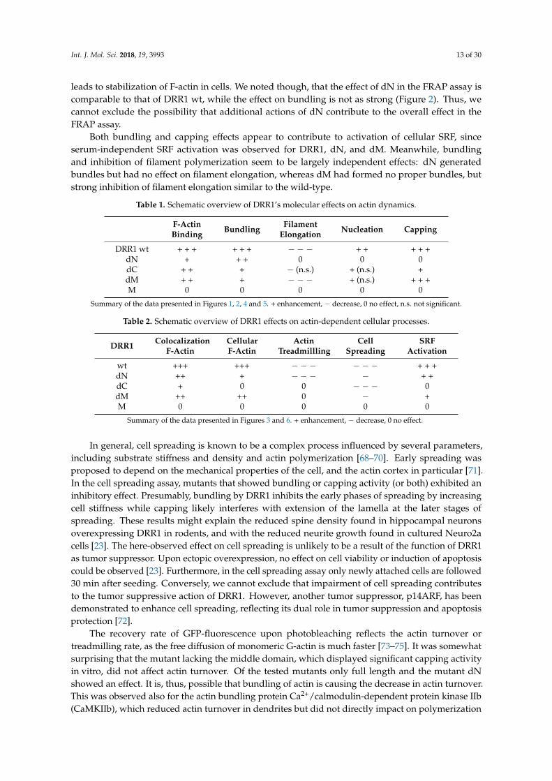
In cells, actin dynamics is shaped by the concerted action of several actin binding proteins.Although the effects observed in vitro with purified compounds may not always reliably predictthe outcome in the cell, the effects on actin dynamics found in cells expressing wt DRR1 and itsmutants were largely congruent with the in vitro results (summaries in Tables 1 and 2). For example,the mutants that exerted proper bundle formation, i.e., DRR1 wt and dN, were the only ones to reduceactin treadmilling in the FRAP experiment, suggesting that it is mainly the bundling activity that
Int. J. Mol. Sci. 2018, 19, 3993 13 of 30
leads to stabilization of F-actin in cells. We noted though, that the effect of dN in the FRAP assay iscomparable to that of DRR1 wt, while the effect on bundling is not as strong (Figure 2). Thus, wecannot exclude the possibility that additional actions of dN contribute to the overall effect in theFRAP assay.
Both bundling and capping effects appear to contribute to activation of cellular SRF, sinceserum-independent SRF activation was observed for DRR1, dN, and dM. Meanwhile, bundlingand inhibition of filament polymerization seem to be largely independent effects: dN generatedbundles but had no effect on filament elongation, whereas dM had formed no proper bundles, butstrong inhibition of filament elongation similar to the wild-type.
Table 1. Schematic overview of DRR1’s molecular effects on actin dynamics.
F-ActinBinding Bundling Filament
Elongation Nucleation Capping
DRR1 wt + + + + + + − − − + + + + +dN + + + 0 0 0dC + + + − (n.s.) + (n.s.) +dM + + + − − − + (n.s.) + + +M 0 0 0 0 0
Summary of the data presented in Figures 1, 2, 4 and 5. + enhancement, − decrease, 0 no effect, n.s. not significant.
Table 2. Schematic overview of DRR1 effects on actin-dependent cellular processes.
DRR1 ColocalizationF-Actin
CellularF-Actin
ActinTreadmillling
CellSpreading
SRFActivation
wt +++ +++ − − − − − − + + +dN ++ + − − − − + +dC + 0 0 − − − 0dM ++ ++ 0 − +M 0 0 0 0 0
Summary of the data presented in Figures 3 and 6. + enhancement, − decrease, 0 no effect.
In general, cell spreading is known to be a complex process influenced by several parameters,including substrate stiffness and density and actin polymerization [68–70]. Early spreading wasproposed to depend on the mechanical properties of the cell, and the actin cortex in particular [71].In the cell spreading assay, mutants that showed bundling or capping activity (or both) exhibited aninhibitory effect. Presumably, bundling by DRR1 inhibits the early phases of spreading by increasingcell stiffness while capping likely interferes with extension of the lamella at the later stages ofspreading. These results might explain the reduced spine density found in hippocampal neuronsoverexpressing DRR1 in rodents, and with the reduced neurite growth found in cultured Neuro2acells [23]. The here-observed effect on cell spreading is unlikely to be a result of the function of DRR1as tumor suppressor. Upon ectopic overexpression, no effect on cell viability or induction of apoptosiscould be observed [23]. Furthermore, in the cell spreading assay only newly attached cells are followed30 min after seeding. Conversely, we cannot exclude that impairment of cell spreading contributesto the tumor suppressive action of DRR1. However, another tumor suppressor, p14ARF, has beendemonstrated to enhance cell spreading, reflecting its dual role in tumor suppression and apoptosisprotection [72].
The recovery rate of GFP-fluorescence upon photobleaching reflects the actin turnover ortreadmilling rate, as the free diffusion of monomeric G-actin is much faster [73–75]. It was somewhatsurprising that the mutant lacking the middle domain, which displayed significant capping activityin vitro, did not affect actin turnover. Of the tested mutants only full length and the mutant dNshowed an effect. It is, thus, possible that bundling of actin is causing the decrease in actin turnover.This was observed also for the actin bundling protein Ca2+/calmodulin-dependent protein kinase IIb(CaMKIIb), which reduced actin turnover in dendrites but did not directly impact on polymerization

Int. J. Mol. Sci. 2018, 19, 3993 14 of 30
and depolymerization kinetics of actin [76]. Since actin treadmilling was shown to consume about halfof the ATP pool in neurons [77], one of DRR1’s physiological roles upon stress could be saving ATPthat might be required for the proper adaptive reaction to stress.
Increased levels of G-actin not only impact SRF, but have also been reported to reduceglucocorticoid receptor (GR)-dependent transcription, possibly through inducing the GR inhibitorc-jun [78]. Accordingly, the F-actin depolymerization factor cofilin 1 has been found to inhibit GRactivity. Thus, it is possible that DRR1 enhances GR activity under certain conditions (constituting afeed-forward mechanism), which may furthermore be cell type-dependent because DRR1 is not onlyexpressed in neurons, but also in other cell types, such as glial cells and various tissues [37,41,79].
Several studies proposed a role of actin dynamics and remodeling in psychiatric disorders suchas depression, based on case-control comparisons and animal models [80–83]. A recent proteomestudy revealed increased levels of F-actin-capping protein subunit beta (CAPZB) in platelets frompatients suffering from major depression in comparison to healthy controls [84]. Other studies reportedchanges of actin regulatory proteins by antidepressants and mood stabilizers [85,86] mutations ingenes of the regulatory network of the actin cytoskeleton appear to be enriched in treatment-resistantmajor depression [87]. Pathway-based methods to genetic data have been suggested to blendbiological information with the power of –omics approaches [83]; we propose that the stress- andglucocorticoid-regulated DRR1 [22,23,47,88] should be included when analyzing the role of the actincytoskeleton in physiology and pathology, particularly in stress-related processes. Furthermore,since actin regulatory factors work in concert, future biochemical investigation of DRR1 shouldinclude the combination with additional actin binding proteins, as this study now firmly establishedDRR1 as an actin-regulatory protein.
4. Materials and Methods
Several of the methods outlined in the following are also described in the Ph.D. thesis of AnjaKretzschmar [89].
4.1. Plasmids
Plasmids for transfection in cell culture were cloned downstream of the Cytomegalovirus (CMV)promoter of the vector pRK5-SV40-MCS. DRR1 mutants were generated by PCR mutagenesis frommurine DRR1 wild-type (wt) construct in pRK5. Cloning of the murine DRR1 wt construct waspreviously described in [23]. The nucleotide sequences of all constructs were confirmed after cloningby Sanger sequencing. For expression of DRR1 proteins N-terminally fused to EGFP or MBP, inserts ofDRR1 wt and mutants were subcloned into the vector pEGFP-C1 (Clontech, Saint-Germain-en-Laye,France) or pMAL-CR1 (New England Biolabs, Ipswich, MA, USA), respectively. Details of the cloningstrategies and primer sequences are available on request.
4.2. Cell Culture and Transfection
HeLa and HEK-293 cells were cultured in Dulbecco’s Modified Eagle Medium (DMEM, LifeTechnologies, Carlsbad, CA, USA) containing 10% fetal bovine serum, 1% sodium pyruvate,and 100 U/mL penicillin and streptomycin at 37 ◦C in a 5% CO2 atmosphere. A confluent 10 cmdish of HEK-293 cells was transfected by electroporation with 15 µg plasmid and cultured for twodays until conduction of the experiments. HeLa cells were transfected using TurboFect (ThermoScientific, Waltham, MA, USA) according to the manufacturer’s instructions and incubated for 24 hafter transfection.
4.3. SDS-PAGE, Colloidal Coomassie Staining, and Immunoblot
Samples were separated on 10, 12, or 15% poly-acrylamide gels with 3.2% stacking gelsand stained with colloidal coomassie brilliant blue G (Sigma-Aldrich, St. Louis, MO, USA) orelectrophoretically transferred onto nitrocellulose membranes (GE Healthcare, Chalfont St Giles,
Int. J. Mol. Sci. 2018, 19, 3993 15 of 30
UK). Immunodetection was performed by blocking the membrane with 5% non-fat milk inTris-buffered saline, supplemented with 0.05% Tween (TBS-T, Sigma-Aldrich) for 1 h at roomtemperature, and then incubated with primary antibody overnight at 4 ◦C. The blots werewashed and probed with the respective horseradish peroxidase- or fluorophor-conjugated secondaryantibody for 3 h at room temperature. All antibodies were diluted in TBS-T with 2% milk powder.The immunoreaction was visualized with ECL detection reagent (Millipore, Darmstadt, Germany)or by fluorescence. The following antibodies were used: rabbit-anti-DRR1 (1:2000, Biogenes,Berlin, Germany, as described in [23]), goat-anti-actin (I-19, 1:2000, Santa Cruz Biotechnology,Dallas, TX, USA), mouse-anti-GFP (B-2, 1:2000, Santa Cruz Biotechnology), rabbit-anti-AKT (1:1000,Cell Signaling, Frankfurt, Germany), rabbit-acetyl-H4 (1:4000, Upstate, Schwalbach, Germany),donkey-anti-rabbit-HRP (1:10,000, Cell Signaling, Cambridge, UK), donkey-anti-goat-HRP (1:10,000,Santa Cruz, Heidelberg, Germany), and Alexa Fluor 488-donkey-anti-mouse (1:5000, Life Technologies).Determination of the relative optical density and quantification of band intensities were performedusing the ImageLab 4.1 Software (Bio-Rad, Munich, Germany).
4.4. Protein Expression and Purification
Recombinant DRR1 proteins were expressed and purified as maltose binding protein (MBP)fusion proteins in order to enhance stability and solubility. In our hands, various efforts to purifyDRR1 without a tag [90] revealed insufficient stability of DRR1 [23]. We observed that with only asmall tag this protein was prone to aggregation at high concentrations and required some urea (1M)and sodium dodecyl sulfate (SDS) (0.1%) to keep it in solution. Similarly, DRR1 turned out to beunstable when the MBP-tag was cleaved off. Therefore, control conditions with buffer only and withMBP only were included in all experiments with recombinant DRR1 proteins. Since there were nodetectable differences in the results between buffer and MBP conditions (see Appendix A Figure A5),the latter is shown in all figures as control. Proteins were expressed in Escherichia coli BL21(DE3)pLysSbacteria (Life Technologies) induced by 0.3 mM isopropyl-beta-D-1-thiogalactopyranoside (IPTG) for2 h at 37 ◦C. The bacterial pellets were lysed by the freeze-thaw method in a dry-ice ethanol bathand then re-suspended in lysis buffer (binding buffer supplemented with protease inhibitor cocktail,1 mg/mL lysozyme, 0.1 mM Phenylmethane sulfonyl fluoride (PMSF) and 1 mM Dithiothreitol (DTT)),incubated on ice for 1 h, and sonicated. The lysates were cleared by centrifugation at 48,400× g for 1 hat 4 ◦C (Beckmann Avanti J-25, Krefeld, Germany) and then filtered through a 0.22 µm syringe filter.The ÄKTA purifier system (General Electrics Healthcare) was used for protein purification with affinitychromatography (MBPTrap HP, 1 mL, GE Healthcare) and gel filtration (Superdex200 10/300 GL,GE Healthcare) as a second step. All buffers used were first filtered through a 0.22 µm filter and thendegassed. Bacterial lysates were loaded on equilibrated MBPTrap columns after clearing and filtrationat a flow rate of 0.5 mL/min with 15 mL binding buffer (20 mM Tris-HCl pH 7.4, 200 mM NaCl, 1 mMEthylenediaminetetraacetic acid (EDTA), 1 mM DTT), washed with 5 mL binding buffer, and elutedwith 10 mL elution buffer (20 mM Tris-HCl pH 7.4, 200 mM NaCl, 1 mM EDTA, 10 mM maltose,1 mM DTT). Samples containing recombinant protein as controlled by SDS-PAGE and Coomassiestaining were pooled and concentrated with Vivaspin 2, MWCO 30 kDa, columns (GE Healthcare).The buffer was changed to Superdex running buffer with (20 mM Tris-HCl pH 7.4, 150 mM NaCl, 1 mMDTT). A Superdex200 10/300 GL column (GE Healthcare) was used for gel filtration at a flow rate of0.5 mL/min. Collected samples were loaded and analyzed with SDS-PAGE, with subsequent poolingof samples containing recombinant protein. Protein concentration was measured with UV absorbanceat 280 nm and with colloidal Coomassie stained SDS-PAGE with a protein standard and densitometry.
Recombinant Profilin2a from mouse tagged with glutathione s-transferase (GST) was expressedfrom E. coli and purified with 2–4 mL glutathion sepharose 4B resin (GE Healthcare) in disposablecolumns. Binding and elution was performed in 50 mM Tris-HCl pH 7.0, 150 nM NaCl, 1 mM EDTA,1 mM DTT. Elution of Profilin was performed by cleaving off the tag overnight at 4 ◦C with PreScission

Int. J. Mol. Sci. 2018, 19, 3993 16 of 30
Protease (GE Healthcare). Protein concentration was determined by UV absorbance at 280 nm. Dialysiswas performed against 20 mM Tris-HCl pH 7.0, 150 mM NaCl, 1 mM EGTA, 1 mM DTT.
All proteins were aliquoted and frozen in liquid nitrogen. Fresh aliquots of recombinant DRR1 orProfilin protein were used for all experiments.
4.5. Co-Immunoprecipitation
For co-immunoprecipitation (CoIP), HEK-293 cells transfected with plasmids expressingEGFP-fusion proteins were lysed with 200 µL ice-cold lysis buffer (10 mM Tris-HCl pH 7.5, 150 mMNaCl, 0.5 mM EDTA, 0.5% NP-40, 1:100 protease inhibitor cocktail P2714 from Sigma-Aldrich).The extract was incubated for 1 h on ice, diluted with 700 µL wash buffer (10 mM Tris-HCl pH7.5, 150 mM NaCl, 0.5 mM EDTA, 1:100 protease inhibitor cocktail), and centrifuged for 10 min at13,000 rpm to remove cell debris. Lysates were incubated with 25 µL GFP-Trap pre-equilibrated agarosebeads (ChromoTek, Planegg-Martinsried, Germany) for 1 h at 4 ◦C. The beads were washed two timeswith 1 mL wash buffer and samples were eluted by incubation for 10 min at 95 ◦C in 50 µL 1× Laemmlisample buffer (1% SDS, 8% glycerol, 32 mM Tris-HCl pH 6.8, 5% mercaptoethanol, bromophenol blue).Fifteen microliters (15 µL) of each input/elution sample were loaded on gels for detection of Co-IPsignals, and 5 µL were loaded for detection of EGFP-fusion-proteins. To calculate relative actin binding,IP and CoIP bands were revealed using an enhanced chemiluminiscence system (Millipore), detectedwith the Chemidoc system (BioRad, Munich, Germany), and quantified by densitometry (using theImageLab 4.1 Software from Bio-Rad) with background signal, corresponding to areas of the membranewithout signal, subtracted. Next, the corrected grey density value of the co-precipitated actin wasreferred to its corresponding value of the precipitated DRR1 protein and the actin/DRR1 ratio wasdefined as “actin binding”. To be able to compare the values between different experiments and blots,the “average actin binding” of all DRR1 proteins (wt, dN, dC, dM, and M) for each experiment wascalculated and “actin binding” of each mutant was normalized to this average actin binding of therespective experiment. Therefore, “1.0” at the y-axis of Figure 1C denotes the (arbitrary value of)average actin binding of the DRR1 proteins and carries no further meaning.
4.6. Dual Luciferase Reporter Assay
For SRF reporter gene assays, Simian virus 40 promoter-driven non-secretory Gaussia luciferaseexpression vector [91] (10 ng per well in a 96-well-plate) was co-transfected in HEK-293 cells with SRFreporter plasmid (25 ng) to correct for transfection efficiency and the respective test plasmids (150 ngper well). The SRF reporter 3DA.luc and mDia1-dDAD plasmids were kind gifts from Robert Grosse(Universität Marburg, Germany). SRF activity was stimulated with 20% FBS or inhibited (0.5% FBS)for 16–18 h twenty-four hours after transfection. Cell lysis was performed with 50 µL passive lysisbuffer (0.2% Triton X-100, 100 mM K2HPO4/KH2PO4 pH 7.8) for 30 min at room temperature. Activityof the firefly luciferase was measured in white microtiter plates in a luminometer ((TriStar LB941Luminometer, Berthold Technologies, Bad Wildbad, Germany)) by adding 50 µL Firefly substratesolution (3 mM MgCl2, 2.4 mM ATP, 120 mM D-Luciferin) to 10 µL lysate. Then, 50 µL of Gaussiasubstrate solution (1.1 M NaCl, 2.2 mM Na2EDTA, 0.22 M K2HPO4/KH2PO4 pH 5.1, 0.44 mg/mLbovine serum albumin (BSA), Coelenterazine 3 µg/mL) was added to the same well to quench thefirefly reaction and measure Gaussia luminescence with a 5 s delay. Firefly luminescence was correctedwith Gaussia values to calculate Firefly activity data. The SRF activity in the serum-stimulated controlwith pRK5 was set to 1 in order to compare different experiments.
4.7. Chemical Crosslinking
HEK-293 cells were transfected with wt and mutant DRR1 expressing plasmids by electroporation.After cultivation for two days, cells were detached, washed, and then incubated in conjugation buffer(PBS with 1 mM EDTA) with either 200 µM crosslinker BMB (1,4-bismaleimidobutane, a crosslinkerwith a spacer arm length of 10.9 Å generating chemical bonds between sulfhydryl groups) or DMSO

Int. J. Mol. Sci. 2018, 19, 3993 17 of 30
as control at 4 ◦C on a shaker for 2 h. Samples were quenched by incubation for 30 min at 4 ◦Cin quenching buffer (10 mM DTT in PBS). Finally, protein extracts were prepared by centrifugingthe cells and resuspension in SDS-lysis (20 mM Tris-HCl pH 7.4, 3.3% sucrose, 0.66% SDS, 1:100protease inhibitor cocktail), short sonication and heating to 95 ◦C for 5 min. Protein concentration wasdetermined with the bicinchoninic acid (BCA) method. A total of 5–10 µg protein were loaded onSDS-PAGE for Western blot analysis.
4.8. Subcellular Fractionation
HEK-293 cells were transfected with wt or mutant DRR1 expressing plasmids; after two daysof cultivation, cells were trypsinized, washed with PBS, and resuspended in 250 µL hypotonic lysisbuffer (10 mM HEPES pH 7.9, 10 mM KCl, 0.5 mM EDTA, 0.1% NP-40, 10% glycerol, 1 mM DTT,and 1:100 protease inhibitor cocktail (freshly added)) per 10 cm dish. After 10 min on ice and briefvortexing, disruption of the outer cell membrane was analyzed in the microscope. Centrifugationwas at 6500 rpm for 30 s at 4 ◦C), followed by transfer of the supernatant (containing the cytosolicproteins) to a fresh tube. The pellet was washed three times with 500 µL hypotonic lysis buffer and thenuclei were lysed by incubating in 200 µL SDS-lysis buffer (1× diluted from 3× which is: 62.5 mMTris-HCl pH 7.4, 10% sucrose, 2% SDS, 1:100 protease inhibitor cocktail (freshly added)) 5 min at95 ◦C and short sonication. After centrifugation, the supernatant was transferred to a fresh tube.Protein concentration of cytosolic and nuclear fractions was determined by BCA and the samples wereanalyzed by SDS-PAGE and Western blot. About 7–10 µg cytosolic fraction and the same volume ofthe corresponding nuclear fraction were loaded.
4.9. HeLa Cell Spreading
HeLa cells were transfected with DRR1 in pEGFP (using TurboFect), harvested on the next dayand replated on fibronectin-coated (50 µg/mL) 12 mm round coverslips in 24-well plates (MerckMillipore, Darmstadt, Germany). Cell spreading was stopped after 30 min of spreading at 37 ◦C byfixing cells with 4% paraformaldehyde for 20 min at room temperature. The actin cytoskeleton wasstained with Alexa Fluore 594-phalloidin and stained cells were placed on glass slides with a drop ofProlong Gold Antifade Medium (Life Technologies). A laser scanning microscope was used for analysis(10×/0.40 NA or 40×/1.15 NA objective, LSM FV-1000, Olympus, Shinjuku, Japan). Up to 10 fieldswere randomly selected with 50–100 cells each and analyzed with ImageJ software. The images werescaled, the phalloidin channel was thresholded (lower threshold level 250, upper threshold level4095 for a 16-bit image) and adjacent cells were separated by the “Watershed” algorithm (controllingmanually for correct cell separation). The thresholded phalloidin channel was used determine cell sizein the original phallidin channel and to measure the mean gray value per cell in the EGFP channelusing the “Analyze Particles” algorithm. Cells with a mean gray value > 500 in the EGFP channelwere determined to be transfected. Cells with a mean gray value of > 500 were defined as transfected.In order to compare different conditions, the mean area of transfected cell was normalized with themean area of untransfected cells in the same condition.
4.10. Fluorescence Recovery after Photobleaching (FRAP)
FRAP was analyzed in HeLa cells plated on 50 µg/mL fibronectin-coated 35 mm glass dishestransfected with TurboFect. Per glass dish, 1 µg GFP-actin was cotransfected with 3 µg (unlabeled)DRR1 constructs in pRK5. After 24 h, the medium was changed to fresh medium. Time-lapse imageframes with 2 s intervals for 5 min were acquired in the confocal microscope (20×/0.8 NA objective,5× zoom, C.A. 200 µm, LSM FV-1000, 2% laser power). Prior to bleaching, five frames were recorded.At an image resolution of 320 × 320 px, bleaching was performed with the circular “TurboTool” for1000 ms at 100% 488-laser power. This led to a bleach of the GFP-fluorescence of about 80%. ImageJsoftware was used to quantify FRAP. First, the mean gray value measured in the bleached area wasdivided through an equally-sized arbitrary non-bleached area within the same cell in each frame to
Int. J. Mol. Sci. 2018, 19, 3993 18 of 30
correct for acquisition photobleaching and possible laser fluctuations. In order to normalize eachfluorescence recovery curve to compare different cells and conditions, the mean gray value of thebleached area immediately after bleaching (C(t0)) was set to 0, while the pre-bleached value (C(pre))was set to one. The mean gray value of each time point was, thus, normalized using the followingformula described in [92]: N(t) = [C(t) − C(t0)]/[C(pre) − C(t0)]. The average gray values in the timelapse imaging was finally averaged across different cells and experiments and plotted as depicted.“N” refers to the number of cells analyzed from 2–3 independent experiments.
4.11. Cellular Stainings
HeLa cells were seeded on 50 µg/mL fibronectin-coated glass coverslips (Merck Millipore),placed in 24-well-plates and transfected using TurboFect as described above. For immunofluorescence,cells were fixed 24 h after transfection with 4% paraformaldehyde in PBS for 20 min at roomtemperature. Cells were permeabilized with 0.1% Triton X-100 for 10 min and blocked with 10%goat serum for 1 h (both in PBS at room temperature). Primary antibodies were diluted 1:200 in 0.1%Triton X-100/PBS and incubated overnight at 4 ◦C. Secondary antibodies were diluted 1:500 in 0.1%Triton X-100/PBS and incubated for 3 h at room temperature. The following antibodies were used:rabbit-anti-DRR1 (Biogenes, Berlin, Germany), Alexa Fluor 647-goat-anti-rabbit (Life Technologies).For staining of actin filaments, cells were incubated with 165 nM AlexaFluor 594, or 546 phalloidin(Life Technologies) in PBS for 20 min at room temperature. Nuclei were counterstained with 1 µg/mLDAPI in PBS for 15 min at room temperature.
4.12. Colocalization Analysis
Images for colocalization analysis were taken with the 40×/1.15 NA objective, 3× zoom, and apinhole of 200 µm. Colocalization analysis was performed in ImageJ with the plugin “Coloc 2”using individual cells as ROI and a point spread function of 4.25. Pearson’s correlation coefficient(PCC) R and Costes p value were calculated using 100 Costes randomizations. For each condition,5–15 randomly-selected cells from two independent experiments were analyzed.
4.13. Quantification of Mean Cellular F-actin Content
Quantification of mean cellular F-actin content was performed in ImageJ. Shortly, the imageswere scaled, the phalloidin channel was thresholded (lower threshold level 150, upper threshold level4095) and adjacent cells were separated by the “Watershed” algorithm. Correct cell separation wasdouble-checked manually. By means of the thresholded phalloidin channel, the mean grey value ofindividual cells in both original channels was measured. Cells with a mean gray value of >500 weredefined as transfected. For each condition, the mean gray value of F-actin in transfected cells wasnormalized to the untransfected cells in order to compare different samples.
4.14. Actin Preparation
G-actin was obtained from rabbit skeletal muscle actin and labeled with pyrene, as describedpreviously [67,93]. All in vitro actin experiments except the F-actin co-sedimentation were performedin the lab of Andreas R. Bausch. In all experiments, “R” refers to the molar ratio of recombinant proteinand actin. Experiments were performed under reducing conditions with 1 mM DTT.
4.15. Pyrene-Actin Polymerization Assay
Actin polymerization was monitored by the increase in fluorescence of 20% pyrenyl-actin at407 nm (excitation at 365 nm) in a fluorescence spectrometer (Jasco FP-8500, Gross-Umstadt, Germany).The final concentration of actin in the reaction was 4 µM. DRR1 proteins were added to G-actin ina constant volume and polymerization was induced by the addition of 1:10 volume of 10× F-buffer
Int. J. Mol. Sci. 2018, 19, 3993 19 of 30
(250 mM Tris-HCl pH 7.4, 250 mM KCl, 40 mM MgCl2, 10 mM EGTA, 10 mM ATP, 10 mM DTT).The polymerization was monitored for 1 h at 21 ◦C with a cycle interval of 5.5 s.
4.16. Actin-Filament Elongation and Nucleation Assay
For visualization of single filament polymerization samples containing 1× F-buffer andrecombinant proteins (in a constant volume) were prepared. Polymerization was induced by theaddition of G-actin (0.5 or 1 µM final concentration). The sample was then immediately pipetted into aflow chamber consisting of two high precision coverslips (60 × 24 mm and 20 × 20 mm, Carl Roth,Karlsruhe, Germany) separated by vacuum grease and placed in a TIRF or confocal microscope (TIRF:Leica DMI6000B, 100×/1.47 NA oil immersion objective, confocal: 63×/1.4 NA oil immersion objective,5× optical zoom, Leica TSC SP5, Solms, Germany). Samples prepared with 10% actin-ATTO488 werevisualized in a TIRF microscope, samples with Alexa Fluor 488-phalloidin were visualized in theconfocal microscope.
To avoid unspecific surface interactions casein was added to the samples in 0.15 mg/mL.The larger coverslips were previously cleaned with a plasma cleaner (40–50 s at 4–6 mbar) andN-ethylmaleimide-modified heavy meromyosin (NEM-HMM, 2.7 µg/mL diluted in F-buffer) wasbound to the surface to keep actin filaments close to the surface during live visualization. The chamberswere washed with 1× F-buffer prior to applying the sample. The time between the addition of actin tothe sample and initiation of the visualization was 2 min. Time-lapse images of polymerization wereacquired for 10 min every 3 s.
The image analysis for single filament elongation rate was performed with ImageJ. In all imagesthe background was subtracted and, subsequently, brightness and contrast were adjusted if necessaryfor ideal visualization. Shortly thereafter, a segmented line was drawn along the filament and plottedtime versus filament length (i.e., fluorescence intensity) with the plugin “multiple kymograph”.The slope of this linear graph corresponds to the filament elongation rate at the barbed end and was asfollows for the MBP control: about 5.6 actin monomers per second in the TIRF assay (0.5 µM actin),and 11.5 actin monomers per second in the confocal assay (1 µM actin), based on the published value of0.0027 µm/actin monomer in actin filaments [94]. The filament elongation rate of the MBP control wasset to 100% in order to compare different experiments. The polymerization speed strongly dependedon the actin preparation. This is in accordance to values described in the literature for ATP-actin atsimilar buffer conditions [95]. Barbed and pointed ends could be easily distinguished in the ImageJgraph in the control, as the elongation rate is much lower at the pointed end. For samples containingDRR1 this was not possible due to the strong inhibition of elongation. Thus, profilin (R = 3) was addedto the samples to block pointed end elongation. Ten filaments from three independent experimentswere measured for each condition.
For nucleation analysis (both in TIRF and confocal assays), filaments in 4–8 frames with 30 sintervals were counted manually from 3–6 independent experiments, respectively. The number offilaments was plotted versus time of polymerization and the slope of the resulting linear graph wasdefined as relative nucleation rate. The MBP control was set to 1. MBP exerted no significant effect onpolymerization or nucleation in comparison to buffer conditions (see Appendix A Figure A5).
4.17. Reconstituted Actin Networks
Samples with 4 µM actin and 2 µM recombinant DRR1 proteins (R = 0.5, wt also in R = 0.1 and 0.25)at a constant volume were prepared for in vitro actin networks and Alexa Fluor 488-phalloidin wasadded to visualized actin (0.08 µM, Life Technologies). Furthermore, casein was added to the samplesin 0.15 mg/mL to avoid unspecific surface interactions. After induction of polymerization by additionof 1:10 volume of 10× F-buffer, the samples were placed into a flow chamber. The chamber wassealed with vacuum grease and samples were polymerized at room temperature for 1.5–2 h protectedfrom light. At equilibrium of polymerization, networks were visualized using a confocal microscope(63×/1.4 NA oil immersion objective, 1× or 3× optical zoom, Leica TSC SP5). Z-stacks of each sample
Int. J. Mol. Sci. 2018, 19, 3993 20 of 30
were taken with a 10 µm depth and slices with 0.38 µm step size and maximum projections of thestaces were generated with ImageJ software. In addition, the background of this maximum projectionswas subtracted and brightness and contrast were automatically adjusted.
4.18. F-Actin Co-Sedimentation Assay
Polymerization of 1 µM G-Actin was induced by the addition of 1:10 volume of 10× F-bufferfor 1 h at room temperature. Freshly thawed recombinant DRR1 proteins were added to the F-actinsamples at 0.5 µM final concentration in a constant volume and incubation on ice was performedfor 30 min to allow proteins to bind to F-actin. The samples were centrifuged at 150.000× g for 1 hat 21 ◦C (Beckmann LB-70M). An aliquot was taken prior to centrifugation (representing “T” = totalprotein) and after centrifugation supernatant and pellet samples were collected (“S” = supernatant,“P” = pellet). Supernatant samples contain G-actin and non-sedimented protein, while F-actin andF-actin-binding proteins are found in the pellet. All samples were loaded on SDS-PAGE, stainedwith colloidal Coomassie and subsequently analyzed by densitometry scanning (ChemiDoc ImagingSystem, Bio-Rad Laboratories, Munich, Germany). Since the pellet fractions are easily contaminated byresidual supernatant proteins, the fraction of co-sedimented protein was calculated by the subtractionof the amount in the supernatant fraction from the amount of total protein determined on the same gelas the more reliable method. To determine the background precipitation of each protein, the proteinswere each centrifuged alone (i.e., without the presence of F-actin) as a control. The amount of pelletedprotein in these samples (−Actin) was subtracted from the co-sedimented protein amount in the sampleswith F-actin (+Actin). This value was then related to (i.e., divided by) the total and is presented as % inFigure 1E. Thus, the amount of DRR1 (wt and mutants) and MBP control co-sedimenting with F-actinwas determined by ([(T+Actin − S+Actin) − (T−Actin − S−Actin)]/T+Actin) × 100. This method was highlyreproducible and consistent.
Control experiments were performed with ddH2O, buffer and MBP at R = 0.5, respectively.No differences were detected in the samples containing MBP vs. buffer and MBP controls are shown inall panels. Both buffer and MBP sample, however, seemed to slightly slow down actin polymerizationin comparison to ddH2O samples (see Appendix A, Figure A5).
4.19. Statistical Analysis
The commercially available program SigmaPlot 14.0 (Erkrath, Germany) was used for statisticalanalysis. A one-way ANOVA (analysis of variants) was performed followed by Bonferroni post hocanalysis for multi-group comparisons. The level of significance was set at p < 0.05. All data arepresented as mean ± SEM. Grubb’s test (with alpha = 0.05) was run to identify significant outliers.
5. Conclusions
This study characterizes the tumor suppressor and stress-regulated protein DRR1 as actin bindingprotein that affects several aspects of actin dynamics such as nucleation, elongation, capping andbundling of F-Actin. We are only beginning to understand the cellular and physiological implications.Through induction of DRR1 stress changes the cellular make-up of actin dynamic regulators.The interplay of DRR1 with the array of other actin binding proteins needs further exploration,as well as the cellular effects that are expected to extend beyond G-actin dependent transcription andcell spreading. Future experiments should also refine the analysis of the structural and functionalchanges of neuronal networks that might contribute to stress-related psychiatric diseases.
Int. J. Mol. Sci. 2018, 19, 3993 21 of 30
Author Contributions: Conceptualization: A.K., J.-P.S., T.R.; data curation: A.K.; formal analysis: A.K.; fundingacquisition: A.R.B., T.R.; investigation: A.K.; methodology: A.K., K.D.; project administration: A.K., T.R.; resources:K.D., A.R.B.; supervision: J.-P.S.; T.R.; validation: A.K.; visualization: A.K.; writing—original draft preparation:A.K., T.R.; writing—review and editing: A.K. J.-P.S., M.M., K.D. M.B.M., A.R.B, T.R.
Funding: This research received no external funding.
Acknowledgments: We are indebted to Christine Wurm, Carina Pelzl, Simone Köhler, and Kurt Schmoller (allTechnical University Munich) for technical help, Angela Oberhofer (Technical University Munich, Germany) forintroduction to TIRF microscopy, and Robert Grosse and Dominique T. Brandt (University of Marburg, Germany)for providing SRF reporter plasmids.
Conflicts of Interest: The authors declare no conflict of interest.
Abbreviations
ANOVA Analysis of variantsATP Adenosine triphosphateBCA Bicinchoninic acidBMB 1,4-BismaleimidobutaneBSA Bovine serum albuminCA3 Cornu Ammonis region 3 (hippocampal region)CaMKIIb Ca2+/calmodulin-dependent protein kinase IIbCMV CytomegalovirusCoIP Co-immunoprecipitation
DRR1Down-regulated in renal cell carcinoma 1 (alsoknown as FAM107A or TU3A)
DTT DithiothreitolEDTA Ethylenediaminetetraacetic acidEGFP Enhanced green fluorescent proteinF-actin Filamentous actinFAM107A Family with sequence similarity 107, member AG-actin Globular actinGFP Green fluorescent proteinHEK-293 Human embryonic kidney 293 cellsMBP Maltose binding proteinPCC Pearson’s correlation coefficientPMSF Phenylmethane sulfonyl fluorideR Molar ratio DRR1 (wt or mutants):actinRT Room temperatureSDS Sodium dodecyl sulfate
SDS-PAGESodium dodecyl sulfate polyacrylamide gelelectrophoresis
SRF Serum response factorTIRF Total internal reflection fluorescenceTU3A Tohoku University cDNA clone A on chromosome 3wt Wild-type

Int. J. Mol. Sci. 2018, 19, 3993 22 of 30
Appendix A
Int. J. Mol. Sci. 2018, 19, 3933 21 of 29
CaMKIIb Ca2+/calmodulin-dependent protein kinase IIb
CMV Cytomegalovirus
CoIP Co-immunoprecipitation
DRR1 Down-regulated in renal cell carcinoma 1 (also known as FAM107A or TU3A)
DTT Dithiothreitol
EDTA Ethylenediaminetetraacetic acid
EGFP Enhanced green fluorescent protein
F-actin Filamentous actin
FAM107A Family with sequence similarity 107, member A
G-actin Globular actin
GFP Green fluorescent protein
HEK-293 Human embryonic kidney 293 cells
MBP Maltose binding protein
PCC Pearson’s correlation coefficient
PMSF Phenylmethane sulfonyl fluoride
R Molar ratio DRR1 (wt or mutants):actin
RT Room temperature
SDS Sodium dodecyl sulfate
SDS-PAGE Sodium dodecyl sulfate polyacrylamide gel electrophoresis
SRF Serum response factor
TIRF Total internal reflection fluorescence
TU3A Tohoku University cDNA clone A on chromosome 3
wt Wild-type
Appendix A
Figure A1. Crosslinking with BMB of DRR1 proteins from HEK-293 cell extracts suggests potential
dimerization of DRR1 wt, dN and dC. (A) Expected molecular weight (MW) of DRR1 monomers and
dimers in kDa; (B) HEK-293 cells transfected with wt or mutant DRR1 (no tag) were incubated with
the cysteine-cysteine crosslinker BMB (+) or DMSO (−) as a control. Lysates were analyzed by SDS-
PAGE and Western blot. Representative Western blots are shown.
Figure A1. Crosslinking with BMB of DRR1 proteins from HEK-293 cell extracts suggests potentialdimerization of DRR1 wt, dN and dC. (A) Expected molecular weight (MW) of DRR1 monomers anddimers in kDa; (B) HEK-293 cells transfected with wt or mutant DRR1 (no tag) were incubated with thecysteine-cysteine crosslinker BMB (+) or DMSO (−) as a control. Lysates were analyzed by SDS-PAGEand Western blot. Representative Western blots are shown.
Int. J. Mol. Sci. 2018, 19, 3933 21 of 29
CaMKIIb Ca2+/calmodulin-dependent protein kinase IIb
CMV Cytomegalovirus
CoIP Co-immunoprecipitation
DRR1 Down-regulated in renal cell carcinoma 1 (also known as FAM107A or TU3A)
DTT Dithiothreitol
EDTA Ethylenediaminetetraacetic acid
EGFP Enhanced green fluorescent protein
F-actin Filamentous actin
FAM107A Family with sequence similarity 107, member A
G-actin Globular actin
GFP Green fluorescent protein
HEK-293 Human embryonic kidney 293 cells
MBP Maltose binding protein
PCC Pearson’s correlation coefficient
PMSF Phenylmethane sulfonyl fluoride
R Molar ratio DRR1 (wt or mutants):actin
RT Room temperature
SDS Sodium dodecyl sulfate
SDS-PAGE Sodium dodecyl sulfate polyacrylamide gel electrophoresis
SRF Serum response factor
TIRF Total internal reflection fluorescence
TU3A Tohoku University cDNA clone A on chromosome 3
wt Wild-type
Appendix A
Figure A1. Crosslinking with BMB of DRR1 proteins from HEK-293 cell extracts suggests potential
dimerization of DRR1 wt, dN and dC. (A) Expected molecular weight (MW) of DRR1 monomers and
dimers in kDa; (B) HEK-293 cells transfected with wt or mutant DRR1 (no tag) were incubated with
the cysteine-cysteine crosslinker BMB (+) or DMSO (−) as a control. Lysates were analyzed by SDS-
PAGE and Western blot. Representative Western blots are shown.
Figure A2. DRR1 wt and mutants display nuclear localization. Cytosolic and nuclear fractions ofHEK-293 cells expressing ectopic DRR1 wt or mutants were analyzed by SDS-PAGE and Western blot.Efficiency of fractionation into cytosol (C) and nucleus (N) was confirmed with antibodies against thecytosolic kinase AKT and the nuclear acetyl-histone H4. Representative Western blots are shown.
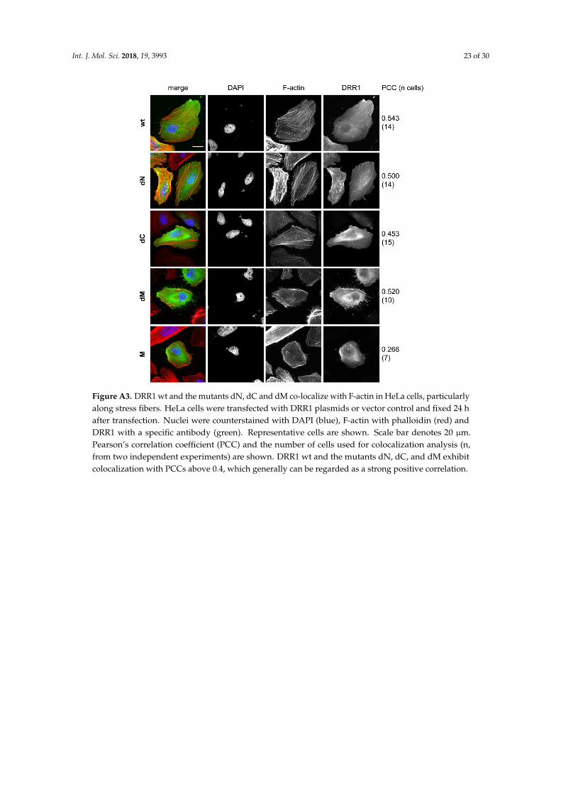
Int. J. Mol. Sci. 2018, 19, 3993 23 of 30
Int. J. Mol. Sci. 2018, 19, 3933 22 of 29
Figure A2. DRR1 wt and mutants display nuclear localization. Cytosolic and nuclear fractions of HEK-
293 cells expressing ectopic DRR1 wt or mutants were analyzed by SDS-PAGE and Western blot.
Efficiency of fractionation into cytosol (C) and nucleus (N) was confirmed with antibodies against the
cytosolic kinase AKT and the nuclear acetyl-histone H4. Representative Western blots are shown.
Figure A3. DRR1 wt and the mutants dN, dC and dM co-localize with F-actin in HeLa cells,
particularly along stress fibers. HeLa cells were transfected with DRR1 plasmids or vector control and
fixed 24 h after transfection. Nuclei were counterstained with DAPI (blue), F-actin with phalloidin (red)
and DRR1 with a specific antibody (green). Representative cells are shown. Scale bar denotes 20 µm.
Pearson’s correlation coefficient (PCC) and the number of cells used for colocalization analysis (n,
from two independent experiments) are shown. DRR1 wt and the mutants dN, dC, and dM exhibit
colocalization with PCCs above 0.4, which generally can be regarded as a strong positive correlation.
Figure A3. DRR1 wt and the mutants dN, dC and dM co-localize with F-actin in HeLa cells, particularlyalong stress fibers. HeLa cells were transfected with DRR1 plasmids or vector control and fixed 24 hafter transfection. Nuclei were counterstained with DAPI (blue), F-actin with phalloidin (red) andDRR1 with a specific antibody (green). Representative cells are shown. Scale bar denotes 20 µm.Pearson’s correlation coefficient (PCC) and the number of cells used for colocalization analysis (n,from two independent experiments) are shown. DRR1 wt and the mutants dN, dC, and dM exhibitcolocalization with PCCs above 0.4, which generally can be regarded as a strong positive correlation.
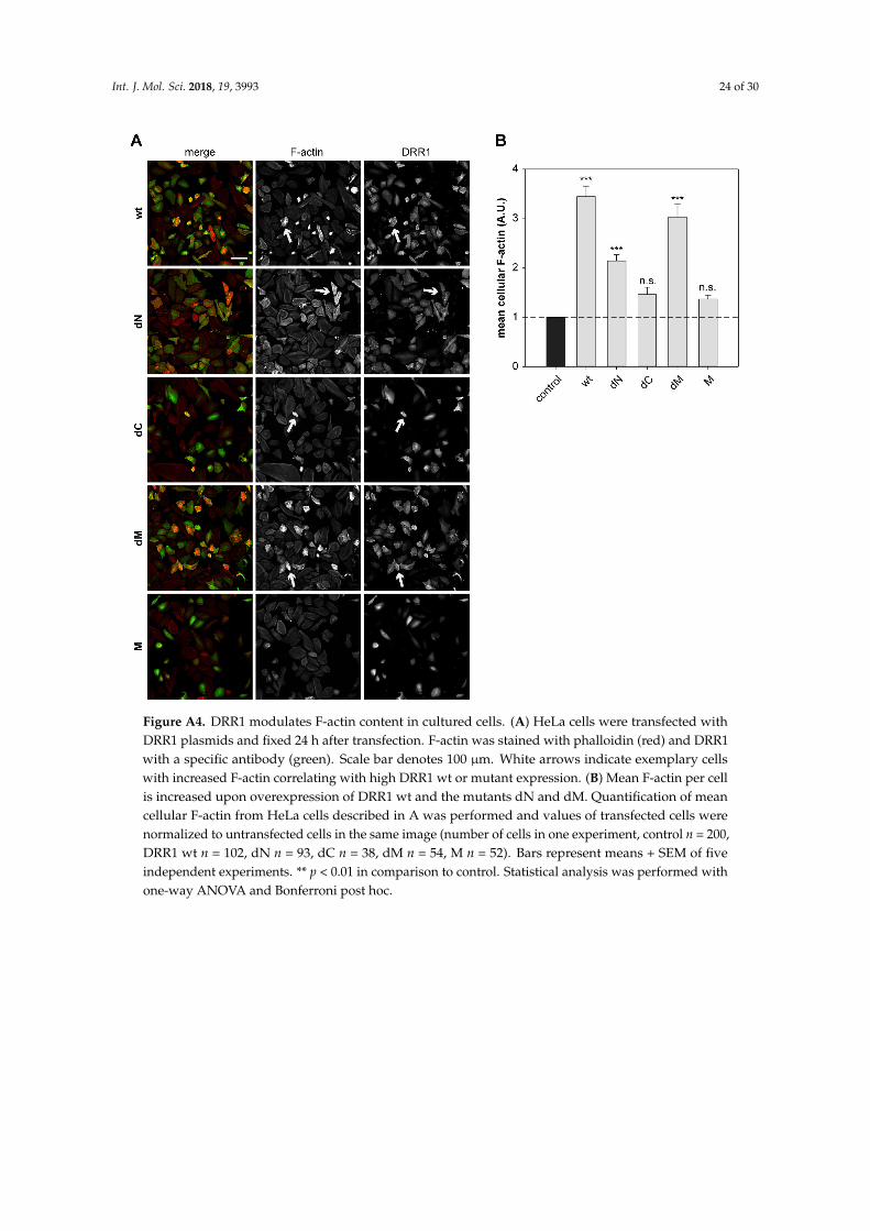
Int. J. Mol. Sci. 2018, 19, 3993 24 of 30
Int. J. Mol. Sci. 2018, 19, 3933 23 of 29
Figure A4. DRR1 modulates F-actin content in cultured cells. (A) HeLa cells were transfected with
DRR1 plasmids and fixed 24 h after transfection. F-actin was stained with phalloidin (red) and DRR1
with a specific antibody (green). Scale bar denotes 100 µm. White arrows indicate exemplary cells
with increased F-actin correlating with high DRR1 wt or mutant expression. (B) Mean F-actin per cell
is increased upon overexpression of DRR1 wt and the mutants dN and dM. Quantification of mean
cellular F-actin from HeLa cells described in A was performed and values of transfected cells were
normalized to untransfected cells in the same image (number of cells in one experiment, control n =
200, DRR1 wt n = 102, dN n = 93, dC n = 38, dM n = 54, M n = 52). Bars represent means + SEM of five
independent experiments. ** p < 0.01 in comparison to control. Statistical analysis was performed with
one-way ANOVA and Bonferroni post hoc.
Figure A4. DRR1 modulates F-actin content in cultured cells. (A) HeLa cells were transfected withDRR1 plasmids and fixed 24 h after transfection. F-actin was stained with phalloidin (red) and DRR1with a specific antibody (green). Scale bar denotes 100 µm. White arrows indicate exemplary cellswith increased F-actin correlating with high DRR1 wt or mutant expression. (B) Mean F-actin per cellis increased upon overexpression of DRR1 wt and the mutants dN and dM. Quantification of meancellular F-actin from HeLa cells described in A was performed and values of transfected cells werenormalized to untransfected cells in the same image (number of cells in one experiment, control n = 200,DRR1 wt n = 102, dN n = 93, dC n = 38, dM n = 54, M n = 52). Bars represent means + SEM of fiveindependent experiments. ** p < 0.01 in comparison to control. Statistical analysis was performed withone-way ANOVA and Bonferroni post hoc.
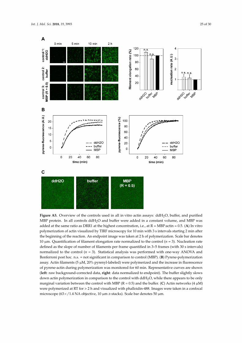
Int. J. Mol. Sci. 2018, 19, 3993 25 of 30
Int. J. Mol. Sci. 2018, 19, 3933 24 of 29
Figure A5. Overview of the controls used in all in vitro actin assays: ddH2O, buffer, and purified MBP
protein. In all controls ddH2O and buffer were added in a constant volume, and MBP was added at
the same ratio as DRR1 at the highest concentration, i.e., at R = MBP:actin = 0.5. (A) In vitro
polymerization of actin visualized by TIRF microscopy for 10 min with 3 s intervals starting 2 min
after the beginning of the reaction. An endpoint image was taken at 2 h of polymerization. Scale bar
denotes 10 µm. Quantification of filament elongation rate normalized to the control (n = 3). Nucleation
rate defined as the slope of number of filaments per frame quantified in 3–5 frames (with 30 s intervals)
normalized to the control (n = 3). Statistical analysis was performed with one-way ANOVA and
Bonferroni post hoc. n.s. = not significant in comparison to control (MBP). (B) Pyrene-polymerization
assay. Actin filaments (5 µM, 20% pyrenyl-labeled) were polymerized and the increase in fluorescence
of pyrene-actin during polymerization was monitored for 60 min. Representative curves are shown (left:
raw background-corrected data, right: data normalized to endpoint). The buffer slightly slows down
actin polymerization in comparison to the control with ddH2O, while there appears to be only marginal
variation between the control with MBP (R = 0.5) and the buffer. (C) Actin networks (4 µM) were
polymerized at RT for > 2 h and visualized with phalloidin-488. Images were taken in a confocal
microscope (63×/1.4 NA objective, 10 µm z-stacks). Scale bar denotes 50 µm.
Figure A5. Overview of the controls used in all in vitro actin assays: ddH2O, buffer, and purifiedMBP protein. In all controls ddH2O and buffer were added in a constant volume, and MBP wasadded at the same ratio as DRR1 at the highest concentration, i.e., at R = MBP:actin = 0.5. (A) In vitropolymerization of actin visualized by TIRF microscopy for 10 min with 3 s intervals starting 2 min afterthe beginning of the reaction. An endpoint image was taken at 2 h of polymerization. Scale bar denotes10 µm. Quantification of filament elongation rate normalized to the control (n = 3). Nucleation ratedefined as the slope of number of filaments per frame quantified in 3–5 frames (with 30 s intervals)normalized to the control (n = 3). Statistical analysis was performed with one-way ANOVA andBonferroni post hoc. n.s. = not significant in comparison to control (MBP). (B) Pyrene-polymerizationassay. Actin filaments (5 µM, 20% pyrenyl-labeled) were polymerized and the increase in fluorescenceof pyrene-actin during polymerization was monitored for 60 min. Representative curves are shown(left: raw background-corrected data, right: data normalized to endpoint). The buffer slightly slowsdown actin polymerization in comparison to the control with ddH2O, while there appears to be onlymarginal variation between the control with MBP (R = 0.5) and the buffer. (C) Actin networks (4 µM)were polymerized at RT for > 2 h and visualized with phalloidin-488. Images were taken in a confocalmicroscope (63×/1.4 NA objective, 10 µm z-stacks). Scale bar denotes 50 µm.
Int. J. Mol. Sci. 2018, 19, 3993 26 of 30
References
1. McEwen, B.S.; Bowles, N.P.; Gray, J.D.; Hill, M.N.; Hunter, R.G.; Karatsoreos, I.N.; Nasca, C. Mechanisms ofstress in the brain. Nat. Neurosci. 2015, 18, 1353–1363. [CrossRef] [PubMed]
2. De Kloet, E.R.; Joels, M.; Holsboer, F. Stress and the brain: From adaptation to disease. Nat. Rev. Neurosci.2005, 6, 463–475. [CrossRef] [PubMed]
3. Fenster, R.J.; Lebois, L.A.M.; Ressler, K.J.; Suh, J. Brain circuit dysfunction in post-traumatic stress disorder:From mouse to man. Nat. Rev. Neurosci. 2018, 19, 535–551. [CrossRef] [PubMed]
4. Cingolani, L.A.; Goda, Y. Actin in action: The interplay between the actin cytoskeleton and synaptic efficacy.Nat. Rev. Neurosci. 2008, 9, 344–356. [CrossRef] [PubMed]
5. Van der Kooij, M.A.; Masana, M.; Rust, M.B.; Müller, M.B. The stressed cytoskeleton: How actin dynamicscan shape stress-related consequences on synaptic plasticity and complex behavior. Neurosci. Biobehav. Rev.2016, 62, 69–75. [CrossRef] [PubMed]
6. Fuchs, E.; Flügge, G. Stress, glucocorticoids and structural plasticity of the hippocampus. Neurosci. Biobehav.Rev. 1998, 23, 295–300. [CrossRef]
7. Kasai, H.; Fukuda, M.; Watanabe, S.; Hayashi-Takagi, A.; Noguchi, J. Structural dynamics of dendritic spinesin memory and cognition. Trends Neurosci. 2010, 33, 121–129. [CrossRef]
8. Yan, Z.; Kim, E.; Datta, D.; Lewis, D.A.; Soderling, S.H. Synaptic Actin Dysregulation, a ConvergentMechanism of Mental Disorders? J. Neurosci. 2016, 36, 11411–11417. [CrossRef]
9. Davidson, A.J.; Wood, W. Unravelling the Actin Cytoskeleton: A New Competitive Edge? Trends Cell Biol.2016, 26, 569–576. [CrossRef]
10. Kuhn, J.R.; Pollard, T.D. Real-time measurements of actin filament polymerization by total internal reflectionfluorescence microscopy. Biophys. J. 2005, 88, 1387–1402. [CrossRef]
11. Harris, E.S.; Higgs, H.N. Biochemical analysis of mammalian formin effects on actin dynamics. MethodsEnzymol. 2006, 406, 190–214. [PubMed]
12. Dominguez, R. Structural insights into de novo actin polymerization. Curr. Opin. Struct. Biol. 2010, 20,217–225. [CrossRef] [PubMed]
13. Mullins, R.D.; Heuser, J.A.; Pollard, T.D. The interaction of Arp2/3 complex with actin: Nucleation, highaffinity pointed end capping, and formation of branching networks of filaments. Proc. Natl. Acad. Sci. USA1998, 95, 6181–6186. [CrossRef] [PubMed]
14. Cooper, J.A.; Pollard, T.D. Effect of capping protein on the kinetics of actin polymerization. Biochemistry 1985,24, 793–799. [CrossRef] [PubMed]
15. Edwards, M.; Zwolak, A.; Schafer, D.A.; Sept, D.; Dominguez, R.; Cooper, J.A. Capping protein regulatorsfine-tune actin assembly dynamics. Nat. Rev. Mol. Cell Biol. 2014, 15, 677–689. [CrossRef] [PubMed]
16. Jayo, A.; Parsons, M. Fascin: A key regulator of cytoskeletal dynamics. Int. J. Biochem. Cell Biol. 2010, 42,1614–1617. [CrossRef]
17. Nakamura, F.; Stossel, T.P.; Hartwig, J.H. The filamins: Organizers of cell structure and function. Cell Adh.Migr. 2011, 5, 160–169. [CrossRef]
18. Sjöblom, B.; Salmazo, A.; Djinovic-Carugo, K. Alpha-actinin structure and regulation. Cell Mol. Life Sci. 2008,65, 2688–2701. [CrossRef]
19. Posern, G.; Treisman, R. Actin’ together: Serum response factor, its cofactors and the link to signaltransduction. Trends Cell Biol. 2006, 16, 588–596. [CrossRef]
20. Esnault, C.; Stewart, A.; Gualdrini, F.; East, P.; Horswell, S.; Matthews, N.; Treisman, R. Rho-actin signalingto the MRTF coactivators dominates the immediate transcriptional response to serum in fibroblasts. GenesDev. 2014, 28, 943–958. [CrossRef]
21. Liebl, C.; Panhuysen, M.; Putz, B.; Trumbach, D.; Wurst, W.; Deussing, J.M.; Muller, M.B.; Schmidt, M.V.Gene expression profiling following maternal deprivation: Involvement of the brain Renin-Angiotensinsystem. Front. Mol. Neurosci. 2009, 2, 1. [CrossRef] [PubMed]
22. Masana, M.; Su, Y.A.; Liebl, C.; Wang, X.D.; Jansen, L.; Westerholz, S.; Wagner, K.V.; Labermaier, C.;Scharf, S.H.; Santarelli, S.; et al. The stress-inducible actin-interacting protein DRR1 shapes social behavior.Psychoneuroendocrinology 2014, 48, 98–110. [CrossRef] [PubMed]
Int. J. Mol. Sci. 2018, 19, 3993 27 of 30
23. Schmidt, M.V.; Schülke, J.P.; Liebl, C.; Stiess, M.; Avrabos, C.; Bock, J.; Wochnik, G.M.; Davies, H.A.;Zimmermann, N.; Scharf, S.H.; et al. Tumor suppressor down-regulated in renal cell carcinoma 1 (DRR1) is astress-induced actin bundling factor that modulates synaptic efficacy and cognition. Proc. Natl. Acad. Sci.USA 2011, 108, 17213–17218. [CrossRef] [PubMed]
24. Yamato, T.; Orikasa, K.; Fukushige, S.; Orikasa, S.; Horii, A. Isolation and characterization of the novel gene,TU3A, in a commonly deleted region on 3p14.3–>p14.2 in renal cell carcinoma. Cytogenet. Cell Genet. 1999,87, 291–295. [CrossRef] [PubMed]
25. Wang, L.; Darling, J.; Zhang, J.S.; Liu, W.; Qian, J.; Bostwick, D.; Hartmann, L.; Jenkins, R.; Bardenhauer, W.;Schutte, J.; et al. Loss of expression of the DRR 1 gene at chromosomal segment 3p21.1 in renal cell carcinoma.Genes Chromosom. Cancer 2000, 27, 1–10. [CrossRef]
26. Udali, S.; Guarini, P.; Ruzzenente, A.; Ferrarini, A.; Guglielmi, A.; Lotto, V.; Tononi, P.; Pattini, P.; Moruzzi, S.;Campagnaro, T.; et al. DNA methylation and gene expression profiles show novel regulatory pathways inhepatocellular carcinoma. Clin. Epigenetics 2015, 7, 43. [CrossRef]
27. Kiwerska, K.; Szaumkessel, M.; Paczkowska, J.; Bodnar, M.; Byzia, E.; Kowal, E.; Kostrzewska-Poczekaj, M.;Janiszewska, J.; Bednarek, K.; Jarmuz-Szymczak, M.; et al. Combined deletion and DNA methylation resultin silencing of FAM107A gene in laryngeal tumors. Sci. Rep. 2017, 7, 5386. [CrossRef]
28. Lawrie, A.; Han, S.; Sud, A.; Hosking, F.; Cezard, T.; Turner, D.; Clark, C.; Murray, G.I.; Culligan, D.J.;Houlston, R.S.; et al. Combined linkage and association analysis of classical Hodgkin lymphoma. Oncotarget2018, 9, 20377–20385. [CrossRef]
29. Pastuszak-Lewandoska, D.; Czarnecka, K.H.; Migdalska-Sek, M.; Nawrot, E.; Domanska, D.; Kiszalkiewicz, J.;Kordiak, J.; Antczak, A.; Gorski, P.; Brzezianska-Lasota, E. Decreased FAM107A Expression in Patients withNon-small Cell Lung Cancer. Adv. Exp. Med. Biol. 2015, 852, 39–48.
30. Mu, P.; Akashi, T.; Lu, F.; Kishida, S.; Kadomatsu, K. A novel nuclear complex of DRR1, F-actin and COMMD1involved in NF-kappaB degradation and cell growth suppression in neuroblastoma. Oncogene 2017, 36,5745–5756. [CrossRef]
31. Asano, Y.; Kishida, S.; Mu, P.; Sakamoto, K.; Murohara, T.; Kadomatsu, K. DRR1 is expressed in thedeveloping nervous system and downregulated during neuroblastoma carcinogenesis. Biochem. Biophys. Res.Commun. 2010, 394, 829–835. [CrossRef] [PubMed]
32. Fèvre-Montange, M.; Champier, J.; Durand, A.; Wierinckx, A.; Honnorat, J.; Guyotat, J.; Jouvet, A. Microarraygene expression profiling in meningiomas: Differential expression according to grade or histopathologicalsubtype. Int. J. Oncol. 2009, 35, 1395–1407. [CrossRef] [PubMed]
33. Kholodnyuk, I.D.; Kozireva, S.; Kost-Alimova, M.; Kashuba, V.; Klein, G.; Imreh, S. Down regulation of 3pgenes, LTF, SLC38A3 and DRR1, upon growth of human chromosome 3-mouse fibrosarcoma hybrids insevere combined immunodeficiency mice. Int. J. Cancer 2006, 119, 99–107. [CrossRef] [PubMed]
34. Liu, Q.; Zhao, X.Y.; Bai, R.Z.; Liang, S.F.; Nie, C.L.; Yuan, Z.; Wang, C.T.; Wu, Y.; Chen, L.J.; Wei, Y.Q. Inductionof tumor inhibition and apoptosis by a candidate tumor suppressor gene DRR1 on 3p21.1. Oncol. Rep. 2009,22, 1069–1075. [PubMed]
35. Vanaja, D.K.; Ballman, K.V.; Morlan, B.W.; Cheville, J.C.; Neumann, R.M.; Lieber, M.M.; Tindall, D.J.;Young, C.Y. PDLIM4 repression by hypermethylation as a potential biomarker for prostate cancer. Clin.Cancer Res. 2006, 12, 1128–1136. [CrossRef]
36. Van den, B.J.; Wolter, M.; Blaschke, B.; Knobbe, C.B.; Reifenberger, G. Identification of novel genesassociated with astrocytoma progression using suppression subtractive hybridization and real-time reversetranscription-polymerase chain reaction. Int. J. Cancer 2006, 119, 2330–2338. [CrossRef] [PubMed]
37. Pollen, A.A.; Nowakowski, T.J.; Chen, J.; Retallack, H.; Sandoval-Espinosa, C.; Nicholas, C.R.; Shuga, J.;Liu, S.J.; Oldham, M.C.; Diaz, A.; et al. Molecular identity of human outer radial glia during corticaldevelopment. Cell 2015, 163, 55–67. [CrossRef]
38. Ma, Y.S.; Wu, Z.J.; Bai, R.Z.; Dong, H.; Xie, B.X.; Wu, X.H.; Hang, X.S.; Liu, A.N.; Jiang, X.H.; Wang, G.R.;et al. DRR1 promotes glioblastoma cell invasion and epithelial-mesenchymal transition via regulating AKTactivation. Cancer Lett. 2018, 423, 86–94. [CrossRef]
39. Le, P.U.; Angers-Loustau, A.; de Oliveira, R.M.; Ajlan, A.; Brassard, C.L.; Dudley, A.; Brent, H.; Siu, V.;Trinh, G.; Molenkamp, G.; et al. DRR drives brain cancer invasion by regulating cytoskeletal-focal adhesiondynamics. Oncogene 2010, 29, 4636–4647. [CrossRef]
Int. J. Mol. Sci. 2018, 19, 3993 28 of 30
40. Quartier, A.; Chatrousse, L.; Redin, C.; Keime, C.; Haumesser, N.; Maglott-Roth, A.; Brino, L.; Le, G.S.;Benchoua, A.; Mandel, J.L.; et al. Genes and Pathways Regulated by Androgens in Human Neural Cells,Potential Candidates for the Male Excess in Autism Spectrum Disorder. Biol. Psychiatry 2018, 84, 239–252.[CrossRef]
41. Wan, B.; Feng, P.; Guan, Z.; Sheng, L.; Liu, Z.; Hua, Y. A severe mouse model of spinal muscular atrophydevelops early systemic inflammation. Hum. Mol. Genet. 2018, 27. [CrossRef] [PubMed]
42. Li, M.D.; Burns, T.C.; Morgan, A.A.; Khatri, P. Integrated multi-cohort transcriptional meta-analysis ofneurodegenerative diseases. Acta Neuropathol. Commun. 2014, 2, 93. [CrossRef] [PubMed]
43. Shao, L.; Vawter, M.P. Shared gene expression alterations in schizophrenia and bipolar disorder. Biol.Psychiatry 2008, 64, 89–97. [CrossRef] [PubMed]
44. Ranno, E.; D’Antoni, S.; Spatuzza, M.; Berretta, A.; Laureanti, F.; Bonaccorso, C.M.; Pellitteri, R.; Longone, P.;Spalloni, A.; Iyer, A.M.; et al. Endothelin-1 is over-expressed in amyotrophic lateral sclerosis and inducesmotor neuron cell death. Neurobiol. Dis. 2014, 65, 160–171. [CrossRef] [PubMed]
45. Murray, L.M.; Lee, S.; Baumer, D.; Parson, S.H.; Talbot, K.; Gillingwater, T.H. Pre-symptomatic developmentof lower motor neuron connectivity in a mouse model of severe spinal muscular atrophy. Hum. Mol. Genet.2010, 19, 420–433. [CrossRef] [PubMed]
46. Shin, J.; Salameh, J.S.; Richter, J.D. Impaired neurodevelopment by the low complexity domain of CPEB4reveals a convergent pathway with neurodegeneration. Sci. Rep. 2016, 6, 29395. [CrossRef] [PubMed]
47. Masana, M.; Westerholz, S.; Kretzschmar, A.; Treccani, G.; Liebl, C.; Santarelli, S.; Dournes, C.; Popoli, M.;Schmidt, M.V.; Rein, T.; et al. Expression and glucocorticoid-dependent regulation of the stress-inducibleprotein DRR1 in the mouse adult brain. Brain Struct. Funct. 2018, 223, 4039–4052. [CrossRef]
48. Masana, M.; Jukic, M.M.; Kretzschmar, A.; Wagner, K.V.; Westerholz, S.; Schmidt, M.V.; Rein, T.; Brodski, C.;Muller, M.B. Deciphering the spatio-temporal expression and stress regulation of Fam107B, the paralog ofthe resilience-promoting protein DRR1 in the mouse brain. Neuroscience 2015, 290, 147–158. [CrossRef]
49. Stankiewicz, A.M.; Goscik, J.; Swiergiel, A.H.; Majewska, A.; Wieczorek, M.; Juszczak, G.R.; Lisowski, P.Social stress increases expression of hemoglobin genes in mouse prefrontal cortex. BMC Neurosci. 2014, 15,130. [CrossRef]
50. Jene, T.; Gassen, N.C.; Opitz, V.; Endres, K.; Muller, M.B.; van der Kooij, M.A. Temporal profiling of an acutestress-induced behavioral phenotype in mice and role of hippocampal DRR1. Psychoneuroendocrinology 2018,91, 149–158. [CrossRef]
51. Lupas, A.N.; Gruber, M. The structure of alpha-helical coiled coils. Adv. Protein Chem. 2005, 70, 37–78.[PubMed]
52. Liu, J.; Rost, B. Comparing function and structure between entire proteomes. Protein Sci. 2001, 10, 1970–1979.[CrossRef] [PubMed]
53. Falahzadeh, K.; Banaei-Esfahani, A.; Shahhoseini, M. The potential roles of actin in the nucleus. Cell J. 2015,17, 7–14. [PubMed]
54. Zhao, X.Y.; Liang, S.F.; Yao, S.H.; Ma, F.X.; Hu, Z.G.; Yan, F.; Yuan, Z.; Ruan, X.Z.; Yang, H.S.; Zhou, Q.; et al.Identification and preliminary function study of Xenopus laevis DRR1 gene. Biochem. Biophys. Res. Commun.2007, 361, 74–78. [CrossRef]
55. Siton-Mendelson, O.; Bernheim-Groswasser, A. Functional Actin Networks under Construction:The Cooperative Action of Actin Nucleation and Elongation Factors. Trends Biochem. Sci. 2017, 42, 414–430.[CrossRef] [PubMed]
56. Li, F.; Higgs, H.N. The mouse Formin mDia1 is a potent actin nucleation factor regulated by autoinhibition.Curr. Biol. 2003, 13, 1335–1340. [CrossRef]
57. Dominguez, R.; Holmes, K.C. Actin structure and function. Annu. Rev. Biophys. 2011, 40, 169–186. [CrossRef]58. Hertzog, M.; Milanesi, F.; Hazelwood, L.; Disanza, A.; Liu, H.; Perlade, E.; Malabarba, M.G.; Pasqualato, S.;
Maiolica, A.; Confalonieri, S.; et al. Molecular basis for the dual function of Eps8 on actin dynamics: Bundlingand capping. PLoS Biol. 2010, 8, e1000387. [CrossRef]
59. Gremm, D.; Wegner, A. Gelsolin as a calcium-regulated actin filament-capping protein. Eur. J. Biochem. 2000,267, 4339–4345. [CrossRef]
60. Schirenbeck, A.; Bretschneider, T.; Arasada, R.; Schleicher, M.; Faix, J. The Diaphanous-related formin dDia2is required for the formation and maintenance of filopodia. Nat. Cell Biol. 2005, 7, 619–625. [CrossRef]
Int. J. Mol. Sci. 2018, 19, 3993 29 of 30
61. Paavilainen, V.O.; Oksanen, E.; Goldman, A.; Lappalainen, P. Structure of the actin-depolymerizing factorhomology domain in complex with actin. J. Cell Biol. 2008, 182, 51–59. [CrossRef]
62. Paavilainen, V.O.; Hellman, M.; Helfer, E.; Bovellan, M.; Annila, A.; Carlier, M.F.; Permi, P.; Lappalainen, P.Structural basis and evolutionary origin of actin filament capping by twinfilin. Proc. Natl. Acad. Sci. USA2007, 104, 3113–3118. [CrossRef] [PubMed]
63. Way, M.; Pope, B.; Weeds, A.G. Evidence for functional homology in the F-actin binding domains of gelsolinand alpha-actinin: Implications for the requirements of severing and capping. J. Cell Biol. 1992, 119, 835–842.[CrossRef] [PubMed]
64. Robinson, R.C.; Mejillano, M.; Le, V.P.; Burtnick, L.D.; Yin, H.L.; Choe, S. Domain movement in gelsolin:A calcium-activated switch. Science 1999, 286, 1939–1942. [CrossRef] [PubMed]
65. McGough, A.; Chiu, W.; Way, M. Determination of the gelsolin binding site on F-actin: Implications forsevering and capping. Biophys. J. 1998, 74, 764–772. [CrossRef]
66. Falzone, T.T.; Lenz, M.; Kovar, D.R.; Gardel, M.L. Assembly kinetics determine the architecture ofalpha-actinin crosslinked F-actin networks. Nat. Commun. 2012, 3, 861. [CrossRef] [PubMed]
67. Schmoller, K.M.; Semmrich, C.; Bausch, A.R. Slow down of actin depolymerization by cross-linkingmolecules. J. Struct. Biol. 2011, 173, 350–357. [CrossRef]
68. Chamaraux, F.; Fache, S.; Bruckert, F.; Fourcade, B. Kinetics of cell spreading. Phys. Rev. Lett. 2005, 94, 158102.[CrossRef]
69. Li, J.; Han, D.; Zhao, Y.P. Kinetic behaviour of the cells touching substrate: The interfacial stiffness guidescell spreading. Sci. Rep. 2014, 4, 3910. [CrossRef]
70. Yauch, R.L.; Felsenfeld, D.P.; Kraeft, S.K.; Chen, L.B.; Sheetz, M.P.; Hemler, M.E. Mutational evidence forcontrol of cell adhesion through integrin diffusion/clustering, independent of ligand binding. J. Exp. Med.1997, 186, 1347–1355. [CrossRef]
71. Cuvelier, D.; Théry, M.; Chu, Y.S.; Dufour, S.; Thiery, J.P.; Bornens, M.; Nassoy, P.; Mahadevan, L. The universaldynamics of cell spreading. Curr. Biol. 2007, 17, 694–699. [CrossRef]
72. Vivo, M.; Fontana, R.; Ranieri, M.; Capasso, G.; Angrisano, T.; Pollice, A.; Calabro, V.; La, M.G. p14ARFinteracts with the focal adhesion kinase and protects cells from anoikis. Oncogene 2017, 36, 4913–4928.[CrossRef] [PubMed]
73. Frost, N.A.; Shroff, H.; Kong, H.; Betzig, E.; Blanpied, T.A. Single-molecule discrimination of discreteperisynaptic and distributed sites of actin filament assembly within dendritic spines. Neuron 2010, 67, 86–99.[CrossRef] [PubMed]
74. Honkura, N.; Matsuzaki, M.; Noguchi, J.; Ellis-Davies, G.C.; Kasai, H. The subspine organization of actinfibers regulates the structure and plasticity of dendritic spines. Neuron 2008, 57, 719–729. [CrossRef][PubMed]
75. Star, E.N.; Kwiatkowski, D.J.; Murthy, V.N. Rapid turnover of actin in dendritic spines and its regulation byactivity. Nat. Neurosci. 2002, 5, 239–246. [CrossRef] [PubMed]
76. Okamoto, K.; Narayanan, R.; Lee, S.H.; Murata, K.; Hayashi, Y. The role of CaMKII as an F-actin-bundlingprotein crucial for maintenance of dendritic spine structure. Proc. Natl. Acad. Sci. USA 2007, 104, 6418–6423.[CrossRef] [PubMed]
77. Bernstein, B.W.; Bamburg, J.R. Actin-ATP hydrolysis is a major energy drain for neurons. J. Neurosci. 2003,23, 1–6. [CrossRef] [PubMed]
78. Rüegg, J.; Holsboer, F.; Turck, C.; Rein, T. Cofilin 1 is revealed as an inhibitor of glucocorticoid receptor byanalysis of hormone-resistant cells 2195. Mol. Cell Biol. 2004, 24, 9371–9382. [CrossRef]
79. Cahoy, J.D.; Emery, B.; Kaushal, A.; Foo, L.C.; Zamanian, J.L.; Christopherson, K.S.; Xing, Y.; Lubischer, J.L.;Krieg, P.A.; Krupenko, S.A.; et al. A transcriptome database for astrocytes, neurons, and oligodendrocytes: Anew resource for understanding brain development and function. J. Neurosci. 2008, 28, 264–278. [CrossRef]
80. Durand, C.M.; Perroy, J.; Loll, F.; Perrais, D.; Fagni, L.; Bourgeron, T.; Montcouquiol, M.; Sans, N. SHANK3mutations identified in autism lead to modification of dendritic spine morphology via an actin-dependentmechanism. Mol. Psychiatry 2012, 17, 71–84. [CrossRef]
81. Nakatani, N.; Ohnishi, T.; Iwamoto, K.; Watanabe, A.; Iwayama, Y.; Yamashita, S.; Ishitsuka, Y.; Moriyama, K.;Nakajima, M.; Tatebayashi, Y.; et al. Expression analysis of actin-related genes as an underlying mechanismfor mood disorders. Biochem. Biophys. Res. Commun. 2007, 352, 780–786. [CrossRef] [PubMed]
Int. J. Mol. Sci. 2018, 19, 3993 30 of 30
82. Nakatani, N.; Aburatani, H.; Nishimura, K.; Semba, J.; Yoshikawa, T. Comprehensive expression analysis ofa rat depression model. Pharmacogenomics J. 2004, 4, 114–126. [CrossRef] [PubMed]
83. Wang, K.; Li, M.; Bucan, M. Pathway-based approaches for analysis of genomewide association studies.Am. J. Hum. Genet. 2007, 81, 1278–1283. [CrossRef]
84. Huang, T.L.; Sung, M.L.; Chen, T.Y. 2D-DIGE proteome analysis on the platelet proteins of patients withmajor depression. Proteome Sci. 2014, 12. [CrossRef]
85. Calabrese, B.; Halpain, S. Lithium prevents aberrant NMDA-induced F-actin reorganization in neurons.Neuroreport 2014, 25, 1331–1337. [CrossRef] [PubMed]
86. Piubelli, C.; Vighini, M.; Mathe, A.A.; Domenici, E.; Carboni, L. Escitalopram affects cytoskeleton andsynaptic plasticity pathways in a rat gene-environment interaction model of depression as revealed byproteomics. Part II: Environmental challenge. Int. J. Neuropsychopharmacol. 2011, 14, 834–855. [CrossRef]
87. O’Dushlaine, C.; Ripke, S.; Ruderfer, D.M.; Hamilton, S.P.; Fava, M.; Iosifescu, D.V.; Kohane, I.S.;Churchill, S.E.; Castro, V.M.; Clements, C.C.; et al. Rare copy number variation in treatment-resistantmajor depressive disorder. Biol. Psychiatry 2014, 76, 536–541. [CrossRef]
88. Frijters, R.; Fleuren, W.; Toonen, E.J.; Tuckermann, J.P.; Reichardt, H.M.; van der, M.H.; van, E.A.; vanLierop, M.J.; Dokter, W.; de, V.J.; et al. Prednisolone-induced differential gene expression in mouseliver carrying wild type or a dimerization-defective glucocorticoid receptor. BMC Genomics 2010, 11, 359.[CrossRef]
89. Kretzschmar, A. The Modulation of Actin Dynamics by the Stress-Induced Protein DRR1 and Antidepressants.Ph.D. Thesis, Ludmig Maximilians University Munich, Munich, Germany, 2015.
90. Zhao, X.Y.; Li, H.X.; Liang, S.F.; Yuan, Z.; Yan, F.; Ruan, X.Z.; You, J.; Xiong, S.Q.; Tang, M.H.; Wei, Y.Q.Soluble expression of human DRR1 (down-regulated in renal cell carcinoma 1) in Escherichia coli andpreparation of its polyclonal antibodies. Biotechnol. Appl. Biochem. 2008, 49, 17–23. [CrossRef]
91. Schülke, J.P.; Wochnik, G.M.; Lang-Rollin, I.; Gassen, N.C.; Knapp, R.T.; Berning, B.; Yassouridis, A.; Rein, T.Differential impact of tetratricopeptide repeat proteins on the steroid hormone receptors. PLoS ONE 2010, 5,e11717. [CrossRef] [PubMed]
92. Rino, J.; Martin, R.M.; Carvalho, T.; Carmo-Fonseca, M. Imaging dynamic interactions between spliceosomalproteins and pre-mRNA in living cells. Methods 2014, 65, 359–366. [CrossRef] [PubMed]
93. Cooper, J.A.; Walker, S.B.; Pollard, T.D. Pyrene actin: Documentation of the validity of a sensitive assay foractin polymerization. J. Muscle Res. Cell Motil. 1983, 4, 253–262. [CrossRef]
94. Liu, X.; Pollack, G.H. Stepwise sliding of single actin and Myosin filaments. Biophys. J. 2004, 86, 353–358.[CrossRef]
95. Pollard, T.D. Rate constants for the reactions of ATP- and ADP-actin with the ends of actin filaments. J. CellBiol. 1986, 103, 2747–2754. [CrossRef] [PubMed]
© 2018 by the authors. Licensee MDPI, Basel, Switzerland. This article is an open accessarticle distributed under the terms and conditions of the Creative Commons Attribution(CC BY) license (http://creativecommons.org/licenses/by/4.0/).


![Interferon-inducible Gene Family 1-8U Expression in …...[CANCER RESEARCH 59, 5927–5931, December 1, 1999] Interferon-inducible Gene Family 1-8U Expression in Colitis-associated](https://img.pdfslide.tips/doc/110x75/5f7765e2aad8b7367718cdc1/interferon-inducible-gene-family-1-8u-expression-in-cancer-research-59-5927a5931.jpg)