Embed Size (px)
Citation preview
The Urinary System
山东大学医学院 解剖教研室李振华
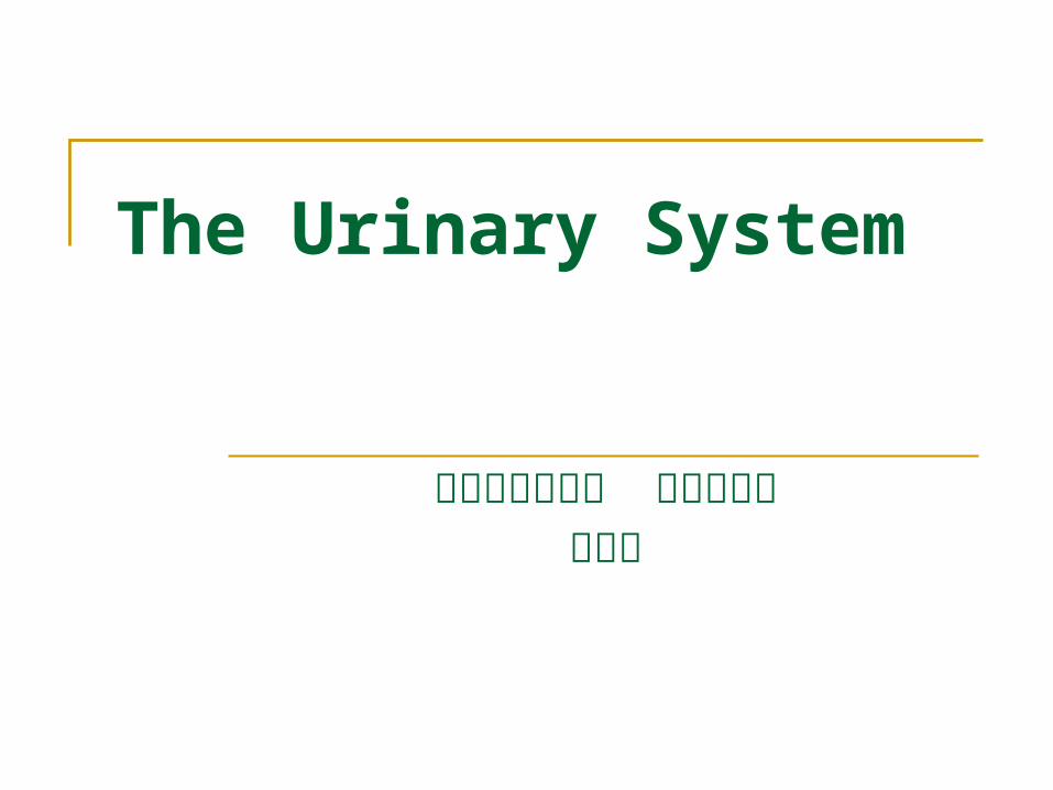
Composition Two kidney - form urin
e Two ureter - conduct u
rine from kidneys to bladder
Bladder - receives and stores urine
Urethra - conducts urine from bladder to exterior of body (discharged)
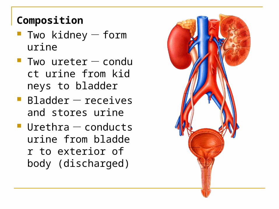
Kidney 肾General features Bean shaped, reddish-brow
n organs Superior extremity - broad
and thin Inferior extremity - narrow
and thick Anterior surface - convex Posterior surface - flat Lateral border - convex
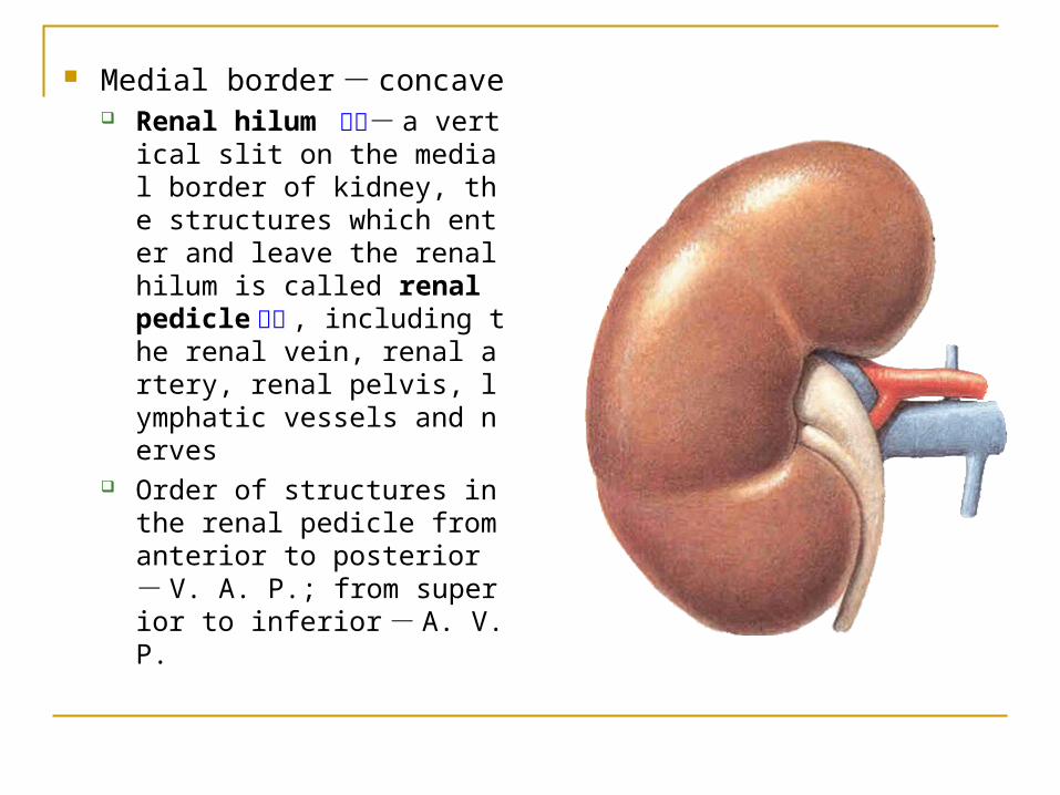
Medial border - concave Renal hilum 肾门- a vertic
al slit on the medial border of kidney, the structures which enter and leave the renal hilum is called renal pedicle 肾蒂 , including the renal vein, renal artery, renal pelvis, lymphatic vessels and nerves
Order of structures in the renal pedicle from anterior to posterior - V. A. P.; from superior to inferior - A. V. P.
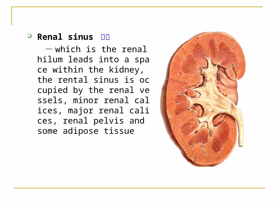
Renal sinus 肾窦 - which is the renal hilum lea
ds into a space within the kidney, the rental sinus is occupied by the renal vessels, minor renal calices, major renal calices, renal pelvis and some adipose tissue
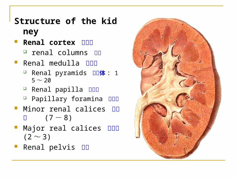
Structure of the kidney Renal cortex 肾皮质
renal columns 肾柱 Renal medulla 肾髓质
Renal pyramids 肾锥体 : 15 ~20
Renal papilla 肾乳头 Papillary foramina 乳头孔
Minor renal calices 肾小盏 (7- 8)
Major real calices 肾大盏 (2 ~ 3)
Renal pelvis 肾盂
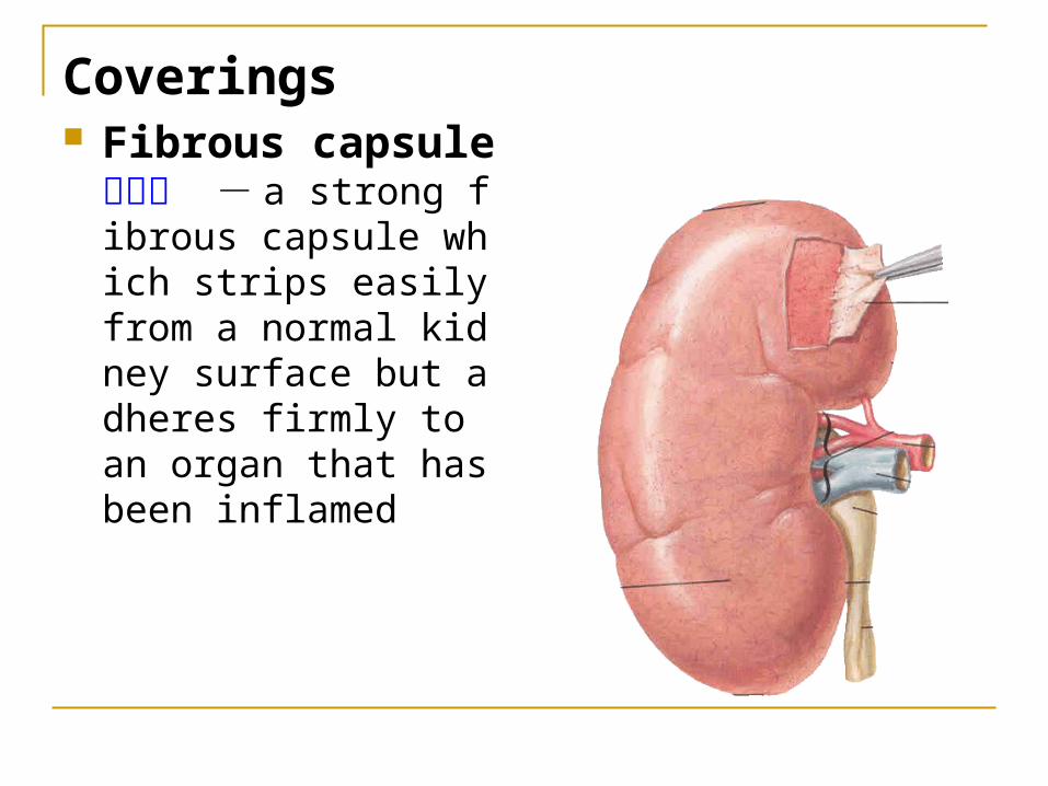
Coverings Fibrous capsule 纤
维囊 - a strong fibrous capsule which strips easily from a normal kidney surface but adheres firmly to an organ that has been inflamed
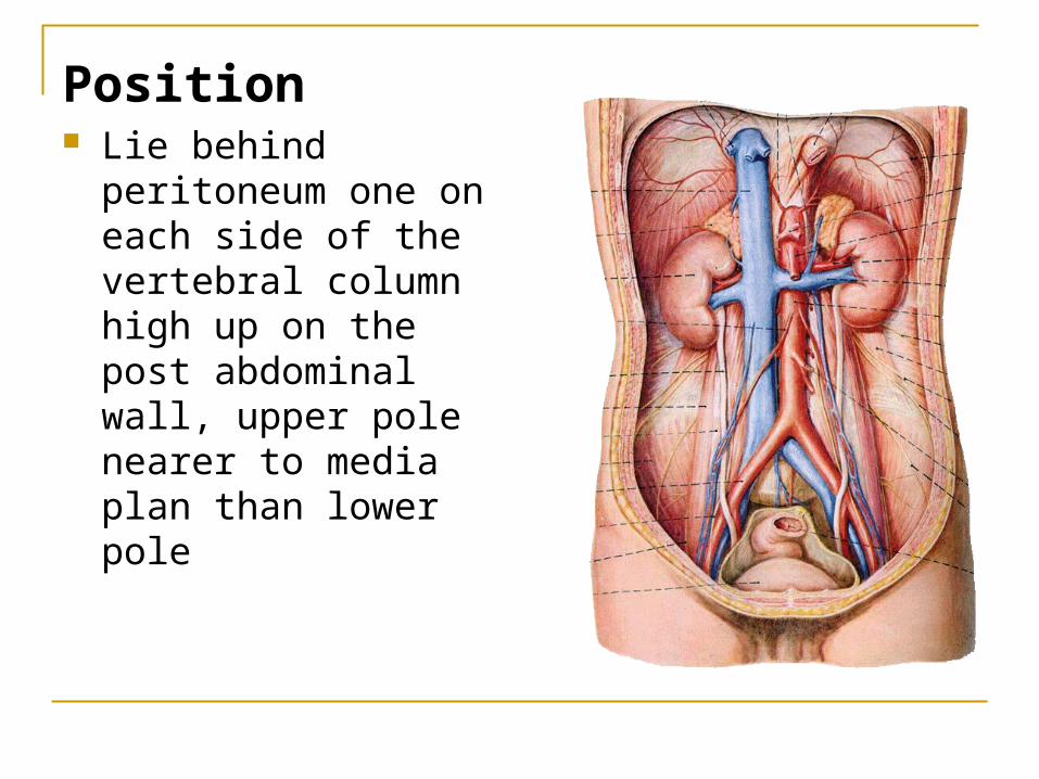
Position Lie behind peritoneum
one on each side of the vertebral column high up on the post abdominal wall, upper pole nearer to media plan than lower pole
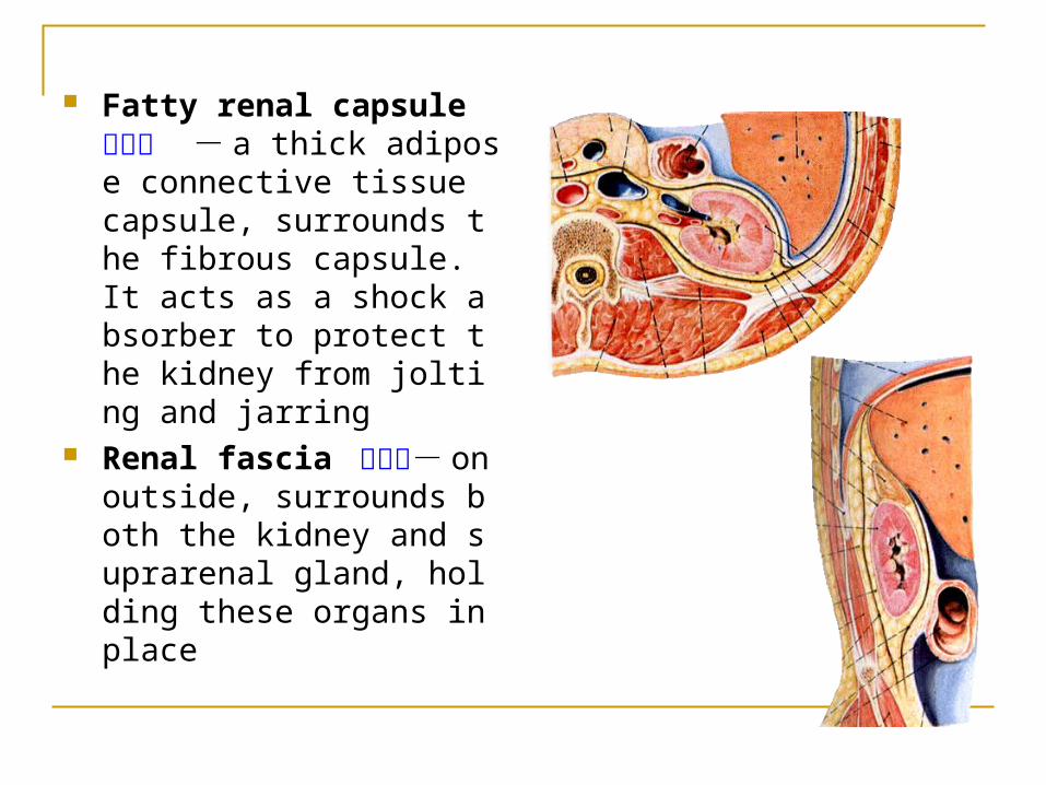
Fatty renal capsule 脂肪囊 - a thick adipose connective tissue capsule, surrounds the fibrous capsule. It acts as a shock absorber to protect the kidney from jolting and jarring
Renal fascia 肾筋膜- on outside, surrounds both the kidney and suprarenal gland, holding these organs in place
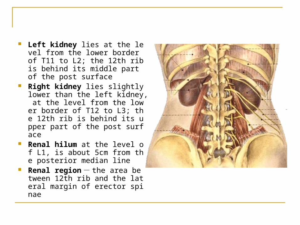
Left kidney lies at the level from the lower border of T11 to L2; the 12th rib is behind its middle part of the post surface
Right kidney lies slightly lower than the left kidney, at the level from the lower border of T12 to L3; the 12th rib is behind its upper part of the post surface
Renal hilum at the level of L1, is about 5cm from the posterior median line
Renal region - the area between 12th rib and the lateral margin of erector spinae
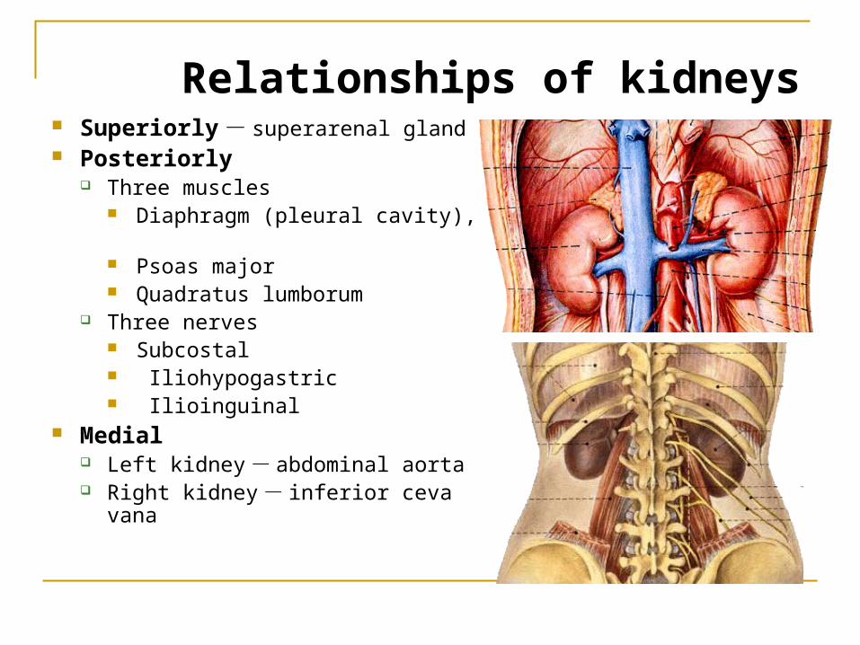
Superiorly - superarenal gland Posteriorly
Three muscles Diaphragm (pleural cavity), Psoas major Quadratus lumborum
Three nerves Subcostal Iliohypogastric Ilioinguinal
Medial Left kidney - abdominal aorta Right kidney - inferior ceva van
a
Relationships of kidneys
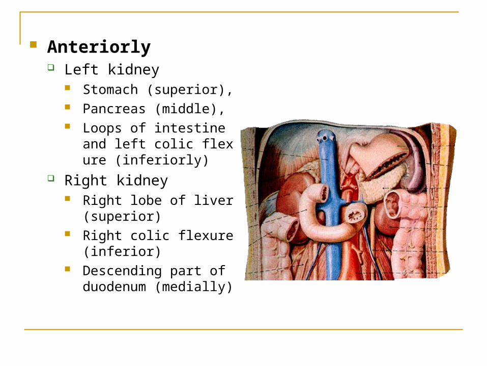
Anteriorly Left kidney
Stomach (superior), Pancreas (middle), Loops of intestine and lef
t colic flexure (inferiorly) Right kidney
Right lobe of liver (superior)
Right colic flexure (inferior)
Descending part of duodenum (medially)
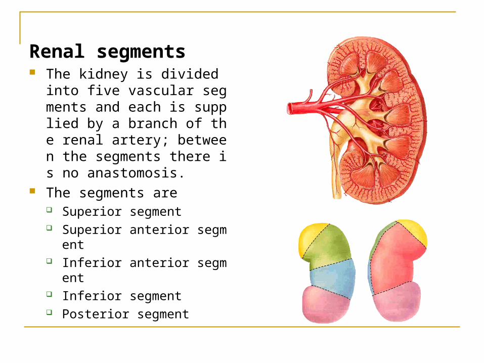
Renal segments The kidney is divided into fiv
e vascular segments and each is supplied by a branch of the renal artery; between the segments there is no anastomosis.
The segments are Superior segment Superior anterior segment Inferior anterior segment Inferior segment Posterior segment
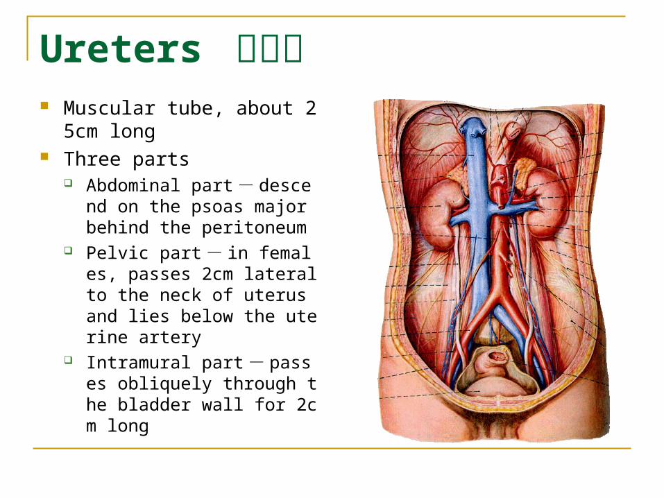
Ureters 输尿管 Muscular tube, about 25cm l
ong Three parts
Abdominal part - descend on the psoas major behind the peritoneum
Pelvic part - in females, passes 2cm lateral to the neck of uterus and lies below the uterine artery
Intramural part - passes obliquely through the bladder wall for 2cm long

Three constrictions At the pelvoureteric junctio
n Where it crosses the pelvi
c inlet and iliac vessels Where it pierces the bladd
er wall obliquely (at intramural part)
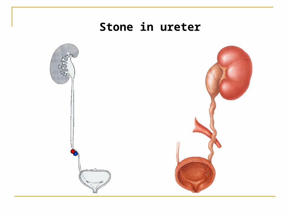
Stone in ureter
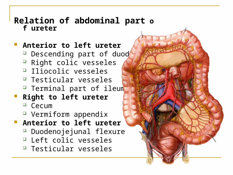
Relation of abdominal part of ureter
Anterior to left ureter Descending part of duodenum Right colic vesseles Iliocolic vesseles Testicular vesseles Terminal part of ileum
Right to left ureter Cecum Vermiform appendix
Anterior to left ureter Duodenojejunal flexure Left colic vesseles Testicular vesseles
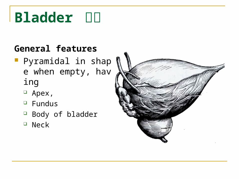
Bladder 膀胱
General features Pyramidal in shape wh
en empty, having Apex, Fundus Body of bladder Neck
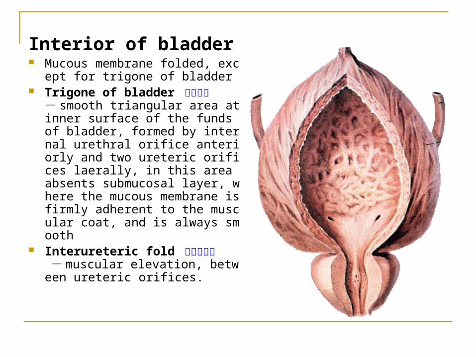
Interior of bladder Mucous membrane folded, exce
pt for trigone of bladder Trigone of bladder 膀胱三角
- smooth triangular area at inner surface of the funds of bladder, formed by internal urethral orifice anteriorly and two ureteric orifices laerally, in this area absents submucosal layer, where the mucous membrane is firmly adherent to the muscular coat, and is always smooth
Interureteric fold 输尿管间襞 - muscular elevation, between ureteric orifices.
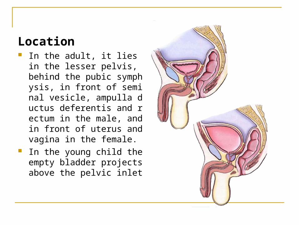
Location In the adult, it lies in the les
ser pelvis, behind the pubic symphysis, in front of seminal vesicle, ampulla ductus deferentis and rectum in the male, and in front of uterus and vagina in the female.
In the young child the empty bladder projects above the pelvic inlet
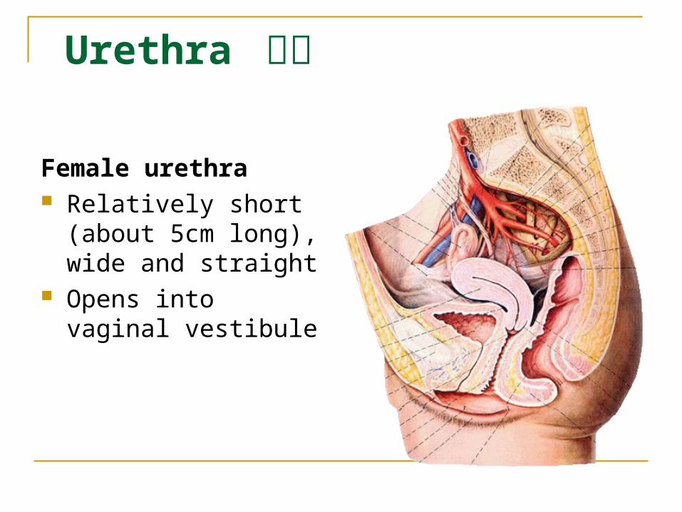
Urethra 尿道
Female urethra Relatively short (about
5cm long), wide and straight
Opens into vaginal vestibule
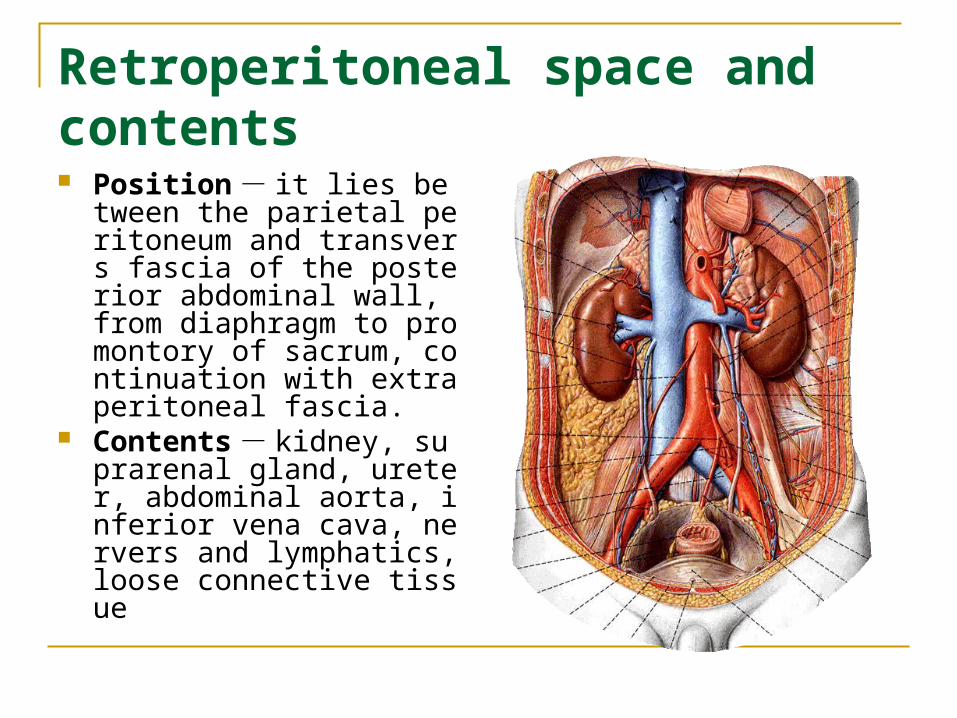
Retroperitoneal space and contents Position - it lies between t
he parietal peritoneum and transvers fascia of the posterior abdominal wall, from diaphragm to promontory of sacrum, continuation with extraperitoneal fascia.
Contents - kidney, suprarenal gland, ureter, abdominal aorta, inferior vena cava, nervers and lymphatics, loose connective tissue
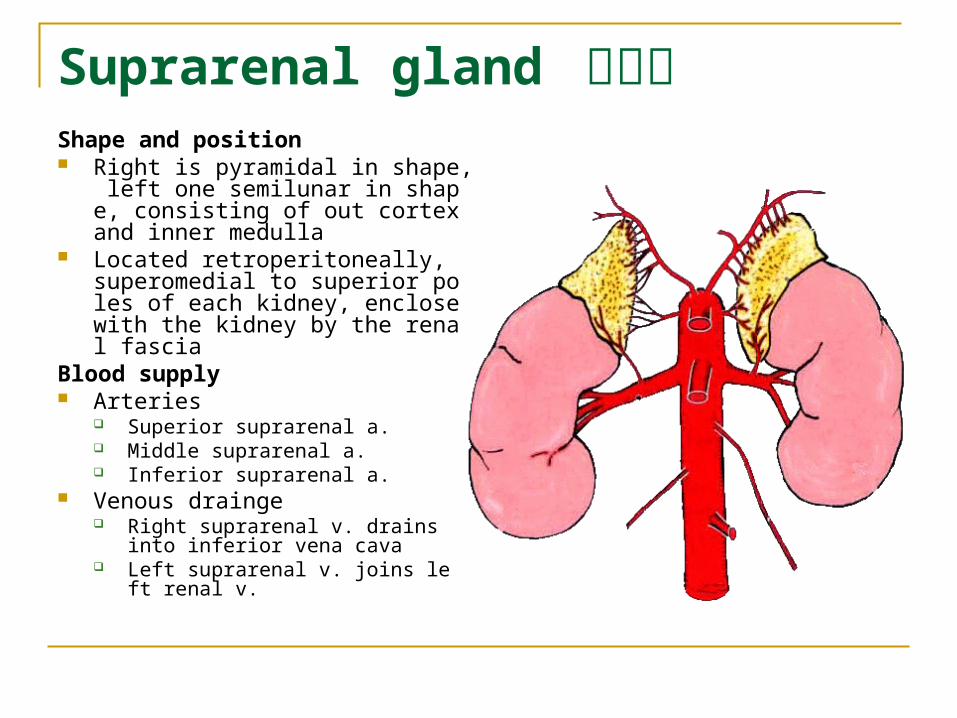
Suprarenal gland 肾上腺Shape and position Right is pyramidal in shape, left one
semilunar in shape, consisting of out cortex and inner medulla
Located retroperitoneally, superomedial to superior poles of each kidney, enclose with the kidney by the renal fascia
Blood supply Arteries
Superior suprarenal a. Middle suprarenal a. Inferior suprarenal a.
Venous drainge Right suprarenal v. drains into inferior
vena cava Left suprarenal v. joins left renal v.
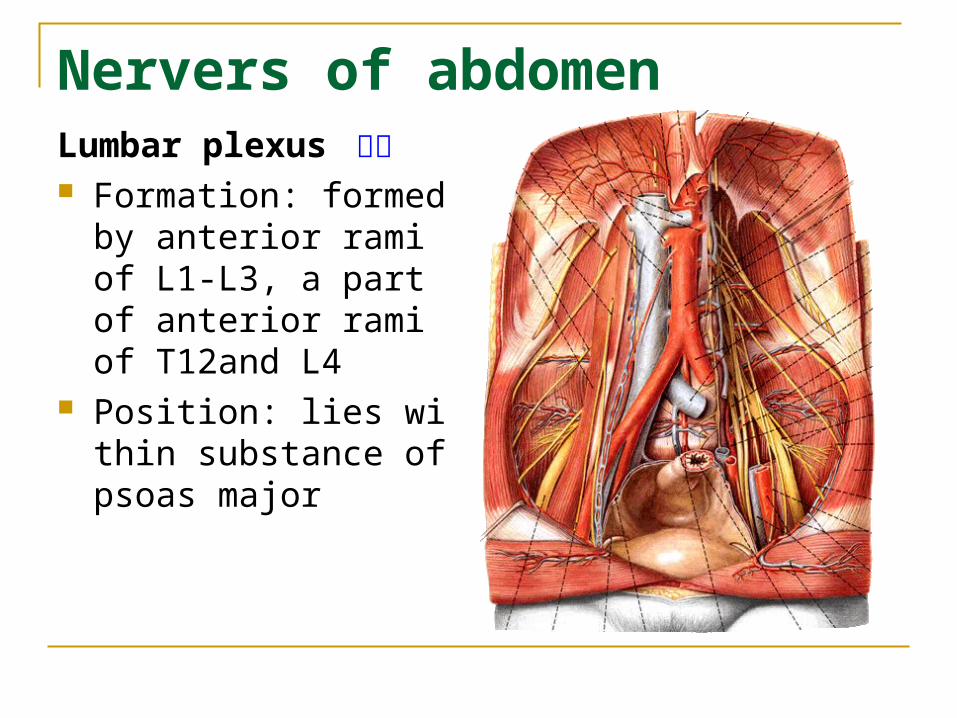
Nervers of abdomen Lumbar plexus 腰丛 Formation: formed by a
nterior rami of L1-L3, a part of anterior rami of T12and L4
Position: lies within substance of psoas major
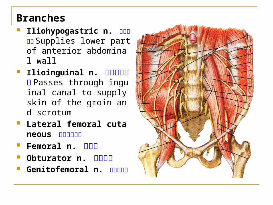
Branches Iliohypogastric n. 髂腹下神经
Supplies lower part of anterior abdominal wall
Ilioinguinal n. 髂腹股沟神经Passes through inguinal canal to supply skin of the groin and scrotum
Lateral femoral cutaneous 股外侧皮神经
Femoral n. 股神经 Obturator n. 闭孔神经 Genitofemoral n. 生殖股神经
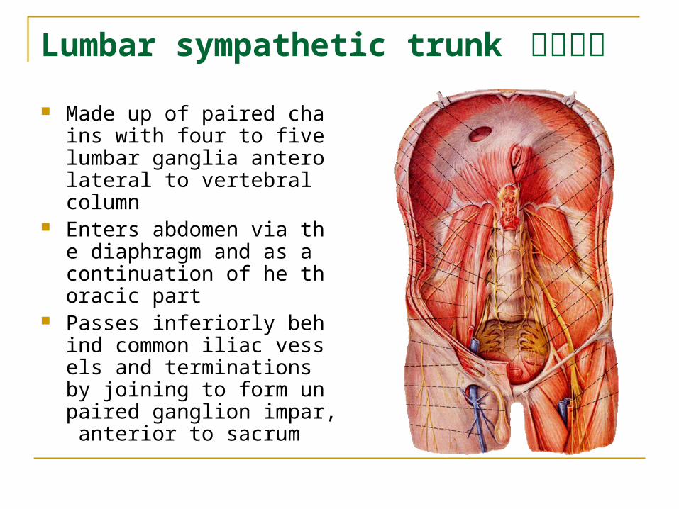
Lumbar sympathetic trunk 腰交感干
Made up of paired chains with four to five lumbar ganglia anterolateral to vertebral column
Enters abdomen via the diaphragm and as a continuation of he thoracic part
Passes inferiorly behind common iliac vessels and terminations by joining to form unpaired ganglion impar, anterior to sacrum
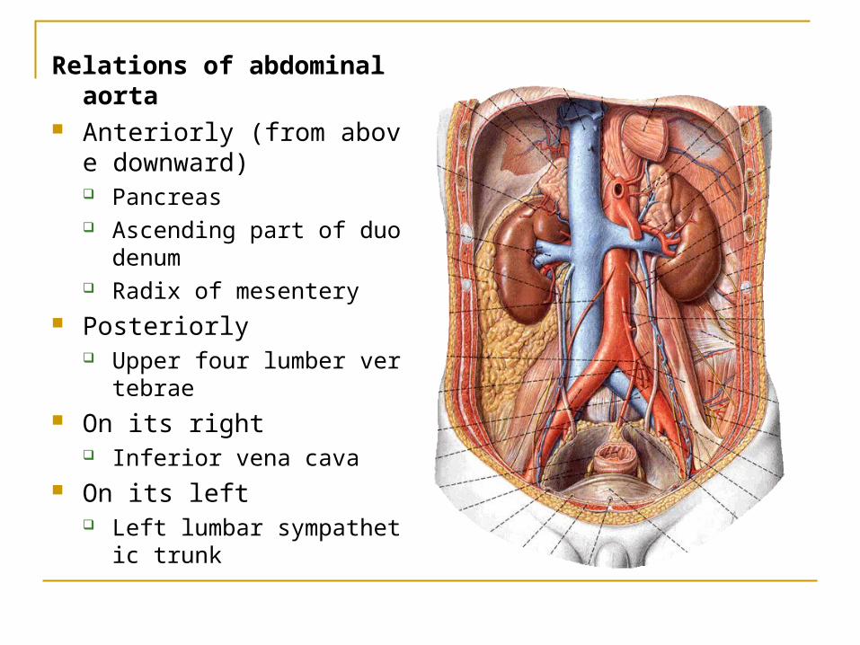
Relations of abdominal aorta Anteriorly (from above down
ward) Pancreas Ascending part of duodenu
m Radix of mesentery
Posteriorly Upper four lumber vertebra
e On its right
Inferior vena cava On its left
Left lumbar sympathetic trunk
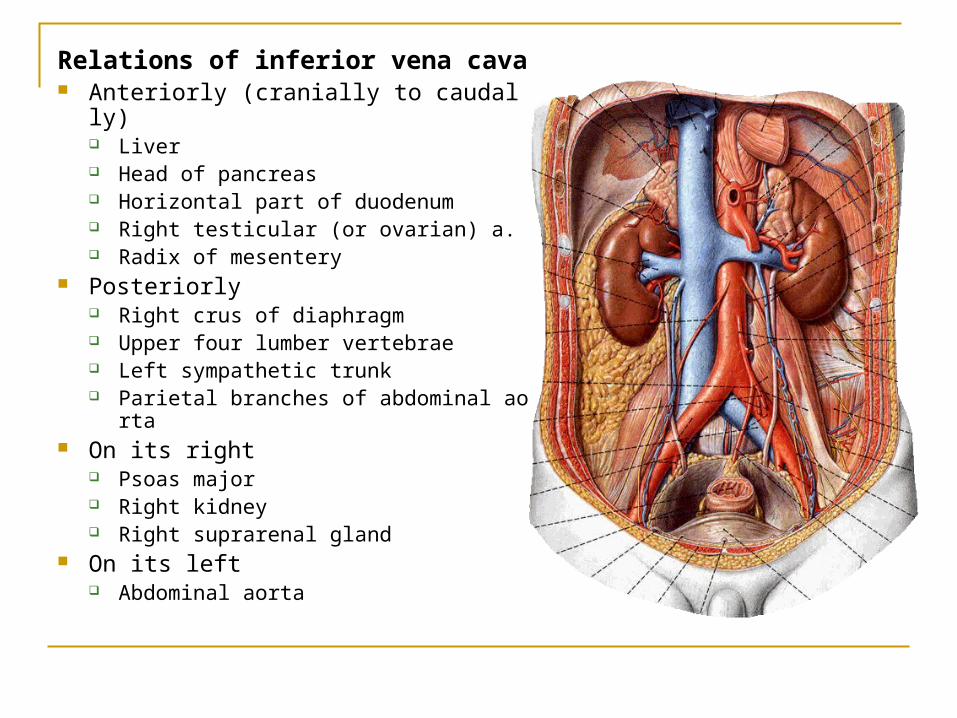
Relations of inferior vena cava Anteriorly (cranially to caudally)
Liver Head of pancreas Horizontal part of duodenum Right testicular (or ovarian) a. Radix of mesentery
Posteriorly Right crus of diaphragm Upper four lumber vertebrae Left sympathetic trunk Parietal branches of abdominal aorta
On its right Psoas major Right kidney Right suprarenal gland
On its left Abdominal aorta