Embed Size (px)
Citation preview
Tissue Engineering of Articular Cartilage Using an Allograft ofCultured Chondrocytes in a Membrane-Sealed AtelocollagenHoneycomb-Shaped Scaffold (ACHMS Scaffold)
Kazunori Masuoka,1 Takashi Asazuma,1 Masayuki Ishihara,2 Masato Sato,3 Hidemi Hattori,2
Miya Ishihara,4 Yasuo Yoshihara,1 Takemi Matsui,2 Bonpei Takase,2 Makoto Kikuchi,4 Koichi Nemoto1
1 Department of Orthopaedic Surgery, National Defense Medical College, 3-2 Namiki, Tokorozawa, Saitama 359-8513, Japan
2 Division of Biomedical Engineering, Research Institute, National Defense Medical College, 3-2 Namiki, Tokorozawa,Saitama 359-8513, Japan
3 Department of Orthopaedic Surgery, Surgical Science, Tokai University School of Medicine, Bohseidai, Isehara,Kanagawa 259-1193, Japan
4 Department of Medical Engineering, National Defense Medical College, 3-2 Namiki, Tokorozawa, Saitama 359-8513, Japan
Received 2 September 2004; revised 15 December 2004; accepted 22 December 2004Published online 15 July 2005 in Wiley InterScience (www.interscience.wiley.com). DOI: 10.1002/jbm.b.30284
Abstract: The aim of this study was to investigate with tissue engineering procedures thepossibility of using atelocollagen honeycomb-shaped scaffolds sealed with a membrane(ACHMS scaffold) for the culturing of chondrocytes to repair articular cartilage defects.Chondrocytes from the articular cartilage of Japanese white rabbits were cultured in ACHMSscaffolds to allow a high-density, three-dimensional culturing for up to 21 days. Although theDNA content in the scaffold increased at a lower rate than monolayer culturing, scanningelectron microscopy data showed that the scaffold was filled with grown chondrocytes andtheir produced extracellular matrix after 21 days. In addition, glycosaminoglycan (GAG)accumulation in the scaffold culture was at a higher level than the monolayer culture.Cultured cartilage in vitro for 14 days showed enough elasticity and stiffness to be handled invivo. An articular cartilage defect was initiated in the patellar groove of the femur of rabbitsand was subsequently filled with the chondrocyte-cultured ACHMS scaffold, ACHMS scaffoldalone, or nonfilled (control). Three months after the operations, histological analysis showedthat only defects inserted with chondrocytes being cultured in ACHMS scaffolds were filledwith reparative hyaline cartilage, and thereby highly expressing type II collagen. These resultsindicate that implantation of allogenic chondrocytes cultured in ACHMS scaffolds may beeffective in repairing articular cartilage defects. © 2005 Wiley Periodicals, Inc. J Biomed Mater ResPart B: Appl Biomater 75B: 177–184, 2005
Keywords: regeneration; articular cartilage; chondrocytes; scaffold; tissue engineering
INTRODUCTION
Articular cartilages show a limited reparative capacity afterinjury. Full-thickness cartilage injuries typically heal usingfibrocartilage. This tissue, however, lacks the biochemicaland mechanical properties necessary to yield a complete anddurable repair.1 Initially, the repair tissue may appear andfunction like hyaline cartilage; it deteriorates with time, pro-voking a degenerative process that may eventually involvethe entire joint.2,3 This inability to reconstitute the cartilagetissue by an intrinsic healing process has promoted studies of
various composite materials in chondrocyte-seeded implantsin the treatment of cartilage lesions.
The implantation of chondrocyte–collagen composites intocartilage defects has proved a promising method of cartilagerepair.4,5 The high-density, three-dimensional culture systemusing the scaffold is able to maintain the phenotype of chondro-cytes and their ability to produce extracellular molecules re-quired for tissue reconstruction.6,7 In fact, chondrocytes culturedin three-dimensional collagen gels produce an extracellular ma-trix consisting of proteoglycans and type II collagen having anarchitecture similar to hyaline cartilage.4,5 However, the colla-gen gel is mechanically too weak to maintain a desired shapeand structure during culturing, and requires some complicatedprocedures to retain the chondrocyte–collagen gel in vivo in the
Correspondence to: M. Ishihara (e-mail: [email protected])
© 2005 Wiley Periodicals, Inc.
177
cartilage lesions. Although numerous tissue engineering ap-proaches have been studied to repair damaged cartilage usingsuch scaffolds as agarose,8 alginate,9 chitosan,10 hyaluronic ac-id,11 fibrin,12 and synthetic polymers,13–15 an improved andmore reliable strategy is desired for cartilage repair.
A new atelocollagen honeycomb-shaped scaffold contain-ing a membrane sealing (ACHMS scaffold) has been reportedto allow a high-density, three-dimensional culturing of vari-ous cells as tissue engineering scaffold.16,17 Furthermore,previous reports indicated the useful application of this scaf-fold as a biomaterial for tissue engineering of intervertebraldiscs using annulus fibrosus (AF) cells.18,19 The AF cellswere able to grow in vitro and maintain its phenotype in theACHMS scaffold. In addition, the AF cells cultured in anACHMS scaffold maintained the viability and ability to pro-duce type II collagen and proteoglycan.17 The main purposeof this work is to evaluate the ACHMS scaffold in a three-dimensional culturing of chondrocyte cells for tissue engi-neering of articular cartilage.
MATERIALS AND METHODS
Preparation of ACHMS Scaffold
Preparation of the atelocollagen honeycomb sponge has beendescribed previously16 and has been obtained from KOKEN Inc.Tokyo, Japan. Briefly, type I atelocollagen from bovine dermisin solution (1%, pH 3) was poured into a shallow flask andexposed to ammonia gas in order to neutralize. The resultingwhite gel was then rinsed with distilled water to wash out theexcessive ammonia and salt produced during the neutralizationreaction. The gel freeze-dried resulted in honeycomb-shapedsponge. The pore diameter of the sponge was controlled bychanging the concentration of collagen solution and ammoniagas. When a high concentration of ammonia gas was used, smallpores were produced, and vice versa. In this study, we used thescaffold with average 350 �m diameter pores. The slices wereirradiated with ultraviolet (UV) light to yield an insoluble hon-eycomb-shaped sponge material.16 To prepare the collagen–membrane sealing, type I atelocollagen solution (1%, pH 3) waspoured into the flask and exposed to ammonia gas to neutralize.The honeycomb-shaped sponges were placed on the producedthin gel and air-dried in a clean bench. After making the mem-brane insoluble by UV irradiation, the honeycomb-shapedsponges with membrane sealing at the bottom side were cut intoround shapes having a diameter of 6 mm using a punch. Finally,the round atelocollagen honeycombs with a membrane sealingwere treated at 130°C for 20 min in vacuum for sterilization.17
Culturing of Chondrocytes from Joints of Rabbits
Articular cartilage slices from joints (hip, knee, and shoulderjoints) were obtained from 4-week-old male Japanese whiterabbits weighing about 1 kg. These cartilage slices wereminced and then digested in F12/DMEM (Iwaki Inc., Tokyo,Japan) containing 0.4% (w/v) pronase E (Kaken Pharmaceu-tical Inc., Tokyo, Japan) for 1 h and F12/DMEM containing
0.025% (w/v) bacterial Collagenase P (Boehringer Mann-heim Co. Ltd., Mannheim, Germany) for 4 h. The digestedtissue was passed through a cell strainer (Becton DickinsonLabware Co. Ltd.) with a pore size of 40 �m. The filtrate wascentrifuged at 1500 rpm for 10 min to separate the cells. Thecells (0.2 mL) were then seeded at a high density (2 � 106
cells/scaffold) on the top (open side) of the round ACHMSscaffold (diameter, 6 mm; thickness, 2 mm) in 96-well plates(Sumitomo Bakelite Corp., Tokyo, Japan) by centrifugationat 500 rpm (45 g) for 5 min and cultured in F12/DMEMsupplemented with 10% fetal bovine serum (Iwaki) and 50�g/mL ascorbic acid (Sigma Corp., St, Louis, MO) at 37°C inan atmosphere of 5% CO2 in air and 100% relative humidity.After the indicated period of incubation, the cultures werestudied by (i) a scanning electron microscope, (ii) determi-nation of the DNA content as indicator of cell proliferation,as well as (iii) determination of glycosaminoglycan (GAG)accumulation as marker of matrix formation.17 Prior to trans-plantation of the chondrocyte-containing ACHMS scaffoldinto an articular cartilage defect in vivo, the chondrocyteswere cultured for 14 days in the scaffold. The cells also havebeen plated at a density of 2 � 106 cells/dish (Falcon culturedish; diameter, 10 cm; Nippon Becton Dickinson Co., Ltd.,Tokyo, Japan) and monolayer cultured under the same con-ditions as described above.
Scanning Electron Microscopy
Samples were fixed in 2.5% glutaraldehyde (Wako PureChemical Corp., Osaka, Japan) and 1% osmium acid (Wako
TABLE I. Histological Grading Scale for Defect Cartilage
Category Points
A. Cell MorphologyHyaline cartilage 0Mostly hyaline cartilage 1Mostly fibrocartilage 2Mostly noncartilage 3Noncartilage only 4
B. Matrix Staining (Metachromasia)Normal (compared with host adjacent cartilage) 0Slightly reduced 1Markedly reduced 2No metachromatic stain 3
C. Surface RegularitySmooth (�3/4) 0Moderate (�1/2–3/4) 1Irregular (1/4–1/2) 2Severely irregular (�1/4) 3
D. Thickness of Cartilage�2/3 01/3–2/3 1�1/3 2
E. Integration of Donor with Host Adjacent CartilageBoth edges integrated 0One edge integrated 1Neither edge integrated 2
Maximum Total 14
This table was extracted from Wakitani et al.’s histological grading scale.22
178 MASUOKA ET AL.
Pure Chemical). After dehydration through a graded series ofwater–ethanol and ethanol–isoamyl acetate solutions, thesamples were critical point-dried (HCP-2, Hitachi Corp., To-kyo, Japan). Samples were subsequently shadowed with goldusing an ion sputter (JFC1100, Nihon Denshi Corp., Tokyo,Japan) and observed with a scanning electron microscope(JSM-840, Nihon Denshi Corp.).17,19
Fluorimetric Assay of DNA
The DNA content in papain digests of the cells and matrixcomponents were determined using a fluorimetric assay.20
Briefly, 15 �L of the papain digest was mixed with a 300 �Lsolution of Hoechst 33258 (Polyscience Corp., Warrrington,PA) in a 96-well fluoroplate (Sumiron R; Sumitomo BakeliteCorp.). Emission and excitation spectra were obtained with aTitertek Multiscan Spectrofluorometer (Lab Systems Corp.,Helsinki, Finland) at 456 nm and 365 nm, respectively. DNAconcentrations were determined against a standard curve ofcalf thymus DNA (Sigma) ranging from 0.625 to 40 �g/mL.
Assay to Determine the GAG Content
To examine the GAG content in cultured chondrocytes in theACHMS scaffold, the cell–matrix component was assayed using
the 1,9-dimethylmethylene blue (DMB) method, as described byFarndale and colleagues.21 Briefly, the cell cultures within thehoneycomb-shaped scaffold on day 2, 7, 14, and 21 were di-gested with 300 �g/mL papain in 50 mM phosphate buffer, pH6.5, containing 2 mM N-acetyl cysteine and 2 mM EDTA at60°C, overnight. Also the cells cultured in a monolayer wereharvested using a scraper and digested with papain as describedabove. The sample solutions (140 �L) were gently mixed withan equal volume of DMB solution in a 96-well microtiter plateand the absorbance at 530 nm was measured immediately usinga Titertec Multiscan Spectrophotometer (Lab Systems). Absor-bance values were quantified against a standard curve of sharkchondroitin sulfate C (Seikagaku Kogyo Corp., Tokyo, Japan),ranging from 0.625 to 20 �g/mL. The absorbance value of thedigestion of the scaffold without cells with papain was definedas 0 point.
Transplantation of Chondrocytes in the ACHMS Scaffold
Twelve Japanese white rabbits (male, 16–18 weeks old,average weight 3 kg) were used in this study. The rabbitswere anesthetized with intramuscular injections of 120 mgketamine and 9 mg xylazine. After a medial parapatellarincision of both legs, each patella was dislocated laterally and
Figure 1. Appearance of the chondrocyte-seeded honeycomb-shaped atelocollagen scaffold withmembrane sealing (ACHMS scaffold). Upper panel: Microscopic appearance of the chondrocyte-seeded ACHMS scaffold (original magnification, 4�). Bar represents 5 mm. Lower panel: Scanningelectron microphotographs of the chondrocyte-seeded ACHMS scaffold (original magnification,100�). Bar represents 300 �m. (A, D) Before seeding the cells, (B, E) cultured for 14 days, (C, F)cultured for 21 days. [Color figure can be viewed in the online issue, which is available at www.inter-science.wiley.com.]
179ACHMS SCAFFOLD
one full thickness cylindrical defect (4 mm in diameter and 3mm deep) was created on the patellar groove of the femur inboth legs using a low-speed drill (Takagi Corp., Niigata,Japan). The rabbits were classified into the following threerecipient groups: (i) the cells-containing scaffold insertiongroup, in which chondrocytes cultured in an ACHMS scaf-fold were allografted into the created full-thickness articularcartilage defect, (ii) the scaffold insertion group, in whichonly the ACHMS scaffold was inserted, and (iii) the nonin-sertion group. Before allograft transplantation, chondrocytescultured in the scaffold and the scaffold without cells werecut to adjust to the size of defects. The implanted scaffolds(with and without chondrocytes) were inserted into the de-fects and fixed without any additional fixation such as fibringlue. After the operation, all animals were allowed to walkfreely without any splints in the cages. These animal exper-iments were approved and carried out following the guide-lines for animal experimentation of the National DefenseMedical College, Tokorozawa, Saitama, Japan.
Histological Evaluations In Vitro and In Vivoand Wakitani’s Scale
The rabbits were sacrificed 3 months after the operation by anoverdose intravenous anesthesia. The distal part of the femurwas excised and fixed with 10% buffered formalin for a periodof 7 days. The chondrocytes cultured in an ACHMS scaffold invitro were also fixed with 10% buffered formalin for 7 days.Each specimen was decalcified with 10% DETA in distilledwater (pH 7.4) for 3 weeks and embedded in paraffin. Sectionsabout 6 �m thick were cut through the grafted area, strippedfrom paraffin, and stained with toluidine blue.
Sections were, after stripping from paraffin, also pre-treated with 0.1 mg/mL actinase E (Kaken PharmaceuticalInc.) in PBS at 37°C for 30 min. And after incubating thesection with 10% pig serum at room temperature for 30 min,thus reducing the degree of nonspecific background staining,the sections were incubated overnight with 50 �g/mL mouseantihuman type II collagen monoclonal antibody (DaiichiFine Chemical Corp., Toyama, Japan) in PBS containing0.1% bovine serum albumin at 4°C. The sections were thenincubated with biotinylated rabbit antimouse immunoglobins(Dako Co.) at dilution of 1:500 for 30 min and followed withperoxidase-conjugated streptavidin (Dako) at dilution of1:500 for 30 min at room temperature. Finally, the sectionswere immunostained by incubating with 20 mg DAB (diami-nobenzidine) and 5 �L hydrogen peroxide (30%) in 100 mLPBS for 5 min at room temperature. Sections were incubatedwith PBS instead of specific primary antibodies and stainedas a negative control.
The sections were evaluated microscopically and scoredaccording to a histological grading scale, consisting of 5categories with a total score ranging from 0 to 14 points asdescribed by Wakitani and colleagues (Table I).22
Statistical Analysis
To analyze quantitative DNA and GAG data, the Mann–Whit-ney U test was carried out. Histological results were analyzedusing ANOVA, followed by the Dunn test as post hoc test. pvalues less than 0.05 were considered statistically significant.
RESULTS
Structure of ACHMS Scaffold
The upper surface of the ACHMS scaffold was found to behoneycomb shaped, as shown in Figures 1(A,D). The diam-eter of the pores could be controlled from about 100 to 1,000�m upon varying the concentration of the collagen solutionand ammonia gas. The lower surface was sealed with thesame atelocollagen membrane. Scaffolds having a diameterof about 6 mm were used in the present study. The honey-comb-shaped parts consisted of thin multiple collagen mem-branes of less than 1 �m in thickness. It was found to beelastic and during the entire cell culture procedure did notdeform or collapse upon forceps handling. Scaffolds could be
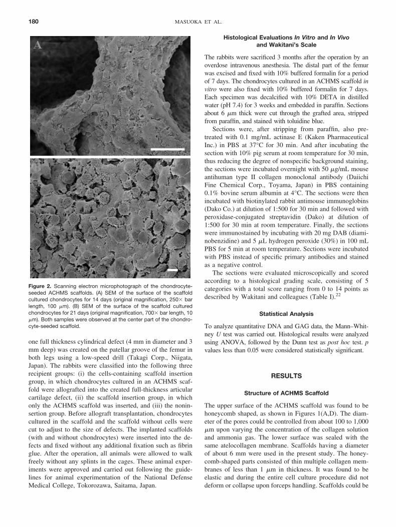
Figure 2. Scanning electron microphotograph of the chondrocyte-seeded ACHMS scaffolds. (A) SEM of the surface of the scaffoldcultured chondrocytes for 14 days (original magnification, 250� barlength, 100 �m). (B) SEM of the surface of the scaffold culturedchondrocytes for 21 days (original magnification, 700� bar length, 10�m). Both samples were observed at the center part of the chondro-cyte-seeded scaffold.
180 MASUOKA ET AL.
easily cut with scissors or sharp knives into a desirable shapebeing maintained during the cell culturing.
Culture of Chondrocytes in ACHMS Scaffolds In Vitro
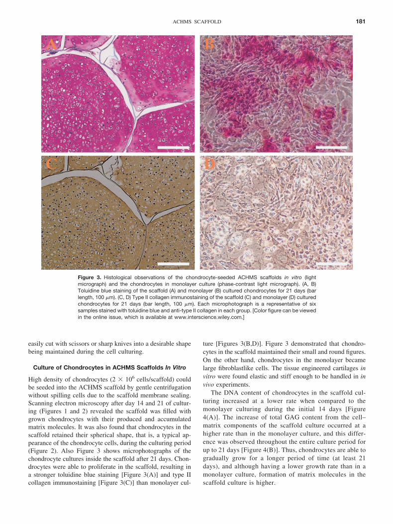
High density of chondrocytes (2 � 106 cells/scaffold) couldbe seeded into the ACHMS scaffold by gentle centrifugationwithout spilling cells due to the scaffold membrane sealing.Scanning electron microscopy after day 14 and 21 of cultur-ing (Figures 1 and 2) revealed the scaffold was filled withgrown chondrocytes with their produced and accumulatedmatrix molecules. It was also found that chondrocytes in thescaffold retained their spherical shape, that is, a typical ap-pearance of the chondrocyte cells, during the culturing period(Figure 2). Also Figure 3 shows microphotographs of thechondrocyte cultures inside the scaffold after 21 days. Chon-drocytes were able to proliferate in the scaffold, resulting ina stronger toluidine blue staining [Figure 3(A)] and type IIcollagen immunostaining [Figure 3(C)] than monolayer cul-
ture [Figures 3(B,D)]. Figure 3 demonstrated that chondro-cytes in the scaffold maintained their small and round figures.On the other hand, chondrocytes in the monolayer becamelarge fibroblastlike cells. The tissue engineered cartilages invitro were found elastic and stiff enough to be handled in invivo experiments.
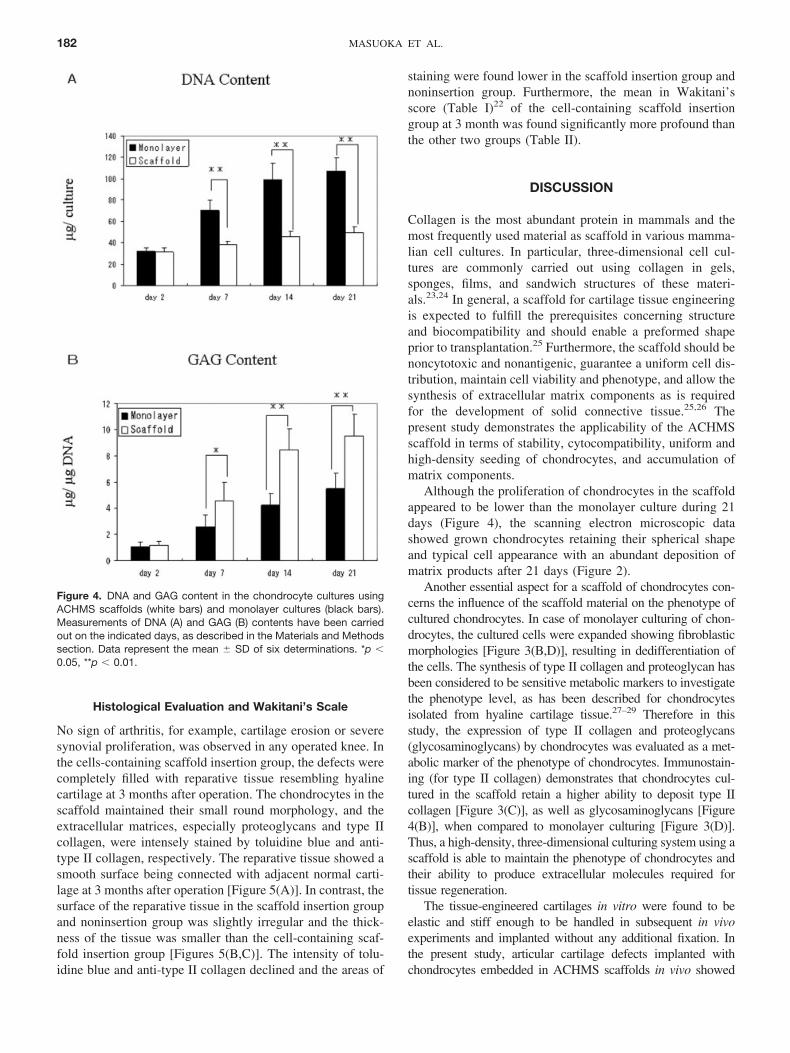
The DNA content of chondrocytes in the scaffold cul-turing increased at a lower rate when compared to themonolayer culturing during the initial 14 days [Figure4(A)]. The increase of total GAG content from the cell–matrix components of the scaffold culture occurred at ahigher rate than in the monolayer culture, and this differ-ence was observed throughout the entire culture period forup to 21 days [Figure 4(B)]. Thus, chondrocytes are able togradually grow for a longer period of time (at least 21days), and although having a lower growth rate than in amonolayer culture, formation of matrix molecules in thescaffold culture is higher.
Figure 3. Histological observations of the chondrocyte-seeded ACHMS scaffolds in vitro (lightmicrograph) and the chondrocytes in monolayer culture (phase-contrast light micrograph). (A, B)Toluidine blue staining of the scaffold (A) and monolayer (B) cultured chondrocytes for 21 days (barlength, 100 �m). (C, D) Type II collagen immunostaining of the scaffold (C) and monolayer (D) culturedchondrocytes for 21 days (bar length, 100 �m). Each microphotograph is a representative of sixsamples stained with toluidine blue and anti-type II collagen in each group. [Color figure can be viewedin the online issue, which is available at www.interscience.wiley.com.]
181ACHMS SCAFFOLD
Histological Evaluation and Wakitani’s Scale
No sign of arthritis, for example, cartilage erosion or severesynovial proliferation, was observed in any operated knee. Inthe cells-containing scaffold insertion group, the defects werecompletely filled with reparative tissue resembling hyalinecartilage at 3 months after operation. The chondrocytes in thescaffold maintained their small round morphology, and theextracellular matrices, especially proteoglycans and type IIcollagen, were intensely stained by toluidine blue and anti-type II collagen, respectively. The reparative tissue showed asmooth surface being connected with adjacent normal carti-lage at 3 months after operation [Figure 5(A)]. In contrast, thesurface of the reparative tissue in the scaffold insertion groupand noninsertion group was slightly irregular and the thick-ness of the tissue was smaller than the cell-containing scaf-fold insertion group [Figures 5(B,C)]. The intensity of tolu-idine blue and anti-type II collagen declined and the areas of
staining were found lower in the scaffold insertion group andnoninsertion group. Furthermore, the mean in Wakitani’sscore (Table I)22 of the cell-containing scaffold insertiongroup at 3 month was found significantly more profound thanthe other two groups (Table II).
DISCUSSION
Collagen is the most abundant protein in mammals and themost frequently used material as scaffold in various mamma-lian cell cultures. In particular, three-dimensional cell cul-tures are commonly carried out using collagen in gels,sponges, films, and sandwich structures of these materi-als.23,24 In general, a scaffold for cartilage tissue engineeringis expected to fulfill the prerequisites concerning structureand biocompatibility and should enable a preformed shapeprior to transplantation.25 Furthermore, the scaffold should benoncytotoxic and nonantigenic, guarantee a uniform cell dis-tribution, maintain cell viability and phenotype, and allow thesynthesis of extracellular matrix components as is requiredfor the development of solid connective tissue.25,26 Thepresent study demonstrates the applicability of the ACHMSscaffold in terms of stability, cytocompatibility, uniform andhigh-density seeding of chondrocytes, and accumulation ofmatrix components.
Although the proliferation of chondrocytes in the scaffoldappeared to be lower than the monolayer culture during 21days (Figure 4), the scanning electron microscopic datashowed grown chondrocytes retaining their spherical shapeand typical cell appearance with an abundant deposition ofmatrix products after 21 days (Figure 2).
Another essential aspect for a scaffold of chondrocytes con-cerns the influence of the scaffold material on the phenotype ofcultured chondrocytes. In case of monolayer culturing of chon-drocytes, the cultured cells were expanded showing fibroblasticmorphologies [Figure 3(B,D)], resulting in dedifferentiation ofthe cells. The synthesis of type II collagen and proteoglycan hasbeen considered to be sensitive metabolic markers to investigatethe phenotype level, as has been described for chondrocytesisolated from hyaline cartilage tissue.27–29 Therefore in thisstudy, the expression of type II collagen and proteoglycans(glycosaminoglycans) by chondrocytes was evaluated as a met-abolic marker of the phenotype of chondrocytes. Immunostain-ing (for type II collagen) demonstrates that chondrocytes cul-tured in the scaffold retain a higher ability to deposit type IIcollagen [Figure 3(C)], as well as glycosaminoglycans [Figure4(B)], when compared to monolayer culturing [Figure 3(D)].Thus, a high-density, three-dimensional culturing system using ascaffold is able to maintain the phenotype of chondrocytes andtheir ability to produce extracellular molecules required fortissue regeneration.
The tissue-engineered cartilages in vitro were found to beelastic and stiff enough to be handled in subsequent in vivoexperiments and implanted without any additional fixation. Inthe present study, articular cartilage defects implanted withchondrocytes embedded in ACHMS scaffolds in vivo showed
Figure 4. DNA and GAG content in the chondrocyte cultures usingACHMS scaffolds (white bars) and monolayer cultures (black bars).Measurements of DNA (A) and GAG (B) contents have been carriedout on the indicated days, as described in the Materials and Methodssection. Data represent the mean � SD of six determinations. *p �0.05, **p � 0.01.
182 MASUOKA ET AL.
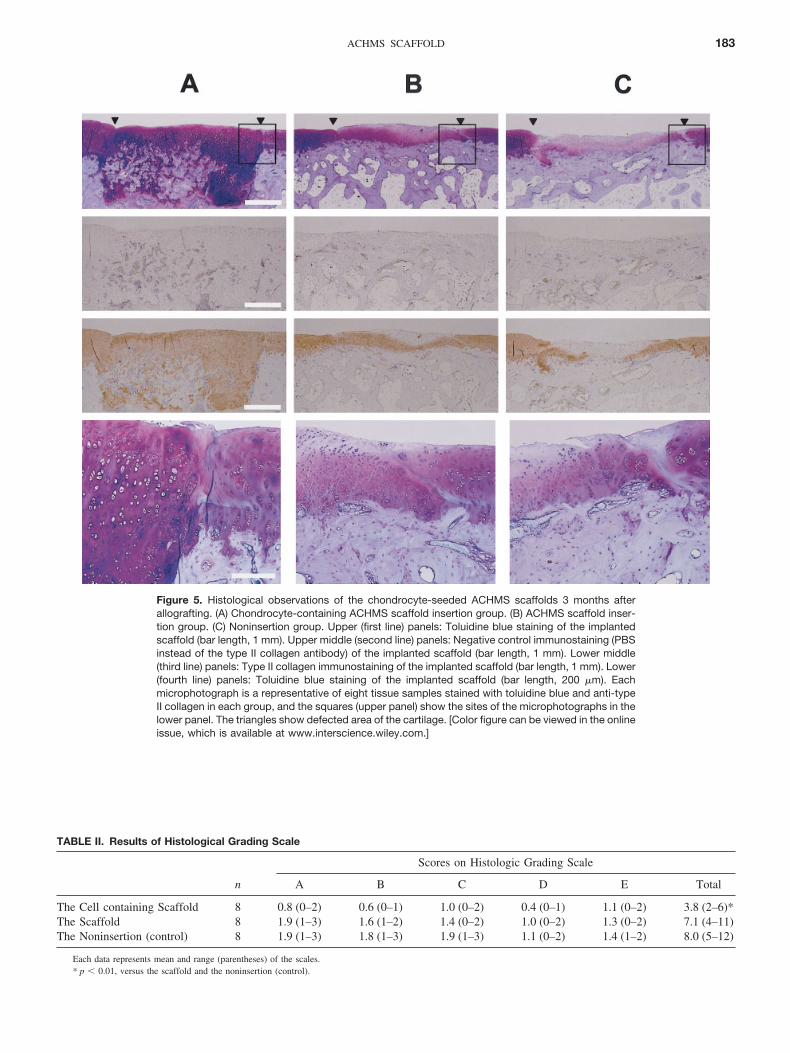
Figure 5. Histological observations of the chondrocyte-seeded ACHMS scaffolds 3 months afterallografting. (A) Chondrocyte-containing ACHMS scaffold insertion group. (B) ACHMS scaffold inser-tion group. (C) Noninsertion group. Upper (first line) panels: Toluidine blue staining of the implantedscaffold (bar length, 1 mm). Upper middle (second line) panels: Negative control immunostaining (PBSinstead of the type II collagen antibody) of the implanted scaffold (bar length, 1 mm). Lower middle(third line) panels: Type II collagen immunostaining of the implanted scaffold (bar length, 1 mm). Lower(fourth line) panels: Toluidine blue staining of the implanted scaffold (bar length, 200 �m). Eachmicrophotograph is a representative of eight tissue samples stained with toluidine blue and anti-typeII collagen in each group, and the squares (upper panel) show the sites of the microphotographs in thelower panel. The triangles show defected area of the cartilage. [Color figure can be viewed in the onlineissue, which is available at www.interscience.wiley.com.]
TABLE II. Results of Histological Grading Scale
n
Scores on Histologic Grading Scale
A B C D E Total
The Cell containing Scaffold 8 0.8 (0–2) 0.6 (0–1) 1.0 (0–2) 0.4 (0–1) 1.1 (0–2) 3.8 (2–6)*The Scaffold 8 1.9 (1–3) 1.6 (1–2) 1.4 (0–2) 1.0 (0–2) 1.3 (0–2) 7.1 (4–11)The Noninsertion (control) 8 1.9 (1–3) 1.8 (1–3) 1.9 (1–3) 1.1 (0–2) 1.4 (1–2) 8.0 (5–12)
Each data represents mean and range (parentheses) of the scales.* p � 0.01, versus the scaffold and the noninsertion (control).
183ACHMS SCAFFOLD
improved histological findings [Figure 5(A) and Table II]. Withgrafted chondrocytes proliferating slowly and forming an extracel-lular matrix similar to hyaline cartilage, thereby maintaining thechondrocyte phenotype. Furthermore, in the implantation sites filledwith the chondrocyte-cultured ACHMS scaffolds, no signs wereobserved of immunologic rejection and degeneration of the repar-ative tissues up to 6 month (data not shown). The goal of regener-ating articular cartilage is not only to achieve anatomic morphology,but also to restore its function. The allografted chondrocytes exhib-ited a proliferated activity resulting in the formation of hyalinecartilage. However, the mechanical strength properties of regener-ated articular cartilage remains to be investigated.
In summary, we used tissue engineering methods to carryout allograft implantations in order to regenerate articularcartilage. The allografted chondrocytes exhibited a prolifer-ated activity showing both in vitro and in vivo extracellularmatrix production, thereby maintaining the phenotype ofchondrocytes. These results suggest the possibility to achieveregeneration of articular cartilage through allografting ofchondrocytes in an ACHMS scaffold.
REFERENCES
1. Coletti JM, Akeson WH, Woo SLY. A comparison of thephysical behavior of normal articular cartilage and the arthro-plasty surface. J Bone Joint Surg 1972;54A:147–160.
2. Buckwalter JA, Mankin HJ. Articular cartilage. 2: degenerationand osteoarthrosis, repair, regeneration, and transplantation.J Bone Joint Surg 1997;79A:612–632.
3. Hunziker EB. Articular cartilage repair: basic science and clin-ical progress. A review of the current status and prospects.Osteoarthritis Cartilage 2002;10:432–463.
4. Wakitani S, Kimura T, Hirooka A, Ochi T, Yoneda M, Yasui N,Owaki H, Ono K. Repair of rabbit articular surfaces withallograft chondrocytes embedded in collagen gel. J Bone JointSurg 1989;71B:74–80.
5. Ochi M, Uchio Y, Kawasaki K, Wakitani S, Iwase J. Trans-plantation of cartilage-like tissue made by tissue engineering inthe treatment of cartilage defects of the knee. J Bone Joint Surg2002;84B:571–578.
6. Liu Y, Chen F, Liu W, Cui L, Sheng Q, Xia W, Wang J, Cui Y,Yang G, Liu D, Wu J, Xu R, Buonocore SD, Cao Y. Repairinglarge porcine full-thickness defects of articular cartilage usingautologous chondrocyte-engineered cartilage. Tissue Eng 2002;8:709–721.
7. Sherwood JK, Riley SL, Palazzolo R, Brown SC, MonkhouseC, Coates M, Griffith LG, Landeen LK, Ratcliffe A. A three-dimentional osteochondral composite scaffold for articular car-tilage repair. Biomaterials 2002;23:4739–4751.
8. Rahfoth B, Weisser J, Sternkopf F, Aiqner T, von der Mark K,Brauer R. Transplantation of allograft chondrocytes embeddedin agarose gel into cartilage defects of rabbits. OsteoarthritisCartilage 1998;6:50–65.
9. Van Susante JL, Buma P, van Osch GJ, Versleyen D, van derKraan PM, van der Berg WB, Homminga GN. Culture ofchondrocytes in alginate and collagen carrier gels. Acta OrthopScand 1995;66:549–556.
10. Lahiji A, Sohrabi A, Hungerford DS, Frondoza CG. Chitosansupports the expression of extracellular matrix protein in humanosteoblasts and chondrocytes. J Biomed Mater Res 2000;51:586–595.
11. Solchaga LA, Dennis JE, Goldberg VM, Caplan AI. Hyaluronicacid-based polymers as cell carrier for tissue-engineered repairof bone and cartilage. J Orthop Res 1999;17:205–213.
12. Fussenegger M, Meinhart J, Hobling W, Kullich W. Funk S,Bernatzky G. Stabilized autologous fibrin-chondrocyte constructsfor cartilage repair in vivo. Ann Plast Surg 2003;51:493–498.
13. Freed LE, Grande DA, Lingbin Z, Emmanual J, Marquis JC,Langer R. Joint resurfacing using allograft chondrocytes andsynthetic biodegradable polymer scaffolds. J Biomed Mater Res1994;28:891–899.
14. Cao Y, Rodriguez A, Vacanti M, Ibarra C, Arevalo C VacantiCA. Comparative study of the use of polyglycolic acid, calciumalginate and pluronics in the engineering of autologous porcinecartilage. J Biomater Sci Polym Ed 1998;9:475–487.
15. Wyre RM, Downes S. An in vitro investigation of the PEMA/THFMA polymer system as a biomaterial for cartilage repair.Biomaterials 2000;21:335–343.
16. Itoh H, Aso Y, Furuse M, Noishiki Y, Miyata T. A honeycombcollagen carrier for cell culture as a tissue engineering scaffold.Artif Organs 2001;25:231–237.
17. Sato M, Asazuma T, Ishihara M, Kikuchi T, Masuoka K,Ichimura S, Kikuchi M, Kurita A, Fujikawa K. An atelocollagenhoneycomb-shaped scaffold with a membrane seal (ACHMS-scaffold) for the culture of annulus fibrosus cells from anintervertebral disc. J Biomed Mater Res 2003;64A:248–256.
18. Sato M, Asazuma T, Ishihara M, Ishihara M, Kikuchi T, Kiku-chi M, Kurita A, Fujikawa K. An experimental study of theregeneration of the intervertebral disc with an allograft of cul-tured annulus fibrosus cells using a tissue-engineering method.Spine 2003;28:548–553.
19. Sato M, Kikuchi M, Ishihara M, Asazuma T, Kikuchi T, MasuokaK, Hattori H, Fujikawa K. Tissue engineering of the intervertebraldisc with cultured annulus fibrosus cells using atelocollagen hon-eycomb-shaped scaffold with a membrane seal (ACHMS scaf-fold). Med Biol Eng Comput 2003;41:365–371.
20. Kim YJ, Sah RL, Doong JY, Grodzinsky AJ. Fluorometricassay of DNA in cartilage explants using Hoechst 33258. AnalBiochem 1988;174:168–176.
21. Farndale RW, Buttle DJ, Barrett AJ. Improved quantitation anddiscrimination of sulphated glycosaminoglycans by use of dimeth-ylmethylene blue. Biochim Biophys Acta 1986;883:173–177.
22. Wakitani S, Goto T, Pineda SJ, Young RG, Mansour JM,Caplan AI, Goldberg VM. Mesenchymal cell-based repair oflarge, full-thickness defects of articular cartilage. J Bone JointSurg (Am) 1994;76:579–592.
23. Pachence JM. Collagen-based devices for soft tissue repair.J Biomed Mater Res 1996;33:35–40.
24. Riesle J, Hollander AP, Langer R, Freed LE, Vunjak-NovakovicG. Collagen in tissue-engineered cartilage: types, structures, andcrosslinks. J Cell Biochem 1998;71:313–327.
25. Freed LE, Marquis JC, Nohria A, Emmanual J, Mikos AG,Langer R. Neocartilage formation in vitro and in vivo usingcells cultured on synthetic biodegradable polymers. J BiomedMater Res 1993;27:11–23.
26. Vacanti CA, Langer R, Schloo B, Vacanti JP. Synthetic poly-mers seeded with chondrocytes provide a template for newcartilage formation. Plast Reconstr Surg 1991;88:753–759.
27. Martin I, Vunjak-Novakovic G, Yang J, Langer R, Freed LE.Mammalian chondrocytes expanded in the presence of fibro-blast growth factor 2 maintain the ability to differentiate andregenerate three-dimensional cartilaginous tissue. Exp Cell Res1999;253;681–688.
28. Loty S, Sautier J-M, Loty C, Boulekbache H, Kokubo T, ForestN. Cartilage formation by fetal rat chondrocytes cultured inalginate beads: a proposed model for investigating tissue-bio-material interactions. J Biomed Mater Res 1998;42:213–222.
29. Frenkel SR, Toolan B, Menche D, Pitman MI, Pachence JM,Chondrocyte transplantation using collagen bilayer matrix forcartilage repair. J Bone Joint Surg 1997;79B:831–836.
184 MASUOKA ET AL.


![ch8 Tissue Engineering english2 [호환 모드]ocw.snu.ac.kr/sites/default/files/NOTE/5326.pdf · 2018-01-30 · for tissue engineering. The mouse used for the study had a defective](https://img.pdfslide.tips/doc/110x75/5f21a69b00f094262310fbe5/ch8-tissue-engineering-english2-eeoeocwsnuackrsitesdefaultfilesnote5326pdf.jpg)