Embed Size (px)
Citation preview
TitleImpact of Sox9 Dosage and Hes1-mediated Notch Signaling inControlling the Plasticity of Adult Pancreatic Duct Cells inMice.
Author(s)
Hosokawa, Shinichi; Furuyama, Kenichiro; Horiguchi,Masashi; Aoyama, Yoshiki; Tsuboi, Kunihiko; Sakikubo,Morito; Goto, Toshihiko; Hirata, Koji; Tanabe, Wataru;Nakano, Yasuhiro; Akiyama, Haruhiko; Kageyama, Ryoichiro;Uemoto, Shinji; Kawaguchi, Yoshiya
Citation Scientific reports (2015), 5
Issue Date 2015-02-17
URL http://hdl.handle.net/2433/196658
Right
This work is licensed under a Creative Commons Attribution4.0 International License. The images or other third partymaterial in this article are included in the article's CreativeCommons license, unless indicated otherwise in the credit line;if the material is not included under the Creative Commonslicense, users will need to obtain permission from the licenseholder in order to reproduce the material. To view a copy ofthis license, visit http://creativecommons.org/licenses/by/4.0/
Type Journal Article
Textversion publisher
Kyoto University
Impact of Sox9 Dosage andHes1-mediated Notch Signaling inControlling the Plasticity of AdultPancreatic Duct Cells in MiceShinichi Hosokawa1,3, Kenichiro Furuyama1,3, Masashi Horiguchi1,3, Yoshiki Aoyama1,3,Kunihiko Tsuboi1,3, Morito Sakikubo1,3, Toshihiko Goto1,3, Koji Hirata1,3, Wataru Tanabe3,5,Yasuhiro Nakano3, Haruhiko Akiyama2, Ryoichiro Kageyama4, Shinji Uemoto1 & Yoshiya Kawaguchi3
1Department of Surgery, Kyoto University Graduate School of Medicine, Kyoto, Japan, 2Department of Orthopaedics, GifuUniversity, Gifu, Japan, 3Department of Clinical Application, Center for iPS cell Research and Application, Kyoto University, Kyoto,Japan, 4Institute for Virus Research, Kyoto University Graduate School of Medicine, Kyoto, Japan, 5Department of Gastroenterologyand Hepatology, Kyoto University Graduate School of Medicine, Kyoto, Japan.
In the adult pancreas, there has been a long-standing dispute as to whether stem/precursor populations thatretain plasticity to differentiate into endocrine or acinar cell types exist in ducts. We previously reported thatadult Sox9-expressing duct cells are sufficiently plastic to supply new acinar cells in Sox9-IRES-CreERT2knock-in mice. In the present study, using Sox9-IRES-CreERT2 knock-in mice as a model, we aimed toanalyze how plasticity is controlled in adult ducts. Adult duct cells in these mice express less Sox9 than dowild-type mice but Hes1 equally. Acinar cell differentiation was accelerated by Hes1 inactivation, butsuppressed by NICD induction in adult Sox9-expressing cells. Quantitative analyses showed that Sox9expression increased with the induction of NICD but did not change with Hes1 inactivation, suggesting thatNotch regulates Hes1 and Sox9 in parallel. Taken together, these findings suggest that Hes1-mediated Notchactivity determines the plasticity of adult pancreatic duct cells and that there may exist a dosage requirementof Sox9 for keeping the duct cell identity in the adult pancreas. In contrast to the extended capability ofacinar cell differentiation by Hes1 inactivation, we obtained no evidence of islet neogenesis fromHes1-depleted duct cells in physiological or PDL-induced injured conditions.
During organogenesis, the plasticity of embryonic cells gradually decreases as lineage separation proceedsand cells differentiate into mature cell types. However, the generation of iPS cells and the direct repro-gramming of some cell types into others clearly show the astonishing plasticity that is retained in adult
cells1,2. The reprogramming can be created by artificially introducing a few transcription factors, and the plasticityof adult cells is shown in several physiological and pathological conditions, including organ maintenance, tissueregeneration and carcinogenesis. Indeed, organ-specific stem/progenitor cells have been identified in adultorgans that continuously supply new cells, such as the skin and gut, where they maintain physiological organhomeostasis3,4. Other reports have shown the dedifferentiation of mature cells into an immature status during theregeneration process after injury5–7. In addition, pathological metaplasia of mature cell types sometimes causesmalignant transformation8–10. However, in contrast with our understanding of the cell differentiation machineryduring embryonic stages, details of the mechanism that controls adult cell plasticity in vivo largely remain to beelucidated.
There has been long-standing debate as to whether physiologically functioning stem/progenitor cell popula-tions exist in the adult ductal compartment of the pancreas11. Several lineage-tracing experiments have beenconducted to follow the fate of adult pancreatic duct cells in vivo, but conflicting results have left this questionunanswered. Solar et al. demonstrated that adult Hnf1b1 cells do not differentiate into acinar or endocrine cells12.In addition, a cell-tracking experiment of adult Hes11 cells found no evidence of acinar or endocrine differ-entiation13. However, these reports do not completely refute the existence of stem/progenitor cells in the adultductal structure, because the expression of neither Hnf1b nor Hes1 represents the entire adult ductal epithelium.We have previously reported that Sox9 is expressed throughout the adult ductal tree and used in lineage-tracing
OPEN
SUBJECT AREAS:
STEM-CELL NICHE
TRANSDIFFERENTIATION
DIFFERENTIATION
Received9 September 2014
Accepted22 January 2015
Published17 February 2015
Correspondence andrequests for materials
should be addressed toY.K. (yoshiyak@cira.
kyoto-u.ac.jp)
SCIENTIFIC REPORTS | 5 : 8518 | DOI: 10.1038/srep08518 1
experiments to demonstrate the continuous supply of new acinarcells from the adult Sox9-expressing ductal component in Sox9-IRES-CreER knock-in (Sox9CreERT2) mice14. Considering the pancre-atic exocrine structure, centroacinar cells that localize at the tip of theSox9-expressing ductal structure are thought to be the best candidatefor acinar progenitors that function in keeping physiological home-ostasis in Sox9CreERT2 mice. However, another lineage-tracing experi-ment using BAC Sox9-CreER transgenic mice provided no evidenceof acinar cell differentiation from adult Sox91 cells15. Therefore,exploration of the mechanism by which new acinar cells are suppliedfrom the Sox9-expressing cells in Sox9CreERT2 mice should provideinsights into the plasticity of adult pancreatic duct/centroacinar cells.
During embryonic stages, several transcription factors and signalscontrol cell differentiation machineries in pancreas organogenesis16.For example, the amounts of expressed Sox9 and Ptf1a have beenshown to influence the differentiation of endocrine and exocrinelineages, respectively17,18. In addition, many reports have revealedthe pivotal role of Notch signaling in pancreas formation: overex-pression of the Notch intracellular domain (NICD) suppresses endo-crine and exocrine differentiation19–21, while inactivation of Hes1, themain effector of Notch signaling, causes inadequate expansion ofpancreatic progenitors and early premature differentiation resultingin hypoplastic pancreas formation22–24. While the effect of the dosageof transcription factors such as Sox9 and Ptf1a has not been fullyinvestigated in the adult organ, that pancreatic regeneration aftercerulein-induced pancreatitis requires the reactivation of Notch sig-naling in mice supports the notion that Notch signaling is involved incontrolling adult pancreatic cell plasticity25. In addition, Kopinke etal. reported that Hes11 duct cells do not normally differentiate intoacinar cells, but do exhibit rapid differentiation into the acinar celltype after inactivation of Rbpj in Hes1-CreER knock-in mice13,26.
In the present study, we aimed to analyze how the differentiationability of Sox91 cells into acinar cells is controlled in Sox9CreERT2
mice. We revealed that Sox9 expression is decreased but that Hes1-mediated Notch signaling is normally conserved in the pancreas ofadult Sox9CreERT2 mice. Hes1-depletion accelerates acinar cell differ-entiation from Sox9-expressing duct cells in Sox9CreERT2 mice,whereas NICD induction suppresses it. In addition, we show thatNotch signaling positively regulates Sox9 and Hes1 in parallel. Basedon these findings, we propose that the strength of Hes1-mediatedNotch signaling and the dosage of Sox9 expression function coop-eratively to control the plasticity of adult pancreatic duct cells.
ResultsPancreatic Sox9 expression is not altered in neonates but isreduced in adult Sox9-IRES-CreER knock-in mice. In Sox9CreERT2
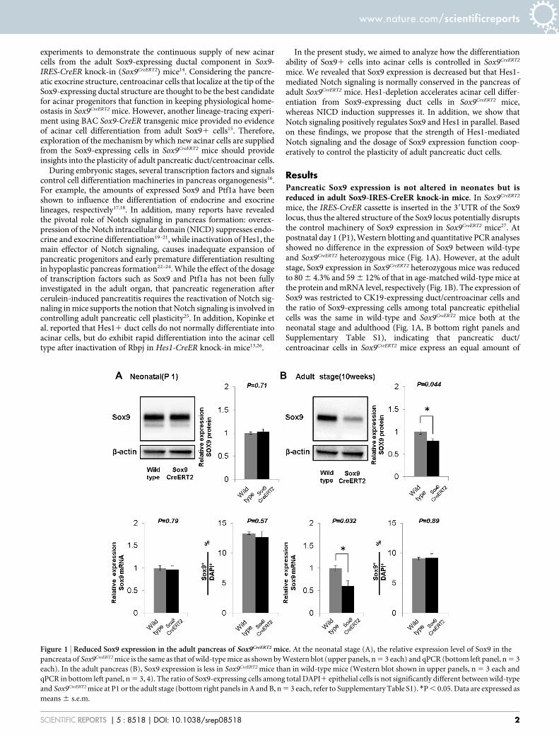
mice, the IRES-CreER cassette is inserted in the 39UTR of the Sox9locus, thus the altered structure of the Sox9 locus potentially disruptsthe control machinery of Sox9 expression in Sox9CreERT2 mice27. Atpostnatal day 1 (P1), Western blotting and quantitative PCR analysesshowed no difference in the expression of Sox9 between wild-typeand Sox9CreERT2 heterozygous mice (Fig. 1A). However, at the adultstage, Sox9 expression in Sox9CreERT2 heterozygous mice was reducedto 80 6 4.3% and 59 6 12% of that in age-matched wild-type mice atthe protein and mRNA level, respectively (Fig. 1B). The expression ofSox9 was restricted to CK19-expressing duct/centroacinar cells andthe ratio of Sox9-expressing cells among total pancreatic epithelialcells was the same in wild-type and Sox9CreERT2 mice both at theneonatal stage and adulthood (Fig. 1A, B bottom right panels andSupplementary Table S1), indicating that pancreatic duct/centroacinar cells in Sox9CreERT2 mice express an equal amount of
Figure 1 | Reduced Sox9 expression in the adult pancreas of Sox9CreERT2 mice. At the neonatal stage (A), the relative expression level of Sox9 in the
pancreata of Sox9CreERT2 mice is the same as that of wild-type mice as shown by Western blot (upper panels, n 5 3 each) and qPCR (bottom left panel, n 5 3
each). In the adult pancreas (B), Sox9 expression is less in Sox9CreERT2 mice than in wild-type mice (Western blot shown in upper panels, n 5 3 each and
qPCR in bottom left panel, n 5 3, 4). The ratio of Sox9-expressing cells among total DAPI1 epithelial cells is not significantly different between wild-type
and Sox9CreERT2 mice at P1 or the adult stage (bottom right panels in A and B, n 5 3 each, refer to Supplementary Table S1). *P , 0.05. Data are expressed as
means 6 s.e.m.
www.nature.com/scientificreports
SCIENTIFIC REPORTS | 5 : 8518 | DOI: 10.1038/srep08518 2
Sox9 in the newborn stage, but less in adulthood than do wild-typemice.
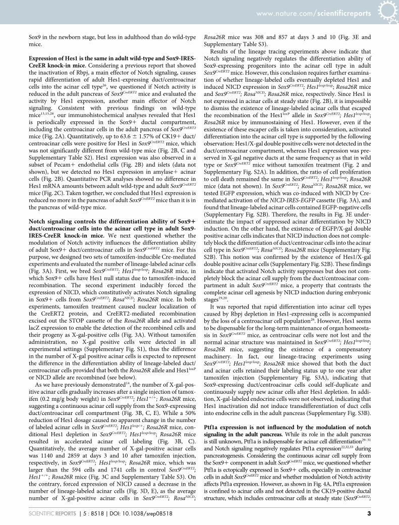
Expression of Hes1 is the same in adult wild-type and Sox9-IRES-CreER knock-in mice. Considering a previous report that showedthe inactivation of Rbpj, a main effector of Notch signaling, causesrapid differentiation of adult Hes1-expressing duct/centroacinarcells into the acinar cell type26, we questioned if Notch activity isreduced in the adult pancreas of Sox9CreERT2 mice and evaluated theactivity by Hes1 expression, another main effector of Notchsignaling. Consistent with previous findings on wild-typemice13,15,28, our immunohistochemical analyses revealed that Hes1is periodically expressed in the Sox91 ductal compartment,including the centroacinar cells in the adult pancreas of Sox9CreERT2
mice (Fig. 2A). Quantitatively, up to 63.6 6 1.57% of CK191 duct/centroacinar cells were positive for Hes1 in Sox9CreERT2 mice, whichwas not significantly different from wild-type mice (Fig. 2B, C andSupplementary Table S2). Hes1 expression was also observed in asubset of Pecam1 endothelial cells (Fig. 2B) and islets (data notshown), but we detected no Hes1 expression in amylase1 acinarcells (Fig. 2B). Quantitative PCR analyses showed no difference inHes1 mRNA amounts between adult wild-type and adult Sox9CreERT2
mice (Fig. 2C). Taken together, we concluded that Hes1 expression isreduced no more in the pancreas of adult Sox9CreERT2 mice than it is inthe pancreas of wild-type mice.
Notch signaling controls the differentiation ability of Sox91duct/centroacinar cells into the acinar cell type in adult Sox9-IRES-CreER knock-in mice. We next questioned whether themodulation of Notch activity influences the differentiation abilityof adult Sox91 duct/centroacinar cells in Sox9CreERT2 mice. For thispurpose, we designed two sets of tamoxifen-inducible Cre-mediatedexperiments and evaluated the number of lineage-labeled acinar cells(Fig. 3A). First, we bred Sox9CreERT2; Hes1loxp/loxp; Rosa26R mice, inwhich Sox91 cells have Hes1 null status due to tamoxifen-inducedrecombination. The second experiment inducibly forced theexpression of NICD, which constitutively activates Notch signalingin Sox91 cells from Sox9CreERT2; RosaNICD; Rosa26R mice. In bothexperiments, tamoxifen treatment caused nuclear localization ofthe CreERT2 protein, and CreERT2-mediated recombinationexcised out the STOP cassette of the Rosa26R allele and activatedlacZ expression to enable the detection of the recombined cells andtheir progeny as X-gal-positive cells (Fig. 3A). Without tamoxifenadministration, no X-gal positive cells were detected in allexperimental settings (Supplementary Fig. S1), thus the differencein the number of X-gal positive acinar cells is expected to representthe difference in the differentiation ability of lineage-labeled duct/centroacinar cells provided that both the Rosa26R allele and Hes1loxP
or NICD allele are recombined (see below).As we have previously demonstrated14, the number of X-gal-pos-
itive acinar cells gradually increases after a single injection of tamox-ifen (0.2 mg/g body weight) in Sox9CreERT2; Hes11/1; Rosa26R mice,suggesting a continuous acinar cell supply from the Sox9-expressingduct/centroacinar cell compartment (Fig. 3B, C, E). While a 50%reduction of Hes1 dosage caused no apparent change in the numberof labeled acinar cells in Sox9CreERT2; Hes1loxp/1; Rosa26R mice, con-ditional Hes1 depletion in Sox9CreERT2; Hes1loxp/loxp; Rosa26R miceresulted in accelerated acinar cell labeling (Fig. 3B, C).Quantitatively, the average number of X-gal-positive acinar cellswas 1140 and 2859 at days 3 and 10 after tamoxifen injection,respectively, in Sox9CreERT2; Hes1loxp/loxp; Rosa26R mice, which waslarger than the 594 cells and 1741 cells in control Sox9CreERT2;Hes11/1; Rosa26R mice (Fig. 3C and Supplementary Table S3). Onthe contrary, forced expression of NICD caused a decrease in thenumber of lineage-labeled acinar cells (Fig. 3D, E), as the averagenumber of X-gal-positive acinar cells in Sox9CreERT2; RosaNICD;
Rosa26R mice was 308 and 857 at days 3 and 10 (Fig. 3E andSupplementary Table S3).
Results of the lineage tracing experiments above indicate thatNotch signaling negatively regulates the differentiation ability ofSox9-expressing progenitors into the acinar cell type in adultSox9CreERT2 mice. However, this conclusion requires further examina-tion of whether lineage-labeled cells eventually depleted Hes1 andinduced NICD expression in Sox9CreERT2; Hes1loxp/loxp; Rosa26R miceand Sox9CreERT2; RosaNICD; Rosa26R mice, respectively. Since Hes1 isnot expressed in acinar cells at steady state (Fig. 2B), it is impossibleto dismiss the existence of lineage-labeled acinar cells that escapedthe recombination of the Hes1loxP allele in Sox9CreERT2; Hes1loxp/loxp;Rosa26R mice by immunostaining of Hes1. However, even if theexistence of these escaper cells is taken into consideration, activateddifferentiation into the acinar cell type is supported by the followingobservation: Hes1/X-gal double positive cells were not detected in theduct/centroacinar compartment, whereas Hes1 expression was pre-served in X-gal negative ducts at the same frequency as that in wildtype or Sox9CreERT2 mice without tamoxifen treatment (Fig. 2 andSupplementary Fig. S2A). In addition, the ratio of cell proliferationto cell death remained the same in Sox9CreERT2; Hes1loxp/loxp; Rosa26Rmice (data not shown). In Sox9CreERT2; RosaNICD; Rosa26R mice, wetested EGFP expression, which was co-induced with NICD by Cre-mediated activation of the NICD-IRES-EGFP cassette (Fig. 3A), andfound that lineage-labeled acinar cells contained EGFP-negative cells(Supplementary Fig. S2B). Therefore, the results in Fig. 3E under-estimate the impact of suppressed acinar differentiation by NICDinduction. On the other hand, the existence of EGFP/X-gal doublepositive acinar cells indicates that NICD induction does not comple-tely block the differentiation of duct/centroacinar cells into the acinarcell type in Sox9CreERT2; RosaNICD; Rosa26R mice (Supplementary Fig.S2B). This notion was confirmed by the existence of Hes1/X-galdouble positive acinar cells (Supplementary Fig. S2B). These findingsindicate that activated Notch activity suppresses but does not com-pletely block the acinar cell supply from the duct/centroacinar com-partment in adult Sox9CreERT2 mice, a property that contrasts thecomplete acinar cell agenesis by NICD induction during embryonicstages19,20.
It was reported that rapid differentiation into acinar cell typescaused by Rbpj depletion in Hes1-expressing cells is accompaniedby the loss of a centroacinar cell population26. However, Hes1 seemsto be dispensable for the long-term maintenance of organ homeosta-sis in Sox9CreERT2 mice, as centroacinar cells were not lost and thenormal acinar structure was maintained in Sox9CreERT2; Hes1loxp/loxp;Rosa26R mice, suggesting the existence of a compensatorymachinery. In fact, our lineage-tracing experiments usingSox9CreERT2; Hes1loxp/loxp; Rosa26R mice showed that both the ductand acinar cells retained their labeling status up to one year aftertamoxifen injection (Supplementary Fig. S3A), indicating thatSox9-expressing duct/centroacinar cells could self-duplicate andcontinuously supply new acinar cells after Hes1 depletion. In addi-tion, X-gal-labeled endocrine cells were not observed, indicating thatHes1 inactivation did not induce transdifferentiation of duct cellsinto endocrine cells in the adult pancreas (Supplementary Fig. S3B).
Ptf1a expression is not influenced by the modulation of notchsignaling in the adult pancreas. While its role in the adult pancreasis still unknown, Ptf1a is indispensable for acinar cell differentiation29–31
and Notch signaling negatively regulates Ptf1a expression22,32,33 duringpancreatogenesis. Considering the continuous acinar cell supply fromthe Sox91 component in adult Sox9CreERT2 mice, we questioned whetherPtf1a is ectopically expressed in Sox91 cells, especially in centroacinarcells in adult Sox9CreERT2 mice and whether modulation of Notch activityaffects Ptf1a expression. However, as shown in Fig. 4A, Ptf1a expressionis confined to acinar cells and not detected in the CK19-positive ductalstructure, which includes centroacinar cells at steady state (Sox9CreERT2;
www.nature.com/scientificreports
SCIENTIFIC REPORTS | 5 : 8518 | DOI: 10.1038/srep08518 3
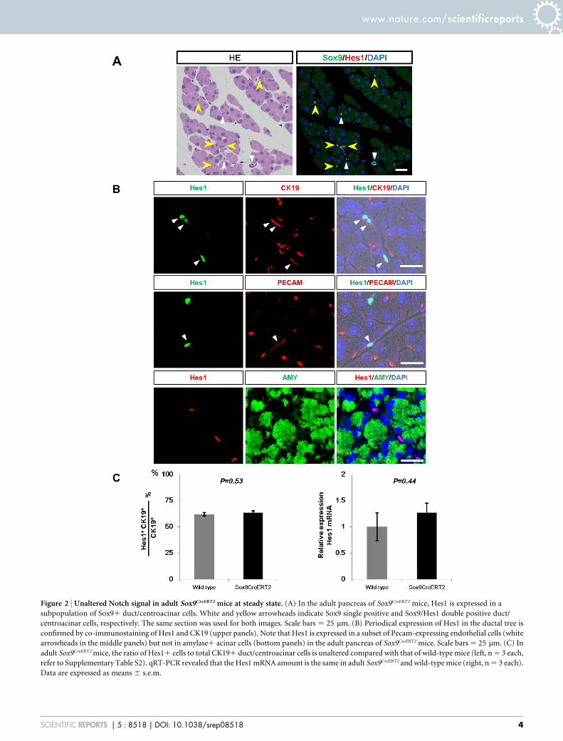
Figure 2 | Unaltered Notch signal in adult Sox9CreERT2 mice at steady state. (A) In the adult pancreas of Sox9CreERT2 mice, Hes1 is expressed in a
subpopulation of Sox91 duct/centroacinar cells. White and yellow arrowheads indicate Sox9 single positive and Sox9/Hes1 double positive duct/
centroacinar cells, respectively. The same section was used for both images. Scale bars 5 25 mm. (B) Periodical expression of Hes1 in the ductal tree is
confirmed by co-immunostaining of Hes1 and CK19 (upper panels). Note that Hes1 is expressed in a subset of Pecam-expressing endothelial cells (white
arrowheads in the middle panels) but not in amylase1 acinar cells (bottom panels) in the adult pancreas of Sox9CreERT2 mice. Scale bars 5 25 mm. (C) In
adult Sox9CreERT2 mice, the ratio of Hes11 cells to total CK191 duct/centroacinar cells is unaltered compared with that of wild-type mice (left, n 5 3 each,
refer to Supplementary Table S2). qRT-PCR revealed that the Hes1 mRNA amount is the same in adult Sox9CreERT2 and wild-type mice (right, n 5 3 each).
Data are expressed as means 6 s.e.m.
www.nature.com/scientificreports
SCIENTIFIC REPORTS | 5 : 8518 | DOI: 10.1038/srep08518 4
Hes11/1; Rosa26R mice). Additionally, it was affected by neither Hes1depletion nor NICD induction, indicating that Notch-mediated Ptf1asuppression is less robust at adult stages than at embryonic stages.Indeed, Ptf1a expression was not detected in X-gal-positive duct cellsof Sox9CreERT2; Hes1loxp/loxp; Rosa26R mice, confirming Hes1 depletion
(Supplementary Fig. S2A). Furthermore, we treated mice with ahigher dosage of tamoxifen (four injections of 0.2 mg/g body weight),which achieved more efficient labeling (approximately 60%, data notshown) of Sox91 duct cells in control mice (Supplementary Fig. S4A).Efficient recombination was confirmed by observing a decrease and
Figure 3 | Notch signaling regulates the ability of duct-to-acinar differentiation in adult Sox9CreERT2 mice. (A) Experimental strategy of tamoxifen-
inducible Cre-mediated lineage tracing (upper panel) and Notch modulation in Sox91 cells (middle panel, Hes1 depletion; lower panel, NICD
induction). (B and D) Representative sections with X-gal staining 3 days and 10 days after single tamoxifen injection. X-gal-positive acinar cells, progenies
of Sox9-expressing duct/centroacinar cells, increased from day 3 to day 10 in all the transgenic mice tested. (C and E) Quantitative analyses of the numbers
of lineage-labeled acinar cells (refer to Supplementary Table S3). Note that conditional Hes1 depletion significantly accelerates acinar cell labeling in
Sox9CreERT2; Hes1loxp/loxp; Rosa26R mice ((C), blue, P 5 0.026 at day 3, P 5 0.006 at day 10), but heterozygous inactivation (Sox9CreERT2; Hes1loxp/1; Rosa26R
mice, shown in green) does not ((C), P 5 0.21 at day 3, P 5 0.65 at day 10). Activation of Notch signaling by NICD induction results in reduced acinar cell
labeling in Sox9CreERT2; RosaNICD; Rosa26R mice compared with Sox9CreERT2; Hes11/1; Rosa26R control mice ((E), red, P 5 0.026 at day 3, P 5 0.007 at day
10). Scale bars 5 100 mm. *P , 0.05, **P , 0.01. Data are expressed as means 6 s.e.m.
www.nature.com/scientificreports
SCIENTIFIC REPORTS | 5 : 8518 | DOI: 10.1038/srep08518 5
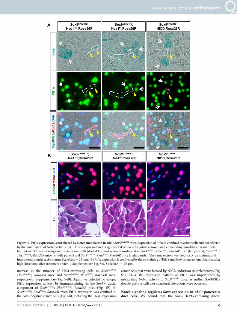
increase in the number of Hes1-expressing cells in Sox9CreERT2;Hes1loxp/loxp; Rosa26R mice and Sox9CreERT2; RosaNICD; Rosa26R mice,respectively (Supplementary Fig. S4B). Again, we detected no ectopicPtf1a expression, at least by immunostaining, in the Sox91 ductalcomponent of Sox9CreERT2; Hes1loxp/loxp; Rosa26R mice (Fig. 4B). InSox9CreERT2; RosaNICD; Rosa26R mice, Ptf1a expression was confined tothe Sox9-negative acinar cells (Fig. 4B), including the Hes1-expressing
acinar cells that were formed by NICD induction (Supplementary Fig.S5). Thus, the expression pattern of Ptf1a was unperturbed bymodulating Notch activity in Sox9CreERT2 mice, as neither Sox9/Ptf1adouble positive cells nor structural alterations were observed.
Notch signaling regulates Sox9 expression in adult pancreaticduct cells. We found that the Sox9/CK19-expressing ductal
Figure 4 | Ptf1a expression is not altered by Notch modulation in adult Sox9CreERT2 mice. Expression of Ptf1a is confined to acinar cells and not affected
by the modulation of Notch activity. (A) Ptf1a is expressed in lineage-labeled acinar cells (white arrows) and surrounding non-labeled acinar cells
but not in CK19-expressing duct/centroacinar cells (dotted line and yellow arrowheads) in Sox9CreERT2; Hes11/1; Rosa26R mice (left panels), Sox9CreERT2;
Hes1loxp/loxp; Rosa26R mice (middle panels) and Sox9CreERT2; RosaNICD; Rosa26R mice (right panels). The same section was used for X-gal staining and
immunostaining in each column. Scale bars 5 25 mm. (B) Ptf1a expression is confirmed by the co-staining of Ptf1a and Sox9 using sections obtained after
high-dose tamoxifen treatment (refer to Supplementary Fig. S4). Scale bars 5 25 mm.
www.nature.com/scientificreports
SCIENTIFIC REPORTS | 5 : 8518 | DOI: 10.1038/srep08518 6
component was structurally impaired by neither Hes1 depletion norforced expression of NICD (Fig. 5A), even at the higher dosage oftamoxifen treatment (four injections of 0.2 mg/g body weight).Further, we did not detect any ectopic Sox9 expression in acinarcells, and the ratio of Sox91 cells among total DAPI1 epithelialcells remained constant with modulating Notch activity (Fig. 5Band Supplementary Table S4). Notably, our Western blotting andquantitative PCR analyses revealed that the expression of Sox9 waselevated by the forced expression of NICD (Fig. 5B). On the otherhand, Hes1 depletion did not influence the amount of Sox9 or thenumber of Sox9-expressing cells, reflecting the asymmetric divisionof Sox91 cells into acinar and Sox91 cells in Sox9CreERT2 mice14.Thus, considering the increased number of Hes1-expressing cellsby NICD induction (Supplementary Fig. S4 and S5), our findingsshow that Notch positively regulates Hes1 and Sox9 in parallel inadult pancreatic duct cells of Sox9CreERT2 mice and that the regulationof Sox9 expression by Notch is mediated in a Hes1-independentmanner in the adult pancreas.
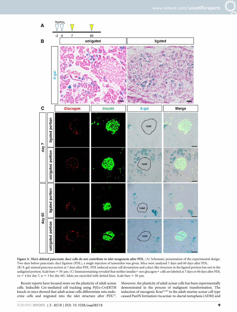
Hes1-depleted duct cells do not show endocrine neogenesis afterpancreatic duct ligation. Another long-standing debate is whetheradult pancreatic duct cells possess plasticity to differentiate into theendocrine lineage. In particular, possible islet neogenesis afterpancreatic duct ligation (PDL) has attracted the interest ofresearchers34,35. We previously reported that beta cell neogenesisfrom Sox9-expressing duct cells was observed in neither thephysiological condition nor the regeneration process after injury,including partial pancreatectomy, cerulein-induced acutepancreatitis, streptozotocin diabetes or PDL, in Sox9CreERT2 mice14.Because Hes1 inactivation in the embryonic pancreas acceleratesendocrine differentiation23, we tested whether Hes1 depletioncombined with PDL prompts adult Sox9-expressing cells todifferentiate into endocrine lineages in Sox9CreERT2 mice. However,lineage tracing analyses detected no X-gal-positive cells in the islets atdays 7 or 60 after PDL in Sox9CreERT2; Hes1loxp/loxp; Rosa26R mice(Fig. 6), indicating that adult Sox9-expressing duct/centroacinarcells are not capable of differentiating into endocrine lineages evenby the combination of Hes1-inactivation and PDL.
DiscussionIn this study, we demonstrated that the plasticity of adult pancreaticduct cells is controlled by the strength of Notch signaling. The dif-ferentiation ability of Sox9-expressing duct/centroacinar cells intoacinar cell types was accelerated by Hes1 inactivation and suppressedby NICD induction in Sox9CreERT2 mice. This effect is consistent withtwo reports by Kopinke et al. that show Hes1-expressing centroaci-nar/terminal duct cells do not normally differentiate into acinar cellsbut will do so rapidly upon the inactivation of Rbpj in Hes1-CreERknock-in mice, which suggests that Notch signaling functions tomaintain the duct cell identity in the adult pancreas13,26. Adding tothat our previous report that showed Sox9-expressing duct/centroa-cinar cells continuously supply new acinar cells in adult Sox9CreERT2
mice14, we hypothesized Hes1-negative/Sox9-positive centroacinarcells are best candidates for the origin of continuous acinar cellsupply in Sox9CreERT2 mice. However, Notch signaling alone cannotexplain why Sox9-expressing cells were reported not to differentiateinto acinar cells in adult BAC Sox9-CreER transgenic mice15. Wetherefore predicted the existence of one or more other mechanismsthat control adult duct cell plasticity.
We propose that the amount of Sox9 expression is pivotal incontrolling adult duct cell plasticity (Fig. 7). The dosage requirementof Sox9 in the formation of the embryonic endocrine pancreas hasbeen previously suggested by autopsy samples from three humancases of campomelic dysplasia, an autosomal dominant disordercaused by SOX9 haploinsufficiency that has been described as ‘‘lessdensely packed epithelial cells within the mesenchymal stroma and
less clearly formed islets36.’’ The dosage requirement has also beenexperimentally supported previously; a Pdx1-Cre-mediated 50%reduction of Sox9 expression caused a decrease in the number ofneurogenin 3 (Ngn3)-expressing endocrine progenitors and lessendocrine formation in Pdx1-Cre; Sox9loxp/1 mice17. However, inthe human campomelic dysplasia cases, little attention was givento the exocrine tissue36, and exocrine pancreatic formation was notimpaired in Pdx1-Cre; Sox9loxp/1 mice17, suggesting that Sox9 dosagehas less effect on exocrine pancreas formation during the embryonicstages. In contrast, our results suggest the involvement of Sox9 dos-age in controlling the plasticity of adult pancreatic duct cells. In thepresent study, we demonstrated that pancreatic Sox9 expression inneonates was the same in wild-type and Sox9CreERT2 mice, whereasadult pancreatic duct cells express less Sox9 in Sox9CreERT2 mice thanin adult wild-type mice. It should be noted that two previouslyreported lineage-tracing experiments using Sox9CreERT2 and BACSox9-CreER transgenic mice are consistent in that embryonicSox91 duct cells were sufficiently plastic to differentiate into acinarand endocrine lineages, an ability that persisted until the perinatalstages up to one week after birth14,15. Provided that the amount ofSox9 expression in BAC Sox9-CreER transgenic mice was kept at thesame level as that of wild-type mice at the newborn stage and adult-hood, reduced Sox9 expression may allow duct cells to differentiateinto acinar cells in adult Sox9CreERT2 mice, whereas higher Sox9expression in BAC Sox9-CreER transgenic mice possibly functionsto maintain the duct cell identity, thereby preventing continuousdifferentiation into acinar cell types.
We also show that the induction of NICD caused elevated Sox9expression, indicating that Sox9 expression is regulated by Notchsignaling in adult Sox9CreERT2 pancreata. This result is consistent withShih et al., who reported that Notch signaling regulates Sox9 express-ion in the embryonic pancreas37. An in vitro tissue culture experimentusing wild-type pancreas at e12.5 further showed that Hes1 express-ion was reduced by the addition of a low-dose c-secretase inhibitor,while Sox9 expression was decreased at a higher dosage, indicatingthat Notch regulates Hes1 and Sox9 in parallel in embryonic pan-creata. Nevertheless, we hypothesized that the reduced Sox9 express-ion in the adult pancreas of Sox9CreERT2 mice at steady state isunconnected with Notch-mediated regulation. Notch activity seemednormal based on the ratio of Hes1-expressing cell numbers to CK-positive ductal epithelia and the amount of Hes1 mRNA was thesame between wild-type and Sox9CreERT2 mice. In Sox9CreERT2 mice,the IRES-CreERT2 cassette is inserted in the 39UTR of the Sox9locus27. We therefore speculate that the altered structure of theSox9 locus is related to the reduction of Sox9 expression38 in the adultpancreas, though the precise machinery has yet to be revealed. Recentreports showed a conserved sequence among species very near thesite of the IRES-CreERT2 cassette insertion, suggesting the existenceof a regulatory element in this region39,40. Because the Sox9 expressionwas normal at the perinatal stage in Sox9CreERT2 mice, Notch-inde-pendent Sox9 regulation using this element, if it exists, would be silentuntil adulthood, when it maintains the Sox9 expression and therebymaintains the identity of the duct cell type. Future studies to dissectthe complete regulatory machinery of Sox9 are required.
Islet neogenesis has long been believed to occur in the ductalcompartment during the tissue restoration process, but we did notobserve evidence of islet regeneration from Sox9-expressing cells in aPDL model. Xu et al. showed that the number of Ngn3-positiveendocrine precursor cells increases after PDL34. Furthermore, inBAC Sox9-CreER transgenic mice, it was reported that de novoNgn3-expressing cells are of duct cell origin, but that they do notexpress other pivotal transcription factors, such as Nkx6.1 and Pax6,and thus are unable to drive the entire differentiation program intoendocrine lineage15. Consistent with this observation, Xiao et al.showed that Ngn3-expressing cells originating from the ducts afterPDL do not differentiate into mature endocrine cells41.
www.nature.com/scientificreports
SCIENTIFIC REPORTS | 5 : 8518 | DOI: 10.1038/srep08518 7
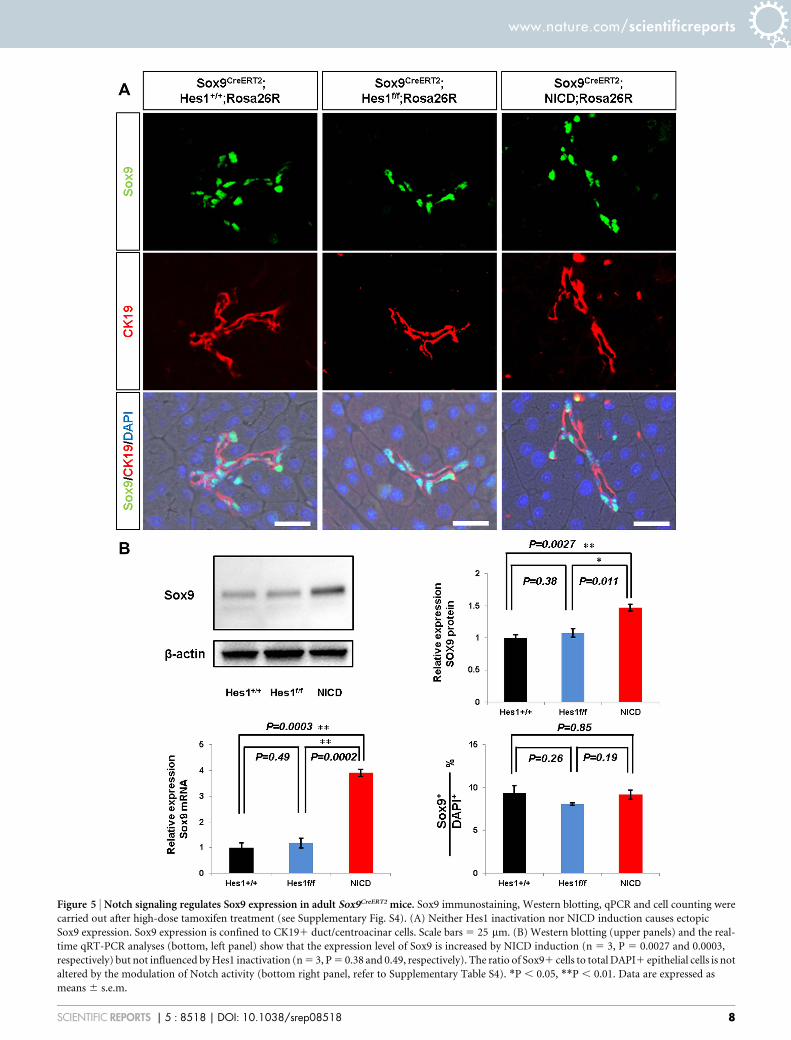
Figure 5 | Notch signaling regulates Sox9 expression in adult Sox9CreERT2 mice. Sox9 immunostaining, Western blotting, qPCR and cell counting were
carried out after high-dose tamoxifen treatment (see Supplementary Fig. S4). (A) Neither Hes1 inactivation nor NICD induction causes ectopic
Sox9 expression. Sox9 expression is confined to CK191 duct/centroacinar cells. Scale bars 5 25 mm. (B) Western blotting (upper panels) and the real-
time qRT-PCR analyses (bottom, left panel) show that the expression level of Sox9 is increased by NICD induction (n 5 3, P 5 0.0027 and 0.0003,
respectively) but not influenced by Hes1 inactivation (n 5 3, P 5 0.38 and 0.49, respectively). The ratio of Sox91 cells to total DAPI1 epithelial cells is not
altered by the modulation of Notch activity (bottom right panel, refer to Supplementary Table S4). *P , 0.05, **P , 0.01. Data are expressed as
means 6 s.e.m.
www.nature.com/scientificreports
SCIENTIFIC REPORTS | 5 : 8518 | DOI: 10.1038/srep08518 8
Recent reports have focused more on the plasticity of adult acinarcells. Inducible Cre-mediated cell tracking using Ptf1a-CreERTMknock-in mice showed that adult acinar cells differentiate into endo-crine cells and migrated into the islet structure after PDL35.
Moreover, the plasticity of adult acinar cells has been experimentallydemonstrated in the process of malignant transformation. Theinduction of oncogenic KrasG12D in the adult murine acinar cell typecaused PanIN formation via acinar-to-ductal metaplasia (ADM) and
Figure 6 | Hes1-deleted pancreatic duct cells do not contribute to islet neogenesis after PDL. (A) Schematic presentation of the experimental design.
Two days before pancreatic duct ligation (PDL), a single injection of tamoxifen was given. Mice were analyzed 7 days and 60 days after PDL.
(B) X-gal-stained pancreas section at 7 days after PDL. PDL induced acinar cell devastation and a duct-like structure in the ligated portion but not in the
unligated portion. Scale bars 5 50 mm. (C) Immunostaining revealed that neither insulin1 nor glucagon1 cells are labeled at 7 days or 60 days after PDL
(n 5 4 for day 7, n 5 3 for day 60). Islets are encircled with dotted lines. Scale bars 5 50 mm.
www.nature.com/scientificreports
SCIENTIFIC REPORTS | 5 : 8518 | DOI: 10.1038/srep08518 9
developed into pancreatic ductal adenocarcinoma, whereas KrasG12D
activation in the duct cell type did not42. Interestingly, Sox9/CPA1double positive cells emerge during the transdifferentiation processof ADM, and simultaneous Sox9 depletion and KrasG12D activationdoes not cause ADM in Ptf1aCreERTM; LSL-KrasG12D; Sox9floxed/floxed;Rosa26YFP mice42. In addition, our preliminary experimental resultsshowed the emergence of Sox9/Ptf1a double positive cells in thetissue restoration after cerulein-induced acute pancreatitis. Thesefindings suggest that cell plasticity might be connected to the co-expression of transcription factors that normally defines specific celltypes: Sox9 in duct cells and Ptf1a in adult acinar cells. In fact, Sox9/Ptf1a double positive cells exist in the early embryonic pancreaticepithelium from which all pancreatic cell types originate15,35,43. Tonote, the Sox9/CPA1 double positive cells and Sox9/Ptf1a doublepositive cells mentioned above accompany tissue structure altera-tions such as ADM, tissue restoration after injury or embryonicorganogenesis, whereas we observed no structural change or Sox9/Ptf1a double positive cells in the present study. So far, a precisemechanism that explains the relationship between the degree of cellplasticity with the co-expression of cell type-specific transcriptionfactors and tissue construction is lacking. Further examination ofduct-originated diseases, such as intraductal neoplasms, that accom-pany with tissue destruction may help reveal such a mechanism.
MethodsMice. To obtain Hes1-conditional knock-out mice, Hes1loxp/1 mice44, Sox9IRES-CreERT2
mice27 and Rosa26LacZ (Rosa26R) mice45 were interbred. Rosa26NICD mice19 andSox9IRES-CreERT2; Rosa26R mice were crossed to produce Sox9IRES-CreERT2; Rosa26NICD;Rosa26R mice. The pancreatic duct ligation (PDL) procedure was performed aspreviously described34. All animal experiments were performed in accordance withthe Kyoto University guidelines for animal experiments and approved by the animalresearch committee of Kyoto University.
Tamoxifen injection. For lineage tracing, adult mice older than 8 weeks of age(approximately 25–30 g in body weight) were injected intraperitoneally withtamoxifen (Sigma T-5648) at 0.2 mg/g body weight. To get higher recombinationefficiency, we injected tamoxifen at 0.2 mg/g body weight 4 times every other day.
Tissue preparation for X-gal staining and paraffin section. X-gal staining andparaffin section were performed as previously described14.
Immunofluorescence. The antigen retrieval procedure was performed with TargetRetrieval Solution pH 6.0 (Dako), Target Retrieval Solution High pH 9.0 (Dako) orProteinase K (Dako) in accordance with the manufacturer’s instructions, followed by
overnight incubation at 4uC with primary antibodies (Supplementary Table S5). Thesections were washed in PBS and incubated for 60 min with secondary antibodies(Supplementary Table S6), followed by counterstaining with 4, 6-diamidino-2-phenylindole (DAPI). Immunostaining for Hes1 and Ptf1a required the use of theTSA amplification Kit (Perkin Elmar). Images were visualized using HS All-in-oneFluorescence Microscopy BZ-9000E (Keyence).
Cell counting. We chose 20 fields at random and counted X-gal-positive acinar cellsin each mouse. For short-term chase in the adult pancreas, we examined 20 fields permouse to count the X-gal-positive acinar cells and averaged them. Cell numbers werecounted by 3200 magnification in 40 fields (at adult stage) and 6 fields (at P1) usingthree sections per mouse and the ratio of Sox91 cells among all DAPI1 epithelialcells was calculated. To determine the ratio of Hes11 cells among CK191 duct cells,cell numbers were counted by 3400 magnification in 6 fields per mouse. All valuesare shown as mean 6 s.e.m. All error bars represent s.e.m. P values were calculatedusing an unpaired Student’s t test (two-tailed); P , 0.05 was considered significant.
RNA isolation and RT-PCR. Total RNA was extracted using the RNeasy Mini Kit(Qiagen). First-strand cDNA synthesis was performed using Rever Tra Ace qPCR RTMaster Mix (Toyobo). Quantitative RT-PCR for Hes1 and Sox9 was performed withTaq Man and SYBR Green. Independent experiments were performed three timesand confirmed. Data were normalized in relation to the expression of GAPDH andHPRT1. The Sox9 and Hes1 probes used were TaqMan Gene Expression Assay nos.Sox9 5 Mm00448840 2 m1, Hes1 5 Mm01342805 2 m1, GAPDH 5 Mm999999152 g1 and HPRT1 5 Mm01545399 2 m1 (Applied Biosystems). For the SYBR GreenDCT method, the Sox9 primer sequences were forward, AGGAAGCTGGCAG-ACCAGTA and reverse, TCCACGAAGGGTCTCTTCTC; and the GAPDH primersequences were forward, GTGTTCCTACCCCCAATGTGT and reverse,ATTGTCATACCAGGAAATGAGCTT. Analysis was performed using the standardcurve and comparative CT methods. P values were calculated using an unpairedStudent’s t test (two-tailed); P , 0.05 was considered significant.
Western blotting. Whole pancreatic tissues were lysed in RIPA buffer (ThermoScientific Pierce) in the presence of protease inhibitor (Roche). Obtained sampleswere electrophoresed in 10% SDS-PAGE gels and transferred to PVDF membrane(Bio-Rad). Membranes were blocked with 5% skim milk in TBS-T and incubated withanti-Sox9 antibody (Millipore 15500) or anti-b-actin antibody (MBL 151000)overnight at 4uC. After washing, membranes were reacted with HRP-conjugatedsecondary antibodies (DAKO) at 152000 dilution. Chemiluminescence was detectedwith Immobilon Western horseradish peroxidase substrate (Thermo ScientificPierce) and visualized with a charge-coupled device camera (Ez-capture). Theintensity of the bands was quantified with imaging analysis software (Image J) usingb-actin as an internal control. P values were calculated using an unpaired Student’s ttest (two-tailed); P , 0.05 was considered significant.
1. Takahashi, K. & Yamanaka, S. Induction of pluripotent stem cells from mouseembryonic and adult fibroblast cultures by defined factors. Cell 126, 663–676(2006).
2. Ladewig, J., Koch, P. & Brustle, O. Leveling Waddington: the emergence of directprogramming and the loss of cell fate hierarchies. Nat Rev Mol Cell Biol 14,225–236 (2013).
3. Barker, N. et al. Identification of stem cells in small intestine and colon by markergene Lgr5. Nature 449, 1003–1007 (2007).
4. Mascre, G. et al. Distinct contribution of stem and progenitor cells to epidermalmaintenance. Nature 489, 257–262 (2012).
5. Jensen, J. N. et al. Recapitulation of elements of embryonic development in adultmouse pancreatic regeneration. Gastroenterology 128, 728–741 (2005).
6. Mu, X., Peng, H., Pan, H., Huard, J. & Li, Y. Study of muscle cell dedifferentiationafter skeletal muscle injury of mice with a Cre-Lox system. PLoS One 6, e16699(2011).
7. Tata, P. R. et al. Dedifferentiation of committed epithelial cells into stem cells invivo. Nature 503, 218–223 (2013).
8. De La O, J. P. et al. Notch and Kras reprogram pancreatic acinar cells to ductalintraepithelial neoplasia. Proc Natl Acad Sci U S A 105, 18907–18912 (2008).
9. Habbe, N. et al. Spontaneous induction of murine pancreatic intraepithelialneoplasia (mPanIN) by acinar cell targeting of oncogenic Kras in adult mice. ProcNatl Acad Sci U S A 105, 18913–18918 (2008).
10. Morris, J. P., Cano, D. A., Sekine, S., Wang, S. C. & Hebrok, M. Beta-catenin blocksKras-dependent reprogramming of acini into pancreatic cancer precursor lesionsin mice. J Clin Invest 120, 508–520 (2010).
11. Kopp, J. L. et al. Progenitor cell domains in the developing and adult pancreas. CellCycle 10, 1921–1927 (2011).
12. Solar, M. et al. Pancreatic exocrine duct cells give rise to insulin-producing betacells during embryogenesis but not after birth. Dev Cell 17, 849–860 (2009).
13. Kopinke, D. et al. Lineage tracing reveals the dynamic contribution of Hes11 cellsto the developing and adult pancreas. Development 138, 431–441 (2011).
14. Furuyama, K. et al. Continuous cell supply from a Sox9-expressing progenitorzone in adult liver, exocrine pancreas and intestine. Nat Genet 43, 34–41 (2011).
15. Kopp, J. L. et al. Sox91 ductal cells are multipotent progenitors throughoutdevelopment but do not produce new endocrine cells in the normal or injuredadult pancreas. Development 138, 653–665 (2011).
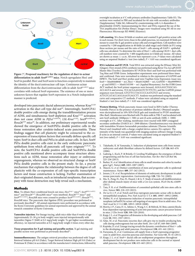
Figure 7 | Proposed machinery for the regulation of duct-to-acinardifferentiation in adult Sox9CreERT2 mice. Notch upregulates Hes1 and
Sox9 in parallel. Hes1 and Sox9 seem to function cooperatively to maintain
the identity of the duct/centroacinar cell type. Continuous acinar
differentiation from the duct/centroacinar cells in adult Sox9CreERT2 mice
correlates with reduced Sox9 expression. The existence of one or more
unknown factors that regulate Sox9 expression in a Notch-independent
manner is predicted.
www.nature.com/scientificreports
SCIENTIFIC REPORTS | 5 : 8518 | DOI: 10.1038/srep08518 10
16. Pan, F. C. & Wright, C. Pancreas organogenesis: from bud to plexus to gland. DevDyn 240, 530–565 (2011).
17. Seymour, P. A. et al. A dosage-dependent requirement for Sox9 in pancreaticendocrine cell formation. Dev Biol 323, 19–30 (2008).
18. Fukuda, A. et al. Reduction of Ptf1a gene dosage causes pancreatic hypoplasia anddiabetes in mice. Diabetes 57, 2421–2431 (2008).
19. Murtaugh, L. C., Stanger, B. Z., Kwan, K. M. & Melton, D. A. Notch signalingcontrols multiple steps of pancreatic differentiation. Proc Natl Acad Sci U S A 100,14920–14925 (2003).
20. Hald, J. et al. Activated Notch1 prevents differentiation of pancreatic acinar cellsand attenuate endocrine development. Dev Biol 260, 426–437 (2003).
21. Esni, F. et al. Notch inhibits Ptf1 function and acinar cell differentiation indeveloping mouse and zebrafish pancreas. Development 131, 4213–4224 (2004).
22. Fukuda, A. et al. Ectopic pancreas formation in Hes1 -knockout mice revealsplasticity of endodermal progenitors of the gut, bile duct, and pancreas. J ClinInvest 116, 1484–1493 (2006).
23. Jensen, J. et al. Control of endodermal endocrine development by Hes-1. NatGenet 24, 36–44 (2000).
24. Sumazaki, R. et al. Conversion of biliary system to pancreatic tissue in Hes1-deficient mice. Nat Genet 36, 83–87 (2004).
25. Siveke, J. T. et al. Notch signaling is required for exocrine regeneration after acutepancreatitis. Gastroenterology 134, 544–555 (2008).
26. Kopinke, D. et al. Ongoing Notch signaling maintains phenotypic fidelity in theadult exocrine pancreas. Dev Biol 362, 57–64 (2012).
27. Soeda, T. et al. Sox9-expressing precursors are the cellular origin of the cruciateligament of the knee joint and the limb tendons. Genesis 48, 635–644 (2010).
28. Stanger, B. Z. et al. Pten constrains centroacinar cell expansion and malignanttransformation in the pancreas. Cancer Cell 8, 185–195 (2005).
29. Krapp, A. et al. The p48 DNA-binding subunit of transcription factor PTF1 is anew exocrine pancreas-specific basic helix-loop-helix protein. EMBO J 15,4317–4329 (1996).
30. Krapp, A. et al. The bHLH protein PTF1-p48 is essential for the formation of theexocrine and the correct spatial organization of the endocrine pancreas. Genes Dev12, 3752–3763 (1998).
31. Kawaguchi, Y. et al. The role of the transcriptional regulator Ptf1a in convertingintestinal to pancreatic progenitors. Nat Genet 32, 128–134 (2002).
32. Schaffer, A. E., Freude, K. K., Nelson, S. B. & Sander, M. Nkx6 transcription factorsand Ptf1a function as antagonistic lineage determinants in multipotent pancreaticprogenitors. Dev Cell 18, 1022–1029 (2010).
33. Horn, S. et al. Mind bomb 1 is required for pancreatic b-cell formation. Proc NatlAcad Sci U S A 109, 7356–7361 (2012).
34. Xu, X. et al. Beta cells can be generated from endogenous progenitors in injuredadult mouse pancreas. Cell 132, 197–207 (2008).
35. Pan, F. C. et al. Spatiotemporal patterns of multipotentiality in Ptf1a-expressingcells during pancreas organogenesis and injury-induced facultative restoration.Development 140, 751–764 (2013).
36. Piper, K. et al. Novel SOX9 expression during human pancreas developmentcorrelates to abnormalities in Campomelic dysplasia. Mech Dev 116, 223–226(2002).
37. Shih, H. P. et al. A Notch-dependent molecular circuitry initiates pancreaticendocrine and ductal cell differentiation. Development 139, 2488–2499 (2012).
38. Mead, T. J. et al. A far-upstream (270 kb) enhancer mediates Sox9 auto-regulation in somatic tissues during development and adult regeneration. NucleicAcids Res 41, 4459–4469 (2013).
39. Dai, L., Zhang, X., Hu, X., Zhou, C. & Ao, Y. Silencing of microRNA-101 preventsIL-1b-induced extracellular matrix degradation in chondrocytes. Arthritis ResTher 14, R268 (2012).
40. Yang, B. et al. MicroRNA-145 regulates chondrogenic differentiation ofmesenchymal stem cells by targeting Sox9. PLoS One 6, e21679 (2011).
41. Xiao, X. et al. Neurogenin3 activation is not sufficient to direct duct-to-beta celltransdifferentiation in the adult pancreas. J Biol Chem 288, 25297–25308 (2013).
42. Kopp, J. L. et al. Identification of Sox9-dependent acinar-to-ductalreprogramming as the principal mechanism for initiation of pancreatic ductaladenocarcinoma. Cancer Cell 22, 737–750 (2012).
43. Zhou, Q. et al. A multipotent progenitor domain guides pancreatic organogenesis.Dev Cell 13, 103–114 (2007).
44. Imayoshi, I., Shimogori, T., Ohtsuka, T. & Kageyama, R. Hes genes andneurogenin regulate non-neural versus neural fate specification in the dorsaltelencephalic midline. Development 135, 2531–2541 (2008).
45. Soriano, P. Generalized lacZ expression with the ROSA26 Cre reporter strain. NatGenet 21, 70–71 (1999).
AcknowledgmentsThis work was supported by the Funding Program for Next Generation World-LeadingResearchers, Japan Society for the Promotion of Science [grant number LS063]. We thankD. Melton for the Rosa-NICD mice, P. Soriano for the Rosa26R mice, T. Sudo for the Hes1antibody, M. Hoshino for the Ptf1a antibody, Developmental Studies Hybridoma Bank(DSHB) for the CK19 antibody, T. Masui for helpful discussions, H.Hirao for technicalsupport, P.Karagiannis for the proofreading and the staff of the Institute of LaboratoryAnimals at Kyoto University for animal care.
Author contributionsY.K. and S.H. designed the study, analyzed the data and prepared the manuscript. S.H.performed the experiments. H.A. and R.K. generated the mice. K.F., M.H., Y.A., K.T., M.S.,T.G., K.H., Y.N. and W.T. provided technical support and discussion. S.U. supervised theproject.
Additional informationSupplementary information accompanies this paper at http://www.nature.com/scientificreports
Competing financial interests: The authors declare no competing financial interests.
How to cite this article: Hosokawa, S. et al. Impact of Sox9 Dosage and Hes1-mediatedNotch Signaling in Controlling the Plasticity of Adult Pancreatic Duct Cells in Mice. Sci.Rep. 5, 8518; DOI:10.1038/srep08518 (2015).
This work is licensed under a Creative Commons Attribution 4.0 InternationalLicense. The images or other third party material in this article are included in thearticle’s Creative Commons license, unless indicated otherwise in the credit line; ifthe material is not included under the Creative Commons license, users will needto obtain permission from the license holder in order to reproduce the material. Toview a copy of this license, visit http://creativecommons.org/licenses/by/4.0/
www.nature.com/scientificreports
SCIENTIFIC REPORTS | 5 : 8518 | DOI: 10.1038/srep08518 11