Embed Size (px)
Citation preview

Title Induced-fit motion of a lid loop involved in catalysis in alginatelyase A1-III.
Author(s)Mikami, Bunzo; Ban, Mizuho; Suzuki, Sachiko; Yoon, Hye-Jin; Miyake, Osamu; Yamasaki, Masayuki; Ogura, Kohei;Maruyama, Yukie; Hashimoto, Wataru; Murata, Kousaku
Citation Acta crystallographica. Section D, Biological crystallography(2012), 68(Part 9): 1207-1216
Issue Date 2012-09
URL http://hdl.handle.net/2433/161048
Right © 2012 International Union of CrystallographyPrinted in Singapore
Type Journal Article
Textversion publisher
Kyoto University

research papers
Acta Cryst. (2012). D68, 1207–1216 doi:10.1107/S090744491202495X 1207
Acta Crystallographica Section D
BiologicalCrystallography
ISSN 0907-4449
Induced-fit motion of a lid loop involved in catalysisin alginate lyase A1-III
Bunzo Mikami,a*‡ Mizuho
Ban,a§ Sachiko Suzuki,b} Hye-Jin
Yoon,a‡‡ Osamu Miyake,b§§
Masayuki Yamasaki,a}} Kohei
Ogura,b‡‡‡ Yukie Maruyama,b
Wataru Hashimotob and
Kousaku Muratab‡
aLaboratory of Applied Structural Biology,
Department of Applied Life Science, Graduate
School of Agriculture, Kyoto University,
Gokasho, Uji, Kyoto 611-0011, Japan, andbLaboratory of Basic and Applied Molecular
Biotechnology, Department of Food Science,
Graduate School of Agriculture, Kyoto
University, Gokasho, Uji, Kyoto 611-0011,
Japan
‡ BM and KM contributed equally to this paper.
§ Present address: Department of Food and
Nutrition, Sanyo Gakuen Junior College,
1-14-1 Hirai, Naka-ku, Okayama 703-8501,
Japan.
} Present address: Research Institute,
Gekkeikan Sake Company Ltd,
300 Katahara-cho, Fushimi-ku,
Kyoto 612-8361, Japan.
‡‡ Present address: School of Chemistry,
Seoul National University, Seoul 151-742,
Republic of Korea.
§§ Present address: Division of Food Nutrition,
Kyoto Junior College, 3370 Nishi-Kotanigaoka,
Fukuchiyama, Kyoto 620-0886, Japan.
}} Present address: Young Researcher
Development Centre (The Hakubi Centre),
Institute for Frontier Medical Sciences, Kyoto
University, Sakyo-ku, Kyoto 606-8397, Japan.
Present address: Department of Molecular
Infectiology, Graduate School of Medicine,
Chiba University, 1-8-1 Inohana, Chuo-ku,
Chiba 260-0856, Japan.
Correspondence e-mail:
# 2012 International Union of Crystallography
Printed in Singapore – all rights reserved
The structures of two mutants (H192A and Y246F) of a
mannuronate-specific alginate lyase, A1-III, from Sphingo-
monas species A1 complexed with a tetrasaccharide substrate
[4-deoxy-l-erythro-hex-4-ene-pyranosyluronate-(mannuronate)2-
mannuronic acid] were determined by X-ray crystallography
at around 2.2 A resolution together with the apo form of the
H192A mutant. The final models of the complex forms, which
comprised two monomers (of 353 amino-acid residues each),
268–287 water molecules and two tetrasaccharide substrates,
had R factors of around 0.17. A large conformational change
occurred in the position of the lid loop (residues 64–85) in
holo H192A and Y246F compared with that in apo H192A.
The lid loop migrated about 14 A from an open form to a
closed form to interact with the bound tetrasaccharide and a
catalytic residue. The tetrasaccharide was bound in the active
cleft at subsites �3 to +1 as a substrate form in which the
glycosidic linkage to be cleaved existed between subsites �1
and +1. In particular, the O� atom of Tyr68 in the closed lid
loop forms a hydrogen bond to the side chain of a presumed
catalytic residue, O� of Tyr246, which acts both as an acid and
a base catalyst in a syn mechanism.
Received 2 December 2011
Accepted 31 May 2012
PDB References: A1-III, H192
mutant, apo, 4e1y; H192
mutant, complexed with
alginate tetrasaccharide, 4f10;
Y246F mutant, complexed
with alginate tetrasaccharide,
4f13.
1. Introduction
The degradation of acid polysaccharides such as alginate
proceeds by a lyase reaction, which produces double-bonded
sugars at the nonreducing end of the product. Among the 21
families (PL-1 to PL-21) of polysaccharide lyases classified
in the CAZY database (Carbohydrate Active Enzymes data-
base; http://www.cazy.org/; Cantarel et al., 2009), the structures
of enzymes from 19 families have been deternined (Garron &
Cygler, 2010; Lombard et al., 2010). In order to elucidate the
mechanism by which these enzymes degrade acid poly-
saccharides, the structures of complexes of many lyases with
ligands including the substrates and/or products of these lyases
have been reported (Garron & Cygler, 2010; Lombard et al.,
2010; Ochiai et al., 2010; Shaya et al., 2010). It has been
reported that the Tyr, His and Asn triad is conserved and plays
an important role in the catalysis of polysaccharide lyases in
the PL-5 (Yoon et al., 2001), PL-7 (Ogura et al., 2008; Yama-
saki et al., 2005; Osawa et al., 2005) and PL-8 (Fethiere et al.,
1999; Lunin et al., 2004; Huang et al., 2001; Li & Jedrzejas,
2001; Mello et al., 2002; Nukui et al., 2003; Rigden & Jedrzejas,
2003) families. There is a structural similarity between the
catalytic domains of PL-5 and PL-8. In alginate lyase (PL-5;
Yoon et al., 2001), chondroitin AC lyase (PL-8; Fethiere et al.,
1999; Lunin et al., 2004; Huang et al., 2001) and xanthan lyase
‡‡‡

(PL-8; Maruyama et al., 2005, 2007) the catalytic residue which
accepts a hydrogen from C5 at the +1 sugar subsite and
donates hydrogen to the glycosidic O atom to be cleaved has
been reported to be a Tyr residue. In contrast, His is the
proton acceptor and Tyr is the proton donor in hyaluronate
lyase (PL-8; Ponnuraj & Jedrzejas, 2000; Li et al., 2001;
Jedrzejas et al., 2002; Li & Jedrzejas, 2001; Mello et al., 2002;
Nukui et al., 2003; Rigden & Jedrzejas, 2003; Rigden et al.,
2006). However, recent analysis of another PL-8 hyaluronate
lyase complexed with a disaccharide suggested that Tyr is
involved in proton abstraction from the substrate (Elmabrouk
et al., 2011).
The alginate lyase A1-III is a �-d-mannuronosyl linkage-
specific (M-M-specific) enzyme that acts on alginate
tetrasaccharide as the minimum substrate and produces
disaccharides and trisaccharides from alginate (Murata et al.,
1993; Yonemoto et al., 1993). The structure of an A1-III crystal
at 1.78 A resolution was reported as the first example of a
family PL-5 enzyme (Yoon et al., 1999). Based on the structure
of A1-III complexed with a trisaccharide product, it was
proposed that Tyr246 is a catalytic residue that is responsible
for both the abstraction and the donation of hydrogen (Yoon
et al., 2001). However, the complex structure was insufficient
in two respects. Firstly, the enzyme was not in an active form,
having a fixed open lid-loop region (residues 64–85) caused by
crystallographic interactions. Secondly, the product sugars did
not bind to cover the subsites on either side of the catalytic
residue (subsites �3 to �1). It is important to overcome
crystallographic artifacts such as symmetry interactions and
the effects of freezing in order to elucidate the conformational
changes and complex formation in the enzymatic process.
An accurate determination of the structure of the mutant
enzyme–substrate complex without crystallographic artifacts
is necessary in order to elucidate the catalytic residue (Dunlop
et al., 2005; Skrzypczak-Jankun et al., 2006).
In this paper, we describe the crystal structures of mutant
alginate lyases (H192A and Y246F) complexed with tetra-
saccharide substrates at around 2.2 A resolution. We thus
determined a new crystal form of the enzyme in which the lid-
loop region can move from an open to a closed form by an
induced-fit motion. For the first time, we were able to deter-
mine the structure of a sugar bound at subsite +1 in the closed
lid-loop form. We used crystals enclosed in capillaries in order
to avoid the effects of freezing.
2. Materials and methods
2.1. Preparation of mutant enzymes
A1-III mutants with mutations of the amino-acid residues in
the active site (Y246F, H192A, G60A, R67A, Y68F and Y80F)
were prepared by site-directed mutagenesis with plasmid
pET3a-A1-III (Yoon et al., 2000) as a template using the
QuikChange Site-Directed Mutagenesis kit (Stratagene, La
Jolla, California, USA) according to the manufacturer’s
instructions. The mutated genes were amplified by PCR.
Escherichia coli strain BL21(DE3)pLysS (Novagen Inc.,
Madison, Wisconsin, USA) was used as a host for expression
of the mutated genes. The mutations were confirmed by
analysis of the nucleotide sequences of the mutated genes. The
transformants of E. coli BL21(DE3)pLysS with the plasmids
containing each mutated gene produced large volumes of
mutant enzymes in soluble form. Purification and activity
assays of the mutant enzymes were performed as described
previously (Yoon et al., 2000).
2.2. Crystallization and structure analysis
Using numerous crystallization screens, we obtained several
crystal forms of the H192A and Y246F mutants in space
groups including that of the original A1-III structure (C2;
Yoon et al., 1999, 2001), P21 and P212121. Among these crystal
forms, only the P212121 crystal had a mobile lid-loop region
without symmetry-packing interactions. The P212121 crystals
of these mutants were obtained by the hanging-drop vapour-
diffusion method against 24%(w/v) PEG 4000, 0.3 M ammo-
nium acetate, 0.1 M sodium citrate pH 5.5 at 293 K with a
protein concentration of around 10 mg ml�1. Prismatic crys-
tals appeared in a month. Holoenzyme crystals were obtained
by soaking these crystals with 12.5–50 mM alginate tetra-
saccharide at 293 K for 30 min. In order to avoid the release of
substrate from the enzyme during freezing, data collection was
carried out by the capillary method at 293 K. The crystals and
a drop of the mother liquor were placed in a glass capillary
sealed with dental wax at both ends. Diffraction data were
collected from apo and holo H192A crystals on an in-house
multi-wire detector (Hi-Star; Bruker, Karlsruhe, Germany)
with Cu K� radiation generated by a rotating-anode generator
(M18XHF; MacScience, Tokyo, Japan) at 293 K. The raw data
images were processed using the SADIE and SAINT software
packages (Bruker). The diffraction data of holo Y246F crys-
tals were collected on the BL38B1 beamline of SPring-8 by the
capillary method at 298 K using a CCD detector (Quantum 4;
ADSC, Poway, California, USA) at a wavelength of 1.0 A. The
collected images were processed with the HKL-2000 package
(Otwinowski & Minor, 1997).
The structures of the H192A and Y246F mutants were
determined by molecular replacement with the CNS_SOLVE
package (Brunger et al., 1998) using wild-type A1-III (PDB
entry 1qaz; Yoon et al., 1999) as a search model. The final
refinement of the structures was carried out by PHENIX
(Adams et al., 2010) using noncrystallographic symmetry
restraints. The models were rebuilt using TURBO-FRODO
(CNRS, France) and Coot (Emsley et al., 2010). The stereo-
chemical quality of the final models was assessed using
PROCHECK (Laskowski et al., 1993) and WHATCHECK
(Hooft et al., 1996). The puckering parameters (Cremer &
Pople, 1975) of the bound alginate tetrasaccharides were
calculated using the PLATON package (Spek, 2003). The
subsite numbers of the bound tetrasaccharides were desig-
nated as �3, �2, �1 and +1 from the reducing end based on
the rule of Davies et al. (1997). The accessible surface area was
calculated by the program NACCESS (Hubbard & Thornton,
1993). The CCP4 package (Winn et al., 2011) was used for the
research papers
1208 Mikami et al. � A1-III mutants Acta Cryst. (2012). D68, 1207–1216

manipulation of data and coordinates. The figures were drawn
using PyMOL (v.1.19; Schrodinger LLC).
2.3. Preparation of alginate tetrasaccharide
Alginate tetrasaccharide was prepared from a digest of
alginate with A1-III by gel filtration as described previously
(Yoon et al., 2001).
2.4. Amino-acid sequencing
The N-terminal protein sequence of mutant alginate lyase
A1-III (M62P) was analyzed by Edman degradation in a
pulsed liquid-phase protein sequencer (Procise 492; Applied
Biosystems) after blotting the SDS gel onto a polyvinylidene
membrane.
2.5. Alignment of amino-acid sequences
The multiple alignment of the amino-acid sequences of
mannuronate-specific alginate lyases from Pseudomonas
syringae (Preston et al., 2000), P. fluorescens (Gimmestad et
al., 2003), P. aeruginosa (Boyd et al., 1993), Cobetia marina
(Swiss-Prot Q9ZNB7) and Azotobacter chroococcum (Pecina
et al., 1999) was performed using ClustalW (Thompson et al.,
1994).
3. Results and discussion
3.1. Mutation of residues in the active site and lid loop ofA1-III
Both the mutant of the assumed catalytic residue, Y246F,
and that of the adjacent residue, H192A, showed large
decreases in Vmax values of the order of 10�5 compared with
the wild-type enzyme (Table 1). The mutants of the residues
on the lid loop (residues 57–90), G60A, M62P, R67A, Y68F
and Y80F, showed Vmax values that were 22–80% lower than
that of the wild-type enzyme. After Y246F and H192A, Y68F
showed the largest effect on the activity, decreasing the
Vmax/Km value to 5.1% of the value for the wild-type enzyme.
Two components of M62P, intact M62P and nicked M62P, were
found during purification of the mutant enzyme. Analysis of
the N-terminal amino acids of the fragments
of the nicked form indicated two sequences
derived from the N-terminus (1MHPFD . . . )
and from the cleaved site (67RYLSE . . . ),
suggesting that amino acids in the vicinity of
Arg66 are deleted in the nicked form owing
to proteolysis during purification. The large
decrease in the activity of the nicked M62P
(of the order of 10�4) suggests the impor-
tance of the lid loop for enzymatic activity.
3.2. Quality of the final models
In the preliminary crystallization
screening several crystal forms were
obtained, but most of them had a fixed open
loop owing to symmetry interactions in the
crystal packing. Even in the present P212121
crystals the open lid loop in one of the two
molecules in the asymmetric unit makes
weak interactions with an adjacent
symmetry molecule. The other problem
arose owing to crystal cooling, which
resulted in dissociation of the substrate from
the molecule, as described previously (Yoon
et al., 2001). In the present work, therefore,
we collected crystallographic data by the
capillary method without cooling. The
refinement of the apo H192A, holo H192A
and holo Y246F structures was carried out
research papers
Acta Cryst. (2012). D68, 1207–1216 Mikami et al. � A1-III mutants 1209
Table 1Kinetic parameters of the mutant enzymes.
EnzymeKm
(mg ml�1)Vmax
(�A235 nm min�1 mg�1)Vmax
(%)
RelativeVmax/Km
(%)
Wild type 0.041 72.7 100 100H192A 0.027 0.0030 0.0033 0.0050Y246F 0.079 0.0024 0.011 0.0057G60A 0.056 21.8 80.0 58.6M62P, intact 0.050 32.0 44.0 42.0M62P, nicked 0.029 0.011 0.015 0.020R67A 0.169 22.5 31.0 7.5Y68F 0.177 16.0 22.0 5.1Y80F 0.038 35.6 49.0 52.9
Figure 1Overall structure of the asymmetric unit of holo Y246F (stereoview). The |Fo| � |Fc| densitymap countered at 3.0� around the bound tetrasaccharide is shown as a red mesh. Molecules Aand B are coloured green and cyan, respectively. The open and closed lid loops in molecule Bare shown in cyan and blue, respectively. The sequence number and subsite number of thebound sugar are labelled. The secondary-structure elements (H1–H12) according to Yoon et al.(1999) are shown in magenta.

by PHENIX, as shown in Table 2. The
final holo-form models contained 2 �
353 amino-acid residues, 270–286 water
molecules and two alginate tetra-
saccharides (Fig. 1). There are 11–14
disordered residues in the structures.
The invisible parts of molecule B in the
holo-form models around residues 66–
77 and two sugars were deleted from the
final model. The R factors (Rfree) for the
data to 2.1–2.2 A resolution were 0.163
(0.206), 0.194 (0.239) and 0.177 (0.214)
for apo H192A, holo H192A and holo
Y246F, respectively. In the Ramachan-
dran plots (Ramachandran & Sasise-
kharan, 1968; Laskowski et al., 1993),
98.8, 99.7 and 99.7% of nonglycine
residues were in the most favoured and
additionally allowed regions, respec-
tively. The absolute coordinate errors
estimated from Luzatti plots (Luzzati,
1952) were 0.20–0.23 A, respectively.
The r.m.s. distances of the two mole-
cules in an asymmetric unit were 0.31,
0.35 and 0.31 A for 343, 340 and 333 C�
atoms for apo H192A, holo H192A and
holo Y246F, respectively. In the struc-
tures of holo H192A and Y246F one
molecule (molecule A) has a closed lid
loop, while the other (molecule B) has an invisible lid loop
that may be alternately opened and closed. Because the
electron-density map of the lid loop and the bound sugar in
molecule B is poor owing to low occupancy and high B factors,
we used the structures of molecule A of holo H192A and
Y246F in the following analysis of enzyme–substrate interac-
tions (Fig. 1).
3.3. Overall structure
A large conformational change of the lid loop (residues 64–
85) was found between the structures of the apo (H192A) and
the holo forms (H192A and Y246F), as shown in Figs. 1 and 2.
The secondary-structure elements (H1–H12) were defined as
described in Yoon et al. (1999). The open lid loop that
protruded into the solvent moved to the closed form by
bending at the middle of L2 (the loop connecting H2 and H3;
residues 44–79) and part of H3 (residues 80–105), with the
result that the active-site cleft was covered (Figs. 1 and 2). The
r.m.s. distance between apo and holo H192A was 0.62 A for
353 C� atoms of molecule A, while that between holo H192A
and holo F246Y was 0.17 A, suggesting that the difference was
caused by the conformational change of the lid loop. Fig. 3(a)
shows B-factor plots for apo and holo H192A (molecule A)
along the sequence together with the r.m.s. distance between
the two molecules. The average B factor of the lid-loop region
(residues 64–85) is high in the apoenzyme (47.4 A2), but
becomes lower in holo H192A (23.1 A2) in accord with the
research papers
1210 Mikami et al. � A1-III mutants Acta Cryst. (2012). D68, 1207–1216
Figure 2Lid-loop motion in the active site of A1-III. The open (red) and closed(green) lid loops are shown together with the bound tetrasaccharide(circles) on the molecular surface after superposition of apo and holoH192A. The sugar residues at subsites +1 to�3 are shown in magenta andred, alternately.
Table 2Data-collection and refinement statistics for mutant A1-III.
Values in parentheses are for the highest resolution shell.
Apo H192A H192A–tetrasaccharide Y246F–tetrasaccharide
Substrate (mM) 0 50 12.5Diffraction data
X-ray source In-house In-house BL38B1, SPring-8Wavelength (A) 1.54 1.54 1.00Detector Hi-Star multiwire Hi-Star multiwire Quantum-4 CCDSpace group P212121 P212121 P212121
Unit-cell parameters (A) a = 65.44, b = 76.99,c = 143.52
a = 65.43, b = 77.60,c = 145.75
a = 65.57, b = 77.56,c = 145.68
Resolution limits (A) 40.6–2.10 (2.22–2.10) 41.2–2.20 (2.28–2.20) 48.7–2.20 (2.24–2.20)Measured reflections 186118 (14476) 213009 (10552) 196763 (9785)hI/�(I)i 12.1 (2.6) 13.2 (2.8) 16.7 (3.0)Unique reflections 41806 (6213) 38230 (3686) 36529 (3116)Completeness (%) 97.0 (93.2) 99.4 (97.5) 96.2 (95.9)Rsym or Rmerge (%) 6.2 (29.8) 8.9 (36.7) 9.8 (48.4)Wilson B (A2) 20.8 17.2 25.7
RefinementResolution range (A) 40.6–2.10 (2.16–2.10) 41.2–2.20 (2.26–2.20) 48.7–2.21 (2.27–2.21)Reflections used 41806 (2601) 38167 (2630) 36472 (2603)Completeness (%) 97.0 (92.4) 99.5 (97.2) 95.9 (91.0)Residues/waters 706/287 693/286 699/270Disordered residues 14 11 12Tetrasaccharide — �M-G-M-M/G-M �M-M-M-M/M-MSugar-occupied sites — +1 to �3/�1 to �2 +1 to �3/�1 to �2Lid-loop position (A/B) Open/open Closed/not visible Closed/not visibleAverage B factor (A2) 29.5 25.2 30.8Bond-length r.m.s.d. (A) 0.012 0.013 0.010Bond-angle r.m.s.d. (�) 1.33 1.37 1.31R factor (%) 16.3 (28.7) 19.4 (31.8) 17.7 (24.8)Rfree (%) 20.6 (36.7) 23.9 (34.8) 21.4 (32.7)

peak in the r.m.s. distance plot. The
maximum movement increases in residues
73–75, reaching 13.4 A between C� atoms at
Gly74.
The two molecules (molecules A and B)
in the asymmetric unit behaved differently
in the holo structures. Molecule A has a
completely closed lid loop, but molecule B
has invisible lid loops (Fig. 1). The two
molecules have different crystal packings.
The open lid loop of molecule B in apo
H192A makes interactions with the adjacent
molecule B including three hydrogen bonds
(Asp79 O�2� � �#Arg276 N�1, 2.8 A; Pro78 O
� � �Wat� � �#Glu229 N, 2.7 and 2.9 A) and
two C—C contacts (Pro82 C�� � �#Glu229 C�,
3.6 A; Pro78 C�� � �#Asn228 C�, 4.0 A). Thus,
the lid loop of molecule B is fixed in an open
position. In contrast, the open lid loops of
molecule A in holo H192A and holo Y246F
make weak interactions with molecule B in
the same asymmetric unit, including a few
van der Waals contacts with C—C distances
less than 4.4 A (Pro75A� � �Asp37B, Pro78A
� � �Ala41B, Pro78A� � �Val336B, Pro82A� � �
Val336B, Thr85A� � �Lys309B and Glu81A
� � �Thr335B). The binding of tetrasaccharide
results in complete and partial lid-loop
closure in molecules A and B, respectively,
in the capillary crystals. However, crystal
cooling resulted in the complete opening of
both lid loops and the release of the bound
tetrasaccharide, possibly owing to shrinkage
of the crystal lattice and the resulting
increase in the above interactions, as
described for other proteins (Dunlop et al.,
2005; Skrzypczak-Jankun et al., 2006). In
addition to cell shrinkage, cooling may
change the pH of the solvent in the crystal.
Moreover, cryoprotectants such as glycerol
and MPD are potent inhibitors of sugar-
related enzymes such as alginate lyase
(Tsitsanou et al., 1999).
3.4. Lid-loop motion
Superposition of the open and closed lid
loops suggests that the conformational
change results in near-rigid-body motion
(Fig. 3c). The r.m.s. deviation of the 22 C�
pairs (residues 64–85) of the open and
closed lid loops is only 0.46 A. At the
N-terminus of the mobile lid loop, Ile64
makes the hinge by changing its main-chain
dihedral angles from (’, ) = (�98�, 116�) in
the open form to (�137�, 126�) and (�132�,
128�) in the H192A and Y246F closed forms,
research papers
Acta Cryst. (2012). D68, 1207–1216 Mikami et al. � A1-III mutants 1211
Figure 3(a) The average B factors and the r.m.s. distance between apo and holo H192A along theamino-acid sequence. The average B factors of apo and holo H192A are indicated by straightand dotted lines, respectively. The r.m.s. distance of C� atoms is indicated by a thick dashedline. The lid-loop region (residues 64–85) has the highest B factor in the apo form and thelargest r.m.s. distance. (b) The r.m.s. distance and the change in accessible surface area ofresidues on the lid loop of H192A are represented by a line and by bars, respectively. (c)Superposition of the open (magenta) and closed (yellow) lid loops of H192A aftersuperposition of 22 C� atoms in the lid loop (residues 64–85) (stereoview).

respectively. In contrast, at the C-terminus
of the lid loop the movement ended gradu-
ally in the middle of the H3 helix through
small changes in dihedral angles from resi-
dues 82 to 85, resulting in an approximately
19� bend of H3 (Fig. 2).
The open–closed movement of the lid
loop decreases the accessible surface area of
582 A2 by covering the active-site cleft. In
particular, the side chains of Arg66, Arg67,
Tyr68, Asn72, His73, Tyr80 and Arg88 are
buried in the closed lid loop (Fig. 3b). Most
of these residues are important for inter-
action with the substrate. The side chains
of Tyr68 and Asn72 make three nascent
hydrogen bonds to residues in the cleft of
holo H192A (Tyr68 O�� � �Tyr246 O�, 2.6 A;
Asn72 O� � �Arg239 N�2, 2.7 A; Asn72 O� � �
Arg239 N", 3.3 A). Because the closed lid
loop completely covers the active-site cleft,
it should be open during the release of the
product and the incorporation of the next
substrate (Fig. 2). The motion of the lid loop
is classified as a fragment motion of the
hinge mechanism (Gerstein & Krebs, 1998).
The fragment region of 22 residues including
part of H3 is larger than the usual !-loop
with an open and closed motion, as repre-
sented by triose phosphate isomerase
(Noble et al., 1993).
3.5. Structure of bound alginatetetrasaccharide
From the density of the bound tetra-
saccharide, it is suggested that the H192A
and Y246F mutants bind different alginate
tetrasaccharides in their active sites (Figs. 4a
and 4b). In the Y246F holo form, the four
sugar residues were identified as
�-mannuronate (M) at subsites between +1
and �2 and a double-bonded sugar (�M)
at subsite �3, while the �-mannuronate at
subsite �2 was replaced by an �-guluronate
(G) in the case of the H192A holo form.
research papers
1212 Mikami et al. � A1-III mutants Acta Cryst. (2012). D68, 1207–1216
Figure 4Interaction of the tetrasaccharide with proteinresidues in H192A and Y246F (stereoview). (a)The structures of apo H192A, holo H192A and holoY246F are shown in pink, cyan and yellow,respectively. Estimated hydrogen bonds are shownas broken lines. (b) Close-up view around the �1and �2 sites after superposition of H192A (cyan)and Y246F (yellow). (c) Close-up view around the+1 sites after superposition of H192A (cyan) andY246F (yellow). The Tyr246 O�
� � �C5, His192 N"2� � �
C5 and Tyr246 O�� � �Tyr68 O� distances are shown
in red.
Because the alginate tetrasaccharide
used in the present experiment is a
mixture mainly consisting of mannur-
onate, the substrate specificity of these
two mutants is likely to be slightly
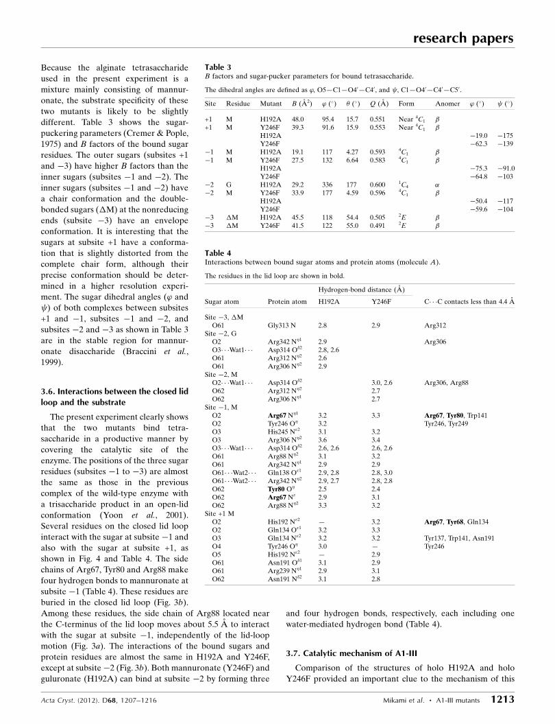
different. Table 3 shows the sugar-
puckering parameters (Cremer & Pople,
1975) and B factors of the bound sugar
residues. The outer sugars (subsites +1
and �3) have higher B factors than the
inner sugars (subsites �1 and �2). The
inner sugars (subsites �1 and �2) have
a chair conformation and the double-
bonded sugars (�M) at the nonreducing
ends (subsite �3) have an envelope
conformation. It is interesting that the
sugars at subsite +1 have a conforma-
tion that is slightly distorted from the
complete chair form, although their
precise conformation should be deter-
mined in a higher resolution experi-
ment. The sugar dihedral angles (’ and
) of both complexes between subsites
+1 and �1, subsites �1 and �2, and
subsites �2 and �3 as shown in Table 3
are in the stable region for mannur-
onate disaccharide (Braccini et al.,
1999).
3.6. Interactions between the closed lidloop and the substrate
The present experiment clearly shows
that the two mutants bind tetra-
saccharide in a productive manner by
covering the catalytic site of the
enzyme. The positions of the three sugar
residues (subsites �1 to �3) are almost
the same as those in the previous
complex of the wild-type enzyme with
a trisaccharide product in an open-lid
conformation (Yoon et al., 2001).
Several residues on the closed lid loop
interact with the sugar at subsite�1 and
also with the sugar at subsite +1, as
shown in Fig. 4 and Table 4. The side
chains of Arg67, Tyr80 and Arg88 make
four hydrogen bonds to mannuronate at
subsite �1 (Table 4). These residues are
buried in the closed lid loop (Fig. 3b).
Among these residues, the side chain of Arg88 located near
the C-terminus of the lid loop moves about 5.5 A to interact
with the sugar at subsite �1, independently of the lid-loop
motion (Fig. 3a). The interactions of the bound sugars and
protein residues are almost the same in H192A and Y246F,
except at subsite�2 (Fig. 3b). Both mannuronate (Y246F) and
guluronate (H192A) can bind at subsite �2 by forming three
and four hydrogen bonds, respectively, each including one
water-mediated hydrogen bond (Table 4).
3.7. Catalytic mechanism of A1-III
Comparison of the structures of holo H192A and holo
Y246F provided an important clue to the mechanism of this
research papers
Acta Cryst. (2012). D68, 1207–1216 Mikami et al. � A1-III mutants 1213
Table 3B factors and sugar-pucker parameters for bound tetrasaccharide.
The dihedral angles are defined as ’, O5—C1—O40—C40, and , C1—O40—C40—C50.
Site Residue Mutant B (A2) ’ (�) � (�) Q (A) Form Anomer ’ (�) (�)
+1 M H192A 48.0 95.4 15.7 0.551 Near 4C1 �+1 M Y246F 39.3 91.6 15.9 0.553 Near 4C1 �
H192A �19.0 �175Y246F �62.3 �139
�1 M H192A 19.1 117 4.27 0.593 4C1 ��1 M Y246F 27.5 132 6.64 0.583 4C1 �
H192A �75.3 �91.0Y246F �64.8 �103
�2 G H192A 29.2 336 177 0.600 1C4 ��2 M Y246F 33.9 177 4.59 0.596 4C1 �
H192A �50.4 �117Y246F �59.6 �104
�3 �M H192A 45.5 118 54.4 0.505 2E ��3 �M Y246F 41.5 122 55.0 0.491 2E �
Table 4Interactions between bound sugar atoms and protein atoms (molecule A).
The residues in the lid loop are shown in bold.
Hydrogen-bond distance (A)
Sugar atom Protein atom H192A Y246F C� � �C contacts less than 4.4 A
Site �3, �MO61 Gly313 N 2.8 2.9 Arg312
Site �2, GO2 Arg342 N�1 2.9 Arg306O3� � �Wat1� � � Asp314 O�2 2.8, 2.6O61 Arg312 N�2 2.6O61 Arg306 N�2 2.9
Site �2, MO2� � �Wat1� � � Asp314 O�2 3.0, 2.6 Arg306, Arg88O62 Arg312 N�2 2.7O62 Arg306 N�1 2.7
Site �1, MO2 Arg67 N�1 3.2 3.3 Arg67, Tyr80, Trp141O2 Tyr246 O� 3.2 Tyr246, Tyr249O3 His245 N"2 3.1 3.2O3 Arg306 N�2 3.6 3.4O3� � �Wat1� � � Asp314 O�2 2.6, 2.6 2.6, 2.6O61 Arg88 N�2 3.1 3.2O61 Arg342 N�1 2.9 2.9O61� � �Wat2� � � Gln138 O"1 2.9, 2.8 2.8, 3.0O61� � �Wat2� � � Arg342 N�2 2.9, 2.7 2.8, 2.8O62 Tyr80 O� 2.5 2.4O62 Arg67 N" 2.9 3.1O62 Arg88 N�2 3.3 3.2
Site +1 MO2 His192 N"2 — 3.2 Arg67, Tyr68, Gln134O2 Gln134 O"1 3.2 3.3O3 Gln134 N"2 3.2 3.2 Tyr137, Trp141, Asn191O4 Tyr246 O� 3.0 — Tyr246O5 His192 N"2 — 2.9O61 Asn191 O�1 3.1 2.9O61 Arg239 N�1 2.9 3.1O62 Asn191 N�2 3.1 2.8

enzyme. In the H192A holo structure, Tyr246 O� makes two
hydrogen bonds to O4 of mannuronate at subsite +1 and O2 of
mannuronate at subsite �1. The distance between Tyr246 O�
and C5 of mannuronate at subsite +1 is only 3.3 A, suggesting
that Tyr246 can extract a hydrogen from the C5 atom (Fig. 5).
In contrast, the side chain of His192 in holo Y246F is situated
at the other side of Tyr246 and makes two hydrogen bonds to
O2 and O5 of mannuronate at subsite +1, with the distance
between His192 N"2 and C5 of mannuronate at subsite +1
being 3.4 A (Figs. 1 and 3c). The mannuronate at subsite +1 in
both holo structures has a near-chair form (Table 3), with the
H atom of C5 facing towards the side chain of Tyr246. It is
suggested from the distance between Tyr246 O� and O4 of
mannuronate at subsite +1 (2.9 A) that Tyr246 O� can also
donate a hydrogen to O4 of the glycoside bond to be cleaved.
The C� � �O�� � �C5 and C� � �O�
� � �O4 angles are 103� and 109�,
respectively (Fig. 3c), suggesting that the reaction coordinates
are almost the same for abstraction and donation. Thus, in this
lyase reaction without conformational change of the side
chain, the side chain of Tyr246 is responsible for both the
abstraction and donation of hydrogen simultaneously, as
shown in the schematic view in Fig. 5. Several positively
charged side chains, Arg239 (3.5 A from Tyr246 O�), Arg67
(4.8 A), Arg306 (6.1 A), His245 (5.4 A) and His192 (6.4 A),
surrounding Tyr246 O� may reduce the pKa of Tyr246 and
facilitate the abstraction and donation of H atoms. For
abstraction of the H atom to occur, the sugar ring must be
distorted with a coplanar C3—C4—C5—O5 configuration
similar to the product double-bonded sugar. The slightly
distorted sugar rings of subsite +1 in both mutant structures
suggest that some additional residues other than Tyr246 and
His192 may be responsible for distortion of the sugar ring at
subsite +1. These residues are estimated to be Asn191 and
Arg239 based on their interaction with mannuronate at
subsite +1. The side-chain atoms of Asn191 and Arg239 are
near O6 and O5 of mannuronate
at subsite +1, respectively (Fig. 3c
and Table 4). These side chains
collaborate with His192 to distort
the sugar ring and assist in the
catalysis conducted by the side
chain of Tyr246. These residues
have also been shown to be
important in chondroitin AC
lyase (PL-8; Lunin et al., 2004;
Huang et al., 2001), hyaluronate
lyase (Li & Jedrzejas, 2001; Mello
et al., 2002; Nukui et al., 2003;
Rigden & Jedrzejas, 2003) and
xanthan lyase (Maruyama et al.,
2005, 2007).
The present experiment also
showed the important role of the
lid loop in the activation of
Tyr246. The side chain of Tyr68
in the closed lid loop makes a
hydrogen bond to the side chain
of Tyr246 (Tyr68 O�� � �Tyr246 O�,
2.6 A) and is near the positively
charged side chain of Arg67
(Tyr68 O�� � �Arg67 N�1, 3.3 A)
(Figs. 3c and 4). We speculate that
when Tyr68 O� donates its
hydrogen to Tyr246 O� to make
the hydrogen bond, the pKa value
of Tyr246 decreases by stabilizing
research papers
1214 Mikami et al. � A1-III mutants Acta Cryst. (2012). D68, 1207–1216
Figure 6Multiple sequence alignment of mannuronate-specific alginate lyases belonging to the PL-5 family. Theposition of the lid loop in A1-III is boxed. Identical residues are shown in bold. The A1-III residuesinvolved in interaction with the substrate are labelled in red. Conserved residues are labelled in orange. Thehelices in A1-III are underlined. The conserved amino-acid residues in the active site of A1-III that are notinvolved in direct interaction with substrate are marked by asterisks.
Figure 5Schematic representation of the catalytic mechanism of alginate lyase A1-III. The side chain of Tyr246 is responsible for simultaneous abstractionand donation of hydrogen without side-chain conformational change. Theside chain of Tyr68 in the closed lid loop forms a hydrogen bond to theside chain of Tyr246 that accelerates proton transfer.

the deprotonated form, thereby accelerating proton transfer.
The results for the mutation of Tyr68 to Phe68 (Table 1)
showed that the side chain of Tyr68 accelerates catalysis by
about 20-fold, which is consistent with this hypothesis.
3.8. Sequence comparison of PL-5 alginate lyase
Fig. 6 shows a sequence alignment of mannuronate-specific
lyases in PL-5. The sequence around Tyr246 and His192 of
A1-III is strongly conserved in the six sequences from various
microorganisms. Although the sequence similarity in the lid
loop region is not as high as the similarity in the regions of
Tyr246 and His192, Tyr68 and Arg88 are well conserved. The
position of Arg67 in A1-III is conserved as an Arg or a Lys
residue. This conservative replacement is also found for Asn72
(Asp), Gly74 (Ala), Asp79 (Asn) and Tyr87 (Phe). These
results suggest that the mobile lid loop of A1-III is functional
throughout the family of PL-5 mannuronate-specific alginate
lyases.
4. Conclusion
The crystal structures of alginate lyase mutants (Y246F and
H192A) complexed with alginate tetrasaccharide have been
explored in this study. The lid loop (residues 64–85) of alginate
lyase A1-III can move with a maximum distance of 13.4 A
from the open form (apoenzyme) to the closed form (holo-
enzyme) in the orthorhombic crystal system. The conforma-
tional change of this loop is a near-rigid-body motion with
hinges around residues 64 and 82–85. The conformational
change is larger than the usual loop motion of the ! loop, as
the lid loop contains a longer loop and part of the �-helix (H3)
region. The closed lid loop interacts with the bound tetra-
saccharide and a catalytic residue. In particular, Tyr68 in the
closed lid loop was expected to activate a presumed catalytic
residue, Tyr246, by forming a hydrogen bond. From alignment
of primary structures of PL-5 mannuronate-specific alginate
lyases, the lid loop seems to be conserved in this family of
enzymes. The tetrasaccharide formed a bond in the active cleft
at subsites�3 to +1 as a substrate form in which the glycosidic
linkage to be cleaved is present between subsites �1 and +1.
Based on the configuration of Tyr246 towards the substrate
sugar, it is concluded that the side chain of Tyr246 acts as both
an acid and a base catalyst in a syn mechanism.
Diffraction data for Y246F were collected at the BL-38B1
station of SPring-8 (Hyogo, Japan) with the approval of JASRI
(Proposal No. C99A24XU-003N). Computation time was
provided by the Supercomputer Laboratory, Institute for
Chemical Research, Kyoto University, Japan. This work was
supported in part by a Grant-in-Aid for Scientific Research
from the Ministry of Education, Science, Sports and Culture of
Japan and by Research Fellowships from the Japan Society
for the Target Protein Project. Part of this work was also
supported by the Program of Basic Research Activities for
Innovative Biosciences (PROBRAIN) of Japan.
References
Adams, P. D. et al. (2010). Acta Cryst. D66, 213–221.Boyd, A., Ghosh, M., May, T. B., Shinabarger, D., Keogh, R. &
Chakrabarty, A. M. (1993). Gene, 131, 1–8.Braccini, I., Grasso, R. P. & Perez, S. (1999). Carbohydr. Res. 317,
119–130.Brunger, A. T., Adams, P. D., Clore, G. M., DeLano, W. L., Gros, P.,
Grosse-Kunstleve, R. W., Jiang, J.-S., Kuszewski, J., Nilges, M.,Pannu, N. S., Read, R. J., Rice, L. M., Simonson, T. & Warren, G. L.(1998). Acta Cryst. D54, 905–921.
Cantarel, B. L., Coutinho, P. M., Rancurel, C., Bernard, T., Lombard,V. & Henrissat, B. (2009). Nucleic Acids Res. 37, D233–D238.
Cremer, D. & Pople, J. A. (1975). J. Am. Chem. Soc. 97, 1354–1358.Davies, G. J., Wilson, K. S. & Henrissat, B. (1997). Biochem. J. 321,
557–559.Dunlop, K. V., Irvin, R. T. & Hazes, B. (2005). Acta Cryst. D61, 80–87.Elmabrouk, Z. H., Vincent, F., Zhang, M., Smith, N. L., Turkenburg,
J. P., Charnock, S. J., Black, G. W. & Taylor, E. J. (2011). Proteins,79, 965–974.
Emsley, P., Lohkamp, B., Scott, W. G. & Cowtan, K. (2010). ActaCryst. D66, 486–501.
Fethiere, J., Eggimann, B. & Cygler, M. (1999). J. Mol. Biol. 288,635–647.
Garron, M. L. & Cygler, M. (2010). Glycobiology, 20, 1547–1573.Gerstein, M. & Krebs, W. (1998). Nucleic Acids Res. 26, 4280–4290.Gimmestad, M., Sletta, H., Ertesvag, H., Bakkevig, K., Jain, S., Suh,
S., Skjak-Braek, G., Ellingsen, T. E., Ohman, D. E. & Valla, S.(2003). J. Bacteriol. 185, 3515–3523.
Hooft, R. W., Vriend, G., Sander, C. & Abola, E. E. (1996). Nature(London), 381, 272.
Huang, W., Boju, L., Tkalec, L., Su, H., Yang, H.-O., Gunay, N. S.,Linhardt, R. J., Kim, Y. S., Matte, A. & Cygler, M. (2001).Biochemistry, 40, 2359–2372.
Hubbard, S. J. & Thornton, J. M. (1993). NACCESS. Department ofBiochemistry and Molecular Biology, University College, London.
Jedrzejas, M. J., Mello, L. V., de Groot, B. L. & Li, S. (2002). J. Biol.Chem. 277, 28287–28297.
Laskowski, R. A., MacArthur, M. W., Moss, D. S. & Thornton, J. M.(1993). J. Appl. Cryst. 26, 283–291.
Li, S. & Jedrzejas, M. J. (2001). J. Biol. Chem. 276, 41407–41416.Li, S., Taylor, K. B., Kelly, S. J. & Jedrzejas, M. J. (2001). J. Biol. Chem.
276, 15125–15130.Lombard, V., Bernard, T., Rancurel, C., Brumer, H., Coutinho, P. M.
& Henrissat, B. (2010). Biochem J. 432, 437–444.Lunin, V. V., Li, Y., Linhardt, R. J., Miyazono, H., Kyogashima, M.,
Kaneko, T., Bell, A. W. & Cygler, M. (2004). J. Mol. Biol. 337,367–386.
Luzzati, V. (1952). Acta Cryst. 5, 802–810.Maruyama, Y., Hashimoto, W., Mikami, B. & Murata, K. (2005). J.
Mol. Biol. 350, 974–986.Maruyama, Y., Mikami, B., Hashimoto, W. & Murata, K. (2007).
Biochemistry, 46, 781–791.Mello, L. V., De Groot, B. L., Li, S. & Jedrzejas, M. J. (2002). J. Biol.
Chem. 277, 36678–36688.Murata, K., Inose, T., Hisano, T., Abe, S., Yonemoto, Y., Yamashita,
T., Takagi, M., Sakaguchi, K., Kimura, A. & Imanaka, T. (1993). J.Ferment. Bioeng. 76, 427–437.
Noble, M. E. M., Zeelen, J. P. & Wierenga, R. K. (1993). Proteins, 16,311–326.
Nukui, M., Taylor, K. B., McPherson, D. T., Shigenaga, M. K. &Jedrzejas, M. J. (2003). J. Biol. Chem. 278, 3079–3088.
Ochiai, A., Yamasaki, M., Mikami, B., Hashimoto, W. & Murata, K.(2010). J. Biol. Chem. 285, 24519–24528.
Ogura, K., Yamasaki, M., Mikami, B., Hashimoto, W. & Murata, K.(2008). J. Mol. Biol. 380, 373–385.
Osawa, T., Matsubara, Y., Muramatsu, T., Kimura, M. & Kakuta, Y.(2005). J. Mol. Biol. 345, 1111–1118.
research papers
Acta Cryst. (2012). D68, 1207–1216 Mikami et al. � A1-III mutants 1215

Otwinowski, Z. & Minor, W. (1997). Methods Enzymol. 276, 307–326.
Pecina, A., Pascual, A. & Paneque, A. (1999). J. Bacteriol. 181, 1409–1414.
Ponnuraj, K. & Jedrzejas, M. J. (2000). J. Mol. Biol. 299, 885–895.Preston, L. A., Wong, T. Y., Bender, C. L. & Schiller, N. L. (2000). J.
Bacteriol. 182, 6268–6271.Ramachandran, G. N. & Sasisekharan, V. (1968). Adv. Protein Chem.
23, 283–438.Rigden, D. J., Botzki, A., Nukui, M., Mewbourne, R. B., Lamani, E.,
Braun, S., von Angerer, E., Bernhardt, G., Dove, S., Buschauer, A.& Jedrzejas, M. J. (2006). Glycobiology, 16, 757–765.
Rigden, D. J. & Jedrzejas, M. J. (2003). J. Biol. Chem. 278, 50596–50606.
Shaya, D., Zhao, W., Garron, M. L., Xiao, Z., Cui, Q., Zhang, Z.,Sulea, T., Linhardt, R. J. & Cygler, M. (2010). J. Biol. Chem. 285,20051–20061.
Skrzypczak-Jankun, E., Borbulevych, O. Y., Zavodszky, M. I.,Baranski, M. R., Padmanabhan, K., Petricek, V. & Jankun, J.
(2006). Acta Cryst. D62, 766–775.Spek, A. L. (2003). J. Appl. Cryst. 36, 7–13.Thompson, J. D., Higgins, D. G. & Gibson, T. J. (1994). Nucleic Acids
Res. 22, 4673–4680.Tsitsanou, K. E., Oikonomakos, N. G., Zographos, S. E., Skamnaki,
V. T., Gregoriou, M., Watson, K. A., Johnson, L. N. & Fleet, G. W.(1999). Protein Sci. 8, 741–749.
Winn, M. D. et al. (2011). Acta Cryst. D67, 235–242.Yamasaki, M., Ogura, K., Hashimoto, W., Mikami, B. & Murata, K.
(2005). J. Mol. Biol. 352, 11–21.Yonemoto, Y., Tanaka, H., Hisano, T., Sakaguchi, K., Abe, S.,
Yamashita, T., Kimura, A. & Murata, K. (1993). J. Ferment. Bioeng.75, 336–342.
Yoon, H.-J., Hashimoto, W., Miyake, O., Murata, K. & Mikami, B.(2001). J. Mol. Biol. 307, 9–16.
Yoon, H.-J., Hashimoto, W., Miyake, O., Okamoto, M., Mikami, B. &Murata, K. (2000). Protein Expr. Purif. 19, 84–90.
Yoon, H.-J., Mikami, B., Hashimoto, W. & Murata, K. (1999). J. Mol.Biol. 290, 505–514.
research papers
1216 Mikami et al. � A1-III mutants Acta Cryst. (2012). D68, 1207–1216