Embed Size (px)
Citation preview

Journal of CellularBiochemistry
FAST TRACKJournal of Cellular Biochemistry 107:622–629 (2009)
Transforming Growth Factor-b3-Induced Smad SignalingRegulates Actin Reorganization During Chondrogenesis ofChick Leg Bud Mesenchymal Cells
Gy
o
*W
R
P
Dongkyun Kim,1 Jungsoo Kim,2y Shin-Sung Kang,2 and Eun-Jung Jin1,3*1Department of Biological Sciences, College of Natural Sciences, Wonkwang University, Iksan, Chunbuk 570-749,South Korea
2Department of Biology, College of Natural Sciences, Kyungpook National University,Daegu 702-701 South Korea
3Institute of Biotechnology, Wonkwang University, Iksan, Chunbuk 570-749, Korea
ABSTRACTEndochondral ossification is characterized by a significant interdependence between cell shape and cytoskeletal organization that
accompanies the onset of chondrogenic signaling. However, the mechanisms mediating these interactions have not been well studied.
Here, treatment with transforming growth factor (TGF)-b3 at a later stage of chondrogenesis led to activation of Smad-2 signaling and the
formation of intense stress fibers, which resulted in suppressing chondrogenic differentiation of leg bud mesenchymal cells. Moreover,
specific siRNA knockdown of Smad-2 reduced TGF-b3-induced stress fibers via physical interactions with b-catenin. In conclusion, our
results indicate that TGF-b3-induced Smad signaling, in conjunction with b-catenin, is involved in the reorganization of the actin
cytoskeleton into a cortical pattern with a concomitant rounding of cells. J. Cell. Biochem. 107: 622–629, 2009. � 2009 Wiley-Liss, Inc.
KEY WORDS: SMAD; b-CATENIN; ACTIN CYTOSKELETON; CHONDROGENIC DIFFERENTIATION
C hondrogenesis is a tightly regulated process in which multi-
potential mesenchymal cells differentiate into chondrocytes
to form cartilage [Hall, 1981; Cancedda et al., 1995; Goldring et al.,
2006]. This process is initiated by commitment to the chondrogenic
lineage and condensation of cells. This is followed by differentiation
into chondrocytes with spatially and temporally regulated expres-
sions of cartilage-specific genes, such as type II collagen, aggrecan
and sulfated proteoglycans [Shinomura et al., 1993], to maintain
the chondrocyte phenotype [Kamiya et al., 2006]. A dramatic
morphological transition in cell shape, from fibroblastoid to
round or polygonal morphologies, is a critical regulatory factor
for chondrogenesis [Zanetti and Solursh, 1984; Yabu et al., 1991;
Benjamin et al., 1994]. In addition, chondrocytes display a primarily
cortical organization of actin filaments, whereas undifferentiated
mesenchymal cells are characterized by a fibrillar organization
[Zhang et al., 2006].
The transforming growth factor (TGF)-b superfamily, comprised of
TGF-bs, bone morphogenetic proteins, activins and related proteins,
rant sponsor: Korea Research Foundation (KRF); Grant number: C00731
This article was published online on 28 May 2009. An error was subsequentnline and print versions to indicate that both have been corrected 8 Jun
Correspondence to: Dr. Eun-Jung Jin, PhD, Department of Biologicalonkwang University, Iksan, Chunbuk 570-749, South Korea. E-mail: jin
eceived 5 January 2009; Accepted 2 April 2009 � DOI 10.1002/jcb.2219
ublished online 28 May 2009 in Wiley InterScience (www.interscience.w
regulates cell function and plays key roles in development, cellular
differentiation, apoptosis, and carcinogenesis [Hoodless et al., 1996;
Itoh et al., 2000; Massague, 2000; Derynck and Zhang, 2003]. In
addition to the BMP/Smad signaling pathway, p38 kinase is also
stimulated by BMP-2 [Iwasaki et al., 1999; Kimura et al., 2000]. BMP-
2 activates MAP kinase kinase kinases, including TAK1. Subse-
quently, a MAP kinase kinase kinase induces MKK3 or MKK6 that
directly phosphorylates and activates p38 kinase [Raingeaud et al.,
1996; Shibuya et al., 1996; Kimura et al., 2000]. The phosphorylated
p38 kinase, in turn, regulates gene expressions in the nucleus. In
addition, BMP-2 treatment also results in increased protein levels of
b-catenin, a known N-cadherin-associated Wnt signal transducer
[Haas and Tuan, 2000; Fischer et al., 2002; Jin et al., 2006]. Taken
together, these phenomena suggests that functional cross-talk occurs
between the BMP-2 and Wnt signaling pathways.
Previously, we demonstrated that an increase of Sox9 protein,
resulting from downregulation of b-catenin/Wnt-7a signaling,
was mediated by p38 MAPK during BMP-2 induced chondrogenesis
.
ly identified. This notice is included in thee 2009.
Sciences, College of Natural Sciences,[email protected]
1 � � 2009 Wiley-Liss, Inc.
iley.com).
622
in chick wing bud mesenchymal cells [Jin et al., 2006]. We also
demonstrated that TGF-b3 downregulated connexin43. This
induced apoptotic cell death via activation of ERK and suppressed
PKC-a activation in leg bud mesenchymal cells [Jin et al., 2008].
However, Smad-dependent TGF-b3 signaling during chondrogenic
differentiation of chick limb bud mesenchymal cells has not been
extensively studied [Hoodless et al., 1996; Heldin et al., 1997; Nakao
et al., 1997; Itoh et al., 2000], even though Smad is the major
mechanism that mediates the TGF-b3 signaling pathway [Nakao
et al., 1997; Miyazono, 2000; Moustakas et al., 2001; Derynck and
Zhang, 2003]. Therefore, in the present study, we investigated the
functional role of Smad during chondrogenic differentiation using
cultured chick leg bud mesenchymal cells. The activation of
Smad signaling was found to act as a negative regulator of the
morphological transitions of cells at a later stage of chondrogenesis.
Furthermore, we found that activation of reorganization of actin
cytoskeleton induced by Smad signaling may occur through an
interaction with b-catenin.
MATERIALS AND METHODS
CELL CULTURE AND TREATMENTS
Mesenchymal cells derived from the distal tips of Hamburger–
Hamilton (HH) stage 22/23 embryo leg buds of fertilized White
Leghorn chicken eggs were micromass cultured as previously
described [Jin et al., 2006]. Briefly, the cells were suspended at a
density of 2� 107 cells/ml in Ham’s F-12 medium containing 10%
fetal bovine serum (FBS), 100 IU/ml penicillin, and 100mg/ml
streptomycin (Gibco Invitrogen, Grand Island, NY). The cells were
plated in 3 drops (15ml each) dispensed into 35-mm culture dishes
or 19 drops (15ml each) into 60-mm Corning culture dishes, and
incubated for 1 h at 378C under 5% CO2 to allow attachment. The
cells were maintained in 1 ml of culture medium after 72 h of culture
in the absence or presence of 5 ng/ml recombinant Human TGF-b3
(R&D, Minneapolis, MN).
TRANSFECTION OF SMAD-2-SPECIFIC SMALL INTERFERING
RNA (siRNA)
The siRNA oligonucleotide (50-CAGGACAGCTGGATGAGCTTGAG-
AA-30) and the corresponding sense oligonucleotide (05-TTCT-
CAAGCTCATCCAGCTGTCCTG-30) were obtained from Invitrogen.
Transfection of the RNA oligonucleotides (50 nM final RNA
concentration) into chick wing bud mesenchymal cells used Fugene
6 (Roche) following the manufacturer’s protocol.
ANALYSIS OF CELL DIFFERENTIATION
Chondrogenesis was measured by Alcian blue staining of sulfated
cartilage glycosaminoglycans. To demonstrate the deposition
of cartilage matrix proteoglycans, representative cultures were
collected at day 5 of incubation and stained with 0.5% Alcian blue
8GX, pH 1.0 [Lev and Spicer, 1964; Hassel and Horigan, 1982].
Alcian-blue-bound sulfated glycosaminoglycans were extracted
with 6 M guanidine–HCl and quantified by measuring the
absorbance of the extracts at 600 nm.
JOURNAL OF CELLULAR BIOCHEMISTRY
SEMI-QUANTITATIVE RT-PCR
Total RNA was isolated using Trizol (Gibco Invitrogen) at days 1, 3,
and 5 of culture. cDNA was synthesized from a 2mg sample of
RNA in 20ml of master mix for reverse transcription containing
200 U/ml Superscript III (Gibco Invitrogen), 5 mM MgCl2, PCR Buffer
II, 1 mM dNTP, 1 U/ml RNase inhibitor, and 2.5 mM oligo dT in
DEPC-treated distilled water. The master mix was incubated in a
Perkin-Elmer GeneAmp PCR system 9600 (Wellesley, MA) at 428Cfor 55 min and 998C for 5 min. Synthesized cDNAs were subjected to
25 cycles of PCR amplification under the following conditions: 948Cdenaturing for 40 s, 558C annealing for 40 s, and 728C extension for
40 s. Subsequently, PCR products were electrophoresed on a 2.0%
agarose gel. Primers for Smad2: 50-ACACTCATTCCATTCCTGAG-30,
50-GAAGGTTTCTCCCACTCTTT-30, for Smad3: 50-AGAACACTAA-
CTTCCCAG-30, 50-GGTTTACAGACTGAGCCAAG-30, for GAPDH:
50-GATGGGTGTCAACCATGAGA AA-30, 50-ATCAAAGGTGGAAGA
ATGGCTG-30.
WESTERN BLOT ANALYSIS
Proteins (30mg) or conditioned media were separated by 10% poly-
acrylamide gel electrophoresis containing 0.1% SDS and transferred
to nitrocellulose membrane (Schleicher and Schuell, Germany). The
membranes were incubated for 1 h at room temperature in blocking
buffer (20 mM Tris–HCl, 137 mM NaCl, pH 8.0, containing 0.1%
Tween and 3% non-fat dry milk), and probed with antibodies against
Smad, pSmad, b-catenin (R&D), glycogen synthase kinase (GSK),
phosphorylated glycogen synthase kinase (pGSK; Calbiochem, La
Jolla, CA), and HSP70 (Stressgene, San Diego, CA). The blots were
developed with a peroxidase-conjugated secondary antibody and
reacted proteins were visualized using the electrochemilumines-
cence (ECL) system (Pierce Biotechnology, Inc., Rockford, MN).
ACTIN STAINING AND IMMUNOCYTOCHEMISTRY
Cells grown on cover slips were treated with various chemicals as
indicated in the figure legends, washed three times with phosphate-
buffered saline (PBS), then fixed and permeabilized as described
above. For actin staining, each culture was stained with Alexa488-
phalloidin (Molecular Probes, Eugene, OR) prepared in PBS con-
taining 1% (v/v) bovine serum albumin for 1 h at room temperature
in a lightproof box. For immunocytochemistry, cultured cells were
double stained with antibodies against Smad2 and b-catenin,
washed three times with PBS, and incubated with secondary
antibody conjugated with either Cy2 (for Smad2 detection) or TRITC
(for b-catenin detection). Cultures were then washed three times
with water and mounted with Gel/Mount (Biomedia, Foster City,
CA). The slides were examined using a confocal microscope (MRC
1024/ES, Bio-Rad Laboratory, CA). Cultures were imaged at scan
speed 8 (1.76ms per voxel, 1 s per section) in 1-mm-thick Z-sections
for a total of 4–10 slices.
CELL VIABILITY AND PROLIFERATION ASSAY
Proliferation of mesenchymal cells was determined by direct
counting of cells from micromass cultures. Control and treated
cultures were maintained for the indicated number of days, cells
were detached with trypsin/EDTA solution and counted in triplicate
using a hemacytometer.
SMAD SIGNALING IN ACTIN ORGANIZATION 623

STATISTICAL ANALYSIS
Results of cell-adhesion experiments were pooled from five replicate
samples derived from three independent experiments and expressed
as means� SEM. One-way analysis of variance (ANOVA) with
Tukey post hoc comparisons of groups was used to test for
significant effects. Differences were considered significant at
P< 0.05.
RESULTS
Pleiotropic members of the TGF-b superfamily are involved with
de novo endochondral bone formation. These factors are soluble
signals for tissue morphogenesis during sculpting of the multi-
cellular mineralized structures of the bone marrow organ
[Ripamonti et al., 2000; Ripamonti, 2004]. To analyze the TGF-
b3-specific signaling, HH stage 22/23 chondroblasts were isolated
from chick leg buds and cultured at a density of 2� 107 cells/ml.
As chondrogenesis progressed, exposing leg bud mesenchymal
cells to TGF-b3 resulted in increased and sustained levels of
unphosphorylated Smad-2 and -3, pGSK-3b, and b-catenin. These
levels decreased at a later stage of chondrogenesis (i.e., day 4 in
the control culture; Fig. 1A). These findings suggested that
downregulation of Smad and Wnt-related signaling might be
required at a later stage of chondrogenesis.
Next, we examined the effects of Smad-2 during the later culture
periods. When cells were exposed to 5 ng/ml of TGF-b at 3–4 days
of culture, the level of Alcian blue staining was reduced in the
micromass culture, which indicated that chondrogenic differentia-
tion had been suppressed. Inhibition of glycosaminoglycan
production by TGF-b3 was confirmed quantitatively by dye
extraction and measurement of the absorbance. Suppressed
chondrogenic differentiation by TGF-b3 was stimulated by
Smad-2 knockdown (Fig. 1B).
Chondrogenic differentiation of mesenchymal cells is regulated
at various stages, including proliferation of chondroblast competent
cells, precartilage condensation, and formation of cartilage nodules
[Quarto et al., 1997; DeLise et al., 2000; Knudson and Knudson,
2001]. Therefore, we examined the effects of siRNA-mediated smad-
2 knockdown on the growth of mesenchymal cells using its specific
siRNA. Cell numbers were assessed at days 3, 4, and 5 after
transfection. Knockdown of smad-2 had little or no effect on the
proliferation of chondroblast competent cells (Fig. 1C). These
findings suggest that the positive role of Smad-2 on chondrogenic
differentiation of leg bud mesenchymal cells occurred indepen-
dently of cell proliferation and/or cellular condensation.
It has previously been shown that reorganization of the actin
cytoskeleton into a cortical pattern with concomitant rounding of
cells and fewer stress fibers, occurred after 4 days of culture [Zakany
et al., 2001]. And our laboratory (2007) also showed that treatment
with cytochalasin D (CD) at later period of culture to disrupt
polymerization of actin cytoskeleton, caused inhibition of chon-
drogenic differentiation indicating that microtubule polymerization
is vital for chondrogenesis. Therefore, we evaluated the blockage of
Smad signaling to determine if it was associated with rearrangement
624 SMAD SIGNALING IN ACTIN ORGANIZATION
of the actin cytoskeleton. Since chondrogenesis of limb mesench-
ymal cells are cell-density dependent, physical pressure from
multilayered culture (micromass culture) would be simply induced
actin reorganization. To investigate this, chondroblasts cultured
in a density-dependent manner (monolayer vs. micromass) were
transfected with Smad-2-specific siRNA in the absence or presence
of 5 ng/ml of TGF-b at 3 days of culture. The levels of actin stress
fibers were markedly intensified after TGF-b3 treatment when
compared to the control culture, pointing to an involvement of the
actin cytoskeleton with Smad signaling. However, knockdown of
Smad-2 at day 3 of culture resulted in decreased levels of TGF-b3-
induced stress fibers as well as a significant increase of cell rounding
compared to control cultures both for monolayer- and micromass-
cultured chondroblasts at 4 days of culture (Fig. 2). These finding
indicate that the physical pressure from increased cell density may
not cause actin cytoskeleton and downregulation of Smad signaling
supports the establishment of chondrocyte-specific cell shape and
actin reorganization.
Knockdown of Smad-2 decreased the TGF-b3-induced levels of
N-cadherin and b-catenin at days 3 and 4 of culture (Fig. 3A). Many
studies have indicated that physical interactions between Smad and
b-catenin occur in a variety of cells [Nishita et al., 2000; Warner
et al., 2005]. Therefore, we examined the physical interactions and
distributions of Smad2 and b-catenin. Immunoprecipitation using
whole lysates of HH stage 23 chick embryos with Smad2 antibody
demonstrated that b-catenin protein co-precipitated with Smad2
(Fig. 3B). Next, non-immune IgG was used, instead of anti-Smad2
antibody as a proper control to demonstrate that the interaction
was specific and not simply due to binding of the agarose beads.
Immunocytochemistry using specific antibodies showed over-
lapping distributions of Smad2 and b-catenin (Fig. 3C). Actin
stress fibers were markedly intensified along with the accumulation
of b-catenin by GSK-3b inhibitor VI, which is a specific inhibitor of
GSK-3b activity (Fig. 3D).
Previously, we demonstrated that overexpression of b-catenin in
primary mesenchymal cells caused a marked inhibition of cellular
condensation and chondrogenesis [Jin et al., 2006]. These results
suggested that a decreased level of b-catenin was required
for precartilage condensation and additional chondrogenic differ-
entiation. The results shown here suggest that TGF-b3-induced
b-catenin, via upregulation of Smad signaling, acts as a negative
regulator for reorganization of the actin cytoskeleton, as well as
inhibition of precartilage condensation.
DISCUSSION
Regulation of actin reorganization and contractility allows cells to
control their shape, movement, division and secretion, which are
vital processes known to be coordinated by the actions of several
signal transduction pathways [Lelkes et al., 1986; Keller and Niggli,
1995; Wang and Newman, 2003].
Rapid reorganization of the actin cytoskeleton is one of the
primary cellular responses to many extracellular signals. TGF-b
induces a rapid reorganization of the actin cytoskeleton in epithelial
cells, whereas a prolonged incubation with TGF-b results in the
JOURNAL OF CELLULAR BIOCHEMISTRY

Fig. 1. Mesenchymal competent cells were prepared as described in Materials and Methods Section. A: Changes in the mRNA and protein levels of (p)Smad2, -3, (p)GSK-3b,
and b-catenin during chondrogenesis were analyzed. B: Cells were transfected with Smad-2-specific siRNA in the absence or presence of 5 ng/ml of TGF-b and then stained with
PNA on day 3 of culture and Alcian blue on day 5 of culture (right panel). Quantification of chondrogenesis was done by measuring the absorbance of bound Alcian blue at
600 nm (left panel). C: The number of viable cells in the control culture was the determined at the indicated days. The results shown are representative of at least four
independent experiments. �Statistically significant differences as compared with the control cells ( P< 0.01).
formation of stress fibers [Bakin et al., 2002; Edlund et al., 2002]
with activation of the Rho family of GTPase, Rac, CDC42, and RhoA
by regulating organization of actin filaments [Jaffe and Hall, 2002]
through TGF-b-mediated signaling including Smads, mitogen-
activated protein kinases (MAPKs), Rho kinases, and Akt/PKB
[Derynck and Zhang, 2003; Roberts and Wakefield, 2003].
Despite the current understanding of the Smad pathways,
the mechanisms by which Smad signals modulate cell shape and
JOURNAL OF CELLULAR BIOCHEMISTRY
the actin cytoskeleton have not yet completely elucidated.
Furthermore, the results of studies conducted to evaluate these
mechanisms have produced conflicting results. Indeed, some studies
have shown that Smad pathway is critical for such responses
[Bakin et al., 2002; Tsai et al., 2007], while others have shown that
non-Smad signaling effectors, such as Rho GTPases, phospholipid
30-kinase, or p38 mitogen-activated protein kinase, play more
important roles for in these responses [Takenawa and Itoh, 2001;
SMAD SIGNALING IN ACTIN ORGANIZATION 625
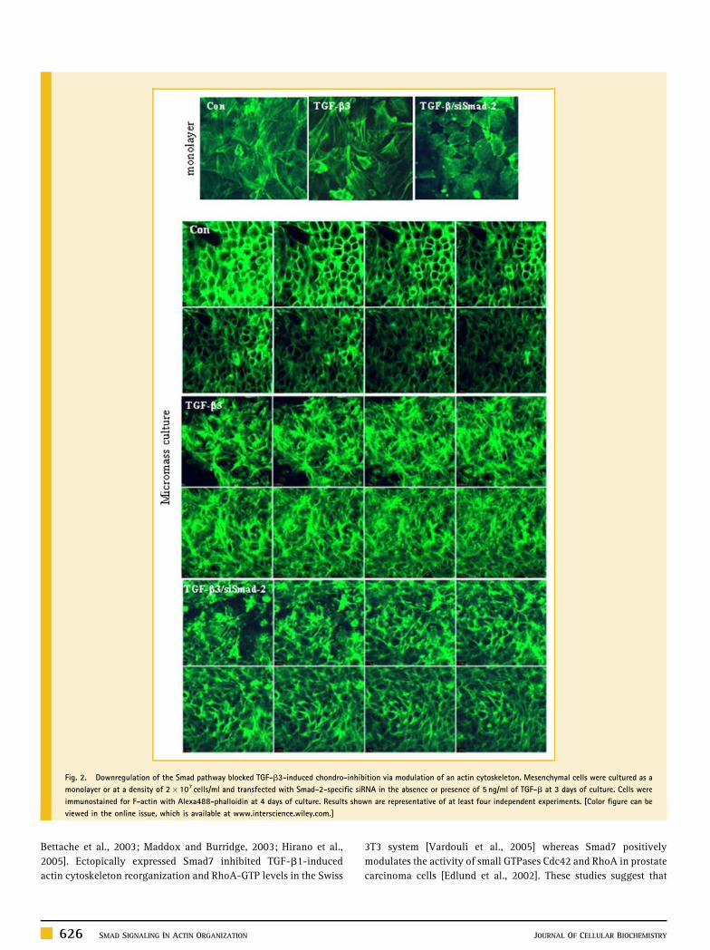
Fig. 2. Downregulation of the Smad pathway blocked TGF-b3-induced chondro-inhibition via modulation of an actin cytoskeleton. Mesenchymal cells were cultured as a
monolayer or at a density of 2� 107 cells/ml and transfected with Smad-2-specific siRNA in the absence or presence of 5 ng/ml of TGF-b at 3 days of culture. Cells were
immunostained for F-actin with Alexa488-phalloidin at 4 days of culture. Results shown are representative of at least four independent experiments. [Color figure can be
viewed in the online issue, which is available at www.interscience.wiley.com.]
Bettache et al., 2003; Maddox and Burridge, 2003; Hirano et al.,
2005]. Ectopically expressed Smad7 inhibited TGF-b1-induced
actin cytoskeleton reorganization and RhoA-GTP levels in the Swiss
626 SMAD SIGNALING IN ACTIN ORGANIZATION
3T3 system [Vardouli et al., 2005] whereas Smad7 positively
modulates the activity of small GTPases Cdc42 and RhoA in prostate
carcinoma cells [Edlund et al., 2002]. These studies suggest that
JOURNAL OF CELLULAR BIOCHEMISTRY

Fig. 3. The Smad pathway affects the actin cytoskeleton in conjunction with b-catenin. A: Cells were transfected with Smad-2-specific siRNA in the absence or presence of
5 ng/ml of TGF-b and analyzed for changes of protein levels for b-catenin and N-cadherin. B: Embryo lysates from HH stage 23 chick immunoprecipitated with anti-Smad2
antibody. Immunoprecipitates were subjected to Western blot analysis with anti-b-catenin antibody. Non-immune IgG (ideally) was used as a negative control.
C: Immunocytochemistry using anti-Smad2 and anti-b-catenin antibodies at 1 day of culture. D: Mesenchymal cells were cultured at a density of 2� 107 cells/ml with
or without 10mM GSK-3b inhibitor VI (GSK-I) at 3-day culture for 24 h. Cells were stained with Alcian blue at day 5 of culture (upper panel) and immunostained for F-actin
with Alexa488-phalloidin at 4 days of culture (lower panel). Results shown are representative of at least four independent experiments. �Statistically significant differences
compared with control cells ( P< 0.01). [Color figure can be viewed in the online issue, which is available at www.interscience.wiley.com.]
Smad signaling on actin cytoskeleton reorganization may be due to
cell type dependent.
In the present study, we addressed the Smad signaling pathway in
the chick leg bud mesenchymal cells that affects reorganization of
the actin cytoskeleton. We found that this reorganization occurs
independently of cell proliferation and/or cellular condensation
Additionally, we found that blocking Smad signaling increased
cell rounding, a typical dramatic characteristic of differentiated
chondrocytes. Furthermore, the coordination and organization of
the cell shape and chondrocyte phenotype by actin was inhibited by
activation Smad by TGF-b3.
Several studies have shown that a- and b-catenin interact
with the actin cytoskeleton [Akiyama and Kawasaki, 2006; Barth
et al., 2006]. Catenin binds to and bundles actin filaments [Rimm
et al., 1995; Pokutta et al., 2002], and interacts with actin binding
proteins, including vinculin [Watabe-Uchida et al., 1998; Weiss
et al., 1998], a-actinin [Knudsen et al., 1995; Hazan et al., 1997],
ZO-1 [Itoh et al., 1997], spectrin [Pradhan et al., 2001], Ajuba [Marie
et al., 2003], afadin [Pokutta et al., 2002], and formin [Kobielak et al.,
2004]. Nishita et al. [2000] identified an endogenous b-catenin/
Smad4 complex, capable of forming a Wnt-dependent complex in
NIH3T3 and L929 cells. In this study, we also observed physical
JOURNAL OF CELLULAR BIOCHEMISTRY
interactions between Smad2 and b-catenin, which suggested the
convergence of these two signaling pathways. This interaction-
induced Wnt-signaling triggered a dense fibrillar actin reorganiza-
tion in chick leg bud mesenchymal cells, which was overcome by
downregulation of the Smad pathway.
In the present study, we investigated the functional role of Smad
during the late stages of chondrogenic differentiation using cultured
chick leg bud mesenchymal cells. Smad was found to be a negative
regulator of the morphological transition of the actin cytoskeleton
in a conjunction with the b-catenin signaling pathway.
ACKNOWLEDGMENTS
This work was supported by Korea Research Foundation (KRF) by
the Korean Government (C00731).
REFERENCES
Akiyama T, Kawasaki Y. 2006. Wnt signaling and the actin cytoskeleton.Oncogene 25(57):7538–7544.
SMAD SIGNALING IN ACTIN ORGANIZATION 627
Bakin AV, Rinehart C, Tomlinson AK, Arteaga CL. 2002. p38 mitogen-activated protein kinase is required for TGF-beta-mediated fibroblastictransdifferentiation and cell migration. J Cell Sci 115(Pt 15):3193–3206.
Barth AI, Nathke IS, Nelson WJ. 2006. Cadherins, catenins and APC protein:Interplay between cytoskeletal complexes and signaling pathways. Curr OpinCell Biol 9(5):683–690.
Benjamin M, Archer CW, Ralphs JR. 1994. Cytoskeleton of cartilage cells.Microsc Res Tech 28(5):372–377.
Bettache N, Baisamy L, Baghdiguian S, Payrastre B, Mangeat P, Bienvenue A.2003. Mechanical constraint imposed on plasma membrane through trans-verse phospholipid imbalance induces reversible actin polymerization viaphosphoinositide 3-kinase activation. J Cell Sci 116(Pt 11):2277–2284.
Cancedda R, Descalzi Cancedda F, Castagnola P. 1995. Chondrocyte differ-entiation. Int Rev Cytol 159:265–358.
DeLise AM, Fischer L, Tuan RS. 2000. Cellular interactions and signaling incartilage development. Osteoarthritis Cartilage 8(5):309–334.
Derynck R, Zhang YE. 2003. Smad-dependent and Smad-independent path-ways in TGF-beta family signaling. Nature 425(6958):577–584.
Edlund S, Landstrom M, Heldin C-H, Aspenstrom P. 2002. Transforminggrowth factor-beta-induced mobilization of actin cytoskeleton requiressignaling by small GTPases Cdc42 and RhoA. Mol Biol Cell 13:902–914.
Fischer L, Boland G, Tuan RS. 2002. Wnt signaling during BMP-2 stimulationof mesenchymal chondrogenesis. J Cell Biochem 84(4):816–831.
Goldring MB, Tsuchimochi K, Ijiri K. 2006. The control of chondrogenesis.J Cell Biochem 97(1):33–44.
Haas AR, Tuan RS. 2000. Murine C3H10T1/2 multipotential cells as an in vitromodel of mesenchymal chondrogenesis. Methods Mol Biol 137:383–389.
Hall BK. 1981. Intracellular and extracellular control of the differentiation ofcartilage and bone. Histochem J 13(4):599–614.
Hassel JR, Horigan EA. 1982. Chondrogenesis: A model developmentalsystem for measuring teratogenic potential of compounds. Teratog CarcinogMutagen 2:325–331.
Hazan RB, Kang L, Roe S, Borgen PI, Rimm DL. 1997. Vinculin is associatedwith the E-cadherin adhesion complex. J Biol Chem 272(51):32448–32453.
Heldin CH, Miyazono K, Ten Dijke P. 1997. TGF-beta signaling from cellmembrane to nucleus through SMAD proteins. Nature 390(6659):465–471.
Hirano S, Sun X, DeGuzman CA, Ransom RF, McLeish KR, Smoyer WE,Shelden EA, Welsh MJ, Benndorf R. 2005. p38 MAPK/HSP25 signalingmediates cadmium-induced contraction of mesangial cells and renal glo-meruli. Am J Physiol Renal Physiol 288(6):F1133–F1143.
Hoodless PA, Haerry T, Abdollah S, Stapleton M, O’Connor MB, Attisano L,Wrana JL. 1996. MADR1, a MAD-related protein that functions in BMP2signaling pathways. Cell 85(4):489–500.
Itoh M, Nagafuchi A, Moroi S, Tsukita S. 1997. Involvement of ZO-1 incadherin-based cell adhesion through its direct binding to alpha catenin andactin filaments. J Cell Biol 138(1):181–192.
Itoh S, Itoh F, Goumans MJ, Ten Dijke P. 2000. Signaling of transforminggrowth factor-beta family members through Smad proteins. Eur J Biochem267(24):6954–6967.
Iwasaki S, Iguchi M, Watanabe K, Hoshino R, Tsujimoto M, Kohno M. 1999.Specific activation of the p38 mitogen-activated protein kinase signalingpathway and induction of neurite outgrowth in PC12 cells by bone mor-phogenetic protein-2. J Biol Chem 274(37):26503–26510.
Jaffe AB, Hall A. 2002. Rho GTPases in transformation and metastasis. AdvCancer Res 84:57–80.
Jin EJ, Lee SY, Choi YA, Jung JC, Bang OS, Kang SS. 2006. BMP-2-enhancedchondrogenesis involves p38 MAPK-mediated down-regulation of Wnt-7apathway. Mol Cells 22(3):353–359.
Jin EJ, Lee SY, Jung JC, Bang OS, Kang SS. 2008. TGF-beta3 inhibitschondrogenesis of cultured chick leg bud mesenchymal cells via down-regulation of connexin 43 and integrin beta4. J Cell Physiol 214(2):345–353.
628 SMAD SIGNALING IN ACTIN ORGANIZATION
Kamiya N, Watanabe H, Habuchi H, Takagi H, Shinomura T, Shimizu K,Kimata K. 2006. Versican/PG-M regulates chondrogenesis as an extracellularmatrix molecule crucial for mesenchymal condensation. Biol Chem 281(4):2390–2400.
Keller H, Niggli V. 1995. Effects of cytochalasin D on shape and fluidpinocytosis in human neutrophils as related to cytoskeletal changes (actin,alpha-actinin and microtubules). Eur J Cell Biol 66(2):157–164.
Kimura N, Matsuo R, Shibuya H, Nakashima K, Taga T. 2000. BMP2-inducedapoptosis is mediated by activation of the TAK1-p38 kinase pathway that isnegatively regulated by Smad6. J Biol Chem 275(23):17647–17652.
Knudsen KA, Soler AP, Johnson KR, Wheelock MJ. 1995. Interaction ofalpha-actinin with the cadherin/catenin cell-cell adhesion complex viaalpha-catenin. J Cell Biol 130(1):67–77.
Knudson CB, Knudson W. 2001. Cartilage proteoglycans. Semin Cell Dev Biol12(2):69–78.
Kobielak A, Pasolli HA, Fuchs E. 2004. Mammalian formin-1 participates inadherens junctions and polymerization of linear actin cables. Nat Cell Biol6(1):21–30.
Lelkes PI, Friedman JE, Rosenheck K, Oplatka A. 1986. Destabilization ofactin filaments as a requirement for the secretion of catecholamines frompermeabilized chromaffin cells. FEBS Lett 208(2):357–363.
Lev R, Spicer SS. 1964. Specific staining of sulfate groups with alcian blue atlow pH. J Histochem Cyctochem 12:309.
Maddox AS, Burridge K. 2003. RhoA is required for cortical retraction andrigidity during mitotic cell rounding. J Cell Biol 160(2):255–265.
Marie H, Pratt SJ, Betson M, Epple H, Kittler JT, Meek L, Moss SJ, Troya-novsky S, Attwell D, Longmore GD, Braga VM. 2003. The LIM protein Ajubais recruited to cadherin-dependent cell junctions through an association withalpha-catenin. J Biol Chem 278(2):1220–1228.
Massague J. 2000. How cells read TGF-beta signals. Nat Rev Mol Cell Biol1(3):169–178.
Miyazono K. 2000. TGF-beta signaling by Smad proteins. Cytokine GrowthFactor Rev 11(1–2):15–22.
Moustakas A, Souchelnytskyi S, Heldin CH. 2001. Smad regulation in TGF-beta signal transduction. J Cell Sci 114(24):4359–4369.
Nakao A, Imamura T, Souchelnytskyi S, Kawabata M, Ishisaki A, Oeda E,Tamaki K, Hanai J, Heldin CH, Miyazono K, Ten Dijke P. 1997. TGF-betareceptor-mediated signaling through Smad2, Smad3 and Smad4. EMBO J16(17):5353–5362.
Nishita M, Hashimoto MK, Ogata S, Laurent MN, Ueno N, Shibuya H, Cho KW.2000. Interaction between Wnt and TGF-beta signaling pathways duringformation of Spemann’s organizer. Nature 403(6771):781–785.
Pokutta S, Drees F, Takai Y, Nelson WJ, Weis WI. 2002. Biochemical andstructural definition of the l-afadin- and actin-binding sites of alpha-catenin. J Biol Chem 277(21):18868–18874.
Pradhan D, Lombardo CR, Roe S, Rimm DL, Morrow JS. 2001. alpha -Cateninbinds directly to spectrin and facilitates spectrin-membrane assembly invivo. J Biol Chem 276(6):4175–4181.
Quarto R, Campanile G, Cancedda R, Dozin B. 1997. Modulation of commit-ment, proliferation, and differentiation of chondrogenic cells in definedculture medium. Endocrinology 138(11):4966–4976.
Raingeaud J, Whitmarsh AJ, Barrett T, Derijard B, Davis RJ. 1996. MKK3- andMKK6-regulated gene expression is mediated by the p38 mitogen-activatedprotein kinase signal transduction pathway. Mol Cell Biol 16(3):1247–1255.
Rimm DL, Koslov ER, Kebriaei P, Cianci CD, Morrow JS. 1995. Alpha 1(E)-catenin is an actin-binding and -bundling protein mediating the attachmentof F-actin to the membrane adhesion complex. Proc Natl Acad Sci USA92(19):8813–8817.
Ripamonti U. 2004. Soluble, insoluble and geometric signals sculpt thearchitecture of mineralized tissues. J Cell Mol Med 8(2):169–180.
JOURNAL OF CELLULAR BIOCHEMISTRY

Ripamonti U, Crooks J, Matsaba T, Tasker J. 2000. Induction of endochondralbone formation by recombinant human transforming growth factor-beta2 inthe baboon (Papioursinus). Growth Factors 17(4):269–285.
Roberts AB, Wakefield LM. 2003. The two faces of transforming growthfactor b in carcinogenesis. Proc Natl Acad Sci USA 100:8621–8623.
Shibuya H, Moriguchi T, Kuroyanagi N, Yamaguchi K, Gotoh Y, Irie K, KanoT, Shirakabe K, Muro Y, Matsumoto K, Nishida E, Hagiwara M. 1996. A novelkinase cascade mediated by mitogen-activated protein kinase kinase 6 andMKK3. J Biol Chem 271(23):13675–13679.
Shinomura T, Nishida Y, Ito K, Kimata K. 1993. cDNA cloning of PG-M, alarge chondroitin sulfate proteoglycan expressed during chondrogenesis inchick limb buds. Alternative spliced multiforms of PG-M and their relation-ships to versican. J Biol Chem 268(19):14461–14469.
Takenawa T, Itoh T. 2001. Phosphoinositides, key molecules for regulationof actin cytoskeletal organization and membrane traffic from the plasmamembrane. Biochim Biophys Acta 1533(3):190–206.
Tsai TT, Guttapalli A, Oguz E, Chen LH, Vaccaro AR, Albert TJ, Shapiro IM,Risbud MV. 2007. Fibroblast growth factor-2 maintains the differentiationpotential of nucleus pulposus cells in vitro: Implications for cell-basedtransplantation therapy. Spine 32(5):495–502.
Vardouli L, Moustakas A, Stournaras C. 2005. LIM-kinase 2 and CofilinPhosphorylation Mediate Actin Cytoskeleton Reorganization Induced byTransforming Growth Factor-b. J Bio Chem 280(12):11448–11457.
Wang Z, Newman WH. 2003. Smooth muscle cell migration stimulatedby interleukin 6 is associated with cytoskeletal reorganization. J Surg Res111(2):261–266.
JOURNAL OF CELLULAR BIOCHEMISTRY
Warner DR, Greene RM, Pisano MM. 2005. Cross-talk between the TGF-betaand Wnt signaling pathways in murine embryonic maxillary mesenchymalcells. FEBS Lett 579(17):3539–3546.
Watabe-Uchida M, Uchida N, Imamura Y, Nagafuchi A, Fujimoto K, UemuraT, Vermeulen S, van Roy F, Adamson ED, Takeichi M. 1998. alpha-Catenin-vinculin interaction functions to organize the apical junctional complex inepithelial cells. J Cell Biol 142(3):847–857.
Weiss EE, Kroemker M, Rudiger AH, Jockusch BM, Rudiger M. 1998. WeissVinculin is part of the cadherin-catenin junctional complex: Complexformation between alpha-catenin and vinculin. J Cell Biol 141(3):755–764.
Yabu M, Takaoka K, Hashimoto J, Fujita H. 1991. Ultramicroscopic aspects ofthe conversion of fibroblasts to chondrocytes in the mouse dorsal subfasciainduced by bone morphogenetic protein (BMP). Arch Histol Cytol 54(1):95–102.
Zakany R, Bako E, Felszeghy S, Hollo K, Balazs M, Bardos H, Gergely P, ModisL. 2001. Okadaic acid-induced inhibition of protein phosphatase 2Aenhances chondrogenesis in chicken limb bud micromass cell cultures. AnatEmbryol (Berl) 203(1):23–34.
Zanetti NC, Solursh M. 1984. Induction of chondrogenesis in limb mesench-ymal cultures by disruption of the actin cytoskeleton. J Cell Biol 99(1 Pt 1):115–123.
Zhang Z, Messana J, Hwang NS, Elisseeff JH. 2006. Reorganization ofactin filaments enhances chondrogenic differentiation of cells derived frommurine embryonic stem cells. Biochem Biophys Res Commun 348(2):421–427.
SMAD SIGNALING IN ACTIN ORGANIZATION 629