Embed Size (px)
Citation preview
LETTERS
TRIM25 RING-finger E3 ubiquitin ligase is essentialfor RIG-I-mediated antiviral activityMichaela U. Gack1,2, Young C. Shin1, Chul-Hyun Joo1,3, Tomohiko Urano4,5, Chengyu Liang1, Lijun Sun6,Osamu Takeuchi7, Shizuo Akira7, Zhijian Chen6, Satoshi Inoue4,5 & Jae U. Jung1
Retinoic-acid-inducible gene-I (RIG-I; also called DDX58) is acytosolic viral RNA receptor that interacts with MAVS (also calledVISA, IPS-1 or Cardif) to induce type I interferon-mediated hostprotective innate immunity against viral infection1–6. Further-more, members of the tripartite motif (TRIM) protein family,which contain a cluster of a RING-finger domain, a B box/coiled-coil domain and a SPRY domain, are involved in various cellularprocesses, including cell proliferation and antiviral activity7. Herewe report that the amino-terminal caspase recruitment domains(CARDs) of RIG-I undergo robust ubiquitination induced byTRIM25 in mammalian cells. The carboxy-terminal SPRY domainof TRIM25 interacts with the N-terminal CARDs of RIG-I; thisinteraction effectively delivers the Lys 63-linked ubiquitin moietyto the N-terminal CARDs of RIG-I, resulting in a marked increasein RIG-I downstream signalling activity. The Lys 172 residue ofRIG-I is critical for efficient TRIM25-mediated ubiquitination andfor MAVS binding, as well as the ability of RIG-I to induce anti-viral signal transduction. Furthermore, gene targeting demon-strates that TRIM25 is essential not only for RIG-Iubiquitination but also for RIG-I-mediated interferon-b produc-tion and antiviral activity in response to RNA virus infection.Thus, we demonstrate that TRIM25 E3 ubiquitin ligase inducesthe Lys 63-linked ubiquitination of RIG-I, which is crucial for thecytosolic RIG-I signalling pathway to elicit host antiviral innateimmunity.
A recent series of studies has identified RIG-I and melanoma dif-ferentiation-associated gene 5 (MDA5; also called IFIH1) as cytosolicreceptors for viral double-stranded RNA and 59 triphosphateRNA2,4,6. RIG-I and MDA5 belong to the DExD/H box RNA helicasefamily, the members of which contain two caspase recruitmentdomains (2CARD) in the N-terminal region and a potential ATP-dependent RNA helicase activity in the C-terminal region8,9. Todecipher the cytosolic RIG-I-mediated antiviral signalling pathway,we attempted to identify cellular proteins associated with theN-terminal 2CARD of RIG-I and MDA5 using mammalian glu-tathione S-transferase (GST) fusion constructs. Polypeptides withapparent molecular masses of 52, 60 and 68 kDa were present spe-cifically in the GST–RIG-I(2CARD) complex but not in the GST–MDA5(2CARD) complex or with GST alone (Fig. 1a). Notably, massspectrometry and immunoblotting showed that these polypeptideswere exclusively identified as ubiquitinated forms of GST–RIG-I(2CARD) (Supplementary Fig. 1a). To confirm RIG-I ubiquitina-tion, HEK293T cells were co-transfected with Flag-tagged full-lengthRIG-I or a RIG-I mutant in which the 2CARD had been deleted (RIG-I(D2CARD)) together with haemagglutinin (HA)-tagged ubiquitin.
RIG-I, but not RIG-I(D2CARD), was extensively ubiquitinated(Fig. 1b and Supplementary Fig. 1b). In addition, anti-HA immuno-blotting detected ubiquitinated Flag-tagged RIG-I as multiple specieswith apparent molecular masses of 120–150 kDa, significantly largerthan unmodified Flag–RIG-I (Fig. 1c, top left panel). Furthermore,Sendai virus infection and/or interferon (IFN)-b treatment resulted inthe markedly increased ubiquitination of endogenously or exogen-ously expressed RIG-I (Fig. 1c and Supplementary Fig. 1c). Theseresults indicate that RIG-I undergoes robust ubiquitination at itsN-terminal 2CARD and that this ubiquitination apparently increaseson viral infection.
To dissect further the ubiquitination of the 2CARD of RIG-I,which contains 18 lysine residues, the in vivo ubiquitinated formsof N-terminal GST-fused and C-terminal Flag-tagged RIG-I(2CARD) were purified and analysed by multi-dimensional liquidchromatography coupled with tandem mass spectrometry (LC/LC-MS/MS; Fig. 1a, d, bands 1–3). Both GST–RIG-I(2CARD) and Flag–RIG-I(2CARD) carried the ubiquitin peptides at Lys 99, 169, 172,181, 190 or 193 (Fig. 1d). Additional mass spectrometry analysisshowed that band 2 and 3 fragments carried the unique, branchedGly-Gly signature peptides primarily with the ubiquitin Lys 63 link-age (Fig. 1d). Furthermore, GST–RIG-I(2CARD) and full-lengthRIG-I were strongly ubiquitinated when HA-tagged wild-type ubi-quitin or a K48R ubiquitin mutant was expressed, whereas theirubiquitination was significantly reduced upon expression of aK63R ubiquitin mutant protein (Supplementary Fig. 2). These resultsindicate that the second CARD of RIG-I is the primary site for Lys 63-linked ubiquitination.
To corroborate the ubiquitination of the 2CARD of RIG-I, sixlysine residues were replaced with arginine (KRR) individuallyand in various combinations; these mutants were then tested for theirubiquitination level. The K172R mutation (alone or together withother mutations) caused near-complete loss of ubiquitination of the2CARD of RIG-I (Fig. 1e and Supplementary Fig. 3a). In contrast,other KRR mutations had little or no effect on ubiquitination of theRIG-I 2CARD (Fig. 1e). As previously shown10, wild-type GST–RIG-I(2CARD) potently induced IFN-b and NF-kB promoter activity(Fig. 1f and Supplementary Fig. 3b). GST–RIG-I(2CARD) mutantscontaining the K172R mutation alone or together with other muta-tions showed markedly reduced IFN-b and NF-kB promoter activa-tion, consistent with their lack of ubiquitination; in contrast, otherGST–RIG-I(2CARD) mutants induced IFN-b and NF-kB promoteractivity as strongly as wild-type GST–RIG-I(2CARD) (Fig. 1f andSupplementary Fig. 3b). These results suggest that Lys 172 is theessential site for RIG-I 2CARD ubiquitination and signalling activity
1Department of Microbiology and Molecular Genetics and Tumor Virology Division, New England Primate Research Center, Harvard Medical School, 1 Pine Hill Drive, Southborough,Massachusetts 01772, USA. 2Institute for Clinical and Molecular Virology, Friedrich-Alexander University Erlangen-Nuremberg, 91054 Erlangen, Germany. 3Department ofMicrobiology, University of Ulsan College of Medicine, Seoul 138-736, South Korea. 4Department of Geriatric Medicine, Graduate School of Medicine, The University of Tokyo, 7-3-1Hongo, Bunkyo, Tokyo 113-8655, Japan. 5Research Center for Genomic Medicine, Saitama Medical School, Saitama 350-124-2, Japan. 6Department of Molecular Biology, University ofTexas Southwestern Medical Center, Dallas, Texas 75390-9148, USA. 7Department of Host Defense, Japan Science and Technology Agency, Osaka 565-0871, Japan.
Vol 446 | 19 April 2007 | doi:10.1038/nature05732
916Nature ©2007 Publishing Group

100
150
kDa
a
50
37
75
25
GS
T
RIG
-I(2
CA
RD
)
(2C
AR
D)
MD
A5
*
*
b
100150
50
37
75
100
150
RIG-IRIG-I
(∆2CARD)
IP:IB:
anti-HA
anti-HA
anti-HA
IP:IB:
WCLIB:
75
100
c
100
150
IP:IB:
IP:IB:
SeV+–
100
150
IB: anti-RIG-I
IB: anti-HA
IB: anti-RIG-I
IB: anti-actin
IB: anti-Ub
IB: anti-HA
+ + – + +– SeV
HA–Ub
IFN-β+ + – – +++ – + + ++
+– + + ++
WCL
f
e
GST PD
GST PD
MetT T E Q R R S L Q A F Q D Y I R K T L D P T Y I L S Y MA P WF R E E EV Q Y I Q A E K N N K G P ME A A T L F L K F L L E L Q E E G W F R G F LD A L D H A G Y S G L Y E A I E S W D F K K I E K L E E Y R L L L K R L QP E F K T R I I P T D I I S D L S E C L I N Q E C E E I L Q I C S T K G MMA GA E K L V E C L L R S D K E N W P K T L K L A L E K E R N K F S EL WI V E K190 193
169 172 181
99
G I K D V E T E D L
RIG-I
200
d
1
kDaFlaganti-
Flaganti-
Flaganti-
Flaganti-
Flaganti-Flaganti-
2CARD Helicase
IP: anti-Flag
IP: anti-RIG-Ianti-
Flag
anti-
HA
IB:
Anti-RIG-I
IB: anti-GST
IB: anti-Ub
Fold
ind
uctio
n
GSTW
TK99R
K169R
K172R
K181R
K190R
K193R
Fold
ind
uctio
n
0
5
10
15
20
25
30
35
0
80
120
160
40R
elat
ive
abun
dan
ce
0
20
40
60
80
100
Time (min)
Band 3Lys 63
Band 2Lys 63
Time (min)Ly
s 29
Lys
48
Lys
29
Lys
48
0 10 20 300 10 20 30
2CARDNS123
WT
K99R
K169R
K172R
K181R
K190R
K193R
*
IFN-β luciferase
NF-κB luciferase
Figure 1 | The 2CARD of RIG-I undergoes robust ubiquitination. a, Silver-stained purified GST fusion complexes. Arrows, unique bands; asterisks,GST fusions. b, c, HEK293T cells transfected with Flag–RIG-I (b, and c, top-left) or Flag–RIG-I(D2CARD) (b) together with HA–ubiquitin were used forimmunoprecipitation (IP) and immunoblotting (IB). WCL, whole celllysate. c, Bottom-left panel: HEK293T cells transfected with Flag–RIG-I andHA–ubiquitin were mock-infected or infected with Sendai virus (SeV).Right: HEK293T cells transfected with HA–ubiquitin were treated (or not)with IFN-b and/or infected, as indicated, with Sendai virus before
immunoprecipitation with anti-RIG-I antibody. d, The red lysine residuesindicate the sites of ubiquitination. Bottom-left panel: Coomassie-blue-stained Flag–RIG-I(2CARD) complex. NS, nonspecific protein. Bottom-right panel: Lys 29/48/63-linked ubiquitination of RIG-I(2CARD)21. e, GSTpull down (PD) of HEK293T cells transfected with GST–RIG-I(2CARD) orKRR mutants. Arrows indicate the ubiquitinated bands. WT, wild type.f, IFN-b and NF-kB promoter activity in GST–RIG-I(2CARD) or KRRmutant transfected cells. The results are expressed as means 6 s.d. (n 5 3).
TRIM25dRING B Box/CCD SPRY
cFlag– RIG-I Overlay
a
– 5α 25 :+ + + :
b
Vect
or
RIG
-I
Vect
orR
IG-I
(2C
AR
D)
50
37
75
75
100
150
25
kDakDa
50
37
25
V5–TRIMFlag–2CARD
HC
IP: anti-V5IB: anti-Flag
IP:anti-V5IB:anti-V5
IP:anti-FlagIB:anti-Flag
IP:anti-FlagIB:anti-TRIM25
WCLIB: anti–Flag
V5–TRIM25
37
2015
25
25
kDa
kDa
Vect
or
RING
B Box
/
CCDSPRY
Vect
or
RING
B Box
/
CCDSPRY
15
2025
37
100
100
IP: anti-V5IB: anti-Flag
WCLIB: anti-Flag
WCLIB: anti-V5
IP: anti-V5IB: anti-Flag
WCLIB: anti-Flag
WCLIB: anti-TRIM25
WCLIB: anti-V5
Figure 2 | Interaction between RIG-I and TRIM25. a, b, WCLs of HEK293Tcells transfected with Flag–RIG-I(2CARD) and V5–TRIM25 or V5–TRIM5-a (a), or with Flag-RIG-I or Flag-RIG-I(2CARD) (b) were used forimmunoprecipitation and immunoblotting, as indicated. c, Confocal imagesof Hela cells transiently transfected with Flag–RIG-I (green) andV5–TRIM25 (red). Arrows indicate representative co-localization between
Flag–RIG-I and V5–TRIM25. Original magnification, 3100. d, WCLs ofHEK293T cells transfected with Flag-tagged RIG-I(2CARD) (top) or full-length Flag–RIG-I (bottom) together with V5-tagged domains of TRIM25were used for immunoprecipitation with V5 antibody, followed byimmunoblotting with anti-Flag. WCLs were used for immunoblotting withanti-Flag and anti-V5 antibodies.
NATURE | Vol 446 | 19 April 2007 LETTERS
917Nature ©2007 Publishing Group
and that the extent of RIG-I 2CARD ubiquitination correlatesstrongly with its signal transduction activity.
Protein purification and mass spectrometry demonstrated thatTRIM25 (also called oestrogen-responsive finger protein (EFP)11)is one of the proteins that associates with Flag–RIG-I(2CARD).TRIM25 has ubiquitin and ISG15 E3 ligase activity and downregu-lates 14-3-3s through proteolysis for cell cycle regulation12,13. Co-immunoprecipitation revealed that RIG-I(2CARD) interacts withTRIM25 but not TRIM5-a, which has a similar structure toTRIM25 and functions as an intracellular inhibitor of retroviral rep-lication7 (Fig. 2a). Furthermore, interaction between Flag-taggedRIG-I or RIG-I(2CARD) and endogenous TRIM25 was readilydetected in HEK293T cells (Fig. 2b). Confocal microscopy revealedthat both RIG-I and TRIM25 exhibited punctate staining throughoutthe cytoplasm and that they co-localized extensively at cytoplasmicperinuclear bodies (Fig. 2c). As with other TRIM family members7,TRIM25 contains a cluster of a RING-finger domain, a B box/coiled-coil domain (B Box/CCD) and a SPRY domain (Fig. 2d). Bindinganalysis revealed that the C-terminal SPRY domain of TRIM25bound to both RIG-I and RIG-I(2CARD) as effectively as full-lengthTRIM25, whereas the RING-finger domain and B Box/CCD did not(Fig. 2d).
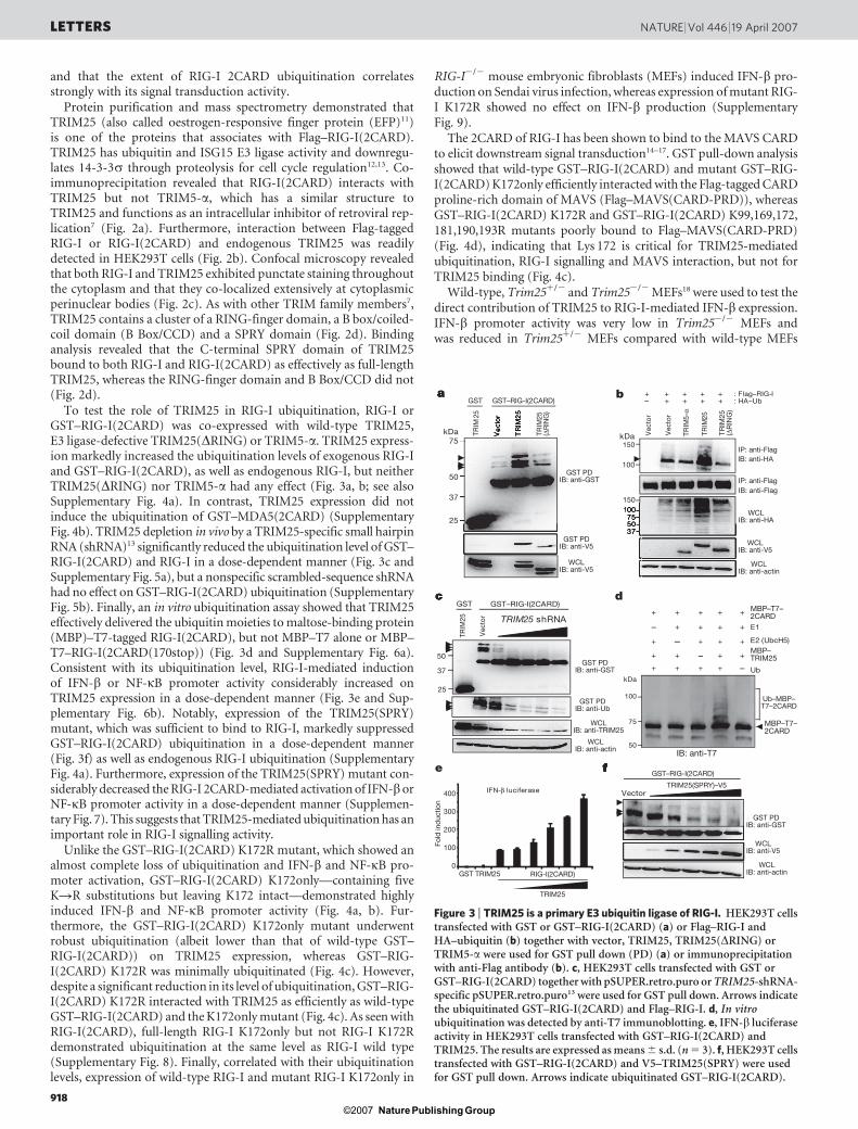
To test the role of TRIM25 in RIG-I ubiquitination, RIG-I orGST–RIG-I(2CARD) was co-expressed with wild-type TRIM25,E3 ligase-defective TRIM25(DRING) or TRIM5-a. TRIM25 express-ion markedly increased the ubiquitination levels of exogenous RIG-Iand GST–RIG-I(2CARD), as well as endogenous RIG-I, but neitherTRIM25(DRING) nor TRIM5-a had any effect (Fig. 3a, b; see alsoSupplementary Fig. 4a). In contrast, TRIM25 expression did notinduce the ubiquitination of GST–MDA5(2CARD) (SupplementaryFig. 4b). TRIM25 depletion in vivo by a TRIM25-specific small hairpinRNA (shRNA)13 significantly reduced the ubiquitination level of GST–RIG-I(2CARD) and RIG-I in a dose-dependent manner (Fig. 3c andSupplementary Fig. 5a), but a nonspecific scrambled-sequence shRNAhad no effect on GST–RIG-I(2CARD) ubiquitination (SupplementaryFig. 5b). Finally, an in vitro ubiquitination assay showed that TRIM25effectively delivered the ubiquitin moieties to maltose-binding protein(MBP)–T7-tagged RIG-I(2CARD), but not MBP–T7 alone or MBP–T7–RIG-I(2CARD(170stop)) (Fig. 3d and Supplementary Fig. 6a).Consistent with its ubiquitination level, RIG-I-mediated inductionof IFN-b or NF-kB promoter activity considerably increased onTRIM25 expression in a dose-dependent manner (Fig. 3e and Sup-plementary Fig. 6b). Notably, expression of the TRIM25(SPRY)mutant, which was sufficient to bind to RIG-I, markedly suppressedGST–RIG-I(2CARD) ubiquitination in a dose-dependent manner(Fig. 3f) as well as endogenous RIG-I ubiquitination (SupplementaryFig. 4a). Furthermore, expression of the TRIM25(SPRY) mutant con-siderably decreased the RIG-I 2CARD-mediated activation of IFN-borNF-kB promoter activity in a dose-dependent manner (Supplemen-tary Fig. 7). This suggests that TRIM25-mediated ubiquitination has animportant role in RIG-I signalling activity.
Unlike the GST–RIG-I(2CARD) K172R mutant, which showed analmost complete loss of ubiquitination and IFN-b and NF-kB pro-moter activation, GST–RIG-I(2CARD) K172only—containing fiveKRR substitutions but leaving K172 intact—demonstrated highlyinduced IFN-b and NF-kB promoter activity (Fig. 4a, b). Fur-thermore, the GST–RIG-I(2CARD) K172only mutant underwentrobust ubiquitination (albeit lower than that of wild-type GST–RIG-I(2CARD)) on TRIM25 expression, whereas GST–RIG-I(2CARD) K172R was minimally ubiquitinated (Fig. 4c). However,despite a significant reduction in its level of ubiquitination, GST–RIG-I(2CARD) K172R interacted with TRIM25 as efficiently as wild-typeGST–RIG-I(2CARD) and the K172only mutant (Fig. 4c). As seen withRIG-I(2CARD), full-length RIG-I K172only but not RIG-I K172Rdemonstrated ubiquitination at the same level as RIG-I wild type(Supplementary Fig. 8). Finally, correlated with their ubiquitinationlevels, expression of wild-type RIG-I and mutant RIG-I K172only in
RIG-I2/2 mouse embryonic fibroblasts (MEFs) induced IFN-b pro-duction on Sendai virus infection, whereas expression of mutant RIG-I K172R showed no effect on IFN-b production (SupplementaryFig. 9).
The 2CARD of RIG-I has been shown to bind to the MAVS CARDto elicit downstream signal transduction14–17. GST pull-down analysisshowed that wild-type GST–RIG-I(2CARD) and mutant GST–RIG-I(2CARD) K172only efficiently interacted with the Flag-tagged CARDproline-rich domain of MAVS (Flag–MAVS(CARD-PRD)), whereasGST–RIG-I(2CARD) K172R and GST–RIG-I(2CARD) K99,169,172,181,190,193R mutants poorly bound to Flag–MAVS(CARD-PRD)(Fig. 4d), indicating that Lys 172 is critical for TRIM25-mediatedubiquitination, RIG-I signalling and MAVS interaction, but not forTRIM25 binding (Fig. 4c).
Wild-type, Trim251/2 and Trim252/2 MEFs18 were used to test thedirect contribution of TRIM25 to RIG-I-mediated IFN-b expression.IFN-b promoter activity was very low in Trim252/2 MEFs andwas reduced in Trim251/2 MEFs compared with wild-type MEFs
f
Vector
c
5037
75100150
100
150
+ + + + + : Flag–RIG-I– + + + + : HA–Ub
Vec
tor
Vec
tor
TRIM
5-α
TRIM
25
ba
Vec
tor
TRIM
25
TRIM
25
TRIM
25(∆
RIN
G)
50
37
75
25
kDakDa
E2 (UbcH5)
E1
Ub+
–
+
+
+
+
+
+
–
+
+
+
+
+
+
+ – + +
+
–
+
+
+
+
50
75
100
kDa
d
e
IFN-β luciferase
50
37
25
Vec
tor
GST–RIG-I(2CARD)
TRIM
25 TRIM25 shRNA
GST
GST PDIB: anti-GST
GST PDIB: anti-V5
IP: anti-FlagIB: anti-HA
IP: anti-FlagIB: anti-Flag
WCLIB: anti-HA
WCLIB: anti-V5
WCLIB: anti-actin
WCLIB: anti-V5
WCLIB: anti-actin
WCLIB: anti-V5
TRIM
25(∆
RIN
G)
Fold
ind
uctio
n
0
100
200
300
400
GST TRIM25
TRIM25
RIG-I(2CARD)
GST PDIB: anti-GST
GST PDIB: anti-GST
GST PDIB: anti-Ub
WCLIB: anti-TRIM25
WCLIB: anti-actin
MBP–T7–2CARD
MBP–T7–2CARD
MBP–TRIM25
Ub–MBP–T7–2CARD
IB: anti-T7
GST–RIG-I(2CARD)
TRIM25(SPRY)–V5
GST GST–RIG-I(2CARD)
Figure 3 | TRIM25 is a primary E3 ubiquitin ligase of RIG-I. HEK293T cellstransfected with GST or GST–RIG-I(2CARD) (a) or Flag–RIG-I andHA–ubiquitin (b) together with vector, TRIM25, TRIM25(DRING) orTRIM5-a were used for GST pull down (PD) (a) or immunoprecipitationwith anti-Flag antibody (b). c, HEK293T cells transfected with GST orGST–RIG-I(2CARD) together with pSUPER.retro.puro or TRIM25-shRNA-specific pSUPER.retro.puro13 were used for GST pull down. Arrows indicatethe ubiquitinated GST–RIG-I(2CARD) and Flag–RIG-I. d, In vitroubiquitination was detected by anti-T7 immunoblotting. e, IFN-b luciferaseactivity in HEK293T cells transfected with GST–RIG-I(2CARD) andTRIM25. The results are expressed as means 6 s.d. (n 5 3). f, HEK293T cellstransfected with GST–RIG-I(2CARD) and V5–TRIM25(SPRY) were usedfor GST pull down. Arrows indicate ubiquitinated GST–RIG-I(2CARD).
LETTERS NATURE | Vol 446 | 19 April 2007
918Nature ©2007 Publishing Group
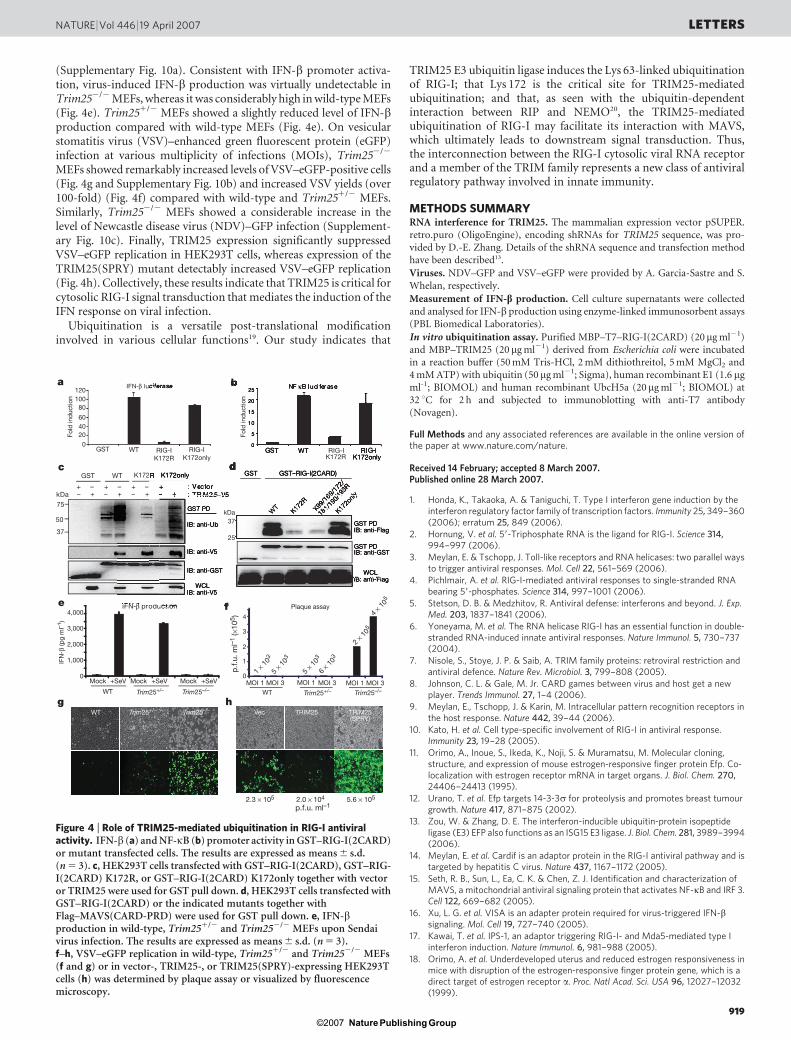
(Supplementary Fig. 10a). Consistent with IFN-b promoter activa-tion, virus-induced IFN-b production was virtually undetectable inTrim252/2 MEFs, whereas it was considerably high in wild-type MEFs(Fig. 4e). Trim251/2 MEFs showed a slightly reduced level of IFN-bproduction compared with wild-type MEFs (Fig. 4e). On vesicularstomatitis virus (VSV)–enhanced green fluorescent protein (eGFP)infection at various multiplicity of infections (MOIs), Trim252/2
MEFs showed remarkably increased levels of VSV–eGFP-positive cells(Fig. 4g and Supplementary Fig. 10b) and increased VSV yields (over100-fold) (Fig. 4f) compared with wild-type and Trim251/2 MEFs.Similarly, Trim252/2 MEFs showed a considerable increase in thelevel of Newcastle disease virus (NDV)–GFP infection (Supplement-ary Fig. 10c). Finally, TRIM25 expression significantly suppressedVSV–eGFP replication in HEK293T cells, whereas expression of theTRIM25(SPRY) mutant detectably increased VSV–eGFP replication(Fig. 4h). Collectively, these results indicate that TRIM25 is critical forcytosolic RIG-I signal transduction that mediates the induction of theIFN response on viral infection.
Ubiquitination is a versatile post-translational modificationinvolved in various cellular functions19. Our study indicates that
TRIM25 E3 ubiquitin ligase induces the Lys 63-linked ubiquitinationof RIG-I; that Lys 172 is the critical site for TRIM25-mediatedubiquitination; and that, as seen with the ubiquitin-dependentinteraction between RIP and NEMO20, the TRIM25-mediatedubiquitination of RIG-I may facilitate its interaction with MAVS,which ultimately leads to downstream signal transduction. Thus,the interconnection between the RIG-I cytosolic viral RNA receptorand a member of the TRIM family represents a new class of antiviralregulatory pathway involved in innate immunity.
METHODS SUMMARYRNA interference for TRIM25. The mammalian expression vector pSUPER.
retro.puro (OligoEngine), encoding shRNAs for TRIM25 sequence, was pro-
vided by D.-E. Zhang. Details of the shRNA sequence and transfection methodhave been described13.
Viruses. NDV–GFP and VSV–eGFP were provided by A. Garcia-Sastre and S.
Whelan, respectively.
Measurement of IFN-b production. Cell culture supernatants were collected
and analysed for IFN-b production using enzyme-linked immunosorbent assays
(PBL Biomedical Laboratories).
In vitro ubiquitination assay. Purified MBP–T7–RIG-I(2CARD) (20mg ml21)
and MBP–TRIM25 (20 mg ml21) derived from Escherichia coli were incubatedin a reaction buffer (50 mM Tris-HCl, 2 mM dithiothreitol, 5 mM MgCl2 and
4 mM ATP) with ubiquitin (50 mg ml21; Sigma), human recombinant E1 (1.6 mg
ml-1; BIOMOL) and human recombinant UbcH5a (20mg ml21; BIOMOL) at
32 uC for 2 h and subjected to immunoblotting with anti-T7 antibody
(Novagen).
Full Methods and any associated references are available in the online version ofthe paper at www.nature.com/nature.
Received 14 February; accepted 8 March 2007.Published online 28 March 2007.
1. Honda, K., Takaoka, A. & Taniguchi, T. Type I interferon gene induction by theinterferon regulatory factor family of transcription factors. Immunity 25, 349–360(2006); erratum 25, 849 (2006).
2. Hornung, V. et al. 59-Triphosphate RNA is the ligand for RIG-I. Science 314,994–997 (2006).
3. Meylan, E. & Tschopp, J. Toll-like receptors and RNA helicases: two parallel waysto trigger antiviral responses. Mol. Cell 22, 561–569 (2006).
4. Pichlmair, A. et al. RIG-I-mediated antiviral responses to single-stranded RNAbearing 59-phosphates. Science 314, 997–1001 (2006).
5. Stetson, D. B. & Medzhitov, R. Antiviral defense: interferons and beyond. J. Exp.Med. 203, 1837–1841 (2006).
6. Yoneyama, M. et al. The RNA helicase RIG-I has an essential function in double-stranded RNA-induced innate antiviral responses. Nature Immunol. 5, 730–737(2004).
7. Nisole, S., Stoye, J. P. & Saib, A. TRIM family proteins: retroviral restriction andantiviral defence. Nature Rev. Microbiol. 3, 799–808 (2005).
8. Johnson, C. L. & Gale, M. Jr. CARD games between virus and host get a newplayer. Trends Immunol. 27, 1–4 (2006).
9. Meylan, E., Tschopp, J. & Karin, M. Intracellular pattern recognition receptors inthe host response. Nature 442, 39–44 (2006).
10. Kato, H. et al. Cell type-specific involvement of RIG-I in antiviral response.Immunity 23, 19–28 (2005).
11. Orimo, A., Inoue, S., Ikeda, K., Noji, S. & Muramatsu, M. Molecular cloning,structure, and expression of mouse estrogen-responsive finger protein Efp. Co-localization with estrogen receptor mRNA in target organs. J. Biol. Chem. 270,24406–24413 (1995).
12. Urano, T. et al. Efp targets 14-3-3s for proteolysis and promotes breast tumourgrowth. Nature 417, 871–875 (2002).
13. Zou, W. & Zhang, D. E. The interferon-inducible ubiquitin-protein isopeptideligase (E3) EFP also functions as an ISG15 E3 ligase. J. Biol. Chem. 281, 3989–3994(2006).
14. Meylan, E. et al. Cardif is an adaptor protein in the RIG-I antiviral pathway and istargeted by hepatitis C virus. Nature 437, 1167–1172 (2005).
15. Seth, R. B., Sun, L., Ea, C. K. & Chen, Z. J. Identification and characterization ofMAVS, a mitochondrial antiviral signaling protein that activates NF-kB and IRF 3.Cell 122, 669–682 (2005).
16. Xu, L. G. et al. VISA is an adapter protein required for virus-triggered IFN-bsignaling. Mol. Cell 19, 727–740 (2005).
17. Kawai, T. et al. IPS-1, an adaptor triggering RIG-I- and Mda5-mediated type Iinterferon induction. Nature Immunol. 6, 981–988 (2005).
18. Orimo, A. et al. Underdeveloped uterus and reduced estrogen responsiveness inmice with disruption of the estrogen-responsive finger protein gene, which is adirect target of estrogen receptor a. Proc. Natl Acad. Sci. USA 96, 12027–12032(1999).
a IFN-β luciferase
c
5–V5
GST PDIB: anti-Flag
WT
GST GST–RIG-I(2CARD)
K172R
K99/1
69/1
72/
181/
190/
193R
K172o
nly
GST PDIB: anti-GST
WCLIB: anti-Flag
GST WT K172R K172only
+ – + – + – + – : Vector– + – + – + – + : TRIM2
GST PD
IB: anti-Ub
IB: anti-V5
IB: anti-GST
WCLIB: anti-V5
50
37
75
kDa
hg
25
20
15
10
5
0GST WT
NF-κB luciferase
e IFN-β production
b
f
0
1
2
3
4
MOI 1 MOI 3 MOI 1 MOI 3 MOI 1 MOI 3
RIG-IK172only
d
120100
80
604020
0GST WT RIG-I
K172RRIG-I
K172onlyRIG-I
K172R
Fold
ind
uctio
n
Fold
ind
uctio
n
IFN
-β (p
g m
l–1)
1,000
0
2,000
3,000
4,000
Mock +SeV Mock +SeV Mock +SeV
WT
WT
Trim25+/–
Trim25+/–
Trim25–/– WT Trim25+/– Trim25–/–
Trim25–/– Vec TRIM25 TRIM25(SPRY)
p.f.
u. m
l–1
(×10
5 )
p.f.u. ml–1
1 × 10³
5 × 10³
2.3 × 10⁵ 2.0 × 10⁴ 5.6 × 10⁵
5 × 10³
2 × 10⁵
4 × 10⁵
6 × 10³
kDa37
25
Plaque assay
Figure 4 | Role of TRIM25-mediated ubiquitination in RIG-I antiviralactivity. IFN-b (a) and NF-kB (b) promoter activity in GST–RIG-I(2CARD)or mutant transfected cells. The results are expressed as means 6 s.d.(n 5 3). c, HEK293T cells transfected with GST–RIG-I(2CARD), GST–RIG-I(2CARD) K172R, or GST–RIG-I(2CARD) K172only together with vectoror TRIM25 were used for GST pull down. d, HEK293T cells transfected withGST–RIG-I(2CARD) or the indicated mutants together withFlag–MAVS(CARD-PRD) were used for GST pull down. e, IFN-bproduction in wild-type, Trim251/2 and Trim252/2 MEFs upon Sendaivirus infection. The results are expressed as means 6 s.d. (n 5 3).f–h, VSV–eGFP replication in wild-type, Trim251/2 and Trim252/2 MEFs(f and g) or in vector-, TRIM25-, or TRIM25(SPRY)-expressing HEK293Tcells (h) was determined by plaque assay or visualized by fluorescencemicroscopy.
NATURE | Vol 446 | 19 April 2007 LETTERS
919Nature ©2007 Publishing Group
19. Haglund, K. & Dikic, I. Ubiquitylation and cell signaling. EMBO J. 24, 3353–3359(2005).
20. Ea, C. K., Deng, L., Xia, Z. P., Pineda, G. & Chen, Z. J. Activation of IKK by TNFarequires site-specific ubiquitination of RIP1 and polyubiquitin binding by NEMO.Mol. Cell 22, 245–257 (2006).
21. Kirkpatrick, D. S., Denison, C. & Gygi, S. P. Weighing in on ubiquitin: the expandingrole of mass-spectrometry-based proteomics. Nature Cell Biol. 7, 750–757 (2005).
Supplementary Information is linked to the online version of the paper atwww.nature.com/nature.
Acknowledgements This work was supported by US Public Health Service grants(J.U.J.), the exchange programme between Harvard Medical School and thegraduate training programme 1071 at the Friedrich-Alexander UniversityErlangen-Nuremberg, Germany (M.U.G.), and a Korea Research Foundation Grant(C.-H.J.). We thank A. Garcia-Sastre, D.-E. Zhang and S. Whelan for providing
reagents, and R. Tomaino and J. Nagel for mass spectrometry. We also thank allmembers of the Tumor Virology Division, New England Primate Research Center,for discussions.
Author Contributions M.U.G. performed all aspects of this study. Y.C.S., C.-H.J.and C.L. assisted in experimental design and in collecting the data. T.U. and S.I.performed the in vitro ubiquitination assay and generated Trim252/2 MEFs. L.S.and Z.C. generated the MAVS construct and RIG-I antibody. T.O. and S.A.generated the RIG-I construct and RIG-I2/2 MEFs. M.U.G. and J.U.J. organized thisstudy and wrote the paper. All authors discussed the results and commented onthe manuscript.
Author Information Reprints and permissions information is available atwww.nature.com/reprints. The authors declare no competing financial interests.Correspondence and requests for materials should be addressed to J.U.J.([email protected]).
LETTERS NATURE | Vol 446 | 19 April 2007
920Nature ©2007 Publishing Group

METHODSCell culture. HEK293T, MEF and Hela cells were cultured in Dulbecco’s modi-
fied Eagle’s medium supplemented with 10% fetal bovine serum, 2 mM
L-glutamine and 1% penicillin-streptomycin (Gibco-BRL). Transient transfec-
tions were performed with FuGENE 6 (Roche), lipofectamine 2000 (Invitrogen),
or calcium phosphate (Clontech) following the manufacturer’s instructions.
Wild-type, Trim251/2 and Trim252/2 MEFs were immortalized with LXSN-
E6/E7 retroviral vector containing human papilloma virus 16 E6 and E7 onco-
genes using a standard protocol of selection with 200mg ml21 of neomycin.
RIG-I2/2 MEFs were infected with pBabe-puro vector, pBabe-puro-RIG-I
wild-type, pBabe-puro-RIG-I K172R, or pBabe-puro-RIG-I K172only retro-
virus, followed by selection with 1 mg ml21 of puromycin.
Plasmid construction. All constructs for transient and stable expression in
mammalian cells were derived from the pEBG GST fusion vector and the pEF-
IRES-Puro expression vector. DNA fragments corresponding to the coding
sequence of the RIG-I and TRIM25 genes were amplified from template DNA
by polymerase chain reaction (PCR) and subcloned into plasmid pEBG between
restriction sites KpnI and NotI or pEF-IRES-puro between AflII and NotI forselection of stable transfectants. V5-tagged TRIM25 and Flag-tagged RIG-I were
expressed from a modified pIRES-puro encoding a C-terminal V5 tag and Flag
tag, respectively. RIG-I mutants were generated by PCR using site-directed
mutagenesis. All constructs were sequenced using an ABI PRISM 377 automatic
DNA sequencer to verify 100% agreement with the original sequence.
In vivo GST pull down, protein purification and mass spectrometry. At 48 h
after transfection with vectors expressing GST, GST–RIG-I(2CARD) or GST–
MDA5(2CARD) fusions, HEK293T cells were collected and lysed with NP40
buffer (50 mM HEPES, pH 7.4, 150 mM NaCl, 1 mM EDTA, 1% (v/v) NP40)
supplemented with a complete protease inhibitor cocktail (Roche). Post-
centrifuged supernatants were pre-cleared with protein A/G beads at 4 uC for
2 h. Pre-cleared lysates were mixed with a 50% slurry of glutathione-conjugated
Sepharose beads (Amersham Biosciences), and the binding reaction was incu-
bated for 4 h at 4 uC. Precipitates were washed extensively with lysis buffer.
Proteins bound to glutathione beads were eluted and separated on a NuPAGE
4–12% Bis-Tris gradient gel (Invitrogen). After Coomassie or silver staining
(Invitrogen), specific protein bands were excised and analysed by ion-trap mass
spectrometry at the Harvard Taplin Biological Mass Spectrometry facility, andamino acid sequences were determined by tandem mass spectrometry and data-
base searches.
Immunoblot analysis and immunoprecipitation assay. For immunoblotting,
polypeptides were resolved by SDS–polyacrylamide gel electrophoresis (SDS–
PAGE) and transferred to a PVDF membrane (Bio-Rad). Immunodetection was
achieved with anti-V5 (1:5,000) (Invitrogen), anti-Flag (1:5,000) (Sigma), anti-
HA (1:5,000), anti-GST (1:10,000) (Sigma), anti-actin (1:10,000) (Abcam), or
anti-TRIM25 (1:2,000) (BD Bioscience) antibodies. The proteins were visualized
by a chemiluminescence reagent (Pierce) and detected by a Fuji Phosphor
Imager.
For immunoprecipitation, cells were collected after 48 h and then lysed in
NP40 buffer supplemented with a complete protease inhibitor cocktail
(Roche). After pre-clearing with protein A/G agarose beads for 2 h at 4 uC,
whole-cell lysates were used for immunoprecipitation with the indicated anti-
bodies. Generally, 1–2mg of commercial antibody was added to 1 ml of cell lysate,
which was incubated at 4 uC for 4–12 h. After addition of protein A/G agarose
beads, the incubation was continued for 2 h. Immunoprecipitates were exten-
sively washed with lysis buffer and eluted with SDS loading buffer by boiling for5 min.
Confocal immunofluorescence microscopy. Eighteen to twenty-four hours
after transfection, cells were fixed with 4% paraformaldehyde for 15 min, per-
meabilized with 0.2% (v/v) Triton X-100 for 15 min, blocked with 10% goat
serum in PBS for 1 h and reacted with diluted primary antibody in 1% goat
serum for up to 2 h at room temperature. After incubation, cells were washed
extensively with PBS, incubated with the appropriate secondary antibody diluted
in 1% goat serum for 1 h at room temperature, and washed three times with PBS.
Confocal microscopy was performed using a Leica TCS SP laser-scanning micro-
scope (Leica Microsystems) fitted with a 3100 Leica objective (PL APO, 1.4NA)
and Leica imaging software. Images were collected at 512 3 512-pixel resolution.
The stained cells were optically sectioned in the z axis, and the images in the
different channels (photo multiplier tubes) were collected simultaneously. The
step size in the z axis varied from 0.2 to 0.5mm to obtain 16 slices per imaged file.
The images were transferred to a Macintosh G4 computer (Apple Computer),
and Photoshop (Adobe) was used to render the images.
doi:10.1038/nature05732
Nature ©2007 Publishing Group