Embed Size (px)
Citation preview

UNIVERSIDADE FEDERAL DE MINAS GERAIS
FACULDADE DE MEDICINA
DEPTO. DE ANATOMIA PATOLÓGICA E MEDICINA LEGAL
Tumores Mistos e Carcinomas Metaplásicos de Glândulas
Mamárias Caninas: Aspectos Comparativos com Tumores de
Glândulas Salivares da Espécie Humana
Marisa Cristina Genelhu Leite Santos
Belo Horizonte – Minas Gerais
2005

MARISA CRISTINA GENELHU LEITE SANTOS
Tumores Mistos e Carcinomas Metaplásicos de Glândulas
Mamárias Caninas: Aspectos Comparativos com Tumores de
Glândulas Salivares da Espécie Humana
Tese apresentada ao Programa de Pós-graduação em
Patologia da Faculdade de Medicina da Universidade
Federal de Minas Gerais, como requisito parcial à obtenção
do título de Doutor em Patologia.
Área de concentração: Patologia Geral
Orientador: Prof. Dr. Geovanni Dantas Cassali
Co-orientador: Prof.a Dr.a Helenice Gobbi
Universidade Federal de Minas Gerais
Belo Horizonte – Minas Gerais
Faculdade de Medicina da UFMG
2005

Ó meu Deus,
Quisestes nutrir-me com Tua divina substância, a mim, pobre criatura, que ao nada
retornaria se Teu Divino olhar não me outorgasse a vida a cada instante...
Vós haveis ido mais longe de minha previsão e eu quero cantar Vossas misericórdias.
Para Vós, unicamente, seja qualquer vitória que me fizerdes adquirir...
Santa Teresa de Lisieux

Aos meus filhos Bernardo, Lucas e Gabriel e ao meu marido Heber. Como uma vez
disse Vinícius de Moraes, vocês são “indispensáveis ao meu equilíbrio vital, porque
fazem parte do mundo que eu, tremulamente, construí e se tornaram alicerces do meu
encanto pela vida”.
Aos meus pais João e Zélia e meus irmãos Giovana e João Luis.
AGRADECIMENTOS
Ao Prof. Dr. Geovanni Dantas Cassali, pela orientação neste trabalho,
admiração e respeito.
À Profa Dra. Helenice Gobbi, por ter acreditado nos meus sonhos e
incentivado de todas as maneiras possíveis para que ele se tornasse realidade.
Ao Pe. Geraldo Hogervorst e Pe. Edson José do Sacramento, verdadeiros
instrumentos dóceis nas mãos do Pai, pelo conforto espiritual concedido a cada
encontro.
Ao Prof. Giovanni Gazzinelli e ao Dr. Rodrigo Corrêa de Oliveira do
Centro de Pesquisas René Rachou CPqRR-FIOCRUZ, por terem observado em
mim a vocação para a pesquisa e incentivado a sua realização.
À Dra. Lucia Alves de Oliveira Fraga e ao Dr. Luiz Cosme Cotta Malaquias
do Núcleo de Pesquisa em Imunologia da Universidade Vale do Rio Doce
(UNIVALE), por serem aqueles amigos e professores que se tornam inesquecíveis e
imortais através da continuidade de sua obra por meio de seus discípulos e
companheiros.
À Maria José Rocha, anjo colocado por Deus no meu caminho para que eu
suportasse as dificuldades e compartilhasse as alegrias deste trabalho.
A todos os colegas do Laboratório de Patologia Comparada do Instituto de
Ciências Biológicas e da Faculdade de Medicina da UFMG, pela acolhida e
prazerosa convivência.
A todos os colegas da UNIVALE, em especial às professoras e amigas
Beatriz Brasileiro e Sílvia Perim e às fiéis colaboradoras, Fátima, Marlucy, Lília,
Ivanete, Nelma, Meire e Elaine: saber que vocês me queriam de volta, era um
bálsamo nas horas difíceis...
Aos meus amigos Luciene Tafuri, Terezinha Marques, Antônio Hugo
Campos, Paôlla Perdigão, Paula Vidigal, Renato Baldan, Sérgio Cardoso e Diele
Carine Barreto Arantes e aos professores Maria Cássia Aguiar, Carlos Alberto
Ribeiro, Lúcia Porto e Mônica Demas Cabral: a colaboração e o apoio de vocês
foram imprescindíveis.
À Fundação de Amparo a Pesquisa do Estado de Minas Gerais
(FAPEMIG), à Coordenação de Aperfeiçoamento de Pessoal de Nível Superior
(CAPES) e à Comissão Institucional de Capacitação Docente e Técnica (CICDT)
da UNIVALE.
E a todos que direta ou indiretamente contribuíram para a realização deste
trabalho.

SUMÁRIO
LISTAS
Lista de Abreviaturas e Siglas................................................................................................09
Lista de Ilustrações.................................................................................................................13
INTRODUÇÃO....................................................................................................................14
I. A CONTRIBUIÇÃO DA PATOLOGIA COMPARADA....................................... 14
I.1. Tumores Mamários Humanos versus Tumores Mamários Caninos. ..............................14
I.2. Tumores Mamários Humanos versus Tumores de Glândula Salivar Humanos…...…...17
I.3. Tumores Mamários Caninos versus Tumores de Glândula Salivar Humanos…............18
II. RECONHECIMENTO DO FENÓTIPO TRANSFORMADO E DOS
COMPONENTES TUMORAIS ATRAVÉS DE MARCADORES
MOLECULARES................................................................................................................20
II.1. Marcadores Mesenquimais ……………………………………………………............21
II.3. Marcadores Epiteliais.....................................................................................................22
II.4. Marcadores Mioepiteliais …………………………………….…………….........…....22
II.5. Complexo E-caderina/β-catenina...................................................................................25
II.6. Receptor de Estrógeno....................................................................................................26
OBJETIVOS........................................................................................................................28
MATERIAIS E MÉTODOS...............................................................................................29
RESULTADOS................................................................................................................... 33
Artigo 1 - Adenoma pleomórfico e carcinoma ex-adenoma pleomórfico de glândulas
salivares – Revisão da literatura.............................................................................................34
Artigo 2 - Immunohistochemical expression of p63 in pleomorphic adenomas and
carcinomas ex-pleomorphic adenomas of salivary glands………………………………....59
Artigo 3 - Immunolocalization of β-catenin in pleomorphic adenomas and carcinomas ex-
pleomorphic adenomas of salivary glands…………………….……………………………67
Artigo 4 - A comparative study between mixed-type tumours from human salivary and
canine mammary glands………………. ……………………………………………….……74
CONCLUSÃO................................................................................................................. .....84
CONSIDERAÇÕES FINAIS......................................................................................... .....85
REFERÊNCIAS BIBLIOGRÁFICAS ......................................................................... .....87
ANEXOS ......................................................................................................................... ..102
Anexo 1 – Parecer do Comitê de Ética em Pesquisa........................................................ ..103
Anexo 2 - Comunicações científicas relacionadas ao trabalho de tese ............................ ..104
8
TRADUÇÕES ÚTEIS PARA A COMPREENSÃO DA TESE
cluster grupo
de novo de origem primária
in situ não invasivo
follow-up seguimento, proservação
multi-steps múltiplos passos
insights novas percepções, novos conceitos
status estado, condição
pool associação
stem cell célula indiferenciada, célula-tronco
splincing clivagem e união de fragmentos de RNA
9
LISTA DE ABREVIATURAS E SIGLAS
μm micrômetro
ΔN Isoforma ΔN (sem a porção amino-terminal) da
proteína p63
α-SMA α-actina de músculo liso (α-smooth muscle actin)
A1 Glândula mamária canina abdominal-cranial
A2 Glândula mamária canina abdominal-cranial
AE1/AE3 Anticorpo anti-pan citoqueratinas humanas (1-8, 10,
14-16 e 19)
AgNORs Regiões organizadoras de nucléolo coradas pela prata
(silver-stained nucleolar organizer regions)
AP Adenoma pleomórfico
APC Gene da polipose adenomatosa do cólon (adenomatosis
polyposis coli)
BMP Proteínas morfogenéticos ósseas (bone morphogenetics
proteins)
BRCA1 Gene do câncer de mama 1 (breast cancer gene 1)
Ca ex-AP Carcinoma ex-adenoma pleomórfico
CD10 Grupo de moléculas de diferenciação de linfócitos 10
(cluster of differentiation 10)
10
CD-RAP Proteína derivada de cartilagem ácido retinóico
específica (cartilage-derived retinoic acid-sensitive
protein)
c-erbB2 Gene celular para receptor do fator de crescimento
epitelial humano 2 ou EGFR-2 (epidermal growth factor
receptor-2) ou HER2 (human epidermal growth factor
receptor 2)
ChM-1 Proteína condromodulina 1 matriz-cartilagem
específica (cartilage-specific matrix chondromodulin-1)
c-myc Oncogene celular da mielocitomatose
(myelocytomatosis celular oncogene)
COOH Porção carbóxi-terminal protéica
CTNNB1 Gene da proteína catenina associada a caderina beta 1
(catenin cadherin-associated protein β1)
DAB Diaminobenzidina
DNA Ácido desoxirribonucléico (desoxi ribonucleic acid)
GSK-3β Proteína quinase (serina-treonina) sintetizadora de
glicogênio (serine-threonine glycogen synthetase kinase-
3β)
h-caldesmona Caldesmona de cadeia pesada
HMGIC Gene da proteína do grupo de alta mobilidade (não
histona cromossomal) – isoforma I-C (High-mobility
group nonhistone chromosomal protein isoform I-C)
I Glândula mamária canina Inguinal
11
Ki-67 Antígeno marcador de proliferação celular Ki-67
K-ras Oncogene celular homólogo do oncogene viral do
sarcoma em ratos Kirsten (Kirsten rat sarcoma viral
oncogene)
LEF Fator aumentador de linfócitos (lynfocyte enhancer
factor)
mdm2 Proteína de célula transformada (transformed 3T3 cell
double minute 2)
MUC Mucina
N-CAM Molécula de adesão celular neuronal (neural-cell
adhesion molecule)
NH2- Porção amino-terminal protéica
NOS Sem outra especificação (not otherwise specified)
p Braço curto de cromossomo (petit)
p105 Proteína p105
p16 Proteína p16
p21 Proteína p21
p53 Proteína p53
p63 Proteína p63
PAAF Punção aspirativa por agulha fina
PCNA Antígeno nuclear de proliferação celular (proliferating
cell nuclear antigen)
PIP Proteína prolactina induzível
PLAG1 Gene do adenoma pleomórfico 1
12
q Braço longo de cromossomo
RB Proteína do gene RB (retinoblastoma)
RE Receptor de estrógeno
RNAm Ácido ribonucléico mensageiro (ribonucleic acid)
RT-PCR Reação em cadeia da polimerase - transcritase reversa
(reverse transcriptase polymerase chain reaction)
S-100 Proteína cérebro específica (brain-specific protein)
SMMHC Miosina de músculo liso de cadeia pesada (smooth-
muscle myosin heavy chain)
T1 Glândula mamária canina torácica-cranial
T2 Glândula mamaria canina torácica-caudal
TA Isoforma transativadora da proteína p63
Tcf Fator de células T (linfócitos) (T-cell factor)
TP53 Gene p53
Wnt Via celular transdutoa de sinais (Wingless)
13
LISTA DE ILUSTRAÇÕES
Figura 1 (Introdução) – Representação esquemática dos cinco pares de mama canina e
suas denominações..........................................................................................................15
Tabela 1 (Materiais e Métodos) – Classificação comparativa entre as glândulas
salivares humanas e as glândulas mamárias caninas, segundo o tamanho das
mesmas............................................................................................................................30
Tabela 2 (Materiais e Métodos) – Anticorpos primários, fontes e diluições utilizados
nos ensaios imuno-histoquímicos....................................................................................31

14
INTRODUÇÃO
I. A CONTRIBUIÇÃO DA PATOLOGIA COMPARADA
Uma estratégia que tem permitido avançar no conhecimento dos diferentes aspectos
da carcinogênese comparada tem sido a utilização de modelos animais (FERNANDES,
1996). O objetivo final do estudo das neoplasias em modelos animais é a busca de maior
entendimento dos fatores responsáveis pela doença no homem e a expectativa de que estes
possam ser identificados, controlados e até mesmo, eliminados. Informações sobre a
ocorrência de tumores espontâneos originam-se principalmente das pesquisas veterinárias
de tumores em animais domésticos, selvagens e naqueles criados em zoológicos e
laboratórios. O estudo dos tumores mais freqüentes em animais pode fornecer dados
epidemiológicos e indicações para a melhor compreensão de sua etiologia e
desenvolvimento, bem como material para investigação biológica e terapêutica
(MARCHANT, 1987).
I.1. Tumores Mamários Humanos versus Tumores Mamários Caninos
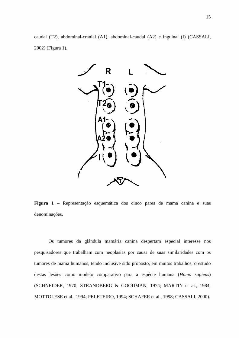
As glândulas mamárias da espécie canina (Canis familiaris) originam-se
embriologicamente pela invaginação de brotos ectodérmicos para o interior do mesoderma
subjacente. Elas se desenvolvem em cinco pares a partir de linhas ou cristas mamárias
bilaterais nas regiões torácica, abdominal e inguinal. A denominação de cada par de
mamas no sentido crânio-caudal da superfície ventral é: torácico-cranial (T1), torácico-
15
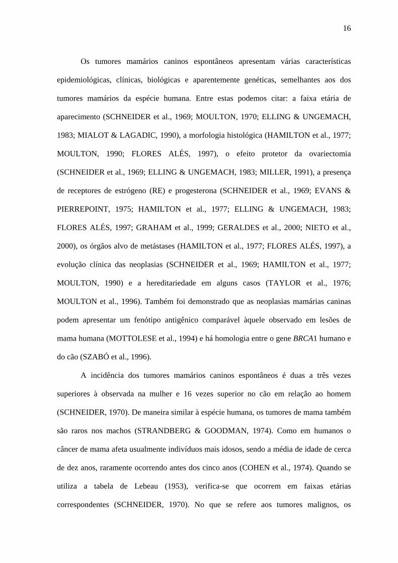
caudal (T2), abdominal-cranial (A1), abdominal-caudal (A2) e inguinal (I) (CASSALI,
2002) (Figura 1).
Figura 1 – Representação esquemática dos cinco pares de mama canina e suas
denominações.
Os tumores da glândula mamária canina despertam especial interesse nos
pesquisadores que trabalham com neoplasias por causa de suas similaridades com os
tumores de mama humanos, tendo inclusive sido proposto, em muitos trabalhos, o estudo
destas lesões como modelo comparativo para a espécie humana (Homo sapiens)
(SCHNEIDER, 1970; STRANDBERG & GOODMAN, 1974; MARTIN et al., 1984;
MOTTOLESE et al., 1994; PELETEIRO, 1994; SCHAFER et al., 1998; CASSALI, 2000).
16
Os tumores mamários caninos espontâneos apresentam várias características
epidemiológicas, clínicas, biológicas e aparentemente genéticas, semelhantes aos dos
tumores mamários da espécie humana. Entre estas podemos citar: a faixa etária de
aparecimento (SCHNEIDER et al., 1969; MOULTON, 1970; ELLING & UNGEMACH,
1983; MIALOT & LAGADIC, 1990), a morfologia histológica (HAMILTON et al., 1977;
MOULTON, 1990; FLORES ALÉS, 1997), o efeito protetor da ovariectomia
(SCHNEIDER et al., 1969; ELLING & UNGEMACH, 1983; MILLER, 1991), a presença
de receptores de estrógeno (RE) e progesterona (SCHNEIDER et al., 1969; EVANS &
PIERREPOINT, 1975; HAMILTON et al., 1977; ELLING & UNGEMACH, 1983;
FLORES ALÉS, 1997; GRAHAM et al., 1999; GERALDES et al., 2000; NIETO et al.,
2000), os órgãos alvo de metástases (HAMILTON et al., 1977; FLORES ALÉS, 1997), a
evolução clínica das neoplasias (SCHNEIDER et al., 1969; HAMILTON et al., 1977;
MOULTON, 1990) e a hereditariedade em alguns casos (TAYLOR et al., 1976;
MOULTON et al., 1996). Também foi demonstrado que as neoplasias mamárias caninas
podem apresentar um fenótipo antigênico comparável àquele observado em lesões de
mama humana (MOTTOLESE et al., 1994) e há homologia entre o gene BRCA1 humano e
do cão (SZABÓ et al., 1996).
A incidência dos tumores mamários caninos espontâneos é duas a três vezes
superiores à observada na mulher e 16 vezes superior no cão em relação ao homem
(SCHNEIDER, 1970). De maneira similar à espécie humana, os tumores de mama também
são raros nos machos (STRANDBERG & GOODMAN, 1974). Como em humanos o
câncer de mama afeta usualmente indivíduos mais idosos, sendo a média de idade de cerca
de dez anos, raramente ocorrendo antes dos cinco anos (COHEN et al., 1974). Quando se
utiliza a tabela de Lebeau (1953), verifica-se que ocorrem em faixas etárias
correspondentes (SCHNEIDER, 1970). No que se refere aos tumores malignos, os
17
carcinomas mais freqüentes na mama humana e na mama canina são, respectivamente, os
carcinomas ductais invasivos e as neoplasias descritas genericamente como
adenocarcinomas (STRANDBERG & GOODMAN, 1974; MARTIN et al., 1984).
Finalmente, quando se considera a freqüência de outras lesões neoplásicas malignas da
mama como, por exemplo, os sarcomas, verifica-se que estes são igualmente raros nas duas
espécies (STRANDBERG & GOODMAN, 1974; ROSEN & OBERMAN, 1993).
I.2. Tumores Mamários Humanos versus Tumores de Glândula Salivar Humanos
Já em 1968, foi relatada uma associação peculiar entre tumores de glândula salivar
e tumores de mama humanos: em um estudo de sobrevida observou-se que mulheres com
tumores de mama tendem significativamente a desenvolver tumores de glândula salivar ou
vice-versa (BERG et al., 1968).
Atualmente, admite-se que existam similaridades bem conhecidas entre as
glândulas mamária e salivar na espécie humana devido à homologia estrutural básica entre
essas glândulas exócrinas (BERG et al., 1968; DUNN et al., 1972; PRIOR &
WATERHOUSE, 1977; ABBEY et al., 1984). Notáveis semelhanças morfológicas entre
certos tumores de glândula salivar e algumas neoplasias de mama têm sido descritas, tais
como aquelas existentes entre o adenocarcinoma polimorfo de baixo grau de malignidade e
o carcinoma lobular invasivo (FREEDMAN & LUMERMAN, 1983; ABERLE et al.,
1985; WENIG & GNEPP, 1991; NICOL & ISKANDAR, 2000), entre o carcinoma de
células acinares e o carcinoma secretor invasivo (DAMIANI et al., 2000; HIROKAWA et
al., 2002), entre o carcinoma epitelial-mioepitelial e o adenomioepitelioma (SEIFERT,
1998; NAGAO et al., 1998; SUGANO et al., 2001). Carcinomas ductais (WICK et al.,
1998; HOANG et al., 2001; SKÁLOVÁ et al., 2001), carcinomas adenóides císticos
(DORI et al., 2000; PIA-FOSCHINI et al., 2003) e os tumores mistos benignos e malignos
18
(adenomas pleomórficos - AP e carcinomas ex-adenomas pleomórficos - Ca ex-AP,
respectivamente) (CHEN, 1990; MORAN et al., 1990; REID-NICHOLSON et al., 2003;
KUMAR et al., 2005; HAYES et al., 2005) podem ser encontrados em ambas as estruturas,
ainda que os prognósticos sejam diferentes.
Alguns aspectos genéticos como expressão de c-erbB2 (CHO et al., 1999;
SKALOVÁ et al., 2001; SCHOLL et al., 2001) e PIP (prolactin-inducible protein)
(CLARK et al., 1999), além de alterações alélicas nos cromossomos 6q, 16q, 17p e 17q
(HOANG et al., 2001) também são compartilhados pelos tumores de mama e glândula
salivar. Sobre a presença de RE em glândula salivar humana, permanece a controvérsia
sobre sua expressão e significado biológico (JEANNON et al., 1999; DORI et al., 2000;
LEIMOLA-VIRTANEN et al., 2000; KUMAGAMI & ONITSUKA, 2001; BJORLING et
al., 2002).
É válido ressaltar que, como na mama humana, o avanço da idade diminui o
volume do parênquima das glândulas salivares, principalmente devido à perda de tecido
acinar e sua substituição por tecido fibroso e adiposo (SCOTT, J. 1977a e b).
I.3. Tumores Mamários Caninos versus Tumores de Glândula Salivar Humanos
Os tumores mistos, lesões incomuns na mama humana, são os mais freqüentes na
mama canina (CASSALI, 2000). Esta, certamente, é uma das importantes diferenças
verificadas entre os tumores mamários do homem e do cão, mas é também uma
característica semelhante ao dos tumores de glândula salivar.
Os tumores mistos caninos são compostos de uma mistura de células epiteliais e
mioepiteliais dispersas em um estroma “mesenquimatoso” (ZHUANG et al., 1997;
SANTOS, 1979), como visto na glândula salivar humana. Os tumores mistos malignos são
menos freqüentes que os benignos e, como na glândula salivar humana, aparecem mais

19
tardiamente (SANTOS, 1979). Para MOULTON et al. (1990) alguns tumores mistos
diagnosticados histologicamente como benignos podem ser potencialmente malignos. Se
tiverem tempo suficiente para crescimento, os tumores mistos caninos podem sofrer
transformação maligna como a que acontece com os AP humanos, numa relação direta
com o tempo de evolução da doença (GNEPP, 1993).
A diversidade histológica vista nos tumores mistos caninos correspondem às
mesmas observadas nos tumores mistos da glândula salivar humana. As células epiteliais e
mioepiteliais perdem um pouco da arquitetura ductal-lobular, ficando dispersas em um
estroma caracteristicamente mixóide, condróide ou ósseo (HAMPE & MISDORP, 1974;
MOULTON, 1990). Os estudos realizados para determinação da histogênese das células
tumorais vêm demonstrando que, provavelmente, estas possuam origem em uma mesma
célula-mãe, dita “intermediária”, geneticamente instável, a qual assumiria expressão
fenotípica comum aos dois tipos, epitelial e miopitelial, e que as células mioepiteliais
seriam responsáveis pela formação de tecidos “estromais” (PELETEIRO, 1994). Essa
hipótese, também levantada sobre a origem dos tumores mistos da glândula salivar humana
(DARDICK et al, 1982; KUMAR et al, 2005), foi reforçada por GÄRTNER et al. (1999)
em um estudo usando imuno-histoquímica e análise de DNA por citometria estática. Por
algum mecanismo ainda desconhecido, uma célula totipotente com capacidade de
diferenciação divergente sofre transformação metaplásica, resultando na aparência
histológica heterogênea desses tumores.
Tais evidências nos levaram a perguntar se a glândula mamária canina não poderia
também ser utilizada como o modelo comparativo para o estudo da carcinogênese nas
glândulas salivares, como proposto para os tumores de mama humana. Para testar essa
hipótese fez-se necessária a avaliação de critérios significativos, tais como, aspectos

20
clínicos, morfologia microscópica e expressão de antígenos relevantes para definição
histogênica nesses dois grupos de lesões.
II. RECONHECIMENTO DO FENÓTIPO TRANSFORMADO E DOS
COMPONENTES TUMORAIS ATRAVÉS DE MARCADORES MOLECULARES
O uso de marcadores moleculares que possam auxiliar na identificação dos
componentes tumorais, compreensão da histogênese e dos processos envolvidos na
carcinogênese é um importante instrumento da patologia humana e veterinária. A
classificação precisa de muitas neoplasias depende essencialmente do encontro de
marcadores antigênicos, só identificáveis na prática, pelas reações com seus anticorpos
específicos (BRASILEIRO-FILHO, 2004; CASSALI et al, 1999a; CASSALI, 2000).
A utilização da técnica de imuno-histoquímica teve enorme impacto no diagnóstico
dos tumores tanto da área humana quanto animal. Vários anticorpos já foram empregados
no diagnóstico histogenético de neoplasias morfologicamente indiferenciadas, na
caracterização da origem e tipo das células constituintes tumorais, na discriminação entre
natureza “benigna” ou “maligna” de proliferações celulares e na avaliação prognóstica de
determinados tumores (LEONG & WRIIGHT, 1987; ALVES et al, 1999; CASSALI et al,
1999b).
Um painel utilizando principalmente marcadores mesenquimais, epiteliais e
mioepiteliais parece ser efetivo para distinguir os diferentes tipos de tumores de glândula
salivar, cujo diagnóstico histomorfológico é, sabidamente, um desafio para o patologista
(ARAÚJO et al., 2000). O mesmo pode-se afirmar para os tumores animais. Vários autores
já demonstraram a reatividade de anticorpos monoclonais humanos para diversos antígenos

21
das glândulas mamárias caninas, estendendo o potencial da imuno-histoquímica também
para a patologia veterinária (MOTTOLESE et al, 1994, FERNANDES, 1996; CASSALI,
2000).
II.1. Marcadores Mesenquimais
A vimentina é o mais ubíquo dos filamentos intermediários do citoesqueleto e se
expressa amplamente desde o início do desenvolvimento embrionário dos mamíferos,
podendo ser detectada tanto em tecidos de origem ectodérmica quanto mesodérmica.
Entretanto, sua expressão vai progressivamente se restringindo a alguns tipos celulares. O
alto grau de insolubilidade da vimentina sugere a sua função estrutural no citoplasma
(MACHADO & FIGUEIREDO, 1996). A vimentina é típica das células mesenquimais,
como fibroblastos, células endoteliais, linfócitos, macrófagos e outras células derivadas do
mesoderma, ainda que algumas de maneira mais escassa (ALVES et al, 1999) e, até o
aparecimento de marcadores mais específicos e sensíveis, era utilizado como marcador
clássico de células mioepiteliais.
Nas células mioepiteliais tanto das glândulas salivares humanas quanto nas
glândulas mamárias caninas, vimentina é co-expressada com algumas citoqueratinas
(CASSALI, 2000). A vimentina está presente nas neoplasias originadas das células
mesenquimais, mas muitos carcinomas humanos podem apresentar positividade,
principalmente aqueles com componente mioepitelial (DUPREY & PAULIN, 1995). Uma
possível explicação para o fato das células mioepiteliais expressarem vimentina seria a
“reversão” para um tipo celular mais primitivo ou embrionário (FERNANDES, 1996).

22
II.3. Marcadores Epiteliais
As citoqueratinas, proteínas constituintes dos filamentos intermediários que
integram o citoesqueleto, são marcadores epiteliais de eleição em mamíferos. Em humanos
são conhecidas pelo menos 20 citoqueratinas cuja expressão varia de acordo com o tipo de
célula, período de desenvolvimento embrionário, grau de diferenciação e microambiente de
crescimento celular, o que permite considerá-las como marcadores de diferenciação. São
utilizadas na caracterização dos carcinomas de origem glandular ou escamosa, bem como
na tentativa de indicar um sítio primário para tumores metastáticos. As células
mioepiteliais também expressam citoqueratinas, mas em menor quantidade que as
epiteliais (ALVES et al., 1999). O anticorpo clone AE1/AE3 reage com um pool de
citoqueratinas humanas (1-8, 10, 14-16 e 19) e outras caninas (MARGARITESCU et al.,
1999-2004; FERNANDES, 1996). Este é o preparado mais amplamente usado na
demonstração da natureza epitelial de tumores morfologicamente indiferenciados (ALVES
et al., 1999). Outras citoqueratinas específicas podem auxiliar também o diagnóstico
diferencial de carcinomas primários e metástases carcinomatosas (ARAÚJO et al., 2000;
NIKITAKIS et al., 2004).
II.4. Marcadores Mioepiteliais
O mioepitélio neoplásico desempenha um papel fundamental na histogênese e nos
processos morfogenéticos responsáveis pela variável aparência histológica dos tumores
mistos (AP e Ca ex-AP) de glândulas salivares humanas (DARDICK et al., 1982; ZARBO
et al., 2000; SAVERA & ZARBO, 2004). BATSAKIS et al., (1983) chegaram a afirmar
que os tumores mistos não ocorrem em tecidos nos quais as células mioepiteliais estejam
ausentes. Além disso, a camada de células mioepiteliais parece ter uma grande
significância como inibidor parácrino da progressão tumoral (STERNLICHT et al., 1997;
23
LAKHANI & O’HARE, 2000). A identificação das células mioepiteliais é de particular
valor diagnóstico nos tumores de mama humanos, pois elas são retidas na maioria das
lesões benignas e perdidas no processo de malignização e invasão (YAZIGI et al., 2000;
BARBARESCHI et al., 2001; KALOF et al., 2004). É de se esperar um comportamento
semelhante nos tumores de glândula salivar e nos tumores mamários caninos, dadas as já
comentadas semelhanças biológicas entre estes grupos de neoplasias.
As células mioepiteliais, como o próprio nome indica, exibem características de
ambas as células epiteliais e da musculatura lisa, além de que, sua heterogeneidade
citomorfológica (células claras, epitelióides, alongadas, plasmocitóides) dificulta sua
correta distinção (SIMPSON, 2002). Essa heterogeneidade se dá, provavelmente, em
decorrência de diferentes estágios de diferenciação (ARAÚJO & RAITZ, 2004). Existem
três tipos de marcadores de células mioepiteliais. O primeiro tipo inclui marcadores de
proteínas de músculo liso tais com como α-SMA (α-smooth muscle actin), SMMHC
(smooth muscle myosin-heavy chain), h-caldesmona e calponina. O segundo tipo é
expresso também nas células epiteliais ductais e inclui citoqueratinas 14, 5 e 17, α1-β1
integrina e metalotioneína. Vimentina, CD10 e S-100 estão no terceiro grupo, cujas
proteínas são expressas também nas células mesenquimais (SAVERA et al., 1997;
FOSCHINI et al., 2000; KALOF et al., 2004; FURUSE et al., 2005). Como visto, nenhum
marcador de células mioepiteliais exibe perfeita sensibilidade e especificidade, sendo
recomendável que uma combinação de marcadores seja usada em casos de dificuldades de
identificação (KALOF et al., 2004).
Investigações recentes mostraram que a proteína p63, além de essencial na
manutenção da população de células precursoras (stem cells) em vários tecidos epiteliais, é
um excelente marcador imuno-histoquímico de células mioepiteliais tanto nos tecidos
glandulares humanos quanto caninos (REIS-FILHO & SCHMITT, 2002; WEBER et al.,
24
2002; BILAL et al., 2003; GAMA et al, 2003; EDWARDS et al., 2004). p63 demonstra
sensibilidade comparável à de outros marcadores mioepiteliais como α-SMA, SMMHC e
calponina, mas uma maior especificidade: as células estromais tais como os
miofibroblastos, as células neurais e as células endoteliais são consistentemente negativas
(BARBARESCHI et al., 2001; WERLING et al., 2002).
A transcrição do gene para p63 se dá a partir de dois promotores distintos e
splicings alternativos responsáveis por duas classes de proteínas que, a despeito de sua
homologia estrutural, possuem funções aparentemente distintas e, pelo menos, seis
isoformas. Três contêm um domínio de transcrição N-terminal, semelhante à p53. Supõe-se
que essa classe transativadora (TA) tenha a habilidade de ativar o gene TP53 induzindo
apoptose e regulando o ciclo celular, agindo como um gene supressor de tumor não
clássico. As outras três isoformas não possuem o domínio NH2- terminal sendo
denominadas ΔN. Essas agiriam através de mecanismos alternativos driblando o controle
do ciclo celular e apoptose em oposição à via TAp63/p53. Os domínios COOH distintos
(α, β e γ) possuem papéis pobremente conhecidos (YANG et al., 1998; LITTLE &
JOCHEMSEN, 2002).
Algumas evidências de que as isoformas ΔN-p63 se associavam às moléculas de β-
catenina, do complexo de adesão celular E-caderina/β-catenina no processo da
carcinogênese in vitro foram descritas por PATTURAJAN et al. (2002) No entanto, essa
hipótese não se confirmou quando foram realizados estudos com neoplasias humanas
(KOGA et al., 2003; REIS-FILHO et al., 2003).
25
II.5. Complexo E-caderina/β-catenina
Um dos primeiros processos que levam um tumor à invasão e metástase é a perda
de adesão celular. Este fato é atribuído a uma desregulação de um ou mais genes e seus
produtos, responsáveis pela adesão intercelular, dentre eles as caderinas e cateninas
(SHIOZAKI et al., 1996). A E-caderina é a principal molécula de adesão do epitélio.
Através de seu domínio extra-citoplasmático, é responsável pela adesão homotípica, em
presença de cálcio, com outra molécula de E-caderina da célula vizinha. Através do seu
domínio citoplasmático, interage diretamente com a β-catenina (HARRINGTON &
SYRIGOS, 2000).
O complexo E-caderina/β-catenina está intimamente envolvido no controle da
diferenciação morfológica (organogênese) e proliferação celular durante o
desenvolvimento, exercendo função primordial na organização e manutenção das
estruturas tissulares (HARRINGTON & SYRIGOS, 2000). Quando a β-catenina não
participa do complexo de adesão, a molécula livre é seqüestrada por um complexo formado
pelos produtos do gene APC (adenomatous polyposis coli), pela molécula GSK-3β (serine-
threonine glycogen synthetase kinase-3β) e por uma proteína adaptadora, inabilitando a
fosforilação e degradação da β-catenina pelo sistema proteolítico celular (JANKOWISKI
et al., 1997). Dessa maneira β-catenina também participa da transdução de sinais, pela via
Wnt (Wingless) (JANKOWSKI et al., 1997; SHIEH et al., 2003). Aumento nas
concentrações de β-catenina livre no citoplasma estimula sua ligação com proteínas tais
como LEF (lynfocyte enhancer factor) e Tcf (T-cell factor). Estes complexos dirigem-se
para o núcleo e agem como co-fatores de transcrição de genes relacionados à proliferação
celular (JANKOWISKI et al., 1997).

26
Pouco se conhece sobre a participação do complexo E-caderina/β-catenina nos
tumores de glândula salivar, mas existem fortes evidências de que esteja associado ao
fenótipo agressivo de muitos desses tumores (ZHANG et al., 2000). Baixos níveis de
expressão E-caderina e β-catenina foram correlacionado positivamente com a
desdiferenciação das células tumorais (ECONOMOPOULOU et al., 2000).
Nos tumores mamários caninos a redução da expressão de E-caderina e sua
distribuição anormal estão relacionadas ao baixo grau de diferenciação desses tumores e
diminuição do tempo de sobrevida (REIS et al, 2003; RESTUCCI et al, 1992; GOMES,
2004).
Vários fatores podem modular a interação do complexo E-caderina/β-catenina
levando a uma redistribuição da β-catenina. Dessa maneira, pode-se dizer que a β-catenina
é uma proteína com dupla função, a qual é determinada por sua localização na membrana
ou no núcleo. Na membrana, representa um importante papel na adesão célula-célula. β-
catenina nuclear aumenta a transcrição de genes ligados à proliferação celular tumoral.
II.6. Receptor de Estrógeno
Células possuidoras de receptores de estrógeno (RE) e sensíveis à sua ação podem
induzir a secreção de fatores mitogênicos e reguladores de crescimento e estimular a
produção de proteases, as quais degradam matriz extracelular e estimulam invasão e
metástase (MILLER, 1990). As células mioepiteliais, tanto das glândulas salivares quanto
das mamárias, humanas e caninas, respondem a estímulos hormonais para expelir saliva e
leite (HAYWARD et al., 1996). Além de que, o status hormonal para RE em alguns
tumores de mama humanos, tem um importante significado prognóstico e terapêutico
(GOBBI et al, 2005).
27
Também nos tumores mamários caninos, a presença de RE é indicativa da
dependência hormonal dessas neoplasias (HAMILTON et al, 1977), havendo correlação
com as características patológicas e comportamento biológico (MAC EWEN et al, 1982).
As glândulas mamária e salivar humanas compartilham as mesmas estruturas ducto-
acinares básicas e uma co-existência de carcinomas em ambos os sítios tem sido reportada
(BERG et al., 1968; DUNN et al., 1972; PRIOR & WATERHOUSE, 1977; ABBEY et al.,
1984; MILLER et al., 1994). Assim, vários estudos procuraram estabelecer uma possível
associação entre a presença de RE nas glândulas salivares e o desenvolvimento de tumores.
Poucos trabalhos foram capazes de demonstrar a presença de RE em glândulas
salivares. Dentre estes podemos citar um estudo bioquímico (DIMERY, 1987), dois
ensaios imuno-histoquímicos (JEANNON et al., 1999; GLAS et al., 2002) e outro,
utilizando RT-PCR (Reverse Transcriptase - Polymerase Chain Reaction). LEIMOLA-
VIRTANEN et al. (2000) conseguiram mostrar atividade transcricional do gene RE através
da presença abundante de RNAm. Entretanto, a imuno-histoquímica pode deixar de
detectar RE devido a múltiplas razões tais como: eficiência relativa na tradução da
proteína, processamento pós-traducional, flutuações na estabilidade protéica e sua
degradação, expressão em níveis muito baixos ou por dificuldades no reconhecimento de
epitopos (LAMEY et al., 1987; MILLER et al., 1994; SHICK et al., 1995; GAFFNEY et
al., 1995; DORI et al., 2000; NASSER et al., 2003).
Assim, os critérios histomorfológicos de diagnóstico, quando associados a
biomarcadores como proteínas reguladoras do ciclo celular, moléculas de adesão e
receptores hormonais podem fornecer novos insights sobre a histogênese, o
comportamento biológico e prognóstico dos tumores em estudo e, em última instância,
direcionar condutas terapêuticas.
28
OBJETIVOS
Considerando a importância da patologia comparada utilizando modelos animais,
propomos abordar o tema estabelecendo os seguintes objetivos:
OBJETIVO GERAL
• Analisar os aspectos histomorfológicos e imuno-histoquímicos dos tumores
mamários caninos (tumores mistos e carcinomas metaplásicos) e compará-los aos
tumores de glândula salivar da espécie humana (adenoma pleomórfico e carcinoma
ex-adenoma pleomórfico) com a finalidade de averiguar seu valor como modelo de
estudo comparativo.
OBJETIVOS ESPECÍFICOS
• Caracterizar imuno-histoquimicamente os elementos epiteliais e mesenquimais dos
tumores mistos da mama canina (tumores mistos benignos e carcinomas
metaplásicos) e dos tumores mistos de glândula salivar humana (adenoma
pleomórfico e carcinoma ex-adenoma pleomórfico) através da expressão dos
seguintes marcadores: AE1-AE-3, vimentina, p63, β-catenina, E-caderina e receptor
de estrógeno.
• Comparar os aspectos histomorfológicos e o perfil imuno-histoquímico dos tumores
mamários caninos (tumores mistos e carcinomas metaplásicos) com os tumores de
glândula salivar humana (adenoma pleomórfico e carcinoma ex-adenoma
pleomórfico) a fim de melhor compreender a histogênese e comportamento
biológico dos mesmos.
29
MATERIAIS E MÉTODOS
1. Caracterização das Amostras
As amostras incluídas em parafina dos tumores mamários de cães sem raça definida
foram obtidas nos arquivos do Laboratório de Patologia Comparada do Instituto de
Ciências Biológicas da Universidade Federal de Minas Gerais. Na análise das amostras
caninas foi empregada a classificação histológica para mama humana (ROSEN &
OBERMAN, 1993) para efeito de comparação. As amostras de tumores mistos de glândula
salivar humana foram obtidas nos departamentos de Anatomia Patológica da Faculdade de
Medicina da UFMG, Centro de tratamento e Pesquisa Hospital do Câncer A. C. Camargo e
Instituto Nacional do Câncer (INCA). O critério de seleção foi baseado no diagnóstico
histopatológico das amostras com exame clínico conhecido. Estas amostras foram revisadas
independentemente por dois observadores e os tumores humanos foram classificados e
subtipados de acordo com a morfologia histológica (SEIFERT & SOBIN, 1991). Os dados
clínicos foram obtidos dos arquivos médicos e veterinários e incluíram idade, gênero e
glândula salivar ou mamária afetada.
As amostras foram caracterizadas clinicamente segundo a idade de aparecimento
dos tumores em ambas as espécies e de acordo com o tamanho da glândula acometida. As
idades dos animais foram convertidas utilizando-se a tabela de Lebeau (1953). De acordo
com essa tabela, os primeiros 2 anos de vida do cão correspondem aos primeiros 24 anos de
vida na espécie humana. Após o segundo ano de vida no animal, cada ano corresponde a 4
anos na espécie humana.
30
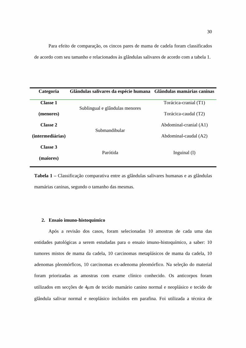
Para efeito de comparação, os cincos pares de mama de cadela foram classificados
de acordo com seu tamanho e relacionados às glândulas salivares de acordo com a tabela 1.
Categoria Glândulas salivares da espécie humana Glândulas mamárias caninas
Classe 1
(menores) Sublingual e glândulas menores
Torácica-cranial (T1)
Torácica-caudal (T2)
Classe 2
(intermediárias) Submandibular
Abdominal-cranial (A1)
Abdominal-caudal (A2)
Classe 3
(maiores) Parótida Inguinal (I)
Tabela 1 – Classificação comparativa entre as glândulas salivares humanas e as glândulas
mamárias caninas, segundo o tamanho das mesmas.
2. Ensaio imuno-histoquímico
Após a revisão dos casos, foram selecionadas 10 amostras de cada uma das
entidades patológicas a serem estudadas para o ensaio imuno-histoquímico, a saber: 10
tumores mistos de mama da cadela, 10 carcinomas metaplásicos de mama da cadela, 10
adenomas pleomórficos, 10 carcinomas ex-adenoma pleomórfico. Na seleção do material
foram priorizadas as amostras com exame clínico conhecido. Os anticorpos foram
utilizados em secções de 4μm de tecido mamário canino normal e neoplásico e tecido de
glândula salivar normal e neoplásico incluídos em parafina. Foi utilizada a técnica de
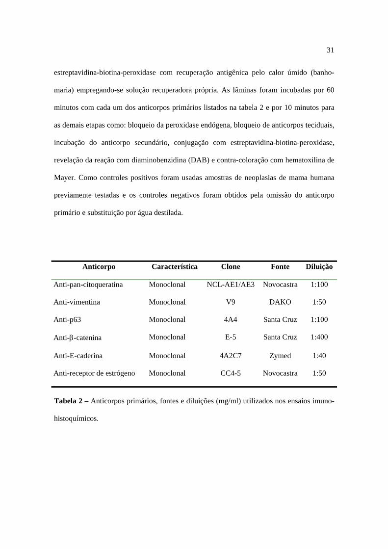
31
estreptavidina-biotina-peroxidase com recuperação antigênica pelo calor úmido (banho-
maria) empregando-se solução recuperadora própria. As lâminas foram incubadas por 60
minutos com cada um dos anticorpos primários listados na tabela 2 e por 10 minutos para
as demais etapas como: bloqueio da peroxidase endógena, bloqueio de anticorpos teciduais,
incubação do anticorpo secundário, conjugação com estreptavidina-biotina-peroxidase,
revelação da reação com diaminobenzidina (DAB) e contra-coloração com hematoxilina de
Mayer. Como controles positivos foram usadas amostras de neoplasias de mama humana
previamente testadas e os controles negativos foram obtidos pela omissão do anticorpo
primário e substituição por água destilada.
Anticorpo Característica Clone Fonte Diluição
Anti-pan-citoqueratina Monoclonal NCL-AE1/AE3 Novocastra 1:100
Anti-vimentina Monoclonal V9 DAKO 1:50
Anti-p63 Monoclonal 4A4 Santa Cruz 1:100
Anti-β-catenina Monoclonal E-5 Santa Cruz 1:400
Anti-E-caderina Monoclonal 4A2C7 Zymed 1:40
Anti-receptor de estrógeno Monoclonal CC4-5 Novocastra 1:50
Tabela 2 – Anticorpos primários, fontes e diluições (mg/ml) utilizados nos ensaios imuno-
histoquímicos.

32
A expressão dos marcadores foi analisada nos diferentes componentes dos tumores.
Para avaliação qualitativa e semiquantitativa, foram empregados critérios morfológicos e
pontos de corte utilizados em estudos prévios sobre tais marcadores: 10% para
citoqueratinas e vimentina, (ARAÚJO et al, 2000; GRAHAM et al, 1999) β-catenina e E-
caderina (SHIEH et al, 2003) e receptor de estrógeno (DORI et al, 2000); para o antígeno
p63 o ponto de corte utilizado foi de 5% (REIS-FILHO et al, 2002b).
3. Análise estatística
Foram realizados testes estatísticos apropriados a cada um dos estudos relatados nos
resultados como artigos.
4. Ética
O projeto de pesquisa para a realização do presente trabalho foi submetido ao
Comitê de Ética em Pesquisa da Universidade Federal de Minas Gerais (COEP/UFMG)
que emitiu parecer favorável à sua execução: ETIC 193/04 (Anexo 1).
33
RESULTADOS
Os resultados, obtidos a partir das amostras de tumores animais e humanos e dos
ensaios realizados para atingir os objetivos propostos, serão apresentados de forma
alternativa, como artigos científicos submetidos a periódicos internacionais e nacionais
Qualis A.
Cada artigo está estruturado com base nas normas do periódico onde foi publicado.
À exceção do primeiro artigo, uma revisão bibliográfica estruturada conforme as normas do
Jornal Brasileiro de Patologia e Medicina Laboratorial (ISSN: 1676 2444), todos os outros
possuem introdução, objetivos, materiais e métodos, resultados, discussão, conclusão e
numeração de páginas próprios.
Após a apresentação desses artigos, seguir-se-á uma conclusão geral, que atende aos
objetivos propostos no início deste trabalho, considerações finais e referências
bibliográficas referentes à introdução geral.

34
Artigo 1 – Adenoma pleomórfico e carcinoma ex-adenoma pleomórfico de glândulas salivares – Revisão da literatura
35
ADENOMA PLEOMÓRFICO E CARCINOMA EX-ADENOMA PLEOMÓRFICO
DE GLÂNDULAS SALIVARES – REVISÃO DA LITERATURA
Pleomorphic adenoma and carcinoma ex-pleomorphic adenoma of salivary glands. A
review
Marisa Genelhu1 Helenice Gobbi2; Geovanni Dantas Cassali3
1. Mestre e doutoranda em Patologia pela Faculdade de Medicina da Universidade Federal
de Minas Gerais (UFMG); professora adjunta e pesquisadora do Núcleo de Pesquisa em
Imunologia da Universidade Vale do Rio Doce (UNIVALE).
2. Professora adjunta e doutora do Departamento de Anatomia Patológica e Medicina
Legal da Faculdade de Medicina da UFMG.
3. Professor doutor do Laboratório de Patologia Comparada do Instituto de Ciências
Biológicas da UFMG.
Esta revisão é parte da tese de doutorado “Tumores mistos e carcinomas metaplásicos da
glândula mamária canina: aspectos comparativos com tumores de glândula salivar da
espécie humana”, desenvolvida no Laboratório de Patologia Comparada, ICB/UFMG.
Apoio: Coordenação para Aperfeiçoamento de Pessoal de Nível Superior através do
Programa de Intercâmbio Internacional Brasil/Portugal (CAPES/GRICES 112/04) e
Fundação de Amparo à Pesquisa de Minas Gerais (FAPEMIG).
36
Resumo
A despeito dos inúmeros trabalhos na literatura com relação aos adenomas pleomórficos e
sua contraparte maligna, os carcinomas ex-adenomas pleomórficos de glândula salivar, a
grande maioria é representada por relatos de caso, devido à raridade do tumor maligno,
suas peculiaridades histomorfológicas e controvérsias sobre histogênese, mecanismos de
transformação maligna e metástases. O presente trabalho tem por objetivo reunir as
informações mais importantes já descritas a respeito dessas lesões, de um modo
sistemático e conciso a fim de facilitar a compreensão global dessas intrigantes neoplasias.
Unitermos: carcinoma ex-adenoma pleomórfico, neoplasias de glândulas salivares, revisão
Abstract
Despite several works in the literature with regard to pleomorphic adenomas (PA) and their
malign counterpart, salivary gland's carcinoma ex pleomorphic adenomas (Ca ex-PA),
most of them are represented by case reports, given how rare malign tumors are, their
histomorphological peculiarities and controversies on histogenesis, malign transformation
and metastasis mechanisms. The present work has the purpose of gathering the most
important pieces of information about lesions, in a systematic and concise way to make it
easy to understand these intriguing neoplasias.
Key Words: carcinoma ex-pleomorphic adenoma, salivary gland tumors, review
37
Introdução - Aspectos Gerais dos Tumores de Glândula Salivar
As glândulas salivares humanas possuem uma morfologia normal relativamente
simples. Entretanto, originam uma variedade surpreendente de tumores malignos e
benignos, sendo que mais de 90% desses são de origem epitelial.(49) Essa variedade tem
sido identificada, categorizada e dinamicamente modificada numa grande diversidade de
tipos histológicos que dificultam a avaliação de casos antigos.(39) De modo geral esses
neoplasmas são relativamente incomuns (representam menos de 2-3% de todos os tumores
e 7% dos tumores de cabeça e pescoço),(36) mas a despeito da baixa incidência, assumem
substancial importância. Apesar de a grande maioria ser de natureza benigna, quando
malignos apresentam um variado comportamento biológico. Conseqüentemente, muitos
destes tumores representam um desafio diagnóstico e terapêutico, como é o caso dos
tumores mistos.
O Tumor Misto ou Adenoma Pleomórfico
Os tumores mistos benignos ou adenomas pleomórficos (AP) são os mais
freqüentes tumores das glândulas salivares humanas (cerca de 90%). Ocorrem mais
comumente na parótida (85%), seguida pelas glândulas submandibular (10%), sublingual e
menores (5%); acometem adultos jovens (4a-5a décadas), com leve predileção pelo gênero
feminino,(88, 33). Clinicamente, é uma lesão de crescimento lento, aparentemente bem
delimitada, que raramente excede 6 cm em seu maior diâmetro. Causa aumento de volume
indolor e pode ser facilmente palpável como uma massa discreta.
Histologicamente, os AP caracterizam-se pela notável diversidade histológica: uma
mistura de células epiteliais ductais e mioepiteliais dispersas em um estroma com variados
graus de tecido mixóide, hialino, condróide, ósseo e até mesmo, adiposo. (74, 42, 94)
38
A Histogênese
O fato de os AP se originarem em glândulas localizadas em uma zona transicional
ontogenética, uma região onde o endoderma e ectoderma se encontram, poderia ser uma
das razões para sua freqüente variabilidade histopatológica.(88, 92) Entretanto, a maioria dos
autores sustenta a hipótese de que, o que representa o princípio básico da heterogeneidade
tecidual desses tumores seja a “estromatização” e transformação do epitélio em
mesênquima.(68, 62, 49) Assim, mesmo que o tumor tenha freqüentemente um componente
“estromal”, com aparência mesenquimatosa, não é verdadeiramente misto, pois não deriva
de mais de uma camada germinativa. (62) As mudanças “estromais” características, seriam
produzidas por células mioepiteliais com propriedades multipotenciais ou epiteliais
progenitoras dos ductos basais que, embora imunofenotipicamente diversas,
compartilhariam as mesmas alterações citogenéticas.(14, 17) Ou seja, as células neoplásicas
parecem possuir natureza clonal, com uma única célula de origem.(26)
A presença de tecidos mixóide, hialino, condróide e ósseo, foi associada à
superexpressão de algumas proteínas pelas células neoplásicas tais como,
glicosaminoglicanos e agregan,(98) BMPs (bone morphogenetics proteins),(55, 99, 50) ChM-1
(cartilage-specific matrix chondromodulin-1),(50) CD-RAP (cartilage-derived retinoic
acid-sensitive protein),(19) lumican(51) e tenascina.(30) Um achado ocasional é a presença de
metaplasia epitelial em elementos escamosos, sebáceos ou oncocíticos.(68)
A Recorrência
A excisão cirúrgica com margens adequadas de ressecção é o tratamento indicado
para se evitar recorrências que ocorrem em cerca de 10% dos casos. A origem da
recorrência dos AP após tratamento cirúrgico é discutível. Além do difícil manejo
cirúrgico devido ao risco de agressão ao nervo facial e/ou profundidade do tumor,(11) a
39
cápsula dos AP parece não ser completamente desenvolvida.(81) O crescimento expansivo
produz projeções digitiformes, o que torna difícil a enucleação do tumor e facilita a
ocorrência de recidivas.(25, 40)
A atividade proliferativa avaliada através da expressão imuno-histoquímica de
PCNA e Ki-67 é marcadamente mais alta no componente epitelial de AP recorrentes
quando comparada à dos tumores não recorrentes, o que sugere que o componente epitelial
seja a origem provável das recorrências.(59)
Com relação ao conteúdo nuclear, os AP com evolução clínica de menos de um ano
geralmente possuem uma população de células diplóide. Em contraste, os tumores com
mais de cinco anos de evolução mostram uma população celular aneuplóide, semelhante à
dos carcinomas.(24, 56) Criscuolo et al.(12) encontraram uma significância prognóstica para a
expressão de AgNORs (silver-stained nucleolar organizer regions) em AP recorrentes e
Hamada et al.(38) atribuíram ao tamanho do tumor (mais que 3 cm) e à expressão de
mucinas (MUC1) um fator de risco para a recorrência de AP.
Múltiplas recidivas locais associadas à incompleta excisão cirúrgica e/ou longo
tempo de evolução do tumor estão diretamente relacionados à transformação maligna do
AP.(60, 66, 52)
O Tumor Misto Maligno ou Carcinoma ex-adenoma Pleomórfico
Relatado pela primeira vez no início dos anos 50,(32) o desenvolvimento de um novo
tumor com propriedades malignas a partir de um AP benigno pré-existente foi denominado
tumor misto maligno, carcinoma em tumor misto ou carcinoma ex-adenoma pleomórfico
(Ca ex-AP).(7, 25) A freqüência com que esse evento de transformação maligna acontece
varia em diferentes séries de 2% a 25%.(85, 70)
40
O Ca ex-AP é um tumor raro, agressivo, tipicamente de alto grau que, em geral,
acomete pacientes acima da 5a década de vida. A média de idade dos pacientes com este
tumor é em torno de 15 a 20 anos acima da dos pacientes com AP.(54, 85, 68, 62) Usualmente,
ocorre em glândulas salivares maiores (ao contrário da maioria dos outros tumores
malignos de glândula salivar) devido à associação ou recorrência do tumor benigno
(AP).(53, 65) Ao exame macroscópico, as lesões são firmes, com evidências de infiltração,
necrose ou hemorragia, dependendo da proporção entre os elementos benigno e maligno.
Nem sempre a porção benigna pode ser identificada. Ao exame microscópico mostram-se
como neoplasias infiltrativas, às vezes com alta celularidade, invasão de parênquima
glandular e extensão para estruturas adjacentes. As células podem formar arranjos sólidos
eosinofílicos, com núcleos pleomórficos e hipercromáticos, cromatina disposta
irregularmente e nucléolos proeminentes.(54, 28) É freqüente o envolvimento perineural,
invasão vascular, áreas de necrose e mitoses atípicas.(53)
A Transformação Maligna
Além do fato de o risco de malignização aumentar com a duração do tumor e o
número de recorrências, outros fatores patológicos de valor preditivo para AP são
pobremente definidos. Os principais achados clínicos no diagnóstico inicial
correlacionados à transformação maligna são idade avançada do paciente e maior tamanho
do tumor.(5) Análise retrospectiva de parâmetros histológicos (angiogênese e
angiotropismo, hialinização, hipercelularidade, necrose, anaplasia celular, maior atividade
mitótica) não são consenso na literatura como critérios preditivos confiáveis para
transformação maligna e metastatização de AP.(93, 5, 59, 64)
Alguns fatores genéticos já foram associados à malignização do AP. Avaliações
citogenéticas demonstraram que a maioria das alterações são translocações e perda de
41
heterozigose que se dão nos cromossomos 8 e 12. O gene alvo no cromossomo 8 é o
PLAG1 (8q12) e no cromossomo 12 é o HMGIC (12q15).(22, 4, 17, 70, 57, 67) A mais comum
alteração encontrada foi t(3;8)(p21;q12),(89, 90) uma translocação capaz de interferir também
na transcrição do gene CTNNB1 (catenin cadherin-associated protein β1).(44, 26) Outras
anormalidades recorrentes que podem ser importantes são: aquisição de cópias extras do
cromossomo 7, deleções nos cromossomos 5 e 17(8) e alterações no cromossomo 6q.(61)
Mutações e amplificações de genes relacionados ao controle do ciclo celular tais como,
RB, K-ras, c-myc, p21,(18, 95, 27) p16,(83) p105,(56) mdm2(3, 70) e, principalmente TP53(69, 95, 96,
3, 64) têm sido encontradas em Ca ex-AP. A despeito dos inúmeros estudos sobre a
expressão imuno-histoquímica da proteína p53 mutada em tumores de glândula salivar,
ainda não há consenso sobre o significado dessa expressão na discriminação do
comportamento biológico nessas lesões.(79)
Mutação no gene c-erb-B2 e superexpressão da proteína receptora para fator de
crescimento epitelial (EGFR-2) ou HER-2/neu também está implicada na iniciação e
progressão tumoral e associada a um pior prognóstico em carcinomas de ducto salivar
(como nos tumores de mama), o que poderia sugerir que os primeiros também pudessem se
beneficiar de imunoterapia com o transtuzumab.(78) Com relação ao Ca ex-AP, o que se
sabe é que este expressa HER-2/neu e AP não.(31, 71) Entretanto, os poucos estudos de
avaliação não foram conclusivos a respeito da utilização da imunoterapia para CA ex-
AP.(36, 21).
A diminuição na expressão de proteínas constituintes da membrana basal, tais como
laminina e colágeno tipo IV,(29) proteínas da matriz extracelular como a tenascina,(30) de
moléculas de adesão tais como N-CAM(72) e as do complexo E-caderina-β-catenina(76)
podem também estar relacionadas aos mecanismos de malignização do AP.
42
O Diagnóstico
Como visto, após o surgimento do tumor benigno e sua evolução com o tempo, as
células podem sofrer transformação maligna sob ação de vários fatores. É possível que,
mesmo os Ca ex-AP diagnosticados como de novo(15) possam se originar de tumores
benignos muito pequenos, profundos e clinicamente indetectáveis. Ou seja, mesmo quando
os tumores primários benignos não são diagnosticados previamente, estes poderiam, em
hipótese, já estar presentes, fazendo com que uma pergunta perdure até hoje: no caso de
diagnóstico de Ca ex-AP, o tumor primário era realmente benigno ou o carcinoma já
existia e não foi detectado?(53, 65)
Para minimizar essa dúvida, um diagnóstico acurado de Ca ex-AP requer a
presença de evidência histológica do tumor benigno em associação com o tumor maligno
e/ou a história clínica de recorrências no mesmo sítio anatômico.(23, 84) A punção aspirativa
por agulha fina (PAAF) é um procedimento diagnóstico bastante usado no pré-operatório
de AP, mas o Ca ex-AP dificilmente é identificado por ele.(46, 2, 87) O exame anátomo-
patológico é obrigatório. FOOTE & FRAZELL(32) estabeleceram que 100 cortes podem ser
necessários para se encontrar um pequeno carcinoma em um AP.
A malignidade dos Ca ex-AP está relacionada à extensão da invasão, à infiltração
de estruturas subjacentes(28) e ao subtipo histológico(86) Incorreções no diagnóstico são
comuns por causa do reduzido resíduo ou ausência do tumor misto benigno e a variedade
de subtipos apresentados(65). O Ca ex-AP pode manifestar um amplo e diferente espectro de
malignidades fenotípicas (Tabela 1) e a identificação do subtipo histológico é
determinante importante para compreensão do comportamento biológico e sua implicação
no prognóstico.(86) Além disso, esses subtipos podem ser agrupados em dois grupos: Ca ex-
AP com diferenciação apenas epitelial (75% dos casos) e Ca ex-AP com um componente
mioepitelial (25%).(1)

43
Tabela 1 – Exemplos de subtipos histológicos encontrados em carcinomas ex-adenomas
pleomórficos (Ca ex-AP) e respectivas referências na literatura.
SUBTIPOS DE Ca ex-AP REFERÊNCIA BIBLIOGRÀFICA
Carcinoma adenóide cístico 93, 65
Carcinoma mucoepidermóide 48, 65
Carcinoma de ducto salivar 31, 2, 65
Adenocarcinoma polimorfo de baixo grau 16
Carcinoma de células escamosas 46
Mioepitelioma maligno 97
Adenocarcinoma NOS (not otherwise specified) 65
Carcinoma epitelial-mioepitelial 65
Carcinoma mioepitelial 65, 77
Carcinoma indiferenciado 65, 37
Carcinoma Sebáceo 10
Carcinoma sarcomatóide 65, 86
Carcinossarcoma verdadeiro 39
Histiocitoma fibroso maligno 62
Rabdomiossarcoma 35
Neurossarcoma 45
Oncocitoma 58
As Categorias de Ca ex-AP
Os Ca ex-AP podem ainda ser divididos em três categorias: o Ca ex-AP
intracapsular, o tumor misto metastatizante, e o verdadeiro carcinossarcoma.(54, 37) A
categoria conhecida por Ca ex-AP intracapsular, in situ ou não invasiva(54, 28, 21) é
caracterizada por áreas malignas completamente circunscritas por AP e sem infiltração.
Teoricamente, essa categoria possuiria um melhor prognóstico. Entretanto, já existe um
44
relato de Ca ex-AP totalmente encapsulado cujo curso clínico foi agressivo com
disseminação metastática.(28)
Os tumores mistos metastatizantes podem metastatizar como verdadeiros
carcinomas, mas os implantes metastáticos apresentam-se com inequívocas características
histológicas benignas, semelhantes ao tumor primário.(93, 29) Por causa da aparência
benigna das metástases, elas podem por vezes, ser erroneamente diagnosticadas como
tumores primários. Por isso, é importante ressaltar que os pacientes acometidos
invariavelmente apresentam um AP primário e a metástase é usualmente precedida de
múltiplos episódios de recorrência.(13) O mecanismo de metástase desse tipo de tumor
permanece desconhecido. Existe a hipótese de que a manipulação cirúrgica possa deslocar
células neoplásicas para espaços vasculares, de onde sairiam para se implantar em
estruturas adjacentes ou à distância.(75) Suportando esse conceito está o fato de que tecidos
de AP têm sido transplantados com sucesso para camundongos, onde as células continuam
a crescer.(9, 6)
O verdadeiro carcinossarcoma ou verdadeiro tumor misto maligno é muito raro e
caracterizado pela transformação em ambos os componentes epitelial e mesenquimal.(82, 15,
39) Histologicamente, o componente sarcomatoso também pode apresentar-se com
fenótipos variáveis como fibrossarcoma, condrossarcoma, osteossarcoma, lipossarcoma,
leiomiossarcoma, rabdomiossarcoma e histiocitoma fibroso maligno.(35,62)
Excepcionalmente a transformação envolve apenas o componente mioepitelial.(77, 20, 97)
As Metástases
Com relação às metástases, suas localizações (principalmente linfonodos, ossos e
pulmões) indicam ambas as vias de disseminação, linfática e hematogênica. Essas são
compostas, na maioria das vezes, de elementos carcinomatosos, exceção feita às metástases

45
de carcinossarcomas verdadeiros cujas metástases podem ter tanto elementos de origem
epitelial ou mesenquimal e do tumor misto metastatizante, cujo implante metastático é
histologicamente idêntico ao tumor primário benigno.(93, 29) Sabe-se também que as
características cariotípicas dos tumores mistos metastatizantes diferem das características
citogenéticas encontradas em AP e outros Ca ex-AP.(43)
O Prognóstico
O Ca ex-AP, uma vez diagnosticado, determina um tempo de sobrevida curto (37%
em 5 anos) e alta taxa de mortalidade (55%).(53, 65) podendo ser considerado o terceiro
tumor de glândula salivar com pior prognóstico em sobrevida, sendo superado apenas pelo
carcinoma indiferenciado e adenocarcinoma NOS.(91) Li Volsi & Persin(54) e Spiro et al.(90)
sugeriram que a presença de linfonodos positivos possa ser o principal fator prognóstico.
Seja qual for sua aparência, categoria ou subtipo, esses tumores devem ser
cuidadosamente manipulados, pois as evidências são argumentos definitivos para
considerar todas as variantes de Ca ex-AP como neoplasmas potencialmente metastáticos e
agressivos.(47, 34, 73, 13, 28)
A Terapêutica
Apesar de que, a possibilidade de recorrência ou de transformação maligna de um
AP dependa não apenas do procedimento cirúrgico, mas de propriedades biológicas
intrínsecas de cada tumor(59) a melhor prevenção contra recorrências e malignização ainda
é a adequada remoção cirúrgica dos tumores benignos o mais precocemente possível, com
margens de segurança e seguida de um prolongado follow-up dos pacientes.(63, 66)
A terapêutica para os tumores malignos inclui excisão total da glândula afetada,
dissecção dos linfonodos cervicais, ressecção de estruturas adjacentes (como por exemplo,
46
o nervo facial), radioterapia e quimioterapia.(84, 65, 41) Um estudo recente mostra um
decréscimo na incidência de Ca ex-AP que pode ser conseqüência do diagnóstico precoce e
rápida remoção da lesão benigna.(91)
A raridade dos Ca ex-AP e sua variabilidade têm dificultado a avaliação do uso de
imunoterapia, mesmo conhecendo o fato de que estes expressam HER-2/neu, ao contrário
dos AP. O benefício, se é que exista, estaria associado ao subtipo histológico dos Ca ex-
AP: tumores que se manifestassem como aqueles originados no ducto intercalar
(carcinoma adenóide cístico, adenocarcinoma NOS, carcinoma de células acinares,
adenocarcinoma polimorfo de baixo grau e carcinoma mioepitelial) não responderiam tão
bem ao transtuzumab quanto aqueles semelhantes aos originados nos ductos secretórios
(carcinoma de ducto salivar, carcinoma mucoepidermóide e carcinoma de células
escamosas).(36)
Conclusão
Em conclusão, a presente revisão mostra os vários fatores até então associados à
transformação maligna do AP em Ca ex-AP, mas ressalta que apenas a precoce e adequada
remoção cirúrgica do tumor benigno é consenso na literatura como procedimento
preventivo da malignização. Além disso, este estudo reforça o conceito de que a
carcinogênese é um complexo processo multi-steps que envolve uma série de fenômenos
moleculares, muitos deles ainda pouco conhecidos.
47
Referencias Bibliográficas
1. ALTEMANI, A. et al. Carcinoma ex-pleomorphic adenoma (CXPA):
immunoprofile of the cells involved in the carcinomatous progression.
Histopathology, v.46, n.6, p.635-41, 2005.
2. ANAND, A.; BROCKIE E.S. Cytomorphological features of salivary duct
carcinoma ex pleomorphic adenoma: diagnosis by fine-needle aspiration
biopsy with histologic correlation. Diagn Cytopathol, v.20, n.6, p.375-8, 1999.
3. ARAÚJO, V.C. et al. Immunohistochemical Mdm2 expression in minor salivary
gland tumours and its relationship to p53 gene status. Oral Oncol, v.36, n.1,
p.67-9, 2000.
4. ASTRÖM, A.K. et al. Conserved mechanism of PLAG1 activation in salivary
gland tumors with and without chromosome 8q12 abnormalities: identification
of SII as a new fusion partner gene. Cancer Res, v.59, n.4, p.918-23, 1999.
5. AUCLAIR, P.L.; ELLIS G.L. Atypical features in salivary gland mixed tumors:
their relationship to malignant transformation. Mod Pathol, v.9, n.6, p.652-7,
1996.
6. BARFOED, C. et al. Human pleomorphic adenomas transplanted to nude mice.
Arch Otolaryngol Head Neck Surg, v.112, n.9, p.946-8, 1986.
7. BEAHRS, O.H. et al. Carcinomatous transformation of mixed tumors of the parotid
gland. AMA Arch Surg, v.75, n.4, p.605-13, 1957.
8. BULLERDIEK, J. et al. Cytogenetic investigation on cell line derived from a
carcinoma arising in a salivary pleomorphic adenoma. Cancer Genet
Cytogenet, v.44, n.2, p.253-62, 1990.
48
9. CASELITZ, J. et al. The pleomorphic adenoma of salivary glands transplanted on
athmymic mice. A lightmicroscopical and immunohistochemical investigation.
Virch Arch A Pathol Anat Histopathol, v.408, n.2-3, p.191-209, 1985.
10. COHN, M.L. et al. Sebaceous carcinoma ex pleomorphic adenoma: a rare
phenotypic occurrence. Ann Diagn Pathol, v.8, n.4, p.224-6, 2004
11. CONLEY, J.; CLAIRMONTT A.A. Facial nerve in recurrent benign pleomorphic
adenoma. Arch Otolaryngol, v.105, n.5, p.247-51, 1979.
12. CRISCUOLO, M. et al. Prognostic significance of nucleolar organizer regions in
recurrent pleomorphic adenomas of salivary glands. Pathologica, v.86, n.6,
p.606-11, 1994.
13. CZADER, M. et al. Metastasizing mixed tumor of the parotid: initial presentation
as a solitary kidney tumor and ultimate carcinomatous transformation at the
primary site. Am J Surg Pathol, v.24, n.8, p.1159-64, 2000.
14. DARDICK, I. et al. Histogenesis of salivary gland pleomorphic adenoma (mixed
tumor) with an evaluation of the role of the myoepithelial cell. Hum Pathol,
v.13, n.1, p.62-75, 1982.
15. DARDICK, I. et al. Ultrastructural contributions to the study of morphological
differentiation in malignant mixed (pleomorphic) tumour of salivary gland.
Head Neck, v.11, n.1, p.5-21, 1989.
16. DARLING, M.R. et al. Polymorphous low-grade adenocarcinoma and adenoid
cystic carcinoma: a review and comparison of immunohistochemical markers.
Oral Oncol, v.38, n.7, p.641-5, 2002.
17. DEBIEC-RYCHTER, M. et al. Histologic localization of PLAG1 (pleomorphic
adenoma gene 1) in pleomorphic adenoma of the salivary gland: cytogenetic
49
evidence of common origin of phenotypically diverse cells. Lab Invest, v.81,
n.9, p.1289-97, 2001.
18. DEGUSHI, H. et al. c-myc, ras p21 and p53 expression in pleomorphic adenoma
and its malignant form of the human salivary glands. Acta Pathol Jpn, v.43,
n.7-8, p.413-22, 1993.
19. DEVLIN, H, SLOAN P. Immunolocalisation of cartilage-derived retinoic acid-
sensitive protein in pleomorphic adenoma of the parotid salivary gland. J Oral
Pathol Med, v.30, n.2, p.87-90, 2001.
20. DI PALMA, S. et al. Malignant myo-epithelioma of the parotid gland arising in a
pleomorphic adenoma. Histopathology, v.19, n.3, p.273-5, 1991.
21. DI PALMA, S. et al. Non-invasive (intracapsular) carcinoma ex pleomorphic
adenoma: recognition of focal carcinoma by HER-2/neu and MIB1
immunohistochemistry. Histopathology, v.46, n.2, p.144-52, 2005.
22. EL-NAGGAR, A.K. et al. Concurrent cytogenetic, interphase fluorescence in situ
hybridization and DNA flow cytometric analyses of a carcinoma ex-
pleomorphic adenoma of parotid gland. Cancer Genet Cytogenet, v.107, n.2,
p.132-6, 1998.
23. ELLIS G.L.; AUCLAIR P.L. Tumors of the salivary glands. In Atlas of Tumor
Pathology. Third Series, fascicle 17. Washington, DC: Armed Forces Institute
of Pathology, 1996.
24. ENEROTH, C.M.; ZETTERBERG, A. Malignancy in pleomorphic adenoma. A
clinical and microspectrophotometric study. Acta Otolaryngol, v.77, n.6,
p.426-32, 1974.
25. ENEROTH, C.M. et al. Carcinoma in pleomorphic adenoma of the parotid gland.
Acta Otolaryngol, v.66, n.6, p.477-92, 1968.
50
26. ENLUND, F. et al. Expression of PLAG1 and HMGIC proteins and fusion
transcripts in radiation-associated pleomorphic adenomas. Int J Oncol, v.20,
n.4, p.713-6, 2002.
27. ETGES, A. et al. Immunohistochemical expression of retinoblastoma pathway
proteins in normal salivary glands in salivary glands tumors. Oral Oncol, v.40,
n.3, p.326-31, 2004.
28. FÉLIX, A. et al. Intracapsular carcinoma ex pleomorphic adenoma. Report of case
with unusual metastatic behaviour. Oral Oncol. v.38, n.1, p.107-10, 2002.
29. FÉLIX, A. et al. Laminin and collagen IV in pleomorphic adenoma and carcinoma
ex-pleomorphic adenoma: an immunohistochemical study. Hum Pathol, v.30,
n.8, p.964-9, 1999.
30. FÉLIX, A. et al. Pleomorphic adenoma and carcinoma ex-pleomorphic adenoma:
immunohistochemical demonstration of the association between tenascin
expression and malignancy. Histopathology, v.45, n.2, p.187-92, 2004.
31. FÉLIX, A. et al. Prognostic significance of biomarkers (c-erbB-2, p53,
proliferating cell nuclear antigen, and DNA content) in salivary duct
carcinoma. Hum Pathol, v.27, n.6, p.561-6, 1996.
32. FERNANDES, MMG. Estudo da patologia dos tumores mamários caninos através
de técnicas de imunoistoquímica e citometria estática. Faculdade de Medicina
da Universidade do Porto, Portugal, 1996. 93p. Dissertação (Mestrado em
Oncobiologia).
33. FOOTE F.W. JR; FRAZELL E.L. Tumors of the major salivary glands. Cancer,
v.6, n.6, p.1065-133, 1953.
34. FRIEDRICH, R.E. et al. Pleomorphic adenoma of the salivary glands: analysis of
94 patients. Anticancer Res, v.25, n.3A, p.1703-5, 2005.

51
35. FUJIMURA, M. et al. Carcinomatous change in the cranial metastasis from a
metastasizing mixed tumor of the salivary gland – case report. Neuro Med
Chir, v.37, n.7, p.546-50, 1997.
36. GANDOUR-EDWARDS, R.F. et al. Carcinosarcoma (malignant mixed tumor) of
the parotid: report of a case with a pure rhabdomyosarcoma component. Head
Neck, v.16, n.4, p.379-82, 1994.
37. Geraldes M, Gartner F, Schmitt F. Immunohistochemical study of hormonal
receptors and cell proliferation in normal canine mammary glands and
spontaneous mammary tumours. Vet Rec. 2000; 146(14): 403-6.
38. GLISSON, B. et al. HER2 expression in salivary gland carcinomas: dependence on
histological subtype. Clin Cancer Res, v.10, n.3, p.944-6, 2004.
39. GNEPP, D.R. Malignant mixed tumors of the salivary glands: a review.
Pathol Annu, v.28, n.1, p.279-328, 1993.
40. HAMADA, T. et al. Mucin expression in pleomorphic adenoma of salivary gland: a
potential role for MUC1 as a marker to predict recurrence. J Clin Pathol, v.57,
n.8, p.813-21, 2004.
41. HARADA, H. Histomorphological investigation regarding to malignant
transformation of pleomorphic adenoma (so-called malignant mixed tumor) of
the salivary gland origin: special reference to carcinosarcoma. Kurume Med J,
v.47, n.4, p.307-23, 2000.
42. HENRIKSSON, G. Recurrent primary pleomorphic adenomas of salivary gland
origin. Intrasurgical rupture, histopathologic features, and pseudopodia.
Cancer, v.82, n.4, p.617-20, 1998.
43. HODGE, C.W. et al. Role of radiotherapy for pleomorphic adenoma. Am J Clin
Oncol, v.28, n.2, p.148-51, 2005.
52
44. JIN, Y. et al. Pleomorphic adenoma with extensive adipose content. Case report.
Histopathology. v.28, n.1, p.87-9, 1996.
45. JIN, Y. et al. Unbalanced chromosomal rearrangements in a metastasizing salivary
gland tumor with benign histology. Cancer Genet Cytogenet, v.102, n.1, p.59-
64, 1998.
46. KAS, K. Et al. Promoter swapping between the genes for a novel zinc finger
protein and beta-catenin in pleomorphic adenomas with t(3;8)(p21;q12)
translocations. Nat Genet, v.15, n.2, p.170-4, 1997. Erratum in: Nat Genet,
v.15, n.4, p.411.
47. KHOCHTALI, H. et al. Neurosarcome de la gland submandibulaire développé sur
un adénome pléomorphe. A propos d’un cas. Rev Stomatol Chir Maxillofac,
v.100, n.2, p.85-7, 1999.
48. KIM, T. et al. Fine needle aspiration diagnosis of malignant mixed tumor
(carcinosarcoma) arising in pleomorphic adenoma of the salivary gland. A
case report. Acta Cytol, v.42, n.4, p.1027-31, 1998.
49. KLIJANIENKO, J. et al. Clinically aggressive metastasizing pleomorphic
adenoma: report of two cases. Head Neck, v19, n.7, p.629-33, 1997.
50. KLIJANIENKO, J. et al. Mucoepidermoid carcinoma ex pleomorphic adenoma:
nonspecific preoperative cytologic findings in six cases. Cancer, v.84, n.4,
p.231-4, 1998.
51. KUMAR, V. et al. Cabeça e Pescoço. In: KUMAR V.; KINGEN M.W. Robbins &
Cotran Patologia – Patológicas das Doenças. 7ed. Rio de Janeiro: Elsevier;
2005. p. 831-36.
53
52. KUSAFUKA, K. et al. Cartilage-specific matrix protein chondromodulin-I is
associated with chondroid formation in salivary pleomorphic adenomas:
immunohistochemical analysis. Am J Pathol, v.158, n.4, p.1465-72, 2001.
53. KUSAFUKA, K. et al. Lumican expression is associated with formation of
mesechyme-like elements in salivary pleomorphic adenomas. Pathol, v.203,
n.4, p.953-60, 2004.
54. LEONETTI, J.P. et al. Recurrent pleomorphic adenoma of the parotid gland.
Otolayngol Head Neck Surg, v.133, n.3, p.319-22, 2005.
55. LEWIS, J.E. et al. Carcinoma ex pleomorphic adenoma: pathologic analysis of 73
cases. Hum Pathol, v.32, n.6, p.596-604, 2001.
56. LI VOLSI, V.A.; PERZIN K.H. Malignant mixed tumors arising in salivary glands.
1. Carcinomas arising in benign mixed tumors: a clinicopathologic study.
Cancer, v.39, p.2209-30, 1977.
57. LIANJIA, Y. et al. An immunohistochemical study of bone morphogenetic protein
in pleomorphic adenoma of the salivary gland. Virch Arch A Pathol Anat
Histopathol. v.422, n.6, p.439-43, 1993.
58. MARTIN, A.R. et al. Proliferative activity and aneuploidy in pleomorphic
adenomas of the salivary glands. Arch Pathol Lab Med, v.118, n.3, p.252-9,
1994.
59. MARTINS, C. et al. PLAG1 gene alterations in salivary gland pleomorphic
adenoma and carcinoma ex-pleomorphic adenoma: a combined study using
chromosome banding, in situ hybridization and immunocytochemistry. Mod
Pathol, v.18, n.8, p.1048-55, 2005.
60. MATSUZAKA, K. et al. Oncocytic tumor in myoepithelioma arising from
grossopalatine gland. Oral Oncol, v.39, n.3, p.306-8, 2003.
54
61. MATTURRI, L. et al. Cell Kinetics of pleomorphic adenomas of the parotid gland.
Oral Oncol Eur J Cancer, v.32B, n.3, p.154-7, 1996.
62. MORBERG, J.G.; ENEROTH C..M. Malignant mixed tumors of the major salivary
glands. Special reference to the histologic structure in metastases. Cancer,
v.21, n.6, p.1198-211, 1968.
63. MORIO, T. DNA copy number changes in carcinoma in pleomorphic adenoma of
the salivary gland: a comparative genomic hybridization study. Pathol Int,
v.52, n.8, p.501-7, 2002.
64. NEVILLE, B.W. et al. Tumores das glândulas salivares. In: NEVILLE B.W. et al.
Patologia Oral e Maxilofacial. 2ed. Rio de Janeiro: Guanabara-Koogan; 2004.
p.369- 417.
65. NIPARKO, J.K. et al. Surgical treatment of recurrent pleomorphic adenoma of the
parotid gland. Arch Otolaryngol Head Neck Surg, v.112, n.11, p.1180-4, 1986.
66. OHTAKÉ, S. et al. Precancerous foci in pleomorphic adenoma of the salivary
gland: recognition of focal carcinoma and atypical tumor cells by p53
immunohistochemistry. J Oral Pathol Med, v.31, n10, p.590-7, 2002.
67. OLSEN, K.D., LEWIS J.E. Carcinoma ex pleomorphic adenoma: a
clinicopathologic review. Head Neck, v.23, n.9, p.705-12, 2001.
68. PATEL, N.; POOLE A. Recurrent benign parotid tumours: the lesson not learnt
yet? Aust N Z J Surg, v.68, n.8, p.562-4, 1998.
69. POETSCH, M. et al. Loss of heterozygosity occurs predominantly, but not
exclusively, in epithelial compartment of pleomorphic adenoma. Neoplasia,
v.7, n.7, p.688-95, 2005.
55
70. REGEZI, J.A.; SCIUBBA J.J. Doenças das glândulas salivares. In: REGEZI. J.A.;
SCIUBBA J.J. Patologia Bucal. Correlações Clinicopatológicas. 3ed. Rio de
Janeiro: Guanabara-Koogan, 2000. p.213-243.
71. RIGHI, P.D. et al. The role of the p53 gene in the malignant transformation of
pleomorphic adenomas of the parotid gland. Anticancer Res, v.14, n.5B,
p.2253-7, 1994.
72. RÖIJER, E. et al. Translocation, deletion/amplification, and expression of HMGIC
and MDM2 in a carcinoma ex pleomorphic adenoma. Am J Pathol, v.160, n.2,
p.433-40. 2002.
73. ROSA, J.C. et al. Immunohistochemical study of c-erbB-2 expression in carcinoma
ex-pleomorphic adenoma. Histopathology, v.28, n.3, p.247-52, 1996.
74. SALEH, E.R.M. et al. Neural adhesion molecule (N-CAM) in pleomorphic
adenoma and carcinoma ex-pleomorphic adenoma. J Oral Pathol Med, v.32,
n.9, p.562-7, 2003.
75. SAMPSON, B.A. et al. Metastasizing mixed tumor of the parotid gland: a rare
tumor with unusually rapid progression in a cardiac transplant recipient. Mod
Pathol, v.11, n.11, p.1142-5, 1998.
76. SEIFERT, G.; SOBIN, L.H. The World Health Organization. International
classification of tumors. Histological typing of salivary gland tumors, 2nd ed.
New York: Spring-Verlag, 1991.
77. SHAABAN, H. et al. Recurrent pleomorphic adenoma of the palate in a child. Br J
Plast Surg, v.54, n.3, p.245-7, 2001.
78. SHIBUYA, Y. et al. Ultrastructural localization of E-cadherin and alpha-/beta-
catenin in adenoid cystic carcinoma. Histopathology, v.35, n.5, p.423-31,
1999.
56
79. SINGH, R.; CAWSON, R.A. Malignant myoepithelial carcinoma
(myoepithelioma) arising in a pleomorphic adenoma of the parotid gland. An
immunohistochemical study and review of the literature. Oral Surg Oral Med
Oral Pathol, v.66, n.1, p.65-70, 1988.
80. SKÁLOVÁ, A. et al. Salivary duct carcinoma: a highly aggressive salivary gland
tumor with HER-2/neu oncoprotein overexpression. Pathol Res Pract, v.197,
n.9, p.621-6, 2001.
81. SOUZA, K.C.N. et al. Expressão imuno-histoquímica de p53 na discriminação do
comportamento biológico dos tumores de glândula salivar. J Bras Patol Med
Lab, v.41, n.3, p.189-95, 2005.
82. SPIRO, R.H. et al. Malignant mixed tumor of salivary origin. A clinicopathologic
study of 146 cases. Cancer, v.39, n.2, p.388-96, 1977.
83. STENNERT, E. et al. Recurrent pleomorphic adenoma of the parotid gland: a
prospective histopathological and immunohistochemical study. Laryngoscope,
v.114, n.1, p.148-63, 2004.
84. STEPHENS, J. et al. True malignant mixed tumors (carcinosarcoma) of salivary
glands. Oral Surg Oral Med Oral Pathol, v.61, n.6, p.597-602, 1986.
85. SUZUKI, H.; FUJIOKA, Y. Deletion of the p16 gene and microsatellite in
carcinoma in pleomorphic adenoma of the parotid gland. Diagn Mol Pathol,
v.7, n.4, p.224-31, 1998.
86. TERHAARD, C.H. et al. The role of radiotherapy in the treatment of malignant
salivary gland tumors. Int J Radiat Oncol Biol Phys, v.61, n.1, p.103-11, 2005.
87. THACKRAY, A.C.; LUCAS, R.B. Tumors of the salivary glands. In: Atlas of
Tumor Pathology. Washington DC: Armed Forces Institute of Pathology;
1983. p107-117.
57
88. TORTOLEDO, M.E. et al. Carcinomas ex pleomorphic adenoma and malignant
mixed tumors. Histomorphologic indexes. Arch Otolaryngol, v.110, n.3,
p.172-6, 1984.
89. VERMA, K., KAPILA K. Role of needle aspiration cytology of pleomorphic
adenomas. Cytopathol, v.13, n.2, p.121-7, 2002.
90. VOZ, M.L. et al. First insight into the molecular basis of pleomorphic adenomas of
the salivary glands. Adv Dent Res, v.14, p.81-3, 2000.
91. VOZ, M.L. et al. PLAG1, the main translocation target in pleomorphic adenoma of
the salivary glands, is a positive regulator of IGF-II. Cancer Res, v.60, n.1,
p.106-13, 2000.
92. VOZ, M.L. et al. The recurrent translocation t(5;8)(p13;q12) in pleomorphic
adenomas results in upregulation of PLAG1 gene expression under control of
the LIFR promoter. Oncogene, v.16, n.11, p.1409-16, 1998.
93. WAHLBERG, P. et al. Carcinoma of the parotid and submandibular glands – a
study of survival in 2465 patients. Oral Oncol, v.38, n.7, p.706-13, 2002.
94. WEBER, A. et al. Expression profiles of p53, p63, and p73 in benign salivary gland
tumors. Virch Arch, v.441, n.5, p.428-36, 2002.
95. WENIG, B.M. et al. Metastasizing mixed tumor of salivary glands. A
clinicopathologic and flow cytometric analysis. Am J Surg Pathol, v.16, n.9,
p.845-58, 1992.
96. XU, H. et al. Pleomorphic adenoma of the submandibular salivary gland with
marked ossification. J Oral Pathol Med, v.32, n.8, p.499-501, 2003.
97. YAMAMOTO, Y. et al. DNA analysis at p53 locus in carcinomas arising from
pleomorphic adenomas of salivary glands: comparison of molecular study and
p53 immunostaining. Pathol Int, v.48, n.4, p.265-72, 1998.
58
98. YAMAMOTO, Y. et al. Mutations associated with carcinomas arising from
pleomorphic adenomas of the salivary glands. Hum Pathol, v.27, n.8, p.782-6,
1996.
99. YOSHIZAKI, T. Malignant myoepithelioma arising from recurrent pleomorphic
adenoma of minor salivary gland. Auris Nasus Larynx, v.29, n.1, p.91-4, 2002.
100. ZHAO, M. et al. Biosynthesis of glycosaminoglycans and agregan by tumor cells in
salivary pleomorphic adenoma: ultrastructural evidence. J Oral Pathol Med,
v.28, n.10, p.442-50, 1999.
101. ZHAO, M. et al. Immunohistochemical demonstration of bone morphogenetic
protein-2 (BMP-2) and type II collagen in pleomorphic adenoma of salivary
glands. J Oral Pathol Med, v.27, n.7, p.293-6, 1998.
59
Artigo 2 - Immunohistochemical expression of p63 in pleomorphic adenomas and carcinomas ex-pleomorphic adenomas of salivary glands
Oral Oncology (2006) 42, 154–160
http://intl.elsevierhealth.com/journals/oron/
Immunohistochemical expression of p63in pleomorphic adenomas and carcinomasex-pleomorphic adenomas of salivary glands
Marisa Cristina Leite Santos Genelhu a,b,d,Helenice Gobbi b, Fernando Augusto Soares c,Antonio Hugo Jose Froes de Marques Campos c,Carlos Alberto Ribeiro b, Geovanni Dantas Cassali a,*
a Laboratorio de Patologia Comparada, Instituto de Ciencias Biologicas,Universidade Federal de Minas Gerais, Belo Horizonte, Brazilb Departamento de Anatomia Patologica e Medicina Legal, Faculdade de Medicina,Universidade Federal de Minas Gerais, Belo Horizonte, Brazilc Departamento de Anatomia Patologica, Hospital do Cancer A. C. Camargo, Sao Paulo, Brazild Nucleo de Pesquisa em Imunologia, Faculdade de Ciencias da Saude,Universidade Vale do Rio Doce, Governador Valadares, Brazil
Received 23 May 2005; accepted 29 June 2005
Summary Alteration of the p63 protein expression has been associated with sev-eral human cancers including salivary gland tumors. We sought to assess the p63expression in pleomorphic adenomas (PA) and carcinoma ex-pleomorphic adenomas(Ca ex-PA), since this protein has been related to myoepithelial phenotype and thebiological behavior of cancer. Ten samples of PA and ten of Ca ex-PA were submit-ted to immunohistochemistry using a monoclonal anti-p63 antibody, clone 4A4. p63was regularly expressed in myoepithelial cells’ nuclei of the normal glandular paren-chyma and in PA, but it was completely absent in five out of the ten samples of Caex-PA. (P < 0.05 by Fisher’s exact test). Apparently, only those cases depicting somemyoepithelial differentiation were positive. Our data suggest that loss of myoepi-thelial differentiation is important in the evolution of Ca ex-PA, and corroboratethe hypothesis that p63 antigen may be a useful marker of myoepithelial cells in sal-ivary glands neoplasms.
�c 2005 Elsevier Ltd. All rights reserved.
KEYWORDSp63;Pleomorphic adenoma;Carcinomaex-pleomorphicadenoma;Salivary gland tumor;Immunohistochemistry
1d
M
368-8375/$ - see front matter �c 2005 Elsevier Ltd. All rights reserved.oi:10.1016/j.oraloncology.2005.06.026
* Corresponding author. Address: Laboratorio de Patologia Comparada, Instituto de Ciencias Biologicas, Universidade Federal deinas Gerais, Av. Antonio Carlos, 6627, Belo Horizonte, Minas Gerais, Brazil. Tel.: +55 31 34992891; fax: +55 31 34992879.E-mail address: [email protected] (G.D. Cassali).

Immunohistochemical expression of p63 in PA and Ca ex-PA of salivary glands 155
Introduction
The p63 protein has been shown to play an essen-tial role in epithelial development and in normalcellular differentiation and may be associated withtumorigenesis in epithelial tissues.1–5 It is also con-sidered a highly sensitive and specific immunohis-tochemical marker of myoepithelial cells.3,4,6–8
Recent studies have reported alterations of p63expression in several human cancers including sali-vary gland carcinomas.1–9
Pleomorphic adenomas (PA) are the most fre-quent benign tumors of the salivary glands.Although monoclonal, PA are composed of epithe-lial, myoepithelial, and stromal components.10,11
Malignant transformation of PA have been re-ported, and it is usually associated with recurrentor longstanding lesions.12–14 In carcinomas ex-pleomorphic adenomas (Ca ex-PA), malignanttransformation is associated with epithelial and/or myoepithelial component resulting in severalhistological patterns.10–15 The transformationrarely involves only the myoepithelial compo-nent.16 These tumors usually present an aggressivecourse and a poor clinical prognosis.14,17,18
There is no consensus regarding the histologicalfeatures of PA that are predictive for maligniza-tion.19,20 Moreover, no conclusive finding was ob-served in several studies of the molecular eventsassociated with this transformation.21–25
The aim of this study was to assess the p63immunohistochemical expression in PA and Ca ex-PA in order to better define the possible role of
Figure 1 Carcinoma ex-pleomorphic adenoma. Note thcomponent (at the bottom) (HE 10x).
p63 in histogenesis, differentiation and aspects ofthe transformation from benign to malignantphenotype.
Materials and methods
Case selection
Ten cases of PA and 10 cases of Ca ex-PA were re-trieved from Department of Pathology, School ofMedicine, Federal University of Minas Gerais, theA. C. Camargo Cancer Hospital, and the NationalInstitute of Cancer. The diagnosis of all cases wereconfirmed by two experienced pathologists(A.H.J.F.M.C. and C.A.R.) and further subtypedaccording to the World Health Organization(WHO) and Armed Forces Institute of Pathology(AFIP) criteria.10,11 Accurate Ca ex-PA diagnosis re-quired the presence of histological evidence of thebenign tumor in association with the malignanttumor and/or the clinical history of recurrencesin the same anatomical site (Fig. 1).
Immunohistochemistry
All samples were fixed in buffered formalin andembedded in paraffin. Briefly, four-micrometersections were obtained from each tumor and dis-pensed on silane-coated slides. They were submit-ted to antigen retrieval solution (DAKO, pH 6.0)before applying the anti-p63 (clone 4A4, dilution
e benign component (at the top) and the malignant
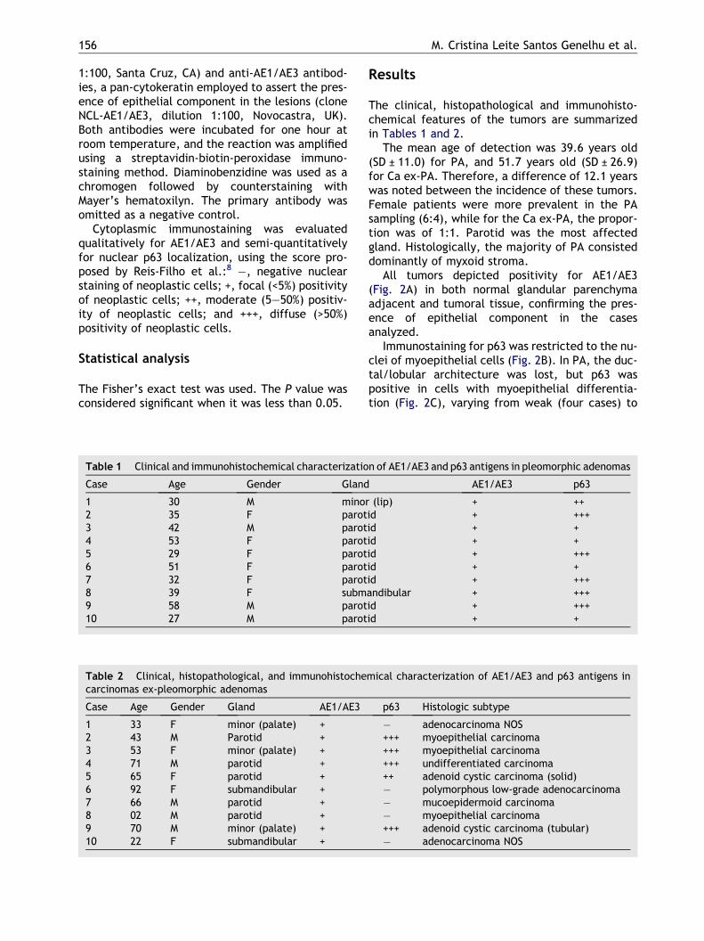
156 M. Cristina Leite Santos Genelhu et al.
1:100, Santa Cruz, CA) and anti-AE1/AE3 antibod-ies, a pan-cytokeratin employed to assert the pres-ence of epithelial component in the lesions (cloneNCL-AE1/AE3, dilution 1:100, Novocastra, UK).Both antibodies were incubated for one hour atroom temperature, and the reaction was amplifiedusing a streptavidin-biotin-peroxidase immuno-staining method. Diaminobenzidine was used as achromogen followed by counterstaining withMayer’s hematoxilyn. The primary antibody wasomitted as a negative control.
Cytoplasmic immunostaining was evaluatedqualitatively for AE1/AE3 and semi-quantitativelyfor nuclear p63 localization, using the score pro-posed by Reis-Filho et al.:8 �, negative nuclearstaining of neoplastic cells; +, focal (<5%) positivityof neoplastic cells; ++, moderate (5–50%) positiv-ity of neoplastic cells; and +++, diffuse (>50%)positivity of neoplastic cells.
Statistical analysis
The Fisher’s exact test was used. The P value wasconsidered significant when it was less than 0.05.
Table 1 Clinical and immunohistochemical characterizatio
Case Age Gender Gland
1 30 M minor2 35 F parot3 42 M parot4 53 F parot5 29 F parot6 51 F parot7 32 F parot8 39 F subm9 58 M parot10 27 M parot
Table 2 Clinical, histopathological, and immunohistochecarcinomas ex-pleomorphic adenomas
Case Age Gender Gland AE1/AE3
1 33 F minor (palate) +2 43 M Parotid +3 53 F minor (palate) +4 71 M parotid +5 65 F parotid +6 92 F submandibular +7 66 M parotid +8 02 M parotid +9 70 M minor (palate) +10 22 F submandibular +
Results
The clinical, histopathological and immunohisto-chemical features of the tumors are summarizedin Tables 1 and 2.
The mean age of detection was 39.6 years old(SD ± 11.0) for PA, and 51.7 years old (SD ± 26.9)for Ca ex-PA. Therefore, a difference of 12.1 yearswas noted between the incidence of these tumors.Female patients were more prevalent in the PAsampling (6:4), while for the Ca ex-PA, the propor-tion was of 1:1. Parotid was the most affectedgland. Histologically, the majority of PA consisteddominantly of myxoid stroma.
All tumors depicted positivity for AE1/AE3(Fig. 2A) in both normal glandular parenchymaadjacent and tumoral tissue, confirming the pres-ence of epithelial component in the casesanalyzed.
Immunostaining for p63 was restricted to the nu-clei of myoepithelial cells (Fig. 2B). In PA, the duc-tal/lobular architecture was lost, but p63 waspositive in cells with myoepithelial differentia-tion (Fig. 2C), varying from weak (four cases) to
n of AE1/AE3 and p63 antigens in pleomorphic adenomas
AE1/AE3 p63
(lip) + ++id + +++id + +id + +id + +++id + +id + +++andibular + +++id + +++id + +
mical characterization of AE1/AE3 and p63 antigens in
p63 Histologic subtype
� adenocarcinoma NOS+++ myoepithelial carcinoma+++ myoepithelial carcinoma+++ undifferentiated carcinoma++ adenoid cystic carcinoma (solid)� polymorphous low-grade adenocarcinoma� mucoepidermoid carcinoma� myoepithelial carcinoma+++ adenoid cystic carcinoma (tubular)� adenocarcinoma NOS
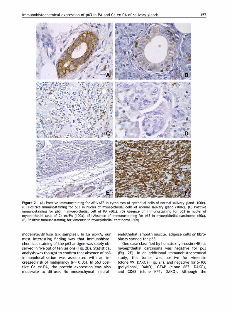
Figure 2 (A) Positive immunostaining for AE1/AE3 in cytoplasm of epithelial cells of normal salivary gland (100x).(B) Positive immunostaining for p63 in nuclei of myoepithelial cells of normal salivary gland (100x). (C) Positiveimmunostaining for p63 in myoepithelial cell of PA (60x). (D) Absence of immunostaining for p63 in nuclei ofmyoepithelial cells of Ca ex-PA (100x). (E) Absence of immunostaining for p63 in myoepithelial carcinoma (60x).(F) Positive immunostaining for vimentin in myoepithelial carcinoma (60x).
Immunohistochemical expression of p63 in PA and Ca ex-PA of salivary glands 157
moderate/diffuse (six samples). In Ca ex-PA, ourmost interesting finding was that immunohisto-chemical staining of the p63 antigen was solely ob-served in five out of ten lesions (Fig. 2D). Statisticalanalysis was thought to confirm that absence of p63immunolocalization was associated with an in-creased risk of malignancy (P < 0.05). In p63 posi-tive Ca ex-PA, the protein expression was alsomoderate to diffuse. No mesenchymal, neural,
endothelial, smooth muscle, adipose cells or fibro-blasts stained for p63.
One case classified by hematoxilyn-eosin (HE) asmyoepithelial carcinoma was negative for p63(Fig. 2E). In an additional immunohistochemicalstudy, this tumor was positive for vimentin(clone V9, DAKO) (Fig. 2F), and negative for S-100(polyclonal, DAKO), GFAP (clone 6F2, DAKO),and CD68 (clone KP1, DAKO). Although the

158 M. Cristina Leite Santos Genelhu et al.
immunohistochemical profile provided was notconclusive, this case was classified as p63 negativemyoepithelial carcinoma based upon its morpho-logical features.
Discussion
Although PA is the most frequent benign tumor ofsalivary glands, the Ca ex-PA, its malignant coun-terpart, is rare. This is due to the fact that timeof tumor evolution and the number of recurrencesare involved in the mechanisms of malignant trans-formation of PA into Ca ex-PA as well as being asso-ciated with clinical disease progression.12–14 Therecurrences are mostly related to incomplete surgi-cal excision, mainly due to pseudopodia pattern ofthose tumors.26 Recently, PA have been earlierdiagnosed and appropriate surgeries reducedmalignant transformation of these benign lesions.27
In this series, clinical characteristics such asage, gender, and type of affected salivary glandwere similar to those reported in the literature,in addition to the gap in time between the emer-gence of PA and of the Ca ex-PA.
Neoplastic myoepithelium is a key cellular par-ticipant in the morphogenesis of the variable histo-logic appearances of many salivary gland tumors.28
Batzakis et al.29 proposed that mixed tumors do notoccur in tissues where the myoepithelial cells areabsent. Barbareschi et al.6 showed that in breasttumors, the identification of myoepithelial cellshas a diagnostic value because, they are retainedin benign lesions and lost during the process ofmalignization and invasion.
Recent research has shown that p63 is a goodmarker of myoepithelial cells even in the presenceof cytomorphological heterogeneity (clear, spin-dle, or plasmacytoid cells). The anti-p63 antibodydemonstrates sensitivity comparable to other myo-epithelial markers, such as a-smooth muscle actin(a-SMA), smooth muscle myosin heavy chain(SMM-HC), and calponin, but it has a greaterspecificity. Stroma cells are consistently nega-tive.3,4,6–8 However, there are few studies evaluat-ing p63 expression in salivary gland tumors.2,4,30
The transcription of the p63 gene begins from twodifferent promoters and further alternative splic-ing. The two main isoforms, TAp63 and DNp63, haveopposite functions, being responsible for cell-cyclearrest and cell proliferation, respectively.31,32 Inour study, using the 4A4 clone, that recognizes allp63 isoforms, all PA showed p63 expression in myo-
epithelial cells nuclei, although those positive cellsbecame dispersed in the tumoral mass.
Our results showed significant variation in p63expression in Ca ex-PA: only 5 cases of malignanttumors were immunostaining to p63, opposite toall cases of PA. This finding allows us to suggestthat Ca ex-PA subtypes can be subdivided intotwo main groups: tumors with myoepithelial differ-entiation (myoepithelial carcinomas and adenoidcystic carcinoma) and those devoid of or withscanty myoepithelial differentiation (mucoepider-moid carcinoma, adenocarcinoma NOS, and poly-morphous low-grade adenocarcinoma) as it wasaccomplished by Pia-Foschini et al.33 with salivarygland-like tumors of the breast.
Coupled with the rarity of myoepithelial carci-noma, the diagnosis is complicated by the consid-erable variability in morphologic features.15 Incase of 8, classified as myoepithelial carcinomaby HE, the spindle cells were negative for p63.The immunohistochemical profile demonstratedthe mesenchymal nature of part of the tumor’smalignant component (vimentin positive). We con-sidered this case a p63 negative myoepithelial car-cinoma: myoepithelial carcinomas may show anabortive myoepithelial phenotype.28
It is possible that p63 positive undifferentiatedcarcinoma (case 4) can be also a myoepithelial car-cinoma. High-grade carcinomas may show myoepi-thelial differentiation even without evidentmyoepithelial morphological features.28 However,for further conclusions an additional immunohisto-chemical panel including other myoepithelialmarkers such as a-SMA, SMMH, calponin and caldes-mon would be necessary.
The identification of Ca ex-PA subtypes is veryimportant due to prognostic implications. Theworst survival rates are related to the undifferenti-ated carcinoma, followed by adenocarcinomaNOS.14 However, in our study, immunostaining forp63 was not useful to identify Ca ex-PA subtypes.In Edwards et al.30 study, p63 staining pattern alsowas not able to distinguish between malignant(adenoid cystic carcinoma, polymorphous low-grade adenocarcinoma) and benign (basal cell ade-noma) salivary gland tumors. In that study, mostcases of adenoid cystic carcinoma and polymor-phous low-grade adenocarcinoma were uniformlypositive for p63. Our case of Ca ex-PA classifiedas polymorphous low-grade adenocarcinoma wasshown to be negative p63 expression. Therefore,p63 is not an ideal marker to distinguish the differ-ent subtypes of Ca ex-PA. In the present study, thisdistinction was accomplished using morphologic
Immunohistochemical expression of p63 in PA and Ca ex-PA of salivary glands 159
parameters. Reis-Filho et al.35 suggest that, poorlydifferentiated carcinomas can be better character-ized by p63 when used together with cytokeratins5/6 and 14.
In spite of the fact that no formal analysis ofspecificity and sensitivity was carried out in thepresent study regarding other myoepithelial mark-ers, our results showed interesting points in com-mon with another authors’ study. Prasad et al.34
studied 135 benign and malignant tumors of sali-vary glands using a-SMA, SMMH and calponin. Theyfound negative results for these markers in poly-morphous low-grade adenocarcinoma, mucoepi-dermoid carcinomas, and adenocarcinoma noother specification (NOS), but results were positivefor adenoid cystic carcinomas and myoepithelialcarcinomas. These findings are in agreement withthose of our study, using p63 in these Ca ex-PAsubtypes.
We also tried to analyze the results consideringthe dual nature of p63 as an oncoprotein throughits inhibition of p53 transactivation functions (DNisoforms) and apoptosis-inducing agent (TA iso-forms).31,32 We can suppose that preservation ofmyoepithelial cells in PA could be related to thehistogenesis of benign tumors, because they wereable to act as progenitors of neoplastic cells (ifthese cells are expressing DNp63). Another possi-bility is that myoepithelial cells of PA could beexpressing TAp63, with its tumor suppressor func-tions. One has indeed invested the myoepitheliumwith great significance as a paracrine inhibitor ofinvasion and thus an inhibitor of tumor progres-sion.36 This hypothesis can be observed in thetransformation from in situ to invasive mammarycarcinoma.37
In the cases of Ca ex-PA we can suppose that theloss of p63 expression could implicate the loss ofthe TAp63 isoforms in the myoepithelial cells or,as already proposed, the tumoral progression wasaddressed for tumors without myoepithelial differ-entiation. However, our data are insufficient to re-late the loss of the expression of this antigen insome Ca ex-PA to the process of malignant trans-formation, since this study does not identify whichisoforms were lost or maintained in the tumors.
In conclusion, our results favor the participationof the myoepithelial cell in the histogenesis ofmixed tumors of salivary gland and suggest that sig-nificant loss of p63 expression is probably involvedin the differentiation of several subtypes of Ca ex-PA. In addition, they corroborate the hypothesisthat the study of p63 antigen may be a useful mar-ker of myoepithelial cells in salivary glands neo-plasms. However, our data are insufficient toinvolve this fact in the malignant transformation
from benign PA to the malignant phenotype of Caex-PA. Further studies using antibodies againstthe different isoforms are necessary for under-standing the biological significance of p63 in sali-vary tumorigenesis.
Acknowledgements
We thank Heitor Paiva, MD, Lucılia Zardo, MD, fromNational Institute of Cancer (INCA), Rio de Janeiro,Brazil, for providing cases of Ca ex-PA, Lucia PortoFonseca de Castro, MD, from School of Medicine ofthe Federal University of Minas Gerais, Brazil, forher scientific assistance, and Sergio Cardoso, PhD,from State University of Montes Claros for havingcritically reviewed the paper.This work was supported in part by Fundacao deAmparo a Pesquisa de Minas Gerais (FAPEMIG),Conselho Nacional de Desenvolvimento Cientıficoe Tecnologico (CNPq) and Coordenacao de Aper-feicoamento de Pessoal de Nıvel Superior (CAPES).
References
1. Di Como CJ, Urist MJ, Babayan I, Drobnjak M, Hedvat CV,Teruya-Felstein J, et al. p63 expression profiles in humannormal and tumor tissues. Clin Cancer Res 2002;8:494–501.
2. Weber A, Langhanki L, Schutz A, et al. Expression profilesof p53, p63, and p73 in benign salivary gland tumors.Virchows Arch 2002;441:428–36.
3. Reis-Filho JS, Schmitt FC. Taking advantage of basicresearch: p63 is a reliable myoepithelial and stem cellmarker. Adv Anat Pathol 2002;9(5):280–9.
4. Bilal H, Handra-Luca A, Bertrand JC, Fouret PJ. p63 isexpressed in basal and myoepithelial cells of human normaland tumor salivary gland tissues. J Histochem Cytochem2003;51:133–9.
5. Bortoluzzi M, Yurgel LS, Dekker NP, Jordan RCK, Regezi JA.Assessment of p63 expression in oral squamous cell carci-nomas and dysplasias. Oral Surg Oral Med Oral Pathol OralRadiol Endod 2004;98(6):698–704.
6. Barbareschi M, Pecciarini L, Cangi MG, Macrı E, Rizzo A,Viale G, et al. p63, a p53 homologue, is a selective nuclearmarker of myoepithelial cells of the human breast. Am JSurg Pathol 2001;25(8):1054–60.
7. Werling RW, Hwang H, Yaziji H, Gown AM. Immunohisto-chemical distinction of invasive from noninvasive breastlesions: a comparative study of p63 versus calponin andsmooth muscle myosin heavy chain. Am J Surg Pathol2003;27(1):82–90.
8. Reis-Filho JS, Torio B, Albergaria A, Schmitt F. p63 expres-sion in normal skin and usual cutaneous carcinomas. J CutanPathol 2002;29:517–23.
9. Park BJ, Lee SJ, Kim JI, Lee SJ, Lee CH, Chang SG, et al.Frequent alteration of p63 expression in human bladdercarcinomas. Cancer Res 2000;60:3370–4.
10. Seifert G, Sobin LH. WHO International classification oftumors. Histological Typing of Salivary Gland Tumors. 2ndEd. Berlin Heidelberg: Spring-Verlag; 1991.
160 M. Cristina Leite Santos Genelhu et al.
11. Ellis GL, Auclair PL. Tumors of the salivary glands. Atlas oftumor pathology. 3rd Ed. Washington, DC: Armed ForcesInstitute of Pathology; 1996.
12. Gnepp DR. Malignant mixed tumors of the salivary glands: areview. Pathol Annu 1993;28(Pt 1):279–328.
13. Li Volsi VA, Perzin KH. Malignant mixed tumors arising insalivary glands. 1. Carcinomas arising in benign mixedtumors: a clinicopathologic study. Cancer 1977;39:2209–30.
14. Spiro RH, Huvos AG, Strong EW. Malignant mixed tumor ofsalivary origin. A clinicopathologic study of 146 cases.Cancer 1977;39:388–96.
15. Said S, Campana J. Myoepithelial carcinoma ex pleomorphicadenoma of salivary glands: a problematic diagnosis. OralSurg Oral Med Oral Pathol Oral Radiol Endod 2005;99:196–201.
16. Singh R, Cawson RA. Malignant myoepithelial carcinoma(myoepithelioma) arising in a pleomorphic adenoma of theparotid gland. An immunohistochemical study and review ofthe literature. Oral Surg Oral Med Oral Pathol 1988;66(1):65–70.
17. Lewis JE, Olsen KD, Sebo TJ. Carcinoma ex pleomorphicadenoma: pathologic analysis of 73 cases. Hum Pathol 2001;32:596–604.
18. Olsen KD, Lewis JE. Carcinoma ex pleomorphic adenoma: aclinicopathologic review. Head Neck 2001;23(9):705–12.
19. Auclair PL, Ellis GL. Atypical features in salivary glandmixed tumors: their relationship to malignant transforma-tion. Mod Pathol 1996;9(6):652–7.
20. Tortolodo ME, Luna MA, Batsakis JG. Carcinomas ex-pleo-morphic adenoma and malignant mixed tumors. Histomorf-ologic indexes. Arch Otolaryngol 1984;110(3):172–6.
21. Righi PD, Li YQ, et al. The role of the p53 gene in themalignant transformation of pleomorphic adenomas of theparotid gland. Anticancer Res 1994;14(5B):2253–7.
22. Costa-Rosa J, Fonseca I, Felix A, Soares J. Immunohisto-chemical study of c-erbB-2 expression in carcinoma ex-pleomorphic adenoma. Histopathology 1996;28:247–52.
23. Roijer E, Nordkvist A, Strom AK, Ryd W, Behrendt M,Bullerdiek J, et al. Translocation, deletion/amplification,and expression of HMGIC and MDM2 in a carcinoma expleomorphic adenoma. Am J Pathol 2002;160:433–40.
24. Voz ML, Mathys J, Hensen K, Pendeville H, Van Valcken-borgh I, Van Huffel C, et al. Microarray screening for targetgenes of the proto-oncogene PLAG1. Oncogene 2004;23(1):179–91.
25. El-Naggar AK, Callender D, Coombes MM, Hurr K, Luna MA,Batsakis JG. Molecular genetic alterations in carcinoma ex-
pleomorphic adenoma: a putative progression model? GenesChromosomes Cancer 2000;27(2):162–8.
26. Henriksson G, Westrin KM, Carlsoo B, Silfversward C.Recurrent primary pleomorphic adenomas of salivary glandorigin. Intrasurgical rupture, histopathologic features, andpseudopodia. Cancer 1998;82:617–20.
27. Stennert E, Wittekindt C, Klussmann JP, Arnold G, Guntinas-Lichius O. Recurrent pleomorphic adenoma of the parotidgland: a prospective histopathological and immunohisto-chemical study. Laryngoscope 2004;114(1):148–63.
28. Savera AT, Zarbo RJ. Defining the role of myoepithelium insalivary gland neoplasia. Adv Anat Pathol 2004;11(2):69–85.
29. Batzakis JG, Kraemer B, Sciubba JJ. The pathology of headand neck tumors: the myoepithelial cell and its participa-tion in salivary gland neoplasia, Part 17. Head Neck Surg1983;5(3):222–33.
30. Edwards PC, Bhuiya T, Kelsch RD. Assessment of p63expression in the salivary gland neoplasms adenoid cysticcarcinoma, polymorphous low-grade adenocarcinoma, andbasal cell and canalicular adenomas. Oral Surg Oral MedOral Pathol Oral Radiol Endod 2004;97(5):613–9.
31. Yang A, Kaghad M, Wang Y, Gillett E, Fleming MD, Dotsch V,et al. p63, a p53 homolog at 3q27-29, encodesmultiple products with transactivating, death-inducing,and dominant-negative activities. Molecular Cell 1998;2:305–16.
32. Little NA, Jochemsen AG. P63. Int J Biochem Cell Biol 2002;34:6–9.
33. Pia-Foschini M, Reis-Filho JS, Eusebi V, Lakhani SR. Salivarygland-like tumours of the breast: surgical and molecularpathology. J Clin Pathol 2003;56:497–506.
34. Prasad AR, Savera AT, Gown AM, Zarbo RJ. The myoepithe-lial immunophenotype in 135 benign and malignant salivarygland tumors other than pleomorphic adenoma. Arch PatholLab Med 1999;123:801–6.
35. Reis-Filho JS, Simpson PT, Martins A, Preto A, Gartner F,Schmitt FC. Distribution of p63, cytokeratins 5/6 andcytokeratin 14 in 51 normal and 400 neoplastic humantissue samples using TARP-4 multi-tumor tissue microarray.Virchows Arch 2003;443(2):122–32.
36. Stemlicht MD, Kedeshian P, Shao ZM, Safarians S, Barsky SH.The human myoepithelial cell is a natural tumor suppressor.Clin Cancer Res 1977;3:1949–58.
37. Kalof AN, Tam D, Beatty B, Cooper K. Immunostaining ofmyoepithelial cells in breast lesions: a comparison of CD10and smooth muscle myosin heavy chain. J Clin Pathol2004;57:625–9.
67
Artigo 3 - Immunolocalization of β-catenin in pleomorphic adenomas and carcinomas ex-pleomorphic adenomas of salivary glands
Immunolocalization of b-Catenin in PleomorphicAdenomas and Carcinomas Ex-pleomorphic Adenomas
of Salivary Glands
Marisa C. L. S. Genelhu, PhD,*w z Helenice Gobbi, PhD,w Diele C. B. Arantes, PhD,ySergio V. Cardoso, PhD,J and Geovanni D. Cassali, PhD*w
Abstract: b-catenin plays a central role in cadherin/catenin cell-
cell adhesion complex and is involved in cell signaling pathway.
Change in b-catenin distribution has been associated with
several human cancers including salivary gland tumors. We
studied the immunolocalization of b-catenin in a series of
pleomorphic adenomas (PA) and carcinomas ex-pleomorphic
adenomas (Ca ex-PA). Ten samples of PA and ten of Ca ex-PA
were evaluated by immunohistochemistry using streptavidin-
biotin-peroxidase technique and a monoclonal antibody against
b-catenin (E-5). Cell membrane/cytoplasmic staining of
b-catenin was observed in normal gland parenchyma, PA, and
in well-differentiated Ca ex-PA. Cytoplasmic/nuclear b-cateninstaining was observed in poorly differentiated carcinomas and,
interestingly, in one case of PA. Our data showed decreased
cell membrane b-catenin expression in higher-grade tumors
suggesting that b-catenin may play an important role in
histologic differentiation and transition to malignant phenotype
of Ca ex-PA.
Key Words: b-catenin, pleomorphic adenoma, carcinoma,
salivary gland, immunohistochemistry
(Appl Immunohistochem Mol Morphol 2007;15:273–278)
Several studies have suggested that E-cadherin/cateninscell-cell adhesion complex is damaged in cancer
cells.1–4 E-cadherins, mainly responsible for cell-celladhesion in most epithelial tissues, are calcium-dependent
transmembrane glycoproteins, located in adherentjunctions. Adhesion occurs through homotypical bind-ings between their extracellular amino-terminal domains.Nevertheless, by means of its cytoplasmic carboxy-terminal tail, E-cadherin is linked to a series of actinicmicrofilaments of cell cytoskeleton through a-catenins,b-catenins, g-catenins and p120.5
In addition to its well-defined role in regulating cell-cell adhesion via interactions with E-cadherin, b-catenin,a 92-kd protein, has a critical role in the highly conservedWnt (Wingless) signaling pathway.6,7 When b-catenindoes not take part in the adhesion complex, the freemolecule is sequestered by a complex formed by APC(adenomatous polyposis coli) gene products, by GSK-3b(serine-threonine glycogen synthetase kinase-3b) moleculeand by an axin adapting protein, disqualifying b-cateninphosphorylation and its degradation by ubiquitin-depen-dent proteolitic system.4,6 Increased free b-catenin con-centrations in cytosol stimulate their link with proteinssuch as lymphocyte enhancer factor and T-cell factor.These complexes are transferred to the nucleus and act ascofactors of genes transcription related to cell prolifera-tion including well-established oncogenes, such as c-myc,Cyclin D1, and matrix metalloproteinase-7.8
This signaling pathway is important during embry-ogenesis when Wnt gene is required to establish andmaintain normal epithelial morphology and tissue andorgan differentiation processes.9 However, in adulttissues, it plays an important role in effective neoplastictransformation when inappropriately activated.6,10
Consistent with b-catenin’s ostensibly independentfunctions in cell adhesion and signaling, distinct pools ofb-catenin exist in cells, including a cadherin-associated(membrane-associated) pool and a cytoplasmic and/ornuclear pool involved in Wnt signaling.8
Adhesion loss is an important step in the progres-sion of several epithelial tumors.11 Cellular redistributionof b-catenin with reduction of membrane expression andnuclear b-catenin accumulation is observed in tumorswith aggressive histopathologic characteristics defined byloss of normal tissue architecture, high grade, infiltrativegrowth, lymph node involvement and metastases.4,5,12–15
A variety of human malignancies have already beenassociated with b-catenin redistribution.3 In salivarygland tumors, studies focusing b-catenin role are scarceCopyright r 2007 by Lippincott Williams & Wilkins
Received for publication October 18, 2005; accepted May 3, 2006.From the *Laboratory of Comparative Pathology, Biological Sciences
Institute; wDepartment of Anatomic Pathology and ForensicMedicine, School of Medicine, Federal University of Minas Gerais;yNewton Paiva University Center, Belo Horizonte; JOral DiagnosisDivision, Department of Dentistry, State University of MontesClaros, and Oral Pathology Laboratory, School of Dentistry,Federal University of Uberlandia; and zImmunology ResearchLaboratory, Vale do Rio Doce University, Governador Valadares,Minas Gerais, Brazil.
Supported in part by grants from CAPES, CNPq, and FAPEMIG,Brazil.
Reprints: Dr Marisa Genelhu, PhD, Nucleo de Pesquisa em Imunologia,Universidade Vale do Rio Doce, Rua Israel Pinheiro, 2000,Governador Valadares, Minas Gerais, Brazil, CEP: 35020-220(e-mail: [email protected]).
RESEARCH ARTICLE
Appl Immunohistochem Mol Morphol � Volume 15, Number 3, September 2007 273
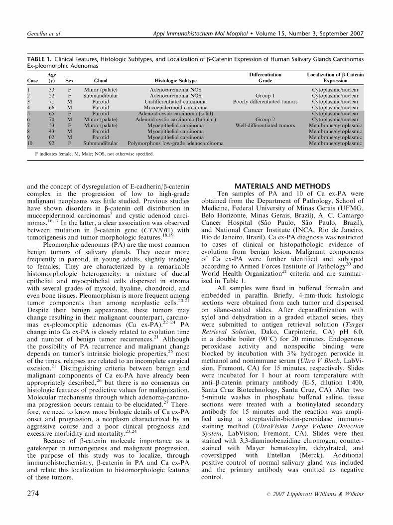
and the concept of dysregulation of E-cadherin/b-catenincomplex in the progression of low to high-grademalignant neoplasms was little studied. Previous studieshave shown disorders in b-catenin cell distribution inmucoepidermoid carcinomas7 and cystic adenoid carci-nomas.16,17 In the latter, a clear association was observedbetween mutation in b-catenin gene (CTNNB1) withtumorigenesis and tumor morphologic features.18,19
Pleomorphic adenomas (PA) are the most commonbenign tumors of salivary glands. They occur morefrequently in parotid, in young adults, slightly tendingto females. They are characterized by a remarkablehistomorphologic heterogeneity: a mixture of ductalepithelial and myoepithelial cells dispersed in stromawith several grades of myxoid, hyaline, chondroid, andeven bone tissues. Pleomorphism is more frequent amongtumor components than among neoplastic cells.20,21
Despite their benign appearance, these tumors maychange resulting in their malignant counterpart, carcino-mas ex-pleomorphic adenomas (Ca ex-PA).22–24 PAchange into Ca ex-PA is closely related to evolution timeand number of benign tumor recurrences.21 Althoughthe possibility of PA recurrence and malignant changedepends on tumor’s intrinsic biologic properties,25 mostof the times, relapses are related to an incomplete surgicalexcision.21 Distinguishing criteria between benign andmalignant components of Ca ex-PA have already beenappropriately described,26 but there is no consensus onhistologic features of predictive values for malignization.Molecular mechanisms through which adenoma-carcino-ma progression occurs remain to be elucidated.27 There-fore, we need to know more biologic details of Ca ex-PAonset and progression, a neoplasm characterized by anaggressive course and a poor clinical prognosis andexcessive morbidity and mortality.23,24
Because of b-catenin molecule importance as agatekeeper in tumorigenesis and malignant progression,the purpose of this study was to localize, throughimmunohistochemistry, b-catenin in PA and Ca ex-PAand relate this localization to histomorphologic featuresof these tumors.
MATERIALS AND METHODSTen samples of PA and 10 of Ca ex-PA were
obtained from the Department of Pathology, School ofMedicine, Federal University of Minas Gerais (UFMG,Belo Horizonte, Minas Gerais, Brazil), A. C. CamargoCancer Hospital (Sao Paulo, Sao Paulo, Brazil),and National Cancer Institute (INCA, Rio de Janeiro,Rio de Janeiro, Brazil). Ca ex-PA diagnosis was restrictedto cases of clinical or histopathologic evidence ofevolution from benign lesion. Malignant componentsof Ca ex-PA were further identified and subtypedaccording to Armed Forces Institute of Pathology20 andWorld Health Organization21 criteria and are summar-ized in Table 1.
All samples were fixed in buffered formalin andembedded in paraffin. Briefly, 4-mm-thick histologicsections were obtained from each tumor and dispensedon silane-coated slides. After deparaffinization withxylol and dehydration in a graded ethanol series, theywere submitted to antigen retrieval solution (TargetRetrieval Solution, Dako, Carpinteria, CA) pH 6.0,in a double boiler (901C) for 20 minutes. Endogenousperoxidase activity and nonspecific binding wereblocked by incubation with 3% hydrogen peroxide inmethanol and nonimmune serum (Ultra V Block, LabVi-sion, Fremont, CA) for 15 minutes, respectively. Slideswere incubated for 1 hour at room temperature withanti–b-catenin primary antibody (E-5, dilution 1:400,Santa Cruz Biotechnology, Santa Cruz, CA). After two5-minute washes in phosphate buffered saline, tissuesections were treated with a biotinylated secondaryantibody for 15 minutes and the reaction was ampli-fied using a streptavidin-biotin-peroxidase immuno-staining method (UltraVision Large Volume DetectionSystem, LabVision, Fremont, CA). Slides were thenstained with 3,3-diaminobenzidine chromogen, counter-stained with Mayer hematoxylin, dehydrated, andcoverslipped with Entellan (Merck). Additionalpositive control of normal salivary gland was includedand the primary antibody was omitted as negativecontrol.
TABLE 1. Clinical Features, Histologic Subtypes, and Localization of b-Catenin Expression of Human Salivary Glands CarcinomasEx-pleomorphic Adenomas
Case
Age
(y) Sex Gland Histologic Subtype
Differentiation
Grade
Localization of b-CateninExpression
1 33 F Minor (palate) Adenocarcinoma NOS Cytoplasmic/nuclear2 22 F Submandibular Adenocarcinoma NOS Group 1 Cytoplasmic/nuclear3 71 M Parotid Undifferentiated carcinoma Poorly differentiated tumors Cytoplasmic/nuclear4 66 M Parotid Mucoepidermoid carcinoma Cytoplasmic/nuclear5 65 F Parotid Adenoid cystic carcinoma (solid) Cytoplasmic/nuclear6 70 M Minor (palate) Adenoid cystic carcinoma (tubular) Group 2 Cytoplasmic/nuclear7 53 F Minor (palate) Myoepithelial carcinoma Well-differentiated tumors Membrane/cytoplasmic8 43 M Parotid Myoepithelial carcinoma Membrane/cytoplasmic9 02 M Parotid Myoepithelial carcinoma Membrane/cytoplasmic10 92 F Submandibular Polymorphous low-grade adenocarcinoma Membrane/cytoplasmic
F indicates female; M, Male; NOS, not otherwise specified.
Genelhu et al Appl Immunohistochem Mol Morphol � Volume 15, Number 3, September 2007
274 r 2007 Lippincott Williams & Wilkins
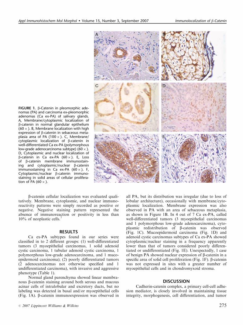
b-catenin cellular localization was evaluated quali-tatively. Membrane, cytoplasmic, and nuclear immuno-reactivity patterns were simply recorded as positive ornegative. Negative staining pattern represented theabsence of immunoreaction or positivity in less than10% of neoplastic cells.7
RESULTSCa ex-PA subtypes found in our series were
classified in to 2 different groups: (1) well-differentiatedtumors (3 myoepithelial carcinomas, 1 solid adenoidcystic carcinoma, 1 tubular adenoid cystic carcinoma, 1polymorphous low-grade adenocarcinoma, and 1 muco-epidermoid carcinoma); (2) poorly differentiated tumors(2 adenocarcinomas not otherwise specified and 1undifferentiated carcinoma), with invasive and aggressivephenotype (Table 1).
Normal gland parenchyma showed linear membra-nous b-catenin staining around both serous and mucousacinar cells of intralobular and excretory ducts, but nolabeling was detected in basal and/or myoepithelial cells(Fig. 1A). b-catenin immunoexpression was observed in
all PA, but its distribution was irregular (due to loss oflobular architecture), occasionally with membrane/cyto-plasmic localization. Membrane expression was alsoobserved in PA with an area of sebaceous metaplasia,as shown in Figure 1B. In 4 out of 7 Ca ex-PA, calledwell-differentiated tumors (3 myoepithelial carcinomasand 1 polymorphous low-grade adenocarcinoma), cyto-plasmic redistribution of b-catenin was observed(Fig. 1C). Mucoepidermoid carcinoma (Fig. 1D) andadenoid cystic carcinomas subtypes of Ca ex-PA showedcytoplasmic/nuclear staining in a frequency apparentlylower than that of tumors considered poorly differen-tiated or undifferentiated (Fig. 1E). Unexpectedly, 1 caseof benign PA showed nuclear expression of b-catenin in aspecific area of solid cell proliferation (Fig. 1F). b-cateninwas not expressed in sites with a greater number ofmyoepithelial cells and in chondromyxoid stroma.
DISCUSSIONCadherin-catenin complex, a primary cell-cell adhe-
sion mediator, is closely involved in maintaining tissueintegrity, morphogenesis, cell differentiation, and tumor
FIGURE 1. b-Catenin in pleomorphic ade-nomas (PA) and carcinoma ex-pleomorphicadenomas (Ca ex-PA) of salivary glands.A, Membrane/cytoplasmic localization ofb-catenin in normal glandular epithelium(60� ). B, Membrane localization with highexpression of b-catenin in sebaceous meta-plasia area of PA (100� ). C, Membrane/cytoplasmic localization of b-catenin inwell-differentiated Ca ex-PA (polymorphouslow-grade adenocarcinoma subtype) (60� ).D, Cytoplasmic and nuclear localization ofb-catenin in Ca ex-PA (60� ). E, Lossof b-catenin membrane immunostain-ing and cytoplasmic/nuclear b-cateninimmunostaining in Ca ex-PA (60� ). F,Cytoplasmic/nuclear b-catenin immuno-staining in solid areas of cellular prolifera-tion of PA (60� ).
Appl Immunohistochem Mol Morphol � Volume 15, Number 3, September 2007 Immunolocalization of b-Catenin
r 2007 Lippincott Williams & Wilkins 275
progression cells.7 Function loss leads malign lesions toescape from their primary sites, degrading extracellularmatrix, acquiring an invasive phenotype and, eventually,metastasizing.11 In addition, cadherin-catenin complex ispart of signal-transduction pathway Wingless/Wnt.2,4,7
b-catenin is an important member of this complex, with atleast 2 functions, which are determined by its localization,in the membrane or nucleus. It plays an important rolein cell adhesion in the membrane. Nuclear b-cateninincreases Wnt gene transcription responsive to cellgrowth.28
Several authors have reported b-catenin subcellularcompartmentalization in different types of cancer, butthe role of this redistribution still has not been studied inPA and its malignant counterpart, Ca ex-PA. It is knownthat such event is associated with the initiation andprogression of tumors but, mechanisms by whichb-catenin molecules are redistributed, remain elusive.This redistribution may be a consequence of mutations inAPC tumor suppressor gene29,30 or mutations in exon 3 ofb-catenin gene itself.31 Posttranslational changes of anyE-cadherin/catenin complex element may also bethe cause of b-catenin nuclear/cytoplasmic expression inneoplasms.32–34
The present manuscript is the first exploratory studyto report association between immunoexpression ofb-catenin in PA and Ca ex-PA of salivary glands.Although the number of cases of Ca ex-PA was limitedbecause of the scarcity of malignant lesions, we reportinteresting data. b-catenin immunolocalization analysis inall PA and, in 4 out of 7 well-differentiated malignanttumors have shown predominantly membrane/cytoplas-mic expressions. In contrast, in 3 malignant tumorsconsidered poorly differentiated or undifferentiated,membrane expression loss and, in some cells, nuclearb-catenin accumulation were observed.
Despite considering well-differentiated malignanttumors, adenoid cystic carcinomas and mucoepidermoidcarcinoma subtypes of Ca ex-PA, have shown cytoplasmic/nuclear staining similarly to poorly differentiated orundifferentiated tumors. Our results have been shown tobe similar to those obtained in immunohistochemicalstudies of mucoepidermoid carcinomas and cystic adenoidcarcinomas carried out by Shieh et al7 and Daa et al,16
respectively. This may be related to the most aggressivebiologic behavior of these tumors, which is possibly due toabnormalities in cellular mechanisms of cell adhesion andsignaling (such as those associated with b-catenin), inaddition to several other regulatory factors of proliferativeand invasive activity regardless Wnt pathway.
In this work, an inverse correlation was foundbetween membrane expression of b-catenin and low-gradedifferentiation and/or more aggressive biological behaviorof tumor cells.35 This result, membrane expression losswith subcellular (nuclear and perinuclear) b-cateninlocalization, is in accordance with those found in theliterature for other tumors such as oral,12,14,33 larynx,34
and esophageal35 squamous cells carcinomas, pancreaticadenocarcinomas,36 gastric carcinomas,37,38 lung carcino-
mas,39 colorectal carcinomas,40,41 endometrial carcino-mas,42,43 hepatocellular carcinomas,44 and bladder,45
renal,46 skin (melanoma),47 thyroid,31 and breast can-cers.48,49
We may suppose that in neoplastic cell membraneof PA and well-differentiated Ca ex-PA, b-cateninmaintains its role as cell adhesion and tumor suppressormolecule. Cytoplasmic localization may both indicatethat after the posttranslational process, the molecule isdriven to the membrane where it links to E-cadherin, andthat b-catenin is free in the cytoplasm, at disposal ofAPC/GSK-3b/axin complex which restrains its degrada-tion and makes its nucleus targeting easier.50 In this case,evidences suggest that b-catenin plays a critical role in thesignaling pathway accounting for gene transcriptionlinked to cell proliferation in malignant tumors. Inaddition, the absence of membrane expression may berelated to tumor progression and Ca ex-PA invasiveproperties.37,51
An interesting datum in this study was the nuclearlocalization of b-catenin in one of the benign tumors. Thisfinding is very important because it may mean an earlymolecular event in the malignant transformation of PA,similar to what may be inferred for malignant tumors.This fact may be relevant in the prognostic of some PAregarding recurrence and malignant transformation.
The role of b-catenin in both Wnt signaling and celladhesion also suggests that targeted disruption of b-catenin-mediated T-cell factor signaling is a possiblestrategy for early chemopreventive intervention to thetumor progression. Some data indicate that sulindac,sulindac sulfone, and indomethacin may modulate sub-cellular localization of b-catenin in vivo, and resulting ineither decreased nuclear compartmentalization or en-hanced localization of b-catenin in plasma membrane.52
Finally, our results suggest that b-catenin immuno-histochemical expression evaluation is of great diagnosticand prognostic value for PA and Ca ex-PA studies, as it isfor several other tumors.53 Changes in b-catenin localiza-tion may be explored, along with conventional clinicaland histopathologic characteristics, distinguishing benigntumors with malignant potential from malignanttumors with more aggressive characteristics of in-vasiveness and metastases.35,53
In conclusion, we have demonstrated changes inb-catenin immunolocalization in PA and Ca ex-PA. Ourdata showed decreased cell membrane b-catenin expres-sion in higher-grade tumors suggesting that b-cateninmay play an important role in histologic differentiationand transition to malignant phenotype of Ca ex-PA. Thepresence of nuclear b-catenin in a benign tumor suggests apossible association of this molecule with early molecularevents of PA malignant transformation into Ca ex-PA.We believe that b-catenin may become a significantprognostic marker for tumors, biologic behavior andoccurrence of local and distant metastases. Responses tosome extracellular modulators may favorably changeb-catenin localization and lead to new therapeuticapproaches for these tumors.
Genelhu et al Appl Immunohistochem Mol Morphol � Volume 15, Number 3, September 2007
276 r 2007 Lippincott Williams & Wilkins
ACKNOWLEDGMENTSThe authors are grateful to Fernando Soares, PhD,
Antonio Campos, PhD, from Department of AnatomicPathology, A.C. Camargo Cancer Hospital, Sao Paulo,Brazil and Heitor Paiva, MD, Lucılia Zardo, MD, fromNational Institute of Cancer (INCA), Rio de Janeiro,Brazil, for providing cases of Ca ex-PA.
REFERENCES1. Wheelock MJ, Johnson KR. Cadherins as modulators of cellular
phenotype. Annu Rev Cell Dev Biol. 2003;19:207–235.2. Nollet F, Geert B, Van Roy F. The role of the E-cadherin/catenin
adhesion complex in the development and progression of cancer.Mol Cell Biol Res Commun. 1999;2:77–85.
3. Van Aken E, De Wever O, Correia da Rocha AS. DefectiveE-cadherin/catenin complexes in human cancer. Virchows Arch.2001;439:725–751.
4. Shiozaki H, Oka H, Inoue M, et al. E-cadherin mediated adhesionsystem in cancer cells. Cancer. 1996;77:1605–1613.
5. Efstathiou JA, Liu D, Wheeler JM, et al. Mutated epithelialcadherin is associated with increased tumorigenicity and loss ofadhesion and of responsiveness to the motogenic trefoil factor 2 incolon carcinoma cells. Proc Natl Acad Sci USA. 1999;96:2316–2321.
6. Jankowski JA, Bruton R, Shepherd N, et al. Cadherin and cateninbiology represent a global mechanism for epithelial cancer progres-sion. Mol Pathol. 1997;50:289–290.
7. Shieh YS, Chang LC, Chiu KC, et al. Cadherin and cateninexpression in mucoepidermoid carcinoma: correlation with histo-pathologic grade, clinical stage, and patient outcome. J Oral PatholMed. 2003;32:297–304.
8. Zhai Y, Wu R, Schwartz DR, et al. Role of b-catenin/T-cell factor-regulated genes in ovarian endometrioid adenocarcinomas.Am J Pathol. 2002;160:1229–1238.
9. Cadigan KM, Nusse R. Wnt signaling: a common theme in animaldevelopment. Genes Devel. 1997;11: F127–F147.
10. Harrington KJ, Syrigos KN. The role of E-cadherin-catenincomplex: more than an intercellular glue? Ann Surg Oncol.2000;7:783–788. Erratum in: Ann Surg Oncol. 2001;8:186.
11. Chow V, Yuen AP, Lam KY, et al. A comparative study of theclinicopathological significance of E-cadherin and catenins (alpha,beta, gamma) expression in the surgical management of oral tongcarcinoma. J Cancer Res Clin Oncol. 2001;127:59–63.
12. Kudo Y, Kitajima S, Ogawa I, et al. Invasion and metastasis of oralcancer cells require methylation of E-cadherin and/or degradation ofmembranous beta-catenin. Clin Cancer Res. 2004;10:5455–5463.
13. Margulis A, Zhang W, Alt-Holland A, et al. E-cadherin suppressionaccelerates squamous cell carcinoma progression in three-dimen-sional, human tissue constructs. Cancer Res. 2005;65:1783–1791.
14. Lo Muzio L, Staibano S, Pannone G, et al. Beta- and gamma-catenin expression in oral squamous cell carcinomas. AnticancerRes. 1999;19:3817–3826.
15. Williams HK, Sanders DS, Jankowski JA, et al. Expression ofcadherins and catenins in oral epithelial dysplasia and squamous cellcarcinoma. J Oral Pathol Med. 1998;27:308–317.
16. Daa T, Kaku N, Kashima K, et al. Expression of beta-catenin,E-cadherin and Cyclin D1 in adenoid cystic carcinomas of thesalivary gland. J Exp Clin Cancer Res. 2005;24:83–87.
17. Zhang ZY, Wu YQ, Zhang WG, et al. The expression ofE-cadherin-catenin complex in adenoid cystic carcinoma of salivaryglands. Chin J Dent Res. 2000;3:36–39.
18. Daa T, Kashima K, Kaku N, et al. Mutations in components of theWnt signaling pathway in adenoid cystic carcinoma. Mod Pathol.2004;17:1475–1482.
19. Shibuya Y, Ri S, Umeda M, et al. Ultrastructural localizationof E-cadherin and a-/b-catenin in adenoid cystic carcinoma.Histopathology. 1999;35:423–431.
20. Ellis GL, Auclair PL. Tumors of the Salivary Glands. Atlas of TumorPathology. 3rd ed. Washington, DC: Armed Forces Institute ofPathology; 1996.
21. Seifert G, Sobin LH. WHO International Classification of Tumors.Histological Typing of Salivary Gland Tumors. 2nd ed. Berlin,Heidelberg: Springer-Verlag; 1991.
22. Gnepp DR. Malignant mixed tumors of the salivary glands:a review. Pathol Annu. 1993;28:279–328.
23. Lewis JE, Olsen KD, Sebo TJ. Carcinoma ex pleomorphicadenoma: pathologic analysis of 73 cases. Hum Pathol. 2001;32:596–604.
24. Olsen KD, Lewis JE. Carcinoma ex pleomorphic adenoma: aclinicopathologic review. Head Neck. 2001;23:705–712.
25. Matturri L, et al. Cell kinetics of pleomorphic adenomas of theparotid gland. Eur J Cancer B Oral Oncol. 1996;32B:154–157.
26. Auclair PL, Ellis GL. Atypical features in salivary gland mixedtumors: their relationship to malignant transformation. Mod Pathol.1996;9:652–657.
27. El Naggar AK, Lovell M, Callender DL, et al. Concurrent cytogenetic,interphase fluorescence in situ hybridization and DNA flow cytometricanalyses of a carcinoma ex-pleomorphic adenoma of parotid gland.Cancer Genet Cytogenet. 1998;107:132–136.
28. Van Nhieu JT, Renard CA, Wei Y, et al. Nuclear accumulation ofmutated b-catenin in hepatocellular carcinoma is associated withincreased cell proliferation. Am J Pathol. 1999;155:703–710.
29. Rubinfeld B, Souza B, Albert I, et al. Association of the APC geneproduct with beta-catenin. Science. 1993 10;262:1731–1734.
30. Hulsken J, Birchmeier W, Behrens J. E-cadherin and APC competefor the interaction with beta-catenin and the cytoskeleton. J CellBiol. 1994;127:2061–2069.
31. Garcia-Rostan G, Camp RL, Herrero A, et al. b-catenin dysregula-tion in thyroid neoplasms: down-regulation, aberrant nuclearexpression, and CTNNB1 exon 3 mutations are markers foraggressive tumor phenotypes and poor prognosis. Am J Pathol.2001;158:987–996.
32. Mareel M, Leroy A. Clinical, cellular, and molecular aspects ofcancer invasion. Physiol Rev. 2003;83:337–376.
33. Tanaka N, Odajima T, Ogi K, et al. Expression of E-cadherin,alpha-catenin, and beta-catenin in the process of lymph nodemetastasis in oral squamous cell carcinoma. Br J Cancer. 2003;89:557–563.
34. Lopez-Gonzalez JS, Cristerna-Sanchez L, Vasquez-Manriquez ME,et al. Localization and level of expression of b-catenin in humanlaryngeal squamous cell carcinoma. Otolaryngol Head Neck Surg.2004;130:89–93.
35. De Castro J, Gamallo C, Palacios J, et al. beta-catenin expressionpattern in oesophageal squamous cell carcinoma. Relationship withclinicopathologic features and clinical outcome. Virchows Arch.2000;437:599–604.
36. Julkunen K, Makinen K, Karja V, et al. alpha-, beta- and chi-catenin expression in human pancreatic cancer. Anticancer Res.2003;23:5043–5047.
37. Nakamura E, Sugihara H, Bamba M, et al. Dynamic alteration ofE-cadherin/catenin complex during cell differentiation and invasionof undifferentiated-type gastric carcinomas. J Pathol. 2005;205:349–358.
38. Kim HS, Hong EK, Park SY, et al. Expression of beta-catenin andE-cadherin in the adenoma-carcinoma sequence of the stomach.Anticancer Res. 2003;23:2863–2868.
39. Rodriguez-Salas N, Palacios J, de Castro J, et al. Beta-cateninexpression pattern in small cell lung cancer: correlation with clinicaland evolutive features. Histol Histopathol. 2001;16:353–358.
40. Hugh TJ, Dillon SA, O’Dowd G, et al. Beta-catenin expression inprimary and metastatic colorectal carcinoma. Int J Cancer. 1999;82:504–511.
41. Wong NACS, Pignatelli M. b-catenin—a linchpin in colorectalcarcinogenesis? Am J Pathol. 2002;160:389–401.
42. Shih HC, Shiozawa T, Miyamoto T, et al. Immunohistochemicalexpression of E-cadherin and beta-catenin in the normal andmalignant human endometrium: an inverse correlation betweenE-cadherin and nuclear beta-catenin expression. Anticancer Res.2004;24:3843–3850.
43. Saegusa M, Hashimura M, Kuwata T, et al. b-catenin simulta-neously induces activation of the p53-p21WAF1 pathway and
Appl Immunohistochem Mol Morphol � Volume 15, Number 3, September 2007 Immunolocalization of b-Catenin
r 2007 Lippincott Williams & Wilkins 277

overexpression of cyclin D1 during squamous differentiationof endometrial carcinoma cells. Am J Pathol. 2004;164:1739–1749.
44. Wong CM, Fan ST, Ng IO. beta-Catenin mutation and over-expression in hepatocellular carcinoma: clinicopathologic andprognostic significance. Cancer. 2001;92:136–145.
45. Shimazui T, Schalken JA, Giroldi LA, et al. Prognostic value ofcadherin-associated molecules (a-, b-, g-catenins and p120cas) inbladder tumors. Cancer Res. 1996;56:4154–4158.
46. Aaltomaa S, Lipponen P, Karja V, et al. The expression andprognostic value of alpha-, beta- and gamma-catenins in renal cellcarcinoma. Anticancer Res. 2004;24:2407–2413.
47. Qi J, Chen N, Wang J, et al. Transendothelial migration ofmelanoma cells involves N-cadherin-mediated adhesion and activa-tion of the b-catenin signaling pathway. Mol Biol Cell. 2005;16:4386–4397.
48. Lin SY, Xia WY, Wang JC, et al. Beta-catenin, a novel prognosticmarker for breast cancer: its roles in cyclin D1 expression and cancerprogression. Proc Natl Acad Sci U S A. 2000;97:4262–4266.
49. Karayiannakis AJ, Nakopoulou L, Gakiopoulou H, et al. Expres-sion patterns of b-catenin in situ and invasive breast cancer. EurJ Surg Oncol. 2001;27:31–36.
50. Klingelhofer J, Troyanovsky RB, Laur OY, et al. Exchange of cateninsin cadherin-catenin complex. Oncogene. 2003;22:1181–1188.
51. Wijnhoven BP, Dinjens WN, Pignatelli M. E-cadherin-catenin cell-cell adhesion complex and human cancer. Br J Surg. 2000;87:992–1005.
52. Clapper ML, Coudry J Chang WC. Beta-catenin-mediated signal-ing: a molecular target for early chemopreventive intervention.Mutat Res. 2004;555:97–105.
53. Montgomery E, Folpe AL. The diagnostic value of b-cateninimmunohistochemistry. Adv Anat Pathol. 2005;12:350–356.
Genelhu et al Appl Immunohistochem Mol Morphol � Volume 15, Number 3, September 2007
278 r 2007 Lippincott Williams & Wilkins

74
Artigo 4 - A comparative study between mixed-type tumours from human salivary and canine mammary glands
BioMed CentralBMC Cancer
ss
Open AcceResearch articleA comparative study between mixed-type tumours from human salivary and canine mammary glandsMarisa CLS Genelhu1,2,3, Sérgio V Cardoso4, Helenice Gobbi2 and Geovanni D Cassali*1Address: 1Laboratory of Comparative Pathology, Biological Sciences Institute, Federal University of Minas Gerais, Belo Horizonte, Minas Gerais, Brazil, 2Department of Pathology and Forensic Medicine, School of Medicine, Federal University of Minas Gerais, Belo Horizonte, Minas Gerais, Brazil, 3Immunology Research Laboratory, Vale do Rio Doce University, Governador Valadares, Minas Gerais, Brazil and 4Department of Dentistry, State University of Montes Claros, and Oral Pathology Laboratory, School of Dentistry, Federal University of Uberlândia, Minas Gerais, Brazil
Email: Marisa CLS Genelhu - [email protected]; Sérgio V Cardoso - [email protected]; Helenice Gobbi - [email protected]; Geovanni D Cassali* - [email protected]
* Corresponding author
AbstractBackground: In comparative pathology, canine mammary tumours have special interest becauseof their similarities with human breast cancer. Mixed tumours are uncommon lesions in the humanbreast, but they are found most frequently in the mammary gland of the female dogs and in thehuman salivary glands. The aim of the study was to compare clinical, morphological andimmunohistochemical features of human salivary and canine mammary gland mixed tumours, inorder to evaluate the latter as an experimental model for salivary gland tumours.
Methods: Ten examples of each mixed tumour type (human pleomorphic adenoma andcarcinomas ex-pleomorphic adenomas and canine mixed tumour and metaplastic carcinoma) wereevaluated. First, clinical and morphologic aspects of benign and malignant variants were comparedbetween the species. Then, streptavidin-biotin-peroxidase immunohistochemistry was performedto detect the expression of cytokeratins, vimentin, p63 protein, estrogen receptor, β-catenin, andE-cadherin.
Results: After standardization, similar age and site distributions were observed in human andcanine tumours. Histological similarities were identified in the comparison of the benign lesions aswell. Metaplastic carcinomas also resembled general aspects of carcinomas ex-pleomorphicadenomas in morphological evaluation. Additionally, immunohistochemical staining furtherpresented similar antigenic expression between lesions.
Conclusion: There are many similar features between human salivary and canine mammary glandmixed tumours. This observation is of great relevance for those interested in the study andmanagement of salivary gland tumours, since canine lesions may constitute useful comparativemodels for their investigations.
Published: 28 November 2007
BMC Cancer 2007, 7:218 doi:10.1186/1471-2407-7-218
Received: 21 November 2006Accepted: 28 November 2007
This article is available from: http://www.biomedcentral.com/1471-2407/7/218
© 2007 Genelhu et al; licensee BioMed Central Ltd. This is an Open Access article distributed under the terms of the Creative Commons Attribution License (http://creativecommons.org/licenses/by/2.0), which permits unrestricted use, distribution, and reproduction in any medium, provided the original work is properly cited.
Page 1 of 9(page number not for citation purposes)
BMC Cancer 2007, 7:218 http://www.biomedcentral.com/1471-2407/7/218
BackgroundAnimal models have been widely used to investigate sev-eral forms of human neoplasias. Because of centuries ofcoexistence with humans in the same environment, dogsare of particular interest as they provide important evolu-tionary information. In addition, both species show greatgenotypic similarities [1]. Thus, spontaneous tumours ofcanine mammary glands have been proposed as compar-ative models for the study of human breast cancer, sincethese lesions share epidemiological, clinical, behaviouraland antigenic features [2-5].
There is also a well-known relationship between the inci-dence of human mammary and salivary glands tumours[6-9]. Morphological similarities have been describedbetween certain tumours of salivary glands and breastneoplasias such as those existing between polymorphouslow-grade adenocarcinoma and invasive lobular carci-noma [10], between acinic cell carcinoma and invasivesecretory carcinoma [11], and between epithelial-myoep-ithelial carcinoma and adenomyoepithelioma [12]. Duc-tal carcinomas [13,14], adenoid cystic carcinomas andmixed tumours with similar patterns may be found inboth organs [15,16].
Mixed tumours are unusual lesions in the human breast[17], but they are frequent in both human salivary andcanine mammary glands [18-20]. In a comparative evalu-ation of the available literature, pleomorphic adenoma(PA) and its malignant counterpart, the carcinomas ex-pleomorphic adenomas (Ca ex-PA) have several interest-ing similarities to benign mixed tumours (MT) and tometaplastic carcinomas (MC) of canine mammary glands.First, all of them are derived from exocrine glands, whichdepict similar tissue architecture. Next, with few varia-tions, both are microscopically characterized by a mixtureof ductal and myoepithelial elements intermingling anapparently mesenchymal stroma of variable constitution[18-20]. In addition, malignant transformation isacknowledged for both for human PA and canine MT, par-ticularly in lesions with long evolution and frequentrecurrences [20-25]. In spite of these similar aspects, to thebest of our knowledge no specific comparative investiga-tion between human salivary and canine mammaryglands tumours is available.
Thus, the present work aimed to perform objective mor-phological microscopic comparison between mixedtumours derived from human salivary and canine mam-mary glands, as well as to evaluate the immunohisto-chemical expression of some relevant antigens in order tocharacterize these two types of neoplastic alterations.
MethodsSamplesTen samples of PA and 10 of Ca ex-PA were obtained fromthe Department of Pathology of School of Medicine, Fed-eral University of Minas Gerais (UFMG, Belo Horizonte,Minas Gerais, Brazil), A. C. Camargo Cancer Hospital(São Paulo, São Paulo, Brazil), and the National CancerInstitute (Rio de Janeiro, Rio de Janeiro, Brazil). Ten sam-ples of MT and 10 of MC of mammary glands of dogswithout defined breed were obtained from the records ofthe Laboratory of Comparative Pathology, Biological Sci-ences Institute, UFMG. Ca ex-PA diagnosis was restrictedto cases with clinical features (such us a previous benigntumour excised from a site in which recurrent malignanttumour), and/or histological evidence of arising in orfrom a benign lesion (identification of at least a focusbenign tumour) [18]. The clinical analyses of the tumoursstudied are summarized in Tables 1, 2, 3, 4. The malignantcomponents of Ca ex-PA were further identified and sub-typed according to World Health Organization (WHO)and Armed Forces Institute of Pathology (AFIP) criteria[18,26] and are summarized in Table 2.
All samples were formalin-fixed, paraffin-embedded, andnew histological sections were independently reviewed bytwo experienced observers to confirm the diagnosis. Clin-ical and demographic data (age, gender, affected salivaryor mammary gland) from affected individuals wereretrieved from medical and veterinary charts.
ImmunohistochemistryTo further compare tumours, immunohistochemicalassays were carried out to detect antigens related to cellu-lar differentiation (cytokeratins, vimentin, and p63),adhesion (E-cadherin and β-catenin), and hormonal sta-tus (estrogen receptor), which have been shown to be rel-evant for the study of human salivary and breast cancer[27-36]. Streptavidin-biotin-peroxidase technique wasused, employing the antibodies described in Table 5.Briefly, 3 μm thick histological sections were deparaffin-
Table 1: Clinical characterization of human salivary glands pleomorphic adenomas. (M – male; F – female)
Case Age Gender Gland
1 30 M minor (lip)2 35 F parotid3 42 M parotid4 53 F parotid5 29 F parotid6 51 F parotid7 32 F parotid8 39 F submandibular9 58 M parotid10 27 M parotid
Page 2 of 9(page number not for citation purposes)

BMC Cancer 2007, 7:218 http://www.biomedcentral.com/1471-2407/7/218
ised in xilol and dehydrated in decreasing alcohol concen-trations. Next, they were submitted to antigenic retrieval(Target Retrieval Solution, pH 6.0, DakoCytomation,Carpinteria, USA) and endogenous peroxidase blocking(3% hydrogen peroxide in methanol). After incubationwith primary antibodies (Table 5) and amplification(Ultra Vision Large Volume Detection System, Lab Vision,Fremont, USA), the reaction was revealed with diami-nobenzidine as chromogen and Mayer haematoxylin ascontrast. As positive controls, sections of normal humansalivary and mammary glands with previously recognizedpositivity for the antigens studied were used. Substitutionof primary antibody for normal human serum constitutedthe negative control.
Finally, morphological analysis of staining was per-formed, and then a semiquantitative protocol was
employed to segregate the cases. For this latter purpose theentire available tumoural tissue in the sections was evalu-ated. Next, it was determined whether the relative numberof positive neoplastic cells was superior ("positive cases")or inferior ("negative cases") to 5% (for the analysis ofp63 and estrogen receptor) [37,38] or 10% (cytokeratins,vimentin, E-cadherin, β-catenin) [29,32] from all of theneoplastic cells in the histological sections evaluated.
Statistical analysisFrequency of positive immunostaining between the fourgroups of lesions was evaluated by Fisher's exact test withvalues of p < 0.05 considered statistically significant.Probability of α-error inferior to 5% was confirmed to besignificant.
ResultsIn both species it was observed that the benign tumoursoccur in the younger individuals' group, while the malig-nant tumours are more frequent in older individuals'group. A slight predominance of female patients (sixcases) was observed for PA, while a homogeneous distri-bution was observed among those patients with Ca ex-PA.In dogs, all lesions affected females.
Histomorphological comparative illustrations are exem-plified in Figure 1. In general, both benign tumours pre-sented formation of ductal structures and also cells withmyoepithelial features, arranged in solid aggregations,
Table 5: Primary antibodies, resources and dilutions used in immunohistochemical assays
Antibody Clone Resource Dilution
Anti-pan-cytokeratin NCL-AE1/AE3 Novocastra 1:100Anti-vimentin V9 DAKO 1:50Anti-p63 4A4 Santa Cruz 1:100Anti-β-catenin E-5 Santa Cruz 1:400Anti-E-cadherin 4A2C7 Zymed 1:40Anti-estrogen receptor CC4–5 Novocastra 1:50
Table 3: Clinical characterization of benign mixed tumours of canine mammaryglands
Case Age Gender Localization
1 5 F Inguinal2 7 F Thoracic-cranial3 9 F Abdominal-caudal4 4 F Abdominal-caudal5 6 F Thoracic-cranial6 3 F Inguinal7 7 F Abdominal-caudal8 5 F Inguinal9 7 F Inguinal10 8 F Inguinal
Table 2: Clinical and histological subtypes of human salivary glands carcinomas ex-pleomorphic adenomas (M – male; F – female)
Case Age (years)
Gender Gland Histological Subtype
1 33 F minor (palate) adenocarcinoma NOS2 22 F submandibular adenocarcinoma NOS3 71 M parotid undifferentiated
carcinoma4 53 F minor (palate) myoepithelial
carcinoma5 43 M Parotid myoepithelial
carcinoma6 02 M parotid myoepithelial
carcinoma7 65 F parotid adenoid cystic
carcinoma (solid)8 70 M minor (palate) adenoid cystic
carcinoma (tubular)9 92 F submandibular polymorphous low-
grade adenocarcinoma10 66 M parotid mucoepidermoid
carcinoma
Table 4: Clinical characterization of malignant mixed tumours of canine mammary glands
Case Age Gender Localization
1 7 F Thoracic-cranial2 13 F Thoracic-caudal3 5 F Abdominal-cranial4 8 F Inguinal5 9 F Thoracic-caudal6 8 F Inguinal7 9 F Inguinal8 9 F Thoracic-cranial9 6 F Thoracic-cranial10 4 F Abdominal-cranial
Page 3 of 9(page number not for citation purposes)

BMC Cancer 2007, 7:218 http://www.biomedcentral.com/1471-2407/7/218
cords, nests, or even isolated, but irregularly dispersed ina predominantly myxoid or myxo-chondroid matrix. MCwere observed to be histomorphologically similar to theadenocarcinoma NOS (not-otherwise specified) or toundifferentiated carcinoma-type Ca ex-PA, since bothwere infiltrative lesions with malignant degenerationareas, characterized by cell pleomorphism (ovoid to poly-hedral cells with clear to hyaline cytoplasm), and hyper-chromatic or vesiculated nuclei with conspicuousnucleoli.
Immunohistochemical assays displayed positive cytoplas-mic localization of cytokeratins in all neoplastic cells fromall lesions. Vimentin was identified in the cytoplasm ofnon-luminal cells of ductal formations, in plasmacytoidand spindle cells of PA and canine MT, in all MC cells, andwas identified diffusely in Ca ex-PA with myoepithelialdifferentiation (those which the malignant componentwas described as myoepithelial, adenoid cystic, and poly-morphous low-grade adenocarcinomas).
All PA presented positive p63 nuclear immunolocaliza-tion in neoplastic luminal, plasmacytoid and spindle
cells, while p63 was found in only five samples of Ca ex-PA (two samples with diagnosis of myoepithelial carci-noma, two with adenoid cystic carcinomas, and one withundifferentiated carcinoma). All canine MT and MCdepicted positive p63 expression, in a similar fashion tothat seen in PA, while the malignant lesion had less posi-tive cells and these presented less intense reaction (Figure2). The decrease in immunopositivity frequency in Ca ex-PA was significantly different regarding all the othergroups (p < 0.05).
β-catenin expressions in neoplastic epithelial cells of bothPA of human salivary gland and MT of canine mammaryglands have shown to be similar in location, to the expres-sion in normal glandular parenchyma (membrane and/orcytoplasmic). Membrane and cytoplasmic β-cateninimmunolocalization was especially frequent in cells ofductal formations in these benign tumours. In malignantlesions, nuclear expression of this protein was also identi-fied (Figure 3). No β-catenin expression was observed inhighly atypical areas of malignant tumours of both speciesor in those with rich myxo-chondroid stroma.
Histopathological aspects of benign and malignant mixed tumours of human salivary and canine mammary glands by haematox-ylin and eosin (HE) stain; original magnification, 10×Figure 1Histopathological aspects of benign and malignant mixed tumours of human salivary and canine mammary glands by haematox-ylin and eosin (HE) stain; original magnification, 10×. (A) Pleomorphic adenoma in human salivary gland; (B) Mixed tumour in canine mammary gland; (C) Carcinoma ex-pleomorphic adenoma in human salivary gland; (D) Metaplastic carcinoma in canine mammary gland.
Page 4 of 9(page number not for citation purposes)
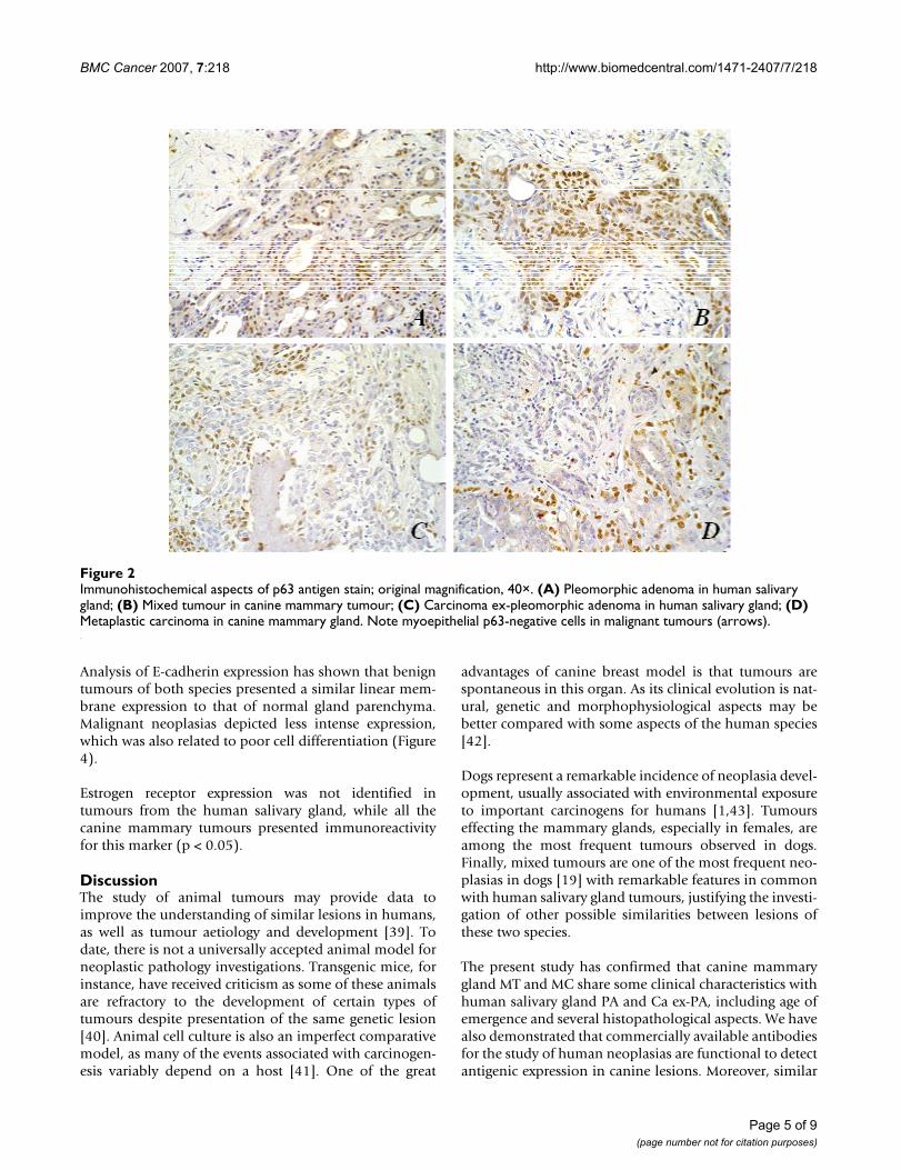
BMC Cancer 2007, 7:218 http://www.biomedcentral.com/1471-2407/7/218
Analysis of E-cadherin expression has shown that benigntumours of both species presented a similar linear mem-brane expression to that of normal gland parenchyma.Malignant neoplasias depicted less intense expression,which was also related to poor cell differentiation (Figure4).
Estrogen receptor expression was not identified intumours from the human salivary gland, while all thecanine mammary tumours presented immunoreactivityfor this marker (p < 0.05).
DiscussionThe study of animal tumours may provide data toimprove the understanding of similar lesions in humans,as well as tumour aetiology and development [39]. Todate, there is not a universally accepted animal model forneoplastic pathology investigations. Transgenic mice, forinstance, have received criticism as some of these animalsare refractory to the development of certain types oftumours despite presentation of the same genetic lesion[40]. Animal cell culture is also an imperfect comparativemodel, as many of the events associated with carcinogen-esis variably depend on a host [41]. One of the great
advantages of canine breast model is that tumours arespontaneous in this organ. As its clinical evolution is nat-ural, genetic and morphophysiological aspects may bebetter compared with some aspects of the human species[42].
Dogs represent a remarkable incidence of neoplasia devel-opment, usually associated with environmental exposureto important carcinogens for humans [1,43]. Tumourseffecting the mammary glands, especially in females, areamong the most frequent tumours observed in dogs.Finally, mixed tumours are one of the most frequent neo-plasias in dogs [19] with remarkable features in commonwith human salivary gland tumours, justifying the investi-gation of other possible similarities between lesions ofthese two species.
The present study has confirmed that canine mammarygland MT and MC share some clinical characteristics withhuman salivary gland PA and Ca ex-PA, including age ofemergence and several histopathological aspects. We havealso demonstrated that commercially available antibodiesfor the study of human neoplasias are functional to detectantigenic expression in canine lesions. Moreover, similar
Immunohistochemical aspects of p63 antigen stain; original magnification, 40×Figure 2Immunohistochemical aspects of p63 antigen stain; original magnification, 40×. (A) Pleomorphic adenoma in human salivary gland; (B) Mixed tumour in canine mammary tumour; (C) Carcinoma ex-pleomorphic adenoma in human salivary gland; (D) Metaplastic carcinoma in canine mammary gland. Note myoepithelial p63-negative cells in malignant tumours (arrows).
Page 5 of 9(page number not for citation purposes)
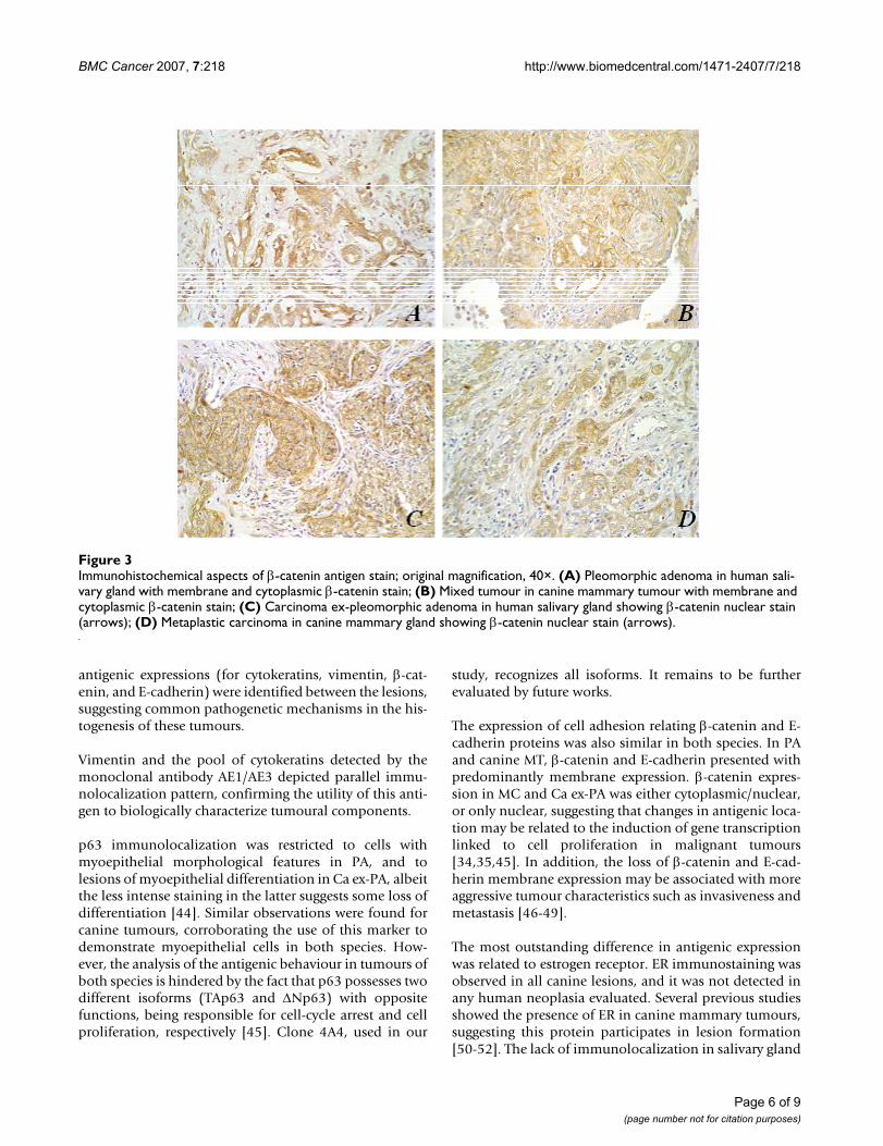
BMC Cancer 2007, 7:218 http://www.biomedcentral.com/1471-2407/7/218
antigenic expressions (for cytokeratins, vimentin, β-cat-enin, and E-cadherin) were identified between the lesions,suggesting common pathogenetic mechanisms in the his-togenesis of these tumours.
Vimentin and the pool of cytokeratins detected by themonoclonal antibody AE1/AE3 depicted parallel immu-nolocalization pattern, confirming the utility of this anti-gen to biologically characterize tumoural components.
p63 immunolocalization was restricted to cells withmyoepithelial morphological features in PA, and tolesions of myoepithelial differentiation in Ca ex-PA, albeitthe less intense staining in the latter suggests some loss ofdifferentiation [44]. Similar observations were found forcanine tumours, corroborating the use of this marker todemonstrate myoepithelial cells in both species. How-ever, the analysis of the antigenic behaviour in tumours ofboth species is hindered by the fact that p63 possesses twodifferent isoforms (TAp63 and ΔNp63) with oppositefunctions, being responsible for cell-cycle arrest and cellproliferation, respectively [45]. Clone 4A4, used in our
study, recognizes all isoforms. It remains to be furtherevaluated by future works.
The expression of cell adhesion relating β-catenin and E-cadherin proteins was also similar in both species. In PAand canine MT, β-catenin and E-cadherin presented withpredominantly membrane expression. β-catenin expres-sion in MC and Ca ex-PA was either cytoplasmic/nuclear,or only nuclear, suggesting that changes in antigenic loca-tion may be related to the induction of gene transcriptionlinked to cell proliferation in malignant tumours[34,35,45]. In addition, the loss of β-catenin and E-cad-herin membrane expression may be associated with moreaggressive tumour characteristics such as invasiveness andmetastasis [46-49].
The most outstanding difference in antigenic expressionwas related to estrogen receptor. ER immunostaining wasobserved in all canine lesions, and it was not detected inany human neoplasia evaluated. Several previous studiesshowed the presence of ER in canine mammary tumours,suggesting this protein participates in lesion formation[50-52]. The lack of immunolocalization in salivary gland
Immunohistochemical aspects of β-catenin antigen stain; original magnification, 40×Figure 3Immunohistochemical aspects of β-catenin antigen stain; original magnification, 40×. (A) Pleomorphic adenoma in human sali-vary gland with membrane and cytoplasmic β-catenin stain; (B) Mixed tumour in canine mammary tumour with membrane and cytoplasmic β-catenin stain; (C) Carcinoma ex-pleomorphic adenoma in human salivary gland showing β-catenin nuclear stain (arrows); (D) Metaplastic carcinoma in canine mammary gland showing β-catenin nuclear stain (arrows).
Page 6 of 9(page number not for citation purposes)
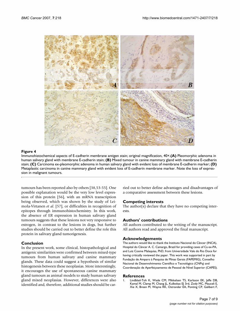
BMC Cancer 2007, 7:218 http://www.biomedcentral.com/1471-2407/7/218
tumours has been reported also by others [38,53-55]. Onepossible explanation would be the very low level expres-sion of this protein [56], with an mRNA transcriptionbeing observed, which was shown by the study of Lei-mola-Virtanen et al. [57], or difficulties in recognition ofepitopes through immunohistochemistry. In this work,the absence of ER expression in human salivary glandtumours suggests that these lesions not very responsive toestrogen, in contrast to the lesions in dogs, but furtherstudies should be carried out to better define the role thisprotein in salivary gland tumorigenesis.
ConclusionIn the present work, some clinical, histopathological andantigenic similarities were confirmed between mixed-typetumours from human salivary and canine mammaryglands. These data could suggest a hypothesis of similarhistogenesis between these neoplasias. More interestingly,it encourages the use of spontaneous canine mammarygland tumours as animal models to study human salivarygland mixed neoplasias. However, differences were alsoidentified and, therefore, additional studies should be car-
ried out to better define advantages and disadvantages ofa comparative assessment between these lesions.
Competing interestsThe author(s) declare that they have no competing inter-ests.
Authors' contributionsAll authors contributed to the writing of the manuscript.All authors read and approved the final manuscript.
AcknowledgementsThe authors would like to thank the Instituto Nacional do Câncer (INCA), Hospital do Câncer A. C. Camargo, Brazil for providing cases of Ca ex-PA, and Luiz Cosme Malaquias, PhD, from Universidade Vale do Rio Doce for having critically reviewed the paper. This work was supported in part by Fundação de Amparo à Pesquisa de Minas Gerais (FAPEMIG), Conselho Nacional de Desenvolvimento Científico e Tecnológico (CNPq) and Coordenação de Aperfeiçoamento de Pessoal de Nível Superior (CAPES).
References1. Lindblad-Toh K, Wade CM, Mikkelsen TS, Karlsson EK, Jaffe DB,
Kamal M, Clamp M, Chang JL, Kulbokas EJ 3rd, Zody MC, Mauceli E,Xie X, Breen M, Wayne RK, Ostrander EA, Ponting CP, Galibert F,
Immunohistochemical aspects of E-cadherin membrane antigen stain; original magnification, 40×Figure 4Immunohistochemical aspects of E-cadherin membrane antigen stain; original magnification, 40×.(A) Pleomorphic adenoma in human salivary gland with membrane E-cadherin stain; (B) Mixed tumour in canine mammary gland with membrane E-cadherin stain; (C) Carcinoma ex-pleomorphic adenoma in human salivary gland with evident loss of membrane E-cadherin marker; (D) Metaplastic carcinoma in canine mammary gland with evident loss of E-cadherin membrane marker. Note the loss of expres-sion in malignant tumours.
Page 7 of 9(page number not for citation purposes)
BMC Cancer 2007, 7:218 http://www.biomedcentral.com/1471-2407/7/218
Smith DR, DeJong PJ, Kirkness E, Alvarez P, Biagi T, Brockman W,Butler J, Chin CW, Cook A, Cuff J, Daly MJ, DeCaprio D, Gnerre S,Grabherr M, Kellis M, Kleber M, Bardeleben C, Goodstadt L, HegerA, Hitte C, Kim L, Koepfli KP, Parker HG, Pollinger JP, Searle SM, Sut-ter NB, Thomas R, Webber C, Baldwin J, Abebe A, Abouelleil A,Aftuck L, Ait-Zahra M, Aldredge T, Allen N, An P, Anderson S, Anto-ine C, Arachchi H, Aslam A, Ayotte L, Bachantsang P, Barry A, BayulT, Benamara M, Berlin A, Bessette D, Blitshteyn B, Bloom T, Blye J,Boguslavskiy L, Bonnet C, Boukhgalter B, Brown A, Cahill P, CalixteN, Camarata J, Cheshatsang Y, Chu J, Citroen M, Collymore A,Cooke P, Dawoe T, Daza R, Decktor K, DeGray S, Dhargay N, Doo-ley K, Dooley K, Dorje P, Dorjee K, Dorris L, Duffey N, Dupes A,Egbiremolen O, Elong R, Falk J, Farina A, Faro S, Ferguson D, FerreiraP, Fisher S, FitzGerald M, Foley K, Foley C, Franke A, Friedrich D,Gage D, Garber M, Gearin G, Giannoukos G, Goode T, Goyette A,Graham J, Grandbois E, Gyaltsen K, Hafez N, Hagopian D, Hagos B,Hall J, Healy C, Hegarty R, Honan T, Horn A, Houde N, Hughes L,Hunnicutt L, Husby M, Jester B, Jones C, Kamat A, Kanga B, Kells C,Khazanovich D, Kieu AC, Kisner P, Kumar M, Lance K, Landers T,Lara M, Lee W, Leger JP, Lennon N, Leuper L, LeVine S, Liu J, Liu X,Lokyitsang Y, Lokyitsang T, Lui A, Macdonald J, Major J, Marabella R,Maru K, Matthews C, McDonough S, Mehta T, Meldrim J, Melnikov A,Meneus L, Mihalev A, Mihova T, Miller K, Mittelman R, Mlenga V, Mul-rain L, Munson G, Navidi A, Naylor J, Nguyen T, Nguyen N, NguyenC, Nguyen T, Nicol R, Norbu N, Norbu C, Novod N, Nyima T,Olandt P, O'Neill B, O'Neill K, Osman S, Oyono L, Patti C, Perrin D,Phunkhang P, Pierre F, Priest M, Rachupka A, Raghuraman S, RameauR, Ray V, Raymond C, Rege F, Rise C, Rogers J, Rogov P, Sahalie J, Set-tipalli S, Sharpe T, Shea T, Sheehan M, Sherpa N, Shi J, Shih D, SloanJ, Smith C, Sparrow T, Stalker J, Stange-Thomann N, Stavropoulos S,Stone C, Stone S, Sykes S, Tchuinga P, Tenzing P, Tesfaye S, Thoulut-sang D, Thoulutsang Y, Topham K, Topping I, Tsamla T, Vassiliev H,Venkataraman V, Vo A, Wangchuk T, Wangdi T, Weiand M, Wilkin-son J, Wilson A, Yadav S, Yang S, Yang X, Young G, Yu Q, Zainoun J,Zembek L, Zimmer A, Lander ES: Genome sequence, compara-tive analysis and haplotype structure of the domestic dog.Nature 2005, 438(7609):803-819.
2. Mottolese M, Morelli L, Agrimi U, Benevolo M, Sciarretta F, Anto-nucci G, Natali PG: Spontaneous canine mammary tumors. Amodel for monoclonal antibody. The unique associationbetween salivary gland cancer and breast cancer diagnosisand treatment of human breast cancer. Lab Invest 1994,71(2):182-7.
3. Strandberg JD, Goodman DG: Animal model of human disease:canine mammary neoplasia. Am J Pathol 1974, 75(1):225-8.
4. Schneider R: Comparison of age, sex, and incidence rates inhuman and canine breast cancer. Cancer 1970, 26(2):419-26.
5. Owen LN: A comparative study of canine and human breastcancer. Invest Cell Pathol 1979, 2(4):257-75.
6. Berg JW, Hutter RV, Foote FW Jr: The unique associationbetween salivary gland cancer and breast cancer. JAMA204(9):771-4. 1968 May 27
7. Dunn JE Jr, Bragg KU, Sautter C, Gardipee C: Breast cancer riskfollowing a major salivary gland carcinoma. Cancer 1972,29(5):1343-6.
8. Prior P, Waterhouse JA: Second primary cancers in patientswith tumours of the salivary glands. Br J Cancer 1977,36(3):362-8.
9. Abbey LM, Schwab BH, Landau GC, Perkins ER: Incidence of sec-ond primary breast cancer among patients with a first pri-mary salivary gland tumor. Cancer 54(7):1439-42. 1984 Oct 1
10. Freedman PD, Lumerman H: Lobular carcinoma of intraoralminor salivary gland origin. Report of twelve cases. Oral SurgOral Med Oral Pathol 1983, 56(2):157-66.
11. Hirokawa M, Sugihara K, Sai T, Monobe Y, Kudo H, Sano N, Sano T:Secretory carcinoma of the breast: a tumour analogous tosalivary gland acinic cell carcinoma? Histopathology 2002,40(3):223-9.
12. Seifert G: Are adenomyoepithelioma of the breast and epithe-lial-myoepithelial carcinoma of the salivary glands identicaltumours? Virchows Arch 1998, 433(3):285-8.
13. Skalova A, Starek I, Kucerova V, Szepe P, Plank L: Salivary duct car-cinoma: a highly aggressive salivary gland tumor with HER-2/neu oncoprotein overexpression. Pathol Res Pract 2001,197(9):621-6.
14. Wick MR, Ockner DM, Mills SE, Ritter JH, Swanson PE: Homolo-gous carcinomas of the breasts, skin, and salivary glands. Ahistologic and immunohistochemical comparison of ductalmammary carcinoma, ductal sweat gland carcinoma, andsalivary duct carcinoma. Am J Clin Pathol 1998, 109(1):75-84.
15. Foschini MP, Reis-Filho JS, Eusebi V, Lakhani SR: Salivary gland-liketumours of the breast: surgical and molecular pathology. JClin Pathol 2003, 56(7):497-506. Erratum in: J Clin Pathol 2003Oct,56(10):804.
16. Hayes MM, Lesack D, Girardet C, Del Vecchio M, Eusebi V: Carci-noma ex-pleomorphic adenoma of the breast. Report ofthree cases suggesting a relationship to metaplastic carci-noma of matrix-producing type. Virchows Arch 2005,446(2):142-9.
17. Kumar PV, Sobhani SA, Monabati A, Talei AR, Shirazi B: Cytologicfindings of a pleomorphic adenoma of the breast: a casereport. Acta Cytol 2004, 48(6):849-52.
18. Ellis GL, Auclair PL: Tumors of the salivary glands. In Atlas oftumor pathology. 3rd Series, Fascicle 17 Volume 39–57. Washington, DC:Armed Forces Institute of Pathology; 1996:228-251.
19. Cohen D, Reif JS, Brodey RS, Keiser H: Epidemiological analysisof the most prevalent sites and types of canine neoplasiaobserved in a veterinary hospital. Cancer Res 1974,34(11):2859-68.
20. Misdorp W, Else RW, Hellmen E, Limpiscomb TP: Histological clas-sification of the mammary tumors of the dog and the cat. InInternational Histological Classification of Tumors of domestic Animals. 2ndSeries Volume 2. Geneva: World Health Organization; 1999.
21. Auclair PL, Ellis GL: Atypical features in salivary gland mixedtumors: their relationship to malignant transformation.Mod Pathol 1996, 9(6):652-7.
22. Li Volsi VA, Perzin KH: Malignant mixed tumors arising in sali-vary glands. I. Carcinomas arising in benign mixed tumors: aclinicopathologic study. Cancer 1977, 39(5):2209-30.
23. Spiro RH, Huvos AG, Strong EW: Malignant mixed tumor of sal-ivary origin: a clinicopathologic study of 146 cases. Cancer1977, 39(2):388-96.
24. Gnepp DR: Malignant mixed tumors of the salivary glands: areview. Pathol Annu 1993, 28 Pt 1:279-328.
25. Lewis JE, Olsen KD, Sebo TJ: Carcinoma ex pleomorphic ade-noma: pathologic analysis of 73 cases. Hum Pathol 2001,32(6):596-604.
26. Seifert G, Sobin LH: Histological typing of salivary glandtumors. In The World Health Organization. International Classificationof Tumors 2nd edition. New York: Spring-Verlag; 1991.
27. de Araujo VC, de Souza SO: Expression of different keratins insalivary gland tumours. Eur J Cancer B Oral Oncol 1996,32B(1):14-8.
28. Reis-Filho JS, Simpson PT, Martins A, Preto A, Gartner F, Schmitt FC:Distribution of p63, cytokeratins 5/6 and cytokeratin 14 in 51normal and 400 neoplastic human tissue samples usingTARP-4 multi-tumor tissue microarray. Virchows Arch 2003,443(2):122-32.
29. Loyola AM, de Souza SO, Araujo NS, Araujo VS: Study of minorsalivary gland mucoepidermoid carcinoma differentiationbased on immunohistochemical expression of cytokeratins,vimentin and muscle-specific actin. Oral Oncol 1998,34(2):112-8.
30. Barbareschi M, Pecciarini L, Cangi MG, Macri E, Rizzo A, Viale G, Dog-lione C: p63, a p53 homologue, is a selective nuclear markerof myoepithelial cells of the human breast. Am J Surg Pathol2001, 25(8):1054-60.
31. Bilal H, Handra-Luca A, Bertrand JC, Fouret PJ: p63 is expressed inbasal and myoepithelial cells of human normal and tumorsalivary gland tissues. J Histochem Cytochem 2003, 51(2):133-9.
32. Shieh YS, Chang LC, Chiu KC, Wu CW, Lee HS: Cadherin and cat-enin expression in mucoepidermoid carcinoma: correlationwith histopathologic grade, clinical stage, and patient out-come. J Oral Pathol Med 2003, 32(5):297-304.
33. Daa T, Kaku N, Kashima K, Nakayama I, Yokoyama S: Expression ofbeta-catenin, E-cadherin and cyclin D1 in adenoid cystic car-cinoma of the salivary gland. J Exp Clin Cancer Res 2005,24(1):83-7.
34. Jankowski JA, Bruton R, Shepherd N, Sanders DS: Cadherin andcatenin biology represent a global mechanism for epithelialcancer progression. Mol Pathol 1997, 50(6):289-90.
Page 8 of 9(page number not for citation purposes)
BMC Cancer 2007, 7:218 http://www.biomedcentral.com/1471-2407/7/218
Publish with BioMed Central and every scientist can read your work free of charge
"BioMed Central will be the most significant development for disseminating the results of biomedical research in our lifetime."
Sir Paul Nurse, Cancer Research UK
Your research papers will be:
available free of charge to the entire biomedical community
peer reviewed and published immediately upon acceptance
cited in PubMed and archived on PubMed Central
yours — you keep the copyright
Submit your manuscript here:http://www.biomedcentral.com/info/publishing_adv.asp
BioMedcentral
35. Nollet F, Berx G, Van Roy F: The role of the E-cadherin/cateninadhesion complex in the development and progression ofcancer. Mol Cell Biol Res Commun 1999, 2(2):77-85.
36. Anderson E: The role of oestrogen and progesterone recep-tors in human mammary development and tumorigenesis.Breast Cancer Res 2002, 4(5):197-201.
37. Reis-Filho JS, Torio B, Albergaria A, Schmitt F: p63 expression innormal skin and usual cutaneous carcinomas. J Cutan Pathol2002, 29(9):517-23.
38. Shick PC, Riordan GP, Foss RD: Estrogen and progesteronereceptors in salivary gland adenoid cystic carcinoma. OralSurg Oral Med Oral Pathol Oral Radiol Endod 1995, 80(4):440-4.
39. Porrelo A, Cardelli P, Spugnini EP: Pet models in cancer research:general principles. J Exp Clin Cancer Res 2004, 23(2):181-93.
40. McLeod KF, Jacks T: Insights into cancer from transgenicmouse models. J Pathol 1999, 187(1):43-60.
41. Rivera EM: Mammary gland culture. In Methods in MammalianEmbryology Edited by: Daniel JC Jr. San Francisco: WH Freeman Com-pany; 1971.
42. Vail DM, MacEwen EG: Spontaneously occurring tumors ofcompanion animals as models for human cancer. Cancer Invest2000, 18(8):781-92.
43. Schneider R, Dorn CR, Taylor DO: Factors influencing caninemammary cancer development and postsurgical survival. JNatl Cancer Inst 1969, 43(6):1249-61.
44. Genelhu MC, Gobbi H, Soares FA, Campos AH, Ribeiro CA, CassaliGD: Immunohistochemical expression of p63 in pleomorphicadenomas and carcinomas ex-pleomorphic adenomas of sal-ivary glands. Oral Oncol 2006, 42(2):154-60.
45. Yang A, Kaghad M, Wang Y, Gillett E, Fleming MD, Dötsch V,Andrews NC, Caput D, McKeon F: p63, a p53 homolog at3q27–29, encodes multiple products with transactivating,death-inducing, and dominant-negative activities. MolecularCell 1998, 2(3):305-16.
46. Shih HC, Shiozawa T, Miyamoto T, Kashima H, Feng YZ, Kurai M,Konishi I: Immunohistochemical expression of E-cadherin andbeta-catenin in the normal and malignant humanendometrium: an inverse correlation between E-cadherinand nuclear beta-catenin expression. Anticancer Res 2004,24(6):3843-50.
47. Harrington KJ, Syrigos KN: The role of E-cadherin-catenin com-plex: more than intercellular glue? Ann Surg Oncol 2000,7(10):783-8. Erratum in: Ann Surg Oncol 2001 Mar, 8(2):186.
48. Shiozaki H, Oka H, Inoue M, Tamura S, Monden M: E-cadherinmediated adhesion system in cancer cells. Cancer 77(8Suppl):1605-13. 1996 Apr 15
49. Genelhu MC, Gobbi H, Arantes DC, Cardoso SV, Cassali GD:Immunolocalization of beta-Catenin in Pleomorphic Adeno-mas and Carcinomas Ex-pleomorphic Adenomas of SalivaryGlands. Appl Immunohistochem Mol Morphol 2007, 15(3):273-278.
50. Hamilton JM, Else RW, Forshaw P: Oestrogen receptors in caninemammary tumours. Vet Rec 101(13):258-60. 1977 Sep 24
51. Graham JC, O'Keefe DA, Gelberg HB: Immunohistochemicalassay for detecting estrogen receptors in canine mammarytumors. Am J Vet Res 1999, 60(5):627-30.
52. Nieto A, Peña L, Perez-Alenza MD, Sanchez MA, Flores JM, CastanoM: Immunohistologic detection of estrogen receptor alpha incanine mammary tumors: clinical and pathologic associa-tions and prognostic significance. Vet Pathol 2000, 37(3):239-47.
53. Miller AS, Hartman GG, Sow-Yeh C, Edmonds PR, Brightman SA,Harwick RD: Estrogen receptor assay in polymorphous low-grade adenocarcinoma and adenoid cystic carcinoma of sali-vary gland origin. An immunohistochemical study. Oral SurgOral Med Oral Pathol 1994, 77(1):36-40.
54. Dori S, Trougouboff P, David R, Buchner A: Immunohistochemi-cal evaluation of estrogen and progesterone receptors inadenoid cystic carcinoma of salivary gland origin. Oral Oncol2000, 36(5):450-3.
55. Jeannon JP, Soames JV, Bell H, Wilson JA: Immunohistochemicaldetection of oestrogen and progesterone receptors in sali-vary tumours. Clin Otolaryngol Allied Sci 1999, 24(1):52-4.
56. Gaffney EV, Pinkstone JA, Eidson JJ: Estrogen receptors in parotidtumors. Endocr Res 1995, 21(3):635-43.
57. Leimola-Virtanen R, Salo T, Toikkanen S, Pulkkinen J, Syrjänen S:Expression of estrogen receptor (ER) in oral mucosa and sal-ivary glands. Maturitas 2000, 36(2):131-7.
Pre-publication historyThe pre-publication history for this paper can be accessedhere:
http://www.biomedcentral.com/1471-2407/7/218/prepub
Page 9 of 9(page number not for citation purposes)

84
CONCLUSÃO
• A análise histológica dos tumores mamários caninos e dos tumores de glândula
salivar humana confirmou a notável semelhança morfológica entre essas neoplasias.
• As semelhanças histomorfológicas foram corroboradas pela caracterização imuno-
histoquímica dos componentes tumorais. A transformação neoplásica do epitélio
está associada com trocas antigênicas similares, o que sugere que, em ambos os
sítios anatômicos (glândulas mamárias e glândulas salivares) e em ambas as
espécies (canina e humana), a tumorigênese possa ser caracterizada por uma via
patogenética comum.
• As disparidades encontradas na expressão dos antígenos p63 e receptor de estrógeno
devem ser melhor investigadas, a fim de esclarecer a influência desses marcadores
no comportamento biológico das neoplasias estudadas.
• Finalmente, os resultados deste trabalho sugerem que os tumores mistos da glândula
mamária canina podem ser utilizados, em alguns aspectos, como modelo
comparativo para o estudo dos tumores mistos da glândula salivar da espécie
humana.
85
CONSIDERAÇÕES FINAIS
Este trabalho pretendeu contribuir para a caracterização de alguns aspectos
moleculares (antigênicos) e demográficos das neoplasias mamárias caninas genericamente
chamadas de tumores mistos, com a finalidade de averiguar o seu valor como modelo
comparativo para o estudo dos tumores mistos das glândulas salivares humanas.
Assim, verificou-se que os marcadores correntemente utilizados para distinguir os
diferentes componentes celulares dos adenomas pleomórficos e carcinomas ex-adenomas
pleomórficos de glândula salivar humana (AE1/AE3 e vimentina) integram as mesmas
estruturas na glândula mamária canina, permitindo reforçar a noção de uma mesma origem
celular monoclonal para os dois grupos de neoplasias estudadas.
Entre os marcadores utilizados não foi possível identificar um que distinguisse
lesões benignas e malignas, mas a utilização dos anticorpos p63, RE, E-caderina e β-
catenina resultou na identificação de semelhanças (E-caderina e β-catenina) e diferenças
(p63 e RE) de marcação entre os tumores mamários caninos e os tumores de glândula
salivar da espécie humana.
Nos casos de E-caderina e β-catenina, os resultados permitiram inferir estreita
relação de localização nuclear com malignidade e “pré-malignidade”, tanto nos tumores
mamários caninos quanto nos de glândula salivar humana. Por outro lado, p63 permitiu a
distinção entre tumores bem diferenciados e pouco diferenciados/indiferenciados somente
nos tumores malignos (Ca ex-AP) de glândula salivar humana.
Apesar de desempenhar um importante papel na carcinogênese mamária canina, o
que pode dar suporte ao uso da terapia hormonal em medicina veterinária, RE parece não
86
ter relevância biológica para os tumores de glândula salivar humana. É importante ressaltar
que devido à transcrição de RNAm, outros estudos com RE em tumores de glândula salivar
humana precisam ser realizados até que se chegue a resultados conclusivos.
Tomados em conjunto, os dados demográficos, clínicos e imuno-histoquímicos
deste estudo sugerem uma possível utilização dos tumores mistos das glândulas mamárias
caninas como modelo de estudo comparativo para os tumores mistos das glândulas
salivares da espécie humana.

87
REFERÊNCIAS BIBLIOGRÁFICAS
1. Abbey LM, Schwab BH, Landau GC, Perkins ER. Incidence of second primary
breast cancer among patients with a first primary salivary gland tumor. Cancer
1984; 54(7): 1439-42.
2. Aberle AM, Abrams AM, Bowe R, Melrose RJ, Handlers JP. Lobular
(polymorphous low-grade) carcinoma of minor salivary glands: a clinicopathologic
study of twenty cases. Oral Surg Oral Med Oral Pathol. 1985; 60: 387-95.
3. Alves VAF, Bacchi CE, Vassalo J. Manual de Imuno-histoquímica. São Paulo:
Sociedade Brasileira de Patologia, 1999.
4. Araújo VC, Raitz R. Estudo do estado de diferenciação da célula mioepitelial nas
neoplasias de glândula salivar. Acta Scientiarum. 2004; 26(2): 345-50.
5. Araújo VC, Souza SOM, Carvalho YR, Araújo NS. Application of
immunohistochemistry to the diagnosis of salivary gland tumors. Appl
Immunohistoch Mol Morph. 2000; 8(2): 196-202.
6. Barbareschi M, Pecciarini L, Cangi MG, Macrì E, Rizzo A, Viale G, Doglione C.
p63, a p53 homologue, is a selective nuclear marker of myoepithelial cells of the
human breast. Am J Surg Pathol. 2001; 25(8): 1054-60.
7. Batzakis JG, Kraemer B, Sciubba JJ. The pathology of head and neck tumors: the
myoepithelial cell and its participation in salivary gland neoplasia, Part 17.
Head Neck Surg. 1983; 5(3): 222-33.
88
8. Berg JW, Hutter RV, Foote FW. The unique association between salivary gland
cancer and breast cancer. JAMA 1968; 204: 771-4.
9. Bilal H, Handra-Luca A, Bertrand JC, Fouret PJ. p63 is expressed in basal and
myoepithelial cells of human normal and tumor salivary gland tissues. J Histochem
Cytochem. 2003; 51:133-9.
10. Björling DE, Beckman M, Clayton MK, Wang ZY. Modulation of nerve growth
factor in peripheral organs by estrogen and progesterone. Neuroscience. 2002;
110(1): 155-67.
11. Brasileiro-Filho G, Barbosa AJA, Miranda D. Métodos de estudo em patologia. In:
Brasileiro-Filho G. Bogliolo – Patologia. 6ed. Rio de Janeiro: Guanabara Koogan,
2004. p.6-18.
12. Cassali GD, Gobbi H, Gärtner F, Schmitt F. Secretory carcinoma of the canine
mammary gland. Vet Pathol. 1999b, 36:601-03.
13. Cassali GD. Canine mammary tumours. A morphological and
immunohistochemistry study: comparative aspects with human breast tumours.
Virch Arch. 1999a; 435(3): 622.
14. Cassali GD. Estudo morfológico, imuno-histoquímico e citométrico de tumores
mamários da cadela. Aspectos comparativos com neoplasias da mama humana.
Escola de Veterinária da Universidade Federal de Minas Gerais. 2000, 73p. Tese
(Doutorado em Ciência Animal).
15. Cassali GD. Patologias da glândula mamária In: Nascimento EF, Lima Santos R.
Patologia da Reprodução dos Animais Domésticos. 2ed. Rio de Janeiro: Guanabara
Koogan; 2002. p.117-133.
89
16. Chen KTK. Pleomorphic adenoma of the breast. Am J Clin Pathol. 1990; 93: 792-4.
17. Cho KJ, Lee YS. Proliferating cell nuclear antigen and c-erbB-2 oncoprotein
expression in adenoid cystic carcinomas of the salivary glands. Head Neck. 1999;
21(5): 414-9.
18. Clark JW, Sell L, Shiu RP, Orr FW, Maitre N, Vary CP, Cole DJ, Watson PH. The
potential role for prolactin-inducible protein (PIP) as a marker of human breast
cancer micrometastasis. Br J Cancer. 1999; 81(6): 1002-8.
19. Cohen D, Reif J, Brooy RS; Keiser H. Epidemiological analysis of the most
prevalent sites and types of canine neoplasia observed in a veterinary hospital.
Cancer Res. 1974; 34:2859-68.
20. Damiani S, Pasquinelli G, Lamovec J, Peterse JL, Eusebi V. Acinic cell carcinoma
of the breast: an immunohistochemical and ultrastructural study. Virch Arch. 2000;
43(1): 74-81.
21. Dardick I, Van Nostrand AW, Phillips MJ. Histogenesis of salivary gland
pleomorphic adenoma (mixed tumor) with an evaluation of the role of the
myoepithelial cell. Hum Pathol. 1982; 13(1): 62-75.
22. Dimery IW, Jones LA, Verjan RP, Raymond AK, Goepfert H, Hong WK. Estrogen
receptors in normal salivary gland and salivary gland carcinoma. Arch Otolaryngol
Head Neck Surg. 1987; 113(10):1082-5.
23. Dori S, Trougouboff P, David R, Buchner A. Immunohistochemical evaluation of
estrogen and progesterone receptors in adenoid cystic carcinoma of salivary gland
origin. Oral Oncol. 2000; 36(5):450-53.
90
24. Dunn J, Bragg K, Sautter C Gardipee C. Breast Cancer risk following a major
salivary gland carcinoma. Cancer 1972; 29:1343-6.
25. Duprey P, Paulin D. What can be learned from intermediate filament gene
regulation in the mouse embryo. Review. Int J Dev Biol. 1995; 39(3): 443-57.
26. Economopoulou P, Hamby A, Odell EW. Expression of E-cadherin, cellular
differentiation and polarity in epithelial salivary neoplasms. Oral Oncol. 2000;
36(6): 15-8.
27. Edwards PC, Bhuiya T, Kelsch RD. Assessment of p63 expression in the salivary
gland neoplasms adenoid cystic carcinoma, polymorphous low-grade
adenocarcinoma, and basal cell and canalicular adenomas. Oral Surg Oral Med Oral
Pathol Oral Radiol Endod. 2004; 97(5):613-9.
28. Elling H, Ungemach FR. Simultaneous occurrence of receptors for estradiol,
progesterone and dihidrotestosterone in canine mammary tumors. J. Cancer Res.
Clin. Oncol. 1983;105:231-37.
29. Evans CR, Pierrepoint CG. Tissue-steroid interactions in canine hormone-
dependent tumors. Vet. Rec. 1975; 13:464-467.
30. Fernandes MMG. Estudo da patologia dos tumores mamários caninos através de
técnicas de imuno-histoquímica e citometria estática. Faculdade de Medicina da
Universidade do Porto, 1996, 93p. Dissertação (Mestrado em Oncobiologia).
31. Flores Alés AJ. Factores epidemiológicos de interés pronóstico en los tumores de
mama en la perra. 1997; 14(5): 273-80.
91
32. Foschini MP, Scarpellini F, Gown AM, Eusebi V. Differential Expression of
Myoepithelial Markers in Salivary, Sweat and Mammary Glands. Int J Surg Pathol.
2000; 8(1): 29-37.
33. Freedman PD, Lumerman H. Lobular carcinoma of intraoral minor salivary gland
origin. Report of twelve cases. Oral Surg Oral Med Oral Pathol. 1983; 56(2): 157-
66.
34. Furuse c, Souza SO, Nunes FD, Magalhães MH, Araújo VC. Myoepithelial cells in
salivary gland neoplasms. Int J Surg Pathol. 2005: 13(1): 57-65.
35. Gaffney EV, Pinkstone JA, Eidson JJ. Estrogen receptors in parotid tumors. Endocr
Res. 1995; 21(3): 635-43.
36. Gama A, Alves A, Gärtner F, Schmitt F. p63: a novel myoepithelial cell marker in
canine mammary tissues. Vet Pathol. 2003; 40(4): 412-20.
37. Gärtner F, Geraldes M, Cassali G, Rema A, Schmitt F. DNA measurement and
immunohistochemical characterization of epithelial and mesenchymal cells in
canine mixed mammary tumours: putative evidence for a common histogenesis. Vet
J. 1999; 158(1): 39-47.
38. Geraldes M, Gärtner F, Schmitt F. An immunohistochemistry study of hormonal
receptors and cell proliferation in normal canine mammary glands and spontaneous
mammary tumors. Vet Rec. 2000; 146(14): 403-06.
39. Glas AS, Hollema H, Nap RE, Plukker JT. Expression of estrogen receptor,
progesterone receptor, and insulin-like growth factor receptor-1 and MIB-1 in
patients with recurrent pleomorphic adenoma of the parotid gland. Cancer 2002;
94(8): 2211-6.

92
40. Gnepp DR. Malignant mixed tumors of the salivary glands: a review.
Pathol Annu. 1993; 28 Pt 1:279-328.
41. Gobbi H, Dupont WD, Parl FF, Schuyler PA, Plummer WD, Olson SJ, Page DL.
Breast cancer risk associated with estrogen receptor expression in epithelial
hyperplasia lacking atypia and adjacent lobular units. Int J Cancer. 2005;
113(5):857-9.
42. Gomes LR. Estudo imuno-histoquímico da expressão de E-caderina e P-caderina
em tumores mamários caninos. Instituto de Ciências Biológicas da Universidade
Federal de Minas Gerais, 2004, 95p. Dissertação (Mestrado em Patologia).
43. Graham JC, O’Keefe DA, Gelberg HB. Immunohistochemical assay for detecting
estrogen receptors in canine mammary tumors. Am J Vet Res. 1999; 60(5): 627-30.
44. Hamilton JM, Else RW, Forshaw P. Oestrogen receptors in canine mammary
tumors. Vet Rec. 1977; 101: 258-60.
45. Hampe JF, Misdorp W. Tumours and dysplasias of the mammary gland. Bull World
Health Organ. 1974; 50(1-2): 111-33.
46. Harrington KJ, Syrigos KN. The role of E-cadherin-catenin complex: more than an
intercellular glue? Ann Surg Oncol. 2000; 7(10):783-8.
47. Hayes MM, Lesack D, Girardet C, Del Vecchio C, Eusebi V. Carcinoma ex-
pleomorphic adenoma of the breast. Report of three cases suggesting a relationship
to metaplastic carcinoma of matrix-producing type. Virch Arch. 2005; 446(2): 142-
9.
93
48. Hayward SW, Brody JR, Cunha GR. An edgewise look at basal epithelial cells;
three-dimensional views of the rat prostate, mammary gland and salivary gland.
Differentiation. 1996; 60: 219-27.
49. Hirokawa M, Sugihara K, Sai T, Monobe Y, Kudo H, Sano N, Sano T. Secretory
carcinoma of the breast: a tumor analogous to salivary gland acinic cell carcinoma?
Histopathology. 2002; 40(3):223-29.
50. Hoang MP, Callender DL, Sola Gallego JJ, Huang Z, Sneige N, Luna MA, Batsakis
JG, El-Naggar AK. Molecular and biomarker analyses of salivary duct carcinomas:
comparison with mammary duct carcinoma. Int. J. Oncol. 2001; 10(4): 865-871.
51. Jankowski JA, Bruton R, Shepherd N, Sanders DS. Cadherin and catenin biology
represent a global mechanism for epithelial cancer progression. Mol Pathol. 1997;
50(6):289-90.
52. Jeannon JP, Soames JV, Bell H, Wilson JA. Immunohistochemical detection of
oestrogen and progesterone receptors in salivary tumours. Clin Otolaryngol. 1999;
24(1):52-4.
53. Kalof AN, Tam D, Beatty B, Cooper K. Immunostaining of myoepithelial cells in
breast lesions: a comparison of CD10 and smooth muscle myosin heavy chain. J
Clin Pathol. 2004; 57:625-29.
54. Koga F, Kawakami S, Kumagai J Takizawa T, Ando N, Arai G, Kageyama Y,
Kihara K. Impaired DeltaNp63 expression associates with reduced beta-catenin and
aggressive phenotypes of urothelial neoplasms. Br J Cancer 2003; 88: 740-47.
55. Kumagami H, Onitsuka T. Estradiol and testosterone in minor salivary glands of
Sjögren’s syndrome. Auris Nasus Larynx. 1993; 20: 137-143.
94
56. Kumar V, Kingen MW. In: Kumar V, Abbas AK, Fausto N. Cabeça e Pescoço.
Robbins & Cotran Patologia – Patológicas das Doenças. 7ed. Rio de Janeiro:
57. Lakhani SR, O’Hare MJ. The mammary myoepithelial cell – Cinderella or ugly
sister? Breast Cancer Res. 2001; 3: 1-4.
58. Lamey PJ, Leake RE, Cowan SK, Soutar DS, McGregor JA, McGregor FM. Steroid
hormone receptors in human salivary gland tumours. J Clin Pathol. 1987; 40(5):
532-4.
59. Lebeau A. L’âge du chien et celui de l’homme essai de statistique sur la mortalité
canine. Bulletin de l’Académie Vétérinaire. 1953; 26:229-32.
60. Leimola-Virtanen R, Salo T, Toikkanen S, Pulkkinen J, Syrjänen S. Expression of
estrogen receptor (ER) in oral mucosa and salivary glands. Maturitas. 2000;
36(2):131-7.
61. Leong ASY, Wright J. The contribution of immunohistochemical staining in tumour
diagnosis. Histopathology 1987; 11(12):1295-305.
62. Little NA, Jochemsen AG. p63. Int J Biochem Cell Biol. 2002; 43:6-9.
63. MacEwen EG, Patnaik AK, Harvey HJ, Panko WB. Estrogen receptors in canine
mammary tumors. Cancer Res. 1982; 42(6): 2255-9.
64. Machado GF, Figueiredo F. Revisão: Filamentos intermediários. Medicina,
Ribeirão Preto; 1996; 29: 104-113.
65. Marchant J. Animal model for tumors of the female genital tract. In: Kurman RJ.
Blaustein’s pathology of female genital tract. 3ed. New York: Springer Verlag,
1987. p.899-924.
95
66. Margaritescu C, Florescu M, Raica M, Simionescu C, Mogoanta L, Preda E. The
immunohistochemical profile of luminal epithelial neoplastic component from
pleomorphic adenomas of salivary glands. Rom J Morphol Embryol. 1999-2004;
45: 97-118.
67. Martin PM, Cotard M, Mialot JP, Andre F, Raynaud JP. Animal model for
hormone-dependent human breast cancer. Relationship between steroid receptor
profiles in canine and feline mammary tumors and survival rate. Cancer Chemother
Pharmacol. 1984; 12(1): 13-17.
68. Mialot M, Lagadic M. Epidémiologie des tumours du chien et du chat. Rec Méd
Vét. 1990; 166(11): 937-47.
69. Miller AS, Hartman GG, Sow-Yeh C, Edmonds PR, Brightman SA, Harwick RD.
Estrogen receptor assay in polymorphous low-grade adenocarcinoma and adenoid
cystic carcinoma of salivary gland origin. An immunohistochemical study. Oral
Surg Oral Med Oral Pathol. 1994; 77:36-40.
70. Miller WR. Oestrogens and breast cancer: biological considerations. Br Med Bull.
1991; 47:470-83.
71. Miller WR. Pathways of hormone metabolism in normal and nonneoplastic breast
tissue. Ann N Y Acad Sci. 1990; 586:53-9.
72. Moran CA, Suster S, Carter D. Benign mixed tumors (pleomorphic adenomas of the
breast. Am J Surg Pathol. 1990; 14(10): 913-21.
73. Mottolese M, Sciarretta F, Antonucci G, Natali PG. Spontaneous canine mammary
tumors. A model for monoclonal antibody diagnosis and treatment of human breast
cancer. Lab Invest. 1994; 71(2):182-87.
96
74. Moulton JE, Rosemblatt LS, Goldman M. Mammary tumors in a colony of beagle
dogs. Vet. Pathol. 1996; 223:741-49.
75. Moulton JE, Taylor DON, Dorn CR, Andersen AC. Canine mammary tumors.
Pathol Vet. 1970; 7(3): 289-320.
76. Moulton JE. Tumors of the mammary glands. In: Moulton JE. Tumors in domestic
animals. 3ed. University of California Press, Berkeley, 1990. p.518-552.
77. Nagao T, Sugano I, Ishida Y, Tajima Y, Matsuzaki O, Cono A, Kondo Y, Nagao K.
Salivary gland malignant myoepithelioma: a clinicopathologic and
immunohistochemical study of ten cases. Cancer 1998; 83: 1292-99.
78. Nasser SM, Faquin WC, Dayal Y. Expression of androgen, estrogen, and
progesterone receptors in salivary gland tumors. Frequent expression of androgen
receptor in a subset of malignant salivary gland tumors. Am J Clin Pathol. 2003;
119(6): 801-6.
79. Nicol KK, Iskandar SS. Lobular carcinoma of the breast metastatic to the oral cavity
mimicking polymorphous low-grade adenocarcinoma of the minor salivary glands.
Arch Pathol Lab Med. 2000; 124(1): 157-9.
80. Nieto A, Peña L, Perez-Alenza MD, Sanchez MA, Flores JM, Castano M.
Immunohistologic detection of estrogen receptor alpha in canine mammary tumors:
clinical and pathologic associations and prognostic significance. Vet Pathol. 2000;
37(3): 239-47.
81. Nikitakis NG, Tosios KI, Papanikolaou VS, Rivera H, Papanikolaou SI, Ioffe OB.
Immunohistochemical expression of cytokeratins 7 and 20 in malignant salivary
glands tumors. Mod Pathol. 2004; 17(4): 407-15.
97
82. Patturajan M, Nomoto S, Sommer M, Fomenkov A, Hibi K, Zangen R, Poliak N,
Califano J, Trink B, Ratovitski E, Sidransky D. DeltaNp63 induces beta-catenin
nuclear accumulation and signaling. Cancer Cell. 2002 May;1(4):369-79.
83. Peleteiro MC. Tumores mamários na cadela e na gata. Revista Portuguesa de
Ciências Veterinárias. 1994; 89(509):10-29.
84. Pia-Foschini M, Reis-Filho JS, Eusebi V, Lakhani S R. Salivary gland-like tumours
of the breast: surgical and molecular pathology. J Clin Pathol. 2003; 56:497–506.
85. Prior P, Waterhouse JAH. Second primary cancers in patients with tumours of the
salivary glands. Br J Cancer 1977; 36:362-7.
86. Reid-Nicholson M, Bleiweiss I, Pace B, Azueta V, Jaffer S. Pleomorphic adenoma
of the breast. A case report and distinction from mucinous carcinoma. Arch Pathol
Lab Med. 2003; 127(4): 474-7.
87. Reis-Filho JS, Schmitt FC. Taking advantage of basic research: p63 is a reliable
myoepithelial and stem cell marker. Adv Anat Pathol. 2002a; 9(5): 280-9.
88. Reis-Filho JS, Simpson PT, Fulford LG, Martins A, Schmitt F. p63-driven nuclear
accumulation of β-catenin is not a frequent event in human neoplasms. Pathol Res
Pract. 2003; 199(12): 785-93.
89. Reis-Filho JS, Torio B, Albergaria A, Schmitt F. p63 expression in normal skin and
usual cutaneous carcinomas. J Cutan Pathol. 2002b; 29:517-23.
90. Restucci B, Papparella S, De Vico, Maiolino P. E cadherin expression in normal
and neoplastic canine mammary gland. J Comp Pathol. 1997;116(2):191-202.
91. Rosen PP, Oberman HA. Tumors of the mammary gland. Washington: Armed
Forces Institute of Pathology, 1993, 390p.

98
92. Santos JA. Patologia Especial dos Animais Domésticos 2ed. Rio de Janeiro:
Interamericana. 1979. p.150-60.
93. Sarli G, Preziosi R, De Tolla L, Brunetti B, Benazzi C. E-cadherin
immunoreactivity in canine mammary tumors. J Vet Diagn Invest. 2004; 16(6):542-
7.
94. Savera AT, Gown AM, Zarbo RJ. Immunolocalization of three novel smooth
muscle-specific proteins in salivary pleomorphic adenoma: assessment of the
morphogenetic role of myoepithelium. Mod Pathol. 1997; 10(11): 1093-100.
95. Savera AT, Zarbo RJ. Defining the role of myoepithelium in salivary gland
neoplasia. Adv Anat Pathol. 2004; 11(2):69-85.
96. Schafer KA, Kelly G, Schrader R, Griffith WC, Muggenburg BA, Tierney LA et al.
A canine model of familial mammary gland neoplasia. Vet Pathol. 1998; 35(3):
168-77.
97. Schneider R, Dorn CR, Taylor DO. Factors influencing canine mammary cancer
development and post surgical survival. J Natl Cancer Inst. 1969; 43(6):1249-61.
98. Schneider R. Comparison of age, Sex and incidence rates in human and canine
breast cancer. Cancer. 1970; 26(2):419-26.
99. Scholl S, Beuzeboc P, Pouillart P. Targenting HER2 in other tumor types. Ann
Oncol. 2001; 12 Suppl1: S81-7.
100. Scott J. A morphometric study of age changes in the histology of the ducts of
human submandibular salivary glands. Arch Oral Biol. 1977; 22(3): 221-7a.
101. Scott J. Quantitative age changes in the histological structure of human
submandibular salivary glands. Arch Oral Biol. 1977; 22(4): 243-9b.
99
102. Seifert G. Are adenomyoepithelioma of the breast and myoepithelial-
myoepithelial carcinoma of the salivary glands identical tumours? Virch Arch.
1998; 433:285-7.
103. Shick PC, Riordan GP, Foss RD. Estrogen and progesterone receptors in salivary
gland adenoid cystic carcinoma. Oral Surg Oral Med Oral Pathol Oral Radiol
Endod. 1995; 80(4): 440-4.
104. Shieh YS, Chang LC, Chiu KC, Wu CW, Lee HS. Cadherin and catenin
expression in mucoepidermoid carcinoma: correlation with histopathologic grade,
clinical stage, and patient outcome. J Oral Pathol Med. 2003; 32(5):297-304.
105. Shiozaki H, Oka H, Inoue M, Tamura S, Monden M. E-cadherin mediated
adhesion system in cancer cells. Cancer. 1996; 77(8 Suppl): 1605-13.
106. Simpson RH, Cope N, Skalova A, Michal M. Malignant adenomyoepithelioma of
the breast with mixed osteogenic, spindle cell, and carcinomatous differentiation.
Am J Surg Pathol. 1998; 22(5): 631-6.
107. Skálová A, Stárek I, Kucerová V, Szépe P, Plank L. Salivary duct carcinoma: a
highly aggressive salivary gland tumors with HER-2/neu oncoprotein
overexpression. Pathol Res Pract. 2001; 197(9):621-26.
108. Sternlicht MD, Kadeshian P, Shao ZN, Safarians S, Barsky SH. The human
myoepithelial cell is a natural tumor suppressor. Clin Cancer Res. 1997; 3:1949-58.
109. Strandberg JD, Goodman DG. Breast cancer – Animal model: canine mammary
neoplasia. Am J Pathol. 1974; 75(1):225-28.
100
110. Sugano I, Nagao T, Tajima Y, Ishida Y, Nagao K, Ooeda Y, Takahashi T.
Malignant adenomyoepithelioma of the breast: non-tubular and matrix-producing
variant. Pathol. Int. 2001; 51(3): 193-9.
111. Szabó C, Wagner LA, Francisco LV, Roach JC, Argonza R, King MC, Ostrander
EA. Human, canine and murine BRCA1 genes sequence comparison among
species. Human Mol. Genet. 1996; 5(9):1289-98.
112. Taylor GN, Shabestari L, Williams J, Mays CW, Angus W, McFarland S.
Mammary neoplasia in a closed beagle colony. Cancer Res. 1976; 36(8):2740-43.
113. Weber A, Langhanki L, Schütz A, Schütz A, Gerstner A, Bootz F, Wittekind C,
Tannapfel A. Expression profiles of p53, p63, and p73 in benign salivary gland
tumors. Virch Arch. 2002; 441(5):428-36.
114. Wenig BM, Gnepp DR. Polymorphous low-grade adenocarcinoma of minor
salivary glands. In: Ellis GL, Auclair PL, Gnepp DR, editors. Surgical pathology of
the salivary glands. Philadelphia: WB Saunders Co; 1991. p.390-411.
115. Werling RW, Hwang H, Yaziji H; Gown AM. Immunohistochemical distinction
of invasive from noninvasive breast lesions: a comparative study of p63 versus
calponin and smooth muscle myosin heavy chain. Am J Surg Pathol. 2003; 27(1):
82-90.
116. Wick MR, Ockner DM, Mills SE, Ritter JH, Swanson PE. Homologous
carcinomas of the breast, skin, and salivary glands. A histologic and
immunohistochemical comparison of ductal mammary carcinoma, ductal sweat
gland carcinomas, and salivary duct carcinoma. Am J Clin Pathol. 1998; 109(1):75-
84.

101
117. Yang A, Kaghad M, Wang Y, Gillett E, Fleming MD, Dötsch V et al. p63, a p53
homolog at 3q27-29, encodes multiple products with transactivating, death-
inducing, and dominant-negative activities. Mol Cell. 1998; 2:305-16.
118. Yazigi H, Gown AM, Sneige N. Detection of stromal invasion in breast cancer:
the myoepithelial markers. Adv Anat Pathol. 2000; 7(2): 100-9.
119. Zarbo RJ, Prasad AR, Regezi JA, Gown AM, Savera AT. Salivary gland basal cell
and canalicular adenomas. Immunohistochemical demonstration of myoepithelial
cell participation and morphogenetic considerations. Arch Pathol Lab med. 2000;
124:401-5.
120. Zhang, ZY, WU YQ, Zhang WG, Tian Z, Cao J. The expression of E-cadherin-
catenin complex in adenoid cystic carcinoma of salivary glands. Chin J Dent Res.
2000 Nov;3(3):36-9.
121. Zhuang Z, Lininger RA, Man Y, Albuquerque A, Merino MJ, Tavassoli FA.
Identical clonality of both components of mammary carcinosarcoma with
differential loss heterozygosity. Mod. Pathol.1997; 10(4): 354-62.

102
102
ANEXOS
103
Anexo 1
104
Anexo 2
Comunicações Científicas Relacionadas ao Trabalho de Tese
Apresentações Orais
• Apresentação Oral de Tema Livre: Tumores Mistos e Carcinomas Metaplásicos de Glândula Mamária da Cadela: Aspectos Comparativos com Tumores de Glândula Salivar da Espécie Humana. GENELHU, M.C.L.S.; GOBBI, H.; SOARES, F.A.; CAMPOS, A.H.J.M.; CASSALI, G.D. X Fórum de Pesquisa Básica e Clínica em Câncer de Cabeça e Pescoço. Centro de Tratamento e Pesquisa Hospital do Câncer, São Paulo, 26-28 de junho de 2003.
• Apresentação Oral de Tema Livre: Tumores mistos e carcinomas metaplásicos
de glândula mamária da cadela: aspectos comparativos com tumores de glândula salivar da espécie humana. GENELHU MCLS, GOBBI H, SOARES FA, CAMPOS AJHFM, CASSALI GD. Simpósio de Pesquisa e Iniciação Científica da Universidade Vale do Rio Doce – UNIVALE, Governador Valadares, 01a 03 de outubro de 2003.
• Apresentação Oral de Tema Livre: Immunohistochemical expression of p63 in
pleomorphic adenomas and carcinomas ex-pleomorphic adenomas of salivary glands. GENELHU MCLS, GOBBI H, SOARES FA, CAMPOS AHJFM, RIBEIRO CA, CASSALI GD. II Intercontinental Congress of Pathology, Foz do Iguaçu, Brasil, 09-13 de junho de 2004.
• Apresentação Oral de Tema Livre: Immunolocalization of β-catenin in
pleomorphic adenomas and carcinomas ex-pleomorphic adenomas of salivary gland. GENELHU MCLS, GOBBI H, ARANTES, DCB, SOARES FA, CAMPOS AHJFM, RIBEIRO CA, CASSALI GD. XXV Congresso Brasileiro de Patologia, Natal, Brasil, 12 a 15 de outubro de 2005.
Apresentação de Pôsteres
• Apresentação do Pôster: Tumores mistos e carcinomas metaplásicos de glândula mamária da cadela: aspectos comparativos com tumores de glândula salivar da espécie humana. GENELHU MCLS, GOBBI H, SOARES FA, CAMPOS AJHFM, CASSALI GD. XVI Congresso Brasileiro de Cancerologia e XIII Congresso Brasileiro de Oncologia Clínica, 26 a 30 de novembro de 2003.
• Apresentação do Pôster: p63, um novo marcador de stem cells/células
mioepiteliais de tumores mistos e carcinomas em tumores mistos da glândula mamária da cadela. COSTA FA, GENELHU MCLS, GOBBI H, CASSALI GD. XII Semana de Iniciação Científica da UFMG, 09 a 12 de dezembro de 2003.
105
• Apresentação do Pôster: Mixed tumors and metaplastic carcinomas of the mammary glands of the female dogs: comparative aspects with human salivary glands tumors. A new model of study. GENELHU MCLS, GOBBI H, SOARES FA, RIBEIRO, CA, CASSALI GD. 12th Annual Meeting of the Portuguese Society of Animal Pathology jointly with 16th Annual Meeting of the Spanish Society of Veterinary Pathology, Vila Nova de Famalicão, Portugal, 2 a 4 junho de 2004.
• Apresentação do Pôster: p63, um novo marcador de stem cells/células
mioepiteliais de tumores mistos e carcinomas em tumores mistos da glândula mamária da cadela. COSTA FA, GENELHU MCLS, GOBBI H, CASSALI GD. 12th Annual Meeting of the Portuguese Society of Animal Pathology jointly with 16th Annual Meeting of the Spanish Society of Veterinary Pathology, Vila Nova de Famalicão, Portugal, 2 a 4 junho de 2004.
• Apresentação do Pôster: Tumores mistos e carcinomas metaplásicos de glândula
mamária da cadela: aspectos comparativos com tumores de glândula salivar da espécie humana. Um novo modelo de estudo. GENELHU MCLS, GOBBI H, SOARES FA, CAMPOS AJHFM, CASSALI GD. VI Simpósio Mineiro de Oncologia, Belo Horizonte, Brasil, 14 a 17 de abril de 2004.
• Apresentação do Pôster: p63, um novo marcador de stem cells/células
mioepiteliais de tumores mistos e carcinomas em tumores mistos da glândula mamária da cadela. COSTA FA, GENELHU MCLS, GOBBI H, CASSALI GD. VI Simpósio Mineiro de Oncologia, Belo Horizonte, Brasil, 14 a 17 de abril de 2004.
• Apresentação do Pôster: Immunohistochemical expression of p63 expression in
pleomorphic adenomas and carcinomas ex-pleomorphic adenomas of salivary glands. GENELHU MCLS, GOBBI H, SOARES FA, CAMPOS AHJFM, RIBEIRO CA, CASSALI GD. Reunião Anual da Sociedade Brasileira de Pesquisa Odontológica – SBPqO, Águas de Lindóia, Brasil, 8 a 12 de setembro de 2004.
Resumos publicados em revistas indexadas:
• Tumores mistos e carcinomas metaplásicos de glândula mamária da cadela: aspectos comparativos com tumores de glândula salivar da espécie humana. GENELHU MCLS, GOBBI H, SOARES FA, CAMPOS AJHFM, CASSALI GD. Revista Prática Hospitalar (ISSN 1679-5512), Suplemento, abril de 2004, p.78.
• p63, um novo marcador de stem cells/células mioepiteliais de tumores mistos e
carcinomas em tumores mistos da glândula mamária da cadela. COSTA FA, GENELHU MCLS, GOBBI H, CASSALI GD. Revista Prática Hospitalar (ISSN 1679-5512), Suplemento, abril de 2004, p.79.
106
• Immunohistochemical expression of p63 expression in pleomorphic adenomas and carcinomas ex-pleomorphic adenomas of salivary glands. GENELHU MCLS, GOBBI H, SOARES FA, CAMPOS AHJFM, RIBEIRO CA, CASSALI GD. Revista Médica do Paraná (ISSN 0100-073-X), Curitiba, 2004; v.62 (n.esp.), p.63.
• Mixed tumors and metaplastic carcinomas of the mammary glands of the female
dogs: comparative aspects with human salivary glands tumors. A new model of study. GENELHU MCLS, GOBBI H, SOARES FA, CAMPOS AHJFM, RIBEIRO, CA, CASSALI GD. Revista Médica do Paraná (ISSN 0100-073-X), Curitiba, 2004; v.62 (n.esp.): 37.
• Immunolocalization of β-catenin expression in pleomorphic adenomas and
carcinomas ex-pleomorphic adenomas of salivary glands. GENELHU MCLS, GOBBI H, SOARES FA, CAMPOS AHJFM, BARRETO DC, RIBEIRO CA, CASSALI GD. Jornal Brasileiro de Patologia e Medicina Laboratorial (ISSN 1676-2444), Rio de Janeiro, 2005; v.41, n.3 (supl): 92.
Imagem publicada em web site
• Hair follicles in derme (área of the parotid gland) in two serial histological cuts: immunohistochemical staining by AE1/AE3 and p63. GENELHU M. Nature Protocols, 2007.

Hair follicles in derme (area of the parotid gland) in two serial histological cuts. Immunuhistochemical staining by AE1/AE3 (A) and p63 (B).