Embed Size (px)
Citation preview

Underglycosylation of IgA1 Hinge Plays a Certain Role forIts Glomerular Deposition in IgA Nephropathy
YOSHIYUKI HIKI,* TOHRU KOKUBO,* HITOO IWASE, † YOSHIHIKO MASAKI, ‡
TAKASHI SANO,* ATSUSHI TANAKA, § KAZUNORI TOMA,§ KYOKO HOTTA,† andYUTAKA KOBAYASHI*Departments of *Medicine,†Biochemistry and‡Animal Experiment, School of Nursing and Medicine, KitasatoUniversity, Sagamihara, Japan; and§Analytical Research Center, Asahi Chemical Industry, Fuji, Japan.
Abstract.This study was performed to isolate and investigatethe IgA1 that could accumulate in glomeruli (glomerulophilicIgA1). IgA1 was fractionated by the electric charge and thereactivity to Jacalin. Serum IgA1 of IgA nephropathy patientswas separated and fractionated using a Jacalin column andsubsequent ion-exchange chromatography. The fractions weredivided into three groups of relatively cationic (C), neutral (N),and anionic (A). IgA1 was also divided into Jacalin low (L),intermediate (I), and high (H) affinity fractions by serial elu-tion using 25, 100, and 800 mM galactose. The left kidneys ofWistar rats were perfused with 2, 5, or 10 mg of each group ofIgA1. The rats were sacrificed 15 min, 30 min, 3 h, or 24 hafter the perfusion. The accumulation of each IgA1 in theglomeruli was then observed by immunofluorescence. The
IgA1 of the fractions N and H separated by the two methodswas definitely accumulated in the rat glomeruli with a similarpattern. The electrophoresis revealed that the macromolecularIgA1 was increased in fraction H compared with other frac-tions. Therefore, Jacalin high-affinity IgA1(fraction H) wasapplied on a diethylaminoethyl column and divided into elec-trically cationic (HC), neutral (HN), and anionic (HA). Onlythe asialo-Galb1,3GalNAc chain was identified in the fractionHN IgA1 by gas-phase hydrazinolysis. Furthermore, the IgA1fraction was strongly recognized by peanut agglutinin, ViciaVillosa lectins, and antisynthetic hinge peptide antibody. Theseresults indicated that the IgA1 molecules having the undergly-cosylated hinge glycopeptide played a certain role in the glo-merular accumulation of IgA1 in IgA nephropathy.
The human IgA1 molecule, which is the predominant subtypeto deposit in the glomeruli in IgA nephropathy (IgAN) (1),is known to have a unique glycosylated hinge, possessingO-glycan side chains (2). Previously, we succeeded in thespecific release of galactosylb1–3N-acetylgalactosamine(Galb1,3GalNAc) side chains from the IgA1 hinge by gas-phase hydrazinolysis (3) and found an increase in the asialo-Galb1,3GalNAc residue in patients with IgAN (4). On theother hand, Allenet al. found a decrease of galactose contentin the IgA1 hinge using Vicia Villosa (VV), which is a lectinspecifically recognizing the terminalN-acetylgalactosamine(GalNAc) residue (5). Tomanaet al. also found the galactose(Gal) deficiency in theO-glycans in the IgA1 hinge usingseveral GalNAc-specific lectins (6). Most recently, we sug-gested the decrease of the Gal and/or GalNAc residues in theIgA1 hinge glycopeptides in IgAN using matrix-assisted laserdesorption/ionization time-of-flight mass spectrometry (7,8).
IgAN discovered by Berger and Hinglais (9) is recognized asa common form of glomerular disease throughout the world
(10). The actual mechanism of the IgA deposition in themesangial area which is characteristic in this disease has notyet been fully understood. The glomerular IgA deposition hasbeen ascribed to the circulating immune complexes containingIgA antibodies (11). On the other hand, Sanchoet al. found anincrease in kidney uptake of soluble heat aggregates of poly-meric IgA (12). We have reported the role of the asialo-Galb1,3GalNAc residue for the formation of macromolecularIgA1 due to the conformational instability of the IgA1 mole-cule (4). Recently, we also observed that the IgA1-IgA1 inter-action that had been found in IgAN (13) was inhibited bythe synthesized IgA1 hinge peptide core as well asGalb1,3GalNAc (14). These observations provided the possi-bility that the glomerular IgA deposition could occur not onlydue to the IgA immune complexes, but also due to the nonim-munologic formation of macromolecular IgA1 induced by theabnormalO-glycosylation in the IgA1 hinge.
Therefore, we hypothesized that patients with IgAN mighthave serum IgA1 possessing a characteristic structure in thehinge region, resulting in its glomerular deposition. The aim ofthe study is to separate the “glomerulophilic” IgA1 from pa-tients with IgAN and to investigate the structure of the IgA1hinge glycopeptides for establishing the hypothesis.
Materials and MethodsPatients
Among the patients with biopsy-proven IgA-N, 10 patients havingsevere glomerular IgA deposits were selected for this study. After
Received April 28, 1998. Accepted October 9, 1998.Correspondence to Dr. Yoshiyuki Hiki, Department of Medicine, School ofNursing, Kitasato University, 2-1-1, Kitasato, Sagamihara-City, Kanagawa,228-0829 Japan. Phone: 81 42 778 8111; Fax: 81 42 778 9428; E-mail:[email protected]
1046-6673/1004-0760$03.00/0Journal of the American Society of NephrologyCopyright © 1999 by the American Society of Nephrology
J Am Soc Nephrol 10: 760–769, 1999

obtaining informed consent, the serum samples were collected at thetime of the diagnosis made by renal biopsy and used for this studyimmediately afterward. Sera from four healthy individuals were alsoused as controls.
MaterialsThe following materials were purchased from the sources indi-
cated: Sephacryl S-300 from Pharmacia Biotech (Uppsala, Sweden);DEAE-Toyopearl from Tosoh Co. (Tokyo, Japan); Jacalin-agarosefrom Vector Laboratories (Burlingame, CA); goat antihuman IgAantibody from BIOSYS S. A. (Compiegne, France).
A rabbit polyclonal antibody against the synthetic IgA1 hingepeptide (PVPSTPPTPSPSTPPTPSPS) (50% ammonium sulfate pre-cipitates) reported in our previous study (15) and peroxidase-conju-gated anti-rabbit IgG antibody were obtained from BIO-Synthesis,Inc. (Lewisville, TX) and Organon Teknika Corp. (West Chester, PA),respectively.
Fractionation of IgA1 by Ion-ExchangeChromatography
If not stated, the affinity chromatography was carried out at roomtemperature (RT). Ten to fifteen milliliters of sera (IgAN patients,n 5 3;healthy controls,n 5 2) from each subject was treated with 50% ammo-nium sulfate precipitation and applied to the Jacalin column (13 20 cm).After the column was thoroughly washed with 0.01 M phosphate buffer,0.15 M NaCl, pH 7.5, containing 0.01% sodium azide (phosphate-buffered saline [PBS]) and 0.8 M glucose, IgA1 bound in the column waseluted with 0.1 M melibiose in PBS solution as described previously (4).
The eluate was dialyzed against 20 mM PB, pH 8.0, overnight(O/N) at 4°C and applied to a DEAE-Toyopearl column (13 40 cm)equilibrated with the same buffer. IgA1 was eluted with a lineargradient elution from 20 to 200 mM PB, pH 8.0. The obtainedrelatively cationic, neutral, and anionic subfractions were separatelycollected, dialyzed against distilled water (D/W), and lyophilized. Asample of the chromatography is shown in Figure 1a.
Figure 1. (A) A sample of the ion-exchange chromatography of IgA1. (B) A sample of the serial elution pattern of IgA1 of Jacalin affinitychromatography.
J Am Soc Nephrol 10: 760–769, 1999 Glomerular Deposition of Underglycosylated IgA1 761
Fractionation of IgA1 by Reactivity to JacalinTen to fifteen milliliters of sera of seven IgAN patients and two
healthy control subjects was treated with 50% ammonium sulfateprecipitation and applied to the Jacalin column. IgA1 molecules werethen divided into Jacalin low-affinity IgA, which was eluted with 25mM galactose (Gal), the Intermediate (eluted with 100 mM Gal), andJacalin high-affinity IgA1 (800 mM Gal). Each fraction was dialyzedagainst D/W and lyophilized. A sample of the serial elution profile isshown in Figure 1b. Jacalin nonbound IgA was isolated from theJacalin pass fraction using a Sepharose 4B column (13 15 cm)coupled with antihuman IgA antibody.
AnimalsExperiments were performed in female Wistar rats weighing ap-
proximately 200 to 250 g, purchased from Shizuoka Jikken Doubutu,(Shizuoka, Japan).
Renal Perfusion of Human IgA1 in RatsThe procedure for the kidney perfusion of rats was basically similar
to that of Davinet al. (16) The left kidney of a rat was exposed bymidline skin incision under ether anesthesia and subsequently inha-lation anesthesia (N2O 0.8 L/min, O2 0.8 L/min, Falothane 2%). Theaorta was clamped above the left renal artery. A branch of the leftrenal artery was canulated from the aorta with a polyethylene tube (24G, Terumo, Tokyo, Japan). The left kidney was injected with 0.3 mlof saline to remove the blood and to certify the perfused part of thekidney. Thereafter, 1 ml of PBS solution containing 2, 5, or 10 mg ofeach IgA1 fraction was injected at an approximate flow rate of 0.2ml/min. After the injection, the clamping of the aorta was removed toreestablish normal blood flow. The rats were sacrificed at various timeintervals (15 min, 30 min, 3 h, 24 h), and the perfused part of thekidneys was removed for immunofluorescence, light, and electronmicroscopy. The dose of the injected IgA and the time intervals aresummarized in Table 1.
Histologic ExaminationRenal tissue was fixed with buffered formalin and embedded in
paraffin, and 4-mm sections were stained with periodic acid-Schiff(PAS). A second portion was embedded in OCT medium (MilesLaboratories, Elkhart, IN) immediately after sampling, frozen in liq-uid nitrogen, and stored at280°C until use. Frozen sections were cutto 2 mm, fixed in absolute acetone for 10 min, and then rinsed in 0.01M phosphate buffer containing 0.15 M saline, pH 7.4 (PBS). Thesections were incubated with 1:20 diluted FITC-labeled goat antihu-man IgA antibody (IgG fraction, Organon Teknika Corp.) with incu-bation for 45 min at RT. The stained sections were rinsed with PBSand observed under a fluorescence photomicroscope (Nikon Corp.,Tokyo, Japan). They were graded as 0 to 31 by the authors (Drs. Hiki
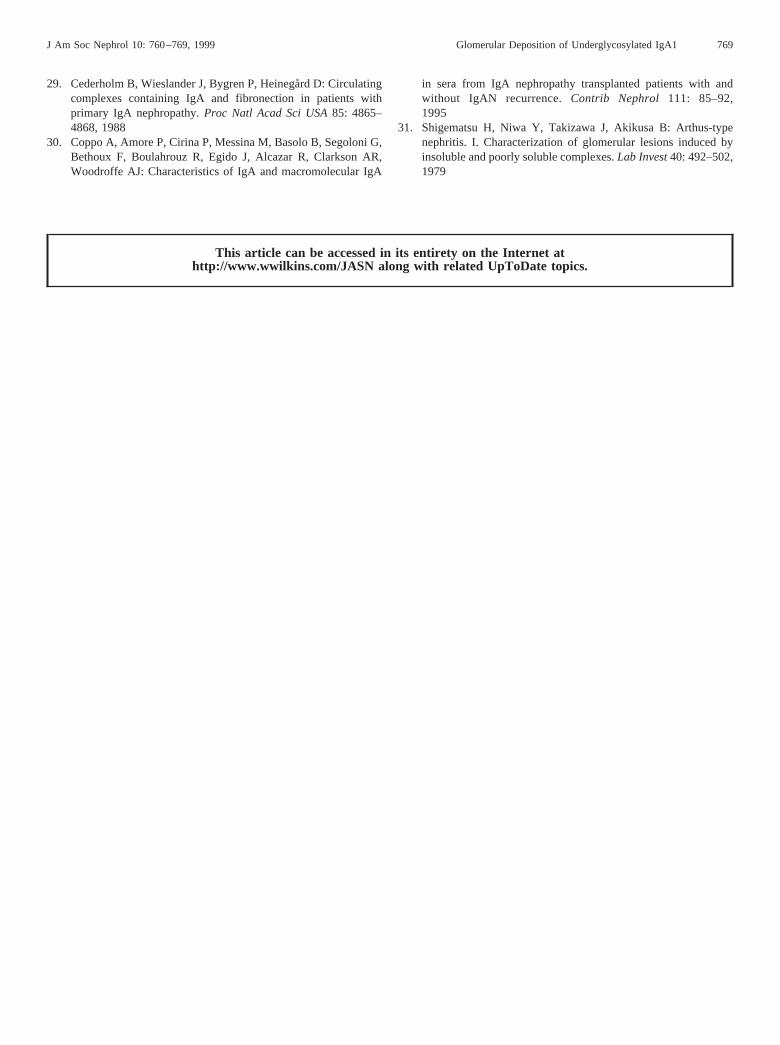
Figure 2. Ion-exchange chromatogram of Jacalin high-affinity IgA1.A definite peak of the neutral fraction is observed.
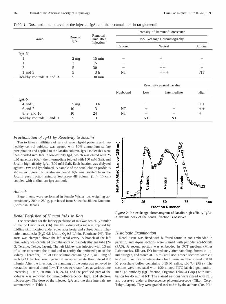
Table 1. Dose and time interval of the injected IgA, and the accumulation in rat glomeruli
Group Dose ofIgA1
RemovalTime afterInjection
Intensity of Immunofluorescence
Ion-Exchange Chromatography
Cationic Neutral Anionic
IgA-N1 2 mg 15 min 2 1 21 2 15 2 11 22 5 30 2 11 21 and 3 5 3 h NT 111 NT
Healthy controls A and B 5 30 min 2 2 2
Reactivity against Jacalin
Nonbound Low Intermediate High
IgA-N4 and 5 5 mg 3 h 2 2 2 116 and 7 10 3 NT 1 2 118, 9, and 10 10 24 NT 2 2 1
Healthy controls C and D 5 3 2 NT NT 2
762 Journal of the American Society of Nephrology J Am Soc Nephrol 10: 760–769, 1999

Kobayashi). A third sample of renal tissue was fixed in 2% glutaral-dehyde and embedded in Epon for electron microscopic examination.
Size Analyses of the IgA1 Molecules by NativePolyacrylamide Gel Electrophoresis
Polyacrylamide gel electrophoresis (PAGE) was performed underthe nonreduced condition by the Phast system (Pharmacia), usingPhastGel gradient 4-15 according to the instructions.
Analysis ofO-Glycans of Jacalin High IgAFractions by Gas-Phase Hydrazinolysis
Because of the positive results of fractions H and N on the histol-ogy, the rest of the injected Jacalin high-affinity IgA1 (fraction H) ofeach IgAN patient was pooled and divided by ion-exchange chroma-tography into cationic (HC), neutral (HN), and anionic (HA) fractionsin the same manner. In this fractionation, a definite peak of the neutralfraction appeared (Figure 2).
The release ofO-glycans from the IgA1 was performed accordingto our previous method of gas-phase hydrazinolysis (3). The solutionscontaining the IgA1 were dried under reduced pressure at 50°C.Approximately 2 mg of dried sample was treated with anhydroushydrazine (Honen Co., Yokohama, Japan) at 65°C for 6 h usingHydraclub S-204 (Honen Co.). After the reaction, hydrazine wasabsorbed in concentrated sulfuric acid under reduced pressure. Thedried sample was suspended in 250ml of saturated sodium bicarbon-ate, and then 25ml of acetic anhydride was added for acetylation.After 30 min of incubation at RT, this treatment was repeated. Todesalt the solution, the sample was applied to a Dowex 50W column(X8, H form, 3 ml) (Muromachi Kagaku Kougyou, Tokyo, Japan) andwashed with 15 ml of distilled water. The eluate containing thereleased oligosaccharides was then dried.
After the dried sample was pyridylaminated as previously reported(17), the pyridylaminated oligosaccharide mixture (PA-sugar) wasanalyzed using a PALPAK type N column (4.63 250 mm, Takara
Shuzo, Tokyo, Japan) at a flow rate of 1.0 ml/min at 40°C. Twosolvents, A and B, were prepared for the fractionation of PA-oligo-saccharides. Solvent A consisted of 50 mM acetic acid adjusted to pH7.3 with triethylamine and acetonitrile (15:85, vol/vol). Solvent B was1 M acetic acid adjusted to pH 7.3 with triethylamine and acetonitrile(50:50, vol/vol). The column was equilibrated with solvent A. Afterthe sample injection, the proportion of solvent B was increasedlinearly to 55% over 110 min. The peaks of each PA-oligosaccharidewere then observed.
Enzyme-Linked Immunosorbent Assay usingAnti-Synthetic IgA1 Hinge Peptide Antibody, PeanutAgglutinin, and Vicia Villosa Lectins
The procedures were carried out according to our previous reports(18). Each well of the microtitration plates (Limbro/Titertek, A FlowGeneral Company, McLean, IL) was coated with 2mg of the IgA1fractions in 100ml of 0.015 M carbonate buffer, pH 9.6, and theunreacted sites were blocked with PBS containing 1% bovine serumalbumin (BSA) (fraction V, Sigma Chemical Co., St. Louis, MO).
One hundred microliters of anti-synthetic IgA1 hinge peptide (anti-sHP) antibody (1:100 dilution in PBS) or peroxidase-labeled peanutagglutinin (PNA) (Seikagaku Co., Tokyo, Japan) or VV lectins (5mg/ml, Sigma Chemical Co.) in PBS was placed in each of theIgA1-coated and noncoated wells. After incubation for 3 h at RT, theplates were washed with PBS containing 0.1% BSA and 0.05%Tween 80 (PBS/BSA/Tween). For the analysis of anti-sHP antibody,100 ml of peroxidase-conjugated anti-rabbit IgG (1:200 in PBS) wasadded to the wells, incubated for 1 h at RT, andthen washed. Allplates were exposed to an enzyme substrate consisting of 0.4 mg/mlO-phenylenediamine dihydrochloride (Sigma Chemical Co.) and0.03% vol/vol of hydrogen peroxide in a solution containing 0.1 Mdisodium hydrogen phosphate and 0.05 M citric acid monohydrate.The developed color was read at 490 nm with a microplate reader(model 450; Bio-Rad Laboratories, Richmond, CA). All analyses
Figure 3.Grading samples of IgA1 deposits in rat glomeruli by immunofluorescence. A,2; B, 1; C, 11; D, 111. Magnification,3460.
J Am Soc Nephrol 10: 760–769, 1999 Glomerular Deposition of Underglycosylated IgA1 763

were performed in triplicate, and each absorbance level of 490 nm wasreduced by that of the IgA1 noncoated well.
ResultsIgA1 Fractions Accumulating in RatGlomeruli and Histology
Definite positive staining was observed in the neutral and theJacalin high-affinity IgA1 fractionated by the ion-exchangeand Jacalin affinity chromatography, respectively (Table 1).No significant deposits were found in the IgA1 among otherfractions in either method.
In the positive samples, IgA was accumulated in the glo-merular capillary lumen and within the mesangial area. Typicalgrading samples of the immunofluorescence (IF) are shown inFigure 3a (2), b (1), c (11), and d (111).
Histologic findings on electron microscopy (EM) and lightmicroscopy (LM) showed the intraluminal precipitates com-patible with the IgA precipitates on IF at 15 and 30 min (Figure
4a). At 3 h, in addition to these precipitates, the definitemesangial deposits, degranulated platelets, and prominent in-filtration of polymorphonuclear cells (EM, Figure 4b; LM,Figure 4c) were observed. At 24 h, the precipitates and thehistologic reaction had declined.
Size Analyses of the IgA1 Molecules by ElectrophoresisThe cationic IgA1 fraction was mainly composed of mono-
mer and dimer. Significant amounts of macromolecular IgA1(approximate MW 1000 kD) were present in the neutral andanionic IgA1 fractions. However, there was no obvious differ-ence in the distribution of the molecular weights between theneutral and anionic IgA1.
On the other hand, it was revealed that the relative contentsof the macromolecule IgA1 was definitely increased in Jacalinhigh-affinity IgA1 compared with other fractions of IgA1(Figure 5).
Figure 4.Typical samples of the histologic findings. (a) Intraluminal precipitates compatible with the IgA precipitates on immunofluorescenceare seen (15-min perfusion;33000). (b) The definite mesangial deposits and degranulated platelets in the lumen are seen (3 h;36000). (c)Typical samples of the histologic findings on light microscopy. The prominent accumulation of polymorphonuclear cells is observed (3 h;periodic acid-Schiff stain;3480).
764 Journal of the American Society of Nephrology J Am Soc Nephrol 10: 760–769, 1999

Analyses of ReleasedO-Glycans from“Glomerulophilic” IgA1 Fraction byGas-Phase Hydrazinolysis
As shown in Figure 6, two major peaks (peak A and B) andtwo minor peaks (peak C and D) were identified. In ourprevious study (19), it had been clarified that peaks A, B, C,and D were asialo-Galb1,3GalNAc, NeuAca2,3Galb1,3GalNAc,Galb1,3(NeuAca2,6) GalNAc and NeuAca2,3Galb1,3(NeuAca2,6)GalNAc, respectively. It was noted that the“glomerulophilic” IgA1 fraction (HN) had only an asialo-Galb1–3GalNAc peak (Figure 6b).
Reactivities of PNA, VV Lectins, and Anti-sHPAntibody to “Glomerulophilic” IgA1
Enzyme-linked immunosorbent assay revealed that all re-agents of anti-sHP antibody, PNA, and VV lectins stronglybound to the “glomerulophilic” IgA1 fraction among the Jaca-lin high-IgA1 fractions (Figure 7). The actual levels of 490-nmabsorbance in each reagent were as follows [(HC)versus(HN,glomerulophilic) versus (HA)]: Anti-sHP antibody: 0.2660.05versus0.946 0.10versus0.016 0.07; PNA: 0.116 0.01versus0.31 6 0.03 versus0.06 6 0.00; VV: 0.01 6 0.01versus0.126 0.01versus0.016 0.01.
DiscussionIn this study, we could isolate and characterize the IgA1
molecules that could accumulate in rat glomeruli. We calledthe IgA “glomerulophilic” IgA1. The “glomerulophilic” IgA1was located in the electrically neutral (N) and Jacalin high-
affinity (H) fractions. For investigating the structure ofO-glycans in the IgA1 hinge, two different approaches, the chem-ical analysis (gas-phase hydrazinolysis) and enzyme-linkedimmunosorbent assay (reactivity of PNA, VV, and anti-sHPantibody), were performed. These analyses provided the resultsreasonably explaining that the hinge of the “glomerulophilic”IgA1 was underglycosylated.
Only an asialo-Galb1,3GalNAc peak was identified in the“glomerulophilic” IgA1 by the analyses of gas-phase hydrazi-nolysis. This was compatible with the finding of the increasedbinding of the “glomerulophilic” IgA1 to PNA lectin, whichrecognizes asialo-Galb1,3GalNAc. These observations wouldreasonably explain our previous comparative studies of theincreased reactivity of Jacalin to IgA1 in the patients withIgAN (20) and the increase in the relative content of asialo-Galb1,3GalNAc in the IgA1 molecule (4).
It was clarified that not only sialic acid but otherO-glycancomponent(s) in the hinge were also lacking in the “glomeru-lophilic” IgA1. The increased binding of the “glomerulophilic”IgA1 to VV lectin suggested the decrease of the galactosecontent resulting in the increase of the unsubstituted GalNAcresidue in the IgA1 hinge because VV lectin binds to GalNAcbut not to Gal residue. The galactose deficiency had beenpointed out in the IgA1 molecules of patients with IgAN byAllen et al. (5) and Tomanaet al. (6). The increased binding ofVV lectin to “glomerulophilic” IgA1 seems to be inconsistentwith the results of PNA lectin and the gas-phase hydrazinoly-sis. However, the phenomenon could occur because PNA lectindoes not recognize sialylated-Galb1,3GalNAc but does recog-
Figure 5. Polyacrylamide gel electrophoresis (native PAGE). The relative contents of the macromolecular IgA1 are definitely increased inJacalin high-affinity IgA1 compared with other fractions of IgA1.
J Am Soc Nephrol 10: 760–769, 1999 Glomerular Deposition of Underglycosylated IgA1 765

nize the asialo-Galb1,3GalNAc residue. If the Galb1,3GalNAcside chains are highly desialylated, PNA lectin could stronglybind to the IgA1 hinge even if the total number ofGalb1,3GalNAc residues is decreased due to the relative in-crease of the unsubstituted GalNAc residue that is recognizedby VV lectin. Similarly, the increased binding of VV lectin tothe “glomerulophilic” IgA1 is not necessarily inconsistent withthe result of the gas-phase hydrazinolysis, because the gas-phase hydrazinolysis provided no information concerning theunsubstituted GalNAc residues, because it could not identifythe unsubstituted GalNAc residues (3,21).
The “glomerulophilic” IgA1 was also strongly recognizedby polyclonal anti-sHP antibody that reacted with the peptidecore of the IgA1 hinge but not withO-glycan side chains. Theresults, therefore, suggested that the hinge region in the “glo-merulophilic” IgA1 was highly deglycosylated because theresults reflected the increased area of the exposed peptide core.The increased reactivity of the antibody was also observedin the IgA1 molecules of the IgAN patients in our recentstudy (15).
The actual reason(s) why the “glomerulophilic” IgA1 islocated in the Jacalin high-affinity fraction is uncertain. Thephenomenon seemed to be inconsistent with the observationthat the hinge glycopeptide of the IgA1 was under-O-glycosy-lated, because Jacalin recognizes theO-glycan residues. Therecould be two possibilities for reasonably explaining the dis-crepancy. The first is the effect of the desialylation ofO-glycans in the hinge. Even if the number ofO-glycan sidechains decreased, the total affinity ofO-glycan to Jacalin couldincrease due to desialylation similar to the binding of PNA. Itwas observed by Hortin and Trimpe (22) that the binding ofO-glycans to Jacalin, although not completely prevented, didappear to be reduced by sialic acid located on the outer side ofthe O-glycan chains. The second possibility is the self-aggre-gation of the “glomerulophilic” IgA1. The result of the nativePAGE indicated that the Jacalin high-affinity IgA1 containshigher amounts of macromolecular IgA1 compared with otherIgA1 fractionated by the Jacalin column. It had been observedthat the reactivity of IgA1 to Jacalin was increased along withthe increase in its molecular weight (20).
The cause(s) of the location of the “glomerulophilic” IgA1in the electrically neutral fraction is also beyond current knowl-edge. The analyses of the molecular weights indicated that thecationic fraction was mainly composed of monomer and dimer,containing few macromolecules of IgA1. On the other hand, alarge amount of macromolecular IgA1 was observed in theneutral and anionic IgA1 fractions. These results suggested thatthe “glomerulophilic” IgA1 was shifted into the relativelycationic range among the macromolecular IgA1. This could bepartially explained by the result of this study that the “glo-merulophilic” IgA1 had no sialic acid in the Galb1,3GalNAcresidues because sialic acid is negatively charged. Consideringthe difference in molecular weights between the cationic IgA1fraction and the “glomerulophilic” IgA1 (neutral) fraction, itwas also suspected that the aggregation itself also influencedthe electric charge of the “glomerulophilic” IgA1 probably dueto the change in its steric structure.
Figure 6. HPLC patterns ofO-glycans from Jacalin high-affinityIgA1. Among the IgA1 divided by a diethylaminoethyl (DEAE)column—Jacalin high and cationic (HC; Panel A), high and neutral(HN; Panel B), and high and anionic (HA; Panel C)—two major peaksof asialo-Galb1,3GalNAc (peak A) and monosialylated (peak B) areidentified. It was noted that the “glomerulophilic” IgA1 fraction (HN)had only an asialo-Galb1,3GalNAc peak (peak A) and no monosia-lylated peak (peak B) at all.
766 Journal of the American Society of Nephrology J Am Soc Nephrol 10: 760–769, 1999

As far as the mechanism of the glomerular IgA deposition inIgAN is concerned, our previous studies clarified the presenceof the IgA-IgA interaction resulting in the formation of mac-romolecular IgA1 (13) and the association of asialo-Galb1,3GalNAc with the conformational instability of IgA1 inIgAN (4). Recently, it was found that desialylation of the IgA1molecule by treatment with sialidase induced the self-aggrega-tion of IgA1 (18). The present study had results consistent withthose reported previously. It was clarified that theGalb1,3GalNAc side chains identified by gas-phase hydrazi-nolysis were only of the asialo-type in the “glomerulophilic”IgA1, and the IgA1 was mainly composed of macromolecularIgA.
In general, it had been established that the mesangial local-ization of the immune complexes was associated with theirmolecular weights ranging up to 1000 kD or greater (23,24).However, the analyses of the molecular weights of IgA1 frac-tionated by ion-exchange chromatography suggested that theglomerular deposition of IgA1 cannot be fully explained onlyby the size of the IgA1 molecules because the anionic IgA1also contained a considerable amount of macromolecular IgA1as did the “glomerulophilic” IgA1 (neutral) fraction. However,the “glomerulophilic” IgA1 was relatively cationic in the mac-romolecular IgA1 fractions. This would be reasonably ex-plained by the fact that the cationic macromolecule tended tobe trapped in the glomeruli rather than the anionic one (25,26).
The increase in the IgA1 having affinity for type IV collagen(27), laminin (28), and fibronectin (29), which composed theglomerular basement membrane and mesangial matrix, hadbeen found in patients with IgAN. Furthermore, Coppoet al.suggested an important role ofO-glycans in the hinge for the
binding of IgA1 to type IV collagen, laminin, and fibronectin(30). Our recent study also showed that the IgA1 moleculesacquired the reactivity with these extracellular matrix proteinswhen they were treated with sialidase. After further treatmentwith b-galactosidase, the IgA1 (agalacto-IgA1) showed thehighest affinity for these proteins compared with those of theasialo- and naked (treated with sialidase,b-galactosidase andN-acetylgalactosaminidase) IgA1 (18). Considering this previ-ous in vitro study, it is reasonable to explain the phenomenaobserved in this study as follows: The desialylation of the IgA1hinge was a prerequisite for the self-aggregation, and further-more underglycosylation, especially undergalactosylation,played a key role(s) in the affinity toward the extracellularmatrix proteins, resulting in the mesangial IgA deposition.
In this study, a mild but definite histologic response due tothe glomerular deposition of IgA1 was observed. At 3 h afterthe perfusion, prominent accumulation of polymorphonuclearcells and degranulated platelets were observed in the glomer-ular capillary lumen. This phenomenon seems to be similar tothat of the study reported by Davinet al. (16), who alsoobserved the histologic response induced by the perfusion ofpolymeric IgA-ConA complexes into the rat kidney. In ourstudy, the histologic reaction did not occur at 15 min and 30min after the perfusion and disappeared after 24 h. These timecourse reactions and histologic characteristics were consistentwith those of the experimental arthus-type nephritis induced byinsoluble and poorly soluble immune complexes reported byShigematsuet al. (31).
This study provided the first information concerning theisolation and the structural characterization of the IgA1 mole-cules that could accumulate in rat glomeruli and induce the
Figure 7. It is noted that all three reagents strongly bound to the “glomerulophilic” IgA1 fraction compared with other Jacalin high-IgA1fractions. (A) Anti-synthetic IgA1 hinge peptide antibody [(HC)versus(HN, glomerulophilic)versus(HA)]: 0.26 6 0.05versus0.946 0.10versus0.016 0.07. (B) PNA: 0.116 0.01versus0.316 0.03versus0.066 0.00. (C) VV: 0.016 0.01versus0.126 0.01versus0.0160.01. Each datum represents the mean6 SD of the absorbance level at 490 nm reduced by the background.
J Am Soc Nephrol 10: 760–769, 1999 Glomerular Deposition of Underglycosylated IgA1 767

histologic reaction. This information may provide further un-derstanding of the actual mechanism of glomerular IgA depo-sition and the histologic reaction in IgAN.
AcknowledgmentsThis study was supported in part by grants from New Energy and
Industrial Technology Development Organization (NEDO) and AsahiChemical Industry. The authors thank Ms. Y. Tanaka and Ms. M.Saitoh for excellent technical assistance. The authors are also gratefulto Professor H. Shigematsu of Shinshu University and the members ofthe Electron Microscope Laboratory Center of Kitasato University fortheir valuable suggestions and assistance with electron microscopyand photography.
References1. Conley ME, Cooper MD, Michael AF: Selective deposition of
immunoglobulin A1 in immunoglobulin A nephropathy, anaphy-lactoid purpura nephritis, and systemic lupus erythematosus.J Clin Invest 66: 1432–1436, 1980
2. Baenziger J, Kornfeld S: Structure of the carbohydrate units ofIgA1 immunoglobulin. II. Structure of theO-glycosidicallylinked oligosaccharide units. J Biol Chem 249: 7270–7281, 1974
3. Iwase H, Ishii-Karakasa I, Fujii E, Hotta K, Hiki Y, KobayashiY: Analysis of glycoform ofO-glycan from human myelomaimmunoglobulin A1 by gas-phase hydrazinolysis following pyri-dylamination of oligosaccharides.Anal Biochem206: 202–205,1992
4. Hiki Y, Iwase H, Kokubo T, Horii A, Tanaka A, Nishikido J,Hotta K, Kobayashi Y: Association of asialo-galactosylb1–3N-acetylgalactosamine on the hinge with a conformational instabil-ity of Jacalin-reactive immunoglobulin A1 in immunoglobulin Anephropathy.J Am Soc Nephrol7: 955–960, 1996
5. Allen AC, Harper SJ, Feehally J: Galactosylation ofN- andO-linked carbohydrate moieties of IgA1 and IgG in IgA nephrop-athy.Clin Exp Immunol100: 470–474,1995
6. Tomana M, Matousovic K, Julian B, Radl J, Konecny K,Mestecky J: Galactose-deficient IgA1 in sera of IgA nephropathypatients is present in complex with IgG.Kidney Int52: 509–516,1997
7. Iwase H, Tanaka A, Hiki Y, Kokubo T, Ishii-Karakasa I, Koba-yashi Y, Hotta K: Estimation of the number of O-linked oligo-saccharides per heavy chain of human serum IgA1 by matrix-assisted laser desorption ionization time-of-flight massspectrometry (MALDI-TOFMS) analysis of the hinge peptide.J Biochem120: 393–397, 1996
8. Hiki Y, Tanaka A, Kokubo T, Iwase H, Nishikido J, Hotta K,Kobayashi Y: Analyses of IgA1 hinge glycopeptides in IgAnephropathy by matrix-assisted laser desorption/ionization time-of-flight mass spectrometry.J Am Soc Nephrol9: 577–582, 1998
9. Berger J, Hinglais N: Le´s depôts intercapillaires d’IgA-IgG J.Urol Nephrol 74: 694–695, 1968
10. Clarkson AR, Woodroffe AJ, Aarons I, Hiki Y, Hale G: IgAnephropathy.Annu Rev Med38: 157–168, 1987
11. Coppo R, Basolo B, Martina G, Rollino C, De Marchi M,Giacchino F, Mazzucco G, Messina M, Piccoli G: Circulatingimmune complexes containing IgA, IgG and IgM in patients withprimary IgA nephropathy and with Henoch-Schönlein nephritis:Correlation with clinical and histologic signs of activity.ClinNephrol18: 230–239, 1982
12. Sancho J, Gonza´lez E, Rivera F, Escanero JF, Egido J: Hepaticand kidney uptake of soluble monomeric and polymeric IgAaggregates.Immunology52: 161–167, 1984
13. Hiki Y, Saitoh M, Kobayashi Y: Serum IgA class anti-IgAantibody in IgA nephropathy.Nephron59: 552–560, 1991
14. Kokubo T, Hiki Y, Iwase H, Horii A, Tanaka A, Nishikido J,Hotta K, Kobayashi Y: Evidence for involvement of IgA1 hingeglycopeptide in the IgA1-IgA1 interaction in IgA nephropathy.J Am Soc Nephrol8: 915–919, 1997
15. Kokubo T, Hiki Y, Iwase H, Horii A, Tanaka A, Nishikido J,Hotta K, Kobayashi Y: Exposed peptide core of IgA1 hingeregion in IgA nephropathy [Abstract].J Am Soc Nephrol8:538A, 1997
16. Davin JC, Dechene C, Mahieu PR: Acute experimental glomer-ulonephritis induced by the glomerular deposition of circulatingpolymeric IgA-concanavalin A complexes [French].Nephrolo-gie 10: 151–155, 1989
17. Iwase H, Ishii-Karakasa I, Urata T, Saito T, Hotta K: Extractionmethod for preparing pyridylamino sugar derivatives and appli-cation to porcine gastric mucus glycoprotein analysis.Anal Bio-chem188: 200–202, 1990
18. Kokubo T, Hiki Y, Iwase H, Tanaka A, Toma K, Hotta K,Kobayashi Y: Protective role of IgA1 glycans against IgA1self-aggregation and adhesion to extracellular matrix proteins.J Am Soc Nephrol9: 2048–2054, 1998
19. Iwase H, Tanaka A, Hiki Y, Kokubo T, Ishii-Karakasa I, Koba-yashi Y, Hotta K: Abundance of Galb1,3GaNAc in O-linkedoligosaccharide on hinge region of polymeric and heat aggre-gated IgA1 from normal human serum.J Biochem120: 92–97,1996
20. Hiki Y, Iwase H, Saitoh M, Saitoh Y, Horii Y, Hotta K, Koba-yashi Y: Reactivity of glomerular and serum IgA1 to Jacalin inIgA nephropathy.Nephron72: 429–435, 1996
21. Patel T, Bruce J, Merry A, Bigge C, Wormald M, Jaques A,Parekh R: Use of hydrazine to release intact and unreduced formof N- and O-linked oligosaccharides from glycoproteins.Bio-chemistry32: 679–693, 1993
22. Hortin GL, Trimpe BL: Lectin affinity chromatography of pro-teins bearing O-linked oligosaccharides: Application of Jacalin-agarose.Anal Biochem188: 271–277, 1990
23. Koyama A, Niwa Y, Shigematsu H, Taniguchi M, Tada T:Studies on passive serum sickness. II. Factors determining thelocalization of antigen-antibody complexes in the murine renalglomerulus.Lab Invest38: 253–262, 1978
24. Germuth FG Jr, Senterfit LB, Dreesman GR: Immune complexdisease. V. The nature of the circulating complexes associatedwith glomerular alterations in the chronic BSA-rabbit system.Johns Hopkins Med J130: 344–357, 1972
25. Kanwar YS, Farquhar MG: Anionic sites in the glomerularbasement membrane:In vivo and in vitro localization to thelaminae rarae by cationic probes.J Cell Biol 81: 137–153, 1979
26. Rennke HG, Patel Y, Venkatachalam MA: Glomerular filtrationof proteins: Clearance of anionic, neutral, and cationic horserad-ish peroxidase in the rat.Kidney Int13: 278–288, 1978
27. van den Wall Bake AW, Kirk KA, Gay RE, Switalski LM, JulianBA, Jackson S, Gay S, Mestecky J: Binding of serum immuno-globulins to collagens in IgA nephropathy and HIV infection.Kidney Int42: 374–382, 1992
28. Shinkai Y, Karai M, Osawa G, Sato M, Koshikawa S: Antimouselaminin antibodies in IgA nephropathy and various glomerulardiseases.Nephron56: 285–296, 1990
768 Journal of the American Society of Nephrology J Am Soc Nephrol 10: 760–769, 1999
29. Cederholm B, Wieslander J, Bygren P, Heinegård D: Circulatingcomplexes containing IgA and fibronection in patients withprimary IgA nephropathy.Proc Natl Acad Sci USA85: 4865–4868, 1988
30. Coppo A, Amore P, Cirina P, Messina M, Basolo B, Segoloni G,Bethoux F, Boulahrouz R, Egido J, Alcazar R, Clarkson AR,Woodroffe AJ: Characteristics of IgA and macromolecular IgA
in sera from IgA nephropathy transplanted patients with andwithout IgAN recurrence.Contrib Nephrol 111: 85–92,1995
31. Shigematsu H, Niwa Y, Takizawa J, Akikusa B: Arthus-typenephritis. I. Characterization of glomerular lesions induced byinsoluble and poorly soluble complexes.Lab Invest40: 492–502,1979
This article can be accessed in its entirety on the Internet athttp://www.wwilkins.com/JASN along with related UpToDate topics.
J Am Soc Nephrol 10: 760–769, 1999 Glomerular Deposition of Underglycosylated IgA1 769