Embed Size (px)
Citation preview
Use of a T Cell Interferon‐γ Release Assay to Evaluate Tuberculosis Risk in Newly QualifiedPhysicians in Singapore Healthcare Institutions • Author(s): C. B. E. Chee, MBBS, FRCP; L. K. Y. Lim, BSc; T. M. Barkham, MBBS, MRC Path;D. R. Koh, MBBS, FRCP; S. O. Lam, RN; L. Shen, PhD; Y. T. Wang, MBBS, FRCPSource: Infection Control and Hospital Epidemiology, Vol. 30, No. 9 (September 2009), pp. 870-875Published by: The University of Chicago Press on behalf of The Society for Healthcare Epidemiologyof AmericaStable URL: http://www.jstor.org/stable/10.1086/599284 .
Accessed: 16/05/2014 14:59
Your use of the JSTOR archive indicates your acceptance of the Terms & Conditions of Use, available at .http://www.jstor.org/page/info/about/policies/terms.jsp
.JSTOR is a not-for-profit service that helps scholars, researchers, and students discover, use, and build upon a wide range ofcontent in a trusted digital archive. We use information technology and tools to increase productivity and facilitate new formsof scholarship. For more information about JSTOR, please contact [email protected].
.
The University of Chicago Press and The Society for Healthcare Epidemiology of America are collaboratingwith JSTOR to digitize, preserve and extend access to Infection Control and Hospital Epidemiology.
http://www.jstor.org
This content downloaded from 195.78.108.169 on Fri, 16 May 2014 14:59:07 PMAll use subject to JSTOR Terms and Conditions

infection control and hospital epidemiology september 2009, vol. 30, no. 9
o r i g i n a l a r t i c l e
Use of a T Cell Interferon-g Release Assay to EvaluateTuberculosis Risk in Newly Qualified Physicians
in Singapore Healthcare Institutions
C. B. E. Chee, MBBS, FRCP; L. K. Y. Lim, BSc; T. M. Barkham, MBBS, MRC Path; D. R. Koh, MBBS, FRCP;S. O. Lam, RN; L. Shen, PhD; Y. T. Wang, MBBS, FRCP
background. Surveillance for latent tuberculosis in high-risk groups such as healthcare workers is limited by the nonspecificity of thetuberculin skin test (TST) in BCG-vaccinated individuals. The Mycobacterium tuberculosis antigen–specific interferon-g release assays (IGRAs)show promise for more accurate latent tuberculosis detection in such groups.
objective. To compare the utility of an IGRA, the T-SPOT.TB assay, with that of the TST in healthcare workers with a high rate ofBCG vaccination.
methods. Two hundred seven medical students from 2 consecutive cohorts underwent the T-SPOT.TB test and the TST in their finalyear of study. Subjects with negative baseline test results underwent repeat testing after working for 1 year as junior physicians in Singapore’spublic hospitals.
results. The baseline TST result was an induration 10 mm or greater in diameter in 177 of the 205 students who returned to havetheir TST results evaluated (86.3%), while the baseline T-SPOT.TB assay result was positive in 9 (4.3%) of the students. Repeat T-SPOT.TBtesting in 182 baseline-negative subjects showed conversion in 9 (4.9%). A repeat TST in 18 subjects with baseline-negative TST resultsdid not reveal any TST result conversion.
conclusions. The high rate of positive baseline TST results in our BCG-vaccinated healthcare workers renders the TST unsuitable asa surveillance tool in this tuberculosis risk group. Use of an IGRA has enabled the detection and treatment of latent tuberculosis in thisgroup. Our T-SPOT.TB conversion rate highlights the need for greater tuberculosis awareness and improved infection control practices inour healthcare institutions.
Infect Control Hosp Epidemiol 2009; 30:870-875
From the Tuberculosis Control Unit (C.B.E.C., L.K.Y.L., S.O.L., Y.T.W.) and the Department of Laboratory Medicine (T.M.B.), Tan Tock Seng Hospital;and Yong Loo Lin School of Medicine, National University of Singapore (D.R.K., S.L.), Singapore.
Received November 14, 2008; accepted March 23, 2009; electronically published July 28, 2009.� 2009 by The Society for Healthcare Epidemiology of America. All rights reserved. 0899-823X/2009/3009-0008$15.00. DOI: 10.1086/599284
It is important to monitor healthcare workers (HCWs) fortuberculosis both because of their occupational risk of latenttuberculosis (LTBI) and because of the threat they may poseto patients in their care if the HCWs develop active tuber-culosis.1-3 In many resource-rich countries with a low inci-dence of tuberculosis, HCWs constitute a high-priority groupfor targeted screening and periodic surveillance with the tu-berculin skin test (TST). Individuals whose TST results con-vert from negative to positive become candidates for LTBItreatment.4,5 The results are also useful in the aggregate: variedconversion rates among HCW job categories or among lo-cations within a healthcare facility or institution serve as animportant indicator of the adequacy of existing infection con-trol measures.6,7 Interpretation of TST results is, however, wellknown to be confounded by infection with nontuberculousmycobacteria and prior inoculation with the BCG vaccine,the most widely used vaccine worldwide.8 Interferon-g release
assays (IGRAs), which measure T cell responses to the My-cobacterium tuberculosis–specific antigens early secreted an-tigenic target 6 (ESAT-6) and culture filtrate protein 10 (CFP-10), have shown superior specificity over the TST inBCG-vaccinated populations and would overcome this lim-itation of the TST.9,10 These assays, available commercially asthe T-SPOT.TB (Oxford Immunotec), QuantiFERON-TBGold (QFT-G), and QuantiFERON-TB Gold In-tube (QFT-G In-tube) (Cellestis) assays, have been approved by the USFood and Drug Administration and endorsed in major na-tional guidelines for use in place of or in addition to the TSTfor LTBI screening.11,12 To date, experience with the QFT-Gor QFT-G In-tube assay for testing of HCWs has been re-ported in the United States, Japan, Germany, Switzerland,Denmark, and India;13-19 conversely, literature pertaining tothe use of the T-SPOT.TB assay in HCWs is scarce. In ad-dition, HCW surveillance is hampered both by a paucity of
This content downloaded from 195.78.108.169 on Fri, 16 May 2014 14:59:07 PMAll use subject to JSTOR Terms and Conditions

igra to evaluate tb risk in junior physicians 871
information and by controversy about the use and interpre-tation of serial IGRA testing.20
Singapore is a country with an intermediate incidence oftuberculosis (incidence, 35 cases per 100,000 residents in2007) with 98% BCG vaccination coverage at birth since the1950s.21 Until 2001, there had been a Ministry of Health policyof BCG revaccination for tuberculin-nonreactive individualsat 12 or 16 years of age. Most Singaporeans born before 1989would thus have received 2 BCG vaccinations.22 The risk ofLTBI among HCWs in Singapore has hitherto not been eval-uated, nor has there been any established practice of LTBIsurveillance for workers in our healthcare settings. This was,in part, a result of the difficulties in interpretation of TSTresults in our population, most of whom have received theBCG vaccination, as well as the priority placed on controllingthe relatively high rates of tuberculosis in the community.Because the community rate of tuberculosis in our countryhas declined over the past 8 years, more attention and re-sources could then be focused toward pockets of M. tuber-culosis transmission such as healthcare settings.23,24
We therefore undertook a study to compare the utility ofa less studied IGRA, the T-SPOT.TB assay, with that of theTST in a group of HCWs with a high rate of BCG vaccination.We chose a group of 2 consecutive cohorts of medical studentsin their final year of study. Those who had negative resultson the inital test were offered repeat testing after their firstyear of work in Singapore’s public hospitals.
methods
The study was approved by the domain-specific review boardof Singapore National Healthcare Group and by the NationalUniversity of Singapore Institutional Review Board. The sub-jects were medical students recruited from 2 consecutive co-horts who were in their final year of study (in 2005–2006and 2006–2007) at the Yong Loo Lin Medical School, NationalUniversity of Singapore, the only medical school in Singaporeat the time. All subjects gave informed consent.
All participants provided information about their demo-graphic characteristics (age, sex, race, history of exposure toM. tuberculosis, residential address) in a self-administeredquestionnaire. They were also examined for the presence andnumber of BCG vaccination scars. Tuberculin skin testingwith 2 TU RT23 purified protein derivative (Statens SerumInstitute, Copenhagen) was performed by experienced nursesfrom the Tuberculosis Control Unit. The two-step TST wasperformed in those whose first TST induration was less than10 mm. The result of the TST was considered positive if theinduration was 10 mm or more in diameter.
Peripheral venous blood was drawn for the T-SPOT.TBassay and transported within 6 hours to the Tan Tock SengHospital microbiology laboratory, where the assay was per-formed according to the manufacturer’s instructions18 bytechnicians who were unaware of the subjects’ identities. Thetest result was considered positive if either or both panel A
(containing ESAT-6 antigen) and panel B (containing CFP-10 antigen) had at least 6 more spots than the negative controland this number was at least twice the number of spots inthe negative control. The test was considered failed if thenegative control spot count was more than 10 or if there wereless than 20 spots in the positive control and both panel Aand B were nonreactive according to the criteria above. Spotswere counted with the EliSpot enzyme-linked immunospotreader system (Autoimmun Diagnostika) and were manuallyverified.
Study participants who had negative initial results on eitheror both tests were invited for repeat testing toward the endof their 1-year internship period. This period comprised 3rotations (each of 4 months duration) in different disciplines,including a compulsory general medical rotation, in the majorpublic hospitals in Singapore. At the time of repeat testing,subjects were asked about known exposures to infectious tu-berculosis patients during the period since their baseline test-ing. These exposures were categorized according to whetherthey occurred within or outside their hospital work environ-ment and whether the workplace exposure was protected orunprotected. Protected exposures referred to encounters withan infectious tuberculosis patient while the HCW used anN95 mask. Unprotected exposures referred to those that oc-curred prior to the establishment of treatment of and isolationmeasures for the (as yet unknown to be infectious) tuber-culosis patient; N95 masks would not have been worn whileattending to these patients.
T-SPOT.TB conversion was defined as the qualitative con-version of a negative to a positive test result, as defined bythe manufacturer’s instructions. TST result conversion wasdefined as an increase in TST induration by 10 mm or greaterfrom a baseline of less than 10 mm or as an increase of theinduration to 15 mm or greater from a baseline of less than10 mm. To determine the test conversion rates for each hos-pital, each subject’s results were attributed to the hospitalwhere he or she was assigned for his or her general medicalrotation.
The demographic characteristics of the students who par-ticipated in this study were compared with those of the restof the students with a x2 test or 2-sample t test for differenttypes of variables. The TST and T-SPOT.TB results at baselineand at 1 year were analyzed descriptively. The difference inthe conversion rates among the hospitals was examined withthe Fisher exact test. All statistical analyses were 2-tailed testsand were performed using the Statistical Package for SocialSciences version 16.0 (SPSS).
results
Two consecutive cohorts of medical students (the graduatingclass of 2006 and of 2007) were invited to participate andrecruited in their final year of study. Two hundred seven ofa total of 460 students consented to participate. Their meanage was 23.6 years (range, 22.2–25.8 years); 117 (56.5%) were
This content downloaded from 195.78.108.169 on Fri, 16 May 2014 14:59:07 PMAll use subject to JSTOR Terms and Conditions

872 infection control and hospital epidemiology september 2009, vol. 30, no. 9
figure 1. Flow chart of tuberculin skin testing and T-SPOT.TB testing in the study subjects.
male; and 190 (91.8%) were Chinese, 6 (2.9%) were Malay,8 (3.9%) were Indian, and 3 (1.4%) were of other ethnicities.There was no significant difference in demographic charac-teristics between the study subjects and those who declinedto participate (data not shown; ). All subjects wereP 1 .05BCG vaccinated: 147 (71.2%) had 2 BCG vaccination scars,while 60 (28.9%) had 1 BCG vaccination scar.
Baseline TST Results
Two hundred five students had initial TST results, while 2students did not return to have their TST indurations eval-uated and were subsequently not retested. Seventy-two of 74students with initial TST indurations of less than 10 mmunderwent 2-step testing; of these, 47 (65.3%) had TST in-durations of 10 mm or greater after the second test in the 2-step TST. The distribution of baseline TST indurations wastherefore as follows: 82 of 205 (40.0%) had TST indurationsof 15 mm or greater, 95 of 205 (46.3%) had TST indurationsof 10–14 mm, and 28 of 205 (13.7%) had TST indurationsof less than 10 mm, including 2 students who did not undergo2-step TST.
Baseline T-SPOT.TB Assay Results
There were 2 failed T-SPOT.TB assays (failure rate, 1%).These were due to failure of the negative control in 1 subject’stest and to an insufficient amount of blood in the other. Ninestudents (4.3%) had positive results on the T-SPOT.TB testat baseline. Of these, 5 had initial TST indurations of 10 mm
or greater and 4 had TST indurations of 10 mm or greaterwith 2-step testing.
Repeat Testing 1 Year after Negative Resultsof Baseline Tests
One hundred ninety-six subjects with negative baseline T-SPOT.TB assay and/or TST results were invited for repeattesting after 1 year of hospital work; 182 (92.9%) consentedto participate. Of these, 164 who initially had positive resultson the TST underwent repeat testing with the T-SPOT.TBtest only, while 18 were retested with both the T-SPOT.TBtest and TST (Figure 1). In nine of 182 (4.9%) subjects, allof whom had positive results on the baseline TST, the T-SPOT.TB assay results converted from negative to positive.All had normal findings on a chest radiograph and wereoffered LTBI treatment. Among the 18 subjects who wereretested with both the T-SPOT.TB test and the TST, neithertest result converted (Figure 1). There were no failed T-SPOT.TB test results among the repeat tests.
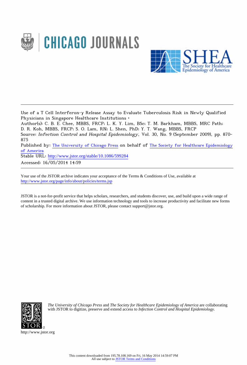
The quantitative responses to the T-SPOT.TB assay accord-ing to the individual panel antigens ESAT-6 and CFP-10 forthe 9 individuals whose TST results converted from negativeto positive are shown in Figure 2. All 9 subjects demonstratedincreases of 6 or more spot-forming cells per pe-52.5 # 10ripheral blood mononuclear cells above negative control intheir response to CFP-10. In contrast, only 3 subjects dem-onstrated at least this degree of response to ESAT-6.
The conversion rates with the T-SPOT.TB test for each of
This content downloaded from 195.78.108.169 on Fri, 16 May 2014 14:59:07 PMAll use subject to JSTOR Terms and Conditions

igra to evaluate tb risk in junior physicians 873
figure 2. Quantitative T cell responses (spot-forming cells perperipheral blood mononuclear cells above negative con-52.5 # 10
trol) to culture filtrate protein 10 and early secreted antigenic target6 in the 9 subjects who demonstrated conversion of the T-SPOT.TBtest results from negative to positive on repeat testing. Each linerepresents a single subject.
the 5 public hospitals (each physician’s results being attrib-uted to the hospital where he or she was assigned for his orher general medical rotation) were 0%, 4.0%, 4.1%, 4.8%,and 7.9%. There was no significant difference in the testconversion rates among the different hospitals ( ).P p .859
Reported Exposure to Infectious Tuberculosis PatientsBetween Baseline and Repeat Testing
One hundred seven (59%) of the 182 retested subjects re-ported no exposure to infectious tuberculosis patients. Four(44%) of the 9 individuals whose T-SPOT.TB test resultsconverted from negative to positive reported unprotected ex-posure. Of the 173 subjects whose results remained negativeafter repeat T-SPOT.TB testing, 71 (41%) reported exposures:50 reported unprotected exposure, 3 reported protected ex-posure, 8 reported exposure outside the hospital (2 of whomalso had unprotected hospital exposure), and 12 reportednonspecified exposure to tuberculosis patients.
discussion
Baseline testing of 205 final-year medical students recruitedfrom 2 consecutive class cohorts revealed a striking discrep-ancy in the positivity rate of the TST (86.3%) compared withthat of the T-SPOT.TB assay (4.3%). Repeat T-SPOT.TB test-ing after 1 year of hospital work in 182 participants who hadbaseline-negative results on the T-SPOT.TB showed test con-version in 9 physicians (4.9%), all of whom had positive TSTresults at baseline.
The high rate of positive baseline results on the TST foundin these final-year medical students was not surprising, giventhat most of them had received the BCG vaccination twice.Two meta-analyses had demonstrated an increased likelihoodof false-positive TST results in those who received BCG vac-cinations after infancy or if the TST was performed less than10–15 years after their last BCG vaccination.25,26 Our findingsare consistent with those in most of the published reports on
the TST and QFT-G test in BCG-vaccinated HCWs. Haradaand coworkers reported a TST positivity rate of 93% (vs aQFT-G test positivity rate of 9.9%) in a cross-sectional studyof HCWs in a Tokyo hospital with a high rate of BCG vac-cination.16 Discordant TST and QFT-G test results were alsoobserved in HCWs in the United States and Germany.13,14 Incontrast, a study on HCWs in India showed good agreementbetween the results of the QFT-G In-tube test and the TST.18
As the vast majority of our HCWs had already (falsely) testedpositive on the TST at baseline, the TST is clearly not usefulas a surveillance tool in our population.
Although we did not have a control group from the generalpopulation for comparison, our 4.9% T-SPOT.TB result con-version rate at 1 year is worrisome in the setting of Singapore’smoderate tuberculosis incidence rate of 35 cases per 100,000resident population per year. We had assumed that the highestrisk of exposure to infectious tuberculosis patients was dur-ing rotations in the general medical wards (compared withrotations in surgical, pediatric, or obstetrics and gynecologywards), and therefore we compared the conversion rates withthe T-SPOT.TB assay according to the hospital in which themedical rotation was undertaken. Infection control protocolsin all Singapore public hospitals mandate that tuberculosispatients whose sputum tests positive for acid-fast bacilli beplaced under airborne infection isolation precautions in neg-ative-pressure rooms and that N95 respirators be worn byattending staff while in the room. Fit testing of N95 respi-rators is also carried out for all new hospital staff. Exposureto persons with unsuspected or undiagnosed infectious tu-berculosis before establishment of airborne isolation is, how-ever, a possibility in busy general hospital medical wards incountries with a moderate to high prevalence of tuberculosis.This is where the risk of M. tuberculosis transmission is causefor worry. In addition, in people who test negative on asputum acid-fast bacilli smear but positive on a culture, itmay take up to several weeks before the diagnosis of tuber-culosis is established on the basis of positive culture results,by which time the episode(s) of exposure would have beenforgotten. Self-reporting of exposures thus results in under-estimation of the true extent of exposure. Indeed, 5 (56%)of our 9 HCWs whose T-SPOT.TB test results converted fromnegative to positive did not report any exposure to infectioustuberculosis patients. Information about the numbers of tu-berculosis patients in the various Singapore public hospitalsis not readily available. Although physicians at one particularhospital’s medical department had a higher likelihood of T-SPOT.TB test result conversion than the others (7.9% vs∼0%–4.8%), this difference was not statistically significant,likely due to the small number of individuals whose T-SPOT.TB test results converted from negative to positive. Ourfindings provide compelling evidence to support the need toheighten tuberculosis awareness and strengthen infectioncontrol measures in our healthcare institutions. Ideally, an-nual or periodic IGRA screening of those working in thehigher risk areas (eg, where the caseload of tuberculosis pa-
This content downloaded from 195.78.108.169 on Fri, 16 May 2014 14:59:07 PMAll use subject to JSTOR Terms and Conditions

874 infection control and hospital epidemiology september 2009, vol. 30, no. 9
tients is highest) should also be instituted. Because a majorlimiting factor would be the high cost of the IGRAs, cost-effectiveness analysis must be performed to guide local policyfor HCWs with respect to this intervention.
To our knowledge, this is the first report on the use of theT-SPOT.TB assay for serial testing of HCWs. An interestingfinding of our study was the higher increase in the quanti-tative response to CFP-10 than that to ESAT-6 in our subjectswhose T-SPOT.TB test results converted from negative topositive. Test result conversion was driven by the responseto CFP-10 in all 9 people whose T-SPOT.TB test results con-verted from negative to positive and who demonstrated clear-cut increases in their responses to this antigen. In contrast,in only 3 subjects did the response to ESAT-6 convert. Thediscrepancy in responses to the 2 M. tuberculosis–specific an-tigens merits further study. It was, unfortunately, not possibleto correlate the T-SPOT.TB test result conversions with thoseof the TST in this study because all of our subjects whose T-SPOT.TB test results converted from negative to positive al-ready had baseline TST indurations of 10 mm or greater andhence were not retested with the TST. We had, however, noreason to doubt that test conversion (rather than randomtest variation) had occurred in our subjects whose T-SPOT.TBtest results converted from negative to positive because theincrease in quantitative response to CFP-10 in all of thesesubjects was by at least 5 spot-forming cells, with none ofthe results in the indeterminate or borderline range (currentlydefined by the manufacturer as 5–7 spot-forming cells abovenegative control).
In conclusion, this study addresses the risk of hospital-acquired tuberculosis among HCWs in Singapore’s publichealthcare institutions. We showed that the TST was not auseful LTBI surveillance tool in our population of youngphysicians, most of whom had received the BCG vaccinationtwice. The T-SPOT.TB assay enabled us to document the riskof LTBI acquisition in this group of HCWs and to offer LTBItreatment to those who would likely benefit. It is hoped thatour findings will raise awareness of the occupational risk oftuberculosis and lead to enhanced infection control measures,where required, in our healthcare institutions.
acknowledgments
We thank all the study participants. We also thank laboratory techniciansChai-Lim Ng and Agampodi Pereira, Dr P.M. Kong, Nurse C.N. Tang, Nurs-ing Officer K.Y. Han, and the nurses of Contact Clinic, Tuberculosis ControlUnit.
Financial support. This study was supported by National Medical Re-search Council Grant 0860/2004, Ministry of Health, Singapore. Oxford Im-munotec provided the T-SPOT.TB assay kits at a reduced price in 2005.
Potential conflicts of interest. All authors report no conflicts of interestrelevant to this article.
Address reprint requests to Dr Cynthia B. E. Chee, TB Control Unit, 144Moulmein Road, Singapore 30809 ([email protected]).
references
1. Menzies D, Fanning A, Yuan L, Fitzgerald M. Tuberculosis among healthcare workers. N Engl J Med 1995; 332:92–98.
2. Sterling TR, Haas DW. Transmission of Mycobacterium tuberculosis fromhealth care workers. N Engl J Med 2006; 355:118–121.
3. Mycobacterium tuberculosis transmission in a newborn nursery and ma-ternity ward—New York City, 2003. MMWR Morb Mortal Wkly Rep2005; 54:1280–1283.
4. Jensen PA, Lambert LA, Iademarco M, Ridzon R. Guidelines for pre-venting the transmission of Mycobacterium tuberculosis in health-caresettings, 2005. MMWR CDC Surveill Summ 2005; 54(RR17):1–141.
5. American Thoracic Society. Targeted tuberculin testing and treatment oflatent tuberculosis infection. Am J Respir Crit Care Med 2000; 161(Suppl4):S221–S247.
6. Baussano I, Bugiani M, Carosso A, et al. Risk of tuberculin conversionamong healthcare workers and the adoption of preventive measures.Occup Environ Med 2007; 64:161–166.
7. Louther J, Rivera P, Feldman J, Villa N, DeHovitz J, Sepkowitz KA. Riskof tuberculin conversion according to occupation among health careworkers at a New York City hospital. Am J Respir Crit Care Med 1997; 156:201–205.
8. Menzies RI. Tuberculin skin testing. In: Reichman LB, Hershfield ES,eds. Tuberculosis: A Comprehensive International Approach. New York,NY: Marcel Dekker; 2000: 279–322.
9. Menzies D, Pai M, Comstock G. Meta-analysis: new tests for the diagnosisof latent tuberculosis infection—areas of uncertainty and recommen-dations for research. Ann Intern Med 2007; 146:340–354.
10. Lalvani A. Diagnosing tuberculosis infection in the 21st century. Chest2007; 131:1898–1906.
11. Centers for Disease Control and Prevention. Guidelines for using theQuantiferon-TB Gold test for detecting Mycobacterium tuberculosis in-fection, United States (published correction appears in MMWR MorbMortal Wkly Rep 2005; 54:1288). MMWR Recomm Rep 2005; 54(RR-15):49–56.
12. National Institute for Health and Clinical Excellence. Clinical Guidelines33. Tuberculosis: clinical diagnosis and management of tuberculosis, andmeasures for its prevention and control. NICE document. London, UK:NICE, 2006:CG33.
13. Pollock NR, Campos-Neto A, Kashino S, et al. Discordant Quanti-FERON-TB Gold test results among US healthcare workers with in-creased risk of latent tuberculosis infection: a problem or solution? In-fect Control Hosp Epidemiol 2008; 29:878–886.
14. Nienhaus A, Schablon A, Le Bacle C, Siano B, Diel R. Evaluation of theinterferon-gamma release assay in healthcare workers. Int Arch OccupEnviron Health 2008; 81:295–300.
15. Stebler A, Iseli P, Muhlemann K, Bodmer T. Whole blood interferon-gamma release assay for baseline tuberculosis screening of healthcareworkers at a Swiss university hospital. Infect Control Hosp Epidemiol2008; 29:681–683.
16. Harada N, Nakajima Y, Higuchi K, Sekiya Y, Rothel J, Mori T. Screen-ing for tuberculosis infection using whole-blood interferon-g and Man-toux testing among Japanese healthcare workers. Infect Control HospEpidemiol 2006; 27:442–448.
17. Soborg B, Andersen AB, Larsen HK, et al. Detecting a low prevalenceof latent tuberculosis among health care workers in Denmark detectedby M. tuberculosis specific IFN-g whole-blood test. Scand J Infect Dis2007; 39:554–559.
18. Pai M, Gokhale K, Joshi R, et al. Mycobacterium tuberculosis infectionin health care workers in rural India: comparison of a whole-bloodinterferon gamma assay with tuberculin skin testing. JAMA 2005; 293:2746–2755.
19. Pai M, Joshi R, Dogra S, et al. Serial testing of health care workers fortuberculosis using interferon-gamma assay. Am J Respir Crit Care Med2006; 174:349–355.
This content downloaded from 195.78.108.169 on Fri, 16 May 2014 14:59:07 PMAll use subject to JSTOR Terms and Conditions

igra to evaluate tb risk in junior physicians 875
20. Pai M, O’Brien R. Serial testing for tuberculosis: can we make sense ofT cell assay conversions and reversions? PLoS Med 2007; 4:e208.
21. Ministry of Health, Singapore. Communicable diseases surveillance inSingapore 2007. Ministry of Health report. Singapore: Ministry of Health,2008.
22. Chee CBE, Soh CH, Boudville IC, Chor SS, Wang YT. Interpretation ofthe tuberculin skin test in Mycobacterium bovis BCG-vaccinated Singa-porean schoolchildren. Am J Respir Crit Care Med 2001; 164:958–961.
23. Uplekar M. Stopping tuberculosis: time to turn urgent attention to hos-pitals. Int J Tuberc Lung Dis 2008; 12:986.
24. Villarino ME, Mazurek G. Tuberculosis contacts, concerns, and con-trols: what matters for healthcare workers? Infect Control Hosp Epidemiol2006; 27:433–435.
25. Wang L, Turner MO, Elwood RK, Schulzer M, FitzGerald JM. A meta-analysis of the effect of BCG vaccination on tuberculin skin test mea-surements. Thorax 2002; 57:804–809.
26. Farhat M, Greenaway C, Pai M, Menzies D. False-positive tuberculinskin tests: what is the absolute effect of BCG and non-tuberculous my-cobacteria? Int J Tuberc Lung Dis 2006; 10:1192–1204.
This content downloaded from 195.78.108.169 on Fri, 16 May 2014 14:59:07 PMAll use subject to JSTOR Terms and Conditions