Embed Size (px)
Citation preview
ZOOTAXA
The subgenus Stegomyia of Aedes in the Afrotropical Region with keys to the species (Diptera: Culicidae)
YIAU-MIN HUANG
Magnolia PressAuckland, New Zealand
700
YIAU-MIN HUANG
The subgenus Stegomyia of Aedes in the Afrotropical Region with keys to the species (Diptera: Culicidae)(Zootaxa 700)
120 pp.; 30 cm.
27 October 2004
ISBN 1-877354-56-2 (Paperback)
ISBN 1-877354-57-0 (Online edition)
FIRST PUBLISHED IN 2004 BY
Magnolia Press
P.O. Box 41383
Auckland 1030
New Zealand
e-mail: [email protected]
http://www.mapress.com/zootaxa/
© 2004 Magnolia Press
All rights reserved.
No part of this publication may be reproduced, stored, transmitted or disseminated, in any form, or by
any means, without prior written permission from the publisher, to whom all requests to reproduce cop-
yright material should be directed in writing.
This authorization does not extend to any other kind of copying, by any means, in any form, and for any
purpose other than private research use.
ISSN 1175-5326 (Print edition)
ISSN 1175-5334 (Online edition)

700
Accepted by A. Whittington: 7 Jul. 2004; published: 27 Oot. 2004 3
ZOOTAXAISSN 1175-5326 (print edition)
ISSN 1175-5334 (online edition)Copyright © 2004 Magnolia Press
Zootaxa 700: 1–120 (2004) www.mapress.com/zootaxa/
The subgenus Stegomyia of Aedes in the Afrotropical Region with keys to the species (Diptera: Culicidae)
YIAU-MIN HUANG Department of Entomology, MRC - 534, Smithsonian Institution, P.O. Box 37012, Washington, D.C. 20013, U.S.A. E-mail: [email protected]
TABLE OF CONTENTS
ABSTRACT . . . . . . . . . . . . . . . . . . . . . . . . . . . . . . . . . . . . . . . . . . . . . . . . . . . . . . . . . . . . . . . . . . . . . . . . . . . 4INTRODUCTION . . . . . . . . . . . . . . . . . . . . . . . . . . . . . . . . . . . . . . . . . . . . . . . . . . . . . . . . . . . . . . . . . . . . . . . 4MATERIALS AND METHODS . . . . . . . . . . . . . . . . . . . . . . . . . . . . . . . . . . . . . . . . . . . . . . . . . . . . . . . . . . . 6Subgenus Stegomyia Theobald . . . . . . . . . . . . . . . . . . . . . . . . . . . . . . . . . . . . . . . . . . . . . . . . . . . . . . . . . . . . . 6 CHARACTERISTICS. . . . . . . . . . . . . . . . . . . . . . . . . . . . . . . . . . . . . . . . . . . . . . . . . . . . . . . . . . . . . . . . . . 6 SYSTEMATICS . . . . . . . . . . . . . . . . . . . . . . . . . . . . . . . . . . . . . . . . . . . . . . . . . . . . . . . . . . . . . . . . . . . . . . 7 AFFINITY . . . . . . . . . . . . . . . . . . . . . . . . . . . . . . . . . . . . . . . . . . . . . . . . . . . . . . . . . . . . . . . . . . . . . . . . . . 15
CHARACTERIZATION OF THE GROUPS IN THE AFROTROPICAL REGION . . . . . . . . . . . . 15THE AEGYPTI GROUP . . . . . . . . . . . . . . . . . . . . . . . . . . . . . . . . . . . . . . . . . . . . . . . . . . . . . . . . . . . . . 15THE AFRICANUS GROUP . . . . . . . . . . . . . . . . . . . . . . . . . . . . . . . . . . . . . . . . . . . . . . . . . . . . . . . . . . . 16THE APICOARGENTEUS GROUP . . . . . . . . . . . . . . . . . . . . . . . . . . . . . . . . . . . . . . . . . . . . . . . . . . . . 16THE DENDROPHILUS GROUP. . . . . . . . . . . . . . . . . . . . . . . . . . . . . . . . . . . . . . . . . . . . . . . . . . . . . . . 16THE METALLICUS GROUP . . . . . . . . . . . . . . . . . . . . . . . . . . . . . . . . . . . . . . . . . . . . . . . . . . . . . . . . . 16
THE POWERI GROUP . . . . . . . . . . . . . . . . . . . . . . . . . . . . . . . . . . . . . . . . . . . . . . . . . . . . . . . . . . . . . . 17THE PSEUDONIGERIA GROUP . . . . . . . . . . . . . . . . . . . . . . . . . . . . . . . . . . . . . . . . . . . . . . . . . . . . . . 17THE SIMPSONI GROUP . . . . . . . . . . . . . . . . . . . . . . . . . . . . . . . . . . . . . . . . . . . . . . . . . . . . . . . . . . . . 17THE GRANTI GROUP . . . . . . . . . . . . . . . . . . . . . . . . . . . . . . . . . . . . . . . . . . . . . . . . . . . . . . . . . . . . . . 17THE SCUTELLARIS GROUP . . . . . . . . . . . . . . . . . . . . . . . . . . . . . . . . . . . . . . . . . . . . . . . . . . . . . . . . . 18THE UNILINEATUS GROUP . . . . . . . . . . . . . . . . . . . . . . . . . . . . . . . . . . . . . . . . . . . . . . . . . . . . . . . . . 18
DISTRIBUTION . . . . . . . . . . . . . . . . . . . . . . . . . . . . . . . . . . . . . . . . . . . . . . . . . . . . . . . . . . . . . . . . . . . . . 18BIONOMICS . . . . . . . . . . . . . . . . . . . . . . . . . . . . . . . . . . . . . . . . . . . . . . . . . . . . . . . . . . . . . . . . . . . . . . . . 19MEDICAL IMPORTANCE . . . . . . . . . . . . . . . . . . . . . . . . . . . . . . . . . . . . . . . . . . . . . . . . . . . . . . . . . . . . 19KEYS TO THE SECTIONS, GROUPS, SUBGROUPS AND SPECIES OF THE SUBGENUS . . . . . . . 22
Males and Females . . . . . . . . . . . . . . . . . . . . . . . . . . . . . . . . . . . . . . . . . . . . . . . . . . . . . . . . . . . . . . . . . . 22THE AEGYPTI GROUP . . . . . . . . . . . . . . . . . . . . . . . . . . . . . . . . . . . . . . . . . . . . . . . . . . . . . . . . . . . 23THE AFRICANUS GROUP . . . . . . . . . . . . . . . . . . . . . . . . . . . . . . . . . . . . . . . . . . . . . . . . . . . . . . . . . 24THE APICOARGENTEUS GROUP . . . . . . . . . . . . . . . . . . . . . . . . . . . . . . . . . . . . . . . . . . . . . . . . . . 25THE DENDROPHILUS GROUP . . . . . . . . . . . . . . . . . . . . . . . . . . . . . . . . . . . . . . . . . . . . . . . . . . . . 25THE POWERI GROUP . . . . . . . . . . . . . . . . . . . . . . . . . . . . . . . . . . . . . . . . . . . . . . . . . . . . . . . . . . . . 27THE PSEUDONIGERIA GROUP . . . . . . . . . . . . . . . . . . . . . . . . . . . . . . . . . . . . . . . . . . . . . . . . . . . . 28THE SIMPSONI GROUP . . . . . . . . . . . . . . . . . . . . . . . . . . . . . . . . . . . . . . . . . . . . . . . . . . . . . . . . . . 28
HUANG4 © 2004 Magnolia Press
700ZOOTAXA Male Genitalia . . . . . . . . . . . . . . . . . . . . . . . . . . . . . . . . . . . . . . . . . . . . . . . . . . . . . . . . . . . . . . . . . . . . . 29
THE AEGYPTI GROUP . . . . . . . . . . . . . . . . . . . . . . . . . . . . . . . . . . . . . . . . . . . . . . . . . . . . . . . . . . . 31THE AFRICANUS GROUP . . . . . . . . . . . . . . . . . . . . . . . . . . . . . . . . . . . . . . . . . . . . . . . . . . . . . . . . . 31THE APICOARGENTEUS GROUP . . . . . . . . . . . . . . . . . . . . . . . . . . . . . . . . . . . . . . . . . . . . . . . . . . 32THE DENDROPHILUS GROUP . . . . . . . . . . . . . . . . . . . . . . . . . . . . . . . . . . . . . . . . . . . . . . . . . . . . 33THE POWERI GROUP . . . . . . . . . . . . . . . . . . . . . . . . . . . . . . . . . . . . . . . . . . . . . . . . . . . . . . . . . . . . 34THE SIMPSONI GROUP . . . . . . . . . . . . . . . . . . . . . . . . . . . . . . . . . . . . . . . . . . . . . . . . . . . . . . . . . . 35
NEW SPECIES OF AEDES (STEGOMYIA) FROM THE AFROTROPICAL REGION . . . . . . . . . . . . . . 36Aedes (Stegomyia) ealaensis New Species . . . . . . . . . . . . . . . . . . . . . . . . . . . . . . . . . . . . . . . . . . . . . . . 36Aedes (Stegomyia) ethiopiensis New Species . . . . . . . . . . . . . . . . . . . . . . . . . . . . . . . . . . . . . . . . . . . . . 39Aedes (Stegomyia) gandaensis New Species . . . . . . . . . . . . . . . . . . . . . . . . . . . . . . . . . . . . . . . . . . . . . . 41Aedes (Stegomyia) hogsbackensis New Species . . . . . . . . . . . . . . . . . . . . . . . . . . . . . . . . . . . . . . . . . . . 43Aedes (Stegomyia) mpusiensis New Species . . . . . . . . . . . . . . . . . . . . . . . . . . . . . . . . . . . . . . . . . . . . . . 47Aedes (Stegomyia) sampi New Species . . . . . . . . . . . . . . . . . . . . . . . . . . . . . . . . . . . . . . . . . . . . . . . . . . 49
ACKNOWLEDGMENTS . . . . . . . . . . . . . . . . . . . . . . . . . . . . . . . . . . . . . . . . . . . . . . . . . . . . . . . . . . . . . . . . 51LITERATURE CITED . . . . . . . . . . . . . . . . . . . . . . . . . . . . . . . . . . . . . . . . . . . . . . . . . . . . . . . . . . . . . . . . . . 52APPENDIX I. Present status of the species of Aedes (Stegomyia) in the Afrotropical Region . . . . . . . . . . 109LIST OF COUNTRY ABBREVIATIONS . . . . . . . . . . . . . . . . . . . . . . . . . . . . . . . . . . . . . . . . . . . . . . . . . . 111APPENDIX II. Distribution list of the species of Aedes (Stegomyia) in the Afrotropical Region . . . . . . . 112INDEX . . . . . . . . . . . . . . . . . . . . . . . . . . . . . . . . . . . . . . . . . . . . . . . . . . . . . . . . . . . . . . . . . . . . . . . . . . . . . . 117
ABSTRACT
The subgenus Stegomyia Theobald of the genus Aedes Meigen in the Afrotropical Region is charac-terized. Eleven species groups are recognized and diagnosed. The taxonomy, distribution, bionom-ics and medical importance of the species of the region are discussed and summarized. Keys andillustrations are provided for the identification of the 11 species groups and 59 species and subspe-cies known to occur in this region. Information on the present status of the species of the AfricanStegomyia is summarized. Six new species: Aedes ealaensis, ethiopiensis, gandaensis, hogsback-ensis, mpusiensis and sampi are recognized. Aedes blacklocki Evans is restored to specific status.One subspecies, denderensis Wolfs is elevated to specific status.
Key words: Mosquitoes, Stegomyia, characteristic, systematics, medical significance, identifica-tion, new species, Afrotropical Region INTRODUCTION
On a worldwide basis, Stegomyia Theobald is one of the most important subgenera ofmosquitoes from the standpoint of transmitting pathogens. Aedes aegypti (Linnaeus) is theclassical vector of urban yellow fever in the African and American tropics and is also theprimary vector of dengue throughout most of the tropical world. Aedes albopictus (Skuse)is also an important vector of dengue. African species in the subgenus Stegomyia havebeen implicated as natural hosts, vectors, and/or reservoirs of eight viruses, six of whichcause human illness (Chikungunya, dengue 1 and 2, Dugbe, Rift Valley fever, yellow feverand Zika). Chikungunya, dengue and yellow fever are the most important arbovirusesassociated with Stegomyia as Huang (1990) has already noted. Various species of Stego-

© 2004 Magnolia Press 5AFROTROPICAL AEDES (STEGOMYIA)
700ZOOTAXAmyia are known to be efficient vectors of arboviruses in most regions of the world.
Throughout the South Pacific Region several species are common vectors of subperiodicfilariasis, although none is yet incriminated in Africa.
Stegomyia is one of the most dominant subgenera of the genus Aedes Meigen in theAfrotropical Region, as indicated by the number of species and variety of types. Atpresent, 59 species and subspecies of Stegomyia are recognized in this region.
The Afrotropical fauna of the subgenus Stegomyia has not been properly defined sinceEdwards' (1932) classification and this has resulted in the incorrect assignment of somespecies to it and exclusion of others. As there is no comprehensive review of the subgenusof the region, this paper is intended to clarify some of these taxonomic problems and alsoto provide a key for identifing the species occurring in the Afrotropical Region.
The subgeneric characters of Stegomyia and its affinities to other aedine subgenera andthe classification of the species groups are discussed. The 11 species groups of this subge-nus, known as the aegypti, africanus, apicoargenteus, dendrophilus, metallicus, poweri,pseudonigeria, simpsoni, granti, scutellaris and unilineatus groups, occurring in the Afro-tropical Region are also characterized. These 11 species groups with their constituent sub-groups and 59 species and subspecies recognized here are listed in Table 1. The role ofmembers of the subgenus in the transmission of pathogens is presented. Keys and illustra-tions for the identification of adults (males and females) and male genitalia of the 11 spe-cies groups and 59 species and subspecies of this region are provided. The fullillustrations of those species that were published previously (Huang 1988b, 1990, 1997)are not included. Information on the present status of the species and their distribution issummarized in appendices I and II.
Of the 59 recognized Afrotropical species and subspecies of Stegomyia mentionedabove, six are new. These six new species are described and important characters are illus-trated. Information on type data, distribution, bionomics, medical importance and a taxo-nomic discussion of each species is presented.
Aedes cozi Cornet 1974 is omitted from the key since it is not a Stegomyia (Huang2001, 2002).
The term "Afrotropical Region" as used here includes continental sub-saharan Africaand immediate offshore islands. At the present time, it comprises the following politicalunits: Angola, Benin, Botswana, Burkina Faso, Burundi, Cabinda, Cameroon, CentralAfrican Republic, Chad, Congo, Democratic Republic of the Congo (Zaire), Djibouti,Equatorial Guinea (Rio Muni), Ethiopia, Fernando Po (part of Equatorial Guinea), Gabon,Gambia, Ghana, Guinea, Guinea-Bissau, Cote d’Ivoire (Ivory Coast), Kenya, Lesotho,Liberia, Malawi, Mali, Mauritania, Mozambique, Namibia, Niger, Nigeria, Rwanda, Sene-gal, Sierra Leone, Somalia, South Africa, Spanish Sahara, Sudan, Swaziland, Tanzania,Togo, Uganda, Zambia, Zanzibar (part of Tanzania), Zimbabwe. This area falls approxi-mately within 35° south to 20° north latitude and 18° west to 52° east longitude (Map 1).

HUANG6 © 2004 Magnolia Press
700ZOOTAXA MATERIALS AND METHODS
This study is based on specimens accumulated by the Medical Entomology Project (MEP)and the Systematics of Aedes Mosquitoes Project (SAMP), Department of SystematicBiology, Entomology Section, National Museum of Natural History, Smithsonian Institu-tion, and upon specimens that were borrowed from individuals and institutions mentionedin the acknowledgments section. All primary types that are pertinent to taxa in this paperhave been studied.
Distributional records are listed in the following order and format: current country(capital letters), administrative divisions where known (italics) and place names (first let-ter capitalized). Place names that could not be located in available gazetteers are spelledaccording to the labels on the specimens.
The terminology follows that of Harbach and Knight (1980, 1982) with the exceptionof "tarsal claws," which is retained for "ungues." The venational terms follow those ofBelkin (1962).
An asterisk (*) following the abbreviations used (M = male, F = female, P = pupa, L =larva and E = egg) indicates that all or some portion of that sex or stage is illustrated.
Subgenus Stegomyia Theobald
Stegomyia Theobald 1901a (June 1), in Howard 1901: 235; Theobald 1901b (July 15): 235;Theobald 1901c (Sept.): 4, App. ii; Theobald 1901d (Nov. 23): 283. Type-species: Culexaegypti Linnaeus 1762 (see Mattingly, Stone and Knight 1962).
Quasistegomyia Theobald 1906: 69. Type-species: Q. unilineatus Theobald 1906,Bahr-el-Ghazal,Sudan; monobasic.
Pseudostegomyia Ludlow 1908: 10. Type-species: Stegomyia gardnerii Ludlow 1905, Bulacao,Mindoro Island, Philippines; monobasic [Apparently an error for Quasistegomyia Theobald1906 (see Theobald 1910: 135)].
Kingia Theobald 1910: 135 (non Schloenbach 1866). Type-species: Stegomyia luteocephala New-stead (Brunetti 1914: 63).
Aniella Enderlein 1923: 26. Type-species: Stegomyia africana Theobald.
CharacteristicsThe subgenus Stegomyia is characterized by the following combination of characters:
ADULT (both sexes). (1) Vertex with all broad, flat decumbent scales, erect forked scalesnot numerous, restricted to occiput; (2) male maxillary palpi not very short, more than 0.5length of proboscis, 5-segmented, segment 4, 5 subequal, slender and with only a fewshort setae, total length of apical 2 segments not very short, at least 0.4 length of theremaining segments; in female about 0.14–0.32 length of proboscis, 3- or sometimes 4-segmented, when present segment 4 minute; (3) maxillary palpi with white scales; (4)acrostichal setae absent; (5) prespiracular setae absent; (6) postspiracular setae present; (7)postprocoxal membrane without scales; (8) scutum with all, or mainly narrow scales; (9)

© 2004 Magnolia Press 7AFROTROPICAL AEDES (STEGOMYIA)
700ZOOTAXAscutellum with broad scales on all lobes; (10) mesopostnotum bare; (11) wing with plume
scales narrow; (12) hindtarsus with basal white band at least on one tarsomere. Male Gen-italia. (13) Aedeagus strongly toothed; (14) claspette well developed, with numeroussetae; (15) gonostylar claw present. Female Genitalia. (16) Insula longer than broad, withminute setae and with 2–10 larger setae on apical 0.25–0.50; (17) cerci short and broad;(18) 3 spermathecae, one larger than the other 2. PUPA. Subgeneric characters not evi-dent. LARVA. (19) Head seta 4-C well developed, branched, closer to 6-C than 5-C,cephalad and mesad of 6-C; (20) 4, 6-C cephalad of antennal base; (21) 6-C cephalad of 5,7-C; (22) seta 12-I not developed; (23) seta 2-VIII distant from 1-VIII; (24) comb scales ina single row; (25) ventral brush (4-X) with 4, 5 pairs of setae on grid; (26) without pre-cratal tufts. This combination of characters differs from other subgenera of Aedes.
SystematicsEdwards (1932) divided the subgenus Stegomyia into four groups, which he desig-
nated as A, B, C and D. In "Group A (aegypti group)" he included 18 species from Africa(except for Ae. aegypti ). Edwards assigned Aedes chemulpoensis Yamada and Ae. masca-rensis MacGregor to Group B (w-albus group). Mattingly (1953) transferred Ae. chemul-poensis and Ae. mascarensis from Group B to Group A (aegypti group). Huang (1974c)redescribed the type-specimens of Ae. chemulpoensis and designated a lectotype for thisspecies and also confirmed the assignment of Ae. chemulpoensis to Group A (aegyptigroup). Aedes amaltheus was described by de Meillon and Lavoipierre (1944) from Liv-ingstone, Zambia (as Northern Rhodesia). Mattingly (1952, 1953) noted difficulty of fit-ting this species into Edwards' (1932) system of grouping. Later, Mattingly (1965) begana revision of the main groups of the subgenus Stegomyia as defined by Edwards (1932)and summarized the characteristic of the species groups (A, B and D) and subgroups. Herecognized three subgroups in Group B (w-albus group) and assigned Ae. amaltheus to hissubgroup B3 (Ae. amaltheus subgroup). Huang (1974a) transferred Ae. amaltheus fromGroup B (Mattingly's Ae. amaltheus subgroup) to Group A (aegypti group) on the basis ofa critical examination of this species (male and female) and comparison with other mem-bers of Groups A, B and C.
Mattingly (1965) subdivided Group A into three subgroups known as Subgroups A1(Ae. aegypti subgroup), A2 (Ae. africanus subgroup) and A3 (Ae. chemulpoensis sub-group). In "Subgroup A1 (Ae. aegypti subgroup)," he included 28 species from the Mas-carenes and Africa (except for Ae. aegypti). Aedes pseudonigeria (Theobald) wasassigned by Mattingly (1965: 22) to his Subgroup A1. In "Subgroup A3 (Ae. chemulpoen-sis subgroup)" he included only one species, Ae. chemulpoensis from Korea and N.E.China. Huang (1988b) removed Ae. pseudonigeria from Mattingly's Subgroup A1 anddefined a new group (pseudonigeria group) for it and three related species. Aedes chemul-poensis from Mattingly's Subgroup A3 was assigned by Huang (1988b: 4) to the pseudoni-geria group. Huang (1990) defined the africanus group, which is practically the same
HUANG8 © 2004 Magnolia Press
700ZOOTAXA complex of species as Mattingly's Subgroup A2 (Ae. africanus subgroup). Huang (1997)
removed Ae. dendrophilus Edwards from Mattingly's Subgroup A1 and defined a newgroup (dendrophilus group) for it and 13 related species. Aedes amaltheus was assignedby Huang (1997: 7) to the dendrophilus group.
The remaining species in Mattingly's Subgroup A1 (Ae. aegypti subgroup) can be fur-ther divided into five species groups, the aegypti, apicoargenteus, metallicus, poweri andsimpsoni groups. These eight groups in the present paper comprise Mattingly's SubgroupA1, Subgroup A2 and Subgroup A3.
Edwards (1932), in his “Group C (scutellaris group),” included 10 species from theOriental and Australasian Regions, Crete and Africa. Huang (1972c) redefined Group C(scutellaris group) and subdivided the scutellaris group into two subgroups, the albopictussubgroup and the scutellaris subgroup. (1) The albopictus subgroup is characterized byhaving the supraalar white line incomplete, not clearly defined and with only narrowscales over the wing root. (2) The scutellaris subgroup is characterized by having thesupraalar white line complete and well developed, with broad flat scales over the wing rootand toward scutellum. Aedes albopictus was assigned by Huang (1972c: 4) to the albopic-tus subgroup. Aedes galloisi Yamada was originally assigned to Group C (scutellarisgroup), by Edwards (1932). Mattingly (1965) transferred it from Group C. to Group B.Huang (1972a) redescribed the type-specimens of Ae. galloisi and designated a lectotypefor this species. Based on the great similarity to members of the scutellaris group, Huang(1972a) transferred Ae. galloisi back to the scutellaris group and placed it in the albopictussubgroup. Aedes granti (Theobald) and Ae. unilineatus (Theobald) were assigned byEdwards to his Group C (scutellaris group).
In the following treatment I recognize three species groups from the AfrotropicalRegion: (1) the granti group is erected for the nominate species, Ae granti (Theobald)1901d, from Socotra, (2) the scutellaris group is represented by the recently introduced Ae.albopictus, and (3) the unilineatus group is erected for the nominate species, Ae. unilinea-tus (Theobald) 1906, from Sudan.
The 59 species and subspecies of the African Stegomyia can be further divided intotwo sections, A and B. (1) Section A is characterized by having the scutum with a distinctpatch of broader crescent-shaped white or yellow scales on the fossal area. It is repre-sented by eight species groups, the aegypti, africanus, apicoargenteus, dendrophilus,metallicus, poweri, pseudonigeria and simpsoni groups. Included also in the aegyptigroup is one Malagasy species, Ae. mascarensis from Mauritius. In addition, one Palearc-tic species, Ae. chemulpoensis from Korea, and N.E. China, is included in the pseudonige-ria group. These two species are not found in the Afrotropical Region and are treated herefor comparison. There is one species, Ae. vinsoni Mattingly, also from Mauritius, that isnot treated here, awaiting more adequate material for study. (2) Section B is characterizedby having the scutum with a long, median longitudinal white stripe of narrow scalesextending from anterior margin to about the level of wing root. It is represented by three
© 2004 Magnolia Press 9AFROTROPICAL AEDES (STEGOMYIA)
700ZOOTAXAspecies groups, the granti, scutellaris and unilineatus groups. Thus, the African Stego-
myia now consists of 11 species groups. These 11 groups with their constituent subgroups,59 species and subspecies are listed in Table 1.
MacGregor (1924: 409) described Aedes (Stegomyia) mascarensis from Mauritius.Edwards (1932) assigned Ae. mascarensis to his Group B (w-albus group), and Mattingly(1953) transferred it from Group B to Group A (aegypti group). After a critical examina-tion of this species, I agree with Mattingly's (1953) assignment of Ae. mascarensis to theaegypti group. The adult of Ae. mascarensis differs from all the members of the aegyptigroup by the absence of white knee-spot on all femora. This same character state of Ae.mascarensis is extremely similar to all the species of the africanus group. However, Ae.mascarensis can be distinguished from all the species in the africanus group by the diag-nostic characters given in the key. The male genitalia of Ae. mascarensis are extremelysimilar to those of Ae. aegypti in having the apical margin of tergum IX with the middlepart deeply concave, with large conical lateral lobes, each with a few very short setae atthe tip. However, Ae. mascarensis can be distinguished from those of Ae. aegypti by thegonostylus, which is not swollen in the middle and strongly elbowed at about apical 0.35(see Fig. 48C). In Ae. aegypti, the gonostylus is somewhat swollen in the middle, with theapical 0.28 rather narrow and curved (see Fig. 35A).
Evans (1925: 121) described Aedes (Stegomyia) blacklocki from Daru, Sierra Leone.Stegomyia fraseri was originally described by Edwards (1912: 11) from a single femalefrom Mpumu Forest, Uganda. Later, Edwards (1917: 210) reported "Since then a goodseries including both sexes has been received by the Imperial Bureau of Entomology fromSouthern Nigeria; unfortunately the names of the locality and collector have been lost.These specimens agree perfectly with the type female." Edwards (1941: 139) consideredAedes (Stegomyia) fraseri (Edwards) as a single species and synonymized Ae. blacklockiwith Ae. fraseri.
A careful study of Evans' type-specimens of blacklocki, Edwards' type-specimen offraseri, and other available material indicates that Ae. blacklocki Evans is a distinct spe-cies. Thus, Ae. blacklocki is removed from synonymy with Ae. fraseri and is restored tospecific status. The adult male and female of Ae. blacklocki are very similar to those ofAe. fraseri but can be easily distinguished from Ae. fraseri as follows: hindfemur withanterobasal 0.20–0.25 white and with a large white spot 0.60–0.64 from base (white spotnot connected with the basal white area). In Ae. fraseri, the hindfemur has a broad, white,anterior stripe on the basal 0.50–0.53.
The male genitalia of Ae. blacklocki are extremely similar to those of Ae. fraseri inhaving the claspette with distal expanded portion square in dorsal aspect (apicomesalangle formed a narrow thumb-like projection, with a 90° basolateral angle), with numer-ous simple setae on the expanded distal portion and bearing 3–4 setae on the apicomesalangle. However, Ae. blackloci can be distinguished from those of Ae. fraseri by the gono-stylar claw, which is rather short and stout. In Ae. fraseri, the gonostylar claw is long and
HUANG10 © 2004 Magnolia Press
700ZOOTAXA slender (see Figs. 38B and 38A). Aedes blacklocki is most similar to Ae. fraseri, and I
consider Ae. blacklocki to be the sister species of Ae. fraseri.Aedes (Stegomyia) denderensis Wolfs (1949: 190) was originally described as a var. of
Aedes (Stegomyia) apicoargenteus (Theobald) from Dender, Costermansville, DEMO-CRATIC REPUBLIC OF THE CONGO (Zaire). Mattingly (1952, 1953) stated that thelarva of Ae. denderensis differs from Ae. apicoargenteus in having an entirely dark siphon.Mattingly (1953: 13) treated Ae. denderensis as a subspecies. However, it is clearly a dis-tinct species. The adult male and female of Ae. denderensis are very similar to those of Ae.apicoargenteus in having the scutellum with broad white scales on midlobe and withbroad, dark scales on lateral lobes. This species can be distinguished easily from Ae. api-coargenteus as follows: hindtarsomere 5 with basal 0.33 white to all white on dorsal sur-face. In Ae. apicoargenteus, the hindtarsomere 5 is all dark. Based on discovery that themale genitalia of Ae. denderensis differ from other species in the apicoargenteus group, Ihave accorded it specific status. The differences are: claspette with distal, expanded por-tion square in dorsal aspect, apicomesal angle forming a broad thumb-like projection, andbasolateral corner rounded, expanded distal portion bearing numerous simple setae andapicomesal angle with 8–9 setae (see Fig. 37A).
Edwards (1932) originally assigned Aedes granti to his Group C (scutellaris group).Knight and Hurlbut (1949) subdivided the scutellaris group into three subgroups known asSubgroup I. scutellaris s. str., Subgroup II. albopictus and Subgroup III. mediopunctatus,and placed Ae. granti in Group C, Subgroup II. (albopictus subgroup). Mattingly (1953:17) considered Ae. granti to be clearly the scutellaris subgroup, which it resembles inpleural markings. The taxonomic position of Ae. granti has been further discussed byMarks (1954: 353). Marks (1954: 353, 382) considered Ae. granti by itself as a separatesubgroup of Group C (scutellaris group). Mattingly and Knight (1956: 100) stated that Ae.granti "... is intermediate in its characters between the Aedes scutellaris Walker and Aedesalbopictus Skuse groups of Stegomyia."
A careful study of Theobald's type-specimen of Stegomyia granti from Dahamish,Sokotra, Leeson's specimens of Aedes (Stegomyia) granti (Theobald) from Mouri,Socotra, and other available material indicates that Ae. granti is clearly a remarkable spe-cies. It differs from all the members of the scutellaris group (the albopictus subgroup andthe scutellaris subgroup) in markings of proboscis, female maxillary palpus, legs, scutel-lum (see Figs. 5B,D and Figs. 7B,C) and wings (costa with white line on basal 0.5–0.6).Based on the discovery that the male genitalia of Ae. granti are differentiated from allother species in the scutellaris group by the claspette, which has the distal elevated portionlong and narrow in dorsal aspect, with numerous simple setae on the elevated distal por-tion and bearing no widened specialized setae, and by the gonostylar claw which is longand slender (see Fig. 49A), I have here placed Ae. granti in a distinct monotypic speciesgroup, the granti group (Aedes granti (Theobald)).
© 2004 Magnolia Press 11AFROTROPICAL AEDES (STEGOMYIA)
700ZOOTAXAThe larva of Ae. granti is very similar to those of all members of the scutellaris group,
in having the comb scales in a single row and not arising from a sclerotized plate, but canbe distinguished from those of the scutellaris group by the basal spine of meso- and meta-pleural setal groups which is strong and bluntly pointed. In this respect it resembles Ae.desmotes (Giles) of the desmotes subgroup, the w-albus group. However, Ae. granti canbe distinguished easily from that of Ae. desmotes by the comb of 9–12 scales in a row,without a sclerotized plate (Leeson and Theodor 1948: 226, 227, Fig.4). In Ae. desmotes,comb of 3–5 scales in a row, which is arising from a sclerotized plate (see Huang 1977a:26, 28, Fig.15C).
The granti group shows the strongest affinities with the scutellaris subgroup of thescutellaris group but can be distinguished easily from the latter by the diagnostic charac-ters given in the key.
Aedes unilineatus was assigned by Edwards (1932) to his Group C (scutellaris group).As noted above, Knight and Hurlbut (1949) subdivided the scutellaris group into threesubgroups and provisionally placed Ae. unilineatus in Group C, Subgroup II. (albopictussubgroup). Examination of type-specimen of Quasistegomyia unilineatus Theobald fromBahr-el-Ghazal, Sudan, type-specimen of Stegomyia gebeleinensis Theobald fromGebelein, Sudan and other available specimens of Aedes (Stegomyia) unilineatus(Theobald) from Erkowit, Sudan, Malawi (Nyasaland), South Africa and Cote d’Ivoire(Ivory Coast), and comparison with other members of the albopictus subgroup shows thatAe. unilineatus is a remarkable species. The adult is very similar to Ae. albopictus, Ae.seatoi Huang and Ae. galloisi in having the scutum with a patch of broad flat white scaleson the lateral margin just before the level of the wing root. It differs from Ae. albopictusand Ae. seatoi in pleural scaling, and in particular in the presence of broad white scales onthe hypostigmal, postspiracular and metameron areas. In this respect it resembles Ae. gal-loisi. However, Ae. unilineatus can be distinguished easily from all other species in thealbopictus subgroup by the midfemur with a large, white spot on anterior surface (seeFigs. 6B,C). Based on the discovery that the male genitalia of Ae. unilineatus are differen-tiated from all other species in the scutellaris group (the albopictus subgroup and thescutellaris subgroup) by the claspette, which is long, slender, with numerous simple setaeand several stouter widened setae on distal part, with a small median mesally directed pro-jection bearing one large seta and with 3–4 smaller setae near to it, and by the gonostylarclaw which is long and slender (see Fig. 49B), I have here placed Ae. unilineatus in a dis-tinct monotypic species group, the unilineatus group (Aedes unilineatus (Theobald)).
The larva of Ae. unilineatus is extremely similar to those of Ae. gardnerii gardnerii(Ludlow), Ae. gardnerii imitator (Leicester) of the w-albus subgroup, the w-albus group,in having the similar shape of the comb scale (with very small and inconspicuous basaldenticles), the ventral brush (4-X) with 4 pairs of unbranched setae, and the basal spine ofmeso- and metapleural setal groups small and straight, but can be distinguished from thoseof Ae. g. gardnerii, g. imitator by the anal segment with complete saddle (Hopkins 1952:
HUANG12 © 2004 Magnolia Press
700ZOOTAXA 158). In Ae. g. gardnerii, saddle is incomplete (see Huang 1977a: 48, 52, Fig. 30C). (The
larva of Ae. g. gardnerii is indistinguishable from that of Ae. g. imitator). The larva of Ae.unilineatus is also extremely similar to that of Ae. albopictus, but can be distinguishedfrom that of Ae. albopictus by having 4d-X which is single, very small, much smaller than4a, b, c-X and without bars, whereas in Ae. albopictus 4d-X is well developed, single andwith bars (see Huang 1972c: 14, Fig.3C).
The unilineatus group shows the strongest affinities with the albopictus subgroup ofthe scutellaris group but can be distinguished easily from the latter by the presence of alarge, white spot on anterior surface of the midfemur.
Savage et al. (1992: 101) reported that “Eggs of Aedes albopictus were collected inoviposition cups from 3 forested areas of Delta State in south-central Nigeria during Sep-tember 1991 as part of a post-yellow fever outbreak investigation. These eggs wereshipped to the Centers for Disease Control in Colorado, where they were reared to theadult stage and identified. This is the first record of breeding populations of Ae. albopictusin continental Africa.” Eleven adults (6 M, 5 F) and six male genitalia slides are in themosquito collection of the USNM. The identity of Nigeria specimens with Ae. albopictus(Skuse) from the Oriental Region are confirmed (see Fig. 35B).
A new species, Aedes ealaensis, from Eala, Democratic Republic of the Congo(Zaire), is recognized. The collection of Ae. denderensis and Ae. ealaensis from the samearea, Eala, Democratic Republic of the Congo (Zaire), suggests that the two species arespecifically distinct. Aedes ealaensis combines some of the features of Ae. denderensisand Ae. apicoargenteus. Difference between the adults of Ae. ealaensis and Ae. denderen-sis, and the adults of Ae. ealaensis and Ae. apicoargenteus, are slight but apparently con-stant. These species form a complex of closely related and very similar mosquitoes withinthe apicoargenteus group.
Two new species: Aedes ethiopiensis, from Ethiopia, and Aedes mpusiensis, fromMont Mpuse, Democratic Republic of the Congo (Zaire), are recognized. There are twoother members of the poweri group: Aedes angustus Edwards, from SW. Uganda, andAedes usambara Mattingly, from Amani, Tanzania (Tanganyika). All these species sharethe following derived characters: (1) posterior dorsocentral pale yellow line of narrowscales present, reaching forward to fuse with the fossal white or pale yellow patch; (2)scutellum with broad white or pale yellow scales on all lobes; (3) midfemur with a large,white spot on anterior surface; (4) hindtibia with a white stripe on ventral surface in basalarea; (5) hindtarsomere 4 almost all white to all white and (6) hindtarsomere 5 all white ondorsal surface. These species form a small group of closely related and very similar mos-quitoes within the poweri group.
A new species, Aedes hogsbackensis, from Hogsback, Cape Province, South Africa, isrecognized. The collection of Ae. poweri (Theobald) and Ae. hogsbackensis from thesame areas, Drakensberg, Natal and Kologha Forest, Cape Province, South Africa, sug-gests that the two species are specifically distinct. The new species, Ae. hogsbackensis, is

© 2004 Magnolia Press 13AFROTROPICAL AEDES (STEGOMYIA)
700ZOOTAXAmost similar to Ae. poweri, and I consider Ae. hogsbackensis to be the sister species of Ae.
poweri. In addition, two new species: Aedes gandaensis, from Ganda, Coast Region of Kenya,
and Aedes sampi, from Mataara (Matahara), Central Region of Kenya, are recognized.The new species, Ae. gandaensis, is most similar to Ae. woodi Edwards, and I consider Ae.gandaensis to be the sister species of Ae. woodi.
TABLE 1. Classification of the subgenus Stegomyia of Aedes in the Afrotropical Region.
GROUP SUBGROUP SPECIES AND SUBSPECIES
1. AEGYPTI 1. aegypti aegypti (Linnaeus)
2. aegypti formosus (Walker) *** mascarensis MacGregor 2. AFRICANUS 3. africanus (Theobald) 4. corneti Huang
5. luteocephalus (Newstead)6. maxgermaini Huang7. neoafricanus Cornet, Valade and Dieng
8. opok Corbet and Van Someren 9. pseudoafricanus Chwatt 10. ruwenzori Haddow and Van Someren3. APICOARGENTEUS 11. apicoargenteus (Theobald) 12. blacklocki Evans 13. denderensis Wolfs 14. ealaensis Huang 15. fraseri (Edwards) 16. schwetzi Edwards 17. soleatus Edwards4. DENDROPHILUS 18. amaltheus De Meillon and Lavoipierre 19. bambusae Edwards 20. deboeri Edwards 21. demeilloni Edwards 22. dendrophilus Edwards 23. hansfordi Huang 24. heischi Van Someren 25. keniensis Van Someren 26. kenyae Van Someren 27. masseyi Edwards
.....continued on the next page
HUANG14 © 2004 Magnolia Press
700ZOOTAXA
*** Species which does not occur in the Afrotropical Region.
TABLE 1 (continued)
GROUP SUBGROUP SPECIES AND SUBSPECIES
28. mattinglyorum Huang 29. muroafcete Huang 30. njombiensis Huang 31. segermanae Huang
5. METALLICUS 32. metallicus (Edwards)6. POWERI 33. angustus Edwards 34. calceatus Edwards 35. chaussieri Edwards 36. contiguus Edwards 37. ethiopiensis Huang 38. hogsbackensis Huang 39. langata Van Someren 40. ledgeri Huang 41. mpusiensis Huang 42. poweri (Theobald) 43. usambara Mattingly7. PSEUDONIGERIA 44. mickevichae Huang 45. pseudonigeria (Theobald) 46. saimedres Huang *** chemulpoensis Yamada 8. SIMPSONI 47. bromeliae (Theobald) 48. gandaensis Huang 49. josiahae Huang 50. kivuensis Edwards 51. lilii (Theobald) 52. sampi Huang 53. simpsoni (Theobald) 54. strelitziae Muspratt 55. subargenteus Edwards 56. woodi Edwards9. GRANTI 57. granti (Theobald) 10. SCUTELLARIS ALBOPICTUS 58. albopictus (Skuse)11. UNILINEATUS
59. unilineatus (Theobald)
© 2004 Magnolia Press 15AFROTROPICAL AEDES (STEGOMYIA)
700ZOOTAXAIn the identification of the species of the subgenus Stegomyia, the adult stages appear
to be more useful than the immature stages. However, it must be remembered that specificdifferences between the members of this subgenus tend to be very slight. Some membersare highly variable in both adult ornamentation and in the immature stages. Althoughmales of all species can be recognized on the basis of morphological features, females andimmatures are extremely difficult or impossible to distinguish in many instances. Themale genitalia of all species are distinct and the most diagnostic feature is the claspette ofthe gonocoxite. In dealing with this structure, special preparations must be made and caretaken to study both lateral and mesal views of the dissected claspette as well as undis-sected aspects.
AffinityThe subgenus Stegomyia possesses some rather important basic characters in common
with the subgenera Aedimorphus Theobald, Albuginosus Reinert, Diceromyia Theobaldand Pseudarmigeres Stone and Knight of the genus Aedes in the Afrotropical Region:male maxillary palpus 5-segmented, aedeagus with conspicuous teeth, claspette devel-oped, female insula longer than broad, larval seta 12-I not developed, and pecten teethpresent. These shared characters indicate the affinity of Stegomyia to these four subgen-era. Of these four subgenera, Stegomyia shares more important characters in both adultand immature stages with Diceromyia than with any other subgenus, suggesting the stron-gest affinities with that subgenus. However, it differs from Diceromyia in the develop-ment of the male maxillary palpus and in the position of seta 4-C of the larva. The malemaxillary palpus of Stegomyia has the total length of the apical two segments not veryshort, at least 0.4 the length of the remaining segments, while in Diceromyia the totallength of the apical two segments is very short, at most 0.3 the length of the remaining seg-ments, or segment 5 is much shorter than segment 4. The larva of Stegomyia has seta 4-Ccephalomesad of 6-C while in Diceromyia, seta 4-C is caudomesad of 6-C.
CHARACTERIZATION OF THE GROUPS IN THE AFROTROPICAL REGION
THE AEGYPTI GROUP DIAGNOSIS. (1) Maxillary palpus with white scales; (2) scutum with dorsocentral
setae; (3) scutum with a distinct patch of broader crescent-shaped white scales on fossalarea; (4) subspiracular area with broad white scales; (5) postspiracular area without scales;(6) paratergite with broad white scales; (7) scutellum with broad white scales on all lobes;(8) white knee-spot present on all femora (except in mascarensis); (9) all tibiae anteriorlydark, without any white band; (10) hindtarsus with a basal white band on tarsomeres 1–4;and (11) hindtarsomere 5 all white.
HUANG16 © 2004 Magnolia Press
700ZOOTAXA THE AFRICANUS GROUP
DIAGNOSIS. (1) Maxillary palpus with white scales; (2) scutum with dorsocentralsetae; (3) scutum with a distinct patch of broad white scales on fossal area; (4) subspiracu-lar area with broad white scales; (5) postspiracular area without scales; (6) paratergite withbroad white scales; (7) white knee-spot absent on all femora; (8) midfemur with 3 large,white patches on anterior surface (on basal, median and apical areas); (9) hindtarsus with abasal white band at least on tarsomeres 1-3; (10) hindtarsomere 4 with a basal white band,or all dark; and (11) hindtarsomere 5 all dark.
THE APICOARGENTEUS GROUPDIAGNOSIS. (1) Maxillary palpus with white scales; (2) scutum with dorsocentral
setae; (3) scutum with a distinct patch of broader crescent-shaped white scales on fossalarea; (4) subspiracular area with broad white scales; (5) postspiracular area without scales;(6) paratergite with broad white scales; (7) white knee-spot absent on forefemur, presenton mid- and hindfemora; (8) midfemur with a large, white spot on anterior surface; (9)hindtibia anteriorly dark, with a white stripe in subbasal area; (10) hindtarsus with a basalwhite band at least on tarsomeres 1–3; (11) hindtarsomere 4 with a basal white band to allwhite; and (12) abdominal basal white band on terga VI–VII rather long, extended to 0.5–0.9 length of tergum.
THE DENDROPHILUS GROUPDIAGNOSIS. (1) Maxillary palpus with white scales; (2) scutum with dorsocentral
setae; (3) scutum with a distinct patch of broader crescent-shaped white or yellow scaleson fossal area; (4) subspiracular area with broad white scales; (5) postspiracular area with-out scales; (6) paratergite with broad white scales; (7) scutellum with broad white scaleson all lobes; (8) white knee-spot absent on forefemur, present at least on midfemur; (9)midfemur without a large, median white spot on anterior surface; (10) hindtibia anteriorlydark, without or with a white stripe in basal area; (11) hindtarsus with a basal white band atleast on tarsomeres 1 and 2, and tarsomere 3 with or without a basal white band; and (12)hindtarsomere 4 with a basal white band to all white.
THE METALLICUS GROUPDIAGNOSIS. (1) Maxillary palpus with white scales; (2) scutun with dorsocentral
setae; (3) scutum with a distinct patch of broader crescent-shaped white scales on fossalarea; (4) prescutellar area with all broad, flat, metallic silvery white scales; (5) subspiracu-lar area with broad white scales; (6) postspiracular area without scales; (7) paratergite withbroad white scales; (8) scutellum with broad white scales on all lobes; (9) white knee-spotabsent on forefemur, present on mid- and hindfemora; (10) midfemur with a large, whitespot on anterior surface; (11) hindtibia anteriorly dark, without a white stripe in basal area;(12) hindtarsus with a basal white band on tarsomeres 1–3; and (13) hindtarsomere 4 alldark.

© 2004 Magnolia Press 17AFROTROPICAL AEDES (STEGOMYIA)
700ZOOTAXA THE POWERI GROUP
DIAGNOSIS. (1) Maxillary palpus with white scales; (2) scutum with dorsocentralsetae; (3) scutum with a distinct patch of broader crescent-shaped white or pale yellowscales on fossal area; (4) subspiracular area with broad white scales; (5) postspiraculararea without or with broad white scales; (6) paratergite with broad white scales; (7) scutel-lum with broad white or pale yellow scales on all lobes; (8) white knee-spot absent onforefemur, present at least on midfemur; (9) midfemur with a large, white spot on anteriorsurface; (10) hindtibia anteriorly dark, without or with a white stripe in basal area; (11)hindtarsus with a basal white band on tarsomeres 1-3; and (12) hindtarsomere 4 almost allwhite to all white.
THE PSEUDONIGERIA GROUPDIAGNOSIS. (1) Maxillary palpus with white scales; (2) scutum with dorsocentral
setae; (3) scutum with a distinct patch of broader crescent-shaped white scales on fossalarea; (4) subspiracular area with broad white scales; (5) postspiracular area without scales;(6) paratergite with broad white scales; (7) scutellum with broad white scales on all lobes;(8) white knee-spot present on all femora; (9) all tibiae with a white band; (10) hindtarsuswith a basal white band at least on tarsomeres 1 and 2, and tarsomere 3 with or without abasal white band; and (11) hindtarsomere 4 all white (African species) or with a basalwhite band (chemulpoensis).
THE SIMPSONI GROUPDIAGNOSIS. (1) Maxillary palpus with white scales; (2) scutum with dorsocentral
setae; (3) scutum with a distinct patch of broader crescent-shaped white scales on fossalarea; (4) scutum with a pair of submedian stripes; (5) prescutellar area without all broad ,flat, metallic silvery white scales; (6) subspiracular area with broad white scales; (7)postspiracular area without scales; (8) paratergite with broad white scales; (9) whiteknee-spot absent on forefemur, present on mid- and hindfemora; (10) midfemur with alarge, white spot on anterior surface; (11) hindtibia anteriorly dark, without or with a whitestripe in basal area; (12) hindtarsus with a basal white band at least on tarsomeres 1- 3;and (13) hindtarsomere 4 with a basal white band to all white, or all dark.
THE GRANTI GROUPDIAGNOSIS. (1) Maxillary palpus with white scales; (2) scutum with dorsocentral
setae; (3) scutum without a distinct patch of broader crescent-shaped white scales on fossalarea; (4) scutum with a long, median longitudinal white stripe of narrow scales externdingfrom anterior margin to about the level of wing root; (5) subspiracular area with broadwhite scales; (6) postspiracular area without scales; (7) paratergite with broad white scales;(8) scutellum with broad white scales on all lobes; (9) white knee-spot present on all fem-ora; (10) all tibiae with a median white line on anterior surface; (11) midfemur with amedian white line on anterior surface; (12) hindtarsus with a basal white band on tarsom-
HUANG18 © 2004 Magnolia Press
700ZOOTAXA eres 2-4; (13) hindtarsomere 5 all white; (14) female fore- and midlegs with tarsal claws
equal, all simple; and (15) male fore- and midlegs with tarsal claws unequal, the larger onetoothed, the smaller one simple.
THE SCUTELLARIS GROUPDIAGNOSIS. (1) Maxillary palpus with white scales; (2) scutum with dorsocentral
setae; (3) scutum without a distinct patch of broader crescent-shaped white scales on fossalarea; (4) scutum with a long, median longitudinal white stripe of narrow scales externdingfrom anterior margin to about the level of wing root; (5) subspiracular area with broadwhite scales; (6) postspiracular area without scales; (7) paratergite with broad white scales;(8) scutellum with broad white scales on all lobes; (9) white knee-spot present on all fem-ora; (10) all tibiae anteriorly dark, without any white line; (11) midfemur without a large,median white spot on anterior surface; (12) hindtarsus with a basal white band on tarsom-eres 1-4; (13) hindtarsomere 5 all white.
THE UNILINEATUS GROUPDIAGNOSIS. (1) Maxillary palpus with white scales; (2) scutum with dorsocentral
setae; (3) scutum without a distinct patch of broader crescent-shaped white scales on fossalarea; (4) scutum with a long, median longitudinal white stripe of narrow scales extendingfrom anterior margin to about the level of wing root; (5) subspiracular and postspiracularareas with broad white scales; (6) hypostigmal and metameron areas with broad whitescales; (7) paratergite with broad white scales; (8) scutellum with broad white scales on alllobes; (9) white knee-spot present on all femora; (10) all tibiae anteriorly dark, without anywhite line; (11) midfemur with a large, white spot on anterior surface; (12) hindtarsus witha basal white band on tarsomeres 1–4; (13) hindtarsomere 5 all white; (14) female fore-and midlegs with tarsal claws equal, all toothed; and (15) male fore- and midlegs with tar-sal claws unequal, all toothed.
DISTRIBUTIONBefore Aedes aegypti and Ae. albopictus, were introduced through commerce into the
New World, Stegomyia was known only from the Old World. Aedes albopictus is nowreported from the United States, Brazil, Mexico and New Zealand (where it has beenrecently introduced). Stegomyia occurs chiefly in the tropical and subtropical zonesthroughout the Old World but is also represented in the southern portion of the PalearcticRegion from Italy and Macedonia (Jagladzlik) eastward through Albania, Greece (Crete),Georgia (Gudauty) to northeast China, Korea (Chemulpo), Russia (Siberia and SakhalinIsland) and Japan (Honshu, Hokkaido).
Members of the African Stegomyia are known only from the Afrotropical Region,except for Ae. aegypti, Ae. albopictus and Ae. unilineatus, which are also known to occurin the Oriental Region. Aedes aegypti and Ae. albopictus are also known to occur in thePapuan, Western Pacific islands, Hawaiian islands and Malagasy, and Ae. aegypti is also
© 2004 Magnolia Press 19AFROTROPICAL AEDES (STEGOMYIA)
700ZOOTAXAknown in the South Pacific and Ae. albopictus is also known in the Palearctic (see Huang
1979a: 39). In Stegomyia, it appears that there are several widely distributed dominantspecies and a number of specialized endemic species. The geographical distribution of thespecies in the Afrotropical Region are given in Appendix II.
BIONOMICSThe immature stages have been found in tree holes, rot holes, bamboo pots, stump
holes, cut bamboos, bamboo stumps, tree forks, leaf axils (Strelitzia, Dracaena, Colocasia,Sansevieria, banana, pineapple, lily, cocoyam, taro), bored bamboos, fern tree, log hole,tree buttress, rock hole, fallen plant part (spathe), wells and artificial containers (plasticbottles, tin cans, old sink, tires). Females of 46 species and subspecies (aegypti, ssp. for-mosus, africanus, luteocephalus, maxgermaini, neoafricanus, opok, pseudoafricanus,ruwenzori, apicoargenteus, denderensis, ealaensis, fraseri, schwetzi, soleatus, amaltheus,bambusae, deboeri, demeilloni, dendrophilus, hansfordi, keniensis, kenyae, masseyi, mat-tinglyorum, segermanae, metallicus, angustus, chaussieri, contiguus, hogsbackensis, lan-gata, ledgeri, usambara, mickevichae, pseudonigeria, saimedres, bromeliae, gandaensis,simpsoni, strelitziae, subargenteus, woodi, granti, albopictus and unilineatus) are knownto bite man.
MEDICAL IMPORTANCEAedes africanus (Theobald) has been recognized as one of the most important virus
vectors in the Afrotropical Region (Haddow 1961). In Uganda, Ae. africanus has beenincriminated as the principal vector of yellow fever in the monkey-to-monkey cycle inSemliki Forest (Haddow, Smithburn et al. 1947; Haddow et al. 1948; Haddow andMahaffy 1949; Smithburn et al. 1949) and from monkey-to-man in Bwamba County (Had-dow 1945; Haddow et al. 1947; Lumsden 1951; Haddow 1968). In Nigeria, Ae. africanuswas shown to be an efficient vector of yellow fever under laboratory conditions (Philip1929, 1930). This species is recognized as a vector of yellow fever in West Africa (Hamonet al. 1971), in Cameroon (Rickenbach et al. 1971 and Germain et al. 1972), in CentralAfrican Republic (Pajot 1972 and Germain, Sureau et al. 1976), and in Nigeria (Bang et al.1979 and Bang et al. 1983).
Aedes luteocephalus from Yaba, Nigeria, is an efficient vector of yellow fever underlaboratory conditions (Bauer 1928). It is recognized as a vector of yellow fever in Westand Central Africa. In Nigeria, Ae. pseudoafricanus has been a proved laboratory vectorof yellow fever (Chwatt 1949). In southeastern Nigeria, Ae. africanus, rather than mon-keys, constitutes the main reservoir of virus in rain forest and forest relicts to the north(Bang et al. 1983). In the southern Sudan savanna of West Africa, Ae. luteocephalus wasreported by Cordellier et al. 1977 as a reservoir of yellow fever virus. Members of theafricanus group are involved in the enzootic-epizootic cycles of yellow fever in primatesin West and Central Africa (Germain, Sureau et al. 1976; Cornet in WHO 1978 and Cornetet al. 1979) and in Uganda (McCare and Kirya 1982).
HUANG20 © 2004 Magnolia Press
700ZOOTAXA Aedes bromeliae is an important vector of yellow fever virus in East Africa. Aedes
simpsoni was incriminated in the transmission of yellow fever during an outbreak inBwamba County, Uganda in 1941 and yellow fever virus were isolated from wild caughtmosquitoes (Ae. simpsoni) from Bwamba, Uganda (Mahaffy et al. 1942). The yellowfever virus has also been isolated from wild caught mosquitoes (Ae. simpsoni) in Ugandaby Smithburn and Haddow (1946). However, the species from which Mahaffy et al. 1942and Smithburn and Haddow (1946) isolated yellow fever virus was not Ae. simpsoni, butAe. bromeliae (see Huang 1986a). Aedes simpsoni (probably Ae. bromeliae) from Nigeriahas been shown to be a laboratory transmitter of yellow fever (Philip 1929). Aedes strelit-
ziae from South Africa can transmit yellow fever virus from one Rhesus monkey toanother under laboratory conditions, as shown by Gillett and Ross (1953).
Several viruses have been isolated from wild-caught Ae. aegypti, africanus, luteoceph-alus, neoafricanus, opok, hansfordi (misidentified as Ae. deboeri ssp.demeilloni) (seeHuang 1997: 8–9), bromeliae (misidentified as Ae. simpsoni)(see Huang 1979b, 1986a)and metallicus from the Afrotropical Region (Table 2).
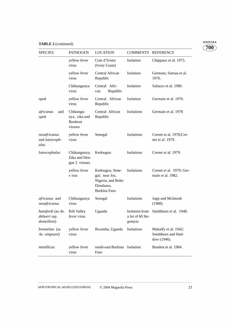
TABLE 2. Isolation of pathogens of actual or potential medical significance from Aedes (Stego-myia) species in the Afrotropical Region
SPECIES PATHOGEN LOCATION COMMENTS REFERENCE
aegypti yellow fever virus
Gambia Isolation Germain et al. 1980.
africanus yellow fever virus
Uganda Isolation Smithburn and Had-dow (1946); Smithburn et al. 1949; Haddow (1968); Kirya et al. 1977.
Chikungunya virus
Uganda Isolation Weinbren et al. 1958; Haddow et al. 1961; McCrae et al. 1971.
Rift Valley fever virus
Uganda Isolation Weinbren et al. 1957.
Zika virus Uganda Isolation Dick et al. 1952; Wein-bren and Williams (1958); Haddow et al. 1964.
yellow fever virus
Ethiopia Isolation Serie et al. 1968.
.....continued on the next page
© 2004 Magnolia Press 21AFROTROPICAL AEDES (STEGOMYIA)
700ZOOTAXA
TABLE 2 (continued)
SPECIES PATHOGEN LOCATION COMMENTS REFERENCE
yellow fever virus
Cote d’Ivoire (Ivory Coast)
Isolation Chippaux et al. 1975.
yellow fever virus
Central African Republic
Isolation Germain, Sureau et al. 1976.
Chikungunya virus
Central Afri-can Republic
Isolation Saluzzo et al. 1980.
opok yellow fever virus
Central African Republic
Isolation Germain et al. 1976.
africanus and opok
Chikungu-nya, zika and Bouboui viruses
Central African Republic
Isolations Germain et al. 1978
neoafricanus and luteoceph-alus
yellow fever virus
Senegal Isolations Cornet et al. 1978;Cor-net et al. 1979.
luteocephalus Chikungunya, Zika and Den-gue 2 viruses
Kedougou Isolations Cornet et al. 1979.
yellow fever v irus
Kedougou, Sene-gal; near Jos, Nigeria; and Bobo Dioulasso, Burkina Faso
Isolations Cornet et al. 1979; Ger-main et al. 1982.
africanus and neoafricanus
Chikungunya virus
Senegal Isolations Jupp and McIntosh (1988)
hansfordi (as Ae. deboeri ssp. demeilloni)
Rift Valley fever virus
Uganda Isolation from a lot of 60 Ste-gomyia
Smithburn et al. 1948.
bromeliae (as Ae. simpsoni)
yellow fever virus
Bwamba, Uganda Isolations Mahaffy et al. 1942; Smithburn and Had-dow (1946).
metallicus yellow fever virus
south-east Burkina Faso
Isolation Baudon et al. 1984.
HUANG22 © 2004 Magnolia Press
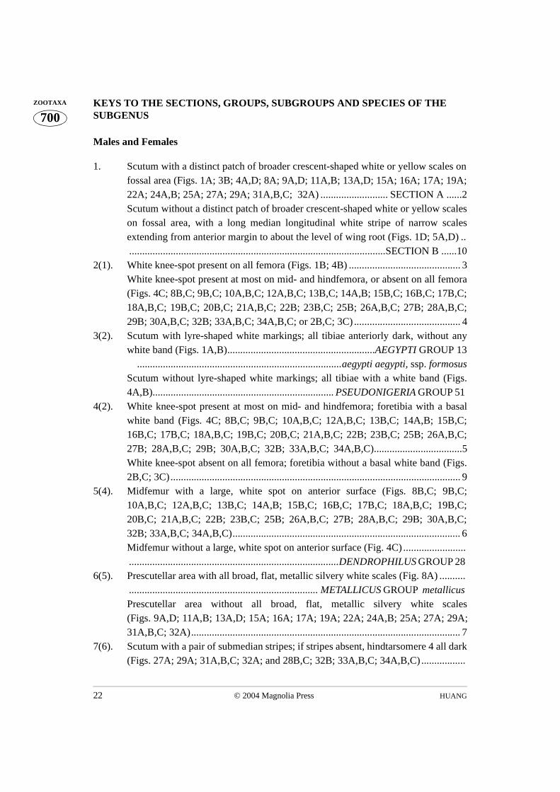
700ZOOTAXA KEYS TO THE SECTIONS, GROUPS, SUBGROUPS AND SPECIES OF THE
SUBGENUS Males and Females
1. Scutum with a distinct patch of broader crescent-shaped white or yellow scales onfossal area (Figs. 1A; 3B; 4A,D; 8A; 9A,D; 11A,B; 13A,D; 15A; 16A; 17A; 19A;22A; 24A,B; 25A; 27A; 29A; 31A,B,C; 32A) .......................... SECTION A ......2Scutum without a distinct patch of broader crescent-shaped white or yellow scaleson fossal area, with a long median longitudinal white stripe of narrow scalesextending from anterior margin to about the level of wing root (Figs. 1D; 5A,D) .....................................................................................................SECTION B ......10
2(1). White knee-spot present on all femora (Figs. 1B; 4B) ........................................... 3White knee-spot present at most on mid- and hindfemora, or absent on all femora(Figs. 4C; 8B,C; 9B,C; 10A,B,C; 12A,B,C; 13B,C; 14A,B; 15B,C; 16B,C; 17B,C;18A,B,C; 19B,C; 20B,C; 21A,B,C; 22B; 23B,C; 25B; 26A,B,C; 27B; 28A,B,C;29B; 30A,B,C; 32B; 33A,B,C; 34A,B,C; or 2B,C; 3C) ......................................... 4
3(2). Scutum with lyre-shaped white markings; all tibiae anteriorly dark, without anywhite band (Figs. 1A,B).........................................................AEGYPTI GROUP 13
...............................................................................aegypti aegypti, ssp. formosusScutum without lyre-shaped white markings; all tibiae with a white band (Figs.4A,B)...................................................................... PSEUDONIGERIA GROUP 51
4(2). White knee-spot present at most on mid- and hindfemora; foretibia with a basalwhite band (Figs. 4C; 8B,C; 9B,C; 10A,B,C; 12A,B,C; 13B,C; 14A,B; 15B,C;16B,C; 17B,C; 18A,B,C; 19B,C; 20B,C; 21A,B,C; 22B; 23B,C; 25B; 26A,B,C;27B; 28A,B,C; 29B; 30A,B,C; 32B; 33A,B,C; 34A,B,C)..................................5White knee-spot absent on all femora; foretibia without a basal white band (Figs.2B,C; 3C)................................................................................................................ 9
5(4). Midfemur with a large, white spot on anterior surface (Figs. 8B,C; 9B,C;10A,B,C; 12A,B,C; 13B,C; 14A,B; 15B,C; 16B,C; 17B,C; 18A,B,C; 19B,C;20B,C; 21A,B,C; 22B; 23B,C; 25B; 26A,B,C; 27B; 28A,B,C; 29B; 30A,B,C;32B; 33A,B,C; 34A,B,C)........................................................................................ 6Midfemur without a large, white spot on anterior surface (Fig. 4C) .........................................................................................................DENDROPHILUS GROUP 28
6(5). Prescutellar area with all broad, flat, metallic silvery white scales (Fig. 8A) ................................................................................... METALLICUS GROUP metallicus
Prescutellar area without all broad, flat, metallic silvery white scales(Figs. 9A,D; 11A,B; 13A,D; 15A; 16A; 17A; 19A; 22A; 24A,B; 25A; 27A; 29A;31A,B,C; 32A)........................................................................................................ 7
7(6). Scutum with a pair of submedian stripes; if stripes absent, hindtarsomere 4 all dark(Figs. 27A; 29A; 31A,B,C; 32A; and 28B,C; 32B; 33A,B,C; 34A,B,C).................
© 2004 Magnolia Press 23AFROTROPICAL AEDES (STEGOMYIA)
700ZOOTAXA.............................................................................................SIMPSONI GROUP 54
Scutum without a pair of submedian stripes; hindtarsomere 4 with a basal whiteband or all white (Figs. 9A,D; 11A,B; 13A,D; 15A; 16A; 17A; 19A; 22A; 24A,B;25A; and 9B,C; 10A,B,C; 12A,B,C; 13B,C; 14A,B; 15B,C; 16B,C; 17B,C;18A,B,C; 19B,C; 20B,C; 21A,B,C; 22B; 23B,C; 25B).......................................... 8
8(7). Hindtibia anteriorly dark, without or with a white stripe on ventral surface in basalarea; abdominal basal white band on terga VI–VII extended at most to 0.4 lengthof tergum (Figs. 16B,C; 17B,C; 18A,B,C; 19B,C; 20B,C; 21A,B,C; 22B; 23B,C;25B; and 22C,D; 25C,D)........................................................POWERI GROUP 41 Hindtibia anteriorly dark, with a subbasal white stripe; abdominal basal whiteband on terga VI–VII rather long, extended to 0.5–0.9 length of tergum (Figs.9B,C; 10A,B,C; 12A,B,C; 13B,C; 14A,B; 15B,C; and 11C) .............................................................................................................. APICOARGENTEUS GROUP 22
9(4). Midfemur with 3 large, white patches on anterior surface (on basal, median andapical areas); hindtarsomere 5 all dark (Fig. 3C)............ AFRICANUS GROUP 15 Midfemur without large, white patches on anterior surface; hindtarsomere 5 all
white (Figs. 2B,C)............................................... AEGYPTI GROUP mascarensis1 10(1). Proboscis with a long white line on dorsal surface; all tibiae anteriorly dark, each
with a median white line on anterior surface (Figs. 5B; 7B,C) ..........................................................................................................................GRANTI GROUP granti
Proboscis dark-scaled, without a long white line on dorsal surface; all tibiae ante-riorly dark, without any white line on anterior surface (Figs. 5C; 6B,C)............. 11
11(10). Midfemur with a large, white spot on anterior surface (Figs. 6B,C)...............................................................................................UNILINEATUS GROUP unilineatusMidfemur without a large, white spot on anterior surface (Fig. 1C) .............................................................................................................SCUTELLARIS GROUP 12
12(11). Supraalar white line incomplete, not clearly defined and with only narrow scalesover wing root (Fig. 1D)........................ (ALBOPICTUS SUBGROUP) albopictusSupraalar white line complete and well developed, with broad flat scales overwing root and toward scutellum (Huang 1972c, Fig.21A; Huang 1979a, Fig. 30B) ............................................................................... (SCUTELLARIS SUBGROUP) Not represented in the Afrotropical Region.
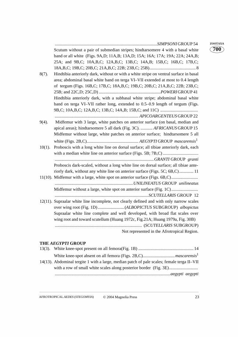
THE AEGYPTI GROUP 13(3). White knee-spot present on all femora(Fig. 1B) .................................................. 14
White knee-spot absent on all femora (Figs. 2B,C).............................mascarensis1
14(13). Abdominal tergite 1 with a large, median patch of pale scales; female terga II–VIIwith a row of small white scales along posterior border (Fig. 3E)...................................................................................................................................aegypti0aegypti
HUANG24 © 2004 Magnolia Press
700ZOOTAXA Abdominal tergite 1 without any median patch of pale scales; female terga II–VII
without a row of small pale scales along posterior border (Fig. 3F) ....sp. formosus1 Malagasy species (Mauritius).
THE AFRICANUS GROUP(male of maxgermaini unknown)15(9). Hindfemur anteriorly with a large pale band at base and with 2 large, white
patches on median and apical areas (Huang 1990, Figs. 8A, B) ....... luteocephalusHindfemur anteriorly without such a pale band at base, or hindfemur anteriorlywith 3 large, white patches on basal, median and apical areas (Huang 1990, Figs.2A, B, C; 4A, B, C; 5C; 6A, B, C; 7B, D; 8C) .................................................... 16
16(15). Posterior dorsocentral yellow line of narrow scales well developed, reaching for-ward to the posterior end of the fossal white patch; lateral lobe of scutellum withbroad dark scales (Huang 1990, Fig. 7C).................................................ruwenzoriPosterior dorsocentral yellow line of narrow scales not developed or, if present,not reaching to the posterior end of the fossal white patch; lateral lobe of scutel-lum with broad white scales (Huang 1990, Figs. 1A; 3A, D; 5A, D; 7A)............ 17
17(16). Posterior dorsocentral yellow or white line of narrow scales present (Huang 1990,Figs. 5A, D; 7A) ................................................................................................... 18
Posterior dorsocentral yellow or white line of narrow scales not developed (Huang1990, Figs. 1A; 3A, D) ......................................................................................... 20
18(17). Fossal white patch rather broad at base along scutal margin; prescutellar line welldeveloped, with narrow yellow scales and with some broad, flat, metallic silverywhite scales posteriorly (Huang 1990, Fig. 7A)............................................... opok Fossal white patch rather narrow at base along scutal margin; prescutellar line notdeveloped or, if present, with only narrow yellow scales (Huang 1990, Figs. 5A, D).............................................................................................................................. 19
19(18). Anterior median white stripe rather long, 2.5–3.0 times as long as wide; hindlegwith tarsal claws equal and simple (Huang 1990, Figs. 5D, E)......pseudoafricanusAnterior median white stripe short and broad, about 2 times as long as long aswide; hindleg with tarsal claws equal and toothed (Huang 1990, Figs. 5A, B) ................................................................................................................... maxgermaini
20(17). Hindtibia with a white stripe on ventral surface in basal 0.20 or more; male fore-and midlegs with tarsal claws unequal, the smaller one toothed, the larger one sim-ple; hindleg with tarsal claws equal and toothed (Huang 1990, Figs. 2A, B, C; 4A;3B, C).................................................................................................................... 21Hindtibia without, or with a very short white stripe on ventral surface in basal 0.08or less; male fore- and midlegs with tarsal claws unequal, all simple; hindlegwith tarsal claws equal and simple (Huang 1990, Figs. 4B, C; 3E, F) .................................................................................................................................. neoafricanus
21(20). Hindfemur with 3 large, white patches on the anterior surface (on basal, median
© 2004 Magnolia Press 25AFROTROPICAL AEDES (STEGOMYIA)
700ZOOTAXAand apical areas) (Huang 1990, Figs. 2C; 4A)...........................................africanus
Hindfemur with at most 2 large, white patches on the anterior surface (on medianand apical areas) (Huang 1990, Figs. 2A, B).................................................corneti
THE APICOARGENTEUS GROUP 22(8). Scutellum with broad white scales on midlobe and with broad dark scales on lat-
eral lobes (Figs. 9A; 11A,B)................................................................................. 23Scutellum with broad white scales on all lobes (Figs. 9D; 13A,D; 15A)............. 25
23(22). Hindtarsomere 5 all dark (Figs. 9B; 10A)........................................apicoargenteusHindtarsomere 5 with a basal white band or all white (Figs. 10C; 12A,B,C) ...... 24
24(23). Hindtarsomere 4 with basal 0.40 or less white on dorsal surface (Figs. 12A,B) ........................................................................................................................ ealaensis Hindtarsomere 4 with basal 0.89 or more white on dorsal surface (Figs. 10C; 12C)............................................................................................................... denderensis
25(22). Hindtarsomere 5 all dark (Figs. 9C; 10B)...................................................schwetziHindtarsomere 5 with a basal white band or all white (Figs. 13B,C; 14A,B; 15B,C).............................................................................................................................. 26
26(25). Scutum with anterior median white spot of narrow scales; female fore- and mid-legs with tarsal claws equal and toothed (Figs. 15A,D).............................. soleatusScutum with anterior median white spot of broad scales; female fore- and midlegswith tarsal claws equal and simple (Figs. 13A,D; 14C) ....................................... 27
27(26). Hindfemur anteriorly with basal 0.20-0.25 white, and with a large white spot0.60-0.64 from base, the white spot not connecting with the basal white area(Figs. 13B; 14A).......................................................................................blacklockiHindfemur anteriorly with a broad white stripe in basal 0.50-0.53 (Figs. 13C; 14B)....................................................................................................................... fraseri
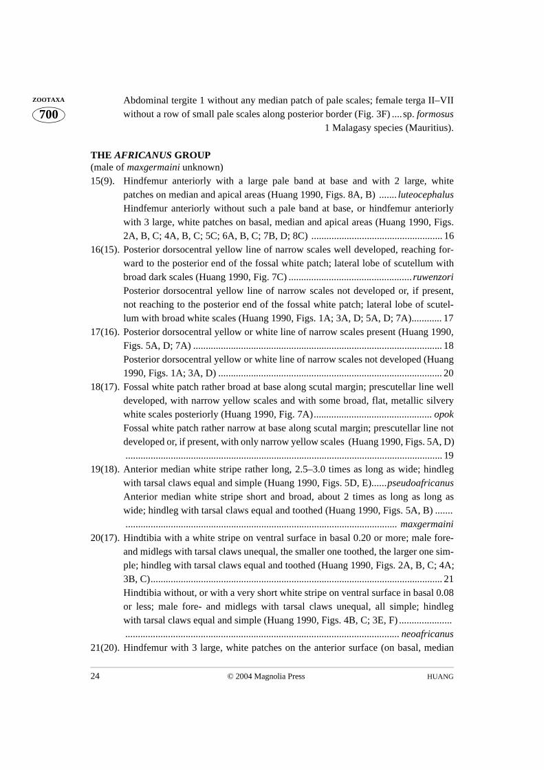
THE DENDROPHILUS GROUP(males of muroafcete and njombiensis unknown) 28(5). Hindtibia with a white stripe on ventral surface in basal area (Huang 1997, Figs.
3A,B,C; 4A,B,C; 5B; 6B,C; 7B,C; 8B,C; 9B,C; 13B,C; 14B,C) ......................... 29Hindtibia without a white stripe on ventral surface in basal area (Huang 1997,Figs. 1B,C; 10B,C; 11B; 12B,C) .......................................................................... 38
29(28). White knee-spot absent on hindfemur, or represented by few pale scales (Huang1997, Figs. 3A,B; 5B).......................................................................................... 30
White knee-spot present and well developed on hindfemur (Huang 1997, Figs. 3C;4A,B,C; 6B,C; 7B,C; 8B,C; 9B,C; 13B,C; 14B,C) .............................................. 31
30(29). Hindtarsomere 5 all dark (Huang 1997, Fig. 5B).................................. muroafceteHindtarsomere 5 with basal 0.50–0.75 white on dorsal surface (Huang 1997, Figs.3A,B).........................................................................................................bambusae
31(29). Hindtarsomere 5 all dark (Huang 1997, Figs. 4B,C)....................................deboeri

HUANG26 © 2004 Magnolia Press
700ZOOTAXA Hindtarsomere 5 with a basal white band or all white (Huang 1997, Figs. 3C; 4A;
6B,C; 7B,C; 8B,C; 9B,C; 13B,C; 14B,C) ............................................................ 3232(31). Scutum with anterior median white spot of broad scales (Huang 1997, Fig. 13A).
...........................................................................................................mattinglyorumScutum with anterior median white spot of narrow scales (Huang 1997, Figs. 5C;7C) ........................................................................................................................33
33(32). Hindleg with tarsal claws equal and toothed (Huang 1997, Figs. 2C,D; 7D,E; 8A,D).............................................................................................................................. 34Hindleg with tarsal claws equal and simple (Huang 1997, Figs. 14A,D) ............ 36
34(33). Female fore-, mid- and hindlegs with tarsal claws equal and toothed (modifiedtooth); male hindleg with tarsal claws equal and toothed (modified tooth) (Huang1997, Figs. 8A,D).....................................................................................hansfordi
Female fore-, mid- and hindlegs with tarsal claws equal and toothed (normaltooth); male hindleg with tarsal claws equal and toothed (normal tooth) (Huang1997, Figs. 7D,E)................................................................................................. 35
35(34) Hindtarsomere 3 with basal 0.2 or less white on dorsal surface; hindtarsomere 5with basal 0.47-0.88 white on dorsal surface (Huang 1997, Figs. 3C; 4A).. kenyaeHindtarsomere 3 with basal 0.32-0.41 white on dorsal surface; hindtarsomere 5 allwhite (female), or with basal 0.84-0.89 white to all white on dorsal surface (male)(Huang 1997, Figs. 7B,C).................................................................... dendrophilus
36(33). Female fore- and midlegs with tarsal claws equal and simple; male fore- and mid-legs with tarsal claws unequal, all simple (Huang 1997, Figs. 14A,D)..................................................................................................................................segermanaeFemale fore- and midlegs with tarsal claws equal and toothed; male fore- and mid-legs with tarsal claws unequal, the smaller one toothed, the larger one simple(Huang 1997, Figs. 6A,D; 9A,D) ........................................................................ 37
37(36). Midtarsomere 1 with a well-marked white stripe on posterior surface; hindtarsom-ere 5 with basal 0.40-0.67 white on dorsal surface (Huang 1997, Figs. 9B,C) ..............................................................................................................................heischi Midtarsomere 1 without a well-marked white stripe on posterior surface; hindtar-somere 5 all white, or all white except tip (Huang 1997, Figs. 6B,C)... demeilloni
38(28). Hindtarsomere 3 with basal 0.11–0.25 white on dorsal surface (Huang 1997, Figs.10B,C; 11B)...........................................................................................................39 Hindtarsomere 3 all dark (Huang 1997, Figs. 1B,C; 12B,C) ............................. 40
39(38). Scutum with anterior median white spot of broad scales (Huang 1997, Fig. 11C) ..................................................................................................................... keniensisScutum with anterior median white spot of narrow scales (Huang 1997, Fig. 11A)...............................................................................................................njombiensis
40(38). Scutum with yellow median stripe, the yellow median stripe connects with theanterior median white spot; male fore- and midlegs with tarsal claws unequal, the
© 2004 Magnolia Press 27AFROTROPICAL AEDES (STEGOMYIA)
700ZOOTAXAsmaller one toothed, the larger one simple (Huang 1997, Figs. 12A,E)...... masseyi
Scutum with white median stripe, the white median stripe does not connect withthe anterior median white spot; male fore- and midlegs with tarsal claws unequal,all toothed (Huang 1997, Figs. 1A,D)......................................................amaltheus
THE POWERI GROUP(males of chaussieri and poweri unknown)41(8). Hindtibia with a white stripe on ventral surface in basal area (Figs. 16B,C; 17B,C;
18A,B,C; 19B,C; 20B,C; 21A,B,C; 22B; 23B,C) ................................................ 42Hindtibia without a white stripe on ventral surface in basal area (Fig. 25B) ............................................................................................................................chaussieri
42(41). Scutum with a large (crescent-shaped) patch of pale yellow scales on fossal area,fossal pale yellow patch with anterior end extending along scutal margin towardsthe median pale yellow stripe (Fig. 16A) ............................................................. 43Scutum with a large patch of broader crescent-shaped white scales on fossal area,fossal white patch without anterior end extending along scutal margin towards theanterior median white spot (Figs. 17A; 19A; 22A; 24A,B) ................................. 44
43(42). White knee-spot absent on hindfemur (Figs. 16B,C).................................angustusWhite knee-spot present and well developed on hindfemur (Fig. 18A)..mpusiensis
44(42). Scutum with anterior median white spot of narrow scales; hindtarsomere 5 allwhite (Figs. 17A,B,C) .......................................................................................... 45
Scutum with anterior median white spot of broad scales; hindtarsomere 5 all dark(Figs. 19A; 22A; 24A,B; 18C; 19B,C; 20B,C; 21A,B,C; 22B; 23B,C) ............... 46
45(44). Midtibia with a white stripe on ventral surface in basal area (Figs. 17B,C) .............................................................................................................................. usambara Midtibia without a white stripe on ventral surface in basal area (Fig. 18B) .......................................................................................................................... .ethiopiensis
46(44). Midtarsomeres 1 and 2 with white stripe on posterior surface (Figs. 24C1,2;24D1,2)..................................................................................................................47Midtarsomeres 1 and 2 without white stripe on posterior surface (Figs. 24C3–6;24D3–5) ............................................................................................................... 48
47(46). Hindtarsomere 3 with basal 0.25 or more white on dorsal surface (Figs. 21A,B) ...................................................................................................................... calceatusHindtarsomere 3 with basal 0.17 or less white on dorsal surface (Figs. 21C; 22B) .......................................................................................................................ledgeri
48(46). Midtibia with a white stripe on ventral surface in basal area (Figs. 18C; 19B,C) 49Midtibia without a white stripe on ventral surface in basal area (Figs. 20B,C;23B,C)...................................................................................................................50
49(48). Female midtarsomere 2 with basal 0.9 or more white on dorsal surface (Fig. 19C) .......................................................................................................................poweriFemale midtarsomere 2 with basal 0.6 or less white on dorsal surface; male mid-
HUANG28 © 2004 Magnolia Press
700ZOOTAXA tarsomere 2 with basal 0.35 or less white on dorsal surface (Figs. 19B; 18C).........
........................................................................................................... hogsbackensis50(48). Midtarsomere 2 with basal 0.4 or less white on dorsal surface (Figs. 23B,C) ........
...................................................................................................................contiguusMidtarsomere 2 with basal 0.66 or more white on dorsal surface (Figs. 20B,C) .......................................................................................................................... langata
THE PSEUDONIGERIA GROUP(males of mickevichae, pseudonigeria and saimedres unknown)51(3). Scutum with anterior median white spot of broad scales (Huang 1988b, Figs. 2A;
3A) ........................................................................................................................52Scutum with anterior median white spot of narrow scales (Huang 1988b, Fig. 2C) .............................................................................................................................. 53
52(51). Hindtarsomere 3 with basal 0.33 white on dorsal surface; hindtarsomere 5 allwhite (Huang 1988b, Fig. 3D)........................................................... pseudonigeriaHindtarsomere 3 with basal 0.25 or less white on dorsal surface; hindtarsomere 5all dark (Huang 1988b, Fig. 2B)............................................................... saimedres
53(51). Hindtarsomere 3 all dark; hindtarsomere 4 all white; hindtarsomere 5 all dark(Huang 1988b, Fig. 2D) ....................................................................... mickevichae
Hindtarsomere 3 with basal 0.33 or more white on dorsal surface; hindtarsomere 4with basal 0.63 or less white on dorsal surface; hindtarsomere 5 all white (Huang
1988b, Fig. 5) .................................................................................. chemulpoensis2
2 Palearctic species (Korea, N.E. China).
THE SIMPSONI GROUP(males of gandaensis, kivuensis and sampi unknown)54(7). Scutellum with broad white scales on midlobe and with broad dark scales on lat-
eral lobes (Fig. 31C) ............................................................................................. 55Scutellum with broad white scales on all lobes (Figs. 27A; 29A; 31A,B; 32A) . 56
55(54). Fore- and midtarsi with a basal white band on tarsomeres 1–5; hindtarsomere 4with basal 0.6 white on dorsal surface (Fig. 26A) ................................. gandaensisFore- and midtarsi with a basal white band on tarsomeres 1,2; hindtarsomere 4 alldark (Figs. 26B,C).......................................................................................... woodi
56(54). Hindtarsomere 4 with a basal white band or all white (Figs. 27B; 28A; 29B;30A,B,C) ...............................................................................................................57Hindtarsomere 4 all dark (Figs. 28B,C; 32B; 33A,B,C; 34A,B,C) ..................... 60
57(56). Hindtarsomere 4 with a basal white band (Figs. 30A,B,C) ................................. 58Hindtarsomere 4 all white (Figs. 27B; 28A; 29B)............................................... 59
58(57). Hindfemur anteriorly with basal 0.33–0.40 white, and with a large white spot about0.67 from base, the white spot not connecting with the basal white area; hindtar-somere 5 with basal 0.40–0.67 white on dorsal surface (Figs. 30B,C) ..................
© 2004 Magnolia Press 29AFROTROPICAL AEDES (STEGOMYIA)
700ZOOTAXA.............................................................................................................subargenteus
Hindfemur anteriorly with a broad white stripe in basal 0.57; hindtarsomere 5 allwhite (Fig. 30A)...............................................................................................sampi
59(57). Hindfemur anteriorly with a broad white stripe in basal 0.50, and with a white spotabout 0.62 from base, the white spot not connecting with the basal white stripe;tergum I without a median white spot (Figs. 29B,C,D)............................. kivuensisHindfemur anteriorly with a broad white stripe in basal 0.60-0.66; tergum I with amedian white spot (Figs. 27B,C,D; 28A)....................................................josiahae
60(56). Hindtibia with a white stripe on ventral surface in basal 0.25-0.30 (Figs. 28B,C) .................................................................................................................. strelitziae
Hindtibia without a white stripe on ventral surface in basal area (Figs. 32B;33A,B,C; 34A,B,C) ............................................................................................. 61
61(60). Fore- and midtarsomere 2 with basal 0.80 or more white on dorsal surface; femalefore- and midlegs with tarsal claws equal and simple (Figs. 32B; 33A; 34A; and 31D) .............................................................................................................simpsoniForetarsomere 2 with basal 0.64 or less white on dorsal surface; midtarsomere 2with basal 0.75 or less white on dorsal surface; female fore- and midlegs with tar-sal claws equal and toothed (Figs. 33B,C; 34B,C; and 31E,F) ........................... 62
62(61). Midtarsomere 2 with basal 0.50 or less white on dorsal surface; hindtarsomere 5with basal 0.50-0.75 white on dorsal surface (Figs. 33B; 34B) .........................liliiMidtarsomere 2 with basal 0.54 or more white on dorsal surface; hindtarsomere 5all white, or all white except tip (Figs. 33C; 34C).................................... bromeliae
Male Genitalia
1. Paraproct with a sternal arm (Figs.35A; 36A,B; 37A,B; 38A,B; 39A,B; 40A,B;41A,B; 42A,B; 43A,B; 44A,B,C,D; 45A,B; 46A,B; 47A,B; 48A,B; 50A; Huang1990, Figs.10C; 13C; 16C; 20A,B; 23A,B; Huang 1997, Figs.15A,B; 16A,B;17A,B; 18A,B; 19A; 20A,B; 21C) ................................................... SECTION A 2Paraproct without a sternal arm (Figs.35B; 49A,B) .......................SECTION B 13
2(1). Apical margin of tergum IX with slightly separated lateral lobe (Figs. 47A; 42A)3Apical margin of tergum IX with well separated lateral lobe (Figs. 35A; 36A,B;37A,B; 38A,B; 39A,B; 40A,B; 41A,B; 42B; 43A,B; 44D; 45A,B; 46A,B; 47B;48A,C; 50A; Huang 1990, Figs. 10C; 13C; 16C; 20A,B; 23A,B; Huang 1977,Figs. 15A,B; 16A,B; 17A,B; 18A,B; 19A;20A,B; 21C) ........................................ 4
3(2). Aedeagus with short teeth only; gonostylar claw very short, about 0.13 length ofgonostylus (Fig. 47A) ...................................... SIMPSONI GROUP subargenteusAedeagus with lateral teeth longer than distal teeth; gonostylar claw rather long,about 0.32 length of gonostylus (Fig. 42A)............... POWERI GROUP contiguus
4(2). Aedeagus with short teeth only; paraproct with sternal arm long and slender; api-cal margin of tergum IX with middle slightly concave to concave (Huang 1990,
HUANG30 © 2004 Magnolia Press
700ZOOTAXA Figs. 10C; 13C; 16C; 20A,B; 23A,B).............................. AFRICANUS GROUP 16
Without this combination of characters . ................................................................ 55(4). Apical margin of tergum IX with middle flat, or slightly concave (Figs. 39A; 42B;
43A,B; 44D) ........................................................................................................... 6Apical margin of tergum IX with middle concave, or deeply concave (Figs.45A,B; 46A,B; 47B; 48A; 35A; 48C; 36A,B; 37A,B; 38A,B; 39B; 40A,B; 41A,B;50A; Huang 1977, Figs. 15A,B; 16A,B; 17A,B; 18A,B; 19A; 20A,B; 21C) ........ 7
6(5). Paraproct with a sternal arm very short, less than 0.4 length of apical arm; gono-stylar claw short and stout; aedeagus with a few very long and slender teeth (Figs.44C,D)...............................................................METALLICUS GROUP metallicus
Paraproct with a sternal arm not very short, about 0.5 to 1.4 length of apical arm;gonostylar claw longer and slender (Figs. 39A; 42B; 43A,B) ............................................................POWERI GROUP 39 calceatus, ledgeri, langata, hogsbackensis
7(5). Lateral lobe of tergum IX with long and strong setae; apical margin of tergum IXwith middle deeply concave and with widely separated lateral lobe; aedeagus withshort teeth only (Figs. 45A,B; 46A,B; 47B; 48A)...............SIMPSONI GROUP 47 ............................................. bromeliae, josiahae, lilii, simpsoni, strelitziae, woodiLateral lobe of tergum IX with short, or median size setae (Figs. 35A; 48C;36A,B; 37A,B; 38A,B; 39B; 40A,B; 41A,B; 50A; Huang 1977, Figs. 15A,B;16A,B; 17A,B; 18A,B; 19A; 20A,B; 21C)............................................................. 8
8(7). Gonostylus rather short, curved (Figs. 35A; 48C; 50A) ........................................ 9Gonostylus simple, elongate (Huang 1977, Fig. 15A,B; 16A,B; 17A,B; 18A,B;19A; 20A,B; 21C; Figs. 40A,B; 41A,B) ............................................................. 10
9(8). Gonostylus short, curved, tapering, with a slender, short gonostylar claw processat apex; lateral lobe of tergum IX with 1–4 very fine short setae; apical margin oftergum IX with middle deeply concave and with widely separated lateral lobe(Figs. 35A; 48C) ...................................................................AEGYPTI GROUP 15
...........................................................aegypti aegypti, ssp. formosus, mascarensis1
Gonostylus short and curved, not tapering, with a short gonostylar claw process atapex; lateral lobe of tergum IX with median size setae; apical margin of tergum IX
with middle concave (Fig. 50)......... PSEUDONIGERIA GROUP chemulpoensis2
10(8). Lateral lobe of tergum IX with 3–5 short setae; gonostylus long, with a long slen-der gonostylar claw process near apex; apical margin of tergum IX with middledeeply concave (Huang 1977, Fig. 15A)... DENDROPHILUS GROUP amaltheusLateral lobe of tergum IX with median size setae (Figs. 36A,B; 37A,B; 38A,B;39B; 40A,B; 41A,B; Huang 1977, Figs. 15B; 16A,B; 17A,B; 18A,B; 19A;20A,B; 21C) ......................................................................................................... 11
1. Malagasy species (Mauritius).2. Palearctic species (Korea, N.E. China).
© 2004 Magnolia Press 31AFROTROPICAL AEDES (STEGOMYIA)
700ZOOTAXA11(10). Gonostylus long, at least 0.66 length of gonocoxite; gonostylar claw very short,
about 0.12–0.13 length of gonostylus; apical margin of tergum IX with middledeeply concave (Figs. 40A,B; 41A,B) ...................................POWERI GROUP 39......................................................... angustus, ethiopiensis, mpusiensis, usambaraWithout this combination of characters ................................................................ 12
12(11). Claspette with apicomesal angle formed a thumb-like projection, or formed aslight projection; gonostylus curved, tapering, with a gonostylar claw process atapex (Figs. 36A,B; 37A,B; 38A,B; 39B) ........... APICOARGENTEUS GROUP 22Claspette without any projecting portion; gonostylus almost straight, not curved,scarcely tapering, with a long slender or short stout gonostylar claw process atapex (Huang 1977, Figs. 15B; 16A,B; 17A,B; 18A,B; 19A; 20A,B; 21C) .............................................................................................DENDROPHILUS GROUP 28bambusae, deboeri, demeilloni, dendrophilus, hansfordi, heischi, keniensis, ken-yae, masseyi, mattinglyorum, segermanae
13(1). Gonostylar claw long, at least 0.25 length of gonostylus; apical margin of tergumIX with middle part produced into a lobe (Figs. 35B; 49A,B) ............................ 14Gonostylar claw rather short, at most 0.20 length of gonostylus; apical margin oftergum IX with conspicuous horn-like median projection (Fig. 35B) ........................................... SCUTELLARIS GROUP (ALBOPICTUS SUBGROUP) albopictus
14(13). Claspette with numerous simple setae on the elevated distal portion and bearing nowidened specialized setae (Fig. 49A)...............................GRANTI GROUP grantiClaspette long, slender, with numerous simple setae and several stouter widenedsetae on distal part, with a small median mesally directed projection bearing onelarge seta and with 3–4 smaller setae near to it (Fig. 49B) .............................................................................................................UNILINEATUS GROUP unilineatus
THE AEGYPTI GROUP 15(9). Gonostylus somewhate swollen in the middle, with apical 0.28 rather narrow and
curved (Fig. 35A).....................................................aegypti aegypti, ssp. formosusGonostylus not swollen in the middle, with strongly elbowed at about apical 0.35
(Fig. 48C) ........................................................................................... mascarensis1
1 Malagasy species (Mauritius).
THE AFRICANUS GROUP(male of maxgermaini unknown)16(4). Claspette with numerous simple setae on the expanded distal portion and bearing
no stronger, spine-like seta on the apicomesal corner (Huang 1990, Figs. 10C;16C) ......................................................................................................................17
Claspette with numerous simple setae on the expanded distal portion and bearing1-3 stronger, spine-like setae on the apicomesal corner (Huang 1990, Figs. 13C;20A, B; 23A, B).................................................................................................... 18
HUANG32 © 2004 Magnolia Press
700ZOOTAXA 17(16). Claspette with distal expanded portion oval in dorsal aspect, with numerous sim-
ple setae on the apicolateral portion, and with some rather short setae on the api-comesal portion (dissected) (Huang 1990, Fig. 16C) ........................ luteocephalusClaspette with distal expanded portion subtriangular in dorsal aspect (narrowstowards the apicolateral angle, becomes broader apicomesally, with apicomesalcorner rounded), with numerous simple setae on the expanded distal portion (dis-sected) (Huang 1990, Fig. 10C) ................................................................ africanus
18(16). Claspette with distal expanded portion subtriangular or oval in dorsal aspect(Huang 1990, Figs. 13C; 20A, B)......................................................................... 19Claspette with distal expanded portion square in dorsal aspect (Huang 1990, Figs.23A, B) ............................................................................................................... 21
19(18). Claspette with distal expanded portion subtriangular in dorsal aspect (narrowstowards the apicomesal angle, becomes broader apicolaterally, with apicolateralcorner rounded), with 1 stronger, spine-like seta on the apicomesal angle (Huang1990, Fig. 13C)..............................................................................................cornetiClaspette with distal expanded portion oval in dorsal aspect (Huang 1990, Figs.20A, B) ................................................................................................................ 20
20(19). Claspette with 2 stronger, spine-like setae on the apicomesal corner; gonostylarclaw rather short and stout, about 5.3 times as long as wide (Huang 1990, Fig.20A) .....................................................................................................neoafricanusClaspette with 3 stronger, spine-like setae on the apicomesal corner; gonostylarclaw long and slender, about 8.5 times as long as wide (Huang 1990, Fig. 20B)......................................................................................................................ruwenzori
21(18). Apicomesal angle of claspette with a narrow thumb-like projection and bearing 2stronger, spine-like setae (Huang 1990, Fig. 23B) ........................pseudoafricanusApicomesal angle of claspette without such a projection, with 1 strong, spine-likeseta on the apicomesal angle and with 1smaller spine-like seta laterad of it(Huang 1990, Fig. 23A) .................................................................................. opok
THE APICOARGENTEUS GROUP22(12). Claspette with distal expanded portion square in dorsal aspect, apicomesal angle
formed a thumb-like projection, with numerous simple setae on the expanded dis-tal portion and bearing no spine-like setae on the apicomesal angle (Figs. 36A,B;37A,B; 38A,B) ..................................................................................................... 23Claspette with distal expanded portion subtriangular in dorsal aspect (narrowstowards apicomesal angle, with apicolateral corner rounded), apicomesal angleformed a slight projection, with numerous simple setae on the expanded distal por-tion and bearing 2 stong, basally widened spine-like setae on the apicomesal angle(Fig. 39B) .................................................................................................... soleatus
23(22). Apicomesal angle of claspette with a thumb-like projection and bearing at its tip arow of 7–9 setae (Figs. 36A,B; 37A,B) ................................................................ 24
© 2004 Magnolia Press 33AFROTROPICAL AEDES (STEGOMYIA)
700ZOOTAXAApicomesal angle of claspette with a narrow thumb-like projection and bearing 3–
4 setae at tip of the projection (Figs. 38A,B) ....................................................... 2724(23). Claspette with apicomesal angle formed a narrower thumb-like projection and
bearing 9 setae at tip of the projection, with apicolateral corner rounded; gonosty-lar claw rather short, at most 0.20 length of gonostylus (Fig. 37B) ......... ealaensis
Claspette with apicomesal angle formed a broader thumb-like projection, with api-colateral angle pointed (Figs. 36A,B; 37A) ......................................................... 25
25(24). Claspette with basolateral corner rounded; gonostylar claw long, at least 0.26length of gonostylus (Fig. 37A) ........................................................... denderensis
Claspette with basolateral corner drawn into a broad beak-like projection (Figs.36A,B) .................................................................................................................26
26(25). Claspette with lateral side rather straight, with 8–9 setae on the apicomesal angle(Fig. 36A) ........................................................................................apicoargenteusClaspette with lateral side rounded, with 7 setae on the apicomesal angle (Fig. 36B) .....................................................................................................................schwetzi
27(23). Gonostylar claw rather short and stout, about 5.5 times as long as wide (Fig. 38B) ..................................................................................................................blacklockiGonostylar claw long and slender, about 7 times as long as wide (Fig. 38A) fraseri
THE DENDROPHILUS GROUP(males of muroafcete and njombiensis unknown)28(12). Claspette simple, short, rounded apically, with numerous simple setae on the
slightly expanded distal portion and bearing no spine-like seta (Huang 1997, Fig. 15A) ..................................................................................................................amaltheusClaspette large, lobed, distal expanded portion square, or subtriangular, or oval indorsal aspect, with numerous simple setae on the expanded distal portion andbearing 1–5 strong, basally widened spine-like setae (Huang 1997, Figs. 15B;16A,B; 17A,B; 18A,B; 19A; 20A,B; 21C) .......................................................... 29
29(28). Claspette with distal expanded portion square in dorsal aspect (narrows towardsapicolateral angle, with basolateral corner rounded) (Huang 1997, Figs. 16B; 18A) .............................................................................................................................. 30Claspette with distal expanded portion subtriangular, or oval in dorsal aspect(Huang 1997, Figs. 15B; 16A; 17A,B; 18B; 19A; 20A,B; 21C) ......................... 31
30(29). Claspette with 2 stronger, basally widened spine-like setae on the apicomesalangle; aedeagus with lateral teeth longer and/or stouter than the others (Huang1997, Fig. 18A) .................................................................................. dendrophilusClaspette with 3 (2–3) stout, basally widened spine-like setae on the apicomesalangle; aedeagus with all rather short teeth (Huang 1997, Fig. 16B) ............ kenyae
31(29). Claspette with distal expanded portion subtriangular in dorsal aspect (Huang 1997,Figs. 15B; 18B)..................................................................................................... 32Claspette with distal expanded portion oval in dorsal aspect (Huang 1997, Figs.
HUANG34 © 2004 Magnolia Press
700ZOOTAXA 16A; 17A,B; 19A; 20A,B; 21C) .......................................................................... 33
32(31). Claspette narrows towards apical angle, with a 90o basolateral angle, with 2 stout,basally widened spine-like setae on the basomesal angle; aedeagus with lateralteeth longer and stouter than the others (Huang 1997, Fig. 18B) ............hansfordi
Claspette narrows towards apical angle, with basolateral corner rounded, with 3(2–4) stout, basally widened spine-like setae on the basomesal angle; aedeaguswith all rather short teeth (Huang 1997, Fig. 15B) ..................................bambusae
33(31). Claspette with lateral and mesal sides more or less parallel (Huang 1997, Figs.16A; 21C) ............................................................................................................ 34Claspette narrows towards apex, broadened at base (Huang 1997, Figs. 17A,B;19A; 20A,B) ........................................................................................................ 35
34(33). Claspette with 2–3 stout, basally widened spine-like setae on the mesal side(Huang 1997, Fig. 16A) ...............................................................................deboeriClaspette with 1 strong, basally widened spine-like seta on the basomesal corner(Huang 1997, Fig. 21C) ....................................................................mattinglyorum
35(33). Aedeagus with 2–3 teeth distinctly longer than the others (Huang 1997, Figs.17A,B; 19A) ........................................................................................................ 36Aedeagus without 2–3 teeth distinctly longer than the others (Huang 1997, Figs.20A,B) ..................................................................................................................38
36(35). Claspette with lateral side rather straight, with mesal side rounded, with 2–3 stout,basally widened spine-like setae on the mesal side (Huang 1997, Fig. 17B)..........................................................................................................................segermanaeClaspette narrows towards apex, broadened at base, with 2–5 stout, basally wid-ened spine-like setae on the mesal side (Huang 1997, Figs. 17A; 19A) ............. 37
37(36). Claspette with 2–3 stout, basally widened spine-like setae on mesal side near the middle(Huang 1997, Fig. 19A) ...............................................................................heischiClaspette with 4 (3–5) stout, basally widened spine-like setae on the mesal side inbasal 0.67 (Huang 1997, Fig. 17A).......................................................... demeilloni
38(35). Claspette with lateral side rather straight, with mesal side rounded, with 3–4 stout,basally widened spine-like setae on the mesal side (Huang 1997, Fig. 20A) ............................................................................................................................ keniensisClaspette narrows towards apex, broadened at base, with 3–4 stout, basally wid-ened spine-like setae on the mesal side (Huang 1997, Fig. 20B) ............... masseyi
THE POWERI GROUP (males of chaussieri and poweri unknown)39(2). Apical margin of tergum IX with slightly separated lateral lobe, each with 12–14
strong setae (Fig. 42A) ............................................................................contiguusApical margin of tergum IX with well separated lateral lobe, each with 12 or lesssetae (Figs. 39A; 40A,B; 41A,B; 42B; 43A,B) .................................................... 40

© 2004 Magnolia Press 35AFROTROPICAL AEDES (STEGOMYIA)
700ZOOTAXA40(39). Apical margin of tergum IX flat, or slightly concave medially; gonostylar claw
long, at least 0.25 length of gonostylus (Figs. 39A; 42B; 43A,B) ...................... 41Apical margin of tergum IX deeply concave medially; gonostylar claw rathershort, at most 0.14 length of gonostylus (Figs. 40A,B; 41A,B) .......................... 44
41(40). Claspette with distal expanded portion subtriangular in dorsal aspect (Figs. 42B;43A,B) ..................................................................................................................42Claspette with distal expanded portion oval in dorsal aspect, with numerous sim-ple setae on the expanded distal portion and bearing 3 stronger, basally wid-ened setae on the apicomesal corner (Fig. 39A) ........................ hogsbackensis
42(41). Claspette (narrows towards apicomesal angle), with 2–4 somewhat stronger,basally widened setae on the apicomesal angle; aedeagus with lateral teeth longerand stouter than distal teeth (Figs. 43A,B) .......................................................... 43Claspette (apicomesal corner formed a slight projection, with apicolateral cornerrounded), with 3 stronger, basally widened setae on the apicomesal corner;aedeagus with all rather short teeth (Fig. 42B) ........................................... langata
43(42). Paraproct with rather short sternal arm, less the length of the apical arm; proctigerwithout cercal setae (Fig. 44A) ..................................................................... ledgeriParaproct with a long sternal arm, as long as or longer than the apical arm; procti-ger with 2–5 long cercal setae (Fig. 44B).................................................. calceatus
44(40). Claspette with distal expanded portion rounded lobe-like in dorsal aspect (Figs. 40A,B) .............................................................................................................................. 45Claspette with distal expanded portion square in dorsal aspect (Figs. 41A,B) .... 46
45(44). Claspette rather small, rounded, with numerous simple setae on the expanded dis-tal portion and bearing 5 strong, basally widened spine-like setae on theapical margin (Fig. 40B) .........................................................................mpusiensis
Claspette large, rounded,with numerous simple setae on the expanded distal por-tion and bearing no spine-like setae (Fig. 40A)..........................................angustus
46(44). Claspette with 4 strong, basally widened spine-like setae on the apical side (Fig.41B) .......................................................................................................ethiopiensisClaspette with 9 strong, basally widened spine-like setae on the apical side andmesal side (Fig. 41A)................................................................................ usambara
THE SIMPSONI GROUP(males of gandaensis, kivuensis and sampi unknown)47(7). Apical margin of tergum IX with low and slightly separated lateral lobe, each with
8-10 strong setae (Fig. 47A)................................................................subargenteusApical margin of tergum IX with middle deeply concave and with widely sepa-rated lateral lobe, each with 3-9 setae (Figs. 45A,B; 46A,B; 47B; 48A) ............. 48
48(47). Claspette large and broad, reaching to 0.54 of gonocoxite, distal expanded portionsquare in dorsal aspect, with numerous simple setae on the expanded distal portionand bearing 2 (1-2) stronger, basally widened spine-like setae on the apicomesal

HUANG36 © 2004 Magnolia Press
700ZOOTAXA angle (Fig. 46B)...........................................................................................josiahae
Claspette large, long, reaching to 0.75-0.90 of gonocoxite, with numerous simplesetae, without any stronger, basally widened spine-like setae on the expanded dis-tal portion (Figs. 45A,B; 46A; 47B; 48A)............................................................ 49
49(48). Claspette long, reaching to 0.90 of gonocoxite, distal expanded portion long andslender, or broad and lobe-like (Figs. 47B; 48A) ................................................. 50Claspette rather short, reaching to 0.75-0.80 of gonocoxite, triangular or subtrian-gular in dorsal aspect (Figs. 45A,B; 46A) ............................................................ 51
50(49). Claspette with distal expanded portion long and slender (narrows towards theapex, becomes broader basally), with numerous simple setae on the expanded dis-tal portion (Fig. 47B)..................................................................................... woodiClaspette with distal expanded portion broad and lobe-like, with numerous simplesetae on the expanded distal lobe (Fig. 48A,B)........................................ strelitziae
51(49). Claspette triangular in dorsal aspect, with mesal angle at middle, apicomesal sideequal to the length of basomesal side (Fig. 45B).................................................liliiClaspette subtriangular in dorsal aspect (Figs. 45A; 46A) .................................. 52
52(51). Claspette with mesal angle closer to base, apicomesal side much longer thanbasomesal side (Fig. 46A)..........................................................................simpsoniClaspette with mesal angle closer to apex, apicomesal side shorter than basomesalside (Fig. 45A).......................................................................................... bromeliae
NEW SPECIES OF AEDES (STEGOMYIA) FROM THE AFROTROPICAL REGION
Aedes (Stegomyia) ealaensis New Species (Figs. 11A,C,D,E; 12A,B; 37B)
FEMALE. Head. Proboscis dark-scaled, without pale scales on ventral surface, longerthan forefemur (1.03–1.09 length of forefemur); maxillary palpus 0.21–0.22 length of pro-boscis, dark, with white scales on entire dorsal surface of palpomere 3; pedicel coveredwith white scales except on dorsal surface; antenna with a few dark scales on flagellomere1; clypeus bare; occiput with few erect forked scales; a row of broad white scales aroundeye margins; vertex with a median stripe of broad white scales, with broad dark scales oneach side interrupted by lateral stripe of broad white scales, followed ventrally by a patchof broad white scales. Thorax (Fig. 11A). Scutum with narrow dark scales, and a distinctmedian stripe of broad white scales on anterior promontory, with a short median longitudi-nal stripe of narrow pale yellowish scales, reaching to prescutellar area, absent in anterior0.50–0.66 of scutum; prescutellar line of narrow pale yellowish scales not present; fossalarea with a large patch of broader crescent-shaped white scales; posterior dorsocentral paleyellowish line of narrow scales present, reaching to posterior 0.33–0.40 of scutum; a patch
© 2004 Magnolia Press 37AFROTROPICAL AEDES (STEGOMYIA)
700ZOOTAXAof narrow white scales on lateral margin just in front of wing root; acrostichal setae absent;
dorsocentral setae present; scutellum with broad white scales on midlobe and with a fewbroad dark scales at apex of midlobe, with broad dark scales on lateral lobe; antepronotumwith broad white scales; postpronotum with a large patch of broad white scales and a fewdark narrow scales dorsally; paratergite with broad white scales; postspiracular area with-out scales; hypostigmal area without scales; patches of broad white scales on propleuron,subspiracular area, upper and lower portions of mesokatepisternum, and on mesepimeron;upper mesokatepisternal scale patch not reaching to anterior corner of mesokatepisternum;upper mesepimeral scale patch connecting with lower mesepimeral scale patch, lowermesepimeral scale patch much reduced, or absent; lower mesepimeron without setae;metameron and mesopostnotum bare. Wing. With dark scales on all veins and without aminute basal spot of white scales on costa; cell R2 2.8–3.7 length of vein R2+3. Halter.
With dark scales. Legs (Figs. 12B; 11D). Coxae with patches of white scales; whiteknee-spot absent on forefemur, present on mid- and hindfemora; forefemur anteriorlydark, with a subapical white stripe on ventral surface in apical 0.33; midfemur with alarge, white spot on anterior surface 0.59–0.62 from base, usually with some white scalesscattered on anterior surface in basal 0.5 and in apical 0.33; hindfemur anteriorly with abroad, white longitudinal stripe in basal 0.58–0.61; foretibia anteriorly dark with a basalwhite band; midtibia anteriorly dark, usually with a basal white spot on posterior surface;hindtibia anteriorly with a white longitudinal stripe on ventral surface in basal 0.43–0.52that narrows 0.20–0.23 from base and not expanded on to anterior surface; foretarsomere 1with basal 0.11-0.14 white on dorsal surface; foretarsomere 2 with basal 0.30–0.42 whiteon dorsal surface; midtarsomere 1 with basal 0.18–0.20 white on dorsal surface; midtar-somere 2 with basal 0.91-0.92 white on dorsal surface; hindtarsus with a basal white bandon tarsomeres 1-5, the ratio of length of white band on dorsal surface to the total length oftarsomere is 0.25–0.27, 0.24–0.30, 0.19–0.23, 0.35–0.40 and 0.27–0.44; fore- and midlegswith tarsal claws equal, all toothed; hindleg with tarsal claws equal, both simple. Abdo-men (Fig. 11C). Tergum I with white scales on laterotergite; terga II–IV with basolateralwhite spots only; terga V-VIII each with a basal white band and basolateral white spotswhich do not connect with the basal white band; basal white band on tergum V usually notcomplete on middle; basal white band on terga VI–VII usually rather long, extending to0.8–0.9 length of tergum; sterna III–VII each with a basal white band.
MALE. Essentially as in the female, differing in the following sexual characters:Head. Maxillary palpus longer than proboscis, predominantly dark, with a white band atbase of palpomeres 2-5, those on palpomeres 4, 5 dorsally incomplete; palpomeres 4, 5subequal, slender, dorsally curved and with only a few short setae; antenna plumose,shorter than proboscis. Wing. Cell R2 2.0–2.2 length of vein R2+3. Legs (Figs. 12A; 11E).
Fore- and midlegs with tarsal claws unequal, the smaller one toothed, the larger one sim-ple. Abdomen. Terga III–VII each with a basal white band and basolateral white spotswhich do not connect with the basal white band; sternum VIII with basolateral white spots.
HUANG38 © 2004 Magnolia Press
700ZOOTAXA Genitalia (Fig. 37B). Gonocoxite 2.1 times as long as wide (width measured 0.5 from
base); claspette large, lobed, distal expanded portion square in dorsal aspect, apicomesalangle formed a thumb-like projection, with numerous simple setae on the expanded distalportion and bearing 9 setae at tip of the projection, with apicolateral corner rounded; gono-stylus rather short, curved, 0.52–0.56 length of gonocoxite, with a short, stout claw pro-cess at apex; paraproct with a sternal arm; cercal setae absent; apical margin of tergum IXconcave medially with 7–8 setae on lateral lobe; sternum IX without setae.
PUPA and LARVA. Unknown. TYPE DATA. Holotype male (MEP Acc. 725/ #3003, Eala, I-1933, Dr. C. Henrard),
with genitalia on slide (MEP Acc. 725, 81/165), Eala (0o 01' N, 18o 30' E), Cuvette-Cen-trale, DEMOCRATIC REPUBLIC OF THE CONGO (Zaire), I-1933 (Dr. C. Henrard).Deposited in the Department of Zoologie, Section d’Entomologie, Musee Royale del’Afrique Centrale, Tervuren, Belgium [CMT]. Allotype female (MEP Acc. 725/ #3003),same data as holotype [CMT]. Paratypes: 3 males and 1 female as follows, (MEP Acc.725): 1 male (#3003), with genitalia on slide (81/166), 2 males (#2800), with genitalia onslides (81/137, 81/138) and 1 female (#3003), with genitalia on slide (81/139), same dataas holotype. Deposited in the CMT.
OTHER MATERIAL EXAMINED. DEMOCRATIC REPUBLIC OF THE CONGO(Zaire). Cuvette-Centrale: Coquilhatville (0° 04' N, 18° 20' E), 10-II-1945, J. Wolfs, 1 F, 1F gen (MEP Acc. 725, 81/140) [CMT]; same data except 1945, J. Wolfs, 1 F (MEP Acc.725) [CMT]; same data except 1-III-1946, J. Wolfs, 1 F, 1 F gen (MEP Acc. 725, 81/171)[CMT]. Haut-Congo: Yangambi (0° 50' N, 24° 15' E), no date, Dr. Parent, 1 M, 1 M gen(MEP Acc. 725, 81/167) [CMT].
DISTRIBUTION. This species is known only from Democratic Republic of theCongo (Zaire).
TAXONOMIC DISCUSSION. Aedes ealaensis is a member of the apicoargenteusgroup. The apicoargenteus group can be distinguished from other Stegomyia species bythe following combination of characters: (1) scutum with a distinct patch of broader cres-cent-shaped white scales on fossal area; (2) white knee-spot absent on forefemur, presenton mid- and hindfemora; (3) midfemur with a large, white spot on anterior surface; (4)hindtibia anteriorly dark, with a white stripe in subbasal area and (5) abdominal basalwhite band on terga VI–VII rather long, extended to 0.5–0.9 length of tergum.
Aedes ealaensis has the scutellum with broad white scales on the midlobe and broaddark scales on the lateral lobes, and can thus be easily distinguished from all other speciesof the apicoargenteus group except Ae. apicoargenteus and Ae. denderensis.
The adult male and female of Ae. ealaensis are very similar to those of Ae. apicoar-genteus, but can be distinguished from those of Ae. apicoargenteus by the hindtarsomere 5with basal 0.27–0.44 white on dorsal surface. In Ae. apicoargenteus, the hindtarsomere 5is entirely dark.
The adult males and females of Ae. ealaensis are easily confused with those of Ae.

© 2004 Magnolia Press 39AFROTROPICAL AEDES (STEGOMYIA)
700ZOOTAXAdenderensis, but can be distinguished by the hindtarsomere 4 with basal 0.35–0.40 white
on dorsal surface and hindtarsomere 5 with basal 0.27–0.44 white on dorsal surface. InAe. denderensis, the hindtarsomere 4 has basal 0.89–0.94 white on dorsal surface andhindtarsomere 5 has basal 0.33 white to all white on dorsal surface.
The male genitalia of Ae. ealaensis are easily differentiated from all other species inthe apicoargenteus group by the claspette, which has the distal expanded portion square indorsal aspect, apicomesal angle formed a thumb-like projection, with numerous simplesetae on the expanded distal portion and bearing 9 setae at the tip of the projection, withapicolateral corner rounded, and by the gonostylar claw, which is short and stout.
Aedes ealaensis is most similar to Ae. denderensis. Based on the present collectiondata, Ae. ealaensis occurs in habitats with altitudes between 333 and 400 m in areas ofyearly rainfall of 177.80 cm.
BIONOMICS. Unknown.MEDICAL IMPORTANCE. Unknown.
Aedes (Stegomyia) ethiopiensis New Species(Figs. 18B; 41B)
MALE. Head. Proboscis dark-scaled, without pale scales on ventral surface, slightlylonger than forefemur; maxillary palpus 5-segmented, about as long as proboscis, predom-inantly dark, with a white band at base of palpomeres 2-5, those on palpomeres 4,5 dor-sally incomplete; palpomeres 4,5 subequal, slender, dorsally curved and with only a fewshort setae; antenna plumose, shorter than proboscis; pedicel covered with white scalesexcept on dorsal surface; clypeus bare; occiput with few erect forked scales; a row ofbroad white scales around eye margins; vertex with a median stripe of broad white scales,with broad dark scales on each side interrupted by lateral stripe of broad white scales, fol-lowed ventrally by a patch of broad white scales. Thorax. Scutum with narrow darkscales, and a distinct median white spot of narrow scales on anterior promontory, with ashort median longitudinal stripe of narrow pale scales, reaching to prescutellar area, absentin anterior 0.66 of scutum; prescutellar line well developed, with narrow white scales, con-necting with median longitudinal stripe at anterior margin of prescutellar area; fossal areawith a large patch of broader crescent-shaped white scales; posterior dorsocentral pale yel-low line of narrow scales present, reaching forward to the posterior end of fossal whitepatch; a patch of narrow white scales on lateral margin just in front of wing root; acros-tichal setae absent; dorsocentral setae present; scutellum with broad white scales on alllobes and with a few broad dark scales at apex of midlobe; antepronotum with broad whitescales; postpronotum with a patch of broad white scales and a few dark narrow scales dor-sally; paratergite with broad white scales; postspiracular area without scales; hypostigmalarea with white scales; patches of broad white scales on propleuron, subspiracular area,upper and lower portions of mesokatepisternum, and on mesepimeron; upper mesokatepis-
HUANG40 © 2004 Magnolia Press
700ZOOTAXA ternal scale patch not reaching to anterior corner of mesokatepisternum; upper mesepime-
ral scale patch connecting with lower mesepimeral scale patch; lower mesepimeronwithout setae; metameron and mesopostnotum bare. Wing. With dark scales on all veinsexcept for a minute basal spot of white scales on costa; cell R2 2.5 length of vein R2+3.
Halter. With dark scales. Legs (Fig. 18B). Coxae with patches of white scales; whiteknee-spot absent on forefemur, present on mid- and hindfemora; forefemur anteriorly witha narrow, white longitudinal stripe on ventral surface in basal 0.4; midfemur with a large,white spot on anterior surface about 0.53 from base; hindfemur anteriorly with a broad,white longitudinal stripe in basal 0.58 that widens about 0.3 from base; foretibia anteriorlydark with a basal white band; midtibia anteriorly dark, without a distinct white longitudi-nal stripe on ventral surface in basal area; hindtibia anteriorly dark, with a white longitudi-nal stripe on ventral surface in basal 0.33; fore- and midtarsi with a basal white band ontarsomeres 1, 2; foretarsomere 1 with basal 0.18 white on dorsal surface; foretarsomere 2with basal 0.20 white on dorsal surface; midtarsomere 1 with basal 0.30–0.33 white ondorsal surface; midtarsomere 2 with basal 0.4 white on dorsal surface; hindtarsus with abasal white band on tarsomeres 1–3, the ratio of length of white band on dorsal surface tothe total length of tarsomere is 0.32, 0.33 and 0.34; hindtarsomere 4 all white except atextreme apex; hindtarsomere 5 all white except at apex on ventral surface; fore- and mid-legs with tarsal claws unequal, all simple; hindlegs with tarsal claws equal, simple. Abdo-men. Tergum I with white scales on laterotergite; tergum II with basolateral white spots;terga III–VII each with a basal white band and basolateral white spots which do not con-nect with the basal white band; sterna IV-VI each with a basal white band; sternum VIIIwith basolateral white spots. Genitalia (Fig. 41B). Gonocoxite 2.3 times as long as wide(width measured 0.5 from base); claspette large, lobed, distal expanded portion square indorsal aspect, with numerous simple setae on the expanded distal portion and bearing 4strong, basally widened spine-like seta on the apical side; gonostylus simple, elongate,about 0.71 length of gonocoxite, with a short, stout claw process at apex; paraproct with asternal arm; cercal setae absent; apical margin of tergum IX deeply concave medially with4–5 median size setae on lateral lobe; sternum IX without setae.
FEMALE, PUPA and LARVA. Unknown.TYPE DATA. Holotype male (MEP Acc. 723, No data except Ethiopia), with genita-
lia on slide (MEP Acc. 723, 80/166), ETHIOPIA. Deposited in the Ecologie Virale, Insti-tut Pasteur, Paris [PIP].
OTHER MATERIAL EXAMINED. ETHIOPIA. Manera (7° 40' N, 36° 50' E),Gamma, Goffa Prov., 16-X-1963, P. Neri, No. 684, 1 M, 1 M gen (MEP Acc. 1036, 97/13)[DVBD].
DISTRIBUTION. This species is known only from Ethiopia.TAXONOMIC DISCUSSION. Aedes ethiopiensis is a member of the poweri group.
The poweri group can be distinguished from other Stegomyia species by the followingcombination of characters: (1) scutum with a distinct patch of broader crescent-shaped
© 2004 Magnolia Press 41AFROTROPICAL AEDES (STEGOMYIA)
700ZOOTAXAwhite or pale yellow scales on fossal area; (2) scutum without a pair of submedian stripes;
(3) white knee-spot absent on forefemur, present at least on midfemur; (4) midfemur witha large, white spot on anterior surface; (5) hindtibia anteriorly dark, without or with awhite stripe on ventral surface in basal area and (6) hindtarsomere 4 almost all white to allwhite.
Aedes ethiopiensis has the scutum with an anteromedial white spot of narrow scales,hindtibia with a white stripe on ventral surface in basal area and hindtarsomere 4 all whiteexcept at extreme apex and hindtarsomere 5 all white on dorsal surface, and can thus beeasily distinguished from all other species of the poweri group except Ae. usambara.
The adult male of Ae. ethiopiensis is very similar to that of Ae. usambara, but can bedistinguished from Ae. usambara by the midtibia without a white stripe on ventral surfacein basal area. In Ae. usambara the midtibia has a white stripe on ventral surface in basalarea.
The male genitalia of Ae. ethiopiensis are differentiated from all other species in thepoweri group by the claspette, which has the distal expanded portion square in dorsalaspect, with numerous simple setae on the expanded distal portion and bearing 4 strong,basally widened, spine-like setae on the apical side, and by the apical margin of tergum IX,which is deeply concave medially and has 4–5 median size setae on each lateral lobe.
The male genitalia of Ae. ethiopiensis are extremely similar to those of Ae. usambarain having the claspette with distal expanded portion square in dorsal aspect, but can be dis-tinguished from those of Ae. usambara by the claspette with 4 strong, basally widened,spine-like setae on the apical side. In Ae. usambara, the claspette has 9 strong, basallywidened, spine-like setae on the apical side and mesal side.
BIONOMICS. Unknown. MEDICAL IMPORTANCE. Unknown.
Aedes (Stegomyia) gandaensis New Species (Fig. 26A)
FEMALE. Head. Proboscis dark-scaled, without pale scales on ventral surface, slightlylonger than forefemur; maxillary palpus 0.28 length of proboscis, dark, with white scaleson apical 0.33; pedicel covered with white scales except on dorsal and ventral surfaces;clypeus bare; occiput with few erect forked scales; a row of broad white scales around eyemargins; vertex with a median stripe of broad white scales, with broad dark scales on eachside interrupted by a lateral stripe of broad white scales, followed ventrally by a patch ofbroad white scales. Thorax. Scutal markings as in Aedes woodi Edwards except most ofthe scales on anterior median area rubbed off; scutum with narrow dark scales, a narrowsubmedian longitudinal stripe of narrow yellowish scales on each side of midline, reachingto prescutellar area and connecting with prescutellar line of narrow yellowish scales; fos-sal area with a patch of broader, crescent-shaped white scales; posterior dorsocentral yel-

HUANG42 © 2004 Magnolia Press
700ZOOTAXA lowish lines present, reaching to posterior 0.33 of scutum; a patch of narrow white scales
on lateral margin just in front of wing root; acrostichal setae absent; dorsocentral setaepresent; scutellum with broad white scales on midlobe, with broad dark scales on laterallobes and with a few broad dark scales at apex of midlobe; antepronotum with broad whitescales; postpronotum with a patch of broad white scales and a few narrow dark scales dor-sally; paratergite with broad white scales; postspiracular area without scales; hypostigmalarea without scales; patches of broad white scales on propleuron, subspiracular area, upperand lower portions of mesokatepisternum, and on mesepimeron; upper mesokatepisternalscale patch not reaching to anterior corner of mesokatepisternum; upper mesepimeral scalepatch connecting with lower mesepimeral scale patch; lower mesepimeron without setae;metameron bare. Wing. With dark scales on all veins except for a minute basal spot ofwhite scales on costa; cell R2 2.5 length of R2+3. Halter. With dark scales. Legs (Fig.
26A). Coxae with patches of white scales; white knee-spot absent on forefemur, presenton mid- and hindfemora; forefemur anteriorly with a narrow, white longitudinal stripe onventral surface in basal 0.33; midfemur with a large white spot on anterior surface about0.60 from base; hindfemur anteriorly with a broad white longitudinal stripe in basal 0.60that widens at base; foretibia anteriorly dark, with a basal white band; mid- and hindtibiaeanteriorly dark; hindtibia without a white stripe at, or near base; foretarsus with a basalwhite band on tarsomeres 1–5, the ratio of length of white band on dorsal surface to thetotal length of tarsomere is 0.20, 0.66, 0.50, 0.33, and 0.66; midtarsus with a basal whiteband on tarsomeres 1–5, the ratio of length of white band on dorsal surface to the totallength of tarsomere is 0.25, 0.50, 0.30, 0.20, and 0.66; hindtarsus with a basal white bandon tarsomeres 1–4, the ratio of length of white band on dorsal surface to the total length oftarsomere is 0.25, 0.33, 0.60, and 0.60; hindtarsomere 5 all white; fore- and midlegs withtarsal claws equal, all toothed; hindleg with tarsal claws equal, both simple. Abdomen.Tergum I with white scales on laterotergite; tergum II with basolateral white spots; tergaIII-VII each with a basal white band and basolateral white spots not connecting with basalwhite band; sterna III-VI each with a basal white band; sternum VII with basolateral whitespots; segment VIII largely retracted.
MALE, PUPA and LARVA. Unknown.TYPE DATA. Holotype female (MEP Acc. 719/ Kenya, Ganda (3° 13' S, 40° 03' E),
2:IX:1953, E.C.C. Van Someren/ Taken biting in bush at 8-12 A.M. with 37 Aedes woodiand 3 Aedes simpsoni), Ganda, KENYA, 2-IX-1953 (E.C.C. Van Someren). Deposited inthe British Museum (Natural History), London [BMNH].
DISTRIBUTION. This species is presently known only from the Type locality,Ganda, Coast Region of Kenya.
TAXONOMIC DISCUSSION. Aedes gandaensis is a member of the simpsoni group.The simpsoni group can be distinguished from other Stegomyia species by the followingcombination of characters: (1) scutum with a distinct patch of broader crescent-shapedwhite scales on fossal area; (2) scutum with a pair of submedian stripes; (3) white knee-
© 2004 Magnolia Press 43AFROTROPICAL AEDES (STEGOMYIA)
700ZOOTAXAspot absent on forefemur, present on mid- and hindfemora and (4) midfemur with a large,
white spot on anterior surface.Aedes gandaensis has the scutellum with broad white scales on the midlobe and broad
dark scales on the lateral lobe, and can thus be easily distinguished from all other speciesof the simpsoni group except Ae. woodi. It is extremely similar to that of Ae. woodi withwhich it has been confused and misidentified, but can be distinguished from Ae. woodi bythe presence of a basal white band on fore- and midtarsomeres 3–5 and hindtarsomere 4with basal 0.60 white on dorsal surface. In Ae. woodi, the fore- and midtarsi have a basalwhite band only on tarsomeres 1, 2 and hindtarsomere 4 is entirely dark.
Based on the present collection data, Ae. gandaensis occurs in habitats with altitudesof <166 m in areas of yearly rainfall of 114.30 cm.
BIONOMICS. The holotype female was taken biting man in the bush between0800-1200 h, in Ganda, Kenya, along with females of the Aedes simpsoni complex andAedes woodi. However, it should be noted that 3 Aedes simpsoni mentioned on thetype-label are Aedes bromeliae (Theobald) (see Huang 1979).
MEDICAL IMPORTANCE. Unknown.
Aedes (Stegomyia) hogsbackensis New Species(Figs. 18C; 19B,D,E; 24C6,D5; 39A; 50B,C)
Aedes (Stegomyia) poweri (Theobald), Muspratt 1953: 83 (M*, F*, L; in part); Muspratt 1956: 72(M, F*, L) (in part).
FEMALE. Head. Proboscis dark-scaled, without pale scales on ventral surface, longerthan forefemur; maxillary palpus about 0.20 length of proboscis, dark, with white scaleson entire dorsal surface of palpomere 3; pedicel covered with white scales except on dorsaland ventral surfaces; antenna with a few dark scales on flagellomere 1; clypeus bare;occiput with few erect forked scales; a row of broad white scales around eye margins; ver-tex with a median stripe of broad white scales, with broad dark scales on each side inter-rupted by lateral stripe of broad white scales, followed ventrally by a patch of broad whitescales. Thorax. Scutum with narrow dark scales, and a distinct median white spot ofbroad scales on anterior promontory, followed by a narrow median longitudinal stripe ofnarrow yellowish scales, reaching to prescutellar area; prescutellar line of narrow yellow-ish scales not present, with only a few narrow yellowish scales; (Drakensberg specimensprescutellar line of narrow yellowish scales usually present, sometimes with only a fewnarrow yellowish scales, or sometimes absent); fossal area with a large patch of broadercrescent-shaped white scales; posterior dorsocentral white line of narrow scales present,reaching to posterior 0.4 of scutum; a patch of narrow white scales on lateral margin just infront of wing root; acrostichal setae absent; dorsocentral setae present; scutellum withbroad white scales on all lobes and with a few broad dark scales at apex of midlobe;
HUANG44 © 2004 Magnolia Press
700ZOOTAXA antepronotum with broad white scales; postpronotum with a small patch of broad white
scales and a few dark narrow scales dorsally; paratergite with broad white scales; postspi-racular area without scales; hypostigmal area without scales; subspiracular area withoutscales; patches of broad white scales on propleuron, upper and lower portions ofmesokatepisternum, and on mesepimeron; upper mesokatepisternal scale patch not reach-ing to anterior corner of mesokatepisternum; upper mesepimeral scale patch connectingwith lower mesepimeral scale patch; lower mesepimeron without setae; metameron with-out broad white scales; mesopostnotum bare. Wing. With dark scales on all veins exceptfor a minute basal spot of white scales on costa; cell R2 2.8 length of vein R2+3. Halter.
With dark and white scales. Legs (Figs. 19B,D; 24C6). Coxae with patches of whitescales; white knee-spot absent on forefemur, present on mid- and hindfemora; forefemuranteriorly with a narrow, white longitudinal stripe on ventral surface in basal 0.36; midfe-mur with a large, white spot on anterior surface about 0.55 from base; midfemur withsome pale scales scattered on anterior surface in basal 0.50; hindfemur anteriorly with abroad, white longitudinal stripe in basal 0.56 that widens 0.14 from base; foretibia anteri-orly dark with a basal white band; midtibia anteriorly dark, with a distinct white longitudi-nal stripe on ventral surface in basal 0.12; hindtibia anteriorly with a white longitudinalstripe on ventral surface in basal 0.22; foretarsomere 1 with basal 0.12 white on dorsal sur-face; foretarsomere 2 with basal 0.23 white on dorsal surface; midtarsomere 1 with basal0.29 white on dorsal surface; midtarsomere 2 with basal 0.55 white on dorsal surface;hindtarsus with a basal white band on tarsomeres 1–3, the ratio of length of white band ondorsal surface to the total length of tarsomere is 0.24, 0.32 and 0.26; hindtarsomere 4 allwhite except at extreme apex; hindtarsomere 5 all dark; fore- and midlegs with tarsalclaws equal, all toothed; hindleg with tarsal claws equal, both simple. Abdomen. TergumI with white scales on laterotergite; terga II–VII each with a basal white band and basolat-eral white spots which do not connect with the basal white band; sterna III–VII each with abasal white band; segment VIII largely retracted.
MALE. Essentially as in the female, differing in the following sexual characters:Head. Maxillary palpus slightly shorter than proboscis, predominantly dark, with a whiteband at base of palpomeres 2–5, those on palpomeres 4,5 dorsally incomplete; palpomeres4,5 subequal, slender, dorsally curved and with only a few short setae; antenna plumose,shorter than proboscis. Thorax. (Drakensberg specimens subspiracular area with broadwhite scales). Wing. Cell R2 about 3.0–0.33 length of vein R2+3. Legs (Figs. 18C; 19E;
24D5). hindfemur anteriorly with a broad white longitudinal stripe in basal 0.57–0.60 thatwidens 0.1–0.2 from base; midtibia anteriorly dark, with a distinct white longitudinalstripe on ventral surface in basal 0.12–0.14; hindtibia anteriorly with a white longitudinalstripe on ventral surface in basal 0.23–0.36; foretarsomere 1 with basal 0.13–0.14 white ondorsal surface; foretarsomere 2 all dark, or with only a few white scales on dorsal surfacein basal area; midtarsomere 1 with basal 0.23–0.24 white on dorsal surface; midtarsomere2 with basal 0.30–0.35 white on dorsal surface; hindtarsus with a basal white band on tar-
© 2004 Magnolia Press 45AFROTROPICAL AEDES (STEGOMYIA)
700ZOOTAXAsomeres 1–3, the ratio of length of white band on dorsal surface to the total length of tar-
somere is 0.25–0.27, 0.22–0.27 and 0.16–0.18; fore- and midlegs with tarsal clawsunequal, the smaller one toothed, the larger one simple. Abdomen. Sternum VIII withbasolateral white spots. Genitalia (Fig. 39A). Gonocoxite 2.2 times as long as wide(width measured 0.5 from base); claspette large, lobed, distal expanded portion oval indorsal aspect, with numerous simple setae on the expanded distal portion and bearing 3stronger, basally widened setae on the apicomesal corner; gonostylus simple, elongate,0.54–0.56 length of gonocoxite, with a long slender claw process at apex; paraproct with asternal arm; cercal setae absent; apical margin of tergum IX slightly concave medially with8–11 setae on each lateral lobe; sternum IX without setae.
PUPA. Essentially as in Ae. ledgeri (Huang 1981, Fig. 5), with the following diagnos-tic characters: Abdomen. Seta 3-II,III usually single (1–2) and barbed, shorter than seg-ment III; 5-IV-VI usually single (1–2) and barbed, not extended beyond posterior marginof following segment; 9-I-VI small, single, simple; 9-VII usually single (1–2) and barbed,9-VII,VIII much longer and stouter than 9-I-VI; 9-VIII usually with 4 branches (2–4) andbarbed. Paddle (Fig. 50B). Oval, about 1.25 times as long as wide; margins with distinctdenticles, without fringe of long hair-like spicules; apex notched. Male genital lobe shortand broad, much shorter than wide.
LARVA. Essentially as in Ae. ledgeri (Huang 1981, Fig. 6), with the following diag-nostic characters: Head. Seta 1-A single, simple. Thorax. Basal spine of meso- and meta-pleural setae groups long, stout, apically pointed. Abdomen. Seta 1-VIII usually with 4branches (3–4), barbed; 3-VIII usually with 5 branches (5–6), barbed; 5-VIII usually with5 branches (4–5), barbed; comb usually with 6 (6–10) scales in a row, each scale with freeportion widened at base and sharply pointed at apex, and with fine denticles at base; saddleincomplete, marginal spicules very small and inconspicuous; seta 1-X with 2 branches,barbed; 2-X usually with 3 branches, rarely 2-branched; 3-X single; 4-X with 4 pairs ofsetae on grid, each seta 2-branched; no precratal tufts; anal papillae about 4.5–5.0 length ofsaddle, sausage-like. Siphon. about 1.4 times as long as wide 0.5 from base, acus absent;with usually 11 (8–12) pecten spines, evenly spaced, each spine with basal denticles, 2–5denticles on the ventral side, and 1–3 smaller denticles on the dorsal side; seta 1-S usuallywith 4 branches (3–4), barbed, inserted beyond apical spine and beyond middle of siphon.
TYPE DATA. Holotype male (MEP Acc. 806/ South Africa 1980, #66-11, Y. M.Huang), with associated larval and pupal skins on slide, with genitalia on slide (81/52),Hogsback (32° 36' S, 27° 01'E), Cape Province, SOUTH AFRICA, collected as larva froma medium size stump hole, about 0.2 m above ground, in a plantation, 7-III-1980 (Y.M.Huang). Deposited in the Smithsonian Institution, Washington, D.C. [USNM]. Allotypefemale (MEP Acc. 806, #66-13), with associated larval and pupal skins on slide, with gen-italia on slide (81/53), same data as holotype [USNM]. Paratypes: 3 males: 1 male (MEPAcc. 806, #66-10), with associated larval and pupal skins on slide, same data as holotype[USNM]; 2 males (MEP Acc. 806, #65-10, -11), with associated larval and pupal skins on

HUANG46 © 2004 Magnolia Press
700ZOOTAXA slides, with genitalia on slides (81/50, 81/51), collected as larvae from a small stump hole,
7-III-1980 (Y.M. Huang), same data as holotype [USNM]. OTHER MATERIAL EXAMINED. SOUTH AFRICA. Cape Province: Kologha
Forest (32° 30' S, 27° 20' E), 1952, J. Muspratt, 1M (SAIMR, CSIR-52, Coll. No. A381E/Aedes (S.) poweri Theo. Det. J.M. 1952) [BMNH]; same data, 2M, 1F (SAIMR/YF/CSIR-52, Coll. No. A379E, A382/E1, A379/E/ Aedes (S.) poweri Theo. Det. J.M. 1952), 2 Mgen, 1 F gen (MEP Acc. 719, 81/42, 81/43, 81/41) [BMNH]; same data, 3M (SAIMR,CSIR-52, Coll. No. A382E/ Aedes (Steg.) poweri Theo. Det. J.M. 1952), 2 M gen (MEPAcc. 724, 81/44, 81/45) [ORSTOM]. Natal, Drakensberg (29° S, 29° E), 1978, J. Mus-pratt, 5M, 3F (0000/78/-, 6, -7, -8, -9, -1, -2, -3/ Aedes (Steg.) poweri (Theo.) Det. J.M.1952), 3 M gen, 1 F gen (MEP Acc. 699, 81/47, 81/48, 81/49, 81/46) [USNM].
DISTRIBUTION. This species is known only from South Africa. TAXONOMIC DISCUSSION. Aedes hogsbackensis, a member of the poweri group,
has the scutum with an anteromedial white spot of broad scales, midtibia with a whitestripe on ventral surface in basal area, hindtibia with a white stripe on ventral surface inbasal area and hindtarsomere 5 all dark, and can thus be easily distinguished from all otherspecies of the poweri group except Ae. poweri.
Aedes hogsbackensis is extremely similar to that of Ae. poweri with which it has beenconfused and misidentified, but can be distinguished from Ae. poweri by: (1) the femalemidtarsomere 2 with basal 0.55 white on dorsal surface; and (2) the male midtarsomere 2with basal 0.30–0.35 white on dorsal surface. In Ae. poweri, the female midtarsomere 2has basal 0.9 white to all white on dorsal surface, and the male of Ae. poweri is not known.
The adult male and female of Ae. hogsbackensis are also very similar to those of Ae.contiguus Edwards, but can be distinguished from those of Ae. contiguus by the midtibiawith a white stripe on ventral surface in basal area. In Ae. contiguus, the midtibia has nowhite stripe on ventral surface in basal area.
The male genitalia of Ae. hogsbackensis are differentiated from all other species in thepoweri group by the claspette, which has the distal expanded portion oval in dorsal aspect,with numerous simple setae on the expanded distal portion and bearing 3 stronger, basallywidened setae on the apicomesal corner, and by the apical margin of tergum IX, which isslightly concave medially and has well separated lateral lobe, each with 8–11 setae.
The male genitalia of Ae. hogsbackensis are very similar to those of Ae. contiguus inhaving the claspette with distal expanded portion oval in dorsal aspect, but can be distin-guished from those of Ae. contiguus by the apical margin of tergum IX with well separatedlateral lobe, each with 8–11 setae. In Ae. contiguus, the apical margin of tergum IX hasslightly separated lateral lobe (very narrow space between them), each with 12–14 strongsetae.
Aedes hogsbackensis is apparently a montane forest species that occurs in habitatswith altitudes between 1,200 and 1,666 m in areas of yearly rainfall of 101.6–139.7 cm.
BIONOMICS. Larvae of Ae. hogsbackensis were collected from stump holes in
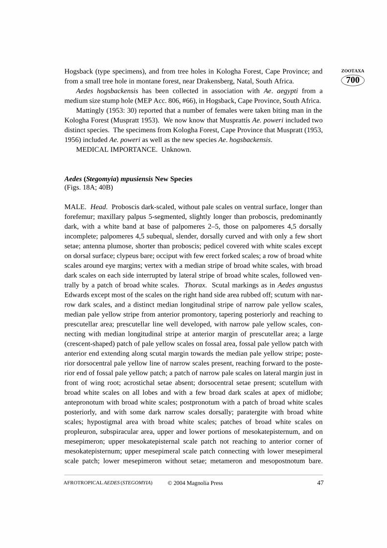
© 2004 Magnolia Press 47AFROTROPICAL AEDES (STEGOMYIA)
700ZOOTAXAHogsback (type specimens), and from tree holes in Kologha Forest, Cape Province; and
from a small tree hole in montane forest, near Drakensberg, Natal, South Africa. Aedes hogsbackensis has been collected in association with Ae. aegypti from a
medium size stump hole (MEP Acc. 806, #66), in Hogsback, Cape Province, South Africa. Mattingly (1953: 30) reported that a number of females were taken biting man in the
Kologha Forest (Muspratt 1953). We now know that Musprattís Ae. poweri included twodistinct species. The specimens from Kologha Forest, Cape Province that Muspratt (1953,1956) included Ae. poweri as well as the new species Ae. hogsbackensis.
MEDICAL IMPORTANCE. Unknown.
Aedes (Stegomyia) mpusiensis New Species (Figs. 18A; 40B)
MALE. Head. Proboscis dark-scaled, without pale scales on ventral surface, longer thanforefemur; maxillary palpus 5-segmented, slightly longer than proboscis, predominantlydark, with a white band at base of palpomeres 2–5, those on palpomeres 4,5 dorsallyincomplete; palpomeres 4,5 subequal, slender, dorsally curved and with only a few shortsetae; antenna plumose, shorter than proboscis; pedicel covered with white scales excepton dorsal surface; clypeus bare; occiput with few erect forked scales; a row of broad whitescales around eye margins; vertex with a median stripe of broad white scales, with broaddark scales on each side interrupted by lateral stripe of broad white scales, followed ven-trally by a patch of broad white scales. Thorax. Scutal markings as in Aedes angustusEdwards except most of the scales on the right hand side area rubbed off; scutum with nar-row dark scales, and a distinct median longitudinal stripe of narrow pale yellow scales,median pale yellow stripe from anterior promontory, tapering posteriorly and reaching toprescutellar area; prescutellar line well developed, with narrow pale yellow scales, con-necting with median longitudinal stripe at anterior margin of prescutellar area; a large(crescent-shaped) patch of pale yellow scales on fossal area, fossal pale yellow patch withanterior end extending along scutal margin towards the median pale yellow stripe; poste-rior dorsocentral pale yellow line of narrow scales present, reaching forward to the poste-rior end of fossal pale yellow patch; a patch of narrow pale scales on lateral margin just infront of wing root; acrostichal setae absent; dorsocentral setae present; scutellum withbroad white scales on all lobes and with a few broad dark scales at apex of midlobe;antepronotum with broad white scales; postpronotum with a patch of broad white scalesposteriorly, and with some dark narrow scales dorsally; paratergite with broad whitescales; hypostigmal area with broad white scales; patches of broad white scales onpropleuron, subspiracular area, upper and lower portions of mesokatepisternum, and onmesepimeron; upper mesokatepisternal scale patch not reaching to anterior corner ofmesokatepisternum; upper mesepimeral scale patch connecting with lower mesepimeralscale patch; lower mesepimeron without setae; metameron and mesopostnotum bare.
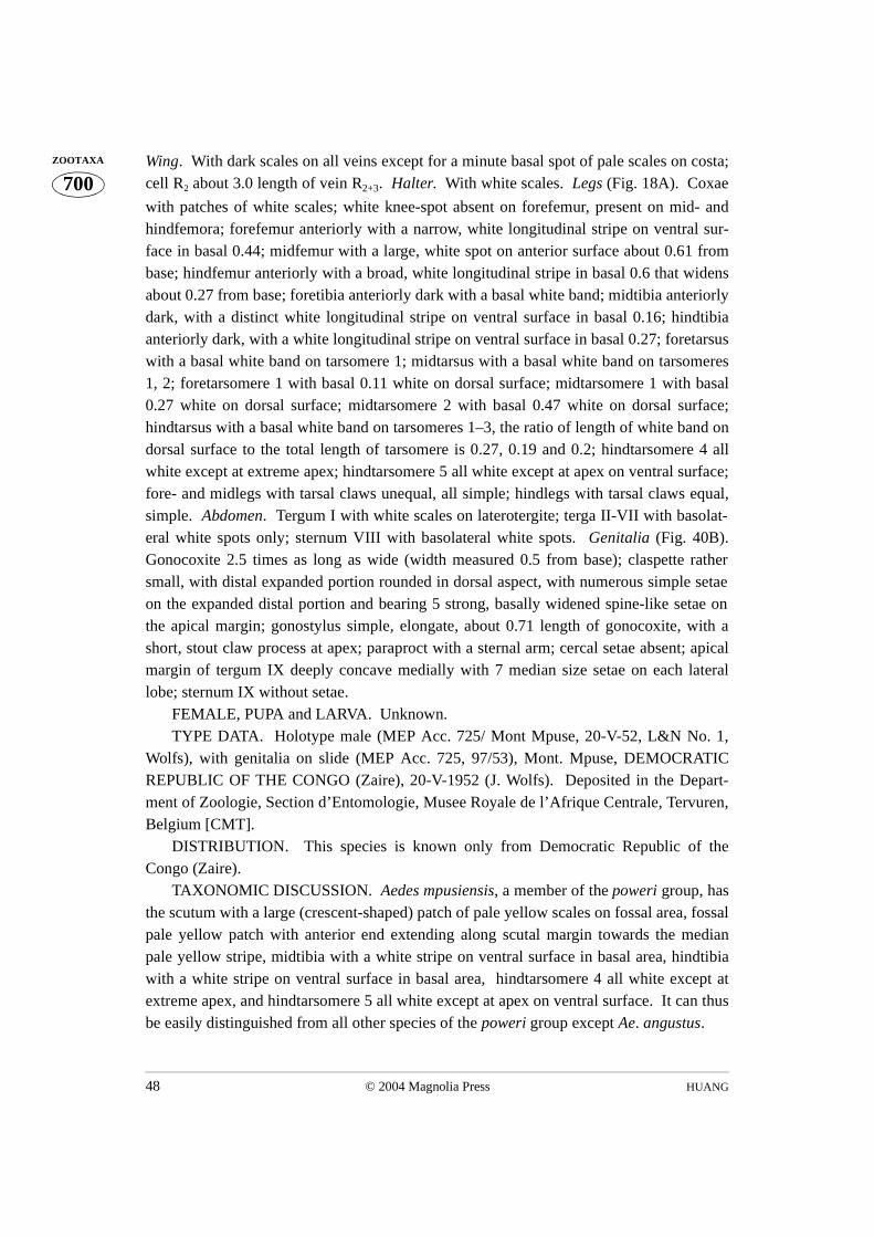
HUANG48 © 2004 Magnolia Press
700ZOOTAXA Wing. With dark scales on all veins except for a minute basal spot of pale scales on costa;
cell R2 about 3.0 length of vein R2+3. Halter. With white scales. Legs (Fig. 18A). Coxae
with patches of white scales; white knee-spot absent on forefemur, present on mid- andhindfemora; forefemur anteriorly with a narrow, white longitudinal stripe on ventral sur-face in basal 0.44; midfemur with a large, white spot on anterior surface about 0.61 frombase; hindfemur anteriorly with a broad, white longitudinal stripe in basal 0.6 that widensabout 0.27 from base; foretibia anteriorly dark with a basal white band; midtibia anteriorlydark, with a distinct white longitudinal stripe on ventral surface in basal 0.16; hindtibiaanteriorly dark, with a white longitudinal stripe on ventral surface in basal 0.27; foretarsuswith a basal white band on tarsomere 1; midtarsus with a basal white band on tarsomeres1, 2; foretarsomere 1 with basal 0.11 white on dorsal surface; midtarsomere 1 with basal0.27 white on dorsal surface; midtarsomere 2 with basal 0.47 white on dorsal surface;hindtarsus with a basal white band on tarsomeres 1–3, the ratio of length of white band ondorsal surface to the total length of tarsomere is 0.27, 0.19 and 0.2; hindtarsomere 4 allwhite except at extreme apex; hindtarsomere 5 all white except at apex on ventral surface;fore- and midlegs with tarsal claws unequal, all simple; hindlegs with tarsal claws equal,simple. Abdomen. Tergum I with white scales on laterotergite; terga II-VII with basolat-eral white spots only; sternum VIII with basolateral white spots. Genitalia (Fig. 40B).Gonocoxite 2.5 times as long as wide (width measured 0.5 from base); claspette rathersmall, with distal expanded portion rounded in dorsal aspect, with numerous simple setaeon the expanded distal portion and bearing 5 strong, basally widened spine-like setae onthe apical margin; gonostylus simple, elongate, about 0.71 length of gonocoxite, with ashort, stout claw process at apex; paraproct with a sternal arm; cercal setae absent; apicalmargin of tergum IX deeply concave medially with 7 median size setae on each laterallobe; sternum IX without setae.
FEMALE, PUPA and LARVA. Unknown.TYPE DATA. Holotype male (MEP Acc. 725/ Mont Mpuse, 20-V-52, L&N No. 1,
Wolfs), with genitalia on slide (MEP Acc. 725, 97/53), Mont. Mpuse, DEMOCRATICREPUBLIC OF THE CONGO (Zaire), 20-V-1952 (J. Wolfs). Deposited in the Depart-ment of Zoologie, Section d’Entomologie, Musee Royale de l’Afrique Centrale, Tervuren,Belgium [CMT].
DISTRIBUTION. This species is known only from Democratic Republic of theCongo (Zaire).
TAXONOMIC DISCUSSION. Aedes mpusiensis, a member of the poweri group, hasthe scutum with a large (crescent-shaped) patch of pale yellow scales on fossal area, fossalpale yellow patch with anterior end extending along scutal margin towards the medianpale yellow stripe, midtibia with a white stripe on ventral surface in basal area, hindtibiawith a white stripe on ventral surface in basal area, hindtarsomere 4 all white except atextreme apex, and hindtarsomere 5 all white except at apex on ventral surface. It can thusbe easily distinguished from all other species of the poweri group except Ae. angustus.
© 2004 Magnolia Press 49AFROTROPICAL AEDES (STEGOMYIA)
700ZOOTAXAThe adult male of Ae. mpusiensis is extremely similar to that of Ae. angustus with
which it has been confused and misidentified. Aedes mpusiensis can be distinguished fromAe. angustus by the white knee-spot present and well developed on the hindfemur. In Ae.angustus, the white knee-spot is absent on the hindfemur.
The adult male of Ae. mpusiensis is also very similar to that of Ae. usambara in havingthe midtibia with a white stripe on ventral surface in basal area, the hindtibia with a whitestripe on ventral surface in basal area, the hindtarsomere 4 almost all white and hindtar-somere 5 all white on dorsal surface. However, Ae. mpusiensis can be distinguished fromAe. usambara by the fossal pale yellow patch with anterior end extending along scutalmargin towards the median pale yellow stripe. In Ae. usambara, the fossal white patch hasno anterior end extending along scutal margin towards the anterior median white spot.
The male genitalia of Ae. mpusiensis are easily differentiated from all other species inthe poweri group by the claspette, which has the distal expanded portion rather smallrounded, lobe-like in dorsal aspect, with numerous simple setae on the expanded distalportion and bearing 5 strong, basally widened, spine-like setae on the apical margin, andby the apical margin of tergum IX, which is deeply concave medially and has 7 mediansize setae on each lateral lobe.
The male genitalia of Ae. mpusiensis are extremely similar to those of Ae. angustus inhaving the claspette with distal expanded portion rounded, lobe-like in dorsal aspect, butcan be distinguished from those of Ae. angustus by the claspette with 5 strong, basallywidened, spine-like setae on the apical margin. In Ae. angustus, the claspette has nostrong, basally widened, spine-like seta.
Aedes mpusiensis is apparently an East African montane forest species. BIONOMICS. Unknown. MEDICAL IMPORTANCE. Unknown.
Aedes (Stegomyia) sampi New Species(Fig. 30A)
FEMALE. Head. Proboscis dark-scaled, without pale scales on ventral surface, as long asforefemur; maxillary palpus 0.23 length of proboscis, dark, with white scales on apical0.50; pedicel covered with white scales except on dorsal and ventral surfaces; clypeusbare; occiput with few erect forked scales; a row of broad white scales around eye mar-gins; vertex with a median stripe of broad white scales, with broad dark scales on each sideinterrupted by a lateral stripe of broad white scales, followed ventrally by a patch of broadwhite scales. Thorax. Scutum with narrow dark scales and a distinct, median white spotof broad scales on anterior promontory, followed by a submedian longitudinal stripe ofnarrow yellowish scales on each side of midline, reaching to prescutellar area and connect-ing with prescutellar line of narrow yellowish scales; fossal area with a large patch ofbroader, crescent-shaped white scales; posterior dorsocentral yellowish lines present,
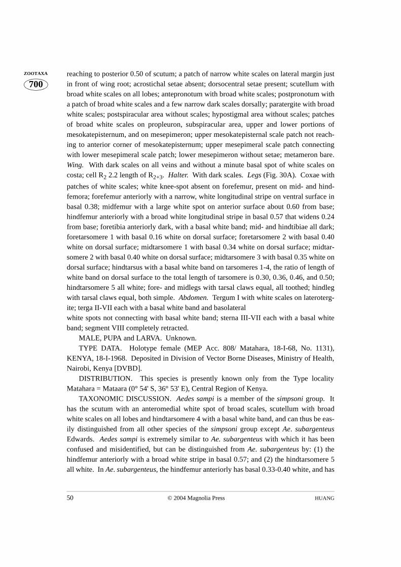
HUANG50 © 2004 Magnolia Press
700ZOOTAXA reaching to posterior 0.50 of scutum; a patch of narrow white scales on lateral margin just
in front of wing root; acrostichal setae absent; dorsocentral setae present; scutellum withbroad white scales on all lobes; antepronotum with broad white scales; postpronotum witha patch of broad white scales and a few narrow dark scales dorsally; paratergite with broadwhite scales; postspiracular area without scales; hypostigmal area without scales; patchesof broad white scales on propleuron, subspiracular area, upper and lower portions ofmesokatepisternum, and on mesepimeron; upper mesokatepisternal scale patch not reach-ing to anterior corner of mesokatepisternum; upper mesepimeral scale patch connectingwith lower mesepimeral scale patch; lower mesepimeron without setae; metameron bare.Wing. With dark scales on all veins and without a minute basal spot of white scales oncosta; cell R2 2.2 length of R2+3. Halter. With dark scales. Legs (Fig. 30A). Coxae with
patches of white scales; white knee-spot absent on forefemur, present on mid- and hind-femora; forefemur anteriorly with a narrow, white longitudinal stripe on ventral surface inbasal 0.38; midfemur with a large white spot on anterior surface about 0.60 from base;hindfemur anteriorly with a broad white longitudinal stripe in basal 0.57 that widens 0.24from base; foretibia anteriorly dark, with a basal white band; mid- and hindtibiae all dark;foretarsomere 1 with basal 0.16 white on dorsal surface; foretarsomere 2 with basal 0.40white on dorsal surface; midtarsomere 1 with basal 0.34 white on dorsal surface; midtar-somere 2 with basal 0.40 white on dorsal surface; midtarsomere 3 with basal 0.35 white ondorsal surface; hindtarsus with a basal white band on tarsomeres 1-4, the ratio of length ofwhite band on dorsal surface to the total length of tarsomere is 0.30, 0.36, 0.46, and 0.50;hindtarsomere 5 all white; fore- and midlegs with tarsal claws equal, all toothed; hindlegwith tarsal claws equal, both simple. Abdomen. Tergum I with white scales on lateroterg-ite; terga II-VII each with a basal white band and basolateral white spots not connecting with basal white band; sterna III-VII each with a basal whiteband; segment VIII completely retracted.
MALE, PUPA and LARVA. Unknown.TYPE DATA. Holotype female (MEP Acc. 808/ Matahara, 18-I-68, No. 1131),
KENYA, 18-I-1968. Deposited in Division of Vector Borne Diseases, Ministry of Health,Nairobi, Kenya [DVBD].
DISTRIBUTION. This species is presently known only from the Type localityMatahara = Mataara (0° 54' S, 36° 53' E), Central Region of Kenya.
TAXONOMIC DISCUSSION. Aedes sampi is a member of the simpsoni group. Ithas the scutum with an anteromedial white spot of broad scales, scutellum with broadwhite scales on all lobes and hindtarsomere 4 with a basal white band, and can thus be eas-ily distinguished from all other species of the simpsoni group except Ae. subargenteusEdwards. Aedes sampi is extremely similar to Ae. subargenteus with which it has beenconfused and misidentified, but can be distinguished from Ae. subargenteus by: (1) thehindfemur anteriorly with a broad white stripe in basal 0.57; and (2) the hindtarsomere 5all white. In Ae. subargenteus, the hindfemur anteriorly has basal 0.33-0.40 white, and has

© 2004 Magnolia Press 51AFROTROPICAL AEDES (STEGOMYIA)
700ZOOTAXAa large white spot about 0.67 from base (the white spot not connecting with the basal white
area), and the hindtarsomere 5 has basal 0.40-0.67 white on dorsal surface.BIONOMICS. Unknown.MEDICAL IMPORTANCE. Unknown.
ACKNOWLEDGMENTS
I wish to express my sincere appreciation to: Dr. Wayne N. Mathis, Department of System-atic Biology, Entomology Section, Smithsonian Institution and Dr. Raymond J. Gagne,Research Entomologist, Systematic Entomology Laboratory, U.S. Department of Agricul-ture, for critically reviewing this manuscript and for their valuable comments; to COLScott W. Gordon, Chief, Department of Entomology, Walter Reed Army Institute ofResearch (WRAIR) and Dr. Richard C. Wilkerson, Walter Reed Biosystematics Unit(WRBU), for providing funding for the publication.
The views expressed in this article are those of the author and do not reflect the officialpolicy or position of the Department of the Army, the Department of Defense..
I am most grateful to Drs. Peter F. Mattingly (deceased), Graham B. White (formerlyof BMNH) and Miss Theresa M. Howard, Department of Entomology, Medical & Veteri-nary Division, The Natural History Museum, London (formerly British Museum (NaturalHistory), England, for the loan of several types and other material in the British Museum[BMNH]; to Drs. J.M.D. Roberts, Davy K. Koech, Mrs. Phoebe A.O. Josiah (formerly ofDVBD) and Mrs. Beth A. Rapuoda, Division of Vector Borne Diseases, Ministry ofHealth, Nairobi, Kenya [DVBD]; to Drs. M. Germain, A. Rickenbach, M. Cornet (for-merly of ORSTOM) and Dr. B. Geoffroy, ORSTOM Department Sante BP 5045, 34032Montpellier Cedex 01 (formerly Services Scientifiques Centraux de l'O.R.S.T.O.M.,Bondy), France [ORSTOM]; to Dr. L. Matile, Museum National d’Histoire Naturelle,Paris [MNHP]; to Dr. F. Rodhain and Miss C. Perez, Ecologie Virale, Institut Pasteur,Paris [PIP]; to Drs. J. Decelle and E. De Coninck, Department of Zoologie, Sectiond'Entomologie, Musee Royale de l'Afrique Centrale, Tervuren, Belgium [CMT]; to Drs.John A. Ledger, Richard Hunt (formerly of SAIMR), and Mrs. Joyce Segerman, Depart-ment of Medical Entomology, The South African Institute for Medical Research, Johan-nesburg, South Africa [SAIM]; to Drs. Margaret K. Thayer, Philip D. Perkins, Departmentof Entomology, Museum of Comparative Zoology, Harvard University, Cambridge, Mas-sachusetts [MCZ]; and to Drs. T. Kurihara, A. Shinohara, Department of Zoology,National Science Museum, Tokyo, Japan, for the loan of the specimens used in this study.I also wish to express my gratitude to Mr. Young T. Sohn (formerly of Medical Entomol-ogy Project), Scientific Illustrator, Department of Systematic Biology, Entomology Sec-tion, Smithsonian Institution, for preparing the illustrations.
HUANG52 © 2004 Magnolia Press
700ZOOTAXA LITERATURE CITED
Bang, Y.H., Brown, A.W.A., Bown, D.N., Onwubiko, A.O., Knudsen, A.B., Lambrecht, F.L. &Arata, A.A. (1979) Summary report on the findings of the WHO Arbovirus Vector ResearchUnit in Southeastern Nigeria, 1973–1978.World Health Organization/Vector Biology and Con-trol/1979, 725, 1–8.
Bang, Y.H., Knudsen, A.B., Onwubiko, A.O. & Bown, D.N. (1983) Seasonal survival of Aedesafricanus (Diptera: Culicidae) in Nigeria. The Journal of Medical Entomology, 20, 128–133.
Baudon, D., Robert, V., Roux, J., Stanghellini, A., Gazin, P., Molez, J.F., Lhuillier, M., Sartholi,J.L., Saluzzo, J.F., Cornet, M., Bibane, L., Some, L., Guiguemde, T.R. & Soudre, B.R. (1984)Epidemic yellow fever in Upper Volta. The Lancet, July 7, 42.
Bauer, J.H. (1928) The transmission of yellow fever by mosquitoes other than Aedes aegypti. TheAmerican Journal of Tropical Medicine, 8, 261–281.
Belkin, J.N. (1962) The mosquitoes of the South Pacific (Diptera, Culicidae). University of Cali-fornia Press, Berkeley and Los Angeles, 2 vols, 608 and 412 pp.
Chippaux, A., Cordellier, R., Courtois B. & Robin, Y. (1975) Une souche de virus amaril isoleed'Aedes africanus en Cote d'Ivoire. Comptes Rendus. Academie des Sciences Serie D, 281, 79–80.
Chwatt, L. J. (1949). Aedes (Stegomyia) pseudoafricanus sp. nov.: a new species of Aedes from thecoast of Nigeria (British West Africa). Nature, London, 163, 808.
Cordellier, R., Germain, M., Hervy, J.P. & Mouchet, J. (1977) Guide partique pour l' etude des vec-teurs de fievre jaune et methodes de lutte. Cahiers de Office de la Recherche Scientifique etTechnique Outre-Mer, Initiations-Documentations Techniques, No. 33, 1–114.
Cornel, A. J. & Hunt, R.H. (1991) Aedes albopictus in Africa? First records of live specimens inimported tires in Cape Town. Journal of the American Mosquito Control Association, 7, 107–108.
Cornet, M. (1974) Aedes (Stegomyia) cozi n. sp., une nouvelle espece de Culicidae au Senegal.Cahiers de Office de la Recherche Scientifique et Technique Outre-Mer Serie EntomologieMedicale et Parasitologie, 11 (1973), 175–180.
Cornet, M., Valade, M. & Dieng, P.Y. (1978) Aedes (Stegomyia) neoafricanus une nouvelle especede moustique capturee au Senegal Oriental (Diptera: Culicidae).
Cahiers de Office de la Recherche Scientifique et Technique Outre-Mer Serie Entomologie Med-icale et Parasitologie, 16, 227–230.
Cornet, M., Robin, Y., Chateau, R., Heme, G., Adam, C., Valade, M., Le Gonidec, G., Jan, C.,Renaudet, J., Dieng, P.L., Bangoura, J.F. & Lorand, A. (1979) Isolements d'arbovirus au Sene-gal oriental a partir de moustiques (1972-1977) et notes sur l'epidemiologie des virus transmispar les Aedes, en particulier du virus amaril. Cahiers de Office de la Recherche Scientifique etTechnique Outre-Mer Serie Entomologie Medicale et Parasitologie, 17, 149–163.
De Meillon, B. (1947) New records and species of biting insects from the Ethiopian Region - II.Journal of the Entomological Society of Southern Africa, 10, 110–124.
De Meillon, B. & Lavoipierre, M. (1944). New records and species of biting insects from the Ethi-opian region. Journal of the Entomological Society of Southern Africa, 7, 38–67.
Dick, G. W. A., Kitchen, S.F. & Haddow, A.J. (1952) Zika virus (1). Isolations and serologicalspecificity. Transactions of the Royal Society of Tropical Medicine and Hygiene, 46, 600–618.
Edwards, F.W. (1912). A synopsis of the species of African Culicidae, other than Anopheles. Bul-letin of Entomological Research, 3, 1–53.
Edwards, F.W. (1917) Notes on Culicidae, with descriptions of new species. Bulletin of Entomo-logical Research, 7, 201–229.
Edwards, F.W. (1932) Diptera. Family Culicidae. In: P. Wytsman (Ed.), Genera Insectorum. Des-met-Verteneuil, Brussels, Fascicle, 194, 258 pp., 5 pl.
© 2004 Magnolia Press 53AFROTROPICAL AEDES (STEGOMYIA)
700ZOOTAXAEdwards, F.W. (1941) Mosquitoes of the Ethiopian region. III. Culicine adults and pupae. British
Museum (Natural History), London, 499 pp.Enderlein, Gunther. (1923) Zur Kenntnis afrikanischer und orientalischer Culiciden. Wiener Ento-
mologische Zeitung, 40, 25–29.Evans, A.M. (1925) Notes on Culicidae collected in Sierra Leone, with descriptions of a new spe-
cies and a new variety. Annals of Tropical Medicine and Parasitology, 19, 119–126.Fontenille, D. & Toto, J.-C. (2001) Aedes (Stegomyia) albopictus (Skuse), a potential new dengue
vector in Southern Cameroon. Emerging Infectious Diseases, 7 (6), 1066–1067. Germain, M., Cordellier, R., Herve, J.-P., Geoffroy, B., Bouchite, B., Ravaonjanahary, C. & Ricken-
bach, A. (1975) Presence en Afrique Centrale et occidentale d' Aedes (Stegomyia) opok Corbetet Van Someren diagnose differentielle de l' espece. Cahiers de Office de la Recherche Scienti-fique et Technique Outre-Mer Serie Entomologie Medicale et Parasitologie, 13, 41–46.
Germain, M., Cornet, M., Mouchet, J., Monath, T.P., Herve, J.-P., Salaun, J.-J., Cordellier, R.,Saluzzo, J.-F., Camicas, J.-L., Hervy, J.-P., Robert, V., Deubel, V., Gonzalez, J.-P., Digoutte, J.-P. & Darwish, D.O. (1982) Recent advances in research regarding sylvatic yellow fever inWest and Central Africa. Bulletin de l’Institut Pasteur, 80, 315–330.
Germain, M., Eouzan, J.P. & Ferrara, L. (1972) Donnees sur les facultes de dispersion de deuxdipteres d'interet medical: Aedes africanus (Theobald) et Simulium damnosum Theobald dansle domaine montagnard du nord du Cameroun occidental. Cahiers de Office de la RechercheScientifique et Technique Outre-Mer Serie Entomologie Medicale et Parasitologie, 10, 291–300.
Germain, M., Francy, D.B., Monath, T.P., Ferrara, L., Bryan, J., Salaun, J.-J., Heme, G., Renaudet,J., Adam, C. & Digoutte, J.-P. (1980) Yellow fever in the Gambia, 1978-1979: Entomologicalaspects and epidemiological correlations. The American Journal of Tropical Medicine andHygiene, 29, 929–940.
Germain, M., Herve, J.P., Cornet, J.P. & Geoffroy, B. (1978) II. Service d'Entomologie medical etd'etude des reservoirs de virus. Paper. Institut Pasteur, Bangui, 1977, 81–123.
Germain, M., Herve, J.P., Sureau, P., Fabre, J., Robin, Y.& Geoffroy, B. (1976) Une souche de virusamaril isolee d'Aedes (Stegomyia) opok Corbet et Van Someren, en Republique Centrafricaine.Cahiers de Office de la Recherche Scientifique et Technique Outre-Mer Serie EntomologieMedicale et Parasitologie, 14, 101–104.
Germain, M., Sureau, P., Herve, J.P., Fabre, J., Mouchet, J., Robin, Y. & Geoffroy, B. (1976) Isole-ments du virus de la fievre jaune a partir d'Aedes du groupe A. africanus (Theobald) en Repub-lique Centrafricaine. Importance des savanes humides et semi-humides en tant que zoned'emergence du virus amaril. Cahiers de Office de la Recherche Scientifique et TechniqueOutre-Mer Serie Entomologie Medicale et Parasitologie, 14, 125–139.
Gillett, J.D. & Ross, R.W. (1953) The laboratory transmission of yellow fever by the mosquitoAedes (Stegomyia) strelitziae Muspratt. Annals of Tropical Medicine and Parasitology, 47(4),367–370.
Haddow, A.J. (1945) The mosquitoes of Bwamba County, Uganda. II. Biting activity with specialreference to the influence of microclimate. Bulletin of Entomological Research, 36, 33–73.
Haddow, A.J. (1961) Studies on the biting habits and medical importance of East African mosqui-tos in the Genus Aedes. II. Subgenera Mucidus, Diceromyia, Finlaya and Stegomyia. Bulletinof Entomological Research, 52, 317–351.
Haddow, A.J. (1968) The natural history of yellow fever in Africa. Royal Society of Edinburgh,Proceedings Section B, 70, 191–227.
Haddow, A.J., Gillett, J.D. & Highton, R.B. (1947) The mosquitoes of Bwamba County, Uganda.V. The vertical distribution and biting-cycle of mosquitoes in rain-forest, with further observa-tions on micro-climate. Bulletin of Entomological Research, 37, 301–330.
Haddow, A.J. & Mahaffy, A.F. (1949) The mosquitoes of Bwamba County, Uganda. VII. Inten-

HUANG54 © 2004 Magnolia Press
700ZOOTAXA sive catching on tree-platforms, with further observation on Aedes (Stegomyia) africanus
Theobald. Bulletin of Entomological Research, 40, 169–178. Haddow, A.J., Smithburn, K.C., Dick, G.W.A., Kitchen, S.F. & Lumsden, W.H.R. (1948) Implica-
tion of the mosquito Aedes (Stegomyia) africanus Theobald in the forest cycle of yellow feverin Uganda. Annals of Tropical Medicine and Parasitology, 42, 218–228.
Haddow, A.J., Smithburn, K.C., Mahaffy, A.F. & Bugher, J.C. (1947) Monkeys in relation to yel-low fever in Bwamba County, Uganda. Transactions of the Royal Society of Tropical Medicineand Hygiene, 40, 677–700.
Haddow, A.J., Williams, M.C. & Woodall, J.P. (1961) Chikungunya near Entebbe, Uganda. Virusisolations from biting flies. East Africa Virus Research Institute Report, Nairobi, 1960–1961,16–17.
Haddow, A.J., Williams, M.C., Woodall, J.P., Simpson, D.I.H. & Goma, L.K.H. (1964) Twelve iso-lations of Zika virus from Aedes (Stegomyia) africanus (Theobald) taken in and above aUganda Forest. Bulletin World Health Organization, 31, 57–69.
Hamon, J., Pichon, G. & Cornet, M. (1971) La transmission du virus amaril en Afrique occidentale.Ecologie, repartition, frequence et controle des vecteurs, et observations concernant l'epidemi-ologie de la fievre jaune. Cahiers de Office de la Recherche Scientifique et Technique Outre-Mer Serie Entomologie Medicale et Parasitologie, 9, 3–60.
Harbach, R.E. & Knight, K.L. (1980) Taxonomists' glossary of mosquito anatomy. Plexus Publishing, Inc., Marlton, N.J. 415 pp.
Harbach, R.E. & Knight, K.L. (1982) Corrections and additions to taxonomists' glossary of mos-quito anatomy. Mosquito Systematics (1981) 13, 201–217.
Hopkins, G.H.E. (1952) Mosquitoes of the Ethiopian region. I. -Larval bionomics of mosquitoesand taxonomy of culicine larvae. 2nd Ed. with notes and addenda by P. F. Mattingly. BritishMuseum (Natural History), London, 355 pp.
Huang, Y.-M. (1972a) Lectotype designation for Aedes (Stegomyia) galloisi Yamada with a note onits assignment to the scutellaris group of species (Diptera: Culicidae). Proceedings of theEntomological Society of Washington, 74, 253–256.
Huang, Y.-M. (1972c) Contributions to the Mosquito Fauna of Southeast Asia. XIV. The subgenusStegomyia of Aedes in Southeast Asia. 1. The scutellaris group of species. Contributions ofthe American Entomological Institute, 9(1), 1–109.
Huang, Y.-M. (1974a) A redescription of Aedes (Stegomyia) amaltheus de Meillon and Lavoipierrewith a note on its assignment to the aegypti group of species (Diptera: Culicidae). MosquitoSystematics, 6, 27–31.
Huang, Y.-M. (1974c) Lectotype designation for Aedes (Stegomyia) chemulpoensis Yamada with anote on its assignmemt to the aegypti group of species (Diptera: Culicidae). Proceedings ofthe Entomological Society of Washington, 76, 208–211.
Huang, Y.-M. (1977a) Medical Entomology Studies. VII. The subgenus Stegomyia of Aedes inSoutheast Asia. II. - The edwardsi group of species. III. - The w-albus group of species.(Diptera: Culicidae). Contributions of the American Entomological Institute (Ann Arbor),14(1), 1–111.
Huang, Y.-M. (1979a) Medical Entomology Studies - XI. The subgenus Stegomyia of Aedes in theOriental Region with keys to the species (Diptera: Culicidae). Contributions of the AmericanEntomological Institute (Ann Arbor). 15(6), 1–79.
Huang, Y.-M. (1979b) Aedes (Stegomyia) simpsoni complex in the Ethiopian Region with lecto-type designation for simpsoni (Theobald) (Diptera: Culicidae). Mosquito Systematics, 11, 221–234.
Huang, Y.-M. (1981) A redescription of Aedes (Stegomyia) calceatus Edwards and description of anew Afrotropical species, Aedes (Stegomyia) ledgeri (Diptera: Culicidae). Mosquito System-atics, 13, 92–113.
© 2004 Magnolia Press 55AFROTROPICAL AEDES (STEGOMYIA)
700ZOOTAXAHuang, Y.-M. (1986a) Aedes (Stegomyia) bromeliae (Diptera: Culicidae), the yellow ever virus vec-
tor in East Africa. The Journal of Medical Entomology, 23, 196–200.Huang, Y.-M. (1988b) The Aedes (Stegomyia) pseudonigeria group with emphasis on the species
from the Afrotropical Region (Diptera: Culicidae). Mosquito Systematics, 20, 1–20. Huang, Y.-M. (1990) The subgenus Stegomyia of Aedes in the Afrotropical Region. I. The africanus
group of species (Diptera: Culicidae). Contributions of the American Entomological Institute(Gainesville, Florida), 26(1), 1–90.
Huang, Y.-M. (1997) The subgenus Stegomyia of Aedes in the Afrotropical Region. II. The den-drophilus group of species (Diptera: Culicidae). Contributions of the American Entomologi-cal Institute (Gainesville, Florida), 29(4), 1–104.
Huang, Y.-M. (2001) A pictorial key for the identification of the subfamilies of Culicidae, Generaof Culicinae, and subgenera of Aedes mosquitoes of the Afrotropical Region (Diptera: Culi-cidae). Proceedings of the Entomological Society of Washington, 103 (1), 1–53.
Huang, Y.-M. (2002) A pictorial key to the mosquito genera of the world, including subgenera ofAedes and Ochlerotatus (Diptera: Culicidae). Center for Insect Systematics, Korea, InsectaKoreana, 19 (1), 1–130.
Jupp, P.G. & McIntosh, B.M. (1988) Chikungunya virus disease. In: T. P. Monath (Ed). The arbovi-ruses: Epidemiology and ecology. Vol. 2. CRC Press, Inc., Boca Raton, Florida, pp.137–157.
Kirya, B.G., Mukwaya, L.G. & Sempala, S.D.K. (1977) A yellow fever epizootic in Zika Forest,Uganda, during 1972: Part I. Virus isolation and sentinel monkeys. Transactions of the RoyalSociety of Tropical Medicine and Hygiene, 71, 254–260.
Knight, K.L. & Hurlbut, H.S. (1949) The mosquitoes of Ponape Island, eastern Carolines. Journalof the Washington Academy of Sciences, 39, 20–34.
Leeson, H.S. & Theodor, O. (1948) Mosquitos of Socotra. Bulletin of Entomological Research, 39,221–229.
Ludlow, C.S. (1905) Mosquito notes. —No. 3. Canadian Entomologist, 37, 94–102.Ludlow, C.S. (1908) The mosquitoes of the Philippine islands. The distribution of certain species,
and their occurrence in relation to the incidence of certain diseases. Ph. D. Thesis, GeorgeWashington University, 65 pp.
Lumsden, W.H.R. (1951) Probable insect vectors of yellow fever virus, from monkey to man, inBwamba County, Uganda. Bulletin of Entomological Research, 42, 317–330.
Macgregor, M.E. (1924) Aedes (Stegomyia) mascarensis, MacGregor:a new mosquito from Mau-ritius. Bulletin of Entomological Research, 14, 409–412.
Mahaffy, A.F., Smithburn, K.C., Jacobs, H.R. & Gillett, J.D. (1942) Yellow fever in westernUganda. Transactions of the Royal Society of Tropical Medicine and Hygiene, 36, 9–20.
Marks, E.N. (1954) A review of the Aedes scutellaris subgroup with a study of variation in Aedespseudoscutellaris (Theobald) (Diptera: Culicidae). Bulletin of the British Museum (NaturalHistory), Entomology, 3(10), 349–414.
Mattingly, P.F. (1952) The sub-genus Stegomyia (Diptera: Culicidae) in the Ethiopian region. I. Apreliminary study of the distribution of species occurring in the West African sub-region withnotes on taxonomy and bionomics. Bulletin of the British Museum (Natural History), Entomol-ogy, London, 2, 235–304.
Mattingly, P.F. (1953) The sub-genus Stegomyia (Diptera: Culicidae) in the Ethiopian region. II.Distribution of species confined to the east and south African sub-region. Bulletin of the Brit-ish Museum (Nattural History), Entomology, London, 3, 1–65.
Mattingly, P.F. (1965) The culicine mosquitoes of the Indomalayan Area. Part VI. Genus AedesMeigen, subgenus Stegomyia Theobald (Groups A, B and D). Trustees of the British Museum(Natural History), London, 67 pp.
Mattingly, P.F. & Knight, K.L. (1956) The mosquitoes of Arabia. I. Bulletin of the British Museum(Natural History), Entomology, 4 (3), 89–141.
HUANG56 © 2004 Magnolia Press
700ZOOTAXA Mattingly, P.F. & Lips, M. (1953) Notes on the Culicini of the Katanga (Diptera, Culicidae). I.
Taxonomy. Revue de Zoologie et de Botanique Africaines, 47, 311–343.Mattingly, P.F., Stone, A. & Knight, K.L. (1962) Culex aegypti Linnaeus, 1762 (Insecta, Diptera);
proposed validation and interpretation under the plenary powers of the species so named. Z. N.(S.) 1216. Bulletin of Zoological Nomenclature, 19, 208–219.
McCrae, A.W.R., Henderson, B.E., Kirya, B.G. & Sempala, S.D.K. (1971) Communications.Chikungunya virus in the Entebbe Area of Uganda: isolations and epidemiology. Transactionsof the Royal Society of Tropical Medicine and Hygiene, 65, 152–168.
McCrae, A.W.R. & Kirya, B.G. (1982) Yellow fever and Zika virus epizootics and enzootics inUganda. Transactions of the Royal Society of Tropical Medicine and Hygiene, 76, 552–562.
Mondet, B. & Montange, L. (1993) Presence en Cote d’ Ivoire, variations saisonnieres et statut epi-demiologique d’ Aedes (Stegomyia) usambara Mattingly, 1953 (Diptera: Culicidae). Annalesde la Societe Entomologique de France, 29(3), 261–267.
Muspratt, J. (1953) Research on South African Culicini (Diptera, Culicidae). II. — Taxonomyrelating to eight species of Aedes. Journal of the Entomological Society of Southern Africa, 16,83–93.
Muspratt, J. (1956) The Stegomyia mosquitoes of South Africa and some neighbouring territories.Memoirs of the Entomological Society of Southern Africa, 4, 1–138.
Pajot, F.X. (1972) Les vecteurs potentiels majeurs du virus amaril en Republique Centrafricaine.Cahiers de Office de la Recherche Scientifique et Technique Outre-Mer Serie EntomologieMedicale et Parasitologie, 10, 111–117.
Philip, C.B. (1929) Preliminary report of further tests with yellow fever transmission by mosqui-toes other than Aedes aegypti. The American Journal of Tropical Medicine, 9, 267–269.
Philip, C.B. (1930) The experimental transmission of yellow fever by mosquitoes. Science, 71,614–615.
Ribeiro, H. & Ramos, H.Da C. (1973) Research on the mosquitoes of Angola VIII. The genusAedes Meigen, 1818 (Diptera: Culicidae). Check-list with new records, keys to females andlarvae, distribution and taxonomic and bioecological notes. Anais do Instituto de Higiene eMedicina Tropical, 1, 107-138.
Rickenbach, A., Ferrara, L., Germain, M., Eouzan, J.P. & Button, J.P. (1971) Quelques donnees surla biologie de trois vecteurs potentiels de fievre jaune: Aedes (Stegomyia) africanus (Theo.), A.(S.) simpsoni (Theo.) et A. (S.) aegypti (L.) dans la region de Yaounde (Cameroun). Cahiersde Office de la Recherche Scientifique et Technique Outre-Mer Serie Entomologie Medicale etParasitologie, 9, 285–299.
Saluzzo, J.-F., Gonzalez, J.-P., Herve, J.-P. & Georges, A.J. (1980) Contribution a l'etude epidemi-ologique des arbovirus en centrafrique: manifestation du virus chikungunya au cours desannees 1978 et 1979. Bulletin. Societe de Pathologie Exotique, 73, 390–399.
Savage, H.M., Ezike, V.I., Nwankwo, A.C.N., Spiegel, R. & Miller, B.R. (1992) First record ofbreeding populations of Aedes albopictus in continental Africa: implications for arboviraltransmission. Journal of the American Mosquito Control Association, 8, 101–103.
Serie, C., Andral, L., Casals, J., Williams, M.C., Bres, P. & Neri, P. (1968) Etudes sur la fievrejaune en Ethiopie. 5. Isolement de souches virales de vecteurs arthropodes. Bulletin WorldHealth Organization, 38, 873–877.
Service, M.W. (1976) Contribution to the knowledge of the mosquitoes (Diptera, Culicidae) ofGabon. Cahiers de Office de la Recherche Scientifique et Technique Outre-Mer Serie Ento-mologie Medicale et Parasitologie, 14, 259–263.
Smithburn, K.C. & Haddow, A.J. (1946) Isolation of yellow fever virus from African mosquitoes.The American Journal of Tropical Medicine, 26, 261–271.
Smithburn, K.C., Haddow, A.J. & Gillett, J.D. (1948) Rift Valley fever. Isolation of the virus fromwild mosquitoes. The British Journal of Experimental pathology, 29, 107–121.
© 2004 Magnolia Press 57AFROTROPICAL AEDES (STEGOMYIA)
700ZOOTAXASmithburn, K.C., Haddow, A.J. & Lumsden, W.H.R. (1949) An outbreak of sylvan yellow fever in
Uganda with Aedes (Stegomyia) africanus Theobald as principal vector and insect host of thevirus. Annals of the Tropical Medicine and Parasitology, 43, 74–80.
Theobald, F.V. (1901a). (June), p. 235, In: Howard, L.O. (Ed.) Mosquitoes: how they live, howthey carry disease, how they are classified, how they may be destroyed. New York. 241 pp.
Theobald, F.V. (1901b) (July), The classification of mosquitoes. Journal of Tropical Medicine, 4,229–235.
Theobald, F.V. (1901c) (Sept.), Notes on a collection of mosquitoes from west Africa, and descrip-tions of new species. Report of the malaria expedition to Nigeria of the Liverpool School ofTropical Medicine and Medical Parasitology. Part II. Filariasis. Memoires Liverpool Schoolof Tropical Medicine, 4 (Appendix), ii, 1–14.
Theobald, F.V. (1901d) (Nov.) A monograph of the Culicidae or mosquitoes. Vol. 1, The BritishMuseum (Natural History), London, 424 pp.
Theobald, F.V. (1906) Report on economic entomology. Part I. Second report on the mosquitoesof Culicidae of the Sudan. Report. Wellcome Tropical Research Laboratories, 2, 67–83.
Theobald, F.V. (1910) A monograph of the Culicidae or mosquitoes. Vol. 5. The British Museum(Natural History), London,. 646 pp., 6 pl.
Weinbren, M.P. & Williams, M.C. (1958) Zika virus: further isolations in the Zika area, and somestudies on the strains isolated. Transactions of the Royal Society of Tropical Medicine andHygiene, 52, 263-268.
Weinbren, M.P., Haddow, A.J. & Williams, M.C. (1958) The occurrence of Chikungunya virus inUganda. I. Isolation from mosquitoes. Transactions of the Royal Society of Tropical Medicineand Hygiene, 52, 253–262.
Weinbren, M.P., Williams, M.C. & Haddow, A.J. (1957) A variant of Rift Valley fever virus. SouthAfrica Medical Journal, 31, 951–957.
Wolfs, J. (1949) Aedes (Stegomyia) apicoargenteus var. denderensis var. n. (Culicidae). Revue deZoologie et de Botanique Africaines, 42, 190–192.
World Health Organization. (1978) Weekly Epidemiological Record. World Health Organization,Geneva, 53, 345–352.
Worth, C.B. & De Meillon, B. (1960) Culicine mosquitoes (Diptera: Culicidae) recorded from theProvince of Mocambique (Portuguese East Africa) and their relationship to arthropod-borneviruses. Anais, Instituto Medicina Tropical, 17, 231–256.
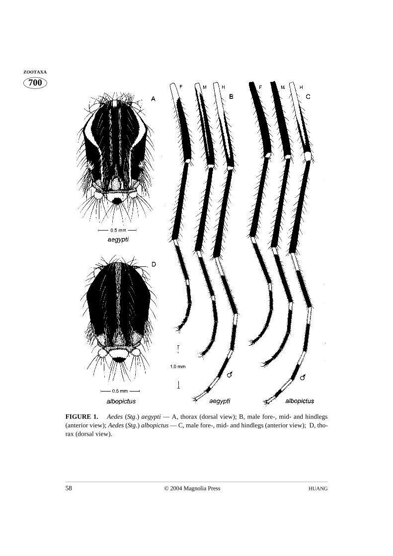
HUANG58 © 2004 Magnolia Press
700ZOOTAXA
FIGURE 1. Aedes (Stg.) aegypti — A, thorax (dorsal view); B, male fore-, mid- and hindlegs(anterior view); Aedes (Stg.) albopictus — C, male fore-, mid- and hindlegs (anterior view); D, tho-rax (dorsal view).
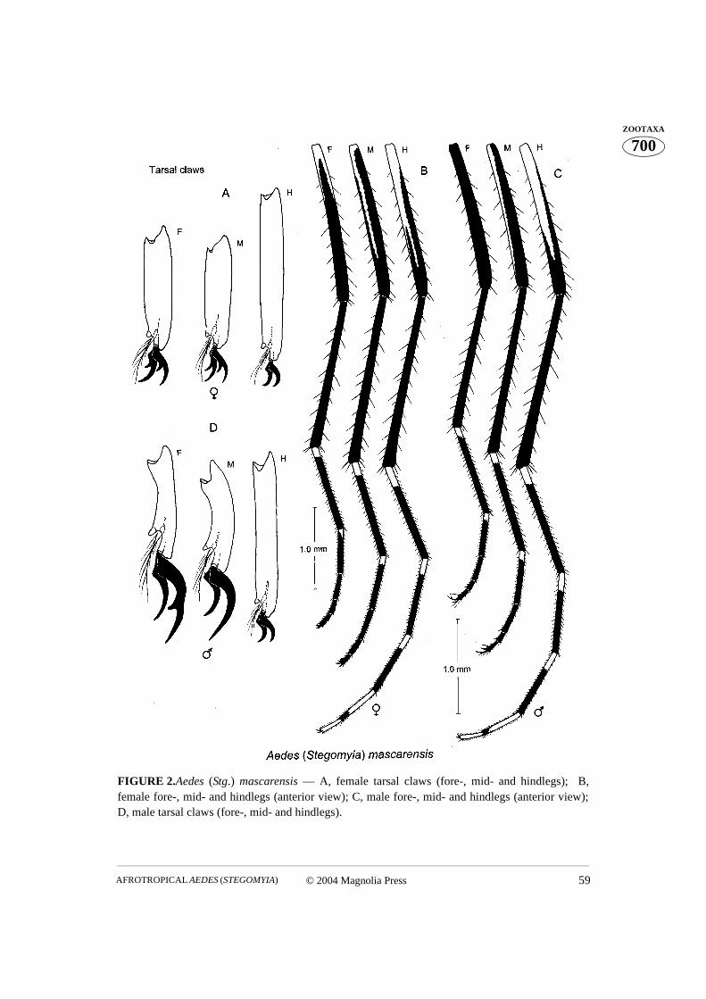
© 2004 Magnolia Press 59AFROTROPICAL AEDES (STEGOMYIA)
700ZOOTAXA
FIGURE 2.Aedes (Stg.) mascarensis — A, female tarsal claws (fore-, mid- and hindlegs); B,female fore-, mid- and hindlegs (anterior view); C, male fore-, mid- and hindlegs (anterior view);D, male tarsal claws (fore-, mid- and hindlegs).
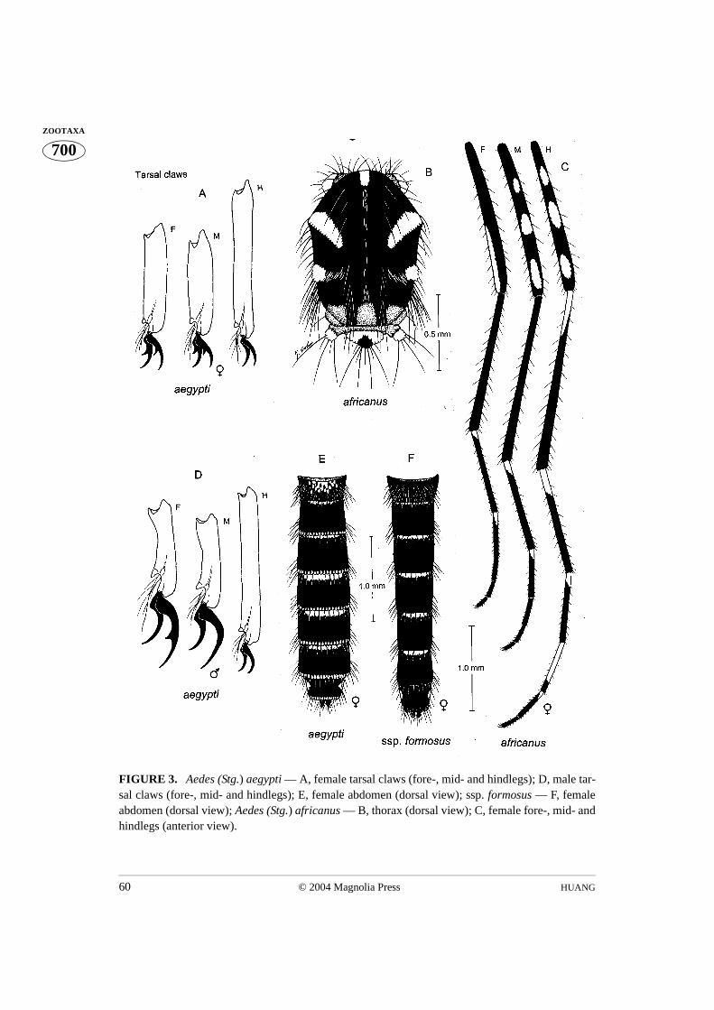
HUANG60 © 2004 Magnolia Press
700ZOOTAXA
FIGURE 3. Aedes (Stg.) aegypti — A, female tarsal claws (fore-, mid- and hindlegs); D, male tar-sal claws (fore-, mid- and hindlegs); E, female abdomen (dorsal view); ssp. formosus — F, femaleabdomen (dorsal view); Aedes (Stg.) africanus — B, thorax (dorsal view); C, female fore-, mid- andhindlegs (anterior view).
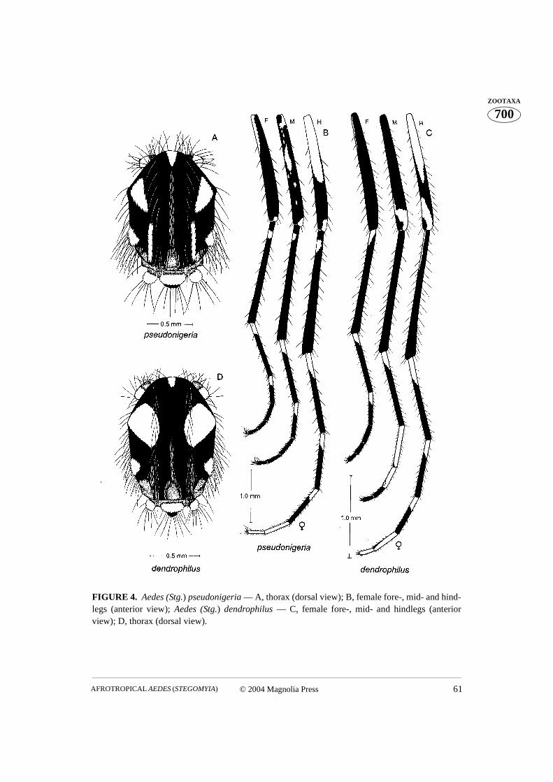
© 2004 Magnolia Press 61AFROTROPICAL AEDES (STEGOMYIA)
700ZOOTAXA
FIGURE 4. Aedes (Stg.) pseudonigeria — A, thorax (dorsal view); B, female fore-, mid- and hind-legs (anterior view); Aedes (Stg.) dendrophilus — C, female fore-, mid- and hindlegs (anteriorview); D, thorax (dorsal view).

HUANG62 © 2004 Magnolia Press
700ZOOTAXA
FIGURE 5.Aedes (Stg.) unilineatus — A, thorax (dorsal view); C, head and proboscis (dorsalview); Aedes (Stg.) granti — B, head and proboscis (dorsal view); D, thorax (dorsal view).
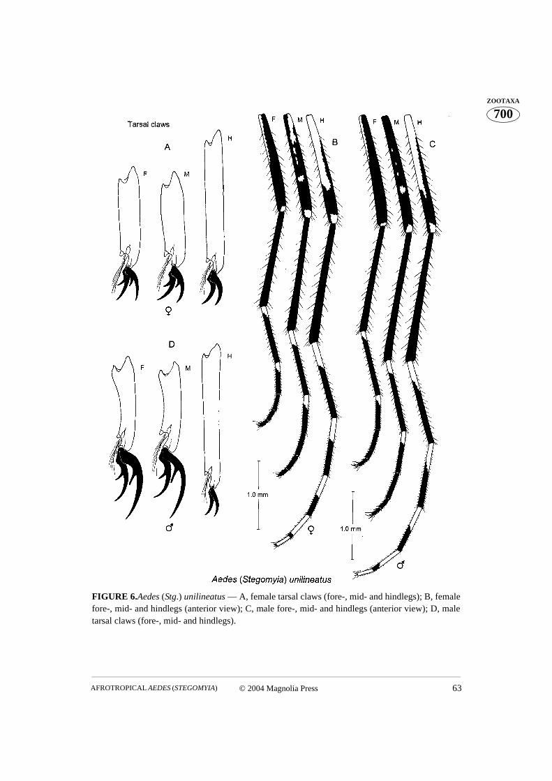
© 2004 Magnolia Press 63AFROTROPICAL AEDES (STEGOMYIA)
700ZOOTAXA
FIGURE 6.Aedes (Stg.) unilineatus — A, female tarsal claws (fore-, mid- and hindlegs); B, femalefore-, mid- and hindlegs (anterior view); C, male fore-, mid- and hindlegs (anterior view); D, maletarsal claws (fore-, mid- and hindlegs).
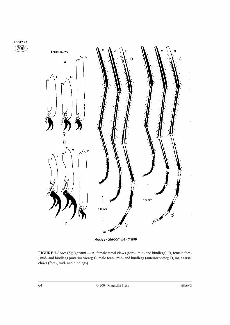
HUANG64 © 2004 Magnolia Press
700ZOOTAXA
FIGURE 7.Aedes (Stg.) granti — A, female tarsal claws (fore-, mid- and hindlegs); B, female fore-, mid- and hindlegs (anterior view); C, male fore-, mid- and hindlegs (anterior view); D, male tarsalclaws (fore-, mid- and hindlegs).

© 2004 Magnolia Press 65AFROTROPICAL AEDES (STEGOMYIA)
700ZOOTAXA
FIGURE 8.Aedes (Stg.) metallicus — A, thorax (dorsal view); B, female fore-, mid- and hindlegs(anterior view); C, male fore-, mid- and hindlegs (anterior view); D, female tarsal claws (fore- andhindlegs); E, male tarsal claws (fore- and hindlegs).

HUANG66 © 2004 Magnolia Press
700ZOOTAXA
FIGURE 9. Aedes (Stg.) apicoargenteus — A, thorax (dorsal view); B, female fore-, mid- andhindlegs (anterior view); Aedes (Stg.) schwetzi — C, female fore-, mid- and hindlegs (anteriorview); D, thorax (dorsal view).
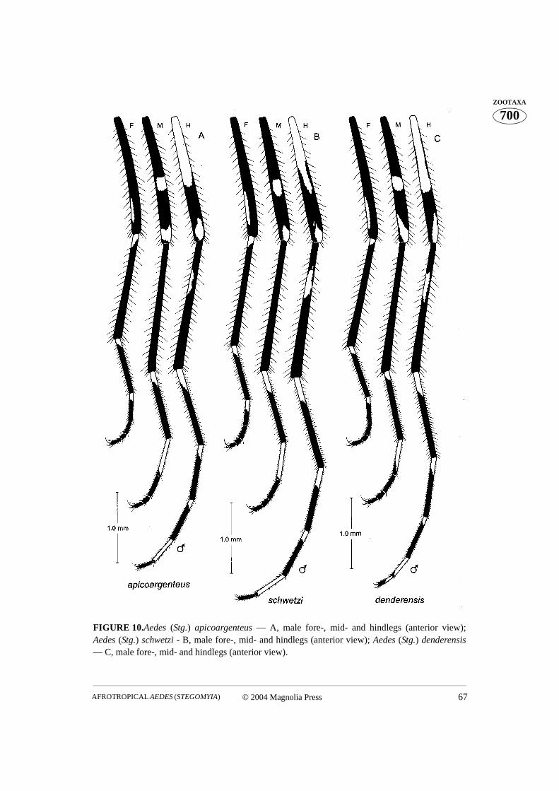
© 2004 Magnolia Press 67AFROTROPICAL AEDES (STEGOMYIA)
700ZOOTAXA
FIGURE 10.Aedes (Stg.) apicoargenteus — A, male fore-, mid- and hindlegs (anterior view);Aedes (Stg.) schwetzi - B, male fore-, mid- and hindlegs (anterior view); Aedes (Stg.) denderensis— C, male fore-, mid- and hindlegs (anterior view).
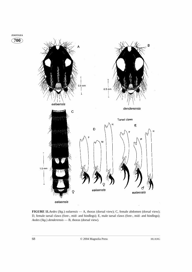
HUANG68 © 2004 Magnolia Press
700ZOOTAXA
FIGURE 11.Aedes (Stg.) ealaensis — A, thorax (dorsal view); C, female abdomen (dorsal view);D, female tarsal claws (fore-, mid- and hindlegs); E, male tarsal claws (fore-, mid- and hindlegs);Aedes (Stg.) denderensis — B, thorax (dorsal view).
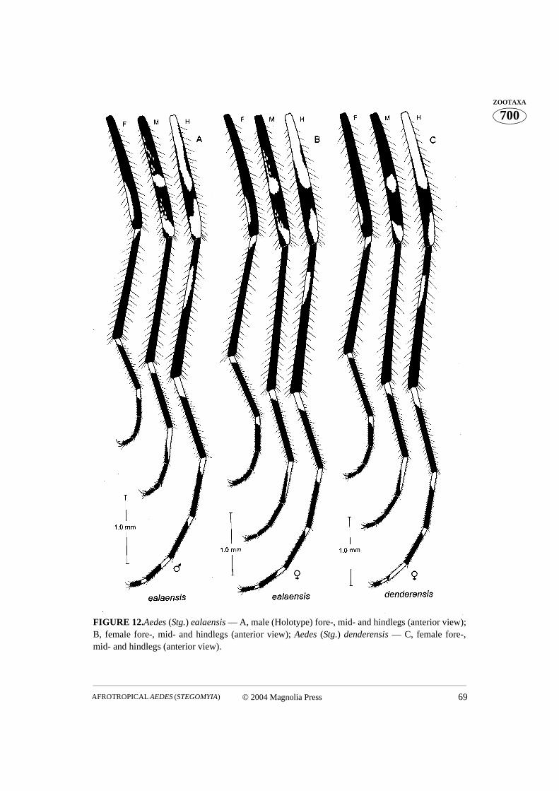
© 2004 Magnolia Press 69AFROTROPICAL AEDES (STEGOMYIA)
700ZOOTAXA
FIGURE 12.Aedes (Stg.) ealaensis — A, male (Holotype) fore-, mid- and hindlegs (anterior view);B, female fore-, mid- and hindlegs (anterior view); Aedes (Stg.) denderensis — C, female fore-,mid- and hindlegs (anterior view).
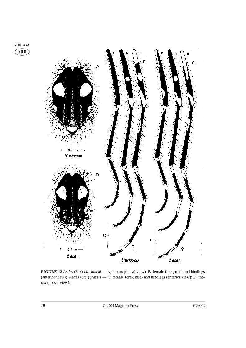
HUANG70 © 2004 Magnolia Press
700ZOOTAXA
FIGURE 13.Aedes (Stg.) blacklocki — A, thorax (dorsal view); B, female fore-, mid- and hindlegs(anterior view); Aedes (Stg.) fraseri — C, female fore-, mid- and hindlegs (anterior view); D, tho-rax (dorsal view).
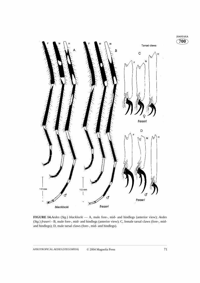
© 2004 Magnolia Press 71AFROTROPICAL AEDES (STEGOMYIA)
700ZOOTAXA
FIGURE 14.Aedes (Stg.) blacklocki — A, male fore-, mid- and hindlegs (anterior view); Aedes(Stg.) fraseri - B, male fore-, mid- and hindlegs (anterior view); C, female tarsal claws (fore-, mid-and hindlegs); D, male tarsal claws (fore-, mid- and hindlegs).

HUANG72 © 2004 Magnolia Press
700ZOOTAXA
FIGURE 15.Aedes (Stg.) soleatus — A, thorax (dorsal view); B, female fore-, mid- and hindlegs(anterior view); C, male fore-, mid- and hindlegs (anterior view); D, female tarsal claws (fore- andhindlegs); E, male tarsal claws (fore- and hindlegs).
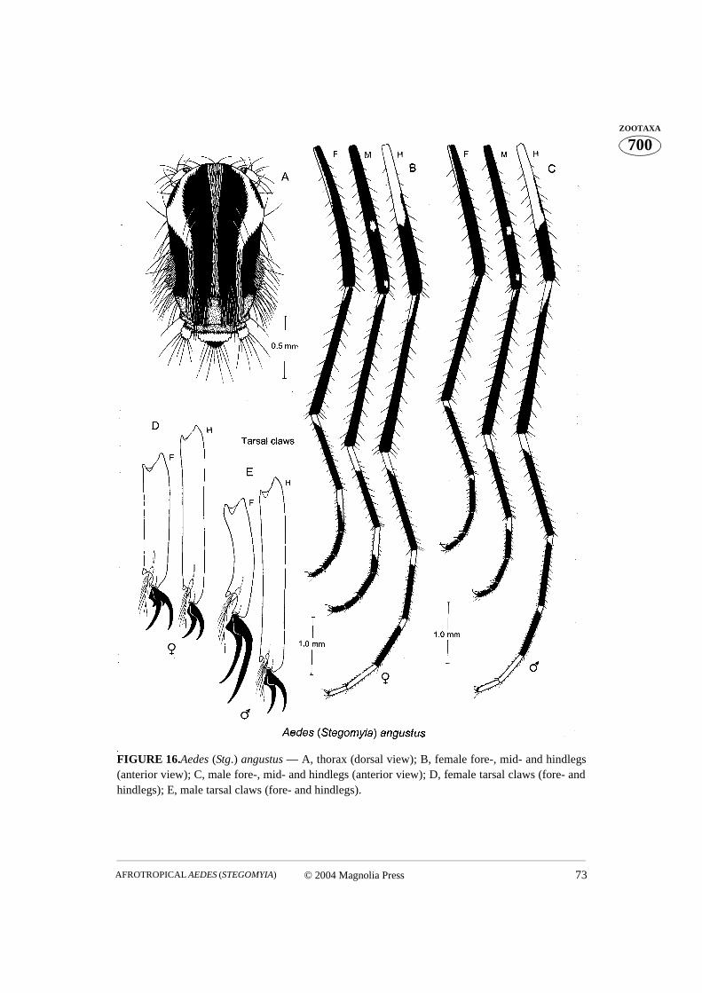
© 2004 Magnolia Press 73AFROTROPICAL AEDES (STEGOMYIA)
700ZOOTAXA
FIGURE 16.Aedes (Stg.) angustus — A, thorax (dorsal view); B, female fore-, mid- and hindlegs(anterior view); C, male fore-, mid- and hindlegs (anterior view); D, female tarsal claws (fore- andhindlegs); E, male tarsal claws (fore- and hindlegs).
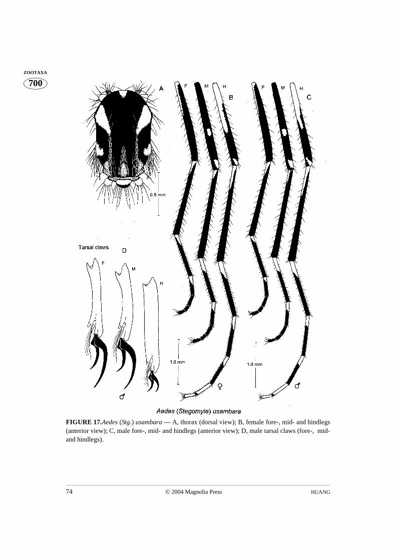
HUANG74 © 2004 Magnolia Press
700ZOOTAXA
FIGURE 17.Aedes (Stg.) usambara — A, thorax (dorsal view); B, female fore-, mid- and hindlegs(anterior view); C, male fore-, mid- and hindlegs (anterior view); D, male tarsal claws (fore-, mid-and hindlegs).

© 2004 Magnolia Press 75AFROTROPICAL AEDES (STEGOMYIA)
700ZOOTAXA
FIGURE 18.Aedes (Stg.) mpusiensis — A, male (Holotype) fore-, mid- and hindlegs (anteriorview); Aedes(Stg.) ethiopiensis — B, male (Holotype) fore-, mid- and hindlegs (anterior view);Aedes (Stg.) hogsbackensis — C, male (Holotype) fore-, mid- and hindlegs (anterior view).
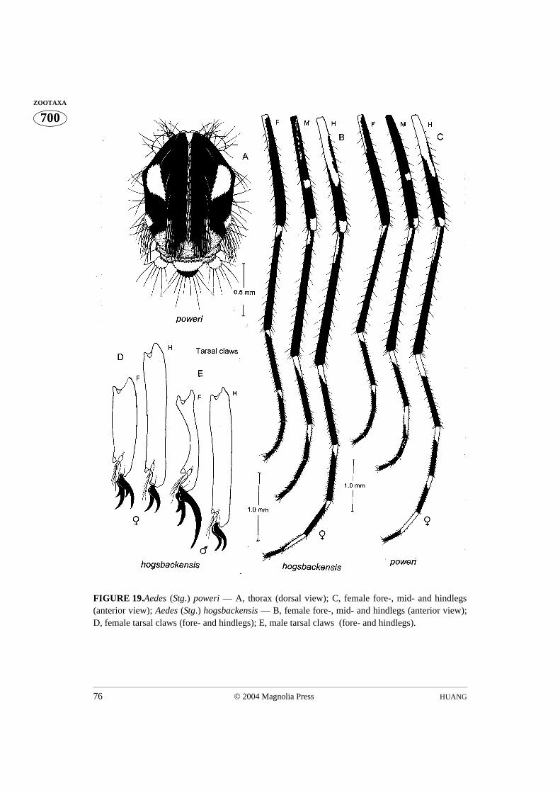
HUANG76 © 2004 Magnolia Press
700ZOOTAXA
FIGURE 19.Aedes (Stg.) poweri — A, thorax (dorsal view); C, female fore-, mid- and hindlegs(anterior view); Aedes (Stg.) hogsbackensis — B, female fore-, mid- and hindlegs (anterior view);D, female tarsal claws (fore- and hindlegs); E, male tarsal claws (fore- and hindlegs).
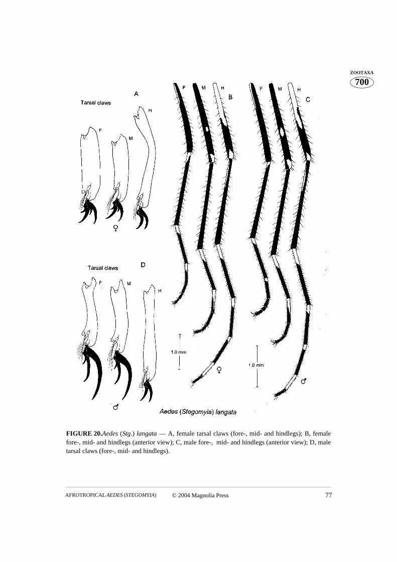
© 2004 Magnolia Press 77AFROTROPICAL AEDES (STEGOMYIA)
700ZOOTAXA
FIGURE 20.Aedes (Stg.) langata — A, female tarsal claws (fore-, mid- and hindlegs); B, femalefore-, mid- and hindlegs (anterior view); C, male fore-, mid- and hindlegs (anterior view); D, maletarsal claws (fore-, mid- and hindlegs).

HUANG78 © 2004 Magnolia Press
700ZOOTAXA
FIGURE 21.Aedes (Stg.) calceatus — A, male fore-, mid- and hindlegs (anterior view); B, femalefore-, mid- and hindlegs (anterior view); Aedes (Stg.) ledgeri — C, female fore-, mid- and hindlegs(anterior view).
© 2004 Magnolia Press 79AFROTROPICAL AEDES (STEGOMYIA)
700ZOOTAXA
FIGURE 22.Aedes (Stg.) ledgeri — A, thorax (dorsal view); B, male fore-, mid- and hindlegs(anterior view); C, male abdomen (dorsal view); D, male abdomen (lateral view).
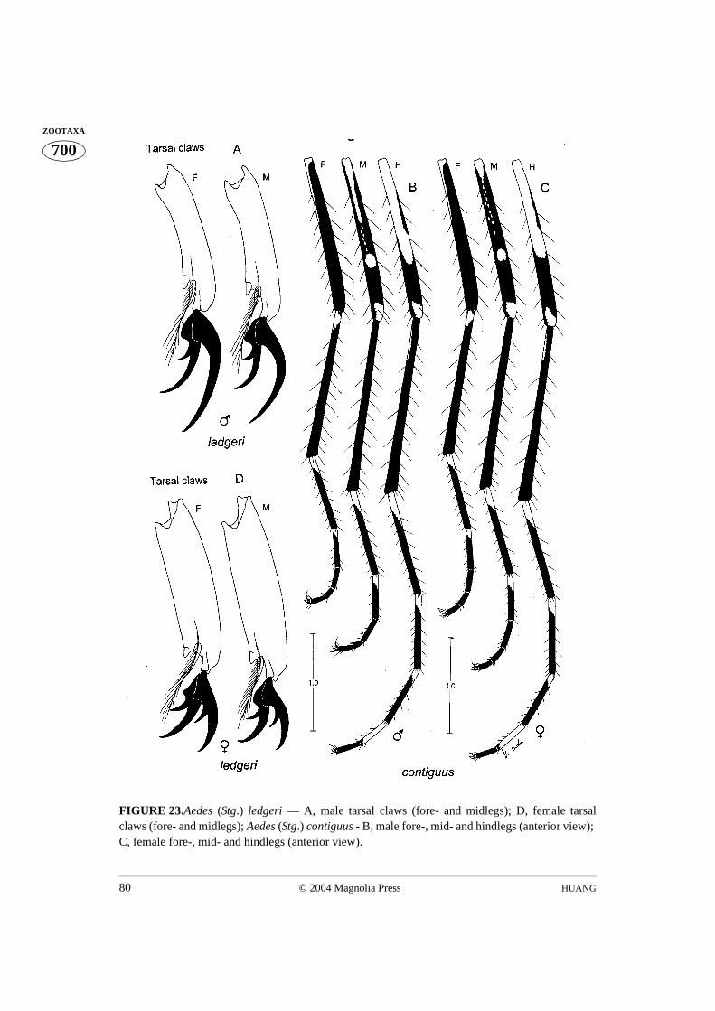
HUANG80 © 2004 Magnolia Press
700ZOOTAXA
FIGURE 23.Aedes (Stg.) ledgeri — A, male tarsal claws (fore- and midlegs); D, female tarsalclaws (fore- and midlegs); Aedes (Stg.) contiguus - B, male fore-, mid- and hindlegs (anterior view); C, female fore-, mid- and hindlegs (anterior view).

© 2004 Magnolia Press 81AFROTROPICAL AEDES (STEGOMYIA)
700ZOOTAXA
FIGURE 24.Aedes (Stg.) calceatus — A, thorax (dorsal view); Aedes (Stg.) contiguus — B, thorax(dorsal view); Aedes (Stg.) calceatus — C1, female midtarsomeres 1, 2 (posterior view); Aedes(Stg.) ledgeri — C2, female midtarsomeres 1, 2 (posterioa view); Aedes (Stg.) contiguus — C3,female midtarsomeres 1, 2 (posterior view); Aedes (Stg.) langata — C4, female midtarsomeres 1, 2(posterior view); Aedes (Stg.) poweri — C5, female midtarsomeres 1, 2 (posterior view); Aedes(Stg.) hogsbackensis — C6, female midtarsomeres 1, 2 (posterior view); Aedes (Stg.) calceatus —D1, male midtarsomeres 1, 2 (posterior view); Aedes (Stg.) ledgeri — D2, male midtarsomeres 1, 2(posterior view); Aedes (Stg.) contiguus — D3, male midtarsomeres 1, 2 (posterior view); Aedes(Stg.) langata — D4, male midtarsomeres 1, 2 (posterior view); Aedes (Stg.) hogsbackensis — D5,male midtarsomeres 1, 2 (posterior view).
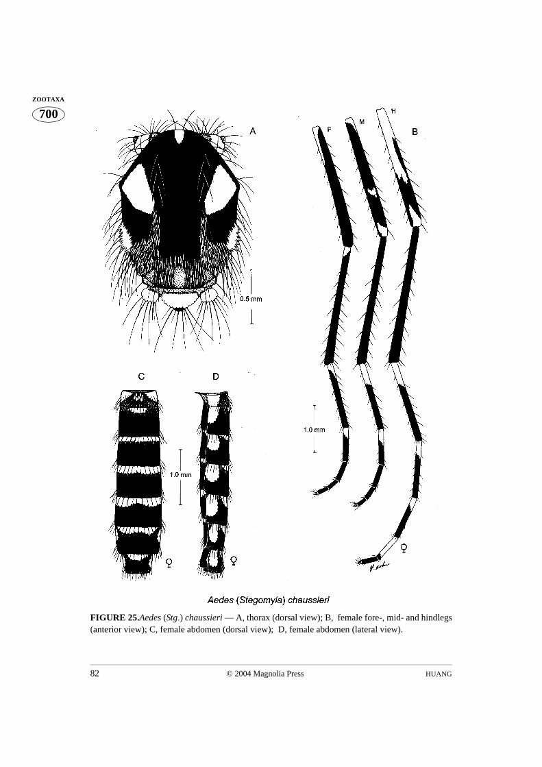
HUANG82 © 2004 Magnolia Press
700ZOOTAXA
FIGURE 25.Aedes (Stg.) chaussieri — A, thorax (dorsal view); B, female fore-, mid- and hindlegs(anterior view); C, female abdomen (dorsal view); D, female abdomen (lateral view).
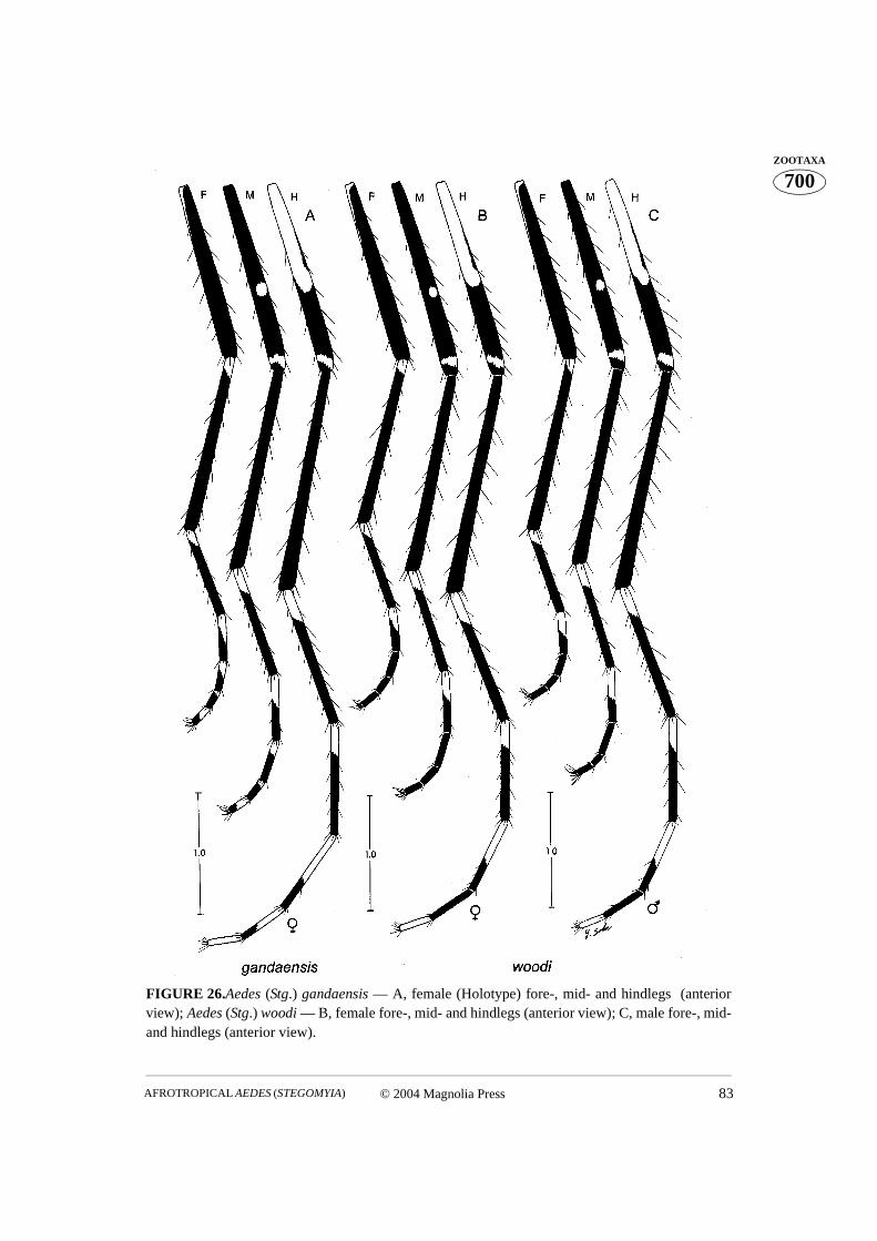
© 2004 Magnolia Press 83AFROTROPICAL AEDES (STEGOMYIA)
700ZOOTAXA
FIGURE 26.Aedes (Stg.) gandaensis — A, female (Holotype) fore-, mid- and hindlegs (anteriorview); Aedes (Stg.) woodi — B, female fore-, mid- and hindlegs (anterior view); C, male fore-, mid-and hindlegs (anterior view).
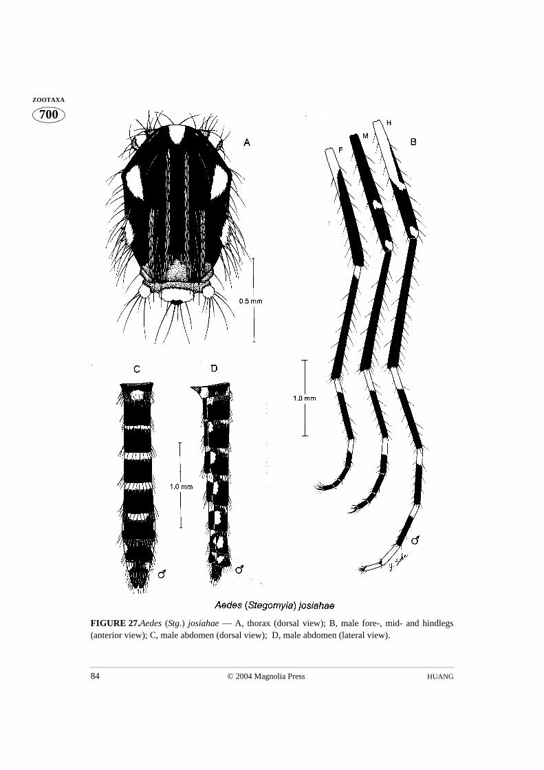
HUANG84 © 2004 Magnolia Press
700ZOOTAXA
FIGURE 27.Aedes (Stg.) josiahae — A, thorax (dorsal view); B, male fore-, mid- and hindlegs(anterior view); C, male abdomen (dorsal view); D, male abdomen (lateral view).

© 2004 Magnolia Press 85AFROTROPICAL AEDES (STEGOMYIA)
700ZOOTAXA
FIGURE 28.Aedes (Stg.) josiahae — A, female fore-, mid- and hindlegs (anterior view); Aedes(Stg.) strelitziae — B, male fore-, mid- and hindlegs (anterior view); C, female fore-, mid- andhindlegs (anterior view).

HUANG86 © 2004 Magnolia Press
700ZOOTAXA
FIGURE 29.Aedes (Stg.) kivuensis — A, thorax (dorsal view); B, female fore-, mid- and hindlegs(anterior view); C, female abdomen (dorsal view); D, female abdomen (lateral view).

© 2004 Magnolia Press 87AFROTROPICAL AEDES (STEGOMYIA)
700ZOOTAXA
FIGURE 30.Aedes (Stg.) sampi — A, female (Holotype) fore-, mid- and hindlegs (anterior view);Aedes (Steg.) subargenteus — B, male fore-, mid- and hindlegs (anterior view); C, female fore-,mid- and hindlegs (anterior view).

HUANG88 © 2004 Magnolia Press
700ZOOTAXA
FIGURE 31.Aedes (Stg.) subargenteus — A, thorax (dorsal view); Aedes (Stg.) strelitziae — B,thorax (dorsal view); Aedes (Stg.) woodi — C, thorax (dorsal view); Aedes (Stg.) simpsoni — D,female tarsal claws (fore- and midlegs); Aedes (Stg.) lilii — E, female tarsal claws (fore- and mid-legs); Aedes (Stg.) bromeliae — F, female tarsal claws (fore- and midlegs).

© 2004 Magnolia Press 89AFROTROPICAL AEDES (STEGOMYIA)
700ZOOTAXA
FIGURE 32.Aedes (Stg.) simpsoni — Lectotype female: A, thorax (dorsal view); B, female fore-,mid- and hindlegs (anterior view); C, female abdomen (dorsal view); D, female abdomen (lateralview).

HUANG90 © 2004 Magnolia Press
700ZOOTAXA
FIGURE 33.Aedes (Stg.) simpsoni — A, female fore-, mid- and hindlegs (anterior view); Aedes(Stg.) lilii — B, female fore-, mid- and hindlegs (anterior view); Aedes (Stg.) bromeliae — C,female fore-, mid- and hindlegs (anterior view).
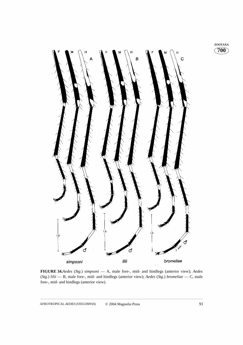
© 2004 Magnolia Press 91AFROTROPICAL AEDES (STEGOMYIA)
700ZOOTAXA
FIGURE 34.Aedes (Stg.) simpsoni — A, male fore-, mid- and hindlegs (anterior view); Aedes(Stg.) lilii — B, male fore-, mid- and hindlegs (anterior view); Aedes (Stg.) bromeliae — C, malefore-, mid- and hindlegs (anterior view).
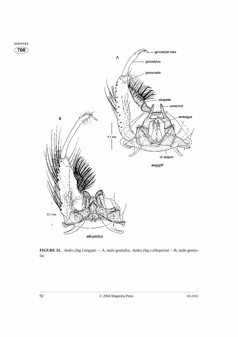
HUANG92 © 2004 Magnolia Press
700ZOOTAXA
FIGURE 35. Aedes (Stg.) aegypti — A, male genitalia; Aedes (Stg.) albopictus —B, male genita-lia.
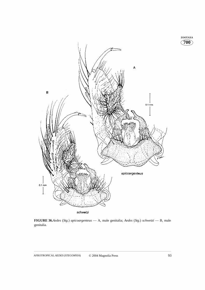
© 2004 Magnolia Press 93AFROTROPICAL AEDES (STEGOMYIA)
700ZOOTAXA
FIGURE 36.Aedes (Stg.) apicoargenteus — A, male genitalia; Aedes (Stg.) schwetzi — B, malegenitalia.

HUANG94 © 2004 Magnolia Press
700ZOOTAXA
FIGURE 37.Aedes (Stg.) denderensis — A, male genitalia; Aedes (Stg.) ealaensis — B, male geni-talia.
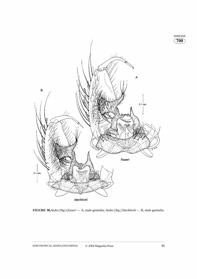
© 2004 Magnolia Press 95AFROTROPICAL AEDES (STEGOMYIA)
700ZOOTAXA
FIGURE 38.Aedes (Stg.) fraseri — A, male genitalia; Aedes (Stg.) blacklocki — B, male genitalia.
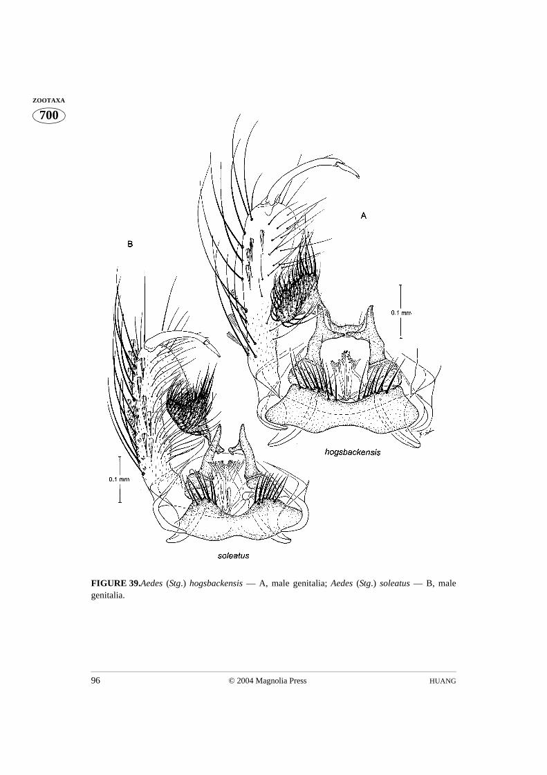
HUANG96 © 2004 Magnolia Press
700ZOOTAXA
FIGURE 39.Aedes (Stg.) hogsbackensis — A, male genitalia; Aedes (Stg.) soleatus — B, malegenitalia.

© 2004 Magnolia Press 97AFROTROPICAL AEDES (STEGOMYIA)
700ZOOTAXA
FIGURE 40.Aedes (Stg.) angustus — A, male genitalia. Aedes (Stg.) mpusiensis — B, male genita-lia;
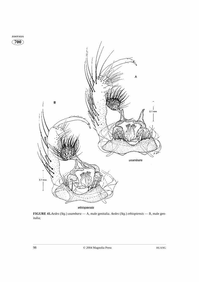
HUANG98 © 2004 Magnolia Press
700ZOOTAXA
FIGURE 41.Aedes (Stg.) usambara — A, male genitalia. Aedes (Stg.) ethiopiensis — B, male gen-italia;
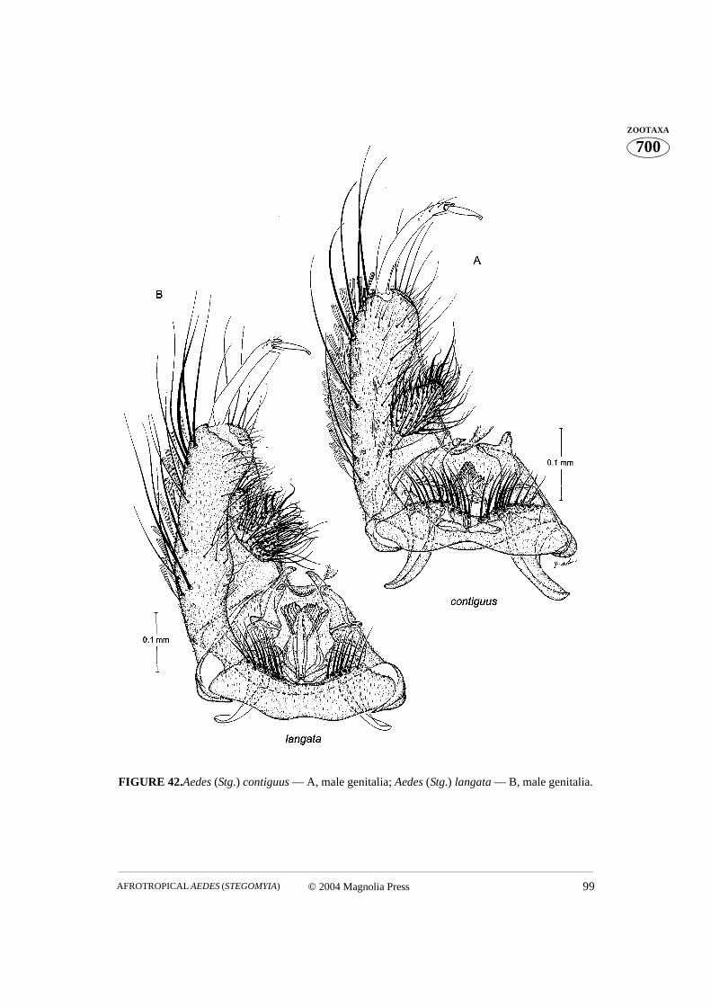
© 2004 Magnolia Press 99AFROTROPICAL AEDES (STEGOMYIA)
700ZOOTAXA
FIGURE 42.Aedes (Stg.) contiguus — A, male genitalia; Aedes (Stg.) langata — B, male genitalia.
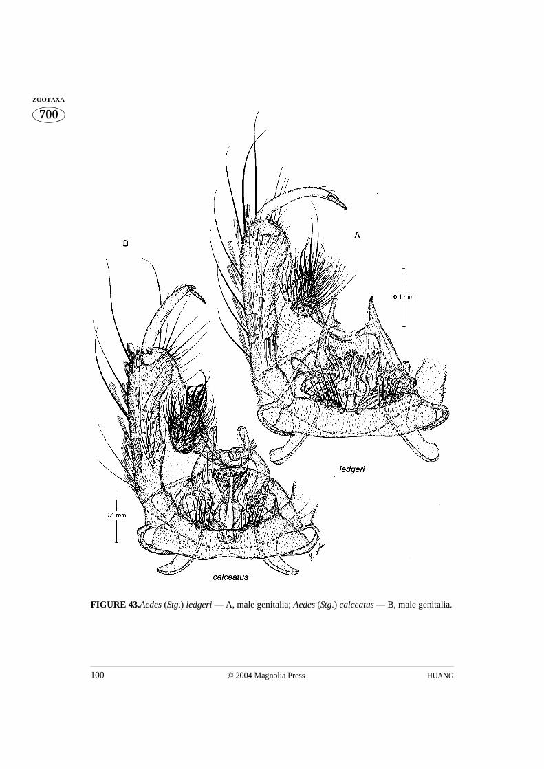
HUANG100 © 2004 Magnolia Press
700ZOOTAXA
FIGURE 43.Aedes (Stg.) ledgeri — A, male genitalia; Aedes (Stg.) calceatus — B, male genitalia.
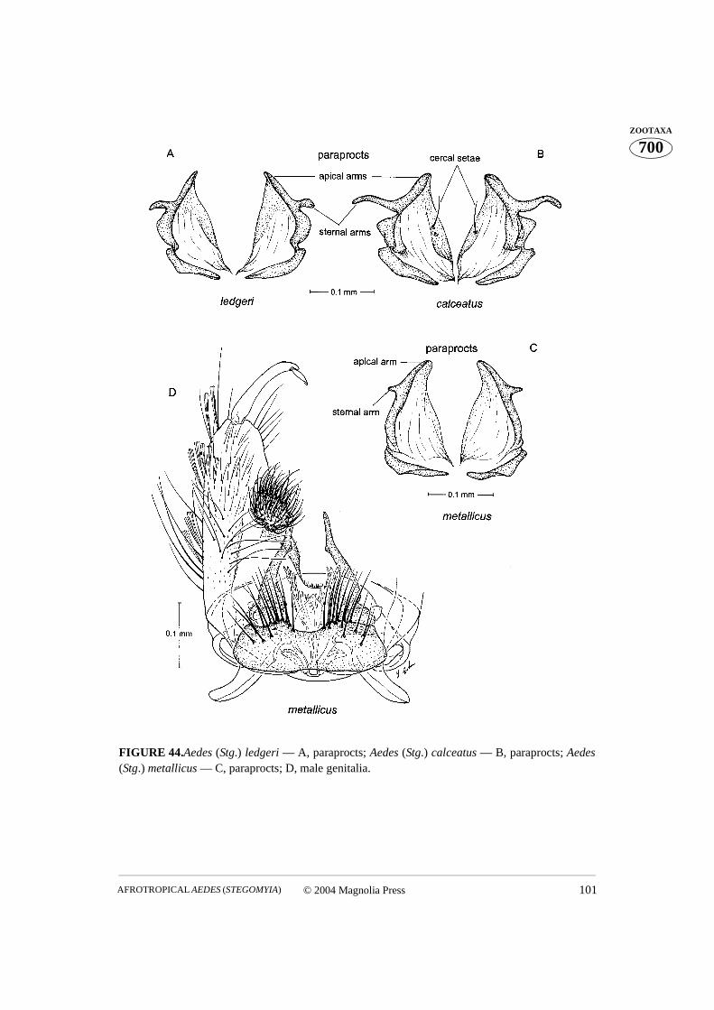
© 2004 Magnolia Press 101AFROTROPICAL AEDES (STEGOMYIA)
700ZOOTAXA
FIGURE 44.Aedes (Stg.) ledgeri — A, paraprocts; Aedes (Stg.) calceatus — B, paraprocts; Aedes(Stg.) metallicus — C, paraprocts; D, male genitalia.
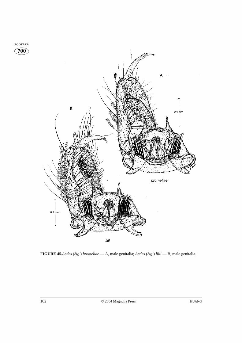
HUANG102 © 2004 Magnolia Press
700ZOOTAXA
FIGURE 45.Aedes (Stg.) bromeliae — A, male genitalia; Aedes (Stg.) lilii — B, male genitalia.
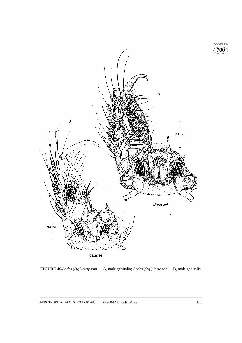
© 2004 Magnolia Press 103AFROTROPICAL AEDES (STEGOMYIA)
700ZOOTAXA
FIGURE 46.Aedes (Stg.) simpsoni — A, male genitalia; Aedes (Stg.) josiahae — B, male genitalia.
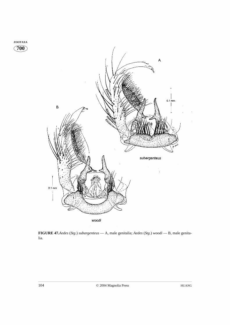
HUANG104 © 2004 Magnolia Press
700ZOOTAXA
FIGURE 47.Aedes (Stg.) subargenteus — A, male genitalia; Aedes (Stg.) woodi — B, male genita-lia.
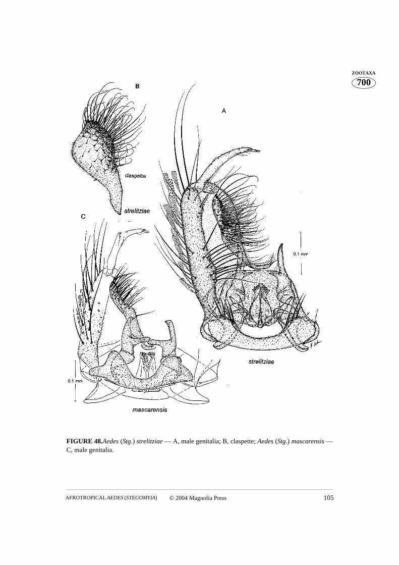
© 2004 Magnolia Press 105AFROTROPICAL AEDES (STEGOMYIA)
700ZOOTAXA
FIGURE 48.Aedes (Stg.) strelitziae — A, male genitalia; B, claspette; Aedes (Stg.) mascarensis —C, male genitalia.
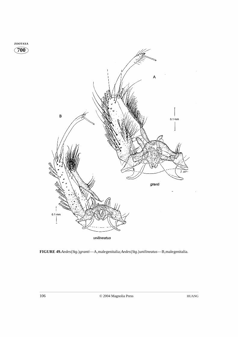
HUANG106 © 2004 Magnolia Press
700ZOOTAXA
FIGURE 49.Aedes (Stg.) granti — A, male genitalia; Aedes (Stg.) unilineatus — B, male genitalia.

© 2004 Magnolia Press 107AFROTROPICAL AEDES (STEGOMYIA)
700ZOOTAXA
FIGURE 50. Aedes (Stg.) chemulpoensis — A, male genitalia. Aedes (Stg.) hogsbackensis — B,larva; C, pupa.
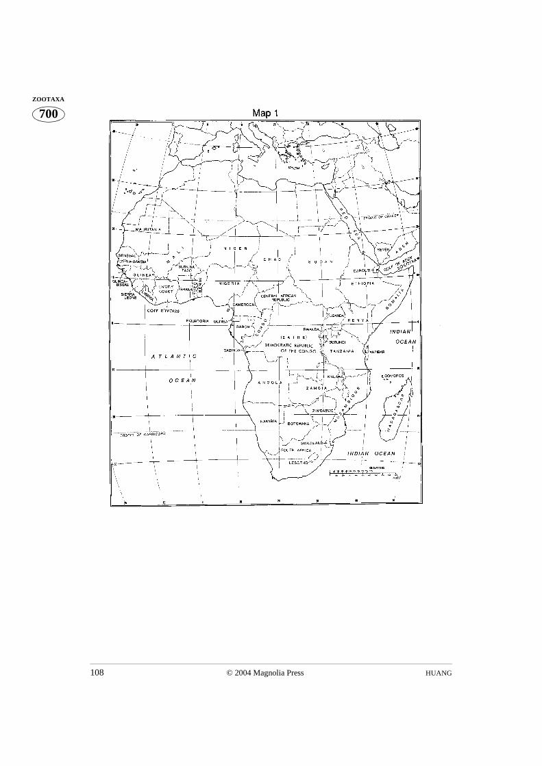
HUANG108 © 2004 Magnolia Press
700ZOOTAXA

© 2004 Magnolia Press 109AFROTROPICAL AEDES (STEGOMYIA)
700ZOOTAXAAPPENDIX I. Present status of the species of Aedes (Stegomyia) in the Afrotropical Region.
SPECIES STAGES BIONOMICS
A P L E
M F
aegypti group
aegypti aegypti X* X* X* X* X* Immature habitats known, female bites man
ssp. formosus — X — — — Immature habitats known, female bites man
mascarensis1 X* X* — X* — Immature habitats known, female bites man
africanus group
africanus X* X* X* X* X* Immature habitats known, female bites man
corneti X* X* X* X* — Immature habitats known, female unknown
luteocephalus X* X* X* X* — Immature habitats known, female bites man
maxgermaini — X* — — — Immature habitats unknown, female bites man
neoafricanus X* X* — X* — Immature habitats known, female bites man
opok X* X* — — — Immature habitats known, female bites man
pseudoafricanus X* X* X* X* — Immature habitats known, female bites man
ruwenzori X* X* X X* — Immature habitats unknown, female bites man
apicoargenteus group
apicoargenteus X* X* — X* — Immature habitats known, female bites man
blacklocki X* X* — — — Immature habitats known, female unknown
denderensis X* X* — X — Immature habitats known, female bites man
ealaensis X* X* — — — Immature habitats unknown, female bites man
fraseri X* X* — X* — Immature habitats known, female bites man
schwetzi X* X* — X — Immature habitats known, female bites man
soleatus X* X* X X* — Immature habitats known, female bites man
dendrophilus group
amaltheus X* X* X* X* — Immature habitats known, female bites man
bambusae X* X* X X — Immature habitats known, female bites man
deboeri X* X* X* X* — Immature habitats known, female bites man
demeilloni X* X* X* X* — Immature habitats known, female bites man
dendrophilus X* X* X* X* — Immature habitats known, female bites man
hansfordi X* X* X* X* — Immature habitats known, female bites man
heischi X* X* X* X* — Immature habitats known, female unknown
keniensis X* X* — X* — Immature habitats known, female bites man
kenyae X* X* — X — Immature habitats known, female bites man
masseyi X* X* — ?X* — Immature habitats known, female bites man
mattinglyorum X* X* X* X* — Immature habitats known, female bites man
muroafcete — X* — — — Immature habitats unknown, female unknown
njombiensis — X* — — — Immature habitats unknown, female unknown
segermanae X* X* — — — Immature habitats known, female bites man
metallicus group
metallicus X* X* X X* — Immature habitats known, female bites man
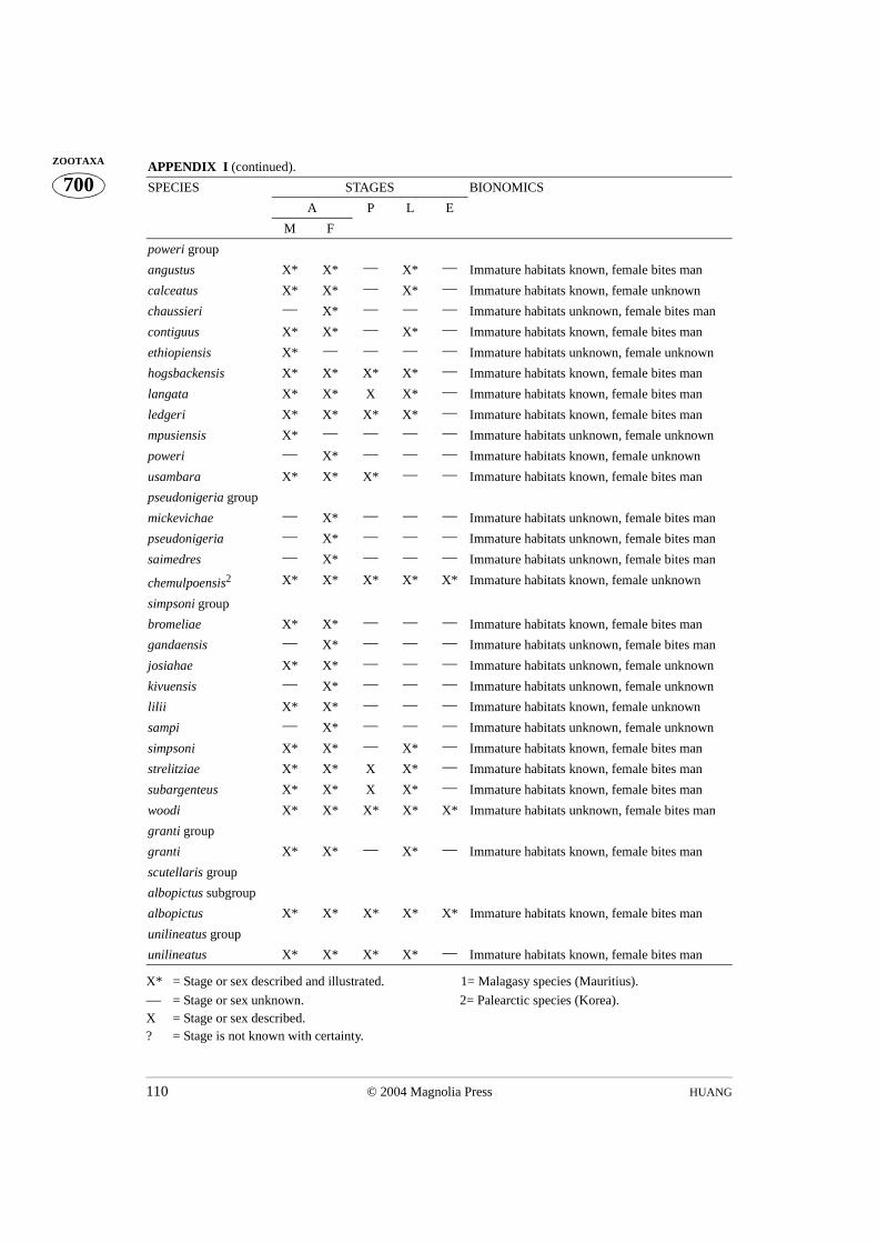
HUANG110 © 2004 Magnolia Press
700ZOOTAXA
X* = Stage or sex described and illustrated. 1= Malagasy species (Mauritius).
— = Stage or sex unknown. 2= Palearctic species (Korea).X = Stage or sex described.? = Stage is not known with certainty.
APPENDIX I (continued).
SPECIES STAGES BIONOMICS
A P L E
M F
poweri group
angustus X* X* — X* — Immature habitats known, female bites man
calceatus X* X* — X* — Immature habitats known, female unknown
chaussieri — X* — — — Immature habitats unknown, female bites man
contiguus X* X* — X* — Immature habitats known, female bites man
ethiopiensis X* — — — — Immature habitats unknown, female unknown
hogsbackensis X* X* X* X* — Immature habitats known, female bites man
langata X* X* X X* — Immature habitats known, female bites man
ledgeri X* X* X* X* — Immature habitats known, female bites man
mpusiensis X* — — — — Immature habitats unknown, female unknown
poweri — X* — — — Immature habitats known, female unknown
usambara X* X* X* — — Immature habitats known, female bites man
pseudonigeria group
mickevichae — X* — — — Immature habitats unknown, female bites man
pseudonigeria — X* — — — Immature habitats unknown, female bites man
saimedres — X* — — — Immature habitats unknown, female bites man
chemulpoensis2 X* X* X* X* X* Immature habitats known, female unknown
simpsoni group
bromeliae X* X* — — — Immature habitats known, female bites man
gandaensis — X* — — — Immature habitats unknown, female bites man
josiahae X* X* — — — Immature habitats unknown, female unknown
kivuensis — X* — — — Immature habitats unknown, female unknown
lilii X* X* — — — Immature habitats known, female unknown
sampi — X* — — — Immature habitats unknown, female unknown
simpsoni X* X* — X* — Immature habitats known, female bites man
strelitziae X* X* X X* — Immature habitats known, female bites man
subargenteus X* X* X X* — Immature habitats known, female bites man
woodi X* X* X* X* X* Immature habitats unknown, female bites man
granti group
granti X* X* — X* — Immature habitats known, female bites man
scutellaris group
albopictus subgroup
albopictus X* X* X* X* X* Immature habitats known, female bites man
unilineatus group
unilineatus X* X* X* X* — Immature habitats known, female bites man
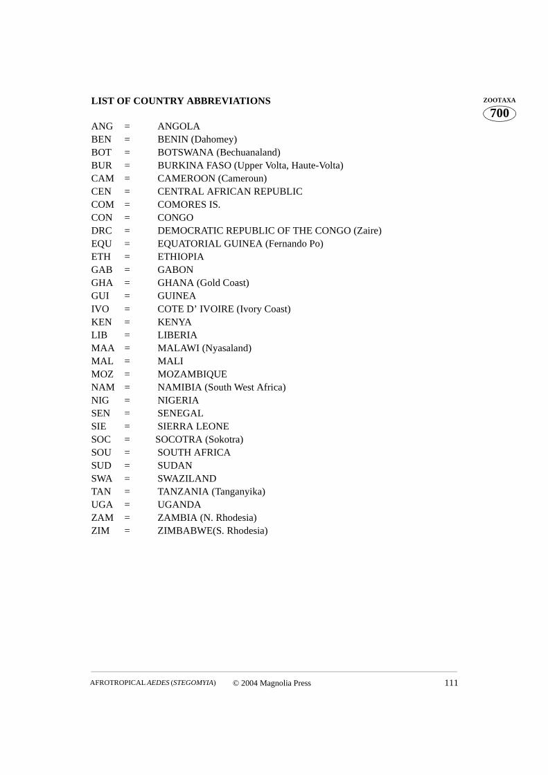
© 2004 Magnolia Press 111AFROTROPICAL AEDES (STEGOMYIA)
700ZOOTAXALIST OF COUNTRY ABBREVIATIONS
ANG = ANGOLABEN = BENIN (Dahomey)BOT = BOTSWANA (Bechuanaland)BUR = BURKINA FASO (Upper Volta, Haute-Volta)CAM = CAMEROON (Cameroun)CEN = CENTRAL AFRICAN REPUBLICCOM = COMORES IS.CON = CONGODRC = DEMOCRATIC REPUBLIC OF THE CONGO (Zaire) EQU = EQUATORIAL GUINEA (Fernando Po)ETH = ETHIOPIAGAB = GABONGHA = GHANA (Gold Coast)GUI = GUINEAIVO = COTE D’ IVOIRE (Ivory Coast)KEN = KENYALIB = LIBERIAMAA = MALAWI (Nyasaland)MAL = MALIMOZ = MOZAMBIQUENAM = NAMIBIA (South West Africa)NIG = NIGERIASEN = SENEGALSIE = SIERRA LEONESOC = SOCOTRA (Sokotra) SOU = SOUTH AFRICASUD = SUDANSWA = SWAZILANDTAN = TANZANIA (Tanganyika)UGA = UGANDAZAM = ZAMBIA (N. Rhodesia)ZIM = ZIMBABWE(S. Rhodesia)
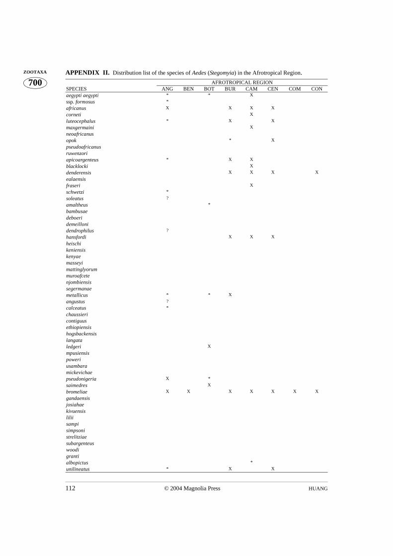
HUANG112 © 2004 Magnolia Press
700ZOOTAXA APPENDIX II. Distribution list of the species of Aedes (Stegomyia) in the Afrotropical Region.
AFROTROPICAL REGIONSPECIES ANG BEN BOT BUR CAM CEN COM CONaegypti aegypti * * X
ssp. formosus *
africanus X X X X
corneti X
luteocephalus * X X
maxgermaini X
neoafricanusopok * X
pseudoafricanusruwenzoriapicoargenteus * X X
blacklocki X
denderensis X X X X
ealaensisfraseri X
schwetzi *
soleatus ?
amaltheus *
bambusaedeboeridemeillonidendrophilus ?
hansfordi X X X
heischikeniensiskenyaemasseyimattinglyorummuroafcetenjombiensissegermanaemetallicus * * X
angustus ?
calceatus *
chaussiericontiguusethiopiensishogsbackensislangataledgeri X
mpusiensispoweriusambaramickevichaepseudonigeria X *
saimedres X
bromeliae X X X X X X X
gandaensisjosiahaekivuensisliliisampisimpsonistrelitziaesubargenteuswoodigrantialbopictus *
unilineatus * X X
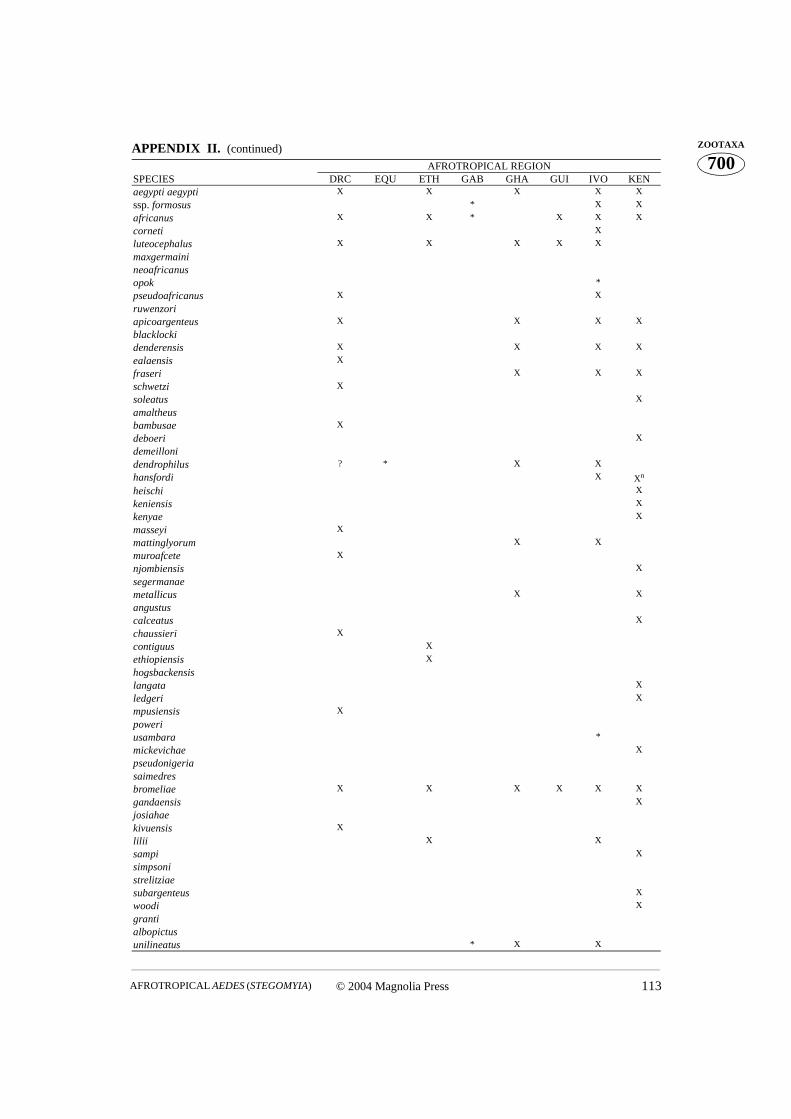
© 2004 Magnolia Press 113AFROTROPICAL AEDES (STEGOMYIA)
700ZOOTAXAAPPENDIX II. (continued)
AFROTROPICAL REGIONSPECIES DRC EQU ETH GAB GHA GUI IVO KENaegypti aegypti X X X X X
ssp. formosus * X X
africanus X X * X X X
corneti X
luteocephalus X X X X X
maxgermainineoafricanusopok *
pseudoafricanus X X
ruwenzoriapicoargenteus X X X X
blacklockidenderensis X X X X
ealaensis X
fraseri X X X
schwetzi X
soleatus X
amaltheusbambusae X
deboeri X
demeillonidendrophilus ? * X X
hansfordi X Xn
heischi X
keniensis X
kenyae X
masseyi X
mattinglyorum X X
muroafcete X
njombiensis X
segermanaemetallicus X X
angustuscalceatus X
chaussieri X
contiguus X
ethiopiensis X
hogsbackensislangata X
ledgeri X
mpusiensis X
poweriusambara *
mickevichae X
pseudonigeriasaimedresbromeliae X X X X X X
gandaensis X
josiahaekivuensis X
lilii X X
sampi X
simpsonistrelitziaesubargenteus X
woodi X
grantialbopictusunilineatus * X X
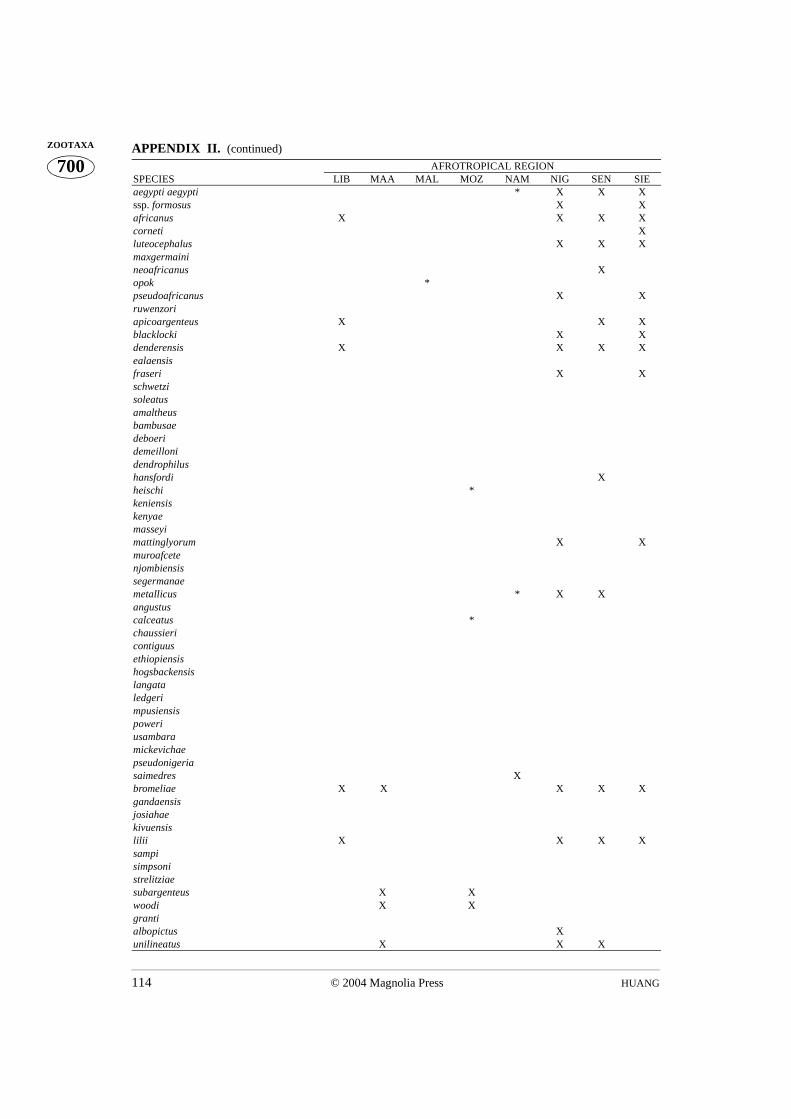
HUANG114 © 2004 Magnolia Press
700ZOOTAXA APPENDIX II. (continued)
AFROTROPICAL REGIONSPECIES LIB MAA MAL MOZ NAM NIG SEN SIEaegypti aegypti * X X Xssp. formosus X Xafricanus X X X Xcorneti Xluteocephalus X X Xmaxgermainineoafricanus Xopok *pseudoafricanus X Xruwenzoriapicoargenteus X X Xblacklocki X Xdenderensis X X X Xealaensisfraseri X Xschwetzisoleatusamaltheusbambusaedeboeridemeillonidendrophilushansfordi Xheischi *keniensiskenyaemasseyimattinglyorum X Xmuroafcetenjombiensissegermanaemetallicus * X Xangustuscalceatus *chaussiericontiguusethiopiensishogsbackensislangataledgerimpusiensispoweriusambaramickevichaepseudonigeriasaimedres Xbromeliae X X X X Xgandaensisjosiahaekivuensislilii X X X Xsampisimpsonistrelitziaesubargenteus X Xwoodi X Xgrantialbopictus Xunilineatus X X X
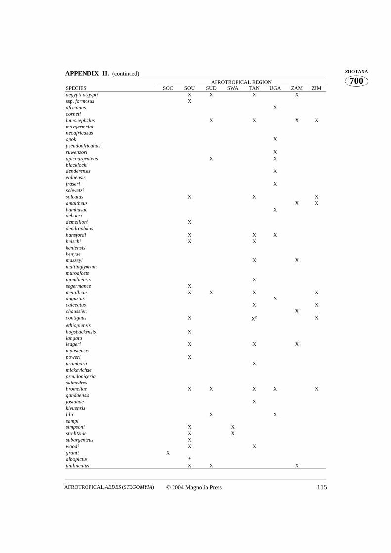
© 2004 Magnolia Press 115AFROTROPICAL AEDES (STEGOMYIA)
700ZOOTAXAAPPENDIX II. (continued)
AFROTROPICAL REGIONSPECIES SOC SOU SUD SWA TAN UGA ZAM ZIMaegypti aegypti X X X Xssp. formosus Xafricanus Xcornetiluteocephalus X X X Xmaxgermainineoafricanusopok Xpseudoafricanusruwenzori Xapicoargenteus X Xblacklockidenderensis Xealaensisfraseri Xschwetzisoleatus X X Xamaltheus X Xbambusae Xdeboeridemeilloni Xdendrophilushansfordi X X Xheischi X Xkeniensiskenyaemasseyi X Xmattinglyorummuroafcetenjombiensis Xsegermanae Xmetallicus X X X Xangustus Xcalceatus X Xchaussieri Xcontiguus X Xn X
ethiopiensishogsbackensis Xlangataledgeri X X Xmpusiensispoweri Xusambara Xmickevichaepseudonigeriasaimedresbromeliae X X X X Xgandaensisjosiahae Xkivuensislilii X Xsampisimpsoni X Xstrelitziae X Xsubargenteus Xwoodi X Xgranti Xalbopictus *unilineatus X X X
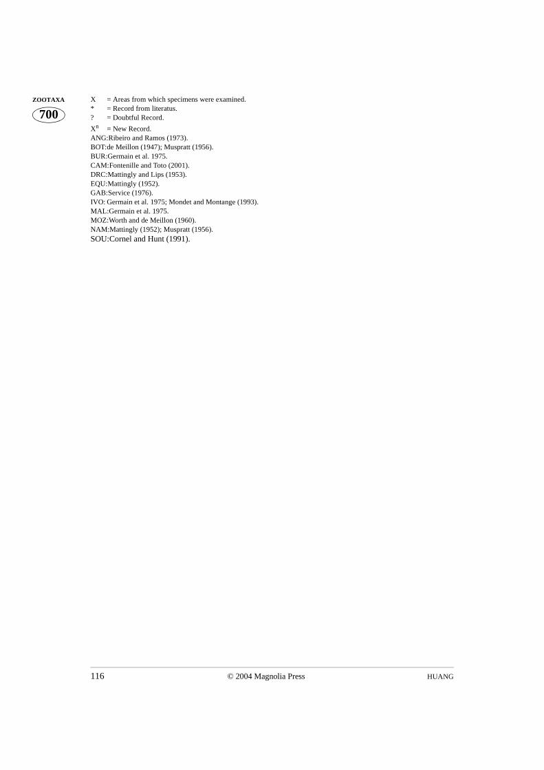
HUANG116 © 2004 Magnolia Press
700ZOOTAXA X = Areas from which specimens were examined.
* = Record from literatus.? = Doubtful Record.
Xn = New Record.ANG:Ribeiro and Ramos (1973).BOT:de Meillon (1947); Muspratt (1956). BUR:Germain et al. 1975.CAM:Fontenille and Toto (2001).DRC:Mattingly and Lips (1953). EQU:Mattingly (1952).GAB:Service (1976).IVO: Germain et al. 1975; Mondet and Montange (1993).MAL:Germain et al. 1975.MOZ:Worth and de Meillon (1960).NAM:Mattingly (1952); Muspratt (1956).SOU:Cornel and Hunt (1991).
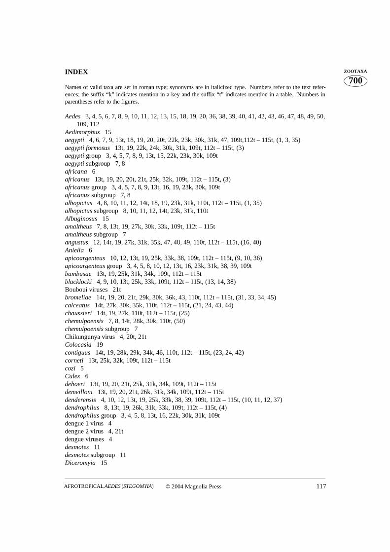
© 2004 Magnolia Press 117AFROTROPICAL AEDES (STEGOMYIA)
700ZOOTAXAINDEX
Names of valid taxa are set in roman type; synonyms are in italicized type. Numbers refer to the text refer-ences; the suffix “k” indicates mention in a key and the suffix “t” indicates mention in a table. Numbers inparentheses refer to the figures.
Aedes 3, 4, 5, 6, 7, 8, 9, 10, 11, 12, 13, 15, 18, 19, 20, 36, 38, 39, 40, 41, 42, 43, 46, 47, 48, 49, 50,109, 112
Aedimorphus 15aegypti 4, 6, 7, 9, 13t, 18, 19, 20, 20t, 22k, 23k, 30k, 31k, 47, 109t,112t – 115t, (1, 3, 35) aegypti formosus 13t, 19, 22k, 24k, 30k, 31k, 109t, 112t – 115t, (3)aegypti group 3, 4, 5, 7, 8, 9, 13t, 15, 22k, 23k, 30k, 109t aegypti subgroup 7, 8africana 6africanus 13t, 19, 20, 20t, 21t, 25k, 32k, 109t, 112t – 115t, (3)africanus group 3, 4, 5, 7, 8, 9, 13t, 16, 19, 23k, 30k, 109tafricanus subgroup 7, 8albopictus 4, 8, 10, 11, 12, 14t, 18, 19, 23k, 31k, 110t, 112t – 115t, (1, 35) albopictus subgroup 8, 10, 11, 12, 14t, 23k, 31k, 110tAlbuginosus 15amaltheus 7, 8, 13t, 19, 27k, 30k, 33k, 109t, 112t – 115tamaltheus subgroup 7angustus 12, 14t, 19, 27k, 31k, 35k, 47, 48, 49, 110t, 112t – 115t, (16, 40)Aniella 6 apicoargenteus 10, 12, 13t, 19, 25k, 33k, 38, 109t, 112t – 115t, (9, 10, 36)apicoargenteus group 3, 4, 5, 8, 10, 12, 13t, 16, 23k, 31k, 38, 39, 109tbambusae 13t, 19, 25k, 31k, 34k, 109t, 112t – 115tblacklocki 4, 9, 10, 13t, 25k, 33k, 109t, 112t – 115t, (13, 14, 38)Bouboui viruses 21tbromeliae 14t, 19, 20, 21t, 29k, 30k, 36k, 43, 110t, 112t – 115t, (31, 33, 34, 45)calceatus 14t, 27k, 30k, 35k, 110t, 112t – 115t, (21, 24, 43, 44)chaussieri 14t, 19, 27k, 110t, 112t – 115t, (25)chemulpoensis 7, 8, 14t, 28k, 30k, 110t, (50)chemulpoensis subgroup 7Chikungunya virus 4, 20t, 21t Colocasia 19contiguus 14t, 19, 28k, 29k, 34k, 46, 110t, 112t – 115t, (23, 24, 42) corneti 13t, 25k, 32k, 109t, 112t – 115tcozi 5Culex 6deboeri 13t, 19, 20, 21t, 25k, 31k, 34k, 109t, 112t – 115tdemeilloni 13t, 19, 20, 21t, 26k, 31k, 34k, 109t, 112t – 115tdenderensis 4, 10, 12, 13t, 19, 25k, 33k, 38, 39, 109t, 112t – 115t, (10, 11, 12, 37)dendrophilus 8, 13t, 19, 26k, 31k, 33k, 109t, 112t – 115t, (4)dendrophilus group 3, 4, 5, 8, 13t, 16, 22k, 30k, 31k, 109tdengue 1 virus 4dengue 2 virus 4, 21tdengue viruses 4desmotes 11desmotes subgroup 11Diceromyia 15
HUANG118 © 2004 Magnolia Press
700ZOOTAXA Dracaena 19
Dugbe virus 4ealaensis 4, 12, 13t, 19, 25k, 33k, 36, 38, 39, 109t, 112t – 115t, (11, 12, 37) ethiopiensis 4, 12, 14t, 27k, 31k, 35k, 39, 40, 41, 42, 110t, 112t – 115t, (18, 41)fraseri 9, 10, 13t, 19, 25k, 33k, 109t, 112t – 115t, (13, 14, 38)galloisi 8, 11gandaensis 4, 13, 14t, 19, 28k, 41, 42, 43, 110t, 112t – 115t, (26)gardnerii 6, 11, 12 gardnerii imitator 11, 12gebeleinensis 11granti 8, 10, 11, 14t, 19, 23k, 31k, 110t, 112t – 115t, (5, 7, 49)granti group 3, 5, 8, 9, 10, 11, 14t, 17, 23k, 31k, 110thansfordi 13t, 19, 20, 21t, 26k, 31k, 34k, 109t, 112t – 115theischi 13t, 26k, 31k, 34k, 109t, 112t – 115thogsbackensis 4, 12, 13, 14t, 19, 28k, 30k, 35k, 43, 46, 47, 110t, 112t – 115t, (18, 19, 24, 39, 50)josiahae 14t, 29k, 30k, 36k, 110t, 112t – 115t, (27, 28, 46)keniensis 13t, 19, 26k, 31k, 34k, 109t, 112t – 115tkenyae 13t, 19, 26k, 31k, 33k, 109t, 112t – 115tKingia 6kivuensis 14t, 29k, 110t, 112t – 115t, (29)langata 14t, 19, 28k, 30k, 35k, 110t, 112t – 115t, (20, 24, 42)ledgeri 14t, 19, 27k, 30k, 35k, 45, 110t, 112t – 115t, (21, 22, 23, 24, 43, 44)lilii 14t, 29k, 30k, 36k, 110t, 112t – 115t, (31, 33, 34, 45)luteocephala 6luteocephalus 13t, 19, 20, 21t, 24k, 32k, 109t, 112t – 115tmascarensis 7, 8, 9, 13t, 23k, 30k, 31k, 109t, (2, 48)masseyi 13t, 19, 27k, 31k, 34k, 109t, 112t – 115tmattinglyorum 14t, 19, 26k, 31k, 34k, 109t, 112t – 115tmaxgermaini 13t, 19, 24k, 109t, 112t – 115tmediopunctatus subgroup 10metallicus 14t, 19, 20, 21t, 22k, 30k, 109t, 112t – 115t, (8, 44)metallicus group 3, 5, 8, 14t, 16, 22k, 30k, 109tmickevichae 14t, 19, 28k, 110t, 112t – 115tmpusiensis 4, 12, 14t, 27k, 31k, 35k, 47, 48, 49, 110t, 112t – 115t, (18, 40)muroafcete 14t, 25k, 109t, 112t – 115tneoafricanus 13t, 19, 20, 21t, 24k, 32k, 109t, 112t – 115tnjombiensis 14t, 26k, 109t, 112t – 115topok 13t, 19, 20, 21t, 24k, 32k, 109t, 112t – 115tpoweri 12, 13, 14t, 27k, 43, 46, 47, 110t, 112t – 115t, (19, 24)poweri group 3, 4, 5, 8, 12, 14t, 17, 23k, 29k, 30k, 31k, 40, 41, 46, 48, 49, 110tPseudarmigeres 15pseudoafricanus 13t, 19, 24k, 32k, 109t, 112t – 115tpseudonigeria 7, 14t, 19, 28k, 110t, 112t – 115t, (4)pseudonigeria group 3, 5, 7, 8, 14t, 17, 22k, 30k, 110tPseudostegomyia 6Quasistegomyia 6, 11 Rift Valley fever virus 4, 20t, 21truwenzori 13t, 19, 24k, 32k, 109t, 112t – 115tsaimedres 14t, 19, 28k, 110t, 112t – 115tsampi 4, 13, 14t, 29k, 49, 50, 110t, 112t – 115t, (30)
© 2004 Magnolia Press 119AFROTROPICAL AEDES (STEGOMYIA)
700ZOOTAXASansevieria 19
schwetzi 13t, 19, 25k, 33k, 109t, 112t – 115t, (9, 10, 36)scutellaris 10scutellaris group 3, 5, 8, 9, 10, 11, 12, 14t, 18, 23k, 31k, 110tscutellaris subgroup 8, 10, 11, 23kseatoi 11segermanae 14t, 19, 26k, 31k, 34k, 109t, 112t – 115tsimpsoni 14t, 19, 20, 21t, 29k, 30k, 36k, 42, 43, 110t, 112t – 115t, (31, 32, 33, 34, 46)simpsoni group 3, 4, 5, 8, 14t, 17, 23k, 29k, 30k, 42, 43, 50, 110tsoleatus 13t, 19, 25k, 32k, 109t, 112t – 115t, (15, 39)Stegomyia 3, 4, 5, 6, 7, 8, 9, 10, 11, 13, 15, 18, 19, 20, 36, 38, 39, 40, 41, 42, 43, 46, 47, 49, 109,
112Strelitzia 19strelitziae 14t, 19, 20, 29k, 30k, 36k, 110t, 112t – 115t, (28, 31, 48)subargenteus 14t, 19, 29k, 35k, 50, 110t, 112t – 115t, (30, 31, 47)unilineatus 6, 8, 11, 12, 14t, 18, 19, 23k, 31k, 110t, 112t – 115t, (5, 6, 49)unilineatus group 3, 5, 8, 9, 11, 12, 14t, 18, 23k, 31k, 110tusambara 12, 14t, 19, 27k, 31k, 35k, 41, 49, 110t, 112t – 115t, (17, 41)vinsoni 8w-albus group 7, 9, 11w-albus subgroup 11woodi 13, 14t, 19, 28k, 30k, 36k, 41, 42, 43, 110t, 112t – 115t, (26, 31, 47)Yellow fever virus 4, 19, 20, 20t, 21tZika virus 4, 20t, 21t
HUANG120 © 2004 Magnolia Press
700ZOOTAXA About the author
Yiau-Min Huang is a research associate/research entomologist at the Department of Ento-mology, Smithsonian Institution, Washington, D.C. She received her bachelor of sciencedegree in Entomo-phytopathology from Taiwan Provincial Chung-Hsing University, Tai-chung, Taiwan, Republic of China, and her M.Sc. and Ph.D. degrees in Entomology at theUniversity of Wisconsin, Madison, Wisconsin, U.S.A. She served as an entomologist atB.P. Bishop Museum, Honolulu, Hawaii, taxonomist and research entomologist in theSoutheast Asia Mosquito Project, Medical Entomology Project and Systematics of AedesMosquitoes Project at the Department of Entomology, Smithsonian Institution, andresearch associate/ research entomologist at the Department of Entomology and Depart-ment of Systematic Biology, Entomology Section, Smithsonian Institution. She alsoserved as a consultant of the World Health Organization on the filariasis vectors of theSouth Pacific and yellow fever vectors in Africa while working for the Southeast AsiaMosquito Project and Medical Entomology Project. She has published a number of scien-tific papers and monographs, primarily on mosquito biosystematics. She identified thespecimens of Aedes albopictus (Skuse), the Asian Tiger Mosquito, from Harris County,Texas which is the first record of breeding populations established in the continentalUnited States. She also has published diagnostic morphological characters to easily iden-tify this species. She received the John N. Belkin Memorial Award in 1999, given by theAmerican Mosquito Control Association (AMCA).