A lipoprotein lipase (LPL)–specific monoclonal antibody, 88B8, that abolishes the binding
of LPL to GPIHBP1
Christopher M. Allan1,*, Mikael Larsson1,*, Xuchen Hu1, Cuiwen He1, Rachel S. Jung1, Alaleh
Mapar1, Constance Voss1, Kazuya Miyashita3, Tetsuo Machida3, Masami Murakami3, Katsuyuki
Nakajima3, André Bensadoun4, Michael Ploug5,6, Loren G. Fong1,‡, Stephen G. Young1,2,‡, and
Anne P. Beigneux1,‡
Departments of 1Medicine and 2Human Genetics, David Geffen School of Medicine, University
of California Los Angeles, Los Angeles, CA 90095; 3Gunma University, Graduate School of
Medicine, Maebashi, Japan; 4Division of Nutritional Science, Cornell University, Ithaca, NY
14853; 5Finsen Laboratory, Rigshospitalet, DK–2200 Copenhagen N, Denmark; and 6Biotech
Research and Innovation Centre (BRIC), University of Copenhagen, DK-220 Copenhagen N,
Denmark
‡Address correspondence to Anne P. Beigneux, Loren G. Fong, or Stephen G. Young, University
of California, Los Angeles, 4506 Gonda Bldg., 695 Charles E. Young Dr. South, Los Angeles,
CA 90095. Tel: (310) 825-4422; Fax: (310) 206-0865; E-mail: [email protected],
[email protected], [email protected]
Running title: A monoclonal antibody that blocks LPL binding to GPIHBP1
*Drs. Larsson and Allan wish to be considered co-first authors.
Abbreviations: GPIHBP1, glycosylphosphatidylinositol-anchored high density lipoprotein binding protein 1; TRLs, triglyceride-rich lipoproteins; DAPI, 4’,6-diamidino-2-phenylindole; HL, hepatic lipase.
by guest, on Septem
ber 17, 2018w
ww
.jlr.orgD
ownloaded from
Christopher M. Allan, Mikael Larsson et al. Page 2 of 2
ABSTRACT
Lipoprotein lipase (LPL) contains two principal domains: an amino-terminal catalytic domain
(residues 1–297) and a carboxyl-terminal domain (residues 298–448) that is important for binding
lipids and binding GPIHBP1 (an endothelial cell protein that shuttles LPL to the capillary lumen).
The LPL sequences required for GPIHBP1 binding have not been examined in detail, but one
study suggested that sequences near LPL’s carboxyl terminus (residues ~403–438) were crucial.
Here, we tested the ability of LPL-specific monoclonal antibodies to block the binding of LPL to
GPIHBP1. One antibody, 88B8, abolished LPL binding to GPIHBP1. Consistent with those
results, antibody 88B8 could not bind to GPIHBP1-bound LPL on cultured cells. Antibody 88B8
bound poorly to LPL proteins with amino acid substitutions that interfered with GPIHBP1
binding (e.g., C418Y, E421K). However, the sequences near LPL’s carboxyl terminus (residues
~403–438) were not sufficient for 88B8 binding; upstream sequences (residues 298–400) were
also required. Additional studies showed that these same sequences are required for LPL binding
to GPIHBP1. In conclusion, we identified an LPL monoclonal antibody that binds to LPL’s
GPIHBP1-binding domain. The binding of both antibody 88B8 and GPIHBP1 to LPL depends on
large segments of LPL’s carboxyl-terminal domain.
Keywords: Chylomicrons, Endothelial cells, Lipids/Chemistry, Lipolysis and fatty acid
metabolism, Triglycerides
by guest, on Septem
ber 17, 2018w
ww
.jlr.orgD
ownloaded from
Christopher M. Allan, Mikael Larsson et al. Page 3 of 3
INTRODUCTION
For more than 50 years, it has been known that lipoprotein lipase (LPL), a triglyceride hydrolase
secreted by myocytes and adipocytes, is crucial for the intravascular processing of triglyceride-
rich lipoproteins (TRLs) (1-3). For most of that time, it was assumed that LPL was attached to the
heparan-sulfate proteoglycans along the lumen of blood vessels (4), but how LPL reached the
lumen of blood vessels was a stubborn mystery. Within the past few years, that mystery has been
solved (5, 6). GPIHBP1, a GPI-anchored protein of capillary endothelial cells, picks up freshly
secreted LPL within the interstitial spaces and shuttles it across endothelial cells to the capillary
lumen (7, 8). In the absence of GPIHBP1, LPL remains in the interstitial spaces and never
reaches the capillary lumen, resulting in an accumulation of plasma TRLs and extremely high
plasma triglyceride levels (“chylomicronemia”) (8). Recent studies showed that GPIHBP1 (and
GPIHBP1-bound LPL) are also crucial for the margination of TRLs along the capillary lumen,
allowing triglyceride hydrolysis to proceed (9).
GPIHBP1 has two main structural features—an amino-terminal acidic domain and a cysteine-
rich three-fingered “LU domain” (7, 10). Recent studies have shown that the LU domain is
primarily responsible for high-affinity binding of LPL, while the acidic domain augments the
interaction and promotes an initial interaction complex between LPL and GPIHBP1 (6, 11). A
variety of missense mutations in GPIHBP1’s LU domain have been identified in patients with
chylomicronemia (12-22), and all of those abolish the ability of GPIHBP1 to bind LPL (6). Most
of these mutations interfere with the formation of disulfide bonds in the LU domain, leading to
disulfide-linked dimers and multimers (23). Alanine-scanning mutagenesis studies showed that
the highly conserved second finger of the three-fingered LU domain is particularly important for
binding LPL (24). Mutagenizing W109 in finger 2 abolishes LPL binding without promoting the
formation of GPIHBP1 dimers/multimers, suggesting that W109 participates directly in binding
LPL (23).
by guest, on Septem
ber 17, 2018w
ww
.jlr.orgD
ownloaded from
Christopher M. Allan, Mikael Larsson et al. Page 4 of 4
In contrast to the situation with GPIHBP1, our understanding of the LPL sequences required
for binding to GPIHBP1 is meager, but the relevant sequences appear to be located in LPL’s
carboxyl-terminal “lipid-binding” domain (residues 298–448) rather than in LPL’s catalytic
domain (residues 1–297). A pair of LPL mutations (C418Y, E421K), first identified in patients
with hypertriglyceridemia (25, 26), interfere with the binding of LPL to GPIHBP1. Mutation of
nearby LPL sequences (residues 403–438) also impaired LPL binding to GPIHBP1 (27). Those
studies were interpreted as showing that sequences near the carboxyl terminus of LPL are
singularly important for mediating LPL binding to GPIHBP1.
Here, we sought to better define LPL sequences that are important for GPIHBP1 binding. As
part of these efforts, we tested the capacity of three LPL-specific monoclonal antibodies (5D2,
88B8, 57A5) (28-30) to block the binding of LPL to GPIHBP1. We reasoned that if we were to
identify a “blocking antibody,” then efforts to define the epitope would lead to new insights into
LPL sequences that are important for LPL binding to GPIHBP1.
by guest, on Septem
ber 17, 2018w
ww
.jlr.orgD
ownloaded from
Christopher M. Allan, Mikael Larsson et al. Page 5 of 5
MATERIAL AND METHODS
Monoclonal antibodies
We examined three LPL-specific mouse monoclonal antibodies (5D2, 57A5, 88B8) (28-30). The
epitope for 5D2 has been studied in detail and is located between residues 380 and 410 in LPL’s
carboxyl-terminal domain (29, 30). mAbs 57A5 and 88B8 were generated against human LPL
and have been used previously in LPL immunoassays (28), but data on the epitopes for these
antibodies have never been reported. Fabʹ′ fragments were prepared with immobilized papain and
Fc fragments removed with Protein A–Sepharose. Monoclonal antibody 5D2 was a gift from Dr.
John Brunzell, and mAbs 57A5 and 88B8 were acquired from Immuno-Biological Laboratories
(Gunma, Japan).
LPL–GPIHBP1 binding assays
Cell-free assay—Secreted versions of wild-type (wt) human GPIHBP1 and GPIHBP1-W109S
were stably expressed in Drosophila S2 cells. Both GPIHBP1 proteins contain a uPAR epitope
tag (23) as well as the epitope for monoclonal antibody 11A12 (31). For the assay, the
conditioned media from Drosophila S2 cells expressing soluble human GPIHBP1 was incubated
for 1 h at 4°C with V5-tagged human LPL (32), with or without monoclonal antibodies (20 µg/ml
final), and agarose beads coated with monoclonal antibody 11A12 (33). After washing the beads,
GPIHBP1 and any GPIHBP1-bound LPL were eluted from the antibody-coated beads by heating
the beads in SDS sample buffer for 5 min at 90°C. The amounts of GPIHBP1 and LPL in the
starting material, unbound fractions, wash fractions, and elution fractions were assessed by
western blotting. Proteins were separated on a 12% NuPAGE SDS-PAGE gel with MES buffer,
followed by transfer to a sheet of nitrocellulose membrane. The membrane was then incubated
with IRdye680-conjugated antibody 11A12 and an IRdye800-conjugated V5-antibody.
Cell-based assay—CHO pgsA-745 cells (2 × 10⁶) were electroporated with 2 µg plasmids
encoding either an S-protein–tagged wild-type human GPIHBP1 or an S-protein-tagged mutant
by guest, on Septem
ber 17, 2018w
ww
.jlr.orgD
ownloaded from
Christopher M. Allan, Mikael Larsson et al. Page 6 of 6
human GPIHBP1 (W109S) and then plated on coverslips in 24-well plates. After 24 h, the cells
were incubated with either V5-tagged human LPL alone, or V5-tagged human LPL with one of
the three mAbs (5D2, 57A5, 88B8) (20 µg/ml) for 1 h at 4°C. The cells were then washed and
processed for western blots or immunofluorescence microscopy. For the western blots, cell
lysates were collected by incubating cells with M-PER Mammalian Protein Extraction Reagent
(ThermoFisher Scientific) with EDTA-free complete protease inhibitor cocktail (Roche) for 5-
min at 4°C, followed by centrifugation at 14,000 × g for 10-min to remove insoluble material.
The nitrocellulose membrane was then blocked for 1 h at room temperature with Odyssey
Blocking Buffer (Li-Cor), and then incubated with an IRdye800–conjugated mouse monoclonal
antibody against the V5 tag (ThermoFisher Scientific; 2.32 µg/mL) and a goat polyclonal
antibody against the S-protein tag (Abcam; 1:1000) followed by an IRdye680–conjugated donkey
anti-goat IgG (Li-Cor). Signals were visualized and quantified with a Li-Cor Odyssey scanner.
For the immunofluorescence microscopy assay, cells were fixed in 3% paraformaldehyde for 15-
min and blocked with 10% donkey serum in PBS/Mg/Ca. Cells were then incubated overnight at
4°C with an Alexa 647–conjugated mouse monoclonal antibody against the V5 tag
(ThermoFisher Scientific; 11.6 µg/mL) and a goat polyclonal antibody against the S-protein tag
(Abcam; 1:800), followed by a 30-min incubation with an Alexa 568–conjugated donkey anti-
goat IgG (ThermoFisher Scientific; 1:800). After washing, the cells were fixed with 3%
paraformaldehyde for 15-min and stained with 4’,6-diamidino-2-phenylindole (DAPI) to
visualize DNA. Images were recorded with an Axiovert 200M microscope and processed with
Zen 2010 software software (all from Zeiss). Within each experiment, the exposure conditions for
each construct were identical.
Testing the binding of hepatic lipase–lipoprotein lipase (HL–LPL) chimeras to GPIHBP1 on
the surface of cultured cells
by guest, on Septem
ber 17, 2018w
ww
.jlr.orgD
ownloaded from
Christopher M. Allan, Mikael Larsson et al. Page 7 of 7
Earlier studies by Wong and coworkers (34) showed that it was possible to express catalytically
active, dimeric hepatic lipase–lipoprotein lipase (HL–LPL) chimeras by exchanging sequences in
HL with the corresponding sequences in LPL. Here, we used that approach to create HL–LPL
chimeras containing human LPL residues 313–448, 330–448, 335–448, 340–448, or 345–448
(Fig. 1). To test the ability of HL, LPL, and the HL–LPL chimeras to bind to GPIHBP1, we used
a “co-plating assay” described previously by Beigneux et al. (24). CHO-K1 cells (1 × 10⁶) were
electroporated with 1.0 mg of either an S-protein–tagged human GPIHBP1 construct or an
expression vector for one of the V5-tagged lipases. The independently transfected cells were then
mixed together and plated on coverslips in 24-well plates. 24 h later, the cells were fixed in 3%
paraformaldehyde. When indicated, the cells were permeabilized with 0.2% Triton X-100. Cells
were blocked with 10% donkey serum in PBS/Mg/Ca and then incubated for 1 h in the blocking
buffer containing a goat antibody against the S-protein tag (Abcam; 1:500) and a mouse
monoclonal antibody against the V5 tag (Invitrogen; 1:100), followed by a 30-min incubation
with an Alexa 568–conjugated donkey anti-goat IgG (Invitrogen; 1:800) and an Alexa 488–
conjugated donkey anti-mouse IgG (Invitrogen; 1:800). After washing, the cells were stained with
DAPI to visualize DNA. Images were recorded with the Axiovert 200M microscope. The
exposure conditions for each construct were identical. In this system, cells that expressed wild-
type GPIHBP1 captured LPL that was secreted by the LPL-transfected cells, thus, GPIHBP1 and
LPL signals colocalized on the merged image (24). HL does not bind to GPIHBP1 (31);
consequently, there was no colocalization of HL and GPIHBP1 signals.
Lipase expression vectors
Human LPL–mouse LPL chimeras, and HL–LPL chimeras (Fig. 1) were created with a PCR-
based method (In-Fusion HD cloning kit, Clontech). Point mutations were introduced with the
QuikChange Lightning kit (Agilent Technologies).
Testing the ability of the monoclonal antibodies to bind to GPIHBP1-bound LPL
by guest, on Septem
ber 17, 2018w
ww
.jlr.orgD
ownloaded from
Christopher M. Allan, Mikael Larsson et al. Page 8 of 8
CHO pgsA-745 cells (2 × 10⁶) were electroporated with 2 µg of a plasmid encoding an S-protein–
tagged wild-type human GPIHBP1 and plated on coverslips in 24-well plates. 24 h later, cells
were incubated with a V5-tagged human LPL or buffer for 1 h at 4ºC. Cells were then washed
and incubated with either buffer or Alexa 568–conjugated mAbs (5D2, 57A5, or 88B8) (20
µg/ml) for 1 h at 4ºC. The cells were then washed and processed for immunocytochemistry. Cells
on coverslips were fixed in 3% paraformaldehyde for 15 min, blocked with 10% donkey serum in
PBS/Mg/Ca, and then incubated overnight at 4°C with an Alexa 647–conjugated mouse
monoclonal antibody against the V5 tag (ThermoFisher Scientific; 11.6 µg/mL) and a goat
polyclonal antibody against the S-protein tag (Abcam; 1:800), followed by a 30-min incubation
with an Alexa 488–conjugated donkey anti-goat IgG (ThermoFisher Scientific; 1:800). After
washing, the cells were fixed with 3% paraformaldehyde for 15 min and stained with DAPI to
visualize DNA. Microscopy was performed as described earlier.
Testing the ability of the monoclonal antibodies to bind to LPL in capillaries of tissue
sections
Wild-type and Lpl–/–MCK-hLPL mice (35) were perfused with PBS followed by 3%
paraformaldehyde; quadriceps muscle was harvested and embedded in OCT on dry ice. Tissue
sections (7 µm) were fixed in methanol at –20ºC for 10 min, permeabilized with 0.2% Triton X-
100 for 5 min, and blocked with 5% donkey serum, 10% FBS, and 0.2% BSA in PBS/Mg/Ca.
Tissues were incubated overnight at 4°C with Alexa 568–conjugated mouse mAbs (5D2, 57A5,
or 88B8) (8 µg/ml) and a rabbit polyclonal antibody against mouse CD31 (Abcam; 1:50),
followed by a 45-min incubation with Alexa 647–conjugated 11A12 antibody (3 µg/mL) and
Alexa 488–conjugated donkey anti-rabbit IgG (ThermoFisher Scientific; 1:200). After washing,
the cells were fixed with 3% paraformaldehyde for 5 min and stained with DAPI to visualize
DNA. Microscopy was performed as described earlier. Mice were fed a chow diet and housed in a
by guest, on Septem
ber 17, 2018w
ww
.jlr.orgD
ownloaded from
Christopher M. Allan, Mikael Larsson et al. Page 9 of 9
barrier facility with a 12 h light-dark cycle. All studies were approved by UCLA’s Animal
Research Committee.
Measurements of LPL activity
Human LPL was prepared from a CHO cell line expressing V5-tagged human LPL (from Dr.
Mark Doolittle, University of California, Los Angeles). LPL was purified by heparin–Sepharose
chromatography on an ÄKTA pure HPLC (GE Healthcare) and eluted with a 0.1–2 M NaCl
gradient in 20 mM NaPO4, pH 7.4. Taurodeoxycholate (final concentration, 5 mM) was added to
the LPL (i.e., fractions eluting at ≥1 M NaCl) before storing at –80°C. The activity of purified
human LPL was compared to that of a known quantity of bovine LPL. Lipase activity was
measured with [3H]triolein that had been incorporated into Intralipid (0.5 µCi [3H]triolein/mg
triglyceride). Human LPL (6 µl, corresponding to the enzymatic activity of 5 ng of bovine LPL)
was added to 200-µl incubation mixtures containing 5 mg of triglyceride/ml in 0.15 M Tris (pH
8.5) containing 0.1 M NaCl, 6% BSA (w/v), 16.7 units of heparin/ml, and 5% (v/v) heat-
inactivated rat serum (as a source of apo-CII). Monoclonal antibodies (20 µg/ml, final
concentration) were added to incubation mixtures 5 min before adding the LPL. Esterase
activities were analyzed by adding 30 µl of the human LPL to 100-µl incubation mixtures of 50
mM Tris, 50 µM 1,2-di-O-lauryl-rac-glycero-3-glutaric acid 6ʹ′-methylresorufin ester (DGGR),
120 mM NaCl, 10 mg/ml BSA, and 0.5% Triton X-100 (pH 7.4). Monoclonal antibodies (200
µg/ml final concentration) were added to incubations 5 min before adding the LPL. Ester
hydrolysis was determined by measuring the increase of resorufin fluorescence at λex 530 nm and
λem 590 nm during the first 15 min of the incubation.
by guest, on Septem
ber 17, 2018w
ww
.jlr.orgD
ownloaded from
Christopher M. Allan, Mikael Larsson et al. Page 10 of 10
RESULTS
Testing the ability of LPL-specific mAbs to block the binding of LPL to GPIHBP1
We used cell-free and cell-based LPL–GPIHBP1 binding assays (18, 23, 31, 33) to test the ability
of three LPL-specific mAbs (5D2, 88B8, 57A5) (29, 30) to block the binding of human LPL to
GPIHBP1. In the cell-free assay, we incubated GPIHBP1, LPL, and an LPL-specific mAb with
agarose beads that had been coated with a GPIHBP1-specific monoclonal antibody (11A12).
After a 1-h incubation, the beads were washed, and the amounts of GPIHBP1 and GPIHBP1-
bound LPL bound to the beads were assessed by western blotting. Monoclonal antibody 88B8
abolished LPL binding to GPIHBP1; thus, no LPL could be eluted from the beads (Fig. 2). The
ability of 88B8 (and 88B8 Fabʹ′ fragments) to block the binding of LPL to GPIHBP1 was
confirmed in additional independent experiments (Figs. S1, S2). mAbs 5D2 and 57A5 did not
abolish the binding of LPL to GPIHBP1, but they did reduce LPL binding (Fig. 2).
Monoclonal antibody 88B8 completely blocked binding of LPL to GPIHBP1 on the surface
of GPIHBP1-transfected cells, as judged by immunofluorescence microscopy and western blots
of cell extracts (Fig. 3, Fig. S3). mAbs 57A5 and 5D2 caused partial inhibition of LPL binding to
GPIHBP1 (Fig. 3, Fig. S3).
Defining the epitopes for LPL-specific mAbs
The 5D2 epitope is located within LPL residues 380–410 (29, 30), but data on the epitopes for
mAbs 57A5 and 88B8 have not been reported. Preliminary western blot studies indicated that
both mAbs 57A5 and 88B8 bind to the carboxyl-terminal region of LPL (residues 298–448) (Fig.
S4). Identical conclusions were reached by performing ELISAs on the medium of cells that had
been transfected with Flag-tagged constructs encoding LPL’s amino- and carboxyl-terminal
domains (Kazuya Miyashita, unpublished data). Because 88B8 and 88B8 Fabʹ′ fragments block
LPL binding to GPIHBP1 and also bound to LPL’s carboxyl-terminal domain, we suspected that
the binding sites for mAb 88B8 and GPIHBP1 on LPL were similar and that LPL mutations
by guest, on Septem
ber 17, 2018w
ww
.jlr.orgD
ownloaded from
Christopher M. Allan, Mikael Larsson et al. Page 11 of 11
known to interfere with GPIHBP1 binding (e.g., C418Y, E421K, C438Y) (27) would also
interfere with 88B8 binding. Indeed, 88B8 bound more avidly to wild-type LPL than to mutant
LPLs harboring C418Y, E421K, or C438Y mutations (Fig. 4A, B). The effect of those mutations
on 5D2 binding was minimal (Fig. 4C, D). As expected, 88B8 bound avidly to human but not
mouse LPL; it bound avidly to a mouse LPL–human LPL chimera containing the entire carboxyl-
terminal domain of human LPL (residues 298–448).
Earlier studies implied LPL’s GPIHBP1-binding domain involved amino acids 403–438 (27),
and the reduced binding of 88B8 to the LPL mutants suggested that the 88B8 epitope might be
located in the same stretch of amino acids. To our surprise, the binding of 88B8 to LPL depended
on upstream sequences within the primary sequence. Monoclonal antibody 88B8 bound weakly to
mLPL–hLPL chimeras containing human LPL residues 330–448 or 370–448, and it failed to bind
to chimeras containing human LPL residues 400–448 or 420–448 (Fig. 5A). The same pattern
was observed for 88B8 Fabʹ′ fragments (Fig. S5). These results suggested that LPL residues 298–
330 are quite relevant to 88B8 binding. Within that region, the only amino acids that differ
between the human and mouse LPL sequences are residues 327–331. Those residues were
important for the 88B8 epitope; when residues 327–331 in human LPL were replaced with the
mouse LPL sequences, the binding of 88B8 was significantly reduced (Fig. 5A).
The finding that 88B8 bound (albeit weakly) to mLPL–hLPL chimeras containing human
LPL residues 330–448 and 370–448 but failed to bind to a chimera with human residues 400–448
indicates that human LPL residues 330–400 are important for 88B8 binding. However, these
results do not mean that residues 400–448 have no role in 88B8 binding, but rather that residues
400–448 are insufficient. First, the C418Y, E421K, and C438Y mutations clearly interfere with
88B8 binding (Fig. 4). Second, introducing human residues 327–403 into the mouse LPL
expression vector resulted in only minimal restoration of 88B8 binding (Fig. 5B), implying that
by guest, on Septem
ber 17, 2018w
ww
.jlr.orgD
ownloaded from
Christopher M. Allan, Mikael Larsson et al. Page 12 of 12
additional sequences at the carboxyl terminus of LPL (i.e., residues 403–448) were important for
the 88B8 epitope.
Monoclonal antibody 57A5 bound avidly to human LPL and to a mLPL–hLPL chimera
containing human residues 298–448 but not to chimeras containing human residues 330–448,
370–448, or 400–448 (Fig. 5C). In contrast to 88B8, 57A5 bound avidly to a mLPL–hLPL
chimera containing human residues 327–403 (Fig. 5D).
Testing the impact of the LPL-specific mAbs on LPL activity
The tryptophan-rich motif within the 5D2 epitope (residues 380–410) is important for the ability
of LPL to hydrolyze long-chain triacylglycerols but not short-chain water-soluble triacylglycerols
(36). Thus, in our studies, antibody 5D2, which binds to the tryptophan-rich motif, inhibited LPL
activity against triolein but not a soluble substrate (Fig. 6). mAbs 88B8 and 57A5 also reduced
LPL activity against triolein but to a lesser degree (Fig. 6). None of the antibodies inhibited LPL
catalytic activity with a water-soluble substrate (Fig. 6).
Assessing LPL sequences relevant to binding GPIHBP1
Earlier studies suggested that the binding of LPL to GPIHBP1 depended on LPL residues 403–
438 (27). However, given that sequences throughout LPL’s carboxyl-terminal domain (residues
298–448) were important for 88B8 binding, we suspected that the same sequences might also
play a role in GPIHBP1 binding. To explore this idea, we tested the ability of hepatic lipase–
lipoprotein lipase (HL–LPL) chimeras to bind to GPIHBP1 on the surface of CHO cells. Wong
and coworkers (34) showed that it was possible to produce HL–LPL chimeras that are secreted
and are catalytically active. We created HL–LPL chimeras containing human LPL residues 313–
448, 330–448, 335–448, 340–448, and 345–448 (Fig. 1, Fig. S6). We then tested the ability of
LPL, HL, and the HL–LPL chimeras to bind to GPIHBP1. We mixed CHO cells that had been
transfected with a lipase construct with CHO cells that had been transfected with GPIHBP1. We
then used immunofluorescence microscopy to assess the binding of the freshly secreted lipases to
by guest, on Septem
ber 17, 2018w
ww
.jlr.orgD
ownloaded from
Christopher M. Allan, Mikael Larsson et al. Page 13 of 13
GPIHBP1 on GPIHBP1-transfected cells. As expected, full-length LPL bound avidly to
GPIHBP1, but HL did not. HL–LPL chimeras containing LPL residues 313–448 and 330–448
bound to GPIHBP1, but chimeras containing LPL residues 335–448, 340–448, and 345–448 did
not (Fig. 7). A western blot experiment showed that cells expressing GPIHBP1 were capable of
binding LPL as well as chimeras containing LPL residues 313–448 and 330–448, but not HL or a
chimera containing LPL residues 335–448 (Fig. S7). Thus, a large segment of LPL’s carboxyl-
terminal domain is required for GPIHBP1 binding—as was the case for 88B8 binding.
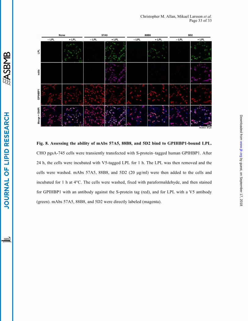
mAb 88B8 cannot bind to GPIHBP1-bound LPL but is still useful for
immunohistochemistry studies
Because 88B8 and GPIHBP1 bind to similar sequences, we suspected that 88B8 would not bind
to GPIHBP1-bound LPL on the surface of CHO cells. Indeed, this was the case (Fig. 8). In
contrast, 5D2 and 57A5 did bind to GPIHBP1-bound LPL. Interestingly, the inability of 88B8 to
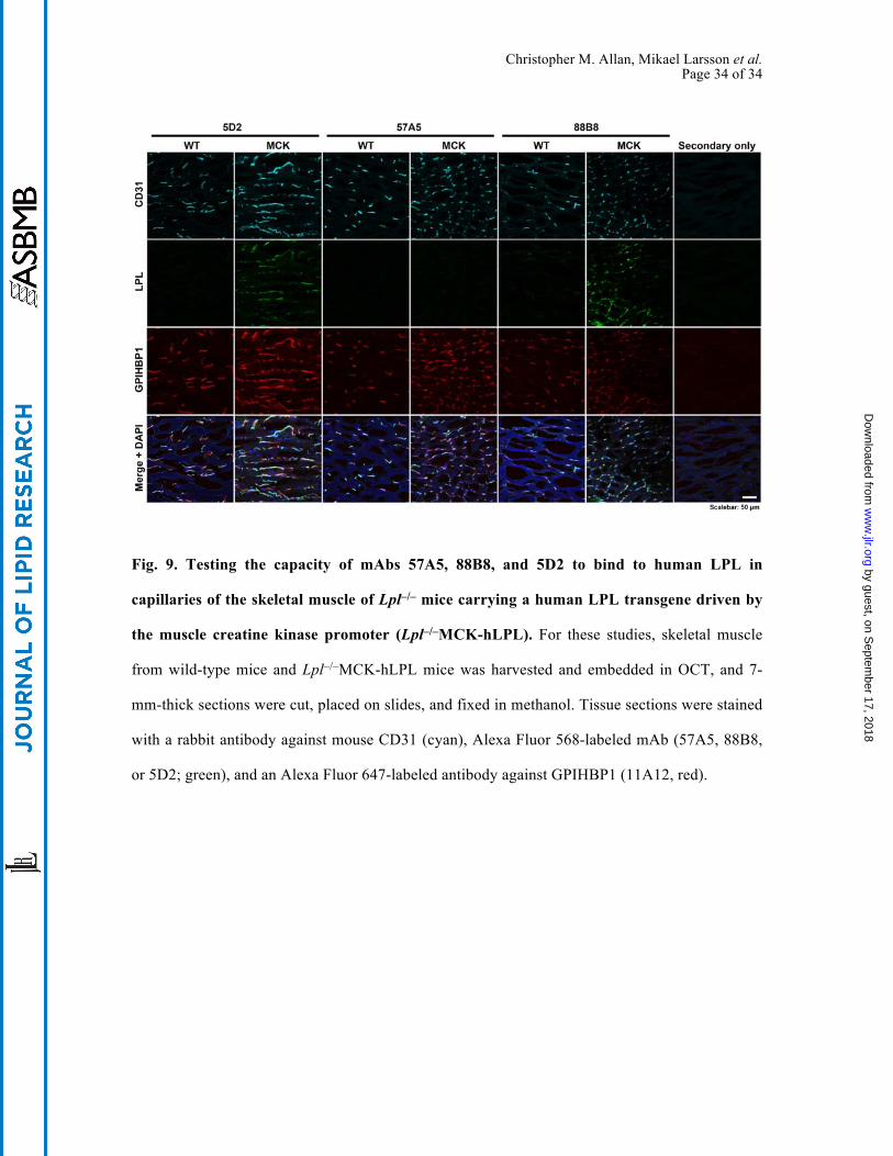
bind to GPIHBP1-bound LPL did not interfere with its utility for immunohistochemistry studies.
Monoclonal antibody 88B8 readily detected human LPL on capillaries after the LPL–GPIHBP1
complex was disrupted by methanol fixation (Fig. 9). Monoclonal antibody 5D2 also was useful
for immunohistochemistry, but 57A5 was not (Fig. 9).
by guest, on Septem
ber 17, 2018w
ww
.jlr.orgD
ownloaded from
Christopher M. Allan, Mikael Larsson et al. Page 14 of 14
DISCUSSION
In the current studies, we identified a human LPL–specific monoclonal antibody, 88B8, that
abolishes the binding of human LPL to GPIHBP1 in both cell-free and cell-based LPL–GPIHBP1
binding assays. Monoclonal antibody 88B8 binding to LPL was impaired by the very same LPL
missense mutations (C418Y, E421K, C438Y) that are known to interfere with the binding of LPL
to GPIHBP1 (27), suggesting that 88B8 and GPIHBP1 binding sites are very similar. We
suspected initially that the LPL sequences required for 88B8 binding would be confined to
residues ~403–438, but this was not the case. Additional upstream sequences (residues 298–400)
proved to be important for 88B8 binding. Monoclonal antibody 88B8 bound avidly to a mLPL–
hLPL chimera containing human LPL residues 298–448 and weakly to chimeras containing
residues 330–448 and 370–448. Monoclonal antibody 88B8 did not bind to a chimera containing
human LPL residues 400–448. The fact that extensive sequences within LPL’s carboxyl-terminal
domain are required for 88B8 binding led us to suspect that the same sequences would be
required for GPIHBP1 binding. Indeed, studies with HL–LPL chimeras showed that a large
portion of LPL’s carboxyl-terminal domain was required for GPIHBP1 binding. HL–LPL
chimeras containing human LPL residues 313–448 and 330–448 bound to GPIHBP1, but
chimeras containing LPL residues 335–448 or 340–448 did not. Thus, the ability of GPIHBP1 to
bind to LPL depends on residues 330–448 and not simply residues 403–438 (as we had originally
suspected). These results add considerably to our understanding of LPL sequences required for
LPL–GPIHBP1 interactions.
To fully understand 88B8–LPL interactions or LPL–GPIHBP1 interactions, co-crystal
structures are required. However, with the mutagenesis-based binding data that are in hand, we
believe that the simplest interpretation is that both 88B8 and GPIHBP1 interact with a complex
epitope that depends on the proper folding of a large portion of LPL’s carboxyl-terminal domain
(residues 298–448). It is equally possible that LPL residues 298–400 are simply required for the
by guest, on Septem
ber 17, 2018w
ww
.jlr.orgD
ownloaded from
Christopher M. Allan, Mikael Larsson et al. Page 15 of 15
proper conformation of a more compact binding site (residues ~400–448). We do not believe that
the absence of GPIHBP1 binding to the HL–LPL chimera containing residues 389–448 means
that residues 389–448 are unimportant for GPIHBP1 binding. First, the C418Y and E421K
mutations abolished GPIHBP1 binding and do so without affecting LPL catalytic activity—
implying that those mutations did not cause global changes in LPL structure. Second, and perhaps
more importantly, Mysling et al. (11) recently reported, using hydrogen–deuterium
exchange/mass spectrometry studies, that the amide hydrogens in LPL residues 419–425 were
protected from deuterium exchange by GPIHBP1 binding (i.e., that the binding of GPIHBP1 to
those LPL sequences limited their accessibility to solvent). In a similar fashion, we believe that
residues 400–448 are relevant to 88B8 binding, despite the fact that 88B8 did not bind to the
mLPL–hLPL chimera containing hLPL residues 400–448. First, 88B8 binding was disrupted by
C418Y, E421K, and C438Y mutations. Second, 88B8 could not bind to the mLPL–hLPL chimera
containing human LPL residues 298–403.
The epitope for 57A5 was simpler: it bound to human LPL residues 298–448 but not to
residues 330–448, implying that residues 298 to 330 were crucial for the epitope. Unlike 88B8,
57A5 and 5D2 did not abolish LPL binding to GPIHBP1, but partial inhibition was clearly
evident. We suspect that the binding of 5D2 and 57A5 locks LPL into a conformation with
reduced affinity for GPIHBP1—or alternatively that these antibodies create a steric hindrance to
GPIHBP1 binding. In earlier studies, 5D2 inhibited LPL activity against triolein but not a soluble
short-chain triacylglycerol (37). We confirmed those findings and found that the same property
applies, at least to some degree, to 88B8 and 57A5. We suspect that the binding of all three
antibodies create a steric hindrance or lock the carboxyl-terminal domain into a suboptimal
conformation for triolein hydrolysis.
Monoclonal antibodies 5D2 and 57A5 had no difficulty binding to GPIHBP1-bound LPL on
the surface of cultured cells, whereas 88B8 did not, reflecting the fact that GPIHBP1 and 88B8
by guest, on Septem
ber 17, 2018w
ww
.jlr.orgD
ownloaded from
Christopher M. Allan, Mikael Larsson et al. Page 16 of 16
have overlapping binding sites. Remarkably, 88B8 was still useful for immunohistochemistry.
Once the LPL–GPIHBP1 complex had been disrupted by methanol fixation, mAb 88B8 readily
bound to the LPL on capillaries, colocalizing with GPIHBP1 and the endothelial cell marker
CD31. Monoclonal antibody 5D2, but not 57A5, also detected LPL in capillaries.
by guest, on Septem
ber 17, 2018w
ww
.jlr.orgD
ownloaded from
Christopher M. Allan, Mikael Larsson et al. Page 17 of 17
ACKNOWLEDGMENTS
This work was supported by grants from the NHLBI (HL090553, HL087228, and HL125335)
and a Transatlantic Network Grant from the Leducq Foundation (12CVD04). Christopher M.
Allan was supported by a Ruth L. Kirschstein National Research Service Award (T32HL69766).
DISCLOSURES
The authors have no financial interests to declare.
by guest, on Septem
ber 17, 2018w
ww
.jlr.orgD
ownloaded from
Christopher M. Allan, Mikael Larsson et al. Page 18 of 18
REFERENCES
1. Korn, E. D. 1955. Clearing factor, a heparin-activated lipoprotein lipase. II. Substrate
specificity and activation of coconut oil. J. Biol. Chem. 215: 15–26.
2. Korn, E. D. 1955. Clearing factor, a heparin-activated lipoprotein lipase. I. Isolation and
characterization of the enzyme from normal rat heart. J. Biol. Chem. 215: 1–14.
3. Havel, R. J., and R. S. Gordon, Jr. 1960. Idiopathic hyperlipemia: metabolic studies in an
affected family. J. Clin. Invest. 39: 1777–1790.
4. Merkel, M., R. H. Eckel, and I. J. Goldberg. 2002. Lipoprotein lipase: genetics, lipid uptake,
and regulation. J. Lipid Res. 43: 1997–2006.
5. Young, S. G., and R. Zechner. 2013. Biochemistry and pathophysiology of intravascular and
intracellular lipolysis. Genes Dev. 27: 459–484.
6. Fong, L. G., S. G. Young, A. P. Beigneux, A. Bensadoun, M. Oberer, H. Jiang, and M. Ploug.
2016. GPIHBP1 and Plasma Triglyceride Metabolism. Trends Endocrinol. Metab. 27: 455–
469.
7. Beigneux, A. P., B. Davies, P. Gin, M. M. Weinstein, E. Farber, X. Qiao, P. Peale, S.
Bunting, R. L. Walzem, J. S. Wong, W. S. Blaner, Z. M. Ding, K. Melford, N. Wongsiriroj,
X. Shu, F. de Sauvage, R. O. Ryan, L. G. Fong, A. Bensadoun, and S. G. Young. 2007.
Glycosylphosphatidylinositol-anchored high density lipoprotein–binding protein 1 plays a
critical role in the lipolytic processing of chylomicrons. Cell Metab. 5: 279–291.
8. Davies, B. S. J., A. P. Beigneux, R. H. Barnes II, Y. Tu, P. Gin, M. M. Weinstein, C.
Nobumori, R. Nyrén, I. J. Goldberg, G. Olivecrona, A. Bensadoun, S. G. Young, and L. G.
Fong. 2010. GPIHBP1 is responsible for the entry of lipoprotein lipase into capillaries. Cell
Metab. 12: 42–52.
by guest, on Septem
ber 17, 2018w
ww
.jlr.orgD
ownloaded from
Christopher M. Allan, Mikael Larsson et al. Page 19 of 19
9. Goulbourne, C. N., P. Gin, A. Tatar, C. Nobumori, A. Hoenger, H. Jiang, C. R. Grovenor, O.
Adeyo, J. D. Esko, I. J. Goldberg, K. Reue, P. Tontonoz, A. Bensadoun, A. P. Beigneux, S.
G. Young, and L. G. Fong. 2014. The GPIHBP1-LPL complex is responsible for the
margination of triglyceride-rich lipoproteins in capillaries. Cell Metab. 19: 849–860.
10. Ioka, R. X., M.-J. Kang, S. Kamiyama, D.-H. Kim, K. Magoori, A. Kamataki, Y. Ito, Y. A.
Takei, M. Sasaki, T. Suzuki, H. Sasano, S. Takahashi, J. Sakai, T. Fujino, and T. T.
Yamamoto. 2003. Expression Cloning and Characterization of a Novel
Glycosylphosphatidylinositol-anchored High Density Lipoprotein-binding Protein, GPI-
HBP1. J. Biol. Chem. 278: 7344–7349.
11. Mysling, S., K. K. Kristensen, M. Larsson, A. P. Beigneux, H. Gardsvoll, L. G. Fong, A.
Bensadouen, T. J. Jorgensen, S. G. Young, and M. Ploug. 2016. The acidic domain of the
endothelial membrane protein GPIHBP1 stabilizes lipoprotein lipase activity by preventing
unfolding of its catalytic domain. eLife 5: e12095.
12. Franssen, R., S. G. Young, F. Peelman, J. Hertecant, J. A. Sierts, A. W. M. Schimmel, A.
Bensadoun, J. J. P. Kastelein, L. G. Fong, G. M. Dallinga-Thie, and A. P. Beigneux. 2010.
Chylomicronemia with low postheparin lipoprotein lipase levels in the setting of GPIHBP1
defects. Circ. Cardiovasc. Genet. 3: 169–178.
13. Olivecrona, G., E. Ehrenborg, H. Semb, E. Makoveichuk, A. Lindberg, M. R. Hayden, P.
Gin, B. S. Davies, M. M. Weinstein, L. G. Fong, A. P. Beigneux, S. G. Young, T. Olivecrona,
and O. Hernell. 2010. Mutation of conserved cysteines in the Ly6 domain of GPIHBP1 in
familial chylomicronemia. J. Lipid Res. 51: 1535–1545.
14. Charriere, S., N. Peretti, S. Bernard, M. Di Filippo, A. Sassolas, M. Merlin, M. Delay, C.
Debard, E. Lefai, A. Lachaux, P. Moulin, and C. Marcais. 2011. GPIHBP1 C89F
neomutation and hydrophobic C-terminal domain G175R mutation in two pedigrees with
severe hyperchylomicronemia. J. Clin. Endocrinol. Metab. 96: E1675–1679.
by guest, on Septem
ber 17, 2018w
ww
.jlr.orgD
ownloaded from
Christopher M. Allan, Mikael Larsson et al. Page 20 of 20
15. Yamamoto, H., M. Onishi, N. Miyamoto, R. Oki, H. Ueda, M. Ishigami, H. Hiraoka, Y.
Matsuzawa, and S. Kihara. 2013. Novel combined GPIHBP1 mutations in a patient with
hypertriglyceridemia associated with CAD. J. Atheroscler. Thromb. 20: 777–784.
16. Rios, J. J., S. Shastry, J. Jasso, N. Hauser, A. Garg, A. Bensadoun, J. C. Cohen, and H. H.
Hobbs. 2012. Deletion of GPIHBP1 causing severe chylomicronemia. J. Inherit. Metab. Dis.
35: 531–540.
17. Coca-Prieto, I., O. Kroupa, P. Gonzalez-Santos, J. Magne, G. Olivecrona, E. Ehrenborg, and
P. Valdivielso. 2011. Childhood-onset chylomicronaemia with reduced plasma lipoprotein
lipase activity and mass: identification of a novel GPIHBP1 mutation. J. Intern. Med.
270:224–228.
18. Plengpanich, W., S. G. Young, W. Khovidhunkit, A. Bensadoun, H. Karnman, M. Ploug, H.
Gardsvoll, C. S. Leung, O. Adeyo, M. Larsson, S. Muanpetch, S. Charoen, L. G. Fong, S.
Niramitmahapanya, and A. P. Beigneux. 2014. Multimerization of
glycosylphosphatidylinositol-anchored high density lipoprotein-binding protein 1 (GPIHBP1)
and familial chylomicronemia from a serine-to-cysteine substitution in GPIHBP1's Ly6
domain. J. Biol. Chem. 289: 19491–19499.
19. Beigneux, A. P., R. Franssen, A. Bensadoun, P. Gin, K. Melford, J. Peter, R. L. Walzem, M.
M. Weinstein, B. S. Davies, J. A. Kuivenhoven, J. J. Kastelein, L. G. Fong, G. M. Dallinga-
Thie, and S. G. Young. 2009. Chylomicronemia with a mutant GPIHBP1 (Q115P) that
cannot bind lipoprotein lipase. Arterioscler. Thromb. Vasc. Biol. 29: 956–962.
20. Gonzaga-Jauregui, C., S. Mir, S. Penney, S. Jhangiani, C. Midgen, M. Finegold, D. M.
Muzny, M. Wang, C. A. Bacino, R. A. Gibbs, J. R. Lupski, R. Kellermayer, and N. A.
Hanchard. 2014. Whole-exome sequencing reveals GPIHBP1 mutations in infantile colitis
with severe hypertriglyceridemia. J. Pediatr. Gastroenterol. Nutr. 59: 17–21.
by guest, on Septem
ber 17, 2018w
ww
.jlr.orgD
ownloaded from
Christopher M. Allan, Mikael Larsson et al. Page 21 of 21
21. Rabacchi, C., S. D’Addato, S. Palmisano, T. Lucchi, S. Bertolini, S. Calandra, and P. Tarugi.
2016. Clinical and genetic features of three patients with familial chylomicronemia due to
mutations in GPIHBP1 gene. J. Clin. Lipidol. (in press)
22. Ariza, M. J., P. L. Martinez-Hernandez, D. Ibarretxe, C. Rabacchi, J. Rioja, C. Grande-
Aragon, N. Plana, P. Tarugi, G. Olivecrona, S. Calandra, and P. Valdivielso. 2016. Novel
mutations in the GPIHBP1 gene identified in 2 patients with recurrent acute pancreatitis. J.
Clin. Lipidol. 10: 92–100 e101.
23. Beigneux, A. P., L. G. Fong, A. Bensadoun, B. S. Davies, M. Oberer, H. Gardsvoll, M.
Ploug, and S. G. Young. 2015. GPIHBP1 missense mutations often cause multimerization of
GPIHBP1 and thereby prevent lipoprotein lipase binding. Circ. Res. 116: 624–632.
24. Beigneux, A. P., B. S. Davies, S. Tat, J. Chen, P. Gin, C. V. Voss, M. M. Weinstein, A.
Bensadoun, C. R. Pullinger, L. G. Fong, and S. G. Young. 2011. Assessing the role of the
glycosylphosphatidylinositol-anchored high density lipoprotein-binding protein 1 (GPIHBP1)
three-finger domain in binding lipoprotein lipase. J. Biol. Chem. 286: 19735–19743.
25. Henderson, H. E., F. Hassan, D. Marais, and M. R. Hayden. 1996. A new mutation destroying
disulphide bridging in the C-terminal domain of lipoprotein lipase. Biochem. Biophys. Res.
Commun. 227: 189–194.
26. Henderson, H., F. Leisegang, F. Hassan, M. Hayden, and D. Marais. 1998. A novel
Glu421Lys substitution in the lipoprotein lipase gene in pregnancy-induced
hypertriglyceridemic pancreatitis. Clin. Chim. Acta 269: 1–12.
27. Voss, C. V., B. S. Davies, S. Tat, P. Gin, L. G. Fong, C. Pelletier, C. D. Mottler, A.
Bensadoun, A. P. Beigneux, and S. G. Young. 2011. Mutations in lipoprotein lipase that
block binding to the endothelial cell transporter GPIHBP1. Proc. Natl. Acad. Sci. U S A 108:
7980–7984.
by guest, on Septem
ber 17, 2018w
ww
.jlr.orgD
ownloaded from
Christopher M. Allan, Mikael Larsson et al. Page 22 of 22
28. Machida, T., K. Miyashita, T. Sone, S. Tanaka, K. Nakajima, M. Saito, K. Stanhope, P.
Havel, H. Sumino, and M. Murakami. 2015. Determination of serum lipoprotein lipase using
a latex particle-enhanced turbidimetric immunoassay with an automated analyzer. Clin. Chim.
Acta 442: 130–135.
29. Chang, S.-F., B. Reich, J. D. Brunzell, and H. Will. 1998. Detailed characterization of the
binding site of the lipoprotein lipase-specific monoclonal antibody 5D2. J. Lipid Res. 39:
2350–2359.
30. Liu, M. S., Y. Ma, M. R. Hayden, and J. D. Brunzell. 1992. Mapping of the epitope on
lipoprotein lipase recognized by a monoclonal antibody (5D2) which inhibits lipase activity.
Biochim. Biophys. Acta 1128: 113–115.
31. Gin, P., A. P. Beigneux, C. Voss, B. S. Davies, J. A. Beckstead, R. O. Ryan, A. Bensadoun,
L. G. Fong, and S. G. Young. 2011. Binding preferences for GPIHBP1, a
glycosylphosphatidylinositol-anchored protein of capillary endothelial cells. Arterioscler.
Thromb. Vasc. Biol. 31: 176–182.
32. Ben-Zeev, O., H. Z. Mao, and M. H. Doolittle. 2002. Maturation of Lipoprotein Lipase in the
Endoplasmic Reticulum. Concurrent Formation of Functional Dimers and Inactive
Aggregates. J. Biol. Chem. 277: 10727–10738.
33. Beigneux, A. P., P. Gin, B. S. J. Davies, M. M. Weinstein, A. Bensadoun, L. G. Fong, and S.
G. Young. 2009. Highly conserved cysteines within the Ly6 domain of GPIHBP1 are crucial
for the binding of lipoprotein lipase. J. Biol. Chem. 284: 30240–30247.
34. Wong, H., R. C. Davis, J. Nikazy, K. E. Seebart, and M. C. Schotz. 1991. Domain exchange:
characterization of a chimeric lipase of hepatic lipase and lipoprotein lipase. Proc. Natl.
Acad. Sci. U S A 88: 11290–11294.
by guest, on Septem
ber 17, 2018w
ww
.jlr.orgD
ownloaded from
Christopher M. Allan, Mikael Larsson et al. Page 23 of 23
35. Sattler, W., S. Levak-Frank, H. Radner, G. M. Kostner, and R. Zechner. 1996. Muscle-
specific overexpression of lipoprotein lipase in transgenic mice results in increased alpha-
tocopherol levels in skeletal muscle. Biochem. J. 318 ( Pt 1): 15–19.
36. Lookene, A., N. B. Groot, J. J. Kastelein, G. Olivecrona, and T. Bruin. 1997. Mutation of
tryptophan residues in lipoprotein lipase. Effects on stability, immunoreactivity, and catalytic
properties. J. Biol. Chem. 272: 766–772.
37. Wong, H., R. C. Davis, T. Thuren, J. W. Goers, J. Nikazy, M. Waite, and M. C. Schotz. 1994.
Lipoprotein lipase domain function. J. Biol. Chem. 269: 10319–10323.
by guest, on Septem
ber 17, 2018w
ww
.jlr.orgD
ownloaded from
Christopher M. Allan, Mikael Larsson et al. Page 24 of 24
FIGURES
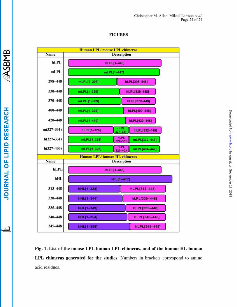
Fig. 1. List of the mouse LPL-human LPL chimeras, and of the human HL-human
LPL chimeras generated for the studies. Numbers in brackets correspond to amino
acid residues.
by guest, on Septem
ber 17, 2018w
ww
.jlr.orgD
ownloaded from
Christopher M. Allan, Mikael Larsson et al. Page 25 of 25
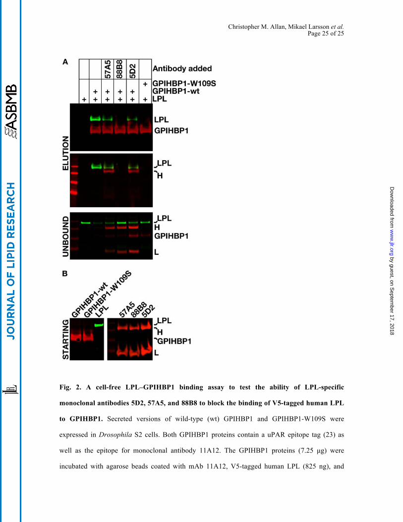
Fig. 2. A cell-free LPL–GPIHBP1 binding assay to test the ability of LPL-specific
monoclonal antibodies 5D2, 57A5, and 88B8 to block the binding of V5-tagged human LPL
to GPIHBP1. Secreted versions of wild-type (wt) GPIHBP1 and GPIHBP1-W109S were
expressed in Drosophila S2 cells. Both GPIHBP1 proteins contain a uPAR epitope tag (23) as
well as the epitope for monoclonal antibody 11A12. The GPIHBP1 proteins (7.25 µg) were
incubated with agarose beads coated with mAb 11A12, V5-tagged human LPL (825 ng), and
by guest, on Septem
ber 17, 2018w
ww
.jlr.orgD
ownloaded from
Christopher M. Allan, Mikael Larsson et al. Page 26 of 26
either no antibody or mAbs 57A5, 88B8, or 5D2 (20 µg/ml final). We used a high molar
concentration of mAbs so as to minimize the impact of differences in the affinity of the three
different mAbs. After a 1 h incubation at 4°C, the beads were washed. GPIHBP1 and GPIHBP1-
bound LPL were then released from the agarose beads by heating the beads in SDS-sample
buffer. (A) Western blots on the “unbound” fraction (proteins that did not bind to the agarose
beads) and “elution” fractions with an IRdye800-V5 antibody (green), an IRdye680–antibody
11A12 (red), and an IRdye680-donkey anti–mouse IgG (red). LPL binding to GPIHBP1 was
inhibited 53.8% with 57A5, 94.9% with 88B8, and 63.5% with 5D2, as judged by quantification
with a Li-Cor scanner. H, heavy chain; L, light chain. (B) Western blots performed on the
“starting material” proteins that were added to the assay (monoclonal antibodies, GPIHBP1,
LPL).
by guest, on Septem
ber 17, 2018w
ww
.jlr.orgD
ownloaded from
Christopher M. Allan, Mikael Larsson et al. Page 27 of 27
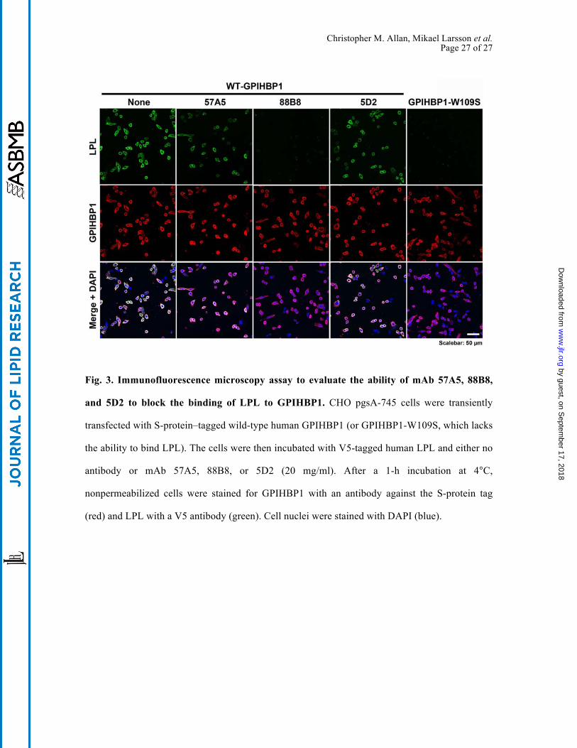
Fig. 3. Immunofluorescence microscopy assay to evaluate the ability of mAb 57A5, 88B8,
and 5D2 to block the binding of LPL to GPIHBP1. CHO pgsA-745 cells were transiently
transfected with S-protein–tagged wild-type human GPIHBP1 (or GPIHBP1-W109S, which lacks
the ability to bind LPL). The cells were then incubated with V5-tagged human LPL and either no
antibody or mAb 57A5, 88B8, or 5D2 (20 mg/ml). After a 1-h incubation at 4°C,
nonpermeabilized cells were stained for GPIHBP1 with an antibody against the S-protein tag
(red) and LPL with a V5 antibody (green). Cell nuclei were stained with DAPI (blue).
by guest, on Septem
ber 17, 2018w
ww
.jlr.orgD
ownloaded from
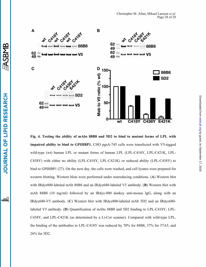
Christopher M. Allan, Mikael Larsson et al. Page 28 of 28
Fig. 4. Testing the ability of mAbs 88B8 and 5D2 to bind to mutant forms of LPL with
impaired ability to bind to GPIHBP1. CHO pgsA-745 cells were transfected with V5-tagged
wild-type (wt) human LPL or mutant forms of human LPL (LPL-C418Y, LPL-C421K, LPL-
C438Y) with either no ability (LPL-C418Y, LPL-C421K) or reduced ability (LPL-C438Y) to
bind to GPIHBP1 (27). On the next day, the cells were washed, and cell lysates were prepared for
western blotting. Western blots were performed under nonreducing conditions. (A) Western blot
with IRdye800-labeled mAb 88B8 and an IRdye680-labeled V5 antibody. (B) Western blot with
mAb 88B8 (10 mg/ml) followed by an IRdye-800 donkey anti-mouse IgG, along with an
IRdye680-V5 antibody. (C) Western blot with IRdye800-labeled mAb 5D2 and an IRdye680-
labeled V5 antibody. (D) Quantification of mAbs 88B8 and 5D2 binding to LPL-C418Y, LPL-
C438Y, and LPL-C421K (as determined by a Li-Cor scanner). Compared with wild-type LPL,
the binding of the antibodies to LPL-C418Y was reduced by 70% for 88B8, 37% for 57A5, and
26% for 5D2.
by guest, on Septem
ber 17, 2018w
ww
.jlr.orgD
ownloaded from
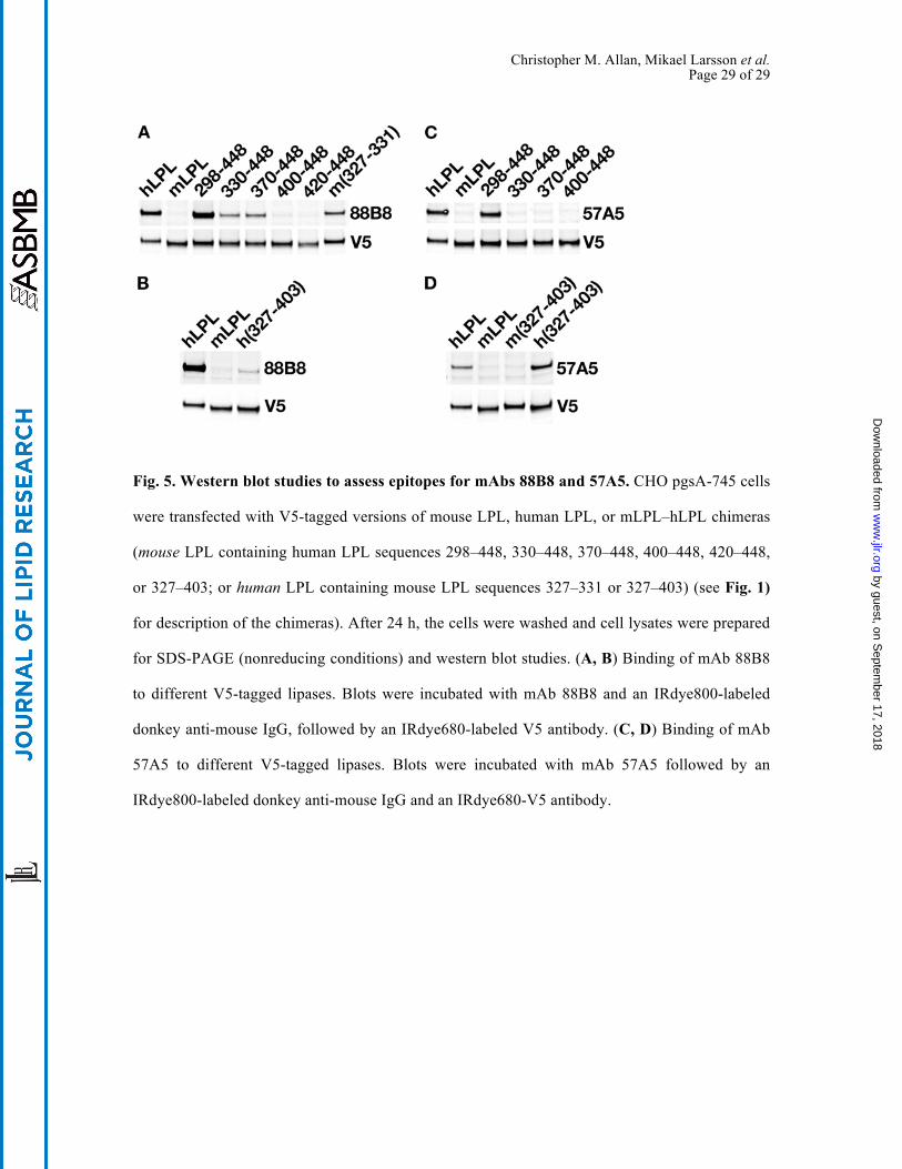
Christopher M. Allan, Mikael Larsson et al. Page 29 of 29
Fig. 5. Western blot studies to assess epitopes for mAbs 88B8 and 57A5. CHO pgsA-745 cells
were transfected with V5-tagged versions of mouse LPL, human LPL, or mLPL–hLPL chimeras
(mouse LPL containing human LPL sequences 298–448, 330–448, 370–448, 400–448, 420–448,
or 327–403; or human LPL containing mouse LPL sequences 327–331 or 327–403) (see Fig. 1)
for description of the chimeras). After 24 h, the cells were washed and cell lysates were prepared
for SDS-PAGE (nonreducing conditions) and western blot studies. (A, B) Binding of mAb 88B8
to different V5-tagged lipases. Blots were incubated with mAb 88B8 and an IRdye800-labeled
donkey anti-mouse IgG, followed by an IRdye680-labeled V5 antibody. (C, D) Binding of mAb
57A5 to different V5-tagged lipases. Blots were incubated with mAb 57A5 followed by an
IRdye800-labeled donkey anti-mouse IgG and an IRdye680-V5 antibody.
by guest, on Septem
ber 17, 2018w
ww
.jlr.orgD
ownloaded from
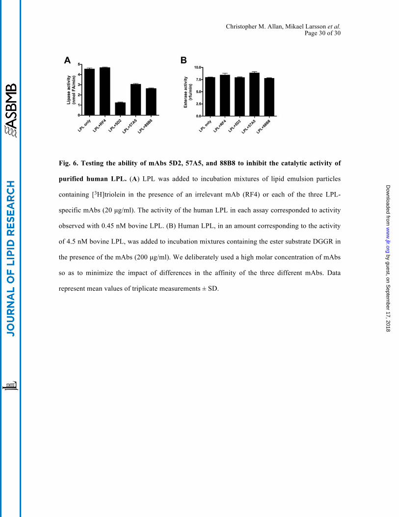
Christopher M. Allan, Mikael Larsson et al. Page 30 of 30
Fig. 6. Testing the ability of mAbs 5D2, 57A5, and 88B8 to inhibit the catalytic activity of
purified human LPL. (A) LPL was added to incubation mixtures of lipid emulsion particles
containing [3H]triolein in the presence of an irrelevant mAb (RF4) or each of the three LPL-
specific mAbs (20 µg/ml). The activity of the human LPL in each assay corresponded to activity
observed with 0.45 nM bovine LPL. (B) Human LPL, in an amount corresponding to the activity
of 4.5 nM bovine LPL, was added to incubation mixtures containing the ester substrate DGGR in
the presence of the mAbs (200 µg/ml). We deliberately used a high molar concentration of mAbs
so as to minimize the impact of differences in the affinity of the three different mAbs. Data
represent mean values of triplicate measurements ± SD.
by guest, on Septem
ber 17, 2018w
ww
.jlr.orgD
ownloaded from
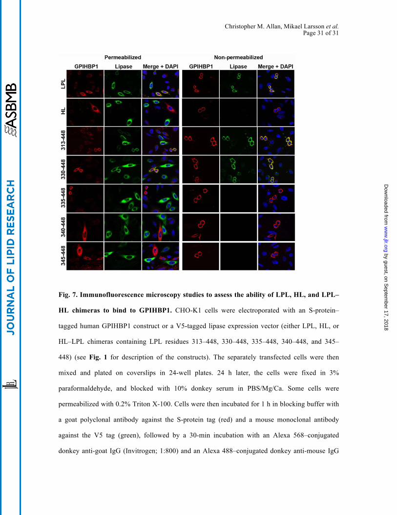
Christopher M. Allan, Mikael Larsson et al. Page 31 of 31
Fig. 7. Immunofluorescence microscopy studies to assess the ability of LPL, HL, and LPL–
HL chimeras to bind to GPIHBP1. CHO-K1 cells were electroporated with an S-protein–
tagged human GPIHBP1 construct or a V5-tagged lipase expression vector (either LPL, HL, or
HL–LPL chimeras containing LPL residues 313–448, 330–448, 335–448, 340–448, and 345–
448) (see Fig. 1 for description of the constructs). The separately transfected cells were then
mixed and plated on coverslips in 24-well plates. 24 h later, the cells were fixed in 3%
paraformaldehyde, and blocked with 10% donkey serum in PBS/Mg/Ca. Some cells were
permeabilized with 0.2% Triton X-100. Cells were then incubated for 1 h in blocking buffer with
a goat polyclonal antibody against the S-protein tag (red) and a mouse monoclonal antibody
against the V5 tag (green), followed by a 30-min incubation with an Alexa 568–conjugated
donkey anti-goat IgG (Invitrogen; 1:800) and an Alexa 488–conjugated donkey anti-mouse IgG
by guest, on Septem
ber 17, 2018w
ww
.jlr.orgD
ownloaded from
Christopher M. Allan, Mikael Larsson et al. Page 32 of 32
(Invitrogen; 1:800). Cell nuclei were visualized with DAPI (blue). Cells expressing wild-type
GPIHBP1 captured LPL secreted by neighboring LPL-expressing cells; hence, the GPIHBP1 and
LPL signals colocalized on the merged image. HL–LPL (313–448) and HL–LPL(330–448) bound
to GPIHBP1. HL and the remaining HL–LPL chimeras did not bind to GPIHBP1 (no
colocalization of GPIHBP1 and the lipase on the merged image). HL–LPL chimeras containing
LPL residues 370–448, 380–448, and 389–448 also failed to bind to GPIHBP1.
by guest, on Septem
ber 17, 2018w
ww
.jlr.orgD
ownloaded from
Christopher M. Allan, Mikael Larsson et al. Page 33 of 33
Fig. 8. Assessing the ability of mAbs 57A5, 88B8, and 5D2 bind to GPIHBP1-bound LPL.
CHO pgsA-745 cells were transiently transfected with S-protein–tagged human GPIHBP1. After
24 h, the cells were incubated with V5-tagged LPL for 1 h. The LPL was then removed and the
cells were washed. mAbs 57A5, 88B8, and 5D2 (20 µg/ml) were then added to the cells and
incubated for 1 h at 4°C. The cells were washed, fixed with paraformaldehyde, and then stained
for GPIHBP1 with an antibody against the S-protein tag (red), and for LPL with a V5 antibody
(green). mAbs 57A5, 88B8, and 5D2 were directly labeled (magenta).
by guest, on Septem
ber 17, 2018w
ww
.jlr.orgD
ownloaded from
Christopher M. Allan, Mikael Larsson et al. Page 34 of 34
Fig. 9. Testing the capacity of mAbs 57A5, 88B8, and 5D2 to bind to human LPL in
capillaries of the skeletal muscle of Lpl–/– mice carrying a human LPL transgene driven by
the muscle creatine kinase promoter (Lpl–/–MCK-hLPL). For these studies, skeletal muscle
from wild-type mice and Lpl–/–MCK-hLPL mice was harvested and embedded in OCT, and 7-
mm-thick sections were cut, placed on slides, and fixed in methanol. Tissue sections were stained
with a rabbit antibody against mouse CD31 (cyan), Alexa Fluor 568-labeled mAb (57A5, 88B8,
or 5D2; green), and an Alexa Fluor 647-labeled antibody against GPIHBP1 (11A12, red).
by guest, on Septem
ber 17, 2018w
ww
.jlr.orgD
ownloaded from
Recommended