Title Chemical Studies on the Regulation of DNA Structures and Z-DNA Specific Reactions( Dissertation_全文 )
Author(s) Kawai, Kiyohiko
Citation Kyoto University (京都大学)
Issue Date 1999-03-23
URL https://doi.org/10.11501/3149549
Right
Type Thesis or Dissertation
Textversion author
Kyoto University
1144
Chemical Studies on the Regulation
of DNA Structures
and Z-DNA Specific Reactions
Kiyohiko Kawai
1999
Preface
The study presented in this thesis has been carried out under the
direction of Professor Isao Saito at the Department of Synthetic
Chemistry and Biological Chemistry of Kyoto University dUring April
1993 to March, 1999. The study is concerned with chemical studies
on the regulation of DNA structures and Z-DNA specific reactions.
The author wishes to express his sincere gratitude to Professor
Isao Saito for his kind guidance, valuable suggestions, and
encouragement throughout this work. The author is deeply
grateful to Professor Hiroshi Sugiyama for his constant advice,
valuable discussions, and encouragement during the course of this
study. The author is also indebted to Associate Professor Kazuhiko
Nakatani for his helpful suggestions. The author is also indebted to
Dr. Yoshikatsu Ito, Dr. Kenzo Fujimoto, Dr. Hisafumi Ikeda and Dr.
Akimitsu Okamoto for their helpful suggestions.
The author is grateful to Mr. Haruo Fujita and Mr. Tadao
Kobatake for the measurements of NMR spectra and mass spectra,
respectively. The author is thankful to Professor Junzo Sunamoto
for the measurements of circular dichroism. The author wishes to
thank Messrs. Atsushi Matsunaga, Kenji Ikudome, Mariko Isomura,
Shuji Ikeda, Katsuhito Kino, Shigeo Matsuda, Yohei Ozeki, Shigenori
Ishihara for their collaboration. The author is also grateful to Junya
Shirai, Yasuki Komeda, Takashi Nakamura, Shinsuke Sando and
other members of Prof. Saito's research group for their helpful
suggestions and hearty encouragement.
The author is also grateful to Dr. Sigeru Ito, Dr. Tsuyoshi
Fujiwara, Dr. Zhi-Fu Tao, Dr. Ayumi Ohsaki, Dr. Lucheng Song and
Mr. Takanori Ohyoshi for their helpful suggestions and hearty
encouragement. The author also thanks to other members of Prof.
Sugiyama's research group.
The author thanks Japan Society for the Promotion of Science
for financial support (Fellowship for Japanese Junior Scientists).
Finally, the author expresses his deep appreciation to his
parents, Mr. Masao Kawai and Mrs. Mitoko Kawai for their constant
assistance and affectionate encouragement.
Kiyohiko Kawai
January, 1999
Contents
pageGeneral Introduction 1
Chapter 1
Chapter 2
Chapter 3
Chapter 4
Chapter 5
Chapter 6
Synthesis, Structure and Thermodynamic
Properties of 8-Methylguanine-Containing
Oligonucleotides: Z-DNA under
Physiological Salt Conditions.... ·.. ·.. ·.. ·.. ········· .. ·.. ·· .. ·.. 9
Effects of Cytosine C5 and Guanine C8
Methylation on the B- to Z-DNA Transition········· 37
Stabilization of Hoogsteen Base-Pairing byIntroducing NHz Group at the C8 Positionof Adenine··· ·· 49
Conformation Dependent Photochemistry of
5-Halouracil-Containing DNA: Stereospecific
2'a-Hydroxylation of Deoxyribose inZ-form DNA· ···················· ·· .. ···· .. · ·65
Intrastrand 2 '~ Hydrogen Abstraction
of 5'-Adjacent Deoxyguanosine byDeoxyuridin-5-yl in Z-form DNA..·· .. ··· .. ··· .. ·.. ·· .. ··· .. 81
Photochemical Halogen-Exchange Reaction of
5-Iodouracil-Containing Oligonucleotides· .. ·..·......93
List of Publications····· .. ····· .... ······ .. ···· .. ·.. ······· .... ···· .. ·· .. ······ .. ·.. ·· .. ····· .... ·· .. ····108
List of Oral Presentations·· ........ ·...... ···· .. ·.. ·.. ·.. ·· .. ······· ....·.. ·· ...... ·· .. ········· ....110
General Introduction
As for its flexible nature as a biopolymer, DNA is able to form a
number of local conformations. In addition to canonical right
handed B-form structure, DNA has been demonstrated to adopt A
form DNA, left handed Z-form DNA, cruciform, parallel stranded
DNA, bent DNA, triplex and quadruplex DNA.! Such
polymorphism of DNA structure has been suggested to play an
important role in a number of transcriptional and replicative
processes.2 N one the less, when and how these local
conformations form remain unresolved, and their detail biological
functions are still unclear.
Understanding the forces responsible for DNA conformational
change would be of fun dam en tal value in exploring the biological
roles of alternative DNA conformations. One useful method to
study the contribution of various thermodynamic forces on DNA
conformations is to examine the effects of chemical modification on
the thermal stability and structures. The chemical modification at
the base,3 sugar moiety! and DNA back boneS have been extensively
studied. For example, Dias et al. used C8 brominated guanine as a
probe for glycosidic conformation in telomeric DNA.3a Strauss et
aI. substituted one of the nonbridging phosphate oxygens with methyl
group for the neutralization of the charge and demonstrated that a
DNA bend is caused by neutralization of one face of DNA helix.Sa
From these results, they have proved the hypothesis that proteins
with cationic surfaces induce substantial DNA bending by
neutralizing phosphates on one DNA face.6
1
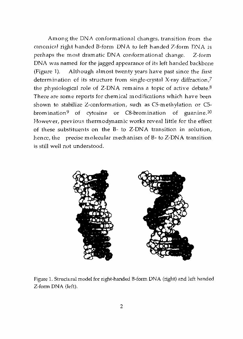
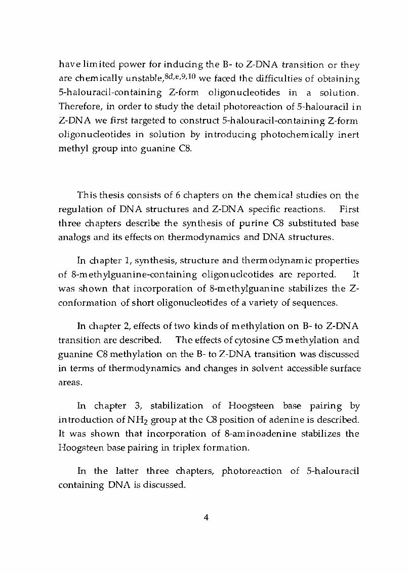
Among the DNA conformational changes, transition from the
canonical right handed B-form DNA to left handed Z-form DNA is
perhaps the most dramatic DNA conformational change. Z-form
DNA was named for the jagged appearance of its left handed backbone
(Figure 1). Although almost twenty years have past since the first
determination of its structure from single-crystal X-ray diffraction,?
the physiological role of Z-DNA remains a topic of active debate.S
There are some reports for chemical modifications which have been
shown to stabilize Z-conformation, such as CS-methylation or CS
bromination9 of cytosine or C8-bromination of guanine. lO
However, previous thermodynamic works reveal little for the effect
of these substituents on the B- to Z-DNA transition in solution,
hence, the precise molecular mechanism of B- to Z-DNA transition
is still well not understood.
Figure 1. Structural model for right-handed B-form DNA (right) and left handed
Z-forrn DNA (left).
2
Chern ical modification which can stabilize a certain DNA
conformation provides opportunities for investigating its
conformation dependent thermodynamic properties, detail structure
and reactivities. Recently, our group reported the construction of
stable parallel stranded DNA by incorporation of 5-methylisocytosine
and isoguanine and the detail structure of parallel stranded DNA was
determined by NMR.ll Since the reactivities of Z-form DNA and
several other local conformations have not been thoroughly
investigated, it is highly desirable to develop a chemical modification
to stabilize such DNA conformations In appropriate short
oligonucleotides in a solution.
Another way to understand the precise biological functions of
DNA local conformations is to have an appropriate detection system
for DNA local structures in a living cell system. Since the DNA
local structures are assumed to appear in a very short period of time in
a living cell, methods which can trap the intermediate structure is
useful. Meyer et al. attached reactive bromoacetoamidopropyl
group to the end of oligonucleotides and trapped the formation of
displacement loop (D-Ioop) with a covalent cross-link.12 However,
such reactive groups are not suitable for a biological system and
therefore development of photo-triggered chemical probe is
anticipated. Previously, our group investigated the photoreactions
of 5-halouracil-containing B-form DNA13 and DNA-RNA hybrid14
and demonstrated that photo-generated uracilyl radical undergoes
hydrogen abstraction in a highly conformation dependent manner.
These results suggested that 5-halouracil may provide important
informations on DNA conformations of biological interest.
Therefore, we examined 5-halouracil as a photochemical probe for the
left handed Z-form DNA. However, since preViously reported
chemical modifications which can stabilizes the Z-conformations
3
have limited power for inducing the B- to Z-DNA transition or they
are chemically unstable,8d,e,9,10 we faced the difficulties of obtaining
5-halouracil-containing Z-form oligonucleotides in a solution.
Therefore, in order to study the detail photoreaction of 5-halouracil in
Z-DNA we first targeted to construct 5-halouracil-containing Z-form
oligonucleotides in solution by introducing photochemically inert
methyl group into guanine C8.
This thesis consists of 6 chapters on the chern ical studies on the
regulation of DNA structures and Z-DNA specific reactions. First
three chapters describe the synthesis of purine C8 substituted base
analogs and its effects on thermodynamics and DNA structures.
In chapter 1, synthesis, structure and thermodynamic properties
of 8-methylguanine-containing oligonucleotides are reported. It
was shown that incorporation of 8-rnethylguanine stabilizes the Z
conformation of short oligonucleotides of a variety of sequences.
In chapter 2, effects of two kinds of methylation on B- to Z-DNA
transition are described. The effects of cytosine C5 methylation and
guanine C8 methylation on the B- to Z-DNA transition was discussed
in terms of thermodynamics and changes in solvent accessible surface
areas.
In chapter 3, stabilization of Hoogsteen base pairIng by
introduction of NH2 group at the C8 position of adenine is described.
It was shown that incorporation of 8-am inoadenine stabilizes the
Hoogsteen base pairing in triplex formation.
In the latter three chapters, photoreaction of 5-halouracil
containing DNA is discussed.
4
In chapter 4, conform ational dependent photochemistry of 5-
halouracil-containing DNA was discussed. Stereospecific 2'a-
hydroxylation of deoxyribose in Z-form DNA is described.
In chapter 5, intrastrand 2'~ Hydrogen Abstraction of 5' -Adjacent
Deoxyguanosine by Deoxyuridin-5-yl in Z-form DN A is discussed.
Incorporation of deuterium into uracil C5 during the C2'
hydroxylation is described.
In chapter 6, photochemical halogen-exchange reaction of 5
iodouracil-containing oligonucleotides is described. It was found
that 5-iodouracil undergoes efficient photo-halogen-exchange
reaction in the presence of halide anions to produce corresponding C5
halogen exchanged uracils.
5
References
1. CozzarellC N. R.; Wang, ]. C. DNA Topology and Its Biological
Effects 1990, Cold Spring Harbor Laboratory Press: New York.
2. (a) Sinden, R. R. DNA Structure and Function 1994, Academic
Press: New York. (b) Travers, A. A. Annu. Rev. Biochem.
1989,58,427.
3. (a) Dias, E.; Battiste, J. L.; Williamson]. R. J. Am. Clzem. Soc.
1994, 116,4479. (b) Kohda, K.; Tsunomoto, H.; Minoura, Y.;
Tanabe, K.; Shibutani, S.; Chem Res. Toxicol. 1996,9, 1278. (c)
Eason, R. G.; Burkhardt, D. M.; Phillips, S. J.; Smith, D. P.; David
S. S. Nucleic Acids Res. 1996,24,890. (d) Scweitzer, B. A.; Kool,
E. T. J. Am. Chem. Soc. 1995,117,1864. (e) Moran, S.; Ren, R.
X.-F.; Rumney, S.; Kool, E. T. J. Am. Chem. Soc. 1997, 119,2056.
(f) Guckian, K. M.; Scweitzer, B. A.; Ren, R. X.-F.; Sheils, C. J.;Paris, P. L.; Tahmasebi, D. C.; Kool, E. T. ]. Am. Chem. Soc. 1996,
118,8182. (g) Moran, S.; Ren, R. X.-F.; Kool, E. T. Proc. Natl.
Acad. Sci. USA 1997, 94, 10506.
4. Ikeda, H.; Fernandez, R.; W ilk, A.; Barchi, J. J.; Huang, X.;
Marquez, V. E. Nucleic Acids Res. 1998,26,2237.
5. (a) Strauss, J. K.; Maher III, 1. J. Science 1994, 266, 1829. (b)
Tarkoy, M.; Leumann, C. Angew. Chem. Int. Ed. Engl. 1993, 32,
1432. (c) Bolli, M.; Litten, C.; Leumann, C. Chem. Bio1.1996, 3,
197. (d) Steffens, R.; Leumann, C. J. J. Am. Chern. Soc. 1997, 119,
11548.
6. Manning, G.; Ebralidse, K. K.; Mirzabekov, A. D.; Wang, A. J.B i 0111 01. Struct. Dynanzics 1989, 6, 877.
7. (a) Wang, A. H. -J.; Quigley, G. J.; Kolpak, F. J.; Crawford,]. L.; van
BOOill, J. H.; van der Marel, G.; Rieh, A. Nature 1979,282, 680.
(b) Drew, H.; Takano, T.; Tanaka, S.; Itakura, K.; Dickerson, R. E.
Nature 1980, 286, 563.
6
8. (a) Kim, Y.-G.; Kim, P. S.; Herbert, A.; Rich, A. Proc. Natl. Acad.
Sci. USA 1997, 94, 12875. (b) Herbert, A.; Lowenhaupt, K.; Spitzer,
J.; Rich, A. Froc. Natl. Acad. Sci. USA 1995, 92, 7550. (c)
Erlanson, D. A.; Mark Glover, J. N., Verdine, G. L. J. Am.
Chem. Soc. 1997, 117,6927. (d) Rich, A.; Nordheim, A.; Wang, A.
H.-J. Ann.Rev.Biochem.1984,53,791. (e) Rich,A. Ann.N.Y. Acad. Sci. 1994, 726, 1.
9. Behe, M., Felsenfeld, G. Froc. Natl. Acad. Sci. USA 1981,78,
1619.
10. (a) Lafer, E. M.; Moller, A.; Nordheim, A.; Stollar, B. D.; Rich, A.
Proc. Natl. Acad. Sci. USA 1981,78,3546. (b) Moller, A.,
Nordheim, A.; Kozlowski, S. A.; Patel, D. J.; Rich, A.
Biochemistry, 1984, 23,54.
11. (a) Sugiyama, H.; Ikeda, S.; Saito. I. J. Am. Chem. Soc. 1996,118,
9994. (b) Yang, X. L.; Sugiyama, H.; Ikeda, S.; Saito. 1.; Wang, A.
H.-J. Biophysical J. 1998, 75, 1163.
12. Gamper, H. B.; Hou, Y.-M.; Stamm, R. M.; Podyminogin, M. A.;
Meyer, R. B. J. Am. Chem. Soc. 1998, 120,2182.
13. (a) Sugiyama, H.; Tsutsumi, Y.; Saito. 1. J. Am. Chem. Soc. 1990,
112, 6720. (b) Sugiyama, H.; Tsutsumi, Y.; Fujimoto, K.; Saito. 1.
J. Am. Chern. Soc. 1993, 115,4443. (c) Sugiyama, H.; Fujimoto, K.;
Saito, 1.; Kawashima, E.; Sekine, T.; Ishida, Y. Tetrahedron Lett.
1996, 37, 1805.
14. Sugiyama, H.; Fujimoto, K.; Saito. 1. Tetrahedron Lett. 1997,38,
8057.
7
Chapter 1
Synthesis, Structure and Thermodynamic Properties of8-Methylguanine-Containing Oligonucleotides:Z-DNA under Physiological Salt Conditions
Abstract
Various oligonucleotides containing 8-methylguanine (m 8G)
have been synthesized and their structures and thermodynamic
properties investigated. Introduction of m 8G into DNA sequences
markedly stabilizes the Z conformation under low salt conditions.
The hexamer d(CGCm8GCGh exhibits a CD spectrum characteristic of
the Z conformation under physiological salt conditions. The
NOE-restrained refinement unequivocally demonstrated that
d(CGCm 8GCGh adopts a Z structure with all guanines in the syn
conformation. The refined NMR structure is very similar to the Z
form crystal structure of d(CGCGCGh, with a root mean square
deviation of 0.6 between the two structures. The contribution of
m 8G to the stabilization of Z-DNA has been estimated from the mid
point NaCl concentrations for the B-Z transition of various m 8G
containing oligomers. The presence of m 8G in d(CGCm 8GeGh
stabilizes the Z-conformation by at least dG :::: -0.8 kcal/mol relative to
the unmodified hexamer. The Z conformation was further
stabilized by increasing the n urnber of m 8Gs incorporated and
destabilized by incorporating syn-A and syn- T, found respectively in
the (A,T)-containing alternating and non-alternating pyrimidine
purine sequences. The results suggest that the chemically less
9
reactive mBC base is a useful agent for studying molecular
in teractions of Z-DNA or other ONA structures that incorporate syn
c conformation.
Introduction
It has been well established that ONA structure has a
remarkable conformational heterogeneity.1,2 Not only does the
biologically relevant B-DNA exhibit considerable local heterogeneity,
dramatically different DNA structures such as Z-DNA have also been
discovered. While the precise biological functions of Z-DNA have
yet to be identified, its role in regulating DNA supercoiling has been
amplydemonstrated.3A A recent study by Rich and colleagues has
shown that chicken double-stranded RNA adenosine deaminase has
strong Z-DNA binding properties.S This enzyme is known to work
near the transcription apparatus, where a high negative supercoiling
density along the DNA chain exist in front of the site of polymerase
action.2 Thus far most of the thermodynamic properties of 2-DNA
have been obtained through the use of supercoiled DNA plasmids
containing various alternating d(C·G)n inserts or their variants.2-4
However, other aspects of 2- DNA have not been thoroughly
investigated, presumably due to the difficulty of obtaining stable 2
form oligonucleotides in a physiological salt solution. Much of the
available experimental data are limited to d(C·C)n oligomers under
non-physiological conditions of high alcohol or high salt
concentrations.6-B While some chemical modifications, such as
CS-methylation or C5-bromination of cytosine9 or CB-bromination of
guanine,lO,ll have been shown to stabilize the Z conformation in
linear DNA oligomers, they have either limited power for inducing
the B-2 transition or they are chemically unstable. Therefore, it is
desirable to have a more convenient and reliable way to stabilize 2
10
tetrazo]e/pyridine
P(N-iPr2l.20(CH2hCN..
form oligomers under low salt conditions by incorporating
chemically and photochemically inert modified bases. We report
herein that the introduction of methyl group at the guanine C8
position produces a stable m 8-modified guanine base and markedly
stabilizes the Z conformation of short oligonucleotides of a variety of
sequences under physiological salt conditions.
Results and Discussion
Although theoretical calculations suggested that methylation at
the guanine C8 position greatly stabilizes the Z conformation by
favoring the syn glycosyl conformation,12 such a property
associated with m 8G-modified DNA has not been examined
experimentally. While introduction of the bulky bromine atom at
the C8 position has been used previously,lO,ll the brominated DNA
suffered the problem of chemical/photochemical instability. It
would be desirable to use the more stable m 8G in DNA to investigated
the molecular basis of a variety of Z conformation-specific reactions at
the oligonucleotide level.
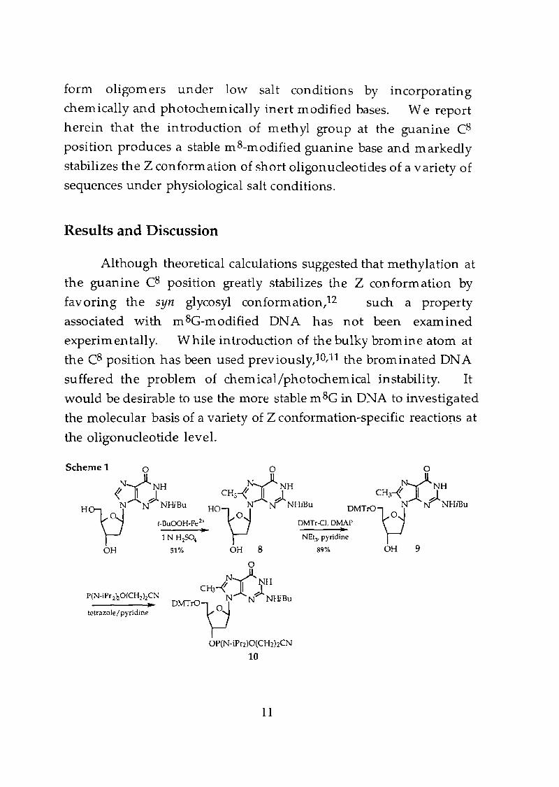
Schemel 0 0 0
t{,t CH,-1YJH CH,-1Y;tH~N N NHiBu HO~N NNHiBu DMTrO\:JN NNHiBu
t-BuOOH-Fe 2+ DMTr-el, DMAP-----l~~ ..1 N H2SO4 NEty pyridine
OH 51% OH 8 89% OH 9
oN0NH
Cl-U-f ~ ~DMTK>~ N NHiBu
OP(N-iPl"2)O(CHz)zCN
10
11
10000
-10000
A OmM
4.52 mM
IS.7mM
26.5 mM
SOmM
214mM
2600 mM
B62°C
I .......52 DCrf' ... ...) ................ 11.
10000,. 1 ... ,
42°CI.~" .......... '~". .... ----- ..~ ~/,/ ..... --------- ~12 DC
/ 1" 1~~ J,/ -----_. 22°C.' \ ,'.,'\ "1/ ~ .. 12 DC'\1;1: .-I 1." .\ .... ,' , :" 2°CI'~' ~ 1 • .'
S/ III"" ~; ...
0 . '... . ' .
-10000
Wavelength (nm)
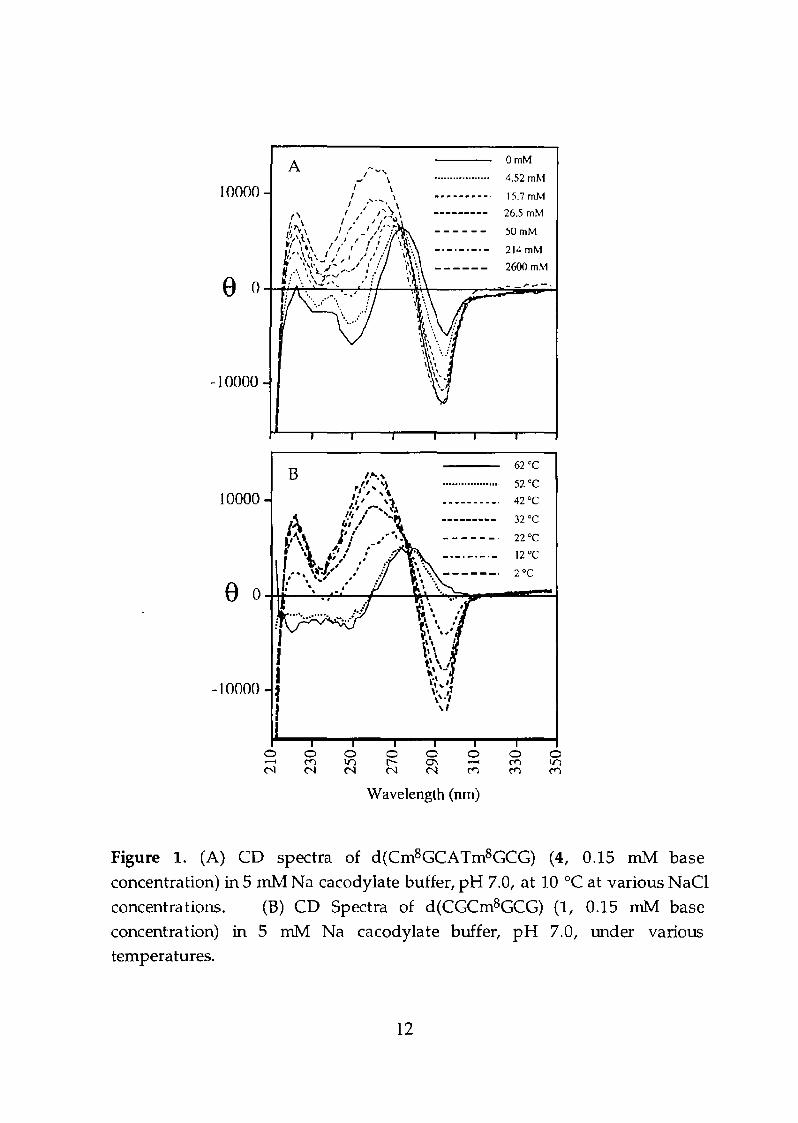
Figure 1. (A) CD spectra of d(Crn8GCATm8GCG) (4, 0.15 rnM base
concentration) in 5 rn1vI Na cacodylate buffer, pH 7,0, at 10°C at various NaCI
concentrations. (B) CD Spectra of d(CGCrn8GCG) (1, 0.15 rnM base
concentration) in 5 rnM Na cacodylate buffer, pH 7.0, under varioustemperatures,
12
The CD spectra of d(CGem 8GCGh (1) at different salt
concentration are shown in Figure 1A at 10°C. The hexamer in a 50
mM NaG solution has the characteristic CD spectrum of Z-DNA.
Without added salt it is in the SS form, as judged by UV and CO
spectroscopy, and is converted to the Z form by increasing salt
concentration, with a mid-point at 45 mM NaG. Since the
respective mid-point NaCI concentration for d(CGCGCGh and
d(mSCGCGmsCGh are 2.6 M13 and 2.0 M,7 it is evident that the C8
methylation of guanine greatly stabilizes the Z conformation.
NMR refinement of Z-DNA. In order to unequivocally
demonstrate that the structure of d(CGCm 8GCGh (1) at 30 mM salt
concentration is Z-DNA, NOE-restrained refinement has been carried
out. 2D-NOESY and TOCSY in 0 20 were used to assign the
resonances of all non-exchangeable protons. Since the structure is
expected to be Z-DNA, as judged from the CD spectrum, the usual
sequential assignment procedure would not be applicable. Indeed,
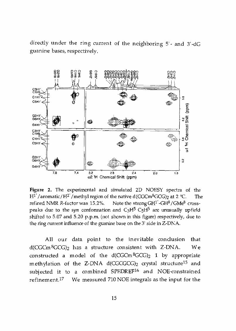
the aromatic-HI' and m8G4 methyl-HI' cross peak region of the 2D
NOESY spectrum (Figure 2) showed only strong intranudeotide
G2HI'-G2H8, G~I'-G~e and G6Hl'-G6H8 cross-peaks, indicative of
the syn conformation of guanine residues. As has been noted
before,6,14 there is no internudeotide connectivity in Z-DNA, in
contrast to that in right-handed B-DNA. The assignment was
subsequently extended to the aromatic-H2' jH2" region and finally to
all regions of the spectrum. The TOCSY data supported the
assignment (data not shown).
The chemical shifts of all resonances are tabulated in Table 1.
Note that all cytidine H2' and HS resonances are unusually upHeld
(--1.7 and 2.6 p.p.m. respectively), analogous to those seen before.6,14
The upHeld shifts are due to the orientation of the sugar moiety of the
de nucleotide in Z-DNA, which places the H2' and HS protons
13
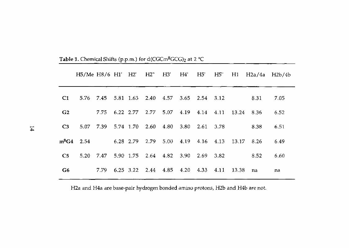
Table 1. Chemical Shifts (p.p.m.) for d(CGCm8GCGh at 2 DC
H5/Me H8/6 HI' H2' H2" H3' H4' H5' H5" HI H2a/4a H2b/4b
C1 5.76 7.45 5.81 1.63 2.40 4.57 3.65 2.54 3.12 8.31 7.05
G2 7.75 6.22 2.77 2.77 5.07 4.19 4.14 4.11 13.24 8.36 6.52
I--" C3 5.07 7.39 5.74 1.70 2.60 4.80 3.80 2.61 3.78 8.38 6.51~
m8G4 2.54 6.28 2.79 2.79 5.00 4.19 4.16 4.13 13.17 8.26 6.49
C5 5.20 7.47 5.90 1.75 2.64 4.82 3.90 2.69 3.82 8.52 6.60
G6 7.79 6.25 3.22 2.44 4.85 4.20 4.33 4.11 13.38 na na
H2a and H4a are base-pair hydrogen bonded amino protons, H2b and H4b are not.
directly under the ring current of the neighboring 5' - and 3' -dGguanine bases, respectively.
""""nnnn nG>"n nnn~~I\)"'(Jl(JlWW ......... 0'1 ........ (JlW~
IIIIIIII I III III~~~~U!~~I\) "'1!:"il~ ~~rq, II> I 1
I
~ <C
ui
~ Ea.s
~~
N'-C.
. ..c.t>
ICC/)
(iju
0>'E
f9 ~\Q)
..c.eO ~~ ~O, -
cJ • G ~
a
• N
@ to
7.8 7.4 3.2 2.8 2.4 2.0 1.6w2 1H Chemical Shift (ppm)
Figure 2. The experimental and simulated 2D NOESY spectra of theHI' /aromatic/H2' Imethyl region of the native d(CGCm8GCG)z at 2°C. Therefined N1'v1R R-factor was 15.2%. Note the strongGHI' -GH8 IGMe8 crosspeaks due to the syn confonnation and C3HS CSHS are unusually upfieldshifted to 5.07 and 5.20 p.p.m (not sho"Wll in this figure) respectively, due tothe ring current influence of the guanine base on the 3' side in Z-DNA.
All our data point to the inevitable conclusion that
d(CCCm 8GCCh has a structure consisten t with Z-DNA. We
constructed a model of the d(CGCm 8GCCh 1 by appropriate
methylation of the Z-DNA d(CGCCCGh crystal structure1S and
subjected it to a combined SPEDREF16 and NOE-constrained
refinement.17 We measured 710 NOE integrals as the input for the
15
NOE-restrained refinement. The refined structure, which has an
NMR R factor of 15.2%, is shown in Figure 3. The NOE-refined
structure is very sim ilar to the d(CGCGCGh Z-DN A structure
determ ined by X-ray crystallography.l5 The Lm .5. difference
between the two structures is only 0.6 A. The cytidine residues are
in the an ti IC2' -endo conformation, whereas the guanosine residues
are in the syn IC3' -e ndo conform ation (except for the 3' -term inal
guanosines, which have a mixed C2'-endo IC3'-endo sugar pucker).
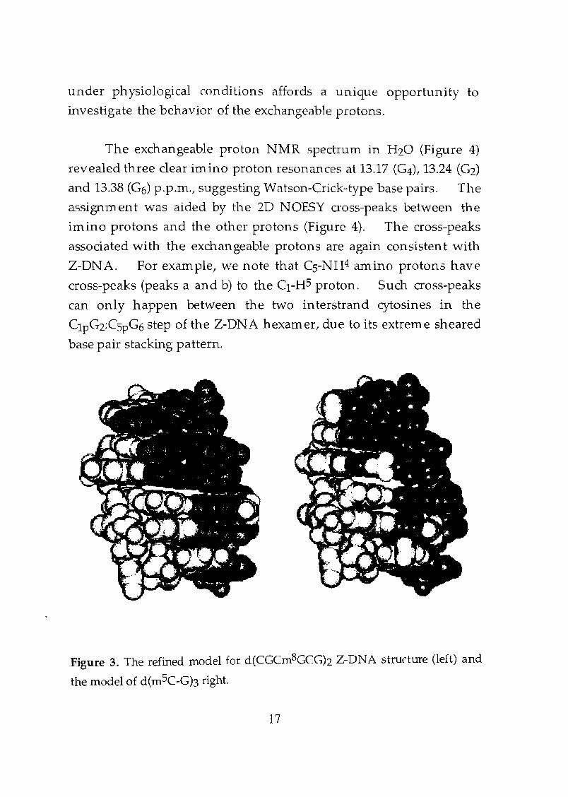
In the rn 8G-modified Z-DNA structure the hydrophobic C8-methyl
groups are located in the periphery of the helix and prominently
exposed to the solvent region. In contrast, in the mSC-modified zONA structure the CS-methyl groups form hydrophobic patches in
the small recessed area of the concave 'major groove' (Figure 3).
The siru ulated NOESY spectra based on the refined model agree with
the observed data (Figure 2). To the best of our knowledge this is
the first example of a refined structure of Z-form DNA by NMR under
physiological salt conditions without added organic solvent or
divalent cation.
Dynamics of Z-DNA. Z-DNA has been shown to have
unusual rigidity.2 The measured Tl relaxation inversion recovery
time (TlIR) of 2.7 s for the d(CGCm 8GCG) helix supports this notion.
For a B-DNA hexamer the averaged T1IR is ~1.7 s. The stiffness of
the Z-DNA double helix is also reflected in the remarkably slow
exchange rate of its various exchangeable protons, including the G
imino, G amino and C amino protons. It has been shown that the
G imino and C amino protons exchange with water in 30 and 50 min,
respectively, whereas the G amino protons exchange in 330 min, at
5 °C and pH 7.18 Our ability to obtain a stable Z-DN A structure
16
under physiological conditions affords a unique opportunity to
investigate the behavior of the exchangeable protons.
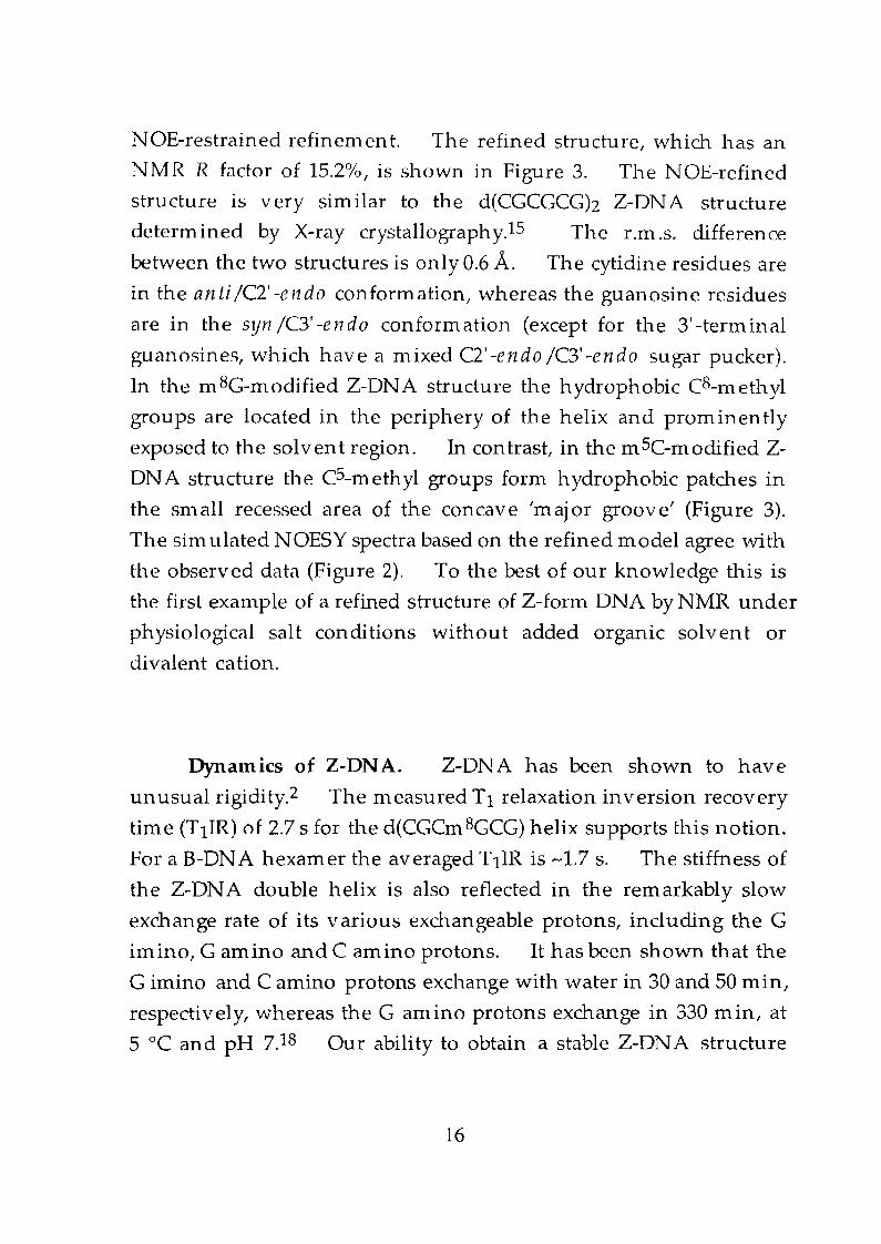
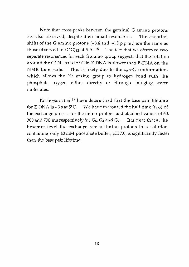
The exchangeable proton NMR spectrum in H20 (Figure 4)
revealed three clear imino proton resonances at 13.17 (G4), 13.24 (G2)
and 13.38 (G6) p.p.m., suggesting Watson-Crick-type base pairs. The
assignment was aided by the 2D NOESY cross-peaks between the
imino protons and the other protons (Figure 4). The cross-peaks
associated with the exchangeable protons are again consistent with
Z-DNA. For exam pIe, we note that CS-NH4 amino protons have
cross-peaks (peaks a and b) to the CI-H5 proton. Such cross-peaks
can only happen between the two interstrand cytosines in the
ClpG2:CSpG6 step of the Z-DN A hexamer, due to its extreme sheared
base pair stacking pattern.
Figure 3. The refined model for d(CGCm8GCGh Z-DNA structure (left) and
the model of d(m5C-Gb right.
17
Note that cross-peaks between the geminal G amino protons
are also observed, despite their broad resonances. The chemical
shifts of the G amino protons (~8.4 and ---6.5 p.p.m.) are the same as
those observed in (C·Ghz at 5 °C.18 The fact that we observed two
separate resonances for each G amino group suggests that the rotation
around the C2-Nz bond of Gin Z-DNA is slower than B-DNA on the
NMR time scale. This is likely due to the syn-G conformation,
which allows the N2 amino group to hydrogen bond with the
phosphate oxygen either directly or through bridging water
molecules.
Kochoyan et al.l8 have determined that the base pair lifetime
for Z-DNA is ~3 s at SoC. We have measured the half-time (h/2) of
the exchange process for the imino protons and obtained values of 60/
300 and 700 ms respectively for G6, G4 and G2. It is clear that at the
hexamer level the exchange rate of imino protons in a solution
containing only 40 mM phosphate buffer, pH 7.0/ is significantly faster
than the base pair lifetime.
18
II
G4H2C3H4 a-----4G2H2CSH4 "'"C1H4 -+ a
c ¢» r...: -EC3H6 = c..
C1H6 0S=co <:0> 0 '" = ce:I" ;:
CSH6 :.cG6H8
U).- roG2H8
u'E
cd Cl Q;Ico .c
0I
0 ® ~
G N
'" b.... s
G4H~ r:"'"~p
...- -0>
~~~~ -~. <la:>
I , I
8.0 7.0 6.0 .....
w1 1HChemical Shift (ppm)
Figure 4. The exchangeable proton 2D NOESY spectra of the HLarornatic
region of d(CGCm8GCGh at 2°C. There are clear NOE cross-peaks (peaks a
and b) between CIH4 amino protons and CSHS protons from the opposite
strand. Such cross-peaks can only happen in a Z-DNA structure. Note
that the guanine N2 amino geminal protons have a broad cross-peak (peaks c
and d).
19
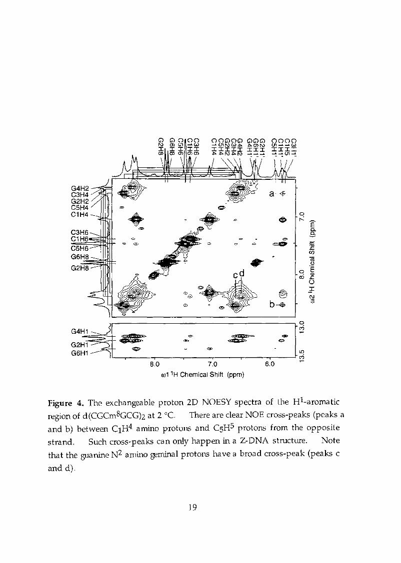
Thermodynamic properties. The effect of m 8G substitution
on the thermodynamic stability of the Z conformation was examined
by measuring the proportions of the Z, B, and 55 forms at various
tern peratures. Figure 1B shows the CD spectra of d(CGCm 8GCGh
(1) in 2.6 M NaCI solution at various temperatures. At 2°C it is
nearly 100% Z-DNA. The proportion of B increased with
increasing tern perature. For com parison, d(CGCGCGh under the
SaIne salt conditions consisted of a 1:1 mixture of Band Z. The
proportions of Z, B, and 55 for a m BG-containing oligomer were
determined at various tenlperatures by means of CD and UV
spectroscopy as previously reported.19 A similar tern perature
dependent B:Z equilibrium has also been observed for
d(CGClnBGCGh at 30 mM salt concentration byNMR spectroscopy as
shown in Figure 5.
20.8%
8.0 7.7 8.0 7.7 8.0 7.7 8.0
1H Chemical Shift (ppm)
7.7 8.0 7.7
Figure 5. Proton 1D N1v1R spectra showing the temperattrre-dependent
equilibrium of the B-Z transition as monitored by the G2H8 ( 7.75 p.p.In. at
2°C) and G6H8 (7.79 p.p.m. at 2°C) protons. The population of the B form
increases from 6.2% at 2 °C to 20.8% at 22 °e.
20
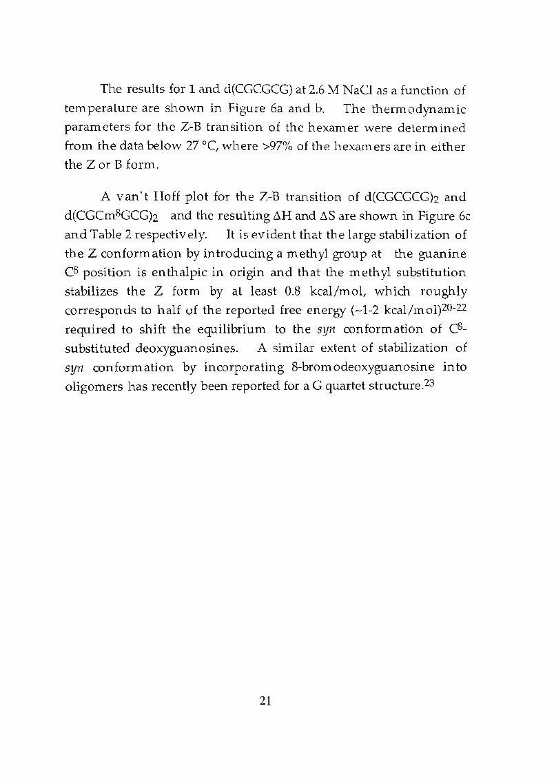
The results for 1 and d(CGCGCG) at 2.6 M NaCI as a function of
temperature are shown in Figure 6a and b. The thern"lodynamic
parameters for the Z-B transition of the hexamer were determined
from the data below 27°C, where >97% of the hexamers are in either
the Z or B form.
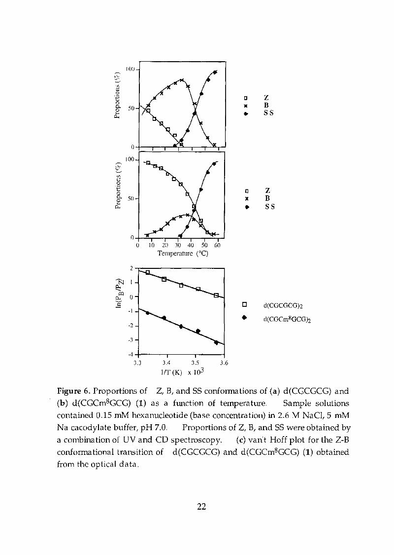
A van't Hoff plot for the Z-B transition of d(CGCGCGh and
d(CGCm8GCGh and the resulting ilH and ilS are shown in Figure 6c
and Table 2 respectively. It is evident that the large stabilization of
the Z conformation by introducing a methyl group at the guanine
C8 position is enthalpic in origin and that the methyl substitution
stabilizes the Z form by at least 0.8 kcal/mol, which roughly
corresponds to half of the reported free energy (-1-2 kcal/mol)20-22
required to shift the equilibrium to the syn conformation of C8
substituted deoxyguanosines. A similar extent of stabilization of
syn conformation by incorporating 8-bromodeoxyguanosine into
oligomers has recently been reported for a G quartet structure.23
21
o-t-=O-r--""'?---r-.----r-'o 10 20 30 40 50 60
Temperature (0C)
0
100~
t'2'-'If)
t:::0'f: c Z00.. 50 X B0...
SS0.. ..
100---t"=-'v,t:::0'2 D z0
)C B0. 500 55... ..P-.
2....-----------.
d(CGCGCG)2o
• d(CGCm8GCGh
3.63.4 3.5
1fT (K) x 103
-2
-I
-3
-4-+---..,....---....--~
3.3
Figure 6. Proportions of Z, B, and SS confonnations of (a) d(CGCGCG) and
(b) d(CGCmSGCG) (1) as a function of temperature. Sample solutions
contained 0.15 rnM hexanucleotide (base concentration) in 2.6 M NaCI, 5 mM
Na cacodylate buffer, pH 7.0, Proportions of Z, B, and 55 were obtained by
a combination of UV and CD spectroscopy. (c) van't Hoff plot for the Z-B
confonnational transition of d(CGCGCG) and d(CGCrnSGCG) (1) obtained
from the optical data.
22
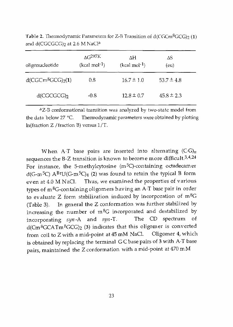
Table 2. Thermodynamic Parameters for Z-B Transition of d(CGCm8GCGh (1)
and d(CGCGCG)z at 2.6 M NaCP
~c297K LlH ~S
oligonucleotide (kcal mol-I) (kcal mol-I) (eu)
d(CGCm8GCGh(1) 0.8 16.7 + 1.0 53.7 + 4.8
d(CGCGCGh -0.8 12.8 + 0.7 45.8 + 2.3
aZ-B conformational transition was analyzed by two-state model from
the data below 27°C. Thermodynamic parameters were obtained by plotting
1n(fraction Z / fraction B) versus 1/T.
W hen A·T base pairs are inserted into alternating (C·G)J1
sequences the B-Z transition is known to become more difficult.3,4,24
For instance, the 5-methylcytosine (m SC)-containing octadecamer
d(G-m 5C) ABrU(G-m SC)4 (2) was found to retain the typical B form
even at 4.0 M NaCl. Thus, we examined the properties of various
types of m 8G-containing oligomers having an A·T base pair in order
to evaluate Z form stabilization induced by incorporation of m 8e
(Table 3). In general the Z conformation was further stabilized by
increasing the number of m 8e incorporated and destabilized by
incorporating syn-A and syn-T. The CD spectrum ofd(Cm 8GCATm 8GCGh (3) indicates that this oligomer is converted
from coil to Z with a mid-point at 45 mM NaCl. OligOlner 4, which
is obtained by replacing the terminal G·C base pairs of 3 with A·T base
pairs, maintained the Z conformation with a mid-point at 470 m M
23
Table 3. Midpoint NaCl Concentration in B-2 Transition of Various 8
Mcthylguaninc-Containing Oligonucleotides
number of residue NaCl
oligonuc1eotidea mBC syn Ab syn Te (mM)
2 0
o 0
o 0
470
120
2450
o
o 2000f
o
o
o
1
2
o
4
o
3
4
o
2
4
4
d(CGCm8CCCh (1)
d(CGCGCG)z
d (SmCGCG5mCGh
dI(GmSC)4A BrU(Gm5C)412 (2)
d(Cm8CCATmBGCGh (3)
d(Tm8GCATm8GCAh (4)
d(Cm8GCATm8CTG) (5)
d(GCGTACAC)
d(CmBGCTCm8CCG) (6)
d(CCm8CAGCmBGC)
ClmBc = 8-Methyl-2'-deoxyguanosine, mSC = 5-methyldeoxycytidine;
bsyn-A confonnation; csyn-T conformation; dtransition from single strand;
ereference 13; freference 7. The data were taken at 10°C.
N aCl. The incorporation of m Be into only one strand is also
capable of stabilizing the Z conformation considerably (oligomer 5).
A non-alternating pyrimidine-purine sequence has been shown to
destabilize the Z conformation due to the energetically disfavored synconfonnation of pyrimidine nucleosides.3A One of the central G·C
base pair of d(CeCGCGCGh can be replaced by a T-A base pair without
significantly increasing the n1id-point NaCI concentration, if the
24
duplex incorporates two m 8e into each strand (oligomer 6). Such a
low salt coneentration requirement of 120 ll1M for an imperfect Z
DNA (out-of-alternation pyrimidine-purine sequence) is remarkable.
Our results suggest that we can now study many heretofore
inaccessible DNA conformations involving Z-DN A, e.g. the B-Z
junction and the Z-Z junction.
Conclusion
The substitution of a methyl group at the guanine C8 position
dramatically stabilizes the Z conformation of short oligonucleotides
of a variety of base sequences. Some of these m 8G-modified
oligomers exist as a stable Z form under physiological salt conditions
without added organic solvent or divalent meta1.3-7 While
significant information on specific chemical reactions for DNA local
structures has been accumulated during the past several years,25-28
considerably less is known about the origin of these specificities.
Incorporation of the mSC moiety into DNA oligomers could be a
powerful tool to examine the molecular basis for many types of Z
conformation-specific reactions at the oligomer level under
physiological salt conditions.
25
Experimental Section
Materials and methods. Pyridine and acetonitrile (HPLC
grade) were dried over calciulTI hydride. 2' -Deoxyguanosine was
purchased from YAMASA Corporation. N ucleoside ~
cyanoethylphosphoramidite reagents (A, G, C, T) were obtained from
Applied BiosystelTIs. Calf intestine alkaline phosphatase (AP, 1000
uni t/ITI L) and snake venom phosphodiesterase (s.v. PDE, 3 unit/m L)
were purchased from Boehringer Mannheim. Silica gel column
chromatography was carried out on Wakogel C-200. Thin layer
chromatography was carried out on a Merck silica gel 60 PFZ54 plate.
N -Isobu tyryl-2' -deoxyguanosine was prepared according to the
published procedure.Z9 1H NMR spectra were recorded on a JEOL
JNM-GX400 spectrometer and a Varian-Gemini 200 spectrometer.
FAB mass were obtained on a JEOL-JMS-SXI02A.
Synthesis of 8-m ethyl-2' -deoxyguanosine 7. In troduction of
methyl group at C8 position of guanine was performed by free radical
methylation method.30 To a solution of 2'-deoxyguanosine (500
mg, 1.89 mmol) and FeS04·7HzO (2.1 g, 7.48 mmol) in 100 mL of 1 N
H2S04, an aqueous solution (100 mL) containing 0.81 mL of 70% t
butyl hydroperoxide (5.81 mmol) was added dropwise over a period of
5 min. After stirring at 0 DC for 40 min, the reaction mixture was
neutralized with saturated KOH solution. The resulting brownish
solution was filtered off and the filtrate was concentrated to dryness.
The residue was triturated with 60 m L of 28% NH40H and 150 m L of
methanol and filted. The filtrate was concentrated to dryness and
subjected to silica gel column chromatography. Elution with 28%
NH40H-methanol (2 : 8) afforded 7 as a white powder: yield 176 mg
(34 0/0). mp 230°C (dec.); IH NMR (DZO, 400 MHz) 82.20 (ddd, 1 H, J
= 14.0, 6.4, 2.6 Hz, 2"), 2.35 (s, 3 H, -8CH3), 2.84 (ddd, 1 H, J = 14.0, 8.0, 6.4
Hz, 2'), 3.69 (dd, 1 H, J = 12.6,4.3 Hz, 5'), 3.75 (dd, 1 H, J = 12.6,3.1 Hz,
26
5"),3.98 (ddd, 1 H, J ::: 4.3,3.4,3.1 Hz} 4'),4.53 (ddd, 1 H, J ::: 6.4, 3.4, 2.6
Hz, 3'), 6.13 (dd, 1 H, J::: 8.0, 6.4 Hz, I'); FABMS (positive ion) m/z
282 (M+H)+; UV (H20) 252.2 nm (e 12,700).
8yn thesis of 800m ethyl-N -isobutyryl-2' -deoxyguanosine 8. To
a solution of N -isobutyryl-2' -deoxyguanosine (1.0 g, 2.97 mmol) and
FeS04·7H20 (6.7 g, 24.1 mmol) in 160 mL of 1 N H2S04, an aqueous
solution (100 m L) containing 2.6 rn L of 70% t-butyl hydroperoxide
(19.0 mmol) was added dropwise over a period of 5 Inin. After
stirring at 0 DC for 60 min, the reaction mixture was neutralized with
saturated KOH solution. After centrifugation of the brown slushy
mixture, the supernatant was separated. The supernatant was
concentrated to dryness and the resulting brownish solid was
triturated three times with 200 mL of methanol. The combined
methanol solution was concentrated and the residue was subjected to
silica gel column chromatography. Elution with CH2Ch-methanol
(9: 1) afforded 8-methyl-N -isobutyryl-2' -deoxyguanosine 8 as a white
powder: yield 527 rng (51%). 8-Methyl-N-isobutyryl-2'-
deoxyguanosine: m p 195 DC (dec.); IH NMR (020) 8 1.21 (d, 6 H, J:::6.9 Hz, -CH(CH3)2), 2.25 (ddd, 1 H, J:::: 13.7,6.9,3.6 Hz, 2"),2.56 (s,3 H}
-SCH3), 2.72 (sep, 1 H, J ::: 6.9 Hz, -eH(CH3h), 3.17 (ddd, 1 H} J=:: 13.7, 7.4,
6.9 Hz, 2'),3.72 (dd, 1 H, J :::: 11.9, 5.4 Hz} 5"),3.74 (dd, 1 H, J::: 11.9, 4.1
Hz,S'), 3.92 (ddd, 1 H, 5.4, 4.1, 3.6 Hz, 4'),4.59 (ddd,1 H, J::: 6.9, 3.6, 3.6
Hz, 3'),6.32 (dd,l H, J :::: 7.4,6.9 Hz, 1'); FABMS (positive ion) m /z
352 (M+H)+.
Synthesis of 8-methyl-N-isobutyryl-51 -()"(dimethoxytrityl)-2'
deoxyguanosine 9. 8 (430 rng, 1.22 mmol) was dried by co
evaporation with dry pyridine three times. The residue was
dissolved in 13 mL of dry pyridine, and then 632 mg (1.83 mmol) of
27
4,4' -dimethoxytrityl chloride (632 mg, 1.83 mmo!), 4
dimethylaminopyridine (3.8 mg, 0.03 mmol) and triethylamine 257
JlL (1.83 mmol) were added to the reaction mixture. The solution
was stirred for 6 h at rOOln tern perature. The reaction mixture was
concen trated, and the residue was extracted with ethyl acetate.
White power was precipitated out from the organic layer and filtered
off. Washing of the precipitate with ethyl acetate and n-hexane
provide 9 as a white powder: yield 713 mg (89%). mp 180°C (dec.);
IH NMR (CD30D, 200 MHz) 81.15 (5,6 H, J = 6.8 Hz, -CH(CH3h), 1.19
(s,3 1-1, J= 6.8 Hz, -CH(CH3h), 2.29 (ddd, 1 H, J = 13.5, 7.2,4.4 Hz, 2' '),
2.55 (s, 3H, -8CH3), 2.63 (sep, 1 H, J = 6.8 Hz, -CH(CH3h), 3.17 (dd, 1 H, J=10.1,3.2 Hz, 5'), 3.25-3.45 (m, 2 H, 2', 5' i), 3.72 (5,3 H, -OCH3), 3.73 (s,3
H, -OCH3), 4.09 (ddd, 1 H, J=7.2,3.4,3.2 Hz, 4'),4.64 (ddd, 1 H, J= 7.2,
4.4, 3.8 Hz, 3'), 6.32 (dd, 1 H, J = 7.2, 6.4 Hz, 1'), 6.63-6.73 (m, 4 H,
aromatic), 7.11-7.36 (In, 9 H, aromatic); HR FABMS (positive ion)
m/z 654.2972 (M+H, 645.2928 calcd for C36H4007Ns).
Syn thesis of 8-m ethyl-N-isobu tyryl-3' -()'[2-cyanoethoxy-(NIN
diisopropylamino)phosphinoJ-5'-()'(dimethoxytrityI)-2'
deoxyguanosine 10. 9 (500 mg, 0.76 mmol) was dried by co
evaporation with pyridine (three times) and redissolved in 5.7 m L of
pyridine. To this solution were added 275 IlL of 2-cyanoethyl
N,N,N ',N '-tetraisopropylphosphorodiamidite (0.95 mmol) and 1.9
mL of 0.5 M tetrazole in acetonitrile and the mixture was stirred over
night at room tenlperature under argon. The reaction mixture was
poured into ice-cooled water and extracted with ethyl acetate. The
organic layer was washed with saturated N aHC03 and dried over
anhydrous NazS04 and concentrated. Crude 10 (580 mg) was used
for automated DNA SYnthesizer without further purification. IH
NMR (CD30D) 8 1.17 (d, 12 H, J = 6.5 Hz, -NCH(CH3h), 1.26 (dd, 6 H, J =
28
6.8,3.4 Hz, -CH(CH3)z), 2.41-2.64 (rn, 3 H, 2', -NCH(CH3)z), 2.57 (s, 3 H,
-8CH3), 2.63-2.77 (rn, 1 H, -CH(CH3)z), 2.69 (t,l H, J =5.9, -OCHz-), 2.84
(t, 1 H, J = 5.9, -OCHz-), 3.28-3.54 (m, 3 H, 2', -CHzCN), 3.72 (5,3 H,
OCH3), 3.73 (5,3 H, -OCH3), 3.54-3.71 (m, 2 H, 5' ,5"),4.07-4.34 (rn, 1 H,
4'),4.09 (ddd, 1 H, J=7.2, 3.4, 3.2 Hz, 4'),4.64-4.83 (rn, 1 H, 3'),6.36 (t, 1
H, J = 7.3 Hz, 1'), 6.60-6.80 (rn, 4 H, aromatic), 7.06-7.44 (m, 9 H,
aromatic); 31p NMR (CD30D) (3 148.48; FABMS (positive ion) nl/z
854 (M+H)+.
Synthesis of deoxynucleotides 1-6. Oligonucleotides were
prepared by the ~-(eyanoethyl)phosphoramidite method on
controlled pore glass supports (1 lImol) by using ABI 381 A DNA
synthesizer. After automated synthesis, the oligomer was detached
from the support by soaking in cone. aqueous ammonia for 1 h at
room tern perature. Deprotection was conducted by heating the
cone. aqueous ammonia for 12 h at 55°C. Aqueous ammonia
was then removed by evaporation, and the crude oligomer was
purified by reverse phase HPLC and lyophilized. Purity and
concentration of all oligonucleotides were determined by com plete
digestion with S.v. PDE and AP to 2'-deoxymononudeosides.
NMR analysis. The NMR solution (1 mM duplex with 0.04
M phosphate buffer, thus 0.06 M Na+, pH 7.0, in DzO) of
d(CGCm 8GCGh was prepared using the established procedure.l6
NMR spectra were collected on a Varian VXR500 500 MHz
spectrometer and processed with FELIX v1.1 on Silicon Graphics IRIS
workstations. The temperature was controlled to be accurate
within 0.01 °C. Tl relaxation experiments were carried out with
29
the standard 180-t-90° inversion-recovery sequence and the average
Tl relaxation time was 2.7 s. The non-exchangeable proton 2D
NOESY spectra were collected at 2°C with a mixing time of 100 ms and
a total recycle delay of 7.0 s. The data were collected by the
States/TPPI technigue31 with 512 t1 increments and 2048 tz complex
points each the average of 16 transients. Apodization of the data in
the t1 and tz dimensions consisted of 8 Hz exponential multiplication
with half of a sine-squared function for the last fourth of the data to
reduce truncation artifacts. Integrals from the non-exchangeable
20 NOE dataset were extracted by evaluation with the observed
cross-peak shapes of each spin in the £1 and b dim ensions. These
shapes were determined by spectral analysis using the program
MYLOR.l6 The exchangeable proton 20 NOESY experiment was
carried out in 90% HzO/IO% DzO solution using the 1-1 pulse
sequence31 as the read sequence, with a mixing time of 100 ms and a
recycle delay of 2.7 s, each data point the average of 24 transients.
The starting model was constructed by Midasplus (UCSF). Forty
cycles of refinelllent of the starting model were then carried out by the
sequence of procedures comprising the SPEOREF package.16 This
includes a full matrix relaxation calculation of the NOEs for the
lllodel with comparison of the experimental and simulated spectra to
deconvolute overlapped areas of the spectra. Minimization of the
residual errors within the program X-PLOR17 was then performed
using conjugate gradient minimization of the NOE-derived force
springs together with the chemical force field. A refined structure
was obtained with the NMR R factor (L INa - Ncl/LNo, where No
and N c are the experimental and calculated NOE cross-peak
intensities respectively) is 15.2%. The optimal rotational
correlation tim e was determ ined to be 6 ns using the procedure
described before.16 The coordinates and related molecular
30
constraints of the refined structure have been deposited in the
Brookhaven Protein Databank (identifiers ITNE and RITNEMR).
CD measurements. Circular dichroism (CD) spectra were
recorded on Jasco J-700 spectrophotometer equipped with a Peltier
tern perature controller. CD spectra of oligonuclotide solutions (0.1
mM duplex in 30 mM phosphate, pH 7.0) were recorded using 1 em
path length cell. CD spectra at different temperatures were recorded
at intervals of 5 DC with a 1 min equilibration period.
Measurement of melting tern perature. Thermal
denaturation profiles were obtained with Jasco V-550
spectrophotom eter equipped with a Peltier tern perature cantroller.
Absorbance of the sam pIes was monitored at 260 nm from 2 to 80 DC
with a heating rate of 1 °C/ min. Experiments with a heating rate of
0.5 DC/min gave the same results, suggesting that thermodynamic
equilibrium had been achieved. The data were normalized to percent
denaturation. A linear least squares analysis of the data gave a
slope of transition and the y-intercept, from which the melting
temperature was calculated.
Analysis of the therm odynam ic data. The proportions of Z,
B, and singlestranded (55) forms in am 8G-containing oligomer were
determined by means of CD and UV spectroscopy as reported.16
Since the molar extinction coefficients of the B and Z forms of the
hexamer were found to be approximately the same, the proportions of
5S relative to that of B and Z at each temperature were estimated by
UV melting experiments at 260 nm. The relative ratio of the
amount of Band Z was determined by the CD ellipticity at 295 nm and
by NMR (vide infra).
31
References
1. Cozzarelli, N. R.; Wang, J. C. DNA Topology and Its Biological
Effects 1990, Cold Spring Harbor Laboratory Press, Cold Spring
Harbor, NY.
2. Sinden , R. R. DNA Structure and Function 1994, Academic
Press, NewYork. NY.
3. Rich, A.; Nordheim, A.; Wang, A.H.-J. Annu.Rev. Bioche111.
1984,53, 791-846.
4. Rich, A. AnnIs NY Acad. Sci. 19941 726, 1-17.
5. I-Ierbert, A.; Lowenhaupt, K.; Spitzer, J.; Rich, A. Pro c. N atl.
Acad. Sci. USA 1995, 92, 7550-7554.
6. Feigon, J.; Wang, A. H.-J.; van der Marel G. A.; Van Boom, J. H.;
Rich A. Nucleic Acids Res. 19841 12, 1243-1263.
7. Tran-Dinh, S.; Taboury, J.; Neurnann, J-M.; Huynh-Dinh, T.;
Genissel, B.; Langlois d' Estaintot, B.; Igolen l J. Bioch em istry
1984, 23, 1362-1371.
8. Feigon l J.; Wang, A. H.-J.; vander Marel G. A.; Van Boom l J. H.;
Rich A. Science 19851 230, 82-84.
9. Behe, M.; Felsenfeld, G. Proc. N atl. Acad. Sci. USA 1981, 78,
1619-1623.
10. Lafer, E. M.; Moller, A.; Nordheim, A.; Stollar, B. D.; Rich, A.
Proc. Natl. Acad. Sci. USA 1981, 78,3546-3550.
11. Moller, A.; Nordheim, A.; Kozlowski, S. A.; Patel, D. J.; Rich, A.
Biochemistry 19841 23,54-62.
12. Van Lier l J. J. C.; Smits, M. T.; Buck, H. M. Eur. J. Biochc111.
1983, 132,55-62.
13. Urata, H.; Shinohara, K.; Ogura, E.; Veda, Y.; Akagi, M. J. Am.
ChC111. Soc. 1991, 113,8174-8175.
14. Orhons, L. P. M.; van der Marel, G. A.; van Boom, J. H.; Altona l C.
Eur. J. Biochem. 1986, 160, 131-139.
32

15. Robinson, H.; Wang, A. H. -J. Bio ch em istry 1992, 31, 3524-
3533.
16. Brunger, A. X-FLOR, (v 3.1) 1993, The Howard Hughes
Medical Institute and Yale University, New Haven, CT.
17. Wang, A. H.-J.; Quigley, G. J.; Kolpak, F. J.; Crawford, J. L.; van
Boom, J. H.; van der Mare!, G. A. and Rieh, A. Nature 1979, 282,
680-682.
18. Kochoyan, M.; Leroy, J. L.; Gueron, M. Bio ch em istry 1990, 29,
4799-4805.
19. Xodo, L.-E.; Manzini, G.; Quadrifoglio, F.; van cler Marel G. A.;
Van Boom, J. H. Biochemistry 1988, 27,6327-6331.
20. Son, T .-D.; Guschlbauer, W.; Gueron, M. J. Am. Ch em. So c.
1972,94,7903-7911.
21. Sarma, R. H.; Lee, C.-H.; Evans, F. E.; Yathindra, N.;
Sundaralingam, M. J. Am. Chem. Soc. 1974,96, 7337-7348.
22. Dudycz, L.; Stolarski, R.; Pless, R.; Shugar, D. N atu rfo rsch ung
C 1979, 34,359-373.
23. Dias, E.; Battiste, J. L.; Williamson, J. R. J. Am. Chem. Soc.
1994,116,4479-4480.
24. Votovova, H.; Matlova, R.; Sponar, J. J. Biom 01. Struct. Dyn.
1994,12, 163-172.
25. Johnston, B. H.; Rich, A. Cell 1985, 42, 713-724.
26. Barton, J. K. Science 1986, 233, 727-734.
27. Kirshenbaum, M. R.; Tribolet, R.; Barton, J. K. Nucleic Acids
Res. 1988, 16,7943-7960.
28. Mei, H.-Y.; Barton, J. K. Proc. N atl. Acad. Sci. USA 1988, 85,
1339-1343.29. Ti, G. S.; Gaffney, B. L.; Jones, R. A. J. Am. Chem. Soc. 1982,
104, 1316
30 M d M N h' K' Kawazoe Y Tetrahedron 1974, 30,. ae a, .; us 1,., ,.
2677-2682.
33
31. States, D. J.; Haberkorn, R. A. and Ruben, D. J. J. Magn.Resonance 1982, 48, 286-292.
34
Chapter 2
Effects of Cytosine C5 and Guanine C8 Methylation onthe B- to Z-DNA Transition
Abstract
The effects of methylation at the cytosine C5 and guanine C8
positions on the B- to Z-DNA transition were investigated.
Thermodynamic parameters for the B- to Z-DNA transition of the
hexamer d(CGCGCGh, 5-methylcytosine (mSC) containing
d(m SCGCGm 5CGh and 8-methylguanine (mSG) containing
d(CGCmSGCGh were determined. It was clearly demonstrated that
stabilization of the Z-conformation by introduction of a methyl group
at the cytosine C5 position is of entropic origin, whereas the methyl
group at the guanine C8 position is enthalpic in origin.
Introduction
It is now well established that DNA structure is polymorphic,
and that many sequence-specific non-B-DNA conformations exist,
often times in response to changes in the environmental conditions.
These DNA local structures are suggested to play an important role in
a number of transcriptional and replicative processes.1 An
understanding of how DNA conformations are affected by the
various substituent groups of the nucleotide bases would help to
extend our understanding of the various mechanism available to
37
control cellular functions of DNA local structures. One of the Inost
dramatic structural transition observed in DNA is that between
right-handed B- and left handed Z-DNA.2 A variety of substituents
are known to stabilizes the Z-conformation including methylation of
cytosine C5 position3 and bromination of guanine C8.4 In
particular, 4-5 % of cytosine residues are methylated in human DNA,
and it now appears that DNA methylation, which causes a
stabilization of Z-conform ation, plays an im portan t part in gene
expression during development.S However, molecular
In echanisIn of the effects of methyl groups on the stability of the Z
conform ation is not well understood. Recently we have reported
that a methyl group at the guanine C8 position dramatically stabilizes
the Z conformation of short oligonucleotides with a variety of
seguences.6 In this chapter, we focused on the thermodynamic
effects of the guanine C8 and cytosine C5 methyl substituent on the B
to Z-DNA transition (Figure 1).
Results and Discussion
As Z-conformation is lnore favorable at low tern perature, B- to
Z-DNA transition was demonstrated to accompanied by favorable
increase in enthalpy and unfavorable decrease in entropy. For
example, the enthalpy and the entropy for B- to Z-DNA transition of
d(CGCGCGh at N aCl 2.6 M condition have been determined to be LlH
= -12.8 kcal mol-I and ilS = -46 eu mol-I, respectively (Table 1).6
Although the origin of enthalpic stabilization is not known, it may
arise from base stacking or hydrogen bonding between the guanine N2
amino group and the phosphate oxygen through bridging water
ll101ecules.6 Unfavorable change in entropy may assigned to the
38
stiffness of Z-DNA or otherwise to the relatively favorable solvationin B-DNA.
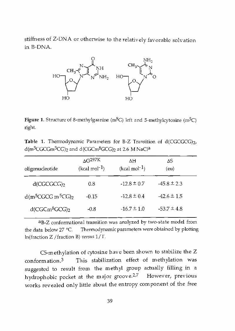
Figure 1. Structure of 8-methylguanine (mSG) left and 5-methykytosine (mSC)
right.
Table 1. Thermodynamic Parameters for B-Z Transition of d(CGCGCGh,
d(mSCGCGm5CG)z and d(CGCm8GCGh at 2.6 M NaCla
~G297K ~H ~S
oligonucleotide (kcal mol-I) (kcal mol-I) (eu)
d(CGCGCGh 0.8 -12.8 + 0.7 -45.8 + 2.3
d(mSCGCG m SCGh -0.15 -12.8 + 0.4 -42.6 + 1.5
d(CGCm8GCGh -0.8 -16.7 + 1.0 -53.7 + 4.8
aB-Z conformational transition was analyzed by two-state model from
the data below 27°C. Thermodynamic parameters were obtained by plotting
In(fraction Z I fraction B) versus 1IT.
CS-methylation of cytosine have been shown to stabilize the Z
conformation.3 This stabilization effect of methylation was
suggested to result from the methyl group actually filling in a
hydrophobic pocket at the major groove.2t7 However, previous
works revealed only little about the entropy component of the free
39
energy of B- to Z-DNA transition and therefore precise molecular
mechanism of the effects of methyl groups on the stability of the Z
conformation is not well understood? Therefore, we studied the
thermodynamics of B- to Z-DNA transition of d(m SCGCGm 5CGh at
NaC12.6 M conditions and compared with unmodified d(CGCGCGh.
While ~H values for these two hexamer were proven to be almost the
same, significant increase in the entropy was observed for
d(m 5CGCGm sCGh. This result clearly demonstrated that the
stabilization of Z-conformation by cytosine C5 methylation is of
entropic origin.
On the other hand, thermodynamic analyses of B- to Z-DNA
transition of d(CGCm BGCGh at N aCI 2.6 M conditions indicated that
the stabilization effects of guanine C8 lnethyl group results from a
large favorable decrease in enthalpy (~H=-3.9 kcal mol-1) and an
unfavorable significant decrease in entropy (~S=-11.1 eu mol-1).
The enthalpic origin of 1.9 kcal/mol by each methyl group at the
guanine C8 position corresponds to the reported free energy (1-2 kcal
mol-I) required to shift the equilibrium to the syn conformation of
C8-substituted deoxyguanosines.B
The contribution of solvent interactions to the stability of
alternative DNA conformations has been previously discussed in
qualitative tenns. A useful semiempirical approach to estimating
the contribution of solvent interactions on molecular structures has
been to calculate the solvent accessible surface (SAS) of the molecule
and of its component parts? The SAS of the hexamer d(CGCGCGh
were calculated in their canonical B- and Z-DN A conformations.
Analogous sets of B-form and Z-form coordinates of
d(nl SCGCGm sCGh and d(CGCm BGCGh were generated by adding
standard geometry methyl group to the four cytosine bases at the C5
position, and to two guanine bases at the C8 position respectively.
40
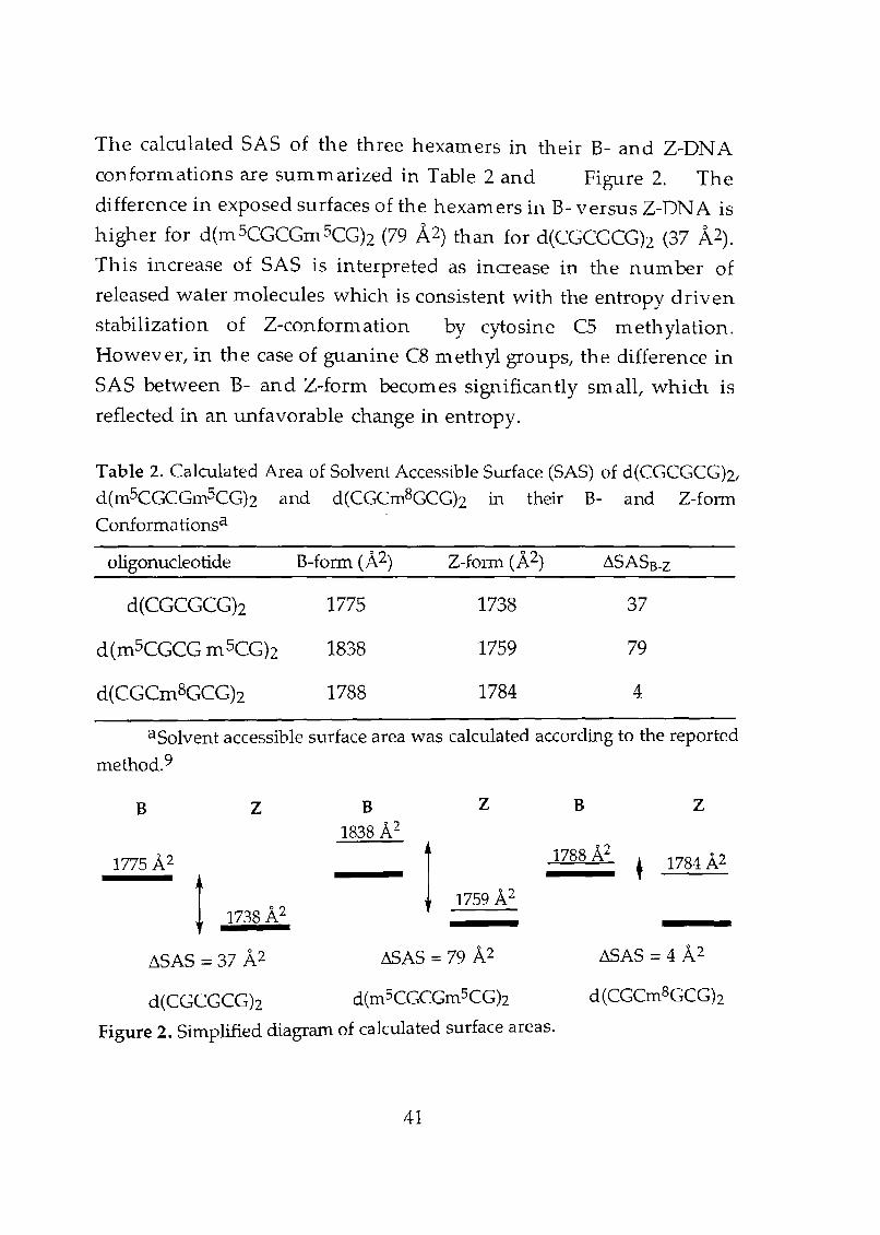
The calculated SAS of the three hexamers in their B- and Z-DNA
conformations are summarized in Table 2 and Figure 2. The
difference in exposed surfaces of the hexamers in B- versus Z-DN A is
higher for d(m sCGCGm SCGh (79 A2) than for d(CGCGCGh (37 A2).
This increase of SAS is interpreted as increase in the number of
released water molecules which is consistent with the entropy driven
stabilization of Z-conformation by cytosine C5 methylation.
However, in the case of guanine C8 methyl groups, the difference in
SAS between B- and Z-form becomes significantly small, which IS
reflected in an unfavorable change in entropy.
Table 2. Calculated Area of Solvent Accessible Surface (SAS) of d(CGCGCG)z,
d(mSCGCGmSCGh and d(CGCm8GCGh in their B- and Z-fonn
Conformationsa
oligonucleotide B-form (A2)
d(CGCGCGh 1775
d(mSCGCG m sCGh 1838
d(CGCm8GCGh 1788
1738
1759
1784
~SASB-Z
37
79
4
aSolvent accessible surface area was calculated according to the reported
method.9
B Z B Z
1838 A2
1775 A2
1t 1759 A21738 A2
~SAS =37 A2 ~AS =79 A2
d(CGCGCGh d(mSCGCGmSCGh
Figure 2. Simplified diagram of calculated surface areas.
41
B z
+ 1784 A2
~AS = 4 A2
d(CGCm8GCGh
Ho et al. have pointed out that potentation of Z-DNA by
cytosine methylation is a solvent effect. They have calculated the
solvent free energies of DNA structures by converting each surface
type to a free energy by applying an atomic solvation parameter that
describes energy required to transfer that surface type from an organic
phase to an aqueous solvent phase? According to their method,
increase in SAS of 42 A2 and decrease in 33 A2 were estilnated to be
1.8 kcal mol-1, and 1.4 kcal mol-1 respectively, which roughly agreed
with our experimental results in this chapter.
Conclusion
The availability of this type of experimentally derived
structural and thermodynamic information on the stabilities of
sequences with various modifications as B- versus Z-DNA makes this
transition an excellent system for studying the contribution of various
thermodynamic forces on macromolecular conformations. In this
chapter, the overall stabilizing effect of the methyl group was
demonstrated to be quite different for cytosine C5 from that for
guanine e8. The stabilization of Z-conformation by a cytosine C5
lllethyl group is prinlary of entropic origin that arises from the
increase in the solvent accessible surface area in the B- to Z-DNA
transition, while methylation at guanine C8 enthalpically stabilize the
Z-conform ation as a consequence of stabilization of syn
conformation.
42
Experimental Section
Materials and methods. Pyridine and acetonitrile (HPLC
grade) were dried over calcium hydride. 2'-Deoxyguanosine was
purchased from YAMASA Corporation. N ucleoside ~
cyanoethylphosphoramidite reagents (A, G, C, T, m 5C) were obtained
from Applied Biosystems. Cyanoethyl phosphoramidite of 8
methyl-2' -deoxyguanosine was prepared by the reported procedure.6
Calf intestine alkaline phosphatase (AP, 1000 unit/m L) and snake
venom phosphodiesterase (s.v. PDE,3 unit/mL) were purchased fromBoehringer Mannheim.
Synthesis of deoxyhexanucleotides. Oligonucleotides were
prepared by the ~-(cyanoethyl)phosphoramidite method on
controlled pore glass supports (1 /lmol) by using ABI 381 A DNA
synthesizer. After automated synthesis, the oligomer was detached
from the support by soaking in cone. aqueous ammonia for 1 h at
room temperature. Deprotection was conducted by heating the cone.
aqueous ammonia for 12 h at 55°C. Aqueous ammonia was then
removed by evaporation, and the crude oligomer was purified by
reverse phase HPLC and lyophilized. Purity and concentration of
all oligonucleotides were determined by com plete digestion with s.v.
PDE and AP to 2'-deoxymononucleosides.
CD Measurements. Circular dichroism (CD) spectra were
recorded on Jasco J-700 spectrophotometer equipped with a Peltier
temperature controller. CD spectra of oligonudotide solutions (0.1
mM duplex in 2.6 M NaCI, 5 mM Na cacodylate, pH 7.0) were
recorded using 1 em path length cell. CD spectra at different
43
temperatures were recorded at intervals of 5 °C with a 1 mIn
equilibration period.
Measurement of melting temperature. Thermal
denaturation profiles were obtained with Jasco V-550
spectrophotometer equipped with a Peltier tern perature controller.
Absorbance of the sam pIes was monitored at 260 nm from 2 to 80°C
with a heating rate of 1°C/min. Experiments with a heating rate of
0.5 DC/min gave the same results, suggesting that thermodynamic
equilibrium had been achieved. The data were normalized to
percent denaturation. A linear least squares analysis of the data
gave a slope of transition and the y-intercept, from which the melting
temperature was calculated.
Analysis of the thermodynamic data. The proportions of Z, B,
and singlestranded (55) forms in a m 8G-containing oligomer were
determined by means of CD and UV spectroscopy as reported.16
Since the molar extinction coefficients of the Band Z forms of the
hexamer were found to be approximately the same, the proportions of
SS relative to that of Band Z at each tern perature were estimated by
UV melting experiments at 260 nm. The relative ratio of the amount
of Band Z was determined by the CD ellipticity at 295 nm.
Calculation of Surface Areas. The method involves first
building models for hexanucleotide sequences in either the B- or the
Z-conformations. The atomic coordinates of sequences as B-DNA
were generated using standard helical parameters for B-DNA. The
44
atomic coordinates of the same sequences as Z-DN A were generated
from the crystal structures of previously crystallized sequences.
Models of mSC and mSC containing sequences were constructed by
adding a sp3 methyl group at the C5 carbon of a cytosine base and at the
C8 carbon of guanine base respectively, using standard distances and
geometries. The solvent accessible surface areas of the DNA
models were calculated by using the Connally rolling ball method
probe radius of 1.4 A.9
45
References1. (a) Cozzarelli, N. R.; Wang, J. C. DNA Topology and Its
Biological Effects 1990, Cold Spring Harbor Laboratory Press, Cold
Spring Harbor, NY. (b) Sinden, R. R. DNA Structure and
Function 1994, Academic Press, NewYork. NY.
2. (a) Rich, A.; Nordheim, A.; Wang, A. H.-J. An 11 u. Rev.
Biochcm. 1984,53,791-846. (b) Rich, A. AnnIs NY Acad. Sci.
1994,726, 1-17.
3. (a) Behe, M.; Felsenfeld, G. Proc. N atl. Acad. Sci. USA 1981, 78,
1619-1623. (b) Tran-Dinh, S.; Taboury, J.; N eum ann, J-M.;
Huynh-Dinh, T.; Genissel, B.; Langlois d'Estaintot, B.; Igolen,. J.
Biochemis try 1984,23, 1362-1371.
4. (a) Lafer, E. M.; Moller, A.; Nordheim, A.; Stollar,. B. D.; Rich, A.
Proc. Natl. Acad. Sci. USA 1981,78, 3546-3550. (b) Moller, A.,
N ordhein1, A.; Kozlowski, S. A.; Patel, D. J., Rich, A.
Biochemistry 1984, 23,54-62.
5. Van der Pleog, L. H. T.; Groffen, J.; Flavell, R. A. Nucleic Acids
Res. 1980, 12, 1243-1263.
6. Sugiyama, H.; Kawai, K.; Matsunaga, A.; Fujimoto, K.; Saito, 1.;
Robinson, H.; Wang, H.-J. Nucleic Acids Res. 1996, 24, 1272
1278.
7. (a) Kagawa, T. F.; Howell, K. T.; Ho, P. S Nucleic Acids Res. 1993,
24,5978-5986. (a) Kagawa, T. F.; Stoddard, D.; Zhou, G.; Ho, P. S
Biochemistry 1989, 28,6642-6651.
8. (a) Son, T.-D.; Guschlbauler, W.; Gueron, M. J. Am. Chem. Soc.
1972, 94, 7903-7911. (b) Sarama, R. H.; Lee, C.-H.; Evans, F. E.;
Yathindra, N.; Sundaralingman, M. J. Am. Chem. Soc. 1974,96,
7337-7348.
9. Connally, M. L. Science 1983,221,709-713.
46
Chapter 3
Stabilization of Hoogsteen Base-Pairing by IntroducingNH2 Group at the C8 Position of Adenine
Abstract
The synthesis and thermodynamic properties of 8
aminodeoxyadenosine (8-amdA)-containing oligonucleotides has
been described. In order to examine the effects of 8-amdA on
Hoogsteen base pair formation, we investigated the stabilities of 8
amdA-containing triplexs. It was found that introduction of 8
amino group leads to the increase in Tm of third strand dissociation.
The results indicate that the incorporation of an amino group at the
C8 position of adenine greatly stabilized the Hoogsteen base pairing in
triplex formation.
Introduction
In addition to the standard Watson-Crick type hydrogen
bonding, nucleoside bases can recognize its com plementary bases by
Hoogsteen-type base pairing,l which is typically known to take place
in a triplex between the purine strand of the Watson-Crick base pair
and a pyrimidine third strand.2 A variety of base modifications was
demonstrated to stabilize the Hoogsteen base pairing in a triplex, but
most modifications were made on the third strand of the triplex for
their potential therapeutic application.3 Since IH-NMR studies on
49
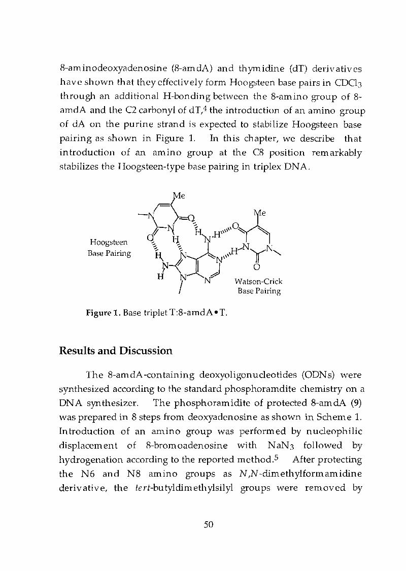
8-aminodeoxyadenosine (8-amdA) and thYmidine (dT) derivatives
have shown that they effectively form Hoogsteen base pairs in CDCl3
through an additional H-bonding between the 8-amino group of 8
amdA and the C2 carbonyl of dT...4 the introduction of an amino group
of ciA on the purine strand is expected to stabilize Hoogsteen base
pairing as shown in Figure 1. In this chapter, we describe that
introduction of an amino group at the C8 position remarkably
stabilizes the Hoogsteen-type base pairing in triplex DNA.
HoogsteenBase Pairing
Figure 1. Base triplet T:8-amdA • T.
Results and Discussion
The 8-amdA-containing deoxyoligonucleotides (ODNs) were
synthesized according to the standard phosphoramdite chemistry on a
DNA synthesizer. The phosphoramidite of protected 8-amciA (9)
was prepared in 8 steps from deoxyadenosine as shown in Scheme 1.
Introduction of an amino group was performed by nucleophilic
displacement of 8-bromoadenosine with NaN3 followed by
hydrogenation according to the reported Inethod.5 After protecting
the N6 and N8 amino groups as N ...N-dimethylformamidine
derivative, the te rt-bu tyldim ethylsilyl groups were removed by
50
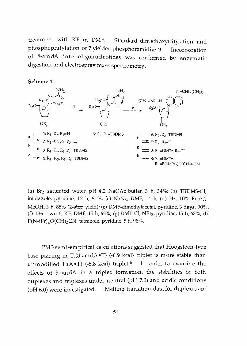
treatment with KF in DMF. Standard dimethoxytritylation and
phosphophitylation of 7 yielded phosphoramidite 9. Incorporation
of 8-amdA into oligonucleotides was confirmed by enzynlatic
digestion and electrospray mass spectrometry.
Scheme 1
NH2
R1-fN/JR~O~ N2~
o~
d ..
NH2
N~N
R;~JlNJ2~
o~
N=CHN(CH3h
(CH3nNc=~tJ'~R20~N
OR3
f C 6: R2, R3=TBDMS
g C 7: R2, R3=H
h C 8: R2=DMTr, R3=H
9: R2=DMTrR3=P(N-iPr2)O(CH2hCN
(a) Br2 saturated water, pH 4.2 NaGAc buffer, 3 h, 34%; (b) TBDMS-CI,imidazole, pyridine, 12 h, 81%; (c) NaN3' DrvtF, 14 h; (d) H2, 10% Pd/C,
MeOH,3 h, 85% (2-step yield); (e) DMF-dimethylacetat pyridine, 3 days, 90%;(f) 18-crown-6, KF, DMF, 15 hi 68%; (g) DMTrCl, NEt3' pyridine, 15 h,63%; (h)P(N-iPrhO(CHhCN, tetrazole, pyridine,5 h, 98%.
PM3 semi-em pirical calculations suggested that Hoogsteen-type
base pairing in T:(8-amdA eT) (-6.9 kcal) triplet is more stable than
unmodified T:(A eT) (-5.8 kcal) triplet.6 In order to examine the
effects of 8-amdA in a triplex formation, the stabilities of both
duplexes and triplexes under neutral (pH 7.0) and acidic conditions
(pH 6.0) were investigated. Melting transition data for duplexes and
51
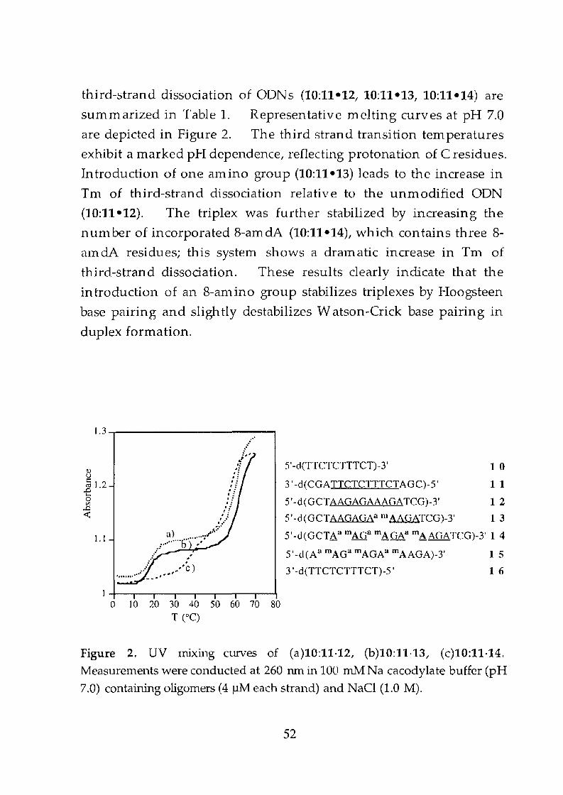
third-strand dissociation of OONs (10:11-12, 10:11-13, 10:11-14) are
summarized in Table 1. Representative melting curves at pH 7.0
are depicted in Figure 2. The third strand transi tion tem peratures
exhibit a marked pH dependence, reflecting protonation of C residues.
Introduction of one amino group (10:11-13) leads to the increase in
Tm of third-strand dissociation relative to the unmodified ODN
(10:11-12). The triplex was further stabilized by increasing the
nun1ber of incorporated 8-am cIA (10:11-14), which contains three 8
amdA residues; this system shows a dralnatic increase in Tm of
third-strand dissociation. These results clearly indicate that the
introduction of an 8-mnino group stabilizes triplexes by Hoogsteen
base pairing and slightly destabilizes Watson-Crick base pairing in
duplex formation.
1.3.....------------,
<tlUI::.e 1.2otil
..0<t:
1.1
../ ..:, ~
.!'o'
'o''o''o'':, :.:, .
:.!//
t ' •••...) ~~...tl ............1) .
I" _~--..' ,.' ,: ../ .·'e)
.............. _ ... -I. ,*'
S'-d(TTCTCTTTCT)-3' 1 0
3'-d(CGATTCTCTTTCTAGC)-S' 11
S'-d(GCTAAGAGAAAGATCG)-3' 1 2
S'-d(GCTAAGAGAa mAAGATCG)-3' 1 3
5'-d(GCTAa mAQa mAGAa mAAGATCG)-3' 1 4
S'-d(Aa mAGa mAGAa mAAGA)-3' 1 5
3 '-d(TTCTCTTTCT)-S' 1 6
14----,--,...---,----,-..,.----,--,...--'1o 10 20 30 40 50 60 70 80
T (OC)
Figure 2. UV mixing curves of (a)10:11·12, (b)10:11·13, (c)10:11·14.
Measurements were conducted at 260 run in 100 mM: Na cacodylate buffer (pH
7.0) containing oligomers (4~ each strand) and NaCl (1.0 M).
52
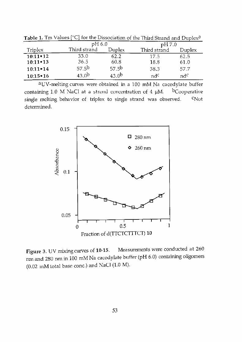
Table 1. Tm Values [DCI for the Dissociation of the Third Strand and Duplexa
pH 6.0 pH 7.0Triplex Third strand Duplex Third strand Duplex10:11-12 33.0 62.2 17.5 62.510:11-13 36.5 60.8 18.8 61.010:11-14 57.5b 57.5b 38.3 57.710:15-16 43.0b 43.0b ndc ndc
aUV-melting curves were obtained in a 100 ffiM Na cacodylate buffer
contairUng 1.0 M NaCl at a strand concentration of 4~. bCooperative
single melting behavior of triplex to single strand was observed. CNot
determined.
0.150 280nm
Q) <> 260nmuI::::CIi
..0H0UJ
..0 0.1-<
0.05
a 0.5 1
Fraction of d(TTCTCTTTCT) 10
Figure 3. UV mixing curves of 10·15. Measurements were conducted at 260
nm and 280 run in 100 mM Na cacodylate buffer (pH 6.0) containing oligomers
(0.02 m1v1 total base conc.) and NaCl (1.0 M).
53
Parallel-stranded duplex with a Hoogsteen base pair was
reported in homopurine-homopyrimidine strands? and 3'-3' and 5'
5' linked system,S therefore, we examined the existence of parallel
stranded duplex formation of 10-15 which forms a stable triplex in the
presence of 16. It was found that 10-15 showed a cooperative single
melting behavior (Tm = 38.2 °C); however, UV mixing curve
experiment indicated that 10-15 forms a 2:1 complex as shown in
Figure 3.9 The results suggest that 15-16 would forms a partially
rn isrnatched triplex. Wang et al. recently showed that 2'-deoxy-3
isoadenine (iA)-containing oligonucleotide d(CG[iA]TCG) forms a B
type anti-parallel duplex with central Hoogsteen-type iA:T base
pairs.10 We next studied the Hoogsteen-type base pairing in an
anti-parallel duplex. Interestingly, octanucleotide d(8-amAT)4
showed a remarkably higher thermal stability (Tm = 25.3 °C)
corn pared to the unm odified oligonucleotide (Tm = 8.3 °C).
Considering that (i) triplex formation is difficult in this purine
pyrimidine alternating sequence and (ii) an 8-amino group itself
destabilizes the Watson-Crick base-pairing, the present d(8-amAT)4
system may form an anti-parallel Hoogsteen-type duplex.
Conclusion
The present results dearly demonstrated that one amino group
at the adenine C8 position greatly stabilizes the Hoogsteen base
pairing in triplex formation by an additional hydrogen bonding.
Incorporation of 8-amdA into DNA will provide a useful tool for
stabilizing nucleic acid local structures which would be of structural
and biological significance.
54
Experimental Section
Materials and methods. Calf intestine alkaline
phosphatase (AP, 1000 units/m L), snake venom phosphodiesterase
(s.v. POE, 3 units/m L) and PI Nuclease (PI, 100 units/m L) were
purchased from Boehringer Mannheim. 2' -Deoxyadenosine was
purchased from Yamasa Shoyu Co. Ltd. Bromine saturated water,
10% palladium on carbon and N,N -dimethylformamide dimethyl
acetal was purchased from N acalai Tesque Co. Sodium azide and
dimethyl amino pyridine were obtained from Wako Pure Chemical
Co. tert-Butyl dimethylchlorosilane and 4,4'-dimethoxytrityl
chloride was purchased from Tokyo Kasei Co. 2-Cyanoethyl
N,N,N ',N '-tetraisopropylphosphorodiamidite was purchased from
Aldrich. The reagents for DNA synthesizer such as 12 solution
(bIHzO/pyridine/tetrahydrofuran, 0.5:9.5:20:70) were purchased from
Applied Biosystems. A-, G-, C-, and T-~
(cyanoethyl)phosphoramidites were purchased from Glen Research.
1H-tetrazole were purchased from Dojin Laboratories. Pyridine was
dried over BaO and distilled before use. DMF was dried over CaH2
and dried before use. Silica gel column chromatography was
carried out on Wakogel C-200. Thin layer chromatography was
carried out on a Merck silica gel60FZ54 plate. IH-NMR spectra were
recorded on Varian GEMINI-200 (200 MHz) or Varian Mercury-400
(400 MHz). HPLC analysis was carried out using PU-980 HPLC
system GASCO, Tokyo) equipped with a CHEMCOBOND 5-0DS-H (4.6
x 150 mm) or a Cosmosil 5CI8-MS column (4.6 x 150 mm).
Detection was carried out at 254 nm.
Synthesis of 8-bromodeoxyadenosine. 2'-Deoxyadenosine (10
g, 0.04 mol) was suspended in aN aOAc buffer (0.5 M, pH 4.2, 150 illL)
with stirring. Then bromine water (200 m L) was added and fully
dissolved solution was obtained. The resulting solution was stirred
55
for 3 hr at room temperature; after 2 hr precipitate began to form.
The color of the solution was discharged by addition of NaHS03 and
the pH of the solution was adjusted 7 with 5N NaOH. The mixture
was filtered and washed with cold water (100 m L), then with
methanol (100 ill L) and dried in vacuo to provide 4.42g (34% yield) of
8-bromodeoxyadenosine as a yellow powder. IH-NMR (400 MHz)
(020) 82.37 (ddd, 1 H, J=14.4/ 8.6, 6.0,2"), 3.00 (ddd, 1 HI J=14.4, 10.1,4.61
2'), 3.78 (dd l 1 H, J=9.2, 3.6, 51), 3.86 (dd, 1 H, J=9.2 1 2.4, 5"), 4.16 (mIl HI
4'), 6.48 (ddl 1 H, J=10.1, 6.01 1'), 8.10 (Sl 1 H, A2). FAB MASS
(positive ion): mle 330 (M+H). Exact mass calcd for
ClOH1203Ns79Br 330.02021 found 330.0186.
Syn thesis of 2' ,S'-bis-o-tert-butyldim ethylsiIiyl-8-
bromodeoxyadenosine. 8-Bromodeoxyadenosine (5.9 g, 17.9 mmol)
was dried by coevaporation with pyridine (20 mL, two times) and
suspended in 60 In L of dry pyridine. To this solution, tert-Butyl
dilnethylchlorosilane (13.5 g, 89.5 mmol) and imidazole (6.09 g, 89.5
In In 01) were added and solution was stirred at room tern perature for
12 hr. After evaporation of the solvent, the residue was extracted
with ethyl acetate followed by silica gel chromatography (hexane:
diethyl ether =1:1) to yield 2' ,5'-bis-O-tert-butyldimethylsiliyl-8
bromodeoxyadenosine as a white powder (6.94 g, 81 0/0). IH-NMR
(200 MHz) (CDC!J) 8 -0.07 (5,3 HI Si-CH3), 0.03 (s/3 H, Si-CH3), 0.12 (5,3
H, Si-CH3)1 0.80 (s/ 9 H, t-Bu),0.91 (5, 9 H, t-Bu), 2.21 (ddd,1 H, J=l1.2,
7.0,4.2/ 2"),3.57-3.71 (m, 2 HI 4', 2'), 3.89 (dd,l H, J=16.2, 3.61 5'), 3.92
(dd, 1 H, J=16.2, 3.4, 5"), 4.85 (ddd, 1 HI J=7.4, 5.8, 3.6, 3'), 5.62 (5, 2 H,
A6NH2), 6.32 (dd l 1 H, J=7.0, 6.6, 1'), 8.23 (s, 1 HI A2). FAB MASS
(positive ion): mle 558 (M+H). Exact mass calcd for
C22H41OJNs79BrSi 558.2009, found 558.1910.
56
Synthesis of 21,5' -bis-().fert-butyldimethylsiliyl-8-
aminodeoxyadenosine. 2' ,5'-Bis-O-tert-butyldimethylsiliyl-8-
bromodeoxyadenosine (1.85 g, 3.32 mmol) was dried by coevaporation
with pyridine (10 mL, two times) and dissolved in 15 mL of dry DMF.
To this solution, NaN3 (735 mg. 11.31 mmol) was added and the
reaction mixture was heated for 17 hr at 75°C. After evaporation of
the solvent, the residue was extracted with diethyl ether. The
residue was dissolved in methanol (100 mL) containing water (10 mL)
and was added 1.0 g of 10% palladium on carbon. This solution was
exposed to hydrogen for 3 hr at 50 psi with stirring at room
temperature. The catalyst was removed by filtration and the filtrate
was evaporated to dryness. The crude solid was purified by silica gel
chromatography, eluting with a 2-4% gradient of MeOH in diethyl
ether to yield 1.17g (yield 85 %) of 2',5' -bis-O-tert-butyldimethylsiliyl
8-aminodeoxyadenosine as a white powder. IH-NMR (200 MHz)
(CDCh) 80.06 (5,6 H, Si-CH3), 0.08 (s, 3 H, Si-CH3), 0.09 (s, 3 H, Si-CH3),
0.88 (5, 9 H, t-Bu), 0.90 (s, 9 H, t-Bu), 2.18 (ddd, 1 H, J::::13.2, 6.4,2.8, 2"),
2.59 (ddd, 1 H, J=13.2, 8.5, 7.4, 2'), 3.77-4.00 (rn, 3 H, 4', 5', 5"), 4.55 (m, 1
H, 3'), 5.13 (5,2 H, A8NH2), 5.45 (s, 2 H, A6NH2), 6.32 (dd, 1 H, J::::8.2, 6.4,
1'), 8.16 (s, 1 H, A2). FAB MASS (positive ion): m Ie 495 (M+H).
Exact mass calcd for C22t4303N6Si2 495.2934, found 495.2944.
Syn thesis of 2,8-[bis-N-(dimethylamino)methylene} 2' ,51-his
o-terf-butyldimethylsiliyl-8-aminodeoxyadenosine. 2',5' -Bis-O-tert
butyldimethylsiliyl-8-aminodeoxyadenosine (1.17 g. 2.37 mmol) was
dried by coevaporation with pyridine (10 ill L, two times) and
dissolved in 10 m L of dry pyridine. To this solution, N ,N
dimethylforman1ide dimethyl acetal (8 mL, excess) was added. After
stirring the reaction mixture at room tern perature for 3 days, the
solvent was evaporated and the residue was purified by silica gel
57
chromatography (dichloromethane: methanol :::::97:3) to afford 1.29 g
(yield 90 (%) of 2,8-fbis-N-(dimethylamino)methylene]-2',5'-bis-O-tert
bu tyldim ethylsiliyl-8-am inodeaxyadenosine. 1H-NMR (200 MHz)
(CDC!J) 0 -0.02 (5,3 H, Si-CH3), 0.01 (5,3 H, Si-CH3), 0.10 (5,6 H, Si-CH3),
0.85 (5,9 H, t-Bu), 0.90 (5,9 H, t-Bu), 2.10 (ddd, 1 H, J::::13.0, 7.0,3.2, 2"),
3.09 (5,3 H, N-CH), 3.10 (5,3 H, N-CH3), 3.12 (5,3 H, N-CH3), 3.20 (5,3 H,
N-CH3), 3.52-3.75 (m, 2 H, 4', 2'), 3.88-4.00 (m, 3 H, 5', 5"), 4.78 (m, 1 H,
3'),6.69 (t, 1 H, }::::7.4, 1'),8.31 (5, 1 H, A2), 8.72 (5, 1 H, N ::::CH), 8.75 (5, 1
H, N::::CH). FAB MASS (positive ion): mle 605 (M+H). Exact
mass calcd for CzsHS303NSSiz 605.3779, found 605.3797.
Synthesis of 2,B-[bis-N-(dim ethylam ino)m ethylenel-B-
aminodeoxyadenosine. 2,8-[Bis-N-(dimethylamino)methylene)
2' ,5' -bis-O-tert-butyldimethylsiliyl-8-aminodeoxyadenosine (1.29 g.
2.14 mlnol) and 18-crown-6 (2.26 g, 8.6 mmol) and KF (497 mg, 8.6
mm 01) were added to dry DMF (10 m L) and the reaction mixture was
stirred for 16 hI. Removal of the solvent under reduced pressure
followed by silica gel chromatography (dichloromethane: methanol
=92:7) to pravied 2,8-[bis-N-(dimethylamino)methylene]-8
aminodeoxy-adenosine as a white powder (0.55 g, 68 % yield). IH
NMR (200 MHz) (DzO) 82.14 (ddd, 1 H, J=12.0, 6.4, 1.9,2'1 2.85 (m, 1 H,
2'), 2.96 (5, 3 H, N -CH3), 2.97 (5,3 H, N -CH3), 3.05 (5, 6 H, N -CH3), 3.65
(dd, 1 H, J=9.7, 3.4, 5"), 3.73 (dd, 1 H, J=9.7, 3.0,5'), 4.00 (m, 1 H, 4'), 4.52
(m, 1 H, 3'), 6.50 (dd, 1 H, J=9.0, 6.4, 1'),8.03 (5, 1 H, A2), 8.28 (s, 1 H,
N=CH), 8.38 (s, 1 H, N=CH). FAB MASS (positive ion): mle 377
(M+H). Exact mass calcd for C16HS03Ns 377.2049, found 377.2034.
58
Synthesis of 8-aminodeoxyadenosine. 2,8-[Bis-N-
(climethylamino)methylene]-8-aminodeoxyadenosine (20 nlg, 0.05
mmol) was deprotected with 28 % Nfi40H for 8 hr at 55°C and
purified with HPLC to yield 8-aminodeoxyadenosine. (14 mg, quant.).
IH-NMR (200 MHz) (D20) 8 2.21 (ddd, 1 H, J=15.0, 6.4, 2.6, 2"), 2.67
(ddd, 1 H, J=15.0, 8.4,6.6,2'),3.73 (m, 2 H, 5',5"),4.00 (m, 1 H, 4'), 4.49
(m, 1 H,3'), 6.24 (dd, 1 H, J=8.7, 6.3, 1'),7.96 (5, 1 H, A2). FAB MASS
(positive ion): m/e 267(M+H). Exact mass calcd for CIOHlS03N 6
267.1205, found 267.1201.
Syn thesis of S'-()..(4,4'-dim ethoxytrityl)-2,8-[bis-N-
(dimethylamino)m ethylenel-8-aminodeoxyadenosine. 2,8-[Bis-N
(dimethylamino)methylene]-8-aminodeoxyadenosine (530 mg, 1.41
mmol) and dimethyl amino pyridine (5 mg. cat.) were dried by
coevaporation with pyridine (10 mL, three times). To this solution
was added 4,4'-dimethoxytrityl chloride (665 mg, 1.96 mmol),
followed by pyridine (8 rnL) and triethylamine (283 JlL, 1.96 rnrnol).
The reaction mixture was stirred at room tern perature for 15 hI.
After evaporation of the solvent, the residue was extracted with ethyl
acetate followed by silica gel chromatography (dichlorornethane:
methanol =95:5) gave 0.6 g (63 % yield) of 5'-0-(4,4' -dimethoxytrityl)
2,8-[bis-N-(dimethylamino)methylene]-8-aminodeoxyadenosine as a
white powder. IH-NMR (200 MHz) (CD03) 82.25 (ddd, 1 H, J=13.2,
7.6,4.6, 2"), 3.04 (s, 3 H, N -CH3), 3.08 (5, 3 H, N -CH3), 3.12 (s, 3 H, N
CH3),3.34 (5,3 H, N-CH3), 3.38-3.51 (m, 3 H, J=9.7, 3.4,2',5',5"),4.01
(ddd, 1 H, J==6.8, 6.0, 5.2,4'), 4.95 (ddd, 1 H, J=6.6, 4.4, 4.0, 3'), 6.64-6.78 (rn,
5 H, methoxyphenyl-o, 1'),7.11-7.42 (m, 9 H, phenyl, methoxyphenyl
m), 8.14 (5, 1 H, A2), 8.70 (s, 1 H, N =CH), 8.73 (s, 1 H, N =CH). FAB
MASS (positive ion): mle 679 (M+H). Exact mass calcd for
C37l£t30 SN 8679.3356, found 679.3381.
59
Synthesis of 5' -0-(4,4' -dimethoxytrity1)-2,8-[bis-N-
(dimethylamino)methylene]-8-aminodeoxyadenosine-3'-()..[(2-
cyanoethyD-N,N-diisopropylphosphoramidite). 5' -0-(4,4'-
dimethoxytrityl)-2,8-[bis-N -(dimethylamino)methylene]-8
aminodeoxyadenosine (400 mg, 0.59 mmol) was coevaporated with
pyridine (3 m L, three times) and dissolved in dry pyridine (3 m L).
To this solution was added 2-cyanoethyl N,N,N',N'
tetraisopropylphosphorodiamidite (212 ~L, 0.73 mmol), and 0.5 M
IH-tetrazole (1.45 m L, solution in dry CH3CN) under an argon
atmosphere. After 5 hr, the reaction mixture was diluted with ethyl
acetate (50 m L) and organic phase was washed with saturated sodiurn
hydrogen carbonate solution (50 mL, two times) and then with water
(50 ill L, two times). The organic layer was dried over with sodium
sulfate and evaporated to dryness to yield 5' -0-(4,4' -dimethoxytrityl)
2,8-[bis-N-(dimethylamino)methylene]-8-aminodeoxyadenosine-3'
0-[(2-cyanoethyl)-N,N-diisopropylphosphoramidite] (480 m g, 98 %).
IH NMR (400 MHz, CDCb) 81.10-1.29 (m, 12 H, CH(CH3h), 2.23-2.38
(m, 1 H, 2"), 2.44-2.63 (m, 2 H, OCH2CH2CN), 3.05 (5, 3 H, N (CH3h),
3.09 (s, 3 H, N (CH3h), 3.13 (s, 3 H, N (CH3h), 3.20 (s, 3 H, N (CH3)z),
3.35-3.76 (n1, 7 H, CH(CH3h), OCH2CH2CN, 2', 5', 5"), 3.74 (s, 6 H,
OCH3), 4.15-4.26 (m, 1 H, 4'), 4.84-4.97 (m, 1 H, 3'), 6.67-6.77 (m, 5 H,
methoxyphenyl-o, 1'), 7.12-7.43 (m, 9 H, phenyl, methoxyphenyl-m),
8.11 (s, 1 H, H2), 8.70 (5, 1 H, NCHN(CH3h), 8.75 70 (s, 1 H,
NCHN(CH3h). 31p NMR (121 MHz, CDCb) 8 149.41, 148.98
(diastereomers). FAB MASS (positive ion): m/e 879 (M+H).
Exact mass ca1cd for Y6H6006N lOP 879.4434, found 879.4458.
Synthesis of oligonucleotides. Oligonucleotides were
prepared by the p-(cyanoethyl)phosphoramidite method on
controlled pore glass supports (1 /lmol) by using ABI 381 A DNA
60
synthesizer. After automated synthesis, the oligomer was detached
from the support, deprotected and purified by HPLC. After
lyophilization, approximately 20 OD of pure oligomer was isolated.
Purity and concentrations of all oligonucleotides were determined by
com plete digestion with S.v. PDE, PI nuclease and AP to 2'
deoxym ononucleosides. The 8-am inodeoxyadenosine containing
oligonucleotides were also identified by electrospray mass
spectrometry: calcd for 13: 4978.3, found 4977.9; calcd for 14: 5008.4,
found 5007.9; calcd for 17: 3163.2, found 3162.4.
Measurement of melting temperature. Thermal
denaturation profiles were obtained with Jasco V -550
spectrophotometer equipped with a Peltier tern perature controller.
The absorbance of the samples was monitored at 260 nm from 2°C to
80°C with a heating rate of 1 °C per minute. Experiments with a
heating rate of 0.5 °C per minute gave the same results as those at 1 °C
per minutes, suggesting that thermodynamic equilibrium was
achieved. The data were normalized to percent denaturation. A
linear least-squares analysis of the data gave a slope of transition and y
in tercept from which the melting tern perature were calculated.
Representative melting curves are depicted in Figure 2.
UV mixing experim en ts. The oligonucleotide (15 and 17)
solutions were prepared in 90:10, 80:20, 70:30, 60:40, 50:50,40:60, 30:70,
20:80, and 10:90 molar ratios in 1 M NaCl containing 100 mM Na
cacodylate buffer (pH 6.0). Two sam pIes containing only one of the
two strands were prepared (100:0 or 0:100) in the same buffer. The
final concentration of the oligonucleotide was 0.02 mM (base conc.).
61
The UV measurements were carried out as described above after
equilibrating each sample at 15 °C for 10-15 min. The UV mixing
curve was shown in Figure 3.
62
References
1. Hoogsteen, K. Acta Crystallogr. 1963, 16,907-916.
2. Thoung, N. T.; Helene, c. A ngew. Chem. Int. Ed. Eng!. 1993,32,
666-690. (b) Moser, H.; Dervan, P. B. Science. 1987,238,645-650.
3. Hildbrand, S.; Leumann, C. Angew. Chem. Int. Ed. Engl. 1996,
35, 1968-1970. (b) Xiang, G.; Bogacki, R.; Mclaughlin, L. W.Nucleic Acids Res. 1996,24, 1963-1970.
4. Kumar, R. K.; Gunjal, A. D.; Ganesh, K. N. Biochem. Biophys.Res. Comm. 1994,204, 788-793.
5. Long, R. A.; Robins~ R. K.; Townsend, 1. B. J. Org. Cll em. 1967,
32, 2751-2756.
6. Leach, A. R.; Kollman, P. A. J. Am. Chem. Soc. 1992, 114, 3675
3683.
7. (a) Lavelle, 1.; Fresco, J. R. Nucleic Acids Res. 1995, 23, 2692
2705. (b) Liu, K.; Miles, H. T.; Fraizer, J.; Sasisekharan, V.
Biochemistry 1993,32, 11802-11809.
8. Kandimalla, E. R.; Agrawal, S. Biochem istry 1996, 35, 15332-
15339.9. (a) Job, P. Ann. Ch im . (Paris) 1928, 9, 113-134. (b) Felsenfeld, G.;
Rich, A. Bio ch em. Bioph ys. Acta 1957,26, 457-468. (c) Plum. G.
E.; Park, Y.-W.; Singleton, S. F.; Dervan, P. B.; Breslauer, K. J.Proc. Natl. Acad. Sci. USA 1990,87,9436-9440.
10. Bhat, B.; Neelin1a.; Leonard, N. J.; Robinson, H.; Wang, A. H·-I·;
J. Am. Chem. Soc. 1996,118,3065-3066.
63
Chapter 4
Confonnation Dependent Photochemistry of 5Halouracil-Containing DNA: Stereospecific 2'aHydroxylation of Deoxyribose in Z-form DNA
Abstract
The photoreaction of Z-form IU-containing octanucleotide
d(CGCGIUGCG) was investigated. It was revealed that the
stereospecific e21 o:-hydroxylation occurs in the Z-form DNA together
with the formation of Clf and C2f oxidation products and a halogen
exchanged product. Under 02-limiting conditions, the yield of both
Clf and C2f oxidation products in the Z-form oligomers decreased
considerably. When the photoreaction was carried out under 1802
atomosphere, the electrospray MS demonstrated the incorporation of180 atom into the C2' hydroxylated product. These results indicate
that atmospheric 02 is the source for the C2f hydroxyl group. Upon
addition of NaI as a reductant of hydroperoxide, a dramatic increase in
the hydroxylated product was observed. These results suggest that
both the C2' oxidation product and hydroxylated product are produced
via a common hydroperoxide intermediate. Upon treatment with
ribonuclease TI, the e2' hydroxylated product was quantitatively
hydrolyzed to d(CGC)rGp and d(UGCG). These results suggest that
this photochemical and enzymatic procedure can be used as a specific
probe for the existence of the local Z-form structure in longer DNA.
65
Introduction
It is well-recognized that DNA has a remarkable structural
heterogeneity) Such local DNA conformations are thought to play
an im portan t biological role in processes such as gene expression by
altering DNA-protein interactions.2 Although the left-handed Z
forIll ONA is one of the characteristic and significan t local structures
of ONA,3 the precise biological functions of Z-DN A have not been
fully understood presumably due to the lack of an appropriate
detection method in a living cell system. Recently, Rich and
colleagues have reported the creation of a Z-form -specific
endonuclease as a probe for Z-DNA.4 The Z-form region is
assumed to appear in a very short period of time in a living cell, and
therefore, utilization of a photochemical reaction which directly
reflects the DNA local conformation would be infinitely useful.
Previously, we have demonstrated that photoirradiation of 5
halouracil-containing B-form and DNA-RNA hybrid
oligonucleotides results in a conformation dependent H abstraction, i.
C., the competitive Cl' and C2' hydrogen abstractions were observed in
the B-fonn DNA, whereas predominant Cl' hydrogen abstraction
occurred in the DNA-RNA hybrid.S Inspection of the molecular
model of Z foril1 DNA suggested that the 5-halouracil in the Z-form
ONA would have a unique photoreactivity compared with B-form
ONA due to differences in the manner that purine bases stacked on
the 3' pyrilnidine base, e. g., the ribose C2P hydrogen of the 5' side
purine is very close to C5 of uracil, whereas Cl' hydrogen is very far
£rOIn C5 of uracil. In addition, the increase in the VV absorption at
308 nIl1 by Z-DNA was expected to enhance the reactivity. In this
study, we examined the photoreaction of the IV-containing Z-form
octanucleotide and found that a novel stereospecific 2' (X
hydroxylation occurs efficiently in the Z-form DNA.
66
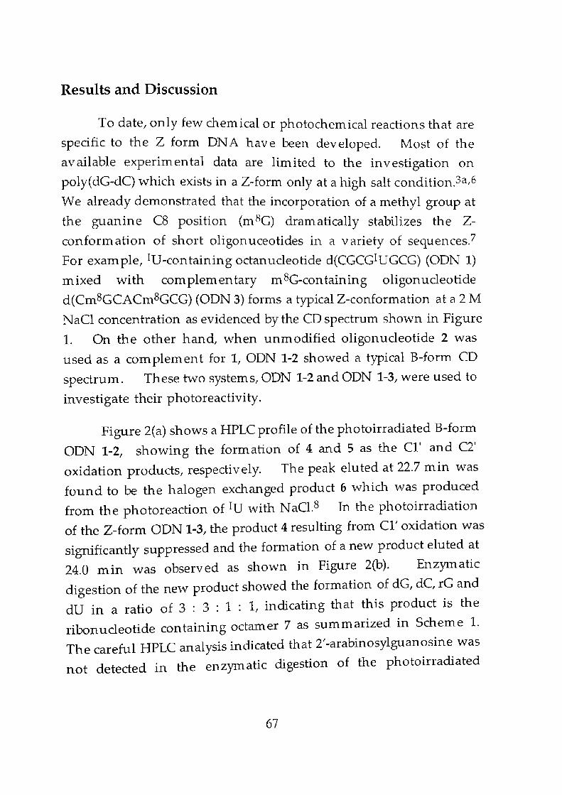
Results and Discussion
To date, only few chemical or photochemical reactions that are
specific to the Z form ONA have been developed. Most of the
available experimental data are limited to the investigation on
poly(dG-dC) which exists in a Z-form only at a high salt condition.3a,6
We already demonstrated that the incorporation of a methyl group at
the guanine C8 position (m 8G) dram atical1y stabilizes the Z
conformation of short oligonuceotides in a variety of sequences?
For example, IV-containing octanucleotide d(CGCGIUGCG) (OON 1)
mixed with complementary m 8G-containing oligonucleotide
d(Cm8GCACm8GCG) (ODN 3) forms a typical Z-conformation at a 2 M
NaCl concentration as evidenced by the CO spectrum shown in Figure
1. On the other hand, when unmodified oligonucleotide 2 was
used as a com plem ent for 1, OON 1-2 showed a typical B-form CD
spectrum. These two systems, OON 1-2 and OON 1-3, were used to
investigate their photoreactivity.
Figure 2(a) shows a HPLC profile of the photoirradiated B-form
ODN 1-2, showing the formation of 4 and 5 as the C11
and C2'
oxidation products, respectively. The peak eluted at 22.7 min was
found to be the halogen exchanged product 6 which was produced
from the photoreaction of IU with NaC1.8 In the photoirradiation
of the Z-form ODN 1-3, the product 4 resulting from C1' oxidation was
significantly suppressed and the formation of a new product eluted at
24.0 min was observed as shown in Figure 2(b). Enzymatic
digestion of the new product showed the formation of dG, dC, rG and
dV in a ratio of 3 : 3 : 1 : 1, indicating that this product is the
ribonucleotide containing octamer 7 as summarized in Scheme 1.
The careful HPLC analysis indicated that 2'-arabinosylguanosine was
not detected in the enzymatic digestion of the photoirradiated
67
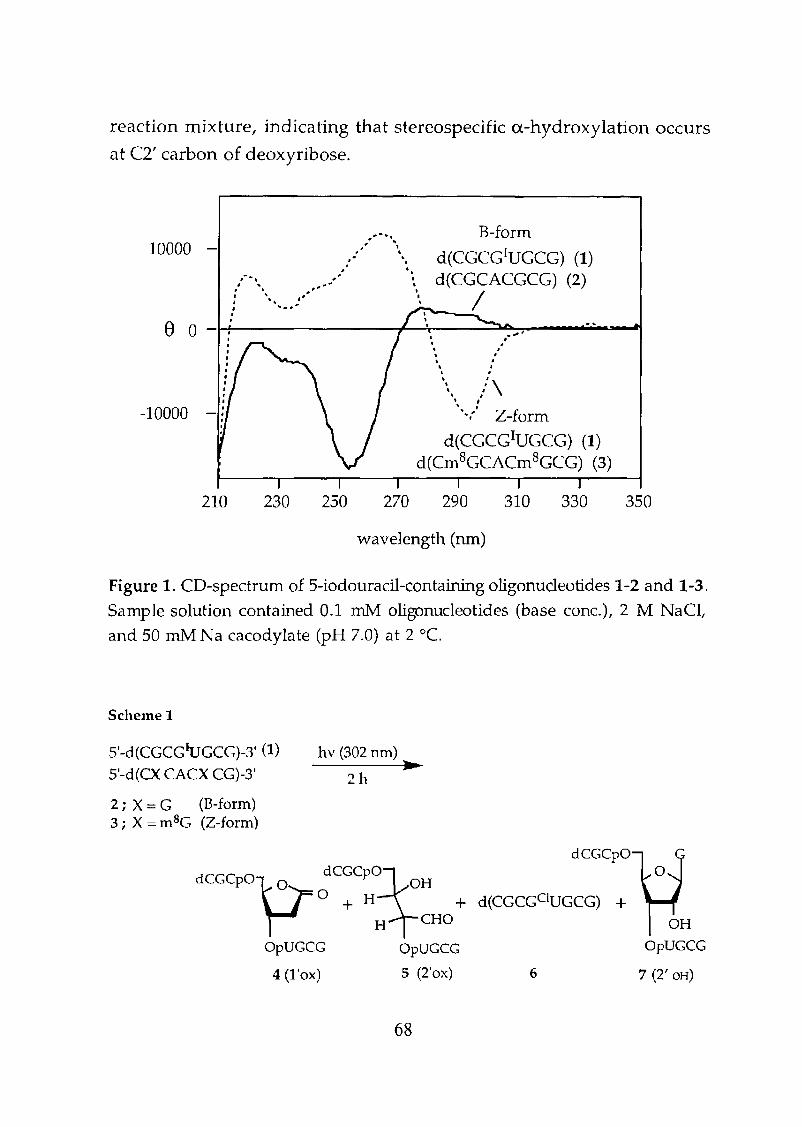
-10000
10000 -
reaction mixture, indicating that stereospecific a-hydroxylation occurs
at C2' carbon of deoxyribose.
.'.', B-form. '. '.... ... d(CGCG1UGCG) (1). .
"".. ...•. ~ d(CGCACGCG) (2)" ... ' '/I ,~ •: ."' ... _: '. " I'_----=---.e a -H-------+----'r-----=::l.A...,":"":"',-_........-.-~
•,..••.
:'\., .",:, Z-form
d(CGCGIUGCG) (1)d(Crn8GCACm8GCG) (3)
210 230 250 270 290 310 330 350
wavelength (nm)
Figure 1. CD~spectrum of 5-iodouracil~containingoligonucleotides 1-2 and 1-3.
Sample solution contained 0.1 :m1\1 oligonucleotides (base conc.), 2 M NaCt
and 50 roM Na cacodylate (pH 7.0) at 2 0c.
Scheme 1
5'-d(CGCGIUGCG)-3' (1)
5 '-d(CX CACX CG)-3'
2 i X:::: G (B-form)3; X:::: mBG (Z-form)
hv (302 !Un) ...2h
dCGCPOvr dCGCPOXH° + H +H CHO
OpUGCG OpUGCG
4 (1 'ox) 5 (2'ox)
68
dCGCpO
d(CGCGCIUGCG) +
6
OHOpUGCG
7 (2' OH)
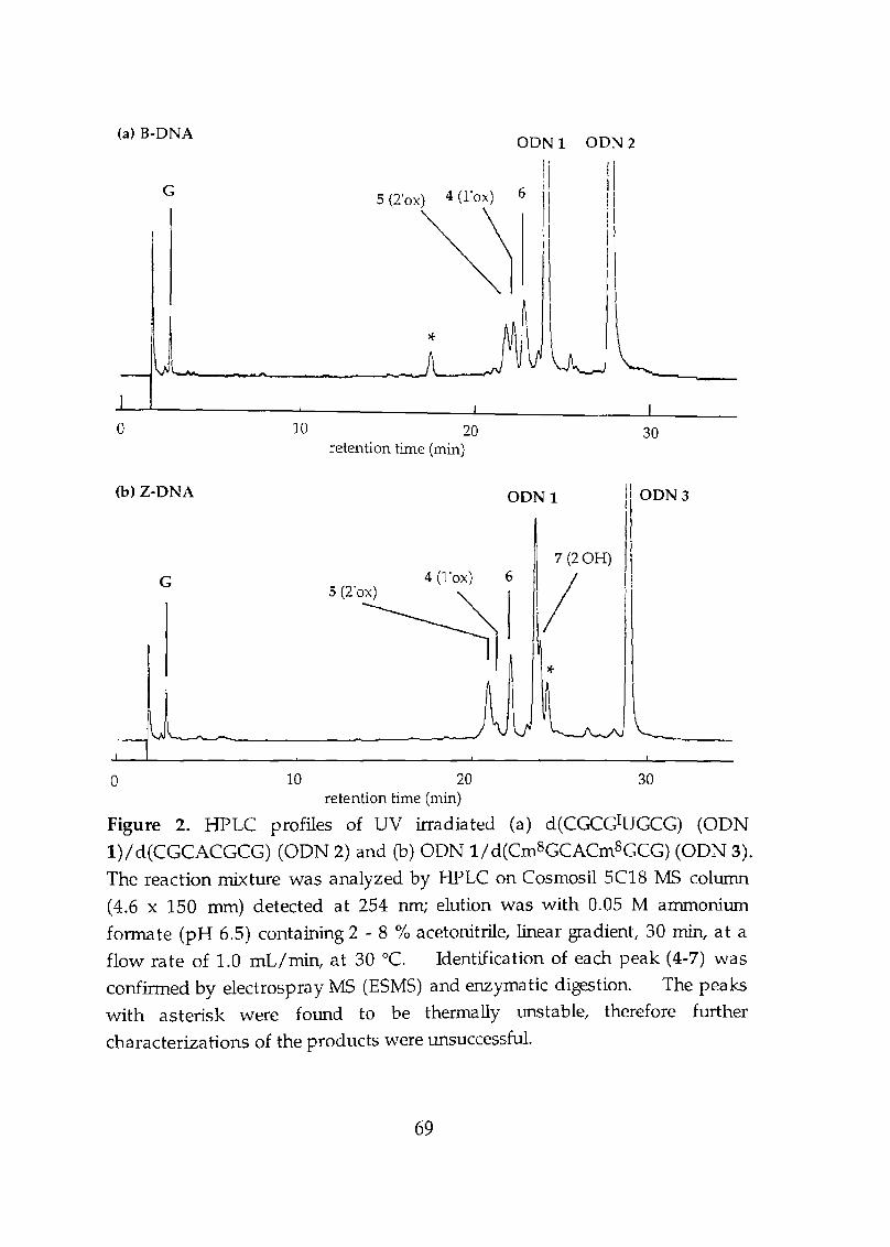
G
10 20retention time (min)
DDNI DDN2
iIiiI
II
30
ODNI(b) Z-DNA
G5(2'0~6
7 (2'OH)
/*
IlODN3
I\I
o 10 20 30retention time (min)
Figure 2. HPLC profiles of UV irradiated (a) d(CGCGIUGCG) (ODN
l)/d(CGCACGCG) (ODN 2) and (b) ODN 1/d(Cm8GCACm8GCG) (ODN 3).
The reaction mixture was analyzed by HPLC on Cosmosil 5C18 MS column
(4.6 x 150 nun) detected at 254 nm; elution was with 0.05 M ammonium
fonnate (pH 6.5) containing 2 - 8 % acetonitrile, linear gradient, 30 min, at a
flow rate of 1.0 mL/min, at 30°C. Identification of each peak (4-7) was
confinned by electrospray MS (ESMS) and enzymatic digestion. The peaks
with asterisk were found to be thermally unstable, therefore further
characterizations of the products were W1SuccessfuL
69
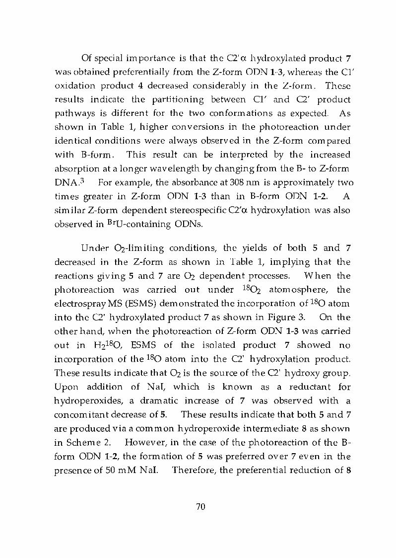
Of special im portance is that the C2' 0: h ydroxylated product 7
was obtained preferentially from the Z-form ODN 1-3, whereas the CI'
oxidation product 4 decreased considerably in the Z-form. These
results indicate the partitioning between CI' and C2' product
pathways is different for the two conformations as expected. As
shown in Table I, higher con versions in the photoreaction under
iden tical conditions were always observed in the Z-form com pared
with B-form. This result can be interpreted by the increased
absorption at a longer wavelength by changing from the B- to Z-form
ONA.3 For example, the absorbance at 308 run is approximately two
times greater in Z-form ODN 1-3 than in B-form ODN 1-2. A
similar Z-form dependent stereospecific C2'o: hydroxylation was also
observed in BrU-containing ODNs.
Under Oz-limiting conditions, the yields of both 5 and 7
decreased in the Z-form as shown in Table 1, implying that the
reactions giving 5 and 7 are Oz dependent processes. When the
photoreaction was carried out under 180z atomosphere, the
electrospray MS (ESMS) demonstrated the incorporation of 180 atom
in to the C2' h ydroxylated product 7 as shown in Figure 3. On the
other hand, when the photoreaction of Z-form OON 1-3 was carried
out in Hz180, ESMS of the isolated product 7 showed no
incorporation of the 180 atom into the C2' hydroxylation product.
These results indicate that Oz is the source of the C2' hydroxy group.
Upon addition of NaI, which is known as a reductant for
h ydroperoxides, a dramatic increase of 7 was observed with a
concom itan t decrease of 5. These results indicate that both 5 and 7
are produced via a common hydroperoxide intermediate 8 as shown
in Scheme 2. However, in the case of the photoreaction of the B
form OON 1-2, the formation of 5 was preferred over 7 even in the
presence of 50 mM N aI. Therefore, the preferential reduction of 8
70
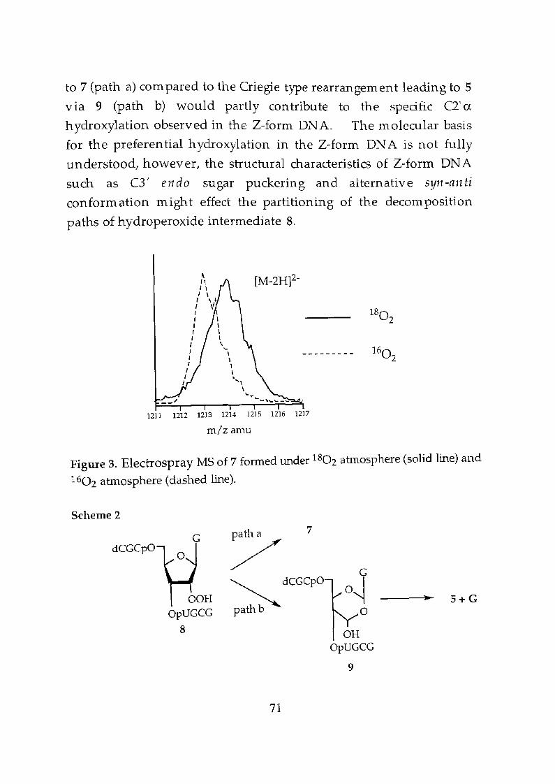
to 7 (path a) com pared to the Criegie type rearrangement leading to 5
via 9 (path b) would partly contribute to the specific C2' ahydroxylation observed in the Z-form DNA. The molecular basis
for the preferential hydroxylation in the Z-form DNA is not fully
understood, however, the structural characteristics of Z-form DNA
such as C3' en do sugar puckering and alternative syl1-an ti
conformation might effect the partitioning of the decomposition
paths of hydroperoxide intermediate 8.
[M-2H]2-
--------- 16()2
1211 1212 1213 1214 1215 1216 1217
m/z amu
Figure 3. Electrospray MS of 7 formed under 1802 atmosphere (solid line) and
1602 atmosphere (dashed line).
7Scheme 2
dCGCpOG
OOHOpUGCG
8
/~dCGCPO
path b
71
OHOpUGCG
9
S+G
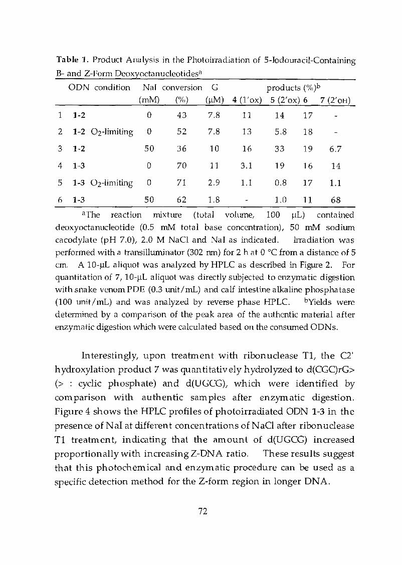
Table 1. Product Analysis in the Photoirradiation of 5-lodouracil-Containing
B- and Z-Form Oeoxyoctanucleotidesa
6.7
14
1.1
18
19
16
17
5.8
33
19
0.8
1.0 11 68
11 14 17
13
16
3.1
1.1
OON condition Nal conversion G products (%)b
4 (l'ox) 5 (2'ox) 6 7 (2'OH)(rnM) (%) (JlM)
1 1-2 a 43 7.8
2 1-2 02-1 inuting 0 52 7.8
3 1-2 50 36 10
4 1-3 a 70 11
5 1-3 02-limiting a 71 2.9
6 1-3 50 62 1.8
ilThe reaction mixture (total volwne, 100 JlL) contamed
deoxyoctanucleotide (0.5 roM total base concentration), 50 rnM sodium
cacodylate (pH 7.0), 2.0 M NaCI and Nal as indicated. Irradiation was
performed with a transilluminator (302 run) for 2 h at a °C from a distance of 5
em. A 10-IlL aliquot was analyzed by HPLC as described in Figure 2. For
quantitation of 7, 10-J.1L aliquot was directly subjected to enzymatic digestion
with snake venom POE (0.3 unit/mL) and calf intestine alkaline phosphatase
(100 unit/mL) and was analyzed by reverse phase HPLC. bYields were
determined by a comparison of the peak area of the authentic material after
enzymatic digestion which were calculated based on the conswned GONs.
Interestingly, upon treatment with ribonuclease TI, the C2'
hydroxylation product 7 was quantitatively hydrolyzed to d(CGC)rG>
(> : cyclic phosphate) and d(UGCG), which were identified by
com parison with authentic sam pIes after enzymatic digestion.
Figure 4 shows the HPLC profiles of photoirradiated ODN 1-3 in the
presence afNaI at different concentrations of NaCl after ribonuclease
TI treatment, indicating that the amount of d(UGCG) increased
proportionally with increasing Z-DNA ratio. These results suggest
that this photochemical and enzymatic procedure can be used as a
specific detection method for the Z-form region in longer DNA.
72
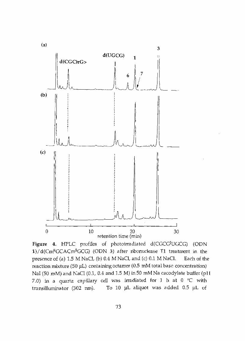
(a)
d(CGC)rG>d(UGCG) 1
3
6 ····f7
(b)
(c)
o 10 20 30retention time (min)
Figure 4. HPLC profiles of photoirradiated d(CGCc;IUGCG) (ODN
1)/d(Cm8GCACm8GCG) (ODN 3) after ribonuclease Tl treatment in the
presence of (a) 1.5 M NaCl, (b) 0.4 M NaCl, and (c) 0.1 M NaCl. Each of the
reaction mixture (50 ilL) containing octamer (0.5 mM total base concentration)
NaI (50 mM) and NaCl (0.1, 0.4 and 1.5 M) in 50 mMNa cacodylate buffer (pH
7.0) in a quartz capillary cell was irradiated for 1 h at 0 °C with
transilluminator (302 run). To 10 ilL aliquot was added 0.5 ilL of
73
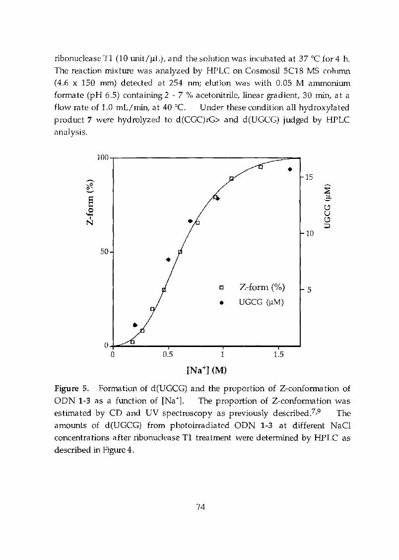
ribonuclease Tl (10 unit/ilL), and the solution was incubated at 37 DC for 4 h.
The reaction mixture was analyzed by HPLC on Cosmosil 5C18 MS column
(4.6 x 150 mm) detected at 254 nm; elution was with 0.05 M ammonium
formate (pH 6.5) containing 2 - 7 % acetonitrile, linear gradient, 30 min, at a
flow rate of 1.0 mL/min, at 40°C. Under these condition all hydroxylated
product 7 were hydrolyzed to d(CGC)rG> and d(UGCG) judged by HPLC
analysis.
5
1.5
Z-form (%)
UGCG (~M)
1
D
•
0.50-+-:;;;...------,.-----__----...,.-.....
o
100
•.- 15~ .-.,0 ::8-e ::L-0 Cl... UI ClN ::>
10
50
[Na+] (M)
Figure 5. Formation of d(UGCG) and the proportion of Z-conformation of
ODN 1-3 as a function of [Na+]. The proportion of Z-confonnation was
estimated by CD and UV spectroscopy as previously described?,9 The
amounts of d(UGCG) from photoirradiated ODN 1-3 at different NaCI
concentrations after ribonuclease Tl treatment were determined by HPLC as
described in Figure 4.
74
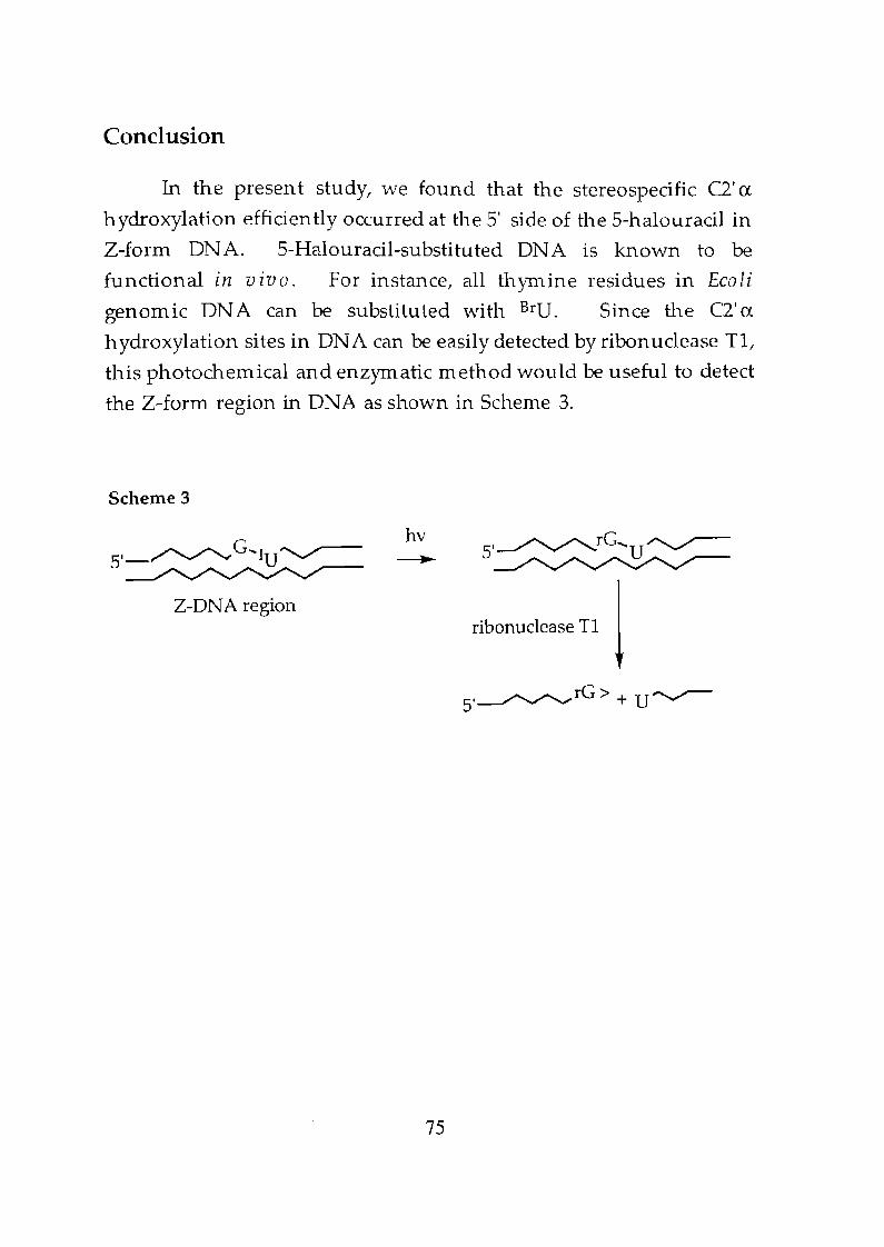
Conclusion
In the present study, we found that the stereospecific Cl' ex
hydroxylation efficiently occurred at the 5' side of the 5-halouracil in
Z-form DNA. 5-Halouracil-substituted DNA is known to be
functional in vivo. For instance, all thymine residues in Ecoli
genomic DNA can be substituted with BrU. Since the Cl' ex
hydroxylation sites in DNA can be easily detected by ribonuclease TI,
this photochemical and enzymatic method would be useful to detect
the Z-form region in DNA as shown in Scheme 3.
Scheme 3
5,_~G"'IU~
Z-DNA region
hv
75
ribonuclease T1 \
5,~rG>+U~
Experimental Section
Materials and methods. Calf in testine alkaline
phosphatase (API 1000 units/mL) and snake venom
phosphodiesterase (s.v. POE, 3 unit/mL) were purchased from
Boehringer MannheiIn. Nuclease P1 (P1, 1000 unit/m L), 5-iodo-2'
deoxyuridine and 5-brolno-2' -deoxyuridine was purchased from
Yarnasa Shoyu Co. Ltd. Ribonuclease T1 (Rnase T1 1 101000
unit/mL) was purchased from Pharmacia Biotech. 2-Cyanoethyl
N ,N,N ',N '-tetraisopropyl-phosphorodiam idite was purchased from
Aldrich. The reagents for DNA synthesizer such as h solution
(Iz/HzO/pyridine/tetrahydrofuran l 0.5:9.5:20:70) A-, G-I C-, and T-B
(cyanoethyl)phosphoramidites were purchased from Applied
Biosystem s. 1H-tetrazole was purchased from Doj in Laboratories.
HPLC analysis was carried out using PU-980 HPLC system GASCO,
Tokyo) equipped with a 01.emcobond 5-0DS-H (4.6 x 150 mm) or a
Cosmosil 5C18-MS column (4.6 x 150 mm). Detection was carried
out at 254 nm. UV irradiation was carried out with a
transilluminator (302 nm l Funakoshi FTI-36M). The ice cooled
reaction nlixture in quartz capillary cell (7 mm diameter) was
irradiated from the distance of 5 em under described conditions.
Electrospray mass spectra were recorded on Perkin-Elmer Sciex API
165 systems.
Synthesis of oligodeoxynucleotides. Oligonucleotides were
prepared by the ~-(cyanoethyl)phosphoramidite method on
controlled pore glass supports (1 Ilmo!) by using ABI 381 A DNA
synthesizer. Cyanoethyl phosphoramidite of 5-iodo-2'
deoxyuridine and 2' -deoxyuridine was prepared according to the
76
standard phosphoramidite chemistry on a DNA synthesizer.
Cyanoethyl phosphoramidite of 8-methyl-2'-deoxyguanosine were
prepared by the reported procedure? After automated synthesis,
the oligomer was detached from the support, deprotected and purified
by HPLC as described previously. After lyophilization,
approximately 20 OD of pure oligomer was isolated. Purity and
concentrations of all oligonucleotides were determined by complete
digestion with S.v. PDE and AP to 2'-deoxymononucleosides.
CD measurements. Circular dichroism (CD) spectra were
recorded on Jasco J-700 spectrophotometer equipped with a Peltier
temperature controller. CD spectra of oligonuclotide solutions (0.1
mM duplex and 2 M N aCl in 50 mM sodium cacodylate, pH 7.0)
were recorded using 1 em path length cell.
Characterization of photoproducts (4), (5) and (6).
Photoproduct (4) and (5) were characterized according to the
published procedure.5b Halogen exchanged product (6) was
characterized according to the published procedure.9 Formation of
photoproducts (4), (5) and (6) was futher confirmed by electrospray
MS. ElectrosprayMS (negative); (4): calcd 2277.5, found 2277.1., (5):
calcd 2265.5, found 2264.8., (6): calcd 2447.1, found 2446.2.
Characterization of product 7. The reaction mixture (total
volume 200 ~L) contained deoxyoligonucleotides d(CGCGIUGCG) (1)/
d(Crn8GCACrn 8GCG) (3) (0.5 mM total base concentration) and 2 M
NaCI in 50 mM sodium cacodylate buffer (pH 7.0) in a quartz capillary
77
cell. After irradiation at 0 °C for 3 h, the resulting mixture was
subjected to HPLC analysis. Analysis was carried out on a Cosmosil
5C18-MS column. Elution was with 0.05 M ammonium formate
containing 2 - 8 % acetonitrile, linear gradient (30 min) at a flow rate of
1.0 mL/min, at 30°C. The HPLC peak at 24.0 min was collected and
the fraction was concentrated. The residue was dissolved in water
and then subjected to enzymatic digestion with S.v. PDE (0.3
unit/m L) and calf intestine AP (100 unit/m L), and the mixture was
subjected to HPLC analysis. HPLC conditions; Cosmosil 5CI8-MS
column; 0.05M ammoniUlll formate containing 2 -10 % acetonitrile,
linear gradient, 20 min; flow rate of 1.0 m L/min. Formation of rG,
dC, dU and dG in a raito of 1:3:1:3 was observed. Product 7 was
further confinned by eleetrospray MS. Electrospray MS (negative);
calcd 2428.6, found 2428.2.
Quantitative analysis of photoirradiated IV-containing B-form
and Z-form oligodeoxynucleotides. The reaction mixture (total
volume 100 ilL) contained 0.5 mM (base concentration) of ODN and
described sodiunl salt in 50 mM sodium cacodylate buffer (pH 7.0) in
quartz capillary cell. After irradiated at 0 °C for 1 h, 10 I-lL of the
aliquot was taken up and subjected to HPLC analysis. Analysis was
carried out on a Cosmosil5C18-MS column; elution was with 0.05 M
ammonium formate containing 2 - 8 % acetonitrile, linear gradient
(30 min) at a flow rate of 1.0 m L/min, at 30°C. Yield of Cl'
oxidation (4), C2' -oxidation products (5), halogen exchanged product
(6) and C2'-hydroxylation product (7) were determined by comparison
of their HPLC peak areas with the authentic samples.
78
References
1. Cozzarelli,N,R.;Wang,J.C. DNA Topologyand Its Biological
Effects 1990, Cold Spring Harbor Laboratory Press: New York.
2, (a) Sinden, R, R. ON A Structure and Function 1994, Academic
Press: New York. (b) Travers, A. A. Annu. Rev. Bioche11l.1989,
58,427.
3. (a) Rieh, A.; Nordheim, A.; Wang, A. H.-J. Ann. Rev. Biochem.
1984,53, 791. (b) Rich, A. Ann. N. Y. Acad. Sci. 1994, 726, 1.
4. (a) Kim, Y.-G.; Kim, P. S.; Herbert, A.; Rich, A. Froc. Natl. Acad.
Sci. USA 1997, 94, 12875. (b) Herbert, A.; Lowenhaupt, K.; Spitzer,
J.; Rich, A. Proc. Natl. Acad. Sci. USA 1995, 92, 7550.
5. (a) Sugiyama, H.; Tsutsumi, Y.; Saito, 1. J. Am. Chem. ·Soc.1990,
112, 6720. (b) Sugiyama, H.; Tsutsumi, Y.; Fujimoto, K.; Saito, 1.
J. Am, Chem. Soc. 1993, 115,4443. (c) Sugiyama, H.; Fujimoto, K.;
Saito, 1.; Kawashima, E.; Sekine, T.; Ishido, Y. Tetrahedron Lett.
1996, 37, 1805. (d) Sugiyama, H.; Fujimoto, K.; Saito, 1.
Tetrahedron Lett. 1997,38,8057.
6. Johnston, B. H.; Rich, A. Cell 1985, 42, 713.
7. Sugiyama, H.; Kawai, K.; Matsunaga, A.; Fujimoto, K.; Saito, 1.;
Robinson, H.; Wang, A. H. -J. Nucleic Acids Res. 1996, 24,
1272'.
S. Kawai, K.; Saito, 1.; Sugiyama, H. to be submitted
9. Xodo, L.-E.; Manzini, G.; Quadrifoglio, F.; van der Marel, G. A.;
van Boom, J. H. Biochemistry 1988,27,6327.
79
Chapter 5
Intrastrand 2'B Hydrogen Abstraction of 5'-AdjacentDeoxyguanosine by Deoxyuridin-5-yl in Z-form DNA
Abstract
In order to identify the site of H abstraction by the deoxyuridin
5-yl radical in Z-form DNA, we exam ined the photoreaction of
d(CGCG(2' ~-D)IUGCG) 2-d/ d(Cm 8CCACm 8GCG) 3 whose G4
residue
was deuterated with the diastereoselectivity of 2' R:2' 5=91:9.
ElectrosprayMS analysis of ribonuclease T1 digested fragments of the
2' a-hydroxylation product 4 demonstrated that 2' a-hydroxylation
results from the abstraction of the 2' ~-H atom by the deoxyuridin-5-yl
in Z-form DNA.
Introduction
DNA local conformations such as A form, Z form, triplex and
DNA-RNA hybrid have been suggested to play an important role in
biological processes) However, the biological relevance of DNA
local conformations is still not well understood due to the lack of a
reliable detection method which is applicable to a living cell. Since
the DNA local structures are assumed to appear in a very short period
of time, utilization of a photoreaction as a DNA conformational probe
would provide important structural information on a short-lived
DNA local structure. Recently, we found that the 2' -deoxyuridin-
81
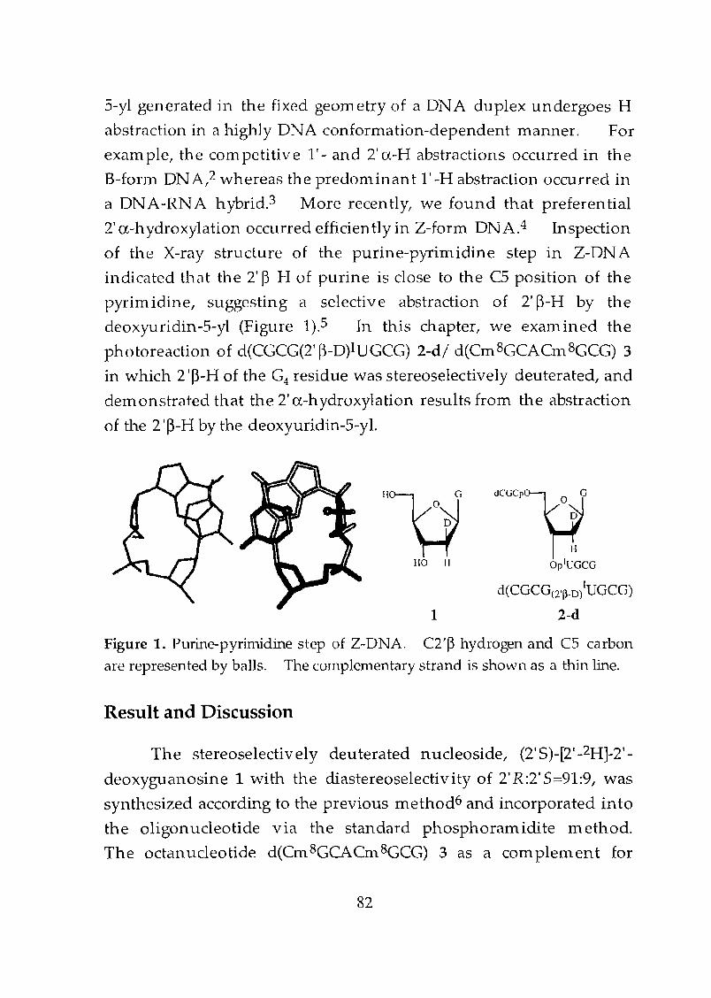
5-yl generated in the fixed geOlnetry of a DNA duplex undergoes H
abstraction in a highly DNA conformation-dependent manner. For
exam pIe, the com petitive 1'- and 2' a-H abstractions occurred in the
B-fonn ON A,2 whereas the predom inan t l' -H abstraction occurred in
a DNA-RNA hybrid.3 More recently, we found that preferential
2' a-hydroxylation occurred efficiently in Z-form DNA.4 Inspection
of the X-ray structure of the purine-pyyim idine step in Z-DNA
indicated that the 2' B H of purine is close to the C5 position of the
pyrimidine, suggesting a selective abstraction of 2' B-H by the
deoxyuridin-5-yl (Figure 1).5 In this chapter, we examined the
photoreaction of d(CGCG(2' B-D)IUGCG) 2-d! d(Cm 8GCACm 8GCG) 3
in which 2 'B-H of the G4 residue was stereoselectively deuterated, and
dernonstrated that the 2' a-hydroxylation results from the abstraction
of the 2'B-H by the deoxyuridin-5-yl.
HO II
1
dCGCP~OJ
IiOplUGCG
d(CGCG(2'~_D)IUGCG)
2-d
Figure 1. Purine-pyrimidine step of Z-DNA. C2'P hydrogen and C5 carbon
are represented by balls. The complementary strand is shown as a thin line.
Result and Discussion
The stereoselectively deuterated nucleoside, (2' 5)-[2' -2H]-2'
deoxyguanosine 1 with the cliastereoselectivity of 2' R:2' 5::::91:9, was
synthesized according to the previous method6 and incorporated into
the oligonucleotide via the standard phosphoramidite method.
The octanucleotide d(Cm8GCACm8GCG) 3 as a complement for
82
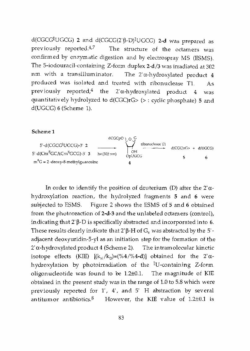
d(CGCGIUGCG) 2 and d(CGCG(2' ~-D)IUGCG) 2-d was prepared as
previously reported.4,7 The structure of the octamers was
confirmed by enzymatic digestion and by electrospray MS (ESMS).
The 5-iodouracil-containing Z-form duplex 2-d/3 was irradiated at 302
nm with a transilluminator. The 2' a-hydroxylated product 4
produced was isolated and treated with ribonuclease Tl. As
prev iously reported,4 the 2' a-hydroxylated product 4 was
quantitatively hydrolyzed to d(CGC)rG> (> : cyclic phosphate) 5 and
d(UGCG) 6 (Scheme 1).
Scheme 1
S'-d(CGCGIUGCG)-3' 2
S'-d(Crn8GCACm8GCG)-3' 3
dCGCPliG_____.. ribonuclease ~1
OHhv (302 nm) OpUGCG
mSC = 2'-deoxy-8-methylguanosine 4
d(CGClrG> + d(UGCG)
5 6
In order to identify the position of deuterium (D) after the 2' a
hydroxylation reaction, the hydrolyzed fragments 5 and 6 were
subjected to ESMS. Figure 2 shows the ESMS of 5 and 6 obtained
from the photoreaction of 2-d-3 and the unlabeled octamers (control),
indicating that 2' ~-D is specifically abstracted and incorporated into 6.
These results clearly indicate that 2' ~-H of G4 was abstracted by the 5'
adjacent deoxyuridin-5-yl as an initiation step for the formation of the
2' a-hydroxylated product 4 (Scheme 2). The intramolecular kinetic
isotope effects (KIE) [(kH /kD)=(%4/%4-d)] obtained for the 2'a
hydroxylation by photoirradiation of the IU-containing Z-form
oligonucleotide was found to be L2±O.L The magnitude of KIE
obtained in the present study was in the range of 1.0 to 5.8 which were
previously reported for 1', 4', and 5' H abstraction by several
antitumor antibiotics.S However, the KIE value of 1.2±0.1 is
83

substantially lower than the value 7.2 which was observed for the
2 'a-I-I abstraction in B-form DNA.2c
Scheme 2
dCGCPO-~') 0
9 H ·~NH-0-- p=o l ~6~N 0
OpGCG
1269.4 (b)1175.6117"\.6
"I', .I •, ., ,,
••••,,,,,,,1268 1269 1270 1271 1272 1173 1174 1175 1176 1177 1178
m/zamu m/z amu
Figure 2. ESMS of (a) d(CGC)rG> 5 and (b) d(UGCG) 6. !vIS of the
products obtained from the deuterated and unlabeled octamers are indicated
by the solid and dashed lines, respectively.
84
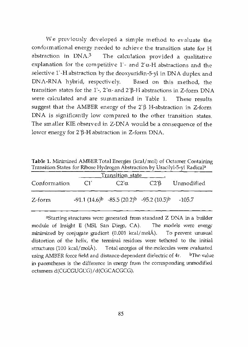
We previously developed a sim pIe method to evaluate the
conform ational energy needed to achieve the transition state for H
abstraction in DNA.3 The calculation provided a qualitative
explanation for the competitive l' - and 2' a-H abstractions and the
selective l' -H abstraction by the deoxyuriclin-5-yl in DN A duplex and
DNA-RNA hybrid, respectively. Based on this method, the
transition states for the 1'-, 2 'a- and 2 '~-H abstractions in Z-form DNA
were calculated and are summarized in Table 1. These results
suggest that the AMBER energy of the 2' ~ H-abstraction in Z-form
DNA is significantly low com pared to the other transition states.
The smaller KIE observed in Z-DNA would be a consequence of the
lower energy for 2 '~-H abstraction in Z-form DNA.
Table 1. Minimized A:MBER Total Energies (kcal/mol) of Octamer ContainingTransition States for Ribose Hydrogen Abstraction by Uracilyl-5-yl Radicala
Transition state
Conformation C1' C2'a C2'~ Unmodified
Z-form -91.1 (14.6)b -85.5 (20.2)b -95.2 (10.5)b -105.7
aStarting structures were generated from standard Z DNA in a builder
module of Insight n (MSI, San Diego, CA). The models were energy
mi.nimjzed by conjugate gradient (0.001 kcal/molA). To prevent unusual
distortion of the helix, the terminal residues were tethered to the initial
structures (100 kcal/molA). Total energies of the molecules were evaluated
using AMBER force field and distance-dependent dielectric of 4r. bThe value
in parentheses is the difference in energy from the corresponding umnodified
octamers d(CGCGUGCG)/ d(CGCACGCG).
85
Conclusion
In this chapter, using stereoselectively deuterated
octanuc1eotide, we clem onstrated that the C2' a-hydroxylation was aconsequence of the reaction of 02 with the C2' radical which was
resulted from 2' ~-H abstraction by the deoxyuridin-5-yl formed in Z
form DNA.
86
Experimental Section
Materials and methods. Calf intestine alkaline
phosphatase (AP, 1000 units/m L) and snake venom
phosphodiesterase (s.v. PDE, 3 unit/m L) were purchased from
Boehringer Mannheim. 5-iodo-2' -deoxyuridine was purchased
from Yamasa Shoyu Co. Ltd. Ribonuclease T1 (Rnase T1, 10,000
unit/m L) was purchased from Pharmacia Biotech. 2-Cyanoethyl
N,N,N ',N '-tetraisopropyl-phosphorodiamidite was purchased from
Aldrich. The reagents for DNA synthesizer such as h solu tion
(I2/H20/pyridine/tetrahydrofuran, 0.5:9.5:20:70) A-, G-, C-, and T-~
(cyanoethyl)phosphoramidites were purchased from Applied
Biosystems. 1H-tetrazole were purchased from Doj in Laboratories.
HPLC analysis was carried out using PU-980 HPLC system (JASCO,
Tokyo) equipped with a Cosmosil 5C18-MS column (4.6 x 150 mm).
Detection was carried out at 254 nm. UV irradiation was carried out
with a transilluminator (302 nm, Funakoshi FTI-36M). The ice
cooled reaction mixture in quartz capillary cell (7 mm diameter) was
irradiated from the distance of 5 an under described conditions.
Electrospray mass spectra were recorded on Perkin-Elmer Sciex API
165 systems.
Synthesis of oligodeoxynucleotides. Oligonucleotides were
prepared by the p-(cyanoethyl)phosphoramidite method on
controlled pore glass supports (1 Ilmol) by using ABI 381 A DNA
synthesizer. Cyanoethyl phosphoramidite of 5-iodo-2'
deoxyuridine and 8-methyl-2'-deoxyguanosine were prepared by the
reported procedure.4 After automated synthesis, the oligomer was
detached from the support, deprotected and purified by HPLC.
87
After lyophilization, approximately 20 OD of pure oligomer was
isolated. Purity and concentrations of all oligonucleotides were
determined by complete digestion with S.v. PDE and AP to 2'
deoxymononucleosides.
Quantitative analysis of photoirradiated IV-containing Z-form
oligodeoxynuc1eotides. The reaction mixture (total volume 100 ~L)
contained 0.5 mM (base concentration) of ODN and 2 M NaG in 50
mM sodium cacodylate buffer (pH 7.0) in a quartz capillary cell.
After irradiated at 0 °C for 2 h, 10-JlL aliquot was directly subjected to
enzymatic digestion with snake venom PDE (0.3 wlit/mL) and calf intestine
alkaline phosphatase (100 unit/mL) and was analyzed by reverse phase
HPLC.
Rnase Tl digestion of 2'-hydoxylation products. To a solution
containing HPLC purified 2'-hydroxylated product (4), Rnase T1,
10,000 unit/ill L was added. After 2 h of incubation at 37°C, the
solution was subjected to HPLC analysis. HPLC analysis was carried
out on a Coslnosil 5C18-MS coluilln. Elution was with 0.05 M
ammonium formate containing 2 - 10 % acetonitrile, linear gradient
(20 illin) at a flow rate of 1.0 m L/min, at 30°C. HPLC peak at 12.4
lnin and 17.0 min were concentrated and subjected to ESMS analysis.
88
References
1. (a) Cozzarelli, N. R.; Wang, J. C. DNA To po 10 glJ and Its
Biological Effects 1990, Cold Spring Harbor Laboratory Press: New
York. (b) Sinden, R. R. DNA Structure and Function 1994,Academic Press: New York. (c) Travers, A. A. Annu. Rev.Biochem. 1989,58,427.
2. (a) Sugiyama, H.; Tsutsumi, Y.; Saito, 1. J. Am. Chem. Soc. 1990,
112, 6720. (b) Sugiyama, H.; Tsutsumi, Y.; Fujimoto, K.; Saito, I.
J. Am. Chem. Soc. 1993,115,4443. (c) Sugiyama, H.; Fujimoto, K.;
Saito, 1.; Kawashima, E.; Sekine, T.; Ishido, Y. Tetrahedron Lett.
1996,37, 1805.
3. Sugiyama, H.; Fujimoto, K.; Saito, 1. Tetrahedron Lett. 1997,38,
8057.
4. Kawai, K.; Saito, 1.; Sugiyama, H. J. Am. Chem. Soc. in press
5. (a) Wang, A. H.-J.; Quigley, G. J.; Kolpak, F. J.; Crawford, J. L.; van
Boom, J. H.; van der Marel, G. A.; Rich, A. Nature 1979,282,
680-686.7. (b) Rich, A.; Nordheim, A.; Wang, A. H.-J. Ann.
Rev. Biochem. 1984,53,791. (c) Rich,A. Ann.N. Y. Acad. Sci.
1994, 726, 1.
6. Kawashima, E.; Aoyama, Y.; Sekine, T.; Miyahara, M.; Radwan,
M. F.; Nakamura, E.; Kainosho, M.; Kyogoku, Y.; Ishido, Y. J.Org. CI1 em. 1995, 60, 6980-6986. (b) Kawashima, E.; Sekine, T.;
Ishido, Y. Nucleoside and Nucleotides 1995, 14,333.
7. Sugiyama, H.; Kawai, K.; Matsunaga, A.; Fujimoto, K.; Saito, 1.;
Robinson, H.; Wang, A. H. -J. N ueIeie Acids Res. 1996, 24,
1272-1278.
8. (a) Kozarich, J. W.; Worth, L.; Frank, B. L.; Christner, D. F.;
Vanderwall, D. E.; Stubbe, J. Science 1989, 245, 1396. (b)
Kappen, L. S.; Goldberg, 1. H.; W u, S. H.; Stubbe, J.; Worth, L.;
Kozarich, J. W. J. Am. C/zem. Soc, 1990, 112, 2797. (c) Frank, B.
89
L.; Worth, 1.; Christner, D. F.; Kozarich, J. W.; Stubbe, J.; Kappen,
1.; Goldberg, 1. H. J. Am. Cl1em. Soc, 1991,113,2271. (d)
Kappen, 1.; Goldberg, 1. H.; Frank, B. L.; Worth, 1.; Christner, D.
F.; Kozarich, J. W.; Stubbe, J. Bio ch e111 istry 1991, 30, 2034. (e)
Hangeland, J. J.; De Voss, J. J.; Heath, J. A.; Townsend, C. A.; Ding,
W.; Ashcroft, J. S.; Ellestad, G. A. J. Am. Chem. Soc, 1992, 114,
9200. (f) Absalon, M. J.; W u, W.; Kozarich, J. W.; Stubbe, J.Bioch em istry, 1995,34, 2076. (g) Greenberg, M. M.; Barvian, M.
R.; Cook, G. P.; Goodman, B. K.; Matray, T. J.; Tronche, C.;
Venkatesan, H. J. Am. Chem. Soc, 1997, 119, 1828.
90
Chapter 6
Photochemical Halogen-Exchange Reaction of5-Iodouracil-Containing Oligonucleotides
Abstract
Photoreactions of 5-iododeoxyuridine (dIU) in aqueous
solutions in the presence of various inorganic salts have been
investigated. In the presence of N aCl and N aBr, dIU undergoes an
efficient photochemical halogen-exchange reaction to give dC1U and
dBrU, respectively.
Introduction
Photoreactions of 5-halouracils are important tools for
investigating nucleic add structures and nucleic acid-protein
interactions) Especially, 5-iodouridine (dIU) has been widely used
as a photo-cross-linking agent, and the photoreactions of dIU
monomer2 or the photo-cross-linking of dIU-containing DNA with
nucleic add binding proteins have been extensively studied.3 dIU
containing DNA has also been known to cause DNA strand breaks via
the creation of alkali labile sites.4 We have been investigating the
molecular mechanism of such DNA lesions and the effect of DNA
conformational change on the photoreactivity of dIU- and dBrU
containing DNA.S During the careful product analysis of the
photoreaction of dIU-containing-oligonucleotides, I have accidentally
93
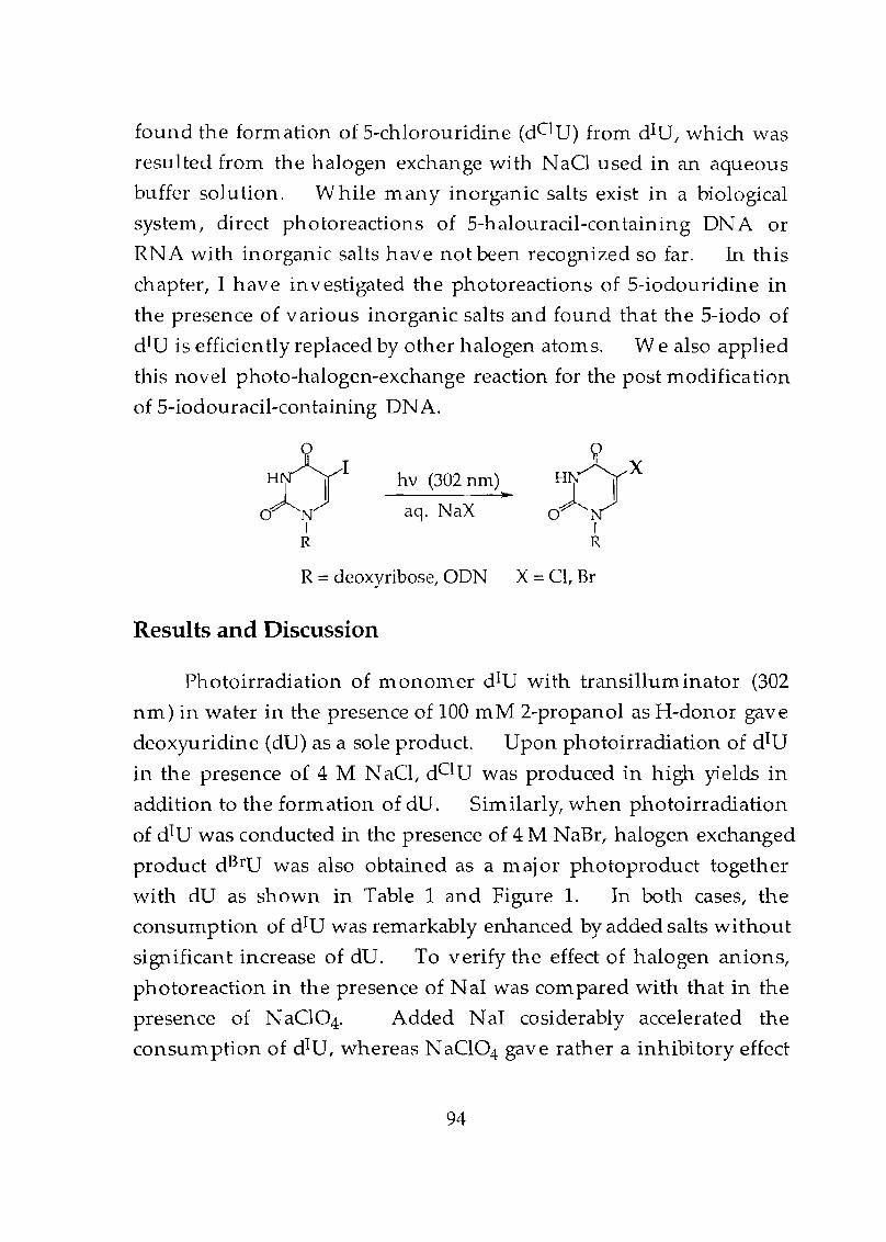
found the formation of 5-chlorouridine (dOU) from dIU, which was
resul ted from the halogen exchange with N aCI used in an aqueous
buffer solution. While many inorganic salts exist in a biological
system, direct photoreactions of 5-halouracil-containing DNA or
RN A with inorganic salts have not been recognized so far. In this
chapter, I have investigated the photoreactions of 5-iodouridine in
the presence of various inorganic salts and found that the 5-iodo of
dIU is efficiently replaced by other halogen atoms. We also applied
this novel photo-halogen-exchange reaction for the post modification
of 5-iodouracil-containing DNA.
hv (302 nm)~
aq. NaX
R =deoxyribose, ODN X=Cl, Br
Results and Discussion
Photoirradiation of 1110nomer dIU with transilluminator (302
nm) in water in the presence of 100 mM 2-propanol as H-donor gave
deoxyuridine (dU) as a sole product. Upon photoirradiation of dIU
in the presence of 4 M N aCI, dO U was produced in high Yields in
addition to the for111ation of dU. Similarly, when photoirradiation
of dIU was conducted in the presence of 4 M NaBr, halogen exchanged
product dBrU was also obtained as a 111 aj or photoproduct together
with dU as shown in Table 1 and Figure 1. In both cases, the
consumption of dIU was remarkably enhanced by added salts without
significant increase of dUo To verify the effect of halogen anions,
photoreaction in the presence of N al was compared with that in the
presence of N aCI04. Added NaT cosiderably accelerated the
consum ption of dIU, whereas N aCI04 gave rather a inhibitory effect
94
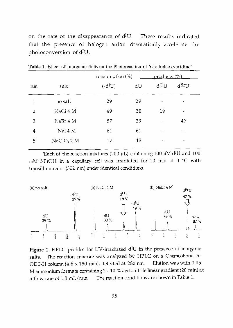
on the rate of the disappearance of dIU. These results indicated
that the presence of halogen anion dramatically accelerate the
photoconversion of dIU.
Table 1. Effect of Inorganic Salts on the Photoreaction of 5-Iododeoxyuridine
consumption (%) products (%)
.nUl salt (-dIU) dU dClU dBrU
1 no salt 29 29
2 NaC14 M 49 30 19
3 NaBr4 M 87 39 47
4 NaI4M 61 61
5 NaCI04 2 M 17 13
aEach of the reaction mixtures (200 ~L) containing 100~ diU and 100
roM i-PrOH in a capillary cell was irradiated for 10 min at 0 °C with
transilluminator (302 run) under identical conditions.
(a) no salt
~9U% I
------')\~--~)
(b) NaCl4M (b) NaBr4 M
Figure 1. HPLC profiles for UV-irradiated diU in the presence of inorganic
salts. The reaction mixture was analyzed by HPLC on a Chemcobond 5
ODS-H column (4.6 x 150 mm), detected at 280 run. Elution was with 0.05
M anunonimn formate containing 2 - 10 % acetonitrile linear gradient (20 min) at
a flow rate of 1.0 mL/rnin. The reaction conditions are shown in Table 1.
95
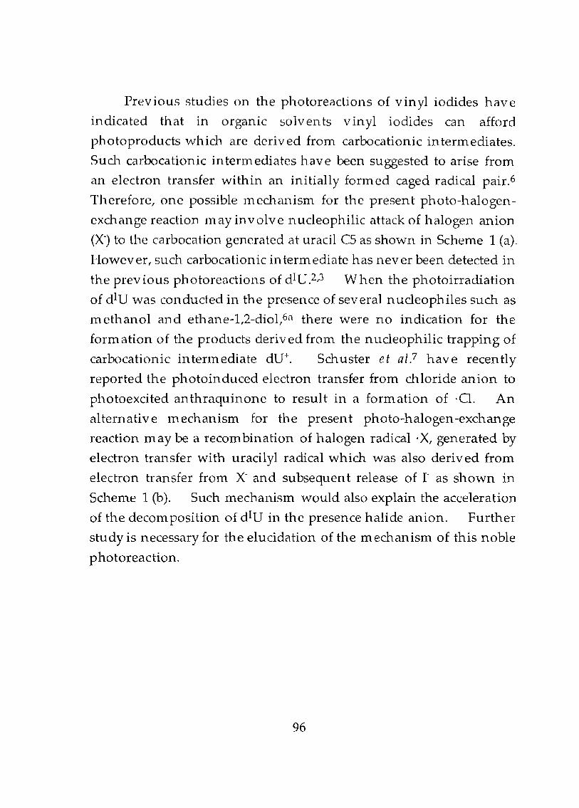
Previous studies on the photoreactions of vinyl iodides have
indicated that in organic solvents vinyl iodides can afford
photoproducts which are derived from carbocationic intermediates.
Such carbocationic intermediates have been suggested to arise from
an electron transfer within an initially formed caged radical pair.6
Therefore, one possible mechanism for the present photo-halogen
exchange reaction may involve nUcleophilic attack of halogen anion
(X·) to the carbacation generated at uracil CS as shown in Scheme 1 (a).
However, such carbocationic intermediate has never been detected in
the prev ious photoreactions of dIU,2,3 W hen the photoirradiation
of dIU was conducted in the presence of several n ucleophiles such as
ll1 ethanol and ethane-l,2-diol,6a there were no indication for the
formation of the products derived from the nucleophilic trapping of
carbocationic intennediate dU+. Sch.uster et al? have recently
reported the photoinduced electron transfer from chloride anion to
photoexcited anthraquinone to result in a formation of ·Cl. An
alternative mechanism for the present photo-halogen-exchange
reaction may be a recombination of halogen radical' X, generated by
electron transfer with uracilyl radical which was also derived from
electron transfer from X' and subsequent release of r- as shown in
Scheme 1 (b). Such ll1echanism would also explain the acceleration
of the decoln position of dIU in the presence halide anion. Further
study is necessary for the elucidation of the mechanism of this noble
photoreaction.
96
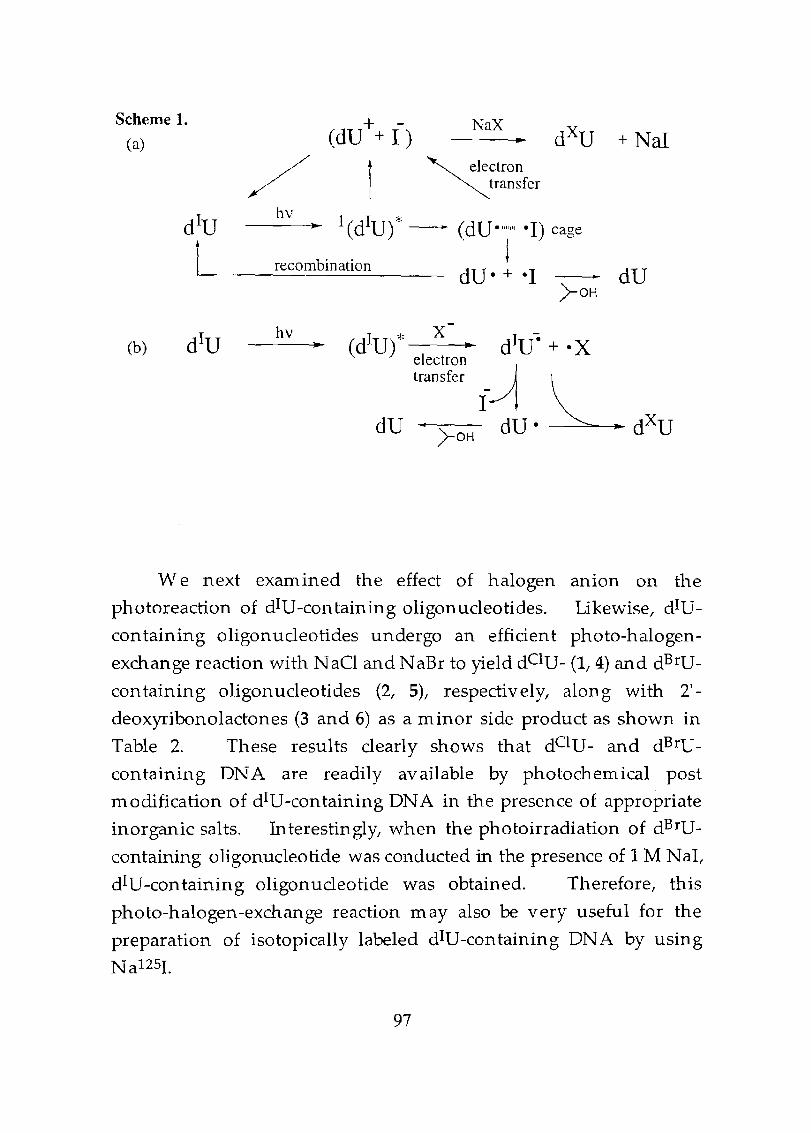
Scheme 1.
(a)
,/+
(dU + I)
1
NaX
~ electron~transfer
bv--....~ 1(dIU)* - (dU"""" -I) cage
b' . ireCOID matlon dU - + -I . dU
)-OH
(b)hv ...
We next examined the effect of halogen anion on the
photoreaction of dIU-containing oligonucleotides. Likewise, dIU
containing oligonucleotides undergo an efficient photo-halogen
exchange reaction with NaCI and NaBr to yield dC1U- (1,4) and dBrU
con taining oligonucleotides (2, 5), respectively, along with 2'
deoxyribonolactones (3 and 6) as a minor side product as shown in
Table 2. These results clearly shows that dC1U- and dBrU
containing DNA are readily available by photochemical post
modification of dIU-containing DNA in the presence of appropriate
inorganic salts. Interestingly, when the photoirradiation of dBrU
containing oligonucleotide was conducted in the presence of 1 M NaI,
dIU-containing oligonucleotide was obtained. Therefore, this
photo-halogen-exchange reaction may also be very useful for the
preparation of isotopically labeled dIU-containing DNA by using
N a125L
97
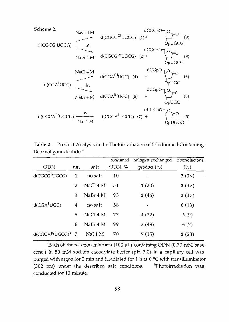
Scheme 2.NaC14 M dCGCPlJO
----------d (CCCCClUGCG) (1) + (3)
d(CCce] UeCG) hv OpUGCG
------... dCGCPlJONaBr4 M d(CGCGBrUCCC) (2) + (3)
OpUGce
NaC14M +dCGPlJo
----------d(CGACIUGC) (4) (6)
d(CGA1UGC) hvGpUGC
------... +dCGPlJoNaBr 4 M d(CGABrUCC) (5) (6)
GpUGC
d(CGCADrUCCG)hv
d(CGCA1UGCG)dCGCPO\JO
(7) + (3)NaIl M OpUGCG
Table 2. Product Analysis in the Photoirradiation of 5-Iodouracil-Containing
Deoxyoligonucleotides"
consumed halogen exchanged ribonolactone
ODN nm salt ODN, % product (%) (%)
d(CGCCIUGCC) 1 no salt 10 3 (3))
2 NaCl4 M 51 1 (20) 3 (3))
3 NaBr 4 M 93 2 (46) 3 (3))
d(CGAIUGC) 4 no salt 58 6 (13)
5 NaC14 M 77 4 (22) 6 (9)
6 NaBr 4 M 99 5 (48) 6 (7)
d(CGCABrUGCG) b 7 NaIl M 70 7 (15) 3 (23)
"Each of the reaction mixtures (100 ~L) containing ODN (0.20 mM base
cone.) in 50 rnIv1 sodiwn cacodylate buffer (pH 7.0) in a capillary cell was
purged with argon for 2 min and irradiated for 1 hat 0 DC with transillwninator
(302 nm) under the described salt conditions. bPhotoirradiation was
canducted for 10 Ininute.
98
Conclusion
In summary, we have demonstrated for the first time that dIU
and dIU-containing DNA undergo an efficient photo-halogen
exchange reaction in the presence of halide ions to produce C-5
halogen exchanged uracils. This efficient photoreaction of 5
halouracils in the presence of inorganic salts should be seriously
taken into consideration when photoinduced crosslinking
experiments using dIU containing DNA are conducted in salt
containing solvent systems.
99
Experimental Section
Materials and methods. Calf intestine alkaline
phosphatase (AP, 1000 units/rn L) and snake venom
phosphodiesterase (sov. POE, 3 unit/m L) were purchased from
Boehringer Mannheim. 5-lodo-2' -deoxyuridine and 5-bromo-2'
deoxyuridine was purchased from Yamasa Shoyu Co. Ltd. 5'-0
dimethoxytrityl-2' -deoxyuridine was prepared according to the
published procedure.S 2-Cyanoethyl N ,N ,N ',N'
tetraisopropylphosphorodiamidite and 5-chloro-2' -deoxyuridine
were purchased from Aldrich. The reagents for DNA synthesizer
such as 12 solution (Iz/H20/pyridine/tetrahydrofuran, 0.5:9.5:20:70)
A-, G-, C-, and T-~-(cyanoethyl)phosphoram idites were purchased
from Applied Biosystems. IH-tetrazole were purchased from Dojin
Laboratories. N aCI, N aBr and N a1 were of puriss grade and were
used as received. HPLC analysis was carried out using PU-980 HPLC
system GASCa, Tokyo) equipped with a Chern cobon d 5-0DS-H (4.6 x
150 Inm) or a Cosmosil5C18-MS column (4.6 x 150 mm). Detection
was carried out at 280 nIn for monOll1er and at 254 nm for ODN.
UV irradiation was carried out with a transilluminator (302 nm,
Funakoshi FTI-36M). The ice cooled reaction mixture in quartz
capillary cell (7 m n1 diameter) was irradiated from the distance of 5 em
under described conditions. Electrospray mass spectra were
recorded on Perkin-Elmer Sciex API 165 systems.
Synthesis of oligodeoxynuc1eotides. Oligonucleotides were
prepared by the ~-(cyanoethyl)phosphoramidite method on
controlled pore glass supports (1 Ilmol) by using ABI 381 A DNA
synthesizer. Cyanoethyl phosphoramidite of 5-iodo-2'-deoxyuridine
was prepared by the reported procedure.S After automated
100
synthesis, the oligom er was detached from the support, deprotected
and purified by HPLC as described previously. After lyophilization,
approximately 20 OD of pure oligomer was isolated. Purity and
concentrations of all oligonucleotides were determined by com plete
digestion with S.v. PDE and AP to 2'-deoxymononucleosides.
Quantitative analysis of photoirradiated dIU. The reaction
mixture (total volume 200 ~L) contained 0.1 mM of dIU ,100 mM
isopropanol and indicated sodium salt in a quartz capillary cell was
irradiated with a transilluminator at a °C for 10 min. After
irradiation, 10 ~L of the aliquot was taken up and subjected to HPLC
analysis. Analysis was carried out on a Chemcobond 5-0DS-H
column; elution was with 0.05 M ammonium formate containing 2
10 % acetonitrile, linear gradient (20 min) at a flow rate of 1.0 mL/min.
Yield of halogen-exchanged product dBrU and dC1U were determined
by com parison of HPLC peak areas of the authentic samples. HPLC
profile of the photoirradiated dIU was shown in Figure 1.
Characterization of photo-halogen exchanged product (1).
The reaction mixture (total volume 200 ~L) contained 0.2 mM (base
concen tration) d(CGCGIUGCG) and 4 M N aO in 5 mM sodium
cacodylate buffer (pH 7.0) in a quartz capillary cell. After irradiated
at a°C for 3 11./ the resulting mixture was subjected to HPLC analysis.
Analysis was carried out on a Cosmosil 5C18-MS column. Elution
was with 0.05 M ammonium formate containing 2 - 8 % acetonitrile,
linear gradient (30 min) at a flow rate of 1.0 mL/min. The HPLC
peak at 23.2 min was collected and the fraction was concentrated.
The residue was dissolved in water and then subjected to enzymatic
101
digestion with s.v. PDE (0.3 unit/m L) and calf intestine AP (100
unit/mL), and the mixture was subjected to HPLC analysis: HPLC
conditions; Cosmosil SC18-MS column; O.OSM ammonium formate
containing 2 - 10 % acetonitrile, linear gradient, 20 min; flow rate of 1.0
111 L/min. The formation of S-chlorodeoxyuridine together with
dC, and dG was observed. The formation of halogen-exchanged
products 1 was confirmed by electrospray MS. Electrospray MS
(negative); 1: calcd 2447.1, found 2446.2.
Characterization of photo-halogen exchanged product (2).
The reaction lnixture (total volume 200 ~L) contained 0.2 mM (base
concentration) d(CGCGIUGCG) and 4 M NaBr in 5 mM sodium
cacodylate buffer (pH 7.0) in a quartz capillary cell. After irradiated
at a°C for 3 h, the resulting mixture was subjected to HPLC analysis.
Analysis was carried out on a Cosmosil SC18-MS column. Elution
was with 0.05 M ammonium formate containing 2 - 8 % acetonitrile,
linear gradien t (30 min) at a flow rate of 1.0 m L/min. The HPLC
peak at 23.6 ill in was collected and the fraction was concentrated.
The residue was dissolved in water and then subjected to enzymatic
digestion with S.v. PDE (0.3 unit/mL) and calf intestine AP (100
unit/In L), and the mixture was subj ected to HPLC analysis: HPLC
conditions; Cosmosil SC18-MS column; 0.05M ammonium formate
containing 2 - 10 % acetonitrile, linear gradient, 20 min; flow rate of 1.0
InL/min. The formation of 5-bromodeoxyuridine together with
dC, and dG was observed. The form ation of halogen-exchanged
products 2 was confirmed by electrospray MS. Electrospray MS
(negative); 2: calcd 2491.5, found 2491.4.
102
Characterization of photo-halogen exchanged product (4).
The reaction mixture (total volume 200 ~L) contained 0.2 mM (base
concentration) d(CGAIUGC) and 4 M NaCl in 5 mM sodium
cacodylate buffer (pH 7.0) in a quartz capillary cell. After i"rradiated
at O°C for 3 h, the resulting mixture was subjected to HPLC analysis.
Analysis was carried out on a Cosmosil 5CI8-MS column. Elution
was with 0.05 M ammonium formate containing 2 - 15 % acetonitrile,
linear gradient (20 min) at a flow rate of 1.0 mL/min. The HPLC
peak at 14.9 m in was collected and the fraction was concen trated.
The residue was dissolved in water and then subjected to enzymatic
digestion with S.v. PDE (0.3 unit/m L) and calf intestine AP (100
unit/mL), and the mixture was subj ected to HPLC analysis. HPLC
conditions; CHEMCOBOND 5-0DS-H column; 0.05M ammonium
formate containing 2 - 10 % acetonitrile, linear gradient, 20 min; flow
rate of 1.0 m L/min. The form ation of 5-chlorodeoxyuridine
together with dC, dA and dG was observed. The formation of
halogen-exchanged products 4 was confirmed by electrospray MS.
Electrospray MS (negative); 4: calcd 1812.7, found 1811.7.
Characterization of photo-halogen exchanged product (5).
The reaction mixture (total volume 200 ~L) contained 0.2 mM (base
concentration) d(CGAIUGC) and 4 1\1 N aBr in 50 mM sodium
cacodylate buffer (pH 7.0) in a quartz capillary cell. After irradiated
at O°C for 3 h, the resulting mixture was subj ected to HPLC analysis.
Analysis was carried out on a Cosmosil 5C18-MS column. Elution
was with 0.05 M ammonium formate containing 2 - 15 % acetonitrile,
linear gradient (20 min) at a flow rate of 1.0 m L/min. The HPLC
peak at 15.4 min was collected and the fraction was concentrated.
The residue was dissolved in water and then subjected to enzymatic
digestion with S.v. PDE (0.3 unit/mL) and calf intestine AP (100
103
unit/m L), and the D1ixture was subjected to HPLC analysis. HPLC
conditions; Chemcobond 5-0DS-H column; 0.05M ammonium
fonnate con taining 2 - 10 % acetonitrile, linear gradien t, 20 m in; flow
rate of 1.0 m L/min. The formation of 5-bromodeoxyuridine
together with dC, dA and dG was observed. The form ation of
halogen-exchanged products 5 was confirmed by electrospray MS.
Electrospray MS (negative); 5: calcd 1857.t found 1855.8.
Quantitative analysis of photoirradiated IV-containing
oligodeoxynucleotide. The reaction mixture (total volume 100 J.lL)
contained 0.2 mM (base concentration) of ODN and sodium salt
indicated in 50 mM sodium cacodylate buffer (pH 7.0) in a quartz
capillary cell. After irradiated at 0 °C for 1 h, 10 J.lL of the aliquot
was taken up and subjected to HPLC analysis. Analysis was carried
out on a Cosmosil 5C18-MS column; elution was with 0.05 M
aInn10nium fonnate containing 2 - 15 % acetonitrile, linear gradient
(20 Inin) for hexamer and 2 - 8 % acetonitrile, linear gradient (30 min)
for octainer and at a flow rate of 1.0 mL/min. Yield of C1 '-oxidation
product and halogen-exchanged product were determined by
con1parison of HPLC peak areas of the authentic samples.
Characterization of photo-halogen exchanged product (7).
The reaction Inixture (total volume 200 J.lL) contained 0.2 mM (base
concentration) d(CGGABrUGCG) and 1 M NaI in 50 mM sodium
cacodylate buffer (pH 7.0) in a quartz capillary cell. After irradiated
at O°C for 30 Inin, the resulting mixture was subj ected to HPLC
analysis. Analysis was carried out on a Cosmosil5C18-MS column.
Elution was with 0.05 M ammonium formate containing 2 - 7 %
104
acetonitrile, linear gradient (30 min) at a flow rate of 1.0 mL/min, at
40°C. The HPLC peak at 24.2 min was collected and the fraction was
concentrated. The formation of halogen-exchanged products 7
was confirmed by coinjection with the authentic sample and by
electrospray MS. Electrospray MS (negative); 5: calcd 2522.5, found
2521.7.
Quantitative analysis of photoirradiated BrU-containing
oligodeoxynuc1eotide. The reaction mixture (total volume 100 ilL)
contained 0.2 mM (base concentration) of ODN and 1 M NaI in 50 mM
sodium cacodylate buffer (pH 7.0) in a quartz capillary cell. After
irradiated at DoC for 10 min, 10 ilL of the aliquot was taken up and
subjected to HPLC analysis. Analysis was carried out on a Cosmosil
5C18-MS column; elution was with 0.05 M ammonium formate
containing 2 - 7 % acetonitrile, linear gradient (30 min) at a flow rate of
1.0 mL/min, at 40 DC. Yield of Cl'-oxidation product and halogen
exchanged product were determ ined by comparison of HPLC peak
areas of the authentic samples.
105
References1. (a) Dickerson, R. E. Methods Enzymol. 1992,211,67. (b) Saito, 1.;
Sugiyama, H.; Morrison, H. Ed. Bioorganic Photochemistry,
John Wiely & Sons, New York 1990, p 317.
2. Norris, C. L.; Meisenheimer, P. L.; Koch, T. H. J. Am. Che111.
Soc. 1996, 118,5796.
3. (a) Willis, M. C.; Hicke, B. J.; Uhlenbeck, O. C.; Cech, T. R.; Koch,
T. H. Science 1993, 262, 1255. (b) Hicke, B. J.; Willis, M. C.; Koch,
T. H.; Cech, T. R. Biochemistry 1994,33,3364.
4. Cadet, J. L.; Vigny, P.; Morrison, H. Ed. Bioorganic
Photochemistry, John Wiely & Sons, New York 1990, p 172.
5. (a) Sugiymua, H.; Tsutsumi, Y.; Saito. 1. J. Am. Chem. Soc. 1990,
112,6720. (b) Sugiyama, H.; Tsutsumi, Y.; Fujimoto, K.; Saito. 1.
f. Am. Chem. Soc. 1993,115,4443. (c) Sugiyama, H.; Fujimoto, K.;
Saito. 1. Tetrahedron Lett. 1997,38, 8057. (d) Kawai, K.; Saito, I;
Sugiyama, H. J. Am. Chem. Soc. in press
6. (a) Kropp, P. J.; McNeely, S. A.; Davis, R. D. J. Am. Chem. Soc.
1983, 105, 6907. (a) Kitamura, T.; Murakami, M.; Kobayashi, S.;
Taniguchi, H. Tetrahedron Lett. 1986, 27, 3385. (b) Kitamura, T.;
Kabashima, T.; Kobayashi, S.; Taniguchi, H. C hem Lett. 1988,
1951. (c) Kitamura, T.; Nakamura, 1.; Kabashima, T.; Kobayashi,
S.; Taniguchi, H. C hem Lett. 1990,9. (d) Lipson, M.; Deniz, A.
A.; Peters, K. S. f. Phys. Chen-I. 1996, 110,3580. (e) Das, P. K.
Chen-I. Rev. 1993,93,119.
7. (a) Armitage, B.; Schuster, G. B. Photochemistry and
Photobiology 1997, 66, 164. (b) Loeff, 1.; Rabani, J.; Treinin, A.;
Linschitz, H. f. Am. Chem. Soc. 1993, 115,8933.
106
List of Publications
Chapter 1
Chapter 2
Chapter 3
Chapter 4
Chapter 5
Synthesis, Structure and Thermodynamic Properties
of 8-Methylguanine-Containing Oligonucleotides: Z
DNA under Physiological Salt Conditions.
Sugiyama, 1-1.; Kawai, K.; Matsunaga, A.; Fujimoto, K.;
Saito, I.; Robinson, H.; Wang, A. H.-J. Nucleic Acids
Res. 1996,24, 1272-1277.
Effects of Methylation on B- to Z-DNA Transition.
Kawai, K.; Sugiyama, H.; Saito, 1. Nucleic Acids
5 ym p. Ser. 1996, 35,87-88.
Stabilization of Hoogsteen Base Pairing byIntroduction of NH2 Group at the C8 Position of
Adenine.
Kawai, K.; Saito, 1.; Sugiyama, H. Tetrahedron Lett.
1998, 39, 5221-5224.
Conformation Dependent Photochemistry of 5
Halouracil Containing DNA: Stereospecific 2 '0:_
Hydroxylation of Deoxyribose in Z-form DNA.
Kawai, K.; Saito, 1.; Sugiyama, H. J. Am. Chem. Soc.
In press
Intrastrand 2'~ Hydrogen Abstraction of 5'-Adjacent
Deoxyguanosine by Deoxyuridin-5-yl in Z-form DNA.Kawai, K.; Saito, 1.; Kawashin1a, E.; Ishida, Y.;
Sugiyama, H. Tetrahedron Lett. m press
108
Chapter 6 Photochemical Halogen-Exchange Reaction of 5
Iodouracil-Containing Oligonucleotides
Kawai, K.; Saito, 1.; Sugiyama, H. To be submitted.
109
List of Oral Presentations
1. "Regulation of B-Z Transition by Introduction of 8-Methyl
deoxyguanosine"
K. Kawai, K. Fujimoto, A. Matsunaga, H. Sugiyama, 1. Saito.
69th Annual Meeting of Chemical Society of Japan, Kyoto, Japan,
March, 1995.
2. "Photoreactivity of BrU-Containing Oligonucleotides in Z-form"
K. Kawai, K. Fujimoto, H. Sugiyama, 1. Saito.
70th Annual Meeting of Chemical Society of Japan, Tokyo, Japan,
March, 1996.
3 "Photoreactivity of Halouracil-Containing Oligonucleotides in
Z-form"
K. Kawai, H. Sugiyama, 1. Saito.
11th Sylnposium on Biofunctional Chemistry, Fukuoka, Japan,
October, 1996.
4. "Photoreactivity of 5-Halouracil in Z-form DNA"
K. Kawai, H. Sugiyama, 1. Saito.
72th Annual Meeting of Chemical Society of Japan, Tokyo, Japan,
March, 1997.
5. "Photoreaction of 5-Halouracil-Containing Z-form DNA"
K. Kawai, H. Sugiyama, 1. Saito.
19th Annual Meeting of The Japanese Society for Photomedicine
and Photobiology, Kobe, Japan, July, 1997.
110
6 "Stabilization of Hoogsteen Base-Pairing by Introduction of NH2Group at Adenine C8"
K. Kawai, H. Sugiyama, I. Saito.
74th Annual Meeting of Chemical Society of Japan, Kyoto, Japan,
March, 1998.
7. "Photoreaction of 5-Halouracil-Containing Z-DNA: :L\1echanistic
Study of Z-form Specific 2'-Hydroxylation"
K. Kawai, I. Saito, H. Sugiyama,
19th Annual Meeting of The Japanese Society for Photomedicine
and Photobiology, Kumamoto, Japan, July, 1998.
8. "Conformation-Dependent Photochemistry of 5-Halouracil
Containing DNA: Specific Ribose 2'-Hydroxylation in Z-form
DNA"
K. Kawai, H. Sugiyama, I. Saito.
25th Symposium on Nucleic Acids Chemistry, Kobe, Japan,
September, 1998.
Other Oral Presentation
1. "Haloboration Polymerization between Boron Trichloride and
Terminal Diynes"
K. Kawai, Y. Sasaki, N. Takizawa, Y. Chujo.
67th Annual Meeting of Chemical Society of Japan, Tokyo, Japan,
March, 1994.
III
Recommended