UNIVERSIDADE FEDERAL DE PERNAMBUCO
CENTRO DE CIÊNCIAS BIOLÓGICAS
PROGRAMA DE PÓS-GRADUAÇÃO EM BIOQUÍMICA E FISIOLOGIA
DOUGLAS HENRIQUE DE HOLANDA ANDRADE
RESÍDUOS DE PROCESSAMENTO DO PINTADO (Pseudoplatystoma
corruscans) COMO FONTE DE PROTEASE ÁCIDA E COLÁGENO
Recife
2015
DOUGLAS HENRIQUE DE HOLANDA ANDRADE
RESÍDUOS DE PROCESSAMENTO DO PINTADO (Pseudoplatystoma
corruscans) COMO FONTE DE PROTEASE ÁCIDA E COLÁGENO
Recife
2015
Tese apresentada ao Programa de Pós-
Graduação em Bioquímica e Fisiologia da
Universidade Federal de Pernambuco como pré-
requisito para a obtenção do grau de doutor em
Bioquímica e Fisiologia.
Orientador: Prof. Dr. Ranilson de Souza Bezerra
Catalogação na fonte
Elaine Barroso
CRB 1728
Andrade, Douglas Henrique de Holanda
Resíduos de processamento do pintado (Pseudoplatystoma corruscans)
como fonte de protease ácida e colágeno/ Douglas Henrique de Holanda
Andrade– Recife: O Autor, 2015.
164 folhas : il., fig., tab.
Orientador: Ranilson de Souza Bezerra
Tese (doutorado) – Universidade Federal de Pernambuco.
Centro de Ciências Biológicas, Bioquímica e Fisiologia, 2015.
Inclui bibliografia e anexos
1. Enzimas proteolíticas 2. Colágeno 3. Peixes I. Bezerra,
Ranilson de Souza (orientador) II. Título
572.76 CDD (22.ed.) UFPE/CCB-2015- 93

RESÍDUOS DE PROCESSAMENTO DO PINTADO (Pseudoplatystoma
corruscans) COMO FONTE DE PROTEASE ÁCIDA E COLÁGENO
Tese apresentada ao Programa de
Pós-Graduação em Bioquímica e Fisiologia
da Universidade Federal de Pernambuco
como pré-requisito para a obtenção do grau
de doutor em Bioquímica e Fisiologia.
Aprovado em 23 / 02 / 2015
BANCA EXAMINADORA
________________________________________________________________
Prof. Dr. Ranilson de Souza Bezerra / UFPE
________________________________________________________________
Prof. Dra. Patrícia Maria Guedes Paiva / UFPE
________________________________________________________________
Prof. Dr. Augusto Cézar Vasconcelos Freitas Júnior / UFPI
________________________________________________________________
Dra Helane Maria Silva da Costa / UFPE
________________________________________________________________
Dra Marina Marcuschi / UFPE

A Deus, a Quem, durante todo o decurso de minha
caminhada, encontrei forças e iluminação para prosseguir.
AGRADECIMENTOS
A Deus, que me dá saúde e força para enfrentar os percalços da vida.
À minha família por todo apoio e incentivo, principalmente nos momentos mais
difíceis. Especialmente a meus pais Cloves e Terezinha, irmãos Leonardo e Dimas e
companheira Fabiana.
Ao CNPq e CAPES pelo financiamento do projeto.
Ao Professor Dr. Ranilson de Souza Bezerra pela oportunidade, confiança e dedicação
na orientação deste trabalho.
Ao Programa de Pós-Graduação em Bioquímica e Fisiologia pela oportunidade e
transmissão de conhecimento.
A todos que integram o LABENZ: Ana Cláudia, Andreia Cybelle, Amália Medeiros,
Augusto Freitas, Caio Assis, Cleópatra Silva, Cyndy Mary, Daniela Campeche, Daniele
Matias, Dárlio Teixeira, Diego Buarque, Fábio Marcel, Flávia Thuanne, Guilherme
Firmino, Helane Costa, Ian Porto, Janilson Felix, Jéssica Vasconcelos, Juliana
Interaminense, Juliana Santos, Juliett Xavier, Kaline Catiely, Karina Ribeiro, Karollina
Lopes, Kelma Souza, Lidiane Cristina, Liliane Moreira, Luiz Swintiskas, Marina
Marchuschi, Marlyete Chagas, Milena Márcia, Natália Albuquerque, Paula Rayane,
Rafael David, Raquel Pereira, Renata Cristina, Renata Nascimento, Robson Coelho,
Ruy Tenório, Thiago Cahú, Vagne Melo, Werlayne Mendes, pelo convívio e troca de
experiências.
À Daniela Campeche, Janilson Felix, Juliett Xavier e Netinho Veras por terem me
ajudado na obtenção dos peixes.
Agradeço a Janilson Felix, Augusto Freitas, Luiz Swintiskas e Flávia Thuanne pela
ajuda nos experimentos de bancada.
Aos Laboratórios de Glicobiologia, Biotecnologia, Farmacologia e Biofísica pelo
socorro nos momentos adversos. Agradeço a José Hélton, Priscilla Sales, Carlos
Eduardo, Raiana Apolinário, Michely Melo, Thiago Napoleão, Emannuel Pontual,
Eryvelton Franco, Sumara, Djanáh.
Aos técnicos e funcionários do Departamento de Bioquímica e Biofísica: Albérico Real,
Miron Oliveira, João Virgínio, Ademar, Helena, Jorge e Fredson por toda ajuda
prestada.
Aos professores da banca de qualificação (Dr. Thiago Napoleão e Dr. Janilson Felix) e
banca avaliadora desta tese pelas sugestões e enriquecimento do trabalho (Dra. Patrícia
Paiva, Dr. Augusto Cézar, Dra. Helane Costa e Dra. Marina Marcuschi).

“Quis mudar tudo. Mudei tudo. Agora, pós-tudo, ex-tudo mudo.”
Augusto de Campos
RESUMO
A presente tese reporta a purificação parcial e caracterização enzimática de uma
protease ácida proveniente do estômago do Pseudoplatystoma corruscans, bem como o
aproveitamento de resíduos deste peixe para aplicação na extração de colágeno. Neste
âmbito, o primeiro capítulo tratou da caracterização enzimática de uma protease ácida
do estômago do pintado e aplicação desta enzima na extração de colágeno da pele de
Oreochromis niloticus. A caracterização com substratos e inibidores específicos sugere
que a protease ácida trata-se de uma pepsina-símile. Esta enzima foi pouco sensível à
exposição a íons metálicos como Cu2+, Cd2+, Hg2+, Ca2+, Al3+, K+, Mg2+ e Ba2+. A
protease demonstrou características interessantes como alta atividade, estabilidade em
pH neutro e elevada temperatura ótima. Adicionalmente colágeno ácido e pepsino-
solúvel (ASC e PSC) foram extraídos da pele de O. niloticus. Pepsina comercial e
pepsina-símile proveniente do estômago do pintado foram utilizadas no processo de
extração e favoreceram para o aumento do rendimento. No capítulo 2, foi realizada uma
purificação parcial da protease ácida do estômago do pintado. Após as três etapas da
purificação (tramento térmico, fracionamento salino e cromatografia de troca iônica)
obteve-se um fator de purificação de 32 vezes. SDS-PAGE mostrou bandas proteicas
entre 30,7 and 94 KDa, possivelmente isoformas da pepsina. O zimograma corroborou a
presença da enzima. Além disso, o uso de inibidor específico produziu evidências de
que a protease trata-se de uma pepsina-símile. A caracterização físico-química da
protease ácida mostrou estabilidade da fração frente ao pH neutro, além de elevada
temperatura ótima. Finalmente, o capítulo três objetivou a extração de colágeno (ácido e
pepsino-solúvel) da pele do pintado, bem como a caracterização dessa proteína,
sugerindo o seu uso como fonte alternativa de colágeno frente aos animais terrestres.
ASC e PSC foram isolados da pele do pintado, evidenciando o aumento do rendimento
da extração com a adição de pepsina. SDS-PAGE e espectro de absorção UV indicaram
que o colágeno estudado foi do tipo I. ASC e PSC apresentaram alta solubilidade em pH
ácido. Na presença de NaCl, PSC exibiu maior solubilidade em relação ao ASC. Diante
dos resultados alcançados neste trabalho, pode-se dizer que os resíduos (pele e vísceras)
do pintado apresentam um grande potencial para aplicações industriais, principalmente
relacionadas à obtenção de colágeno.
Palavras-chave: Colágeno; Protease digestiva; Purificação; Víscera de peixes.
ABSTRACT
The present work reports on the partial purification and enzymatic characterization of a
acid protease from Pseudoplatystoma corruscans stomach as well as the use of this
waste for fish collagen extraction. In this context, chapter one treated the enzymatic
characterization of an acidic protease from spotted sorubim stomach and application of
this enzyme in collagen extraction from Oreochromis niloticus skin. The
characterization with substrates and specific inhibitors suggests that it is the acid
protease pepsin-like. This enzyme was not sensitive to the exposure to metal ions such
as Cu2+, Cd2+, Hg2+, Ca2+, Al3+, K+, Mg2+ and Ba2+. The protease showed interesting
features like high activity, stability at neutral pH and high optimum temperature.
Additionally, acid and pepsin soluble collagen (ASC and PSC) were extracted from the
skin of O. niloticus. Commercial pepsin and pepsin-like from the spotted sorubim
stomach were used in the extraction process and favored to increase the yield. In chapter
two, it was performed a partial purification of acid protease from the spotted sorubim
stomach. After the three steps purification it was obtained a 32 fold purification factor.
SDS-PAGE revealed protein bands of 30.7 and 94 kDa, possibly pepsin isoforms. The
zymography confirmed the presence of the enzyme. Furthermore, the use of specific
inhibitor produced evidence that the protease is pepsin-like. The physicochemical
characterization of acid protease showed stable fraction to neutral pH and high optimum
temperature. Finally, chapter three brings the collagen extraction (acid and pepsin
soluble) from the spotted sorubim skin as well the characterization this protein,
suggesting its use as an alternative source of collagen front of land animals. ASC and
PSC were isolated from the spotted sorubim skin showing increased extraction yield
with the addition of pepsin. SDS-PAGE and UV absorption spectrum indicate that
collagen studied is type I. ASC and PSC showed high solubility in acid pH. In the
presence of NaCl, PSC exhibited higher solubility compared to ASC. With the results
reported in the present thesis, it can be said that spotted sorubim wastes (skin and
visceras) show great potential for industrial applications, mainly related to obtaining
collagen as an alternative source.
Keywords: Collagen; Digestive protease; Purification; Fish viscera.
LISTA DE ILUSTRAÇÕES
Figura Descrição Página
1 Produção aquícola mundial...............................................................................17
2 Produção mundial de pescados referente à captura em águas interiores...........18
3 Exemplar de Pseudoplatystoma corruscans......................................................19
4 Bacia do Rio São Francisco...............................................................................20
5 Reação de catálise enzimática. Enzima (E), Substrato (S), Produto (P),
constante de velocidade (k).....................................................................
6 Reação de catálise enzimática. Enzima (E), Substrato (S), Produto (P),
constante de velocidade (k).................................................................
7 Classificação das proteases: Endoproteases clivam ligações peptídicas
dentro da proteína (1). Exoproteases, mais especificamente as
aminopeptidases, clivam resíduos localizados na posição N-terminal
da proteína (2).....................................................................................
8 Centro ativo de uma serinoprotease...................................................................29
9 Sítio de hidrólise específico para tripsina..........................................................29
10 Sítio de hidrólise específica para quimotripsina................................................30
11 (A) Estrutura tridimensional da pepsina do bacalhau-do-Atlântico
(Gadus morhua) e (B) pepsinogênio do peixe mandarim-ouro
(Sinipercascherzeri)............................................................................
12 Desenho esquemático da organização do colágeno na sua estrutura
supermolecular......................................................................................
13 Representação esquemática da estrutura da hélice tripla do colágeno...............35
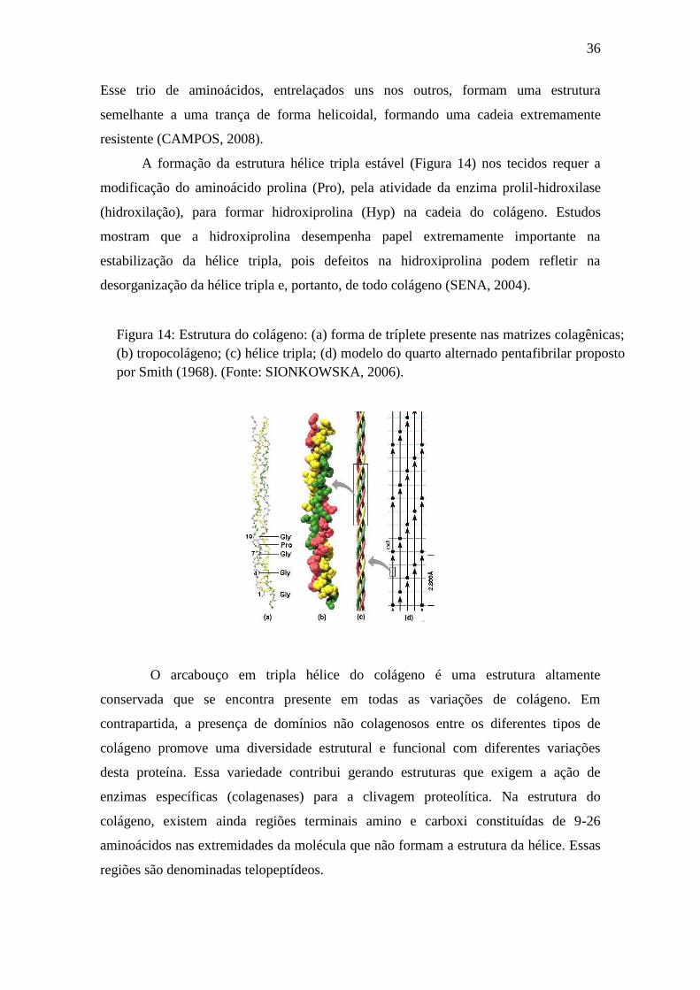
14 Estrutura do colágeno: (a) forma de tríplete presente nas matrizes
colagênicas; (b) tropocolágeno; (c) hélice tripla; (d) modelo do quarto
alternado pentafibrilar...........................................................................
Capítulo 1: Spotted sorubim (Pseudoplatystoma corruscans)
viscera as a source of acid protease for collagen extraction
1 Effect of temperature and pH on the acid digestive protease from P.
corruscans. (A) Optimum temperature for activity of enzyme. (B)
Thermal stability of enzyme, after 30 min of incubation in the
...........25
...............27
...............28
................32
..............34
..............36
temperature range 25–80°C. (C) pH optimum for activity of enzyme,
utilizing various buffers in the pH range 1.5-6.5. (D) pH stability of
enzyme, after incubation of 30 min in the pH range 2.0-
10.5.......................................................................................................
2 Zymogram of protein activity from stomach of P. corruscans. The
enzyme activity occurred in the presence of 2% hemoglobin as
substrate. The zymogram was performed in the absence of SDS and
12.5% (w/v) separating gel. The gel was stained for protein overnight in
a solution containing 0.25% (w/v) Coomassie Brilliant Blue, 10% (v/v)
acetic acid and 25% methanol. The background of the gel was destained
by washing in a solution containing 10% (v/v) acetic acid and 25%
methanol (v/v) .....................................................................................
Capítulo 2: Extraction, partial purification and characterization of
acid protease from viscera of spotted sorubim (Pseudoplatystoma
corruscans)
1 Chromatogram of purification on DEAE-Cellulose column: (●)
Absorbance (280 nm); (-) NaCl step gradient......................................
2 A) SDS-PAGE electrophoresis 12.5%. B) Zymogram 14% using 2%
hemoglobin as substrate........................................................................
3 A) Effect of pH on the activity of semi-purified enzyme from the
stomach of P. corruscans. The enzymatic activity assay was performed
at different pH (1.5 to 7.5). Glycine-HCl buffer pH 1.5-3.5; Citrate-
phosphate buffer pH 4.0-7.5; Tris-HCl buffer pH 8.0-9.0. B) Effect of
pH on the stability of semi-purified enzyme from P. corruscans
stomach. The enzyme was incubated for 30 min. at various pH (1.5 to
9.0). Subsequently, the enzymatic activity assay was conducted at 37°C.
The activity was performed using 2% hemoglobin as
substrate................................................................................................
4 A) Effect of temperature on the activity of semi-purified acid protease
from P. corruscans stomach. Enzymatic activity assays were performed
in the temperature range from 25 to 75°C. B) Effect of temperature on
the stability of semi-purified enzyme from P. corruscans stomach. The
...............87
...............88
...............110
..............110
...............111
enzyme was incubated for 30 min. at various temperatures (25 to 75°C).
After cooling enzyme was performed enzymatic activity assay at 37°C.
The activity was performed using 2% hemoglobin as substrate.
..............................................................................................................
Capítulo 3: Isolation and characterization of skin collagen of
spotted sorubim (Pseudoplatystoma corruscans)
1 SDS-PAGE patterns of acid soluble collagen (ASC) and pepsin soluble
collagen (PSC) from the Pseudoplatystoma corruscans skin. M, high-
molecular weight markers; I, type I collagen from calf skin.
...............................................................................................................
2 Relative solubility (%) of ASC and PSC from Pseudoplatystoma
corruscans skin in 0.5 M acetic acid at different pHs. ........................
3 Relative solubility (%) of ASC and PSC from Pseudoplatystoma
corruscans skin in 0.5 M acetic acid with different NaCl concentrations.
...............................................................................................................
..............112
............133
............134
............134
LISTA DE TABELAS
Tabela Descrição Página
1 Sistemática Filogenética do pintado............................................................19
2 Classificação das enzimas segundo a IUBMB............................................25
Capítulo 1: Spotted sorubim (Pseudoplatystoma corruscans)
viscera as a source of acid protease for collagen extraction
1 Specific proteolytic and amylolytic activities from the
Pseudoplatystoma corruscans stomach. The results are represented
by mean ± standart deviation (n=3)...............................................
2 Inhibitory effect on the activity of proteases from P. corruscans
stomach using 2% hemoglobin (w.v-1) as substrate at 37ºC for 30
minutes. The results are represented by mean ± standart deviation
(n=3). .............................................................................................
3 Effects of metal ions on the activity of acid digestive protease from
P. corruscans stomach...................................................................
4 Yield of collagen from the skin of Oreochromus niloticus..............
Capítulo 2: Extraction, partial purification and
characterization of acid protease from viscera of spotted
sorubim (Pseudoplatystoma corruscans)
1 A summary of the purification of acid digestive protease from P.
corruscans stomach..........................................................................
2 Effect of inhibitors on the activity of semi-purified acid protease
from P. corruscans stomach. ...........................................................
Capítulo 3: Isolation and characterization of skin collagen of
spotted sorubim (Pseudoplatystoma corruscans)
1 The extraction yields of acid and pepsin soluble collagens from
fish skin............................................................................................
..............83
..............84
..............85
............86
............108
............109
............135
LISTA DE ABREVIATURAS E SÍMBOLOS
ASC Colágeno ácido solúvel
Asp Ácido aspártico
BApNA N-α-benzoil-L-arginina-p-nitoanilida
β-ME β-Mercaptoetanol
BSE Encefalopatia espongiforme bovina
DEAE-cellulose Diethylaminoethyl cellulose
DMSO Dimetilsulfóxido
D.N.O.C.S. Departamento Nacional de Obras Contra a Seca
DNSA Ácido 3,5 - dinitrossalicílico
E-64 L-3-carboxitrans- 2, 3-epoxi-propionil-L-leucin-4-guanidino-
butilamida
EC Comitê enzimático
EDTA Etileno-diamina-tetra-acético
ES Complexo Enzima-Substrato
FA Febre aftosa
FAO Organização das nações Unidas para Alimentação e Agricultura
FMD Febre aftosa
Gly Glicina
Hyp Hidroxiprolina
IUBMB União Internacional de Bioquímica e Biologia Molecular
k Constante de velocidade
kDa Quilo Daltons
mM Milimolar
nm Nanômetro
MPA Ministério da Pesca e Aquicultura
PGs Pepsinogênios
pH Potencial Hidrogeniônico
pI Ponto isoelétrico
PMSF Fenilmetanossufonilfluoreto
Pro Prolina
PSC Colágeno pepsin solúvel
RNA Ácido ribonucleico
SAPNA Succinil-alanina-alanina-prolina-fenilalanina-p-nitroanilida
SDS Dodecil sulfato de sódio
SDS-PAGE Eletroforese em gel de poliacrilamida com dodecil sulfato de
sódio
TAME Tosil-arginina-metil-éster
TLCK N-p-tosil-L-lisina clorometil cetona
TPCK N-tosil-L-fenilalanina clorometil cetona
TSE Encefalopatia espongiforme transmissível
UFPE Universidade Federal de Pernambuco
U/mg Unidades de atividade enzimática por miligrama
U/mL Unidades por mililitro
UV-vis Ultravioleta visível
v/v Volume/volume
w/v Peso/volume
µg Micrograma
µL Microlitro
SUMÁRIO
1. Introdução................................................................................................................15
2. Revisão Da Literatura.............................................................................................17
2.1 Panorama da aquicultura e produção de pintado (Pseudoplatystoma
corruscans)................................................................................................................17
2.2 Pintado (Pseudoplatystoma corruscans)...........................................................19
2.2.1 Bioecologia do P. corruscans...................................................................21
2.2.2 Importância econômica do Pseudoplatystoma corruscans....................22
2.3 Utilização de resíduos da aquicultura..............................................................23
2.4 Enzimas...............................................................................................................24
2.4.1 Carboidrases.............................................................................................26
2.4.2 Proteases digestivas de peixes.................................................................27
2.5 Pepsina.........................................................................................................30
2.5.1 Aplicabilidade industrial da pepsina......................................................32
2.6 Colágeno..............................................................................................................33
2.6.1 Estrutura molecular do colágeno...........................................................34
2.6.2 Tipos de colágeno.....................................................................................37
2.6.3 Fontes de colágeno...................................................................................37
2.6.4 Aplicações do uso do colágeno................................................................38
3. Objetivos....................................................................................................................40
3.1 Geral....................................................................................................................40
3.2 Específicos...........................................................................................................40
Referências....................................................................................................................41
Capítulo 1......................................................................................................................56
Capítulo 2......................................................................................................................89
Capítulo 3....................................................................................................................113
Considerações finais...................................................................................................136
Anexos..........................................................................................................................137
15
1. INTRODUÇÃO
A produção aquícola mundial tem passado por um constante crescimento devido
a um aumento na demanda por produtos pesqueiros. Estima-se que as atividades de
pesca extrativa e aquicultura em 2013 atingiram um novo recorde mundial, com 160
milhões de toneladas contra as 157 milhões de toneladas do ano anterior (FAO, 2014).
No entanto, com a estagnação da quantidade de pescado proveniente da captura, a
aquicultura vem assumindo, nos últimos anos, a responsabilidade de atender à demanda
por produtos aquícolas, através do aumento da utilização de espécies e tecnologias
adequadas. Segundo as projeções da FAO, os aquicultores produziram 70 milhões de
toneladas de pescado no ano passado, 44% da produção total mundial (FAO, 2014).
A aquicultura brasileira, só há pouco tempo, vem dando ênfase para espécies de
peixes nativos. Na procura por novidades para atender ao mercado, os produtores têm
buscado espécies inovadoras, que tenham boa produtividade e aceitação para a pesca e o
consumo. Dentre as espécies nativas, várias são aquelas com esse potencial, sem,
contudo, terem sido estudadas suficientemente. Espécies carnívoras, como o pintado
(Pseudoplatystoma corruscans), têm despertando interesse dos pesquisadores e
produtores de peixes, principalmente devido ao valor comercial, à qualidade da carne e
às características esportivas para a pesca que apresentam (LUZ et al., 2001).
A produção de pintado teve o primeiro registro nacional em 1998, com 329
toneladas, e em onze anos cresceu 546%, atingindo a marca de 2.126,7 toneladas em
2009. É importante ressaltar que o crescimento da produção no período de 2006 a 2009
foi de 94%, mostrando-se bastante consistente e expressivo, com média anual de 19,4%
(IBAMA, 2010). Apesar de ter alcançado um crescimento vertiginoso, a criação desta
espécie em cativeiro ainda apresenta limitações, sobretudo relacionadas à sua nutrição.
A falta de dados concretos sobre o hábito alimentar e as exigências nutricionais nas
diferentes fases de crescimento fazem com que as deficiências na alimentação e na
nutrição desse peixe sejam responsáveis por altos índices de mortalidade (CREPALDI
et al., 2006a).
Em contrapartida, o aumento da produção pesqueira é proporcional às
concentrações de resíduos deixados através do processamento do pescado. Em geral os
resíduos são descartados no ambiente e causam sérios problemas de poluição. Essa
crescente poluição ambiental e o reconhecimento de que o uso dos recursos biológicos é
limitado tem enfatizado a necessidade de utilização de subprodutos da indústria
pesqueira (MONTE, 2013).
16
As vísceras dos peixes estão entre os resíduos que não são consumidos pelos
humanos e correspondem por cerca de 10% do peso total do animal (SIMPSON e
HAARD, 1987). De acordo com Bezerra et al. (2000), as vísceras dos peixes são
conhecidas por serem ricas em proteases. Proteases ácidas como a pepsina representam
uma alternativa para o uso em certos processos biotecnológicos: coagulação do leite
(TAVARES et al., 1997), produção de silagem (GILDBERG et al., 2004) e extração de
colágeno (BENJAKUL et al., 2010a). Devido a sua ampla aplicabilidade em processos
biotecnológicos, pepsinas têm sido extraídas, purificadas e caracterizadas a partir de
vísceras de peixes (WU et al., 2009; NALINANON et al., 2010; KHALED et al., 2011).
Em relação ao aproveitamento de resíduos pesqueiros, o número de trabalhos
objetivando a extração de colágeno a partir de organismos aquáticos tem crescido
consideravelmente. Colágeno da pele de várias espécies de peixes tem sido extraído e
caracterizado, como Ictalurus punctatus (LIU et al., 2007), Lutjanus lutjanus
(NALINANON et al., 2007), Monoceros aluterus (AHMAD et al., 2010), Nemipterus
hexodon (NALINANON et al., 2011), Diodon holocanthus (HUANG et al., 2011),
Pangasianodon hypophthalmus (SINGH et al., 2011), Evenchelys macrura
(VEERURAJ et al., 2013), Hypophthalmichthys molitrix (SAFANDOWSKA et al.,
2013) e Acipensern schrenckii (VEERURAJ et al., 2014).
Sendo assim, o presente trabalho aborda o estudo das vísceras do pintado com
enfoque na obtenção de biomoléculas ativas e aplicação biotecnológica na extração de
colágeno. Dessa forma, pode-se fornecer conhecimento da sua fisiologia digestiva,
contribuindo para o entendimento da capacidade enzimática digestiva, bem como
propiciar a utilização dos resíduos do seu processamento, agregando valor ao pescado.
17
2. REVISÃO DA LITERATURA
2.1 Panorama da aquicultura e produção de pintado
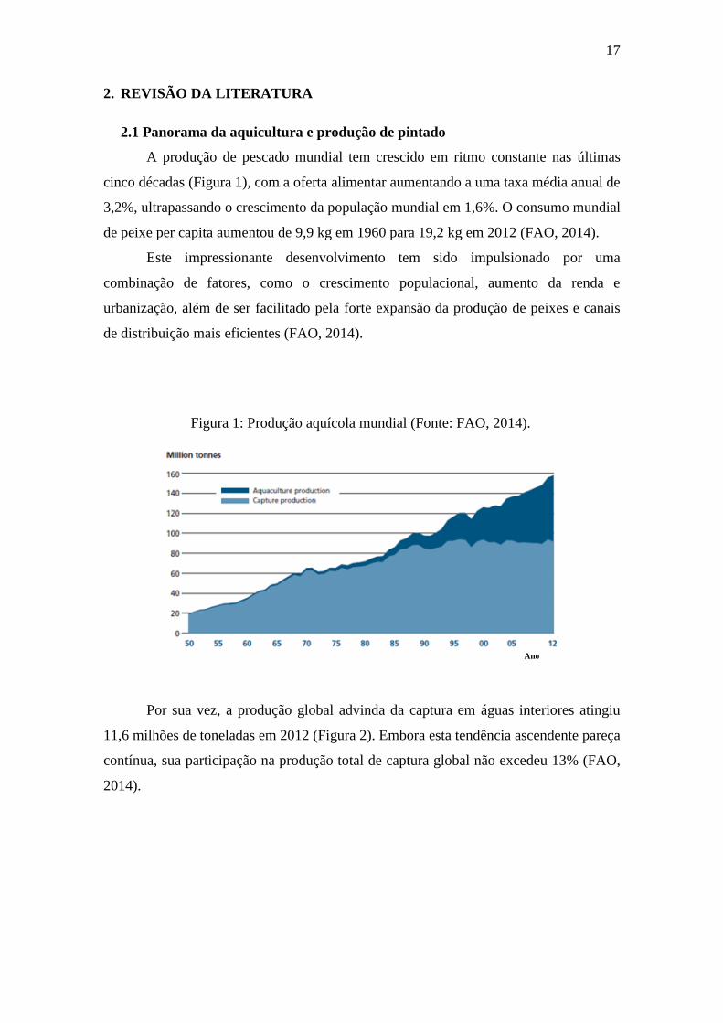
A produção de pescado mundial tem crescido em ritmo constante nas últimas
cinco décadas (Figura 1), com a oferta alimentar aumentando a uma taxa média anual de
3,2%, ultrapassando o crescimento da população mundial em 1,6%. O consumo mundial
de peixe per capita aumentou de 9,9 kg em 1960 para 19,2 kg em 2012 (FAO, 2014).
Este impressionante desenvolvimento tem sido impulsionado por uma
combinação de fatores, como o crescimento populacional, aumento da renda e
urbanização, além de ser facilitado pela forte expansão da produção de peixes e canais
de distribuição mais eficientes (FAO, 2014).
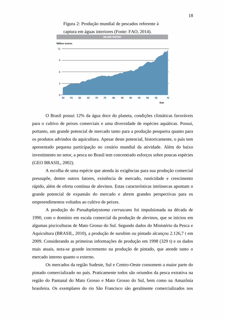
Por sua vez, a produção global advinda da captura em águas interiores atingiu
11,6 milhões de toneladas em 2012 (Figura 2). Embora esta tendência ascendente pareça
contínua, sua participação na produção total de captura global não excedeu 13% (FAO,
2014).
Ano
Figura 1: Produção aquícola mundial (Fonte: FAO, 2014).
18
O Brasil possui 12% da água doce do planeta, condições climáticas favoráveis
para o cultivo de peixes comerciais e uma diversidade de espécies aquáticas. Possui,
portanto, um grande potencial de mercado tanto para a produção pesqueira quanto para
os produtos advindos da aquicultura. Apesar deste potencial, historicamente, o país tem
apresentado pequena participação no cenário mundial da atividade. Além do baixo
investimento no setor, a pesca no Brasil tem concentrado esforços sobre poucas espécies
(GEO BRASIL, 2002).
A escolha de uma espécie que atenda às exigências para sua produção comercial
pressupõe, dentre outros fatores, existência de mercado, rusticidade e crescimento
rápido, além de oferta contínua de alevinos. Estas características intrínsecas apontam o
grande potencial de expansão do mercado e abrem grandes perspectivas para os
empreendimentos voltados ao cultivo de peixes.
A produção do Pseudoplatystoma corruscans foi impulsionada na década de
1990, com o domínio em escala comercial da produção de alevinos, que se iniciou em
algumas pisciculturas de Mato Grosso do Sul. Segundo dados do Ministério da Pesca e
Aquicultura (BRASIL, 2010), a produção de surubim ou pintado alcançou 2.126,7 t em
2009. Considerando as primeiras informações de produção em 1998 (329 t) e os dados
mais atuais, nota-se grande incremento na produção de pintado, que atende tanto o
mercado interno quanto o externo.
Os mercados da região Sudeste, Sul e Centro-Oeste consomem a maior parte do
pintado comercializado no país. Praticamente todos são oriundos da pesca extrativa na
região do Pantanal do Mato Grosso e Mato Grosso do Sul, bem como na Amazônia
brasileira. Os exemplares do rio São Francisco são geralmente comercializados nos
Ano
Figura 2: Produção mundial de pescados referente à
captura em águas interiores (Fonte: FAO, 2014).
19
centros consumidores próximos aos locais de captura, visto que a quantidade desse
peixe nessa bacia caiu drasticamente (KUBITZA et al.,1998).
2.2 Pintado (Pseudoplatystoma corruscans)
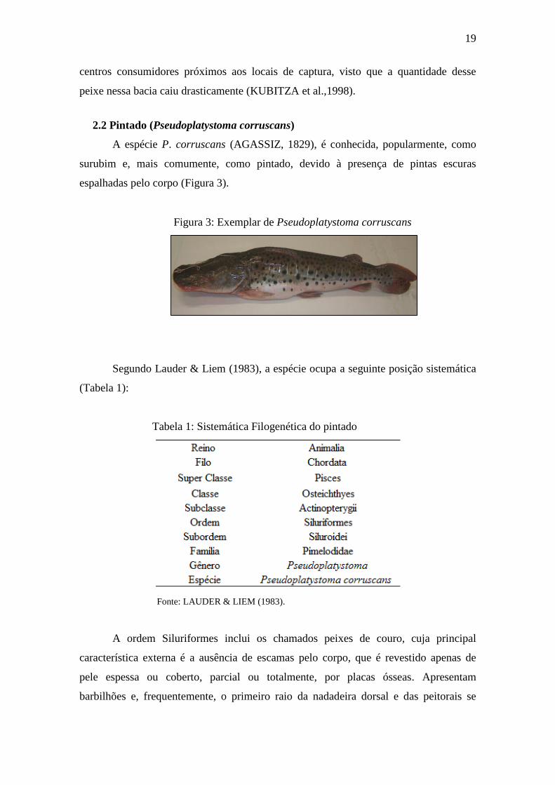
A espécie P. corruscans (AGASSIZ, 1829), é conhecida, popularmente, como
surubim e, mais comumente, como pintado, devido à presença de pintas escuras
espalhadas pelo corpo (Figura 3).
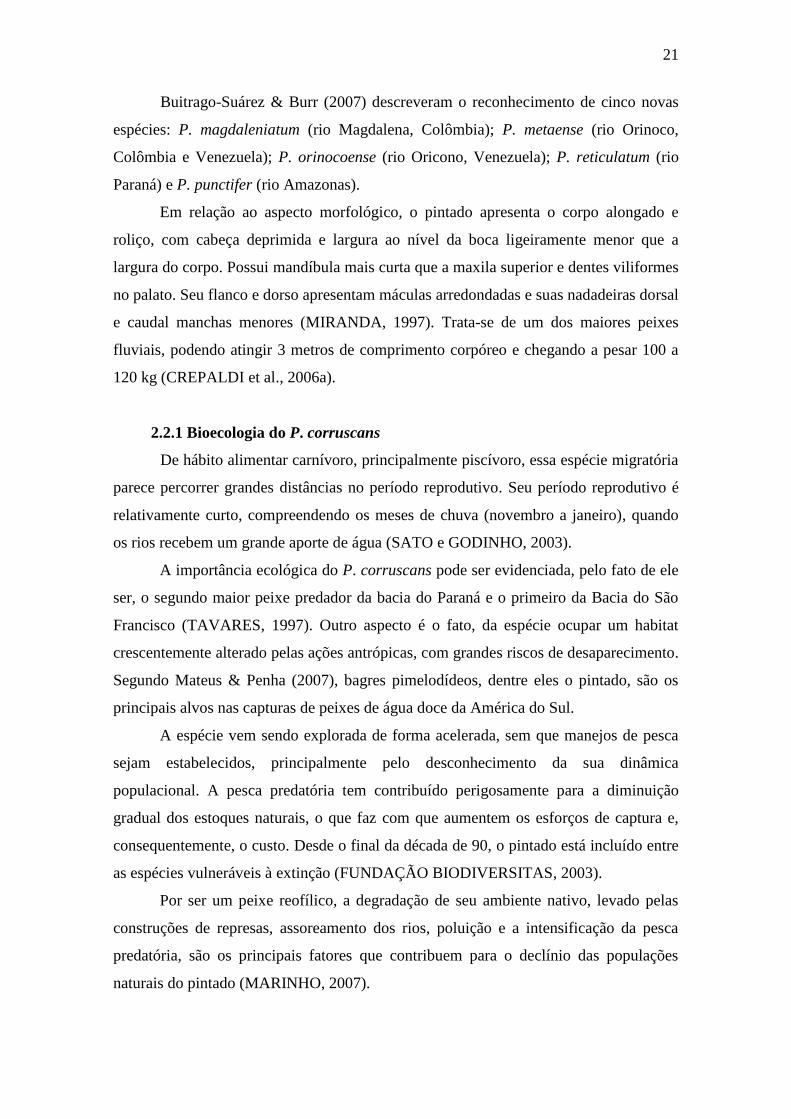
Segundo Lauder & Liem (1983), a espécie ocupa a seguinte posição sistemática
(Tabela 1):
Tabela 1: Sistemática Filogenética do pintado
A ordem Siluriformes inclui os chamados peixes de couro, cuja principal
característica externa é a ausência de escamas pelo corpo, que é revestido apenas de
pele espessa ou coberto, parcial ou totalmente, por placas ósseas. Apresentam
barbilhões e, frequentemente, o primeiro raio da nadadeira dorsal e das peitorais se
Fonte: LAUDER & LIEM (1983).
Figura 3: Exemplar de Pseudoplatystoma corruscans
20
constitui de um acúleo forte e pungente (MIRANDA et al., 1997). Segundo Kubitza et
al. (1998), a ordem Siluriformes engloba mais de 2.200 espécies de bagres espalhados
por todos os continentes.
A subordem Siluroidei é composta por um significativo número de gêneros e
espécies dulcícolas de hábitos noturnos e dieta variada. Compreende treze famílias na
região neotropical, das quais sete ocorrem no rio São Francisco, dentre elas a
Pimelodidae, que é a mais numerosa da subordem e tem como característica distinta a
presença de três pares de barbilhões próximos à boca (MARINHO, 2007).
O gênero Pseudoplatystoma (BLEEKER, 1862), que inclui os maiores peixes da
família Pimelodidae, é exclusivamente de água doce e pode ser encontrado nas
principais bacias hidrográficas sul-americanas. Sua distribuição inclui os rios
Amazonas, Orinoco, São Francisco, bem como no sistema do rio da Prata formado
pelos rios Paraná, Paraguai e Uruguai (ROMAGOSA et al., 2003).
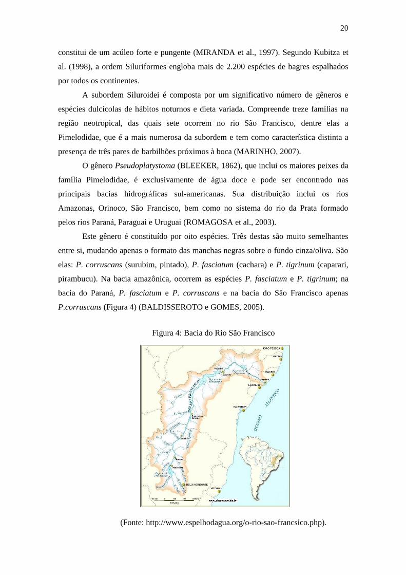
Este gênero é constituído por oito espécies. Três destas são muito semelhantes
entre si, mudando apenas o formato das manchas negras sobre o fundo cinza/oliva. São
elas: P. corruscans (surubim, pintado), P. fasciatum (cachara) e P. tigrinum (caparari,
pirambucu). Na bacia amazônica, ocorrem as espécies P. fasciatum e P. tigrinum; na
bacia do Paraná, P. fasciatum e P. corruscans e na bacia do São Francisco apenas
P.corruscans (Figura 4) (BALDISSEROTO e GOMES, 2005).
Figura 4: Bacia do Rio São Francisco
(Fonte: http://www.espelhodagua.org/o-rio-sao-francsico.php).
21
Buitrago-Suárez & Burr (2007) descreveram o reconhecimento de cinco novas
espécies: P. magdaleniatum (rio Magdalena, Colômbia); P. metaense (rio Orinoco,
Colômbia e Venezuela); P. orinocoense (rio Oricono, Venezuela); P. reticulatum (rio
Paraná) e P. punctifer (rio Amazonas).
Em relação ao aspecto morfológico, o pintado apresenta o corpo alongado e
roliço, com cabeça deprimida e largura ao nível da boca ligeiramente menor que a
largura do corpo. Possui mandíbula mais curta que a maxila superior e dentes viliformes
no palato. Seu flanco e dorso apresentam máculas arredondadas e suas nadadeiras dorsal
e caudal manchas menores (MIRANDA, 1997). Trata-se de um dos maiores peixes
fluviais, podendo atingir 3 metros de comprimento corpóreo e chegando a pesar 100 a
120 kg (CREPALDI et al., 2006a).
2.2.1 Bioecologia do P. corruscans
De hábito alimentar carnívoro, principalmente piscívoro, essa espécie migratória
parece percorrer grandes distâncias no período reprodutivo. Seu período reprodutivo é
relativamente curto, compreendendo os meses de chuva (novembro a janeiro), quando
os rios recebem um grande aporte de água (SATO e GODINHO, 2003).
A importância ecológica do P. corruscans pode ser evidenciada, pelo fato de ele
ser, o segundo maior peixe predador da bacia do Paraná e o primeiro da Bacia do São
Francisco (TAVARES, 1997). Outro aspecto é o fato, da espécie ocupar um habitat
crescentemente alterado pelas ações antrópicas, com grandes riscos de desaparecimento.
Segundo Mateus & Penha (2007), bagres pimelodídeos, dentre eles o pintado, são os
principais alvos nas capturas de peixes de água doce da América do Sul.
A espécie vem sendo explorada de forma acelerada, sem que manejos de pesca
sejam estabelecidos, principalmente pelo desconhecimento da sua dinâmica
populacional. A pesca predatória tem contribuído perigosamente para a diminuição
gradual dos estoques naturais, o que faz com que aumentem os esforços de captura e,
consequentemente, o custo. Desde o final da década de 90, o pintado está incluído entre
as espécies vulneráveis à extinção (FUNDAÇÃO BIODIVERSITAS, 2003).
Por ser um peixe reofílico, a degradação de seu ambiente nativo, levado pelas
construções de represas, assoreamento dos rios, poluição e a intensificação da pesca
predatória, são os principais fatores que contribuem para o declínio das populações
naturais do pintado (MARINHO, 2007).
22
2.2.2 Importância econômica do Pseudoplatystoma corruscans
No Brasil, o pintado está entre as espécies de peixes de água doce com grande
potencial de consumo, representativos na produção pesqueira de águas interiores,
devido às suas grandes proporções e à qualidade de sua carne (BENÍTEZ, 2003). A
grande aceitação pelos consumidores decorre do fato da espécie apresentar carne
saborosa, com baixo teor de gordura e ausência de espinhos intramusculares, o que o
torna adequado aos mais variados preparos. Estas características atendem as
preferências de mercado e fazem da carne do pintado um produto com grandes
possibilidades de exportação (INOUE et al., 2008).
A valorização econômica e social da espécie tem sido demonstrada não só pela
representatividade das capturas, mas também devido à importância quanto à
possibilidade de seu emprego na piscicultura empresarial. Além da sua grande estima
popular, o pintado é também valioso e muito apreciado pelos pescadores esportivos. A
agressividade e o grande porte que alcançam fizeram com que conquistassem
rapidamente esse mercado. Nesse tipo de comércio, o quilo do peixe tende a alcançar
maiores valores, tornando-se uma alternativa interessante para os piscicultores.
Camargo e Petrere (2001) confirmaram a importância dessa espécie na pesca artesanal
devido ao seu alto valor de mercado.
No Brasil existe grande número de espécies que têm potencial para o cultivo,
pois preenchem os requisitos necessários para a escolha de uma espécie adequada para a
piscicultura. Dentre elas, o P. corruscans tem se destacado por apresentar características
comerciais e zootécnicas desejáveis, como rápido crescimento e eficiente conversão
alimentar. As características e os rendimentos de carcaça (CREPALDI et al., 2008) e a
capacidade de obtenção de gametas por meio da hipofisação (CREPALDI et al., 2006b)
reforçam a qualificação da espécie para a piscicultura industrial. Por apresentar estas
vantagens, a produção de pintado vem crescendo no Brasil (PILECCO et al., 2008).
Entretanto, apesar de possuir grande potencial zootécnico, o cultivo desta
espécie apresenta limitações relacionadas ao manejo alimentar, por ser carnívora,
podendo se alimentar de ração quando condicionada. Um exemplo dessa lacuna é a
dificuldade para introduzir o alimento artificial na dieta das larvas e juvenis. Além
disso, devido ao hábito noturno, a espécie só se alimenta durante o dia quando
submetida a treinamento (BALDISSEROTO e GOMES, 2005).
No momento já há tecnologia adequada para a reprodução do pintado e obtenção
de larvas, mas o maior desafio está na mudança da fase de larva para juvenil. Por
23
apresentar hábito alimentar piscívoro, aliado ao desconhecimento de técnicas de manejo
alimentar adequadas, nessa fase ocorre um acentuado canibalismo, levando à redução da
taxa de sobrevivência durante a criação inicial (ALVARADO et al., 2003).
No entanto, muito trabalho ainda tem que ser realizado para a elaboração de um
pacote tecnológico, principalmente nas fases mais complicadas do cultivo: a larvicultura
e o treinamento alimentar. Segundo Sato et al. (1997) o P. corruscans requer cuidados
extremos, principalmente nas fases iniciais, das quais, embora seja grande o número de
indivíduos produzidos numa única desova, os organismos são extremamente frágeis.
2.3 Utilização de resíduos da aquicultura
O aumento da produção pesqueira e, consequentemente, do volume de pescado
processado mundialmente, tem gerado uma grande quantidade de resíduos e
subprodutos. Por serem produtos altamente perecíveis, os peixes apresentam uma
necessidade significativa de processamento, o que gera grande quantidade de resíduos
líquidos (águas residuais) e sólidos (pele, ossos, vísceras, nadadeiras e cabeças)
(DOODE, 1996).
Os resíduos da indústria pesqueira apresentam uma composição rica em
compostos orgânicos e inorgânicos, o que gera preocupação relativa aos potenciais
impactos ambientais negativos decorrentes da disposição deste material diretamente no
ambiente (SEIBEL & SOARES, 2003). Tal fato representa um desafio para empresários
e comunidade científica interessados no desenvolvimento sustentável da aquicultura.
Como já é sabido, a redução do uso inconsciente da matéria prima além de evitar
desperdícios e promover a reciclagem dos resíduos, garante processos mais econômicos
e com menor impacto ambiental.
Assim, preocupados com problemas ambientais, pesquisadores em todo o mundo
vêm desenvolvendo diversos esforços para obtenção de métodos que possibilitem a
transformação desses materiais em co-produtos com valor agregado
(ARVANITOYANNIS e KASSAVETI, 2008). A obtenção de biomoléculas com ampla
aplicabilidade tecnológica a partir de resíduos de peixes tem sido uma necessidade por
parte do setor industrial. Biomoléculas como enzimas, polissacarídeos e colágeno
provenientes das sobras do processamento de peixes têm propiciado a criação de novos
setores industriais, contribuindo também com a sustentabilidade econômica e ambiental
da indústria pesqueira (COSTA, 2012).

24
Os resíduos da indústria pesqueira são passíveis de aplicação em diversos
segmentos industriais, entre eles: obtenção de silagem de peixe (ARRUDA et al., 2006),
com potencial para utilização como fonte proteica em rações (BORGHESI et al., 2007);
produção de fertilizantes ou produtos químicos (CAVALCANTE JÚNIOR et al., 2005);
iscas e artesanatos (BANCO do NORDESTE, 1999); na produção de biodiesel/biogás
(KATO et al., 2004); produtos dietéticos (quitosana) (GILDBERG e STENBERG,
2001); pigmentos naturais (SACHINDRA et al., 2006); imobilização de cromo
(OZAWA et al., 2003); embalagens de alimentos (quitosana, gelatina e colágeno)
(GÓMEZ-ESTACA et al., 2009); obtenção de enzimas, principalmente proteases
(ESPÓSITO et al., 2010; MARCUSCHI et al., 2010; SILVA et al., 2011; FREITAS-
JÚNIOR et al., 2012; COSTA et al., 2013) e na indústria farmacêutica e de cosméticos
(colágeno) (BENJAKUL et al., 2010b; LIU et al., 2012).
2.4 Enzimas
Enzimas são biomoléculas catalisadoras que atuam diminuindo o nível de
energia de ativação, implicando no aumento da velocidade das reações bioquímicas. A
eficiência das enzimas em catalisar reações é tal que a velocidade de uma reação pode
ser aumentada em até 1020 vezes (HARVEY et al., 2009).
Todas as enzimas conhecidas, com exceção de certos RNAs catalíticos, são
proteínas, e estão presente em todos os organismos vivos, sendo essenciais, tanto para a
manutenção, como para o crescimento e a diferenciação celular. As enzimas digestórias
agem em sequências organizadas e catalisam centenas de reações sucessivas, pelas quais
as moléculas de nutrientes são degradadas. Essas biomoléculas catalisadoras não
reagem quimicamente com as substâncias sobre as quais atuam, nem alteram o
equilíbrio das reações (NELSON e COX, 2004).
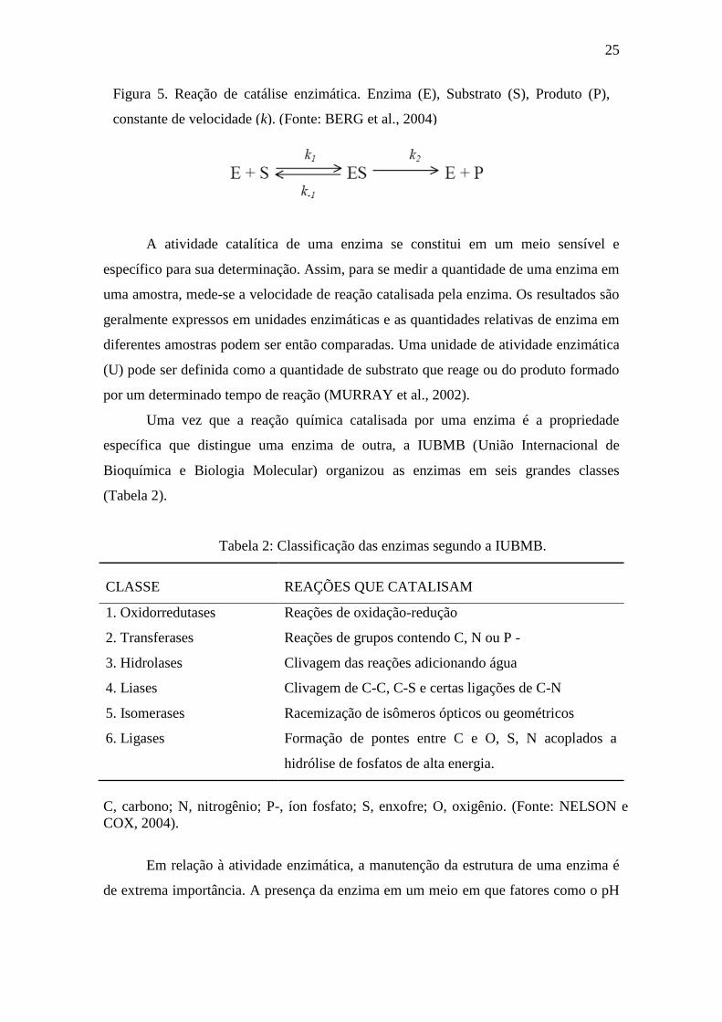
De uma maneira geral, uma enzima liga-se ao seu substrato formando um
complexo Enzima-Substrato (ES), de caráter transitório (Figura 5). Provavelmente,
apenas uma fração da molécula, denominada sítio ativo, é a responsável pela ligação da
enzima ao substrato, e essa fração determina a especificidade enzimática (NELSON e
COX, 2004). Podem existir também outras regiões da cadeia polipeptídica que são
sensíveis à presença de determinadas espécies químicas, modulando a atividade da
enzima. Tais regiões são denominadas centros alostéricos (BERG et al., 2004).
25
A atividade catalítica de uma enzima se constitui em um meio sensível e
específico para sua determinação. Assim, para se medir a quantidade de uma enzima em
uma amostra, mede-se a velocidade de reação catalisada pela enzima. Os resultados são
geralmente expressos em unidades enzimáticas e as quantidades relativas de enzima em
diferentes amostras podem ser então comparadas. Uma unidade de atividade enzimática
(U) pode ser definida como a quantidade de substrato que reage ou do produto formado
por um determinado tempo de reação (MURRAY et al., 2002).
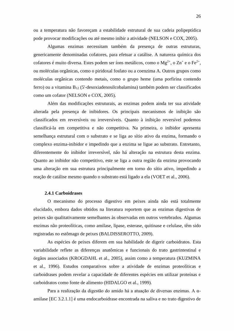
Uma vez que a reação química catalisada por uma enzima é a propriedade
específica que distingue uma enzima de outra, a IUBMB (União Internacional de
Bioquímica e Biologia Molecular) organizou as enzimas em seis grandes classes
(Tabela 2).
Tabela 2: Classificação das enzimas segundo a IUBMB.
CLASSE REAÇÕES QUE CATALISAM
1. Oxidorredutases Reações de oxidação-redução
2. Transferases Reações de grupos contendo C, N ou P -
3. Hidrolases Clivagem das reações adicionando água
4. Liases Clivagem de C-C, C-S e certas ligações de C-N
5. Isomerases Racemização de isômeros ópticos ou geométricos
6. Ligases Formação de pontes entre C e O, S, N acoplados a
hidrólise de fosfatos de alta energia.
C, carbono; N, nitrogênio; P-, íon fosfato; S, enxofre; O, oxigênio. (Fonte: NELSON e
COX, 2004).
Em relação à atividade enzimática, a manutenção da estrutura de uma enzima é
de extrema importância. A presença da enzima em um meio em que fatores como o pH
Figura 5. Reação de catálise enzimática. Enzima (E), Substrato (S), Produto (P),
constante de velocidade (k). (Fonte: BERG et al., 2004)
26
ou a temperatura não favoreçam a estabilidade estrutural de sua cadeia polipeptídica
pode provocar modificações ou até mesmo inibir a atividade (NELSON e COX, 2005).
Algumas enzimas necessitam também da presença de outras estruturas,
genericamente denominadas cofatores, para efetuar a catálise. A natureza química dos
cofatores é muito diversa. Estes podem ser íons metálicos, como o Mg2+, o Zn+ e o Fe2+,
ou moléculas orgânicas, como o piridoxal fosfato ou a coenzima A. Outros grupos como
moléculas orgânicas contendo metais, como o grupo heme (uma porfirina contendo
ferro) ou a vitamina B12 (5'-desoxiadenosilcobalamina) também podem ser classificados
como um cofator (NELSON e COX, 2005).
Além das modificações estruturais, as enzimas podem ainda ter sua atividade
alterada pela presença de inibidores. Os principais mecanismos de inibição são
classificados em reversíveis ou irreversíveis. Quanto à inibição reversível podemos
classificá-la em competitiva e não competitiva. Na primeira, o inibidor apresenta
semelhança estrutural com o substrato e se liga ao sítio ativo da enzima, formando o
complexo enzima-inibidor e impedindo que a enzima se ligue ao substrato. Entretanto,
diferentemente do inibidor irreversível, não há alteração na estrutura desta enzima.
Quanto ao inibidor não competitivo, este se liga a outra região da enzima provocando
uma alteração em sua estrutura principalmente em torno do sítio ativo, impedindo a
reação de catálise mesmo quando o substrato está ligado a ela (VOET et al., 2006).
2.4.1 Carboidrases
O mecanismo do processo digestivo em peixes ainda não está totalmente
elucidado, embora dados obtidos na literatura reportem que as enzimas digestivas de
peixes são qualitativamente semelhantes às observadas em outros vertebrados. Algumas
enzimas não proteolíticas, como amilase, lipase, esterase, quitinase e celulase, têm sido
registradas no estômago de peixes (BALDISSEROTTO, 2009).
As espécies de peixes diferem em sua habilidade de digerir carboidratos. Esta
variabilidade reflete as diferenças anatômicas e funcionais do trato gastrintestinal e
órgãos associados (KROGDAHL et al., 2005), assim como a temperatura (KUZMINA
et al., 1996). Estudos comparativos sobre a atividade de enzimas proteolíticas e
carboidrases podem revelar a capacidade de diferentes espécies em utilizar proteínas e
carboidratos como fonte de alimento (HIDALGO et al., 1999).
Para a realização da digestão do amido há a atuação de diversas enzimas. A α-
amilase [EC 3.2.1.1] é uma endocarboidrase encontrada na saliva e no trato digestivo de
27
animais vertebrados (SALEH et al., 2005), responsável pela hidrólise de ligações
glicosídicas α (1,4), no amido e glicogênio. Nesse processo são produzidos
oligossacarídeos, α-dextrinas e maltose, que são hidrolisados à glicose pela ação
complementar da α-glicosidase [EC 3.2.1.20], da sacarase-isomaltase [EC 3.2.1.48] e da
α-dextrinase [EC 3.2.1.20]. Dentre essas, a α-glicosidase está diretamente relacionada à
exo-hidrólise de ligações glicosídicas α (1,4) da maltose e demais oligossacarídeos
formados após a atuação da α-amilase (ROSAS et al., 2000).
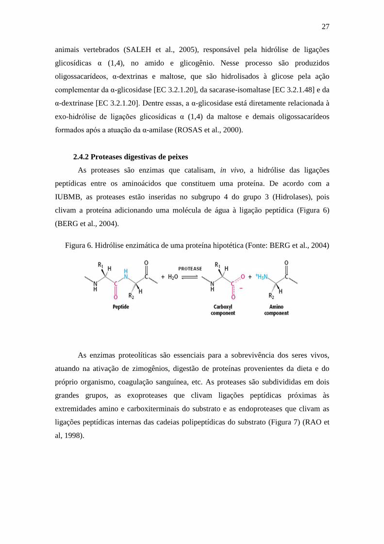
2.4.2 Proteases digestivas de peixes
As proteases são enzimas que catalisam, in vivo, a hidrólise das ligações
peptídicas entre os aminoácidos que constituem uma proteína. De acordo com a
IUBMB, as proteases estão inseridas no subgrupo 4 do grupo 3 (Hidrolases), pois
clivam a proteína adicionando uma molécula de água à ligação peptídica (Figura 6)
(BERG et al., 2004).
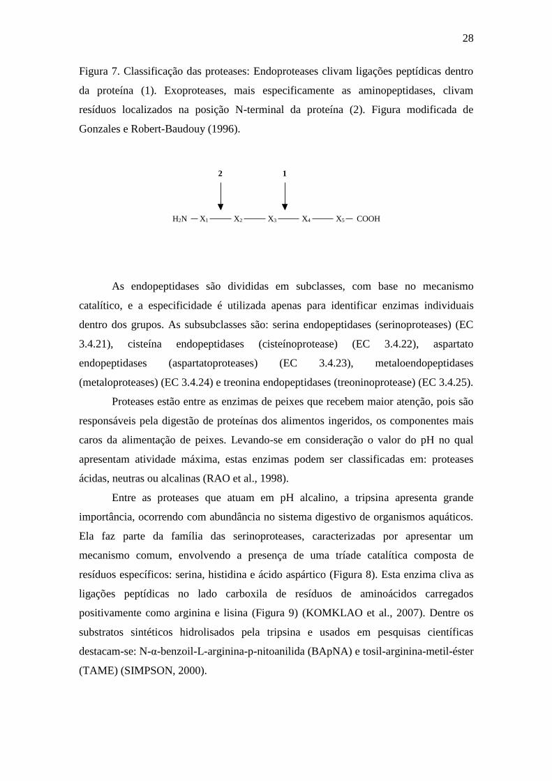
As enzimas proteolíticas são essenciais para a sobrevivência dos seres vivos,
atuando na ativação de zimogênios, digestão de proteínas provenientes da dieta e do
próprio organismo, coagulação sanguínea, etc. As proteases são subdivididas em dois
grandes grupos, as exoproteases que clivam ligações peptídicas próximas às
extremidades amino e carboxiterminais do substrato e as endoproteases que clivam as
ligações peptídicas internas das cadeias polipeptídicas do substrato (Figura 7) (RAO et
al, 1998).
Figura 6. Hidrólise enzimática de uma proteína hipotética (Fonte: BERG et al., 2004)
28
Figura 7. Classificação das proteases: Endoproteases clivam ligações peptídicas dentro
da proteína (1). Exoproteases, mais especificamente as aminopeptidases, clivam
resíduos localizados na posição N-terminal da proteína (2). Figura modificada de
Gonzales e Robert-Baudouy (1996).
X1H2N COOHX2 X3 X4 X5
12
As endopeptidases são divididas em subclasses, com base no mecanismo
catalítico, e a especificidade é utilizada apenas para identificar enzimas individuais
dentro dos grupos. As subsubclasses são: serina endopeptidases (serinoproteases) (EC
3.4.21), cisteína endopeptidases (cisteínoprotease) (EC 3.4.22), aspartato
endopeptidases (aspartatoproteases) (EC 3.4.23), metaloendopeptidases
(metaloproteases) (EC 3.4.24) e treonina endopeptidases (treoninoprotease) (EC 3.4.25).
Proteases estão entre as enzimas de peixes que recebem maior atenção, pois são
responsáveis pela digestão de proteínas dos alimentos ingeridos, os componentes mais
caros da alimentação de peixes. Levando-se em consideração o valor do pH no qual
apresentam atividade máxima, estas enzimas podem ser classificadas em: proteases
ácidas, neutras ou alcalinas (RAO et al., 1998).
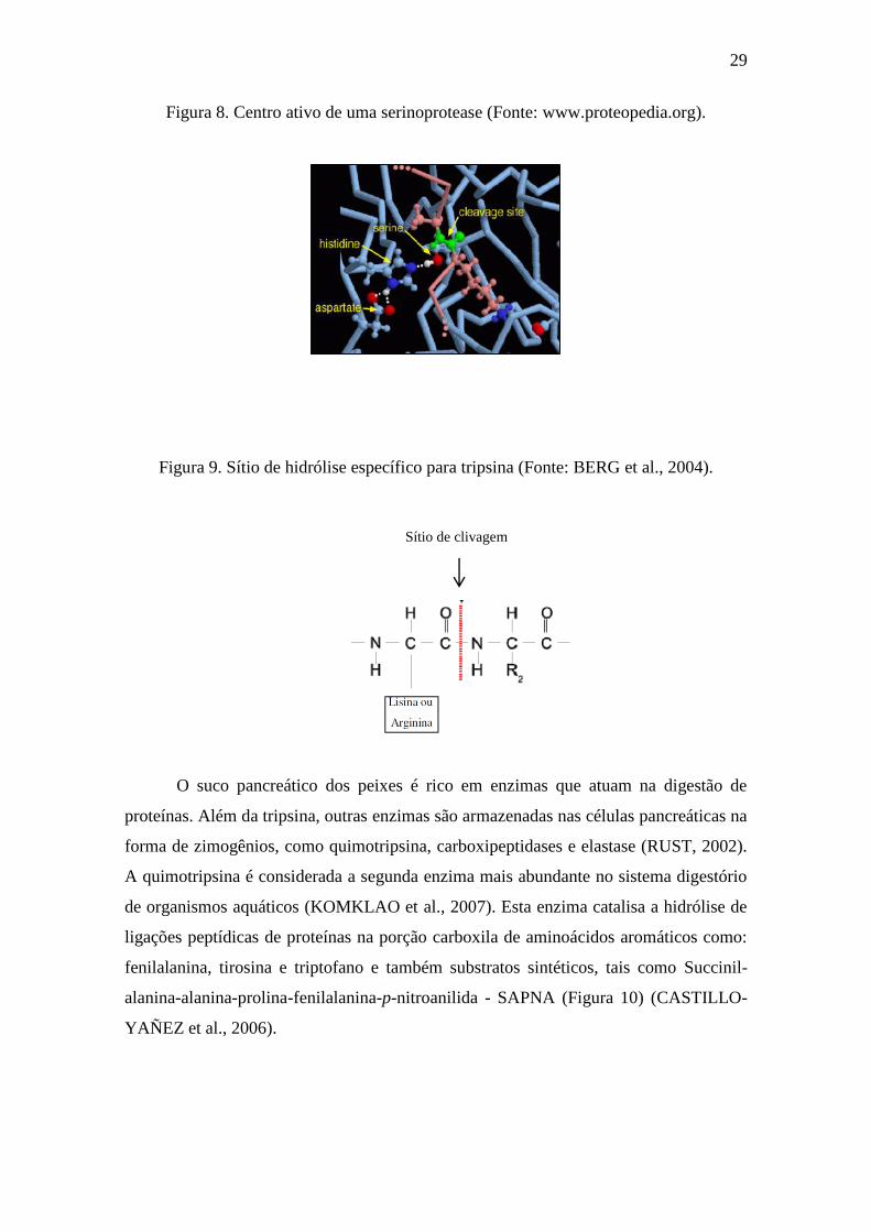
Entre as proteases que atuam em pH alcalino, a tripsina apresenta grande
importância, ocorrendo com abundância no sistema digestivo de organismos aquáticos.
Ela faz parte da família das serinoproteases, caracterizadas por apresentar um
mecanismo comum, envolvendo a presença de uma tríade catalítica composta de
resíduos específicos: serina, histidina e ácido aspártico (Figura 8). Esta enzima cliva as
ligações peptídicas no lado carboxila de resíduos de aminoácidos carregados
positivamente como arginina e lisina (Figura 9) (KOMKLAO et al., 2007). Dentre os
substratos sintéticos hidrolisados pela tripsina e usados em pesquisas científicas
destacam-se: N-α-benzoil-L-arginina-p-nitoanilida (BApNA) e tosil-arginina-metil-éster
(TAME) (SIMPSON, 2000).
29
Figura 8. Centro ativo de uma serinoprotease (Fonte: www.proteopedia.org).
Figura 9. Sítio de hidrólise específico para tripsina (Fonte: BERG et al., 2004).
O suco pancreático dos peixes é rico em enzimas que atuam na digestão de
proteínas. Além da tripsina, outras enzimas são armazenadas nas células pancreáticas na
forma de zimogênios, como quimotripsina, carboxipeptidases e elastase (RUST, 2002).
A quimotripsina é considerada a segunda enzima mais abundante no sistema digestório
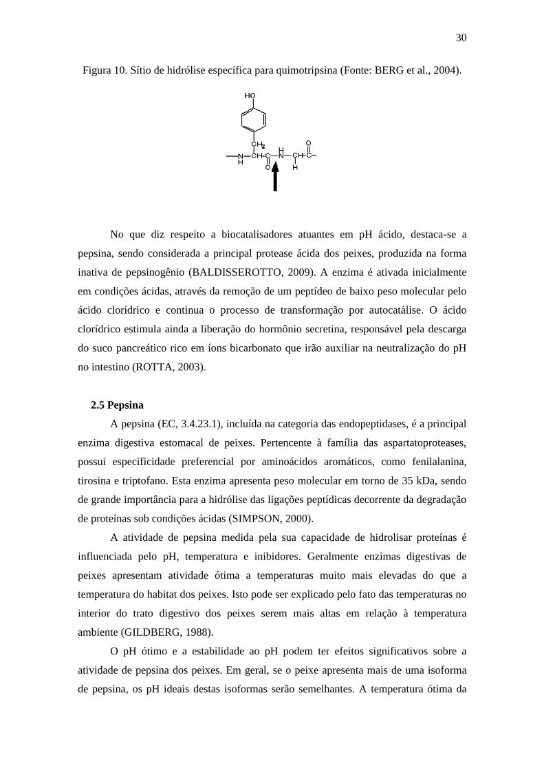
de organismos aquáticos (KOMKLAO et al., 2007). Esta enzima catalisa a hidrólise de
ligações peptídicas de proteínas na porção carboxila de aminoácidos aromáticos como:
fenilalanina, tirosina e triptofano e também substratos sintéticos, tais como Succinil-
alanina-alanina-prolina-fenilalanina-p-nitroanilida - SAPNA (Figura 10) (CASTILLO-
YAÑEZ et al., 2006).
Sítio de clivagem
30
Figura 10. Sítio de hidrólise específica para quimotripsina (Fonte: BERG et al., 2004).
No que diz respeito a biocatalisadores atuantes em pH ácido, destaca-se a
pepsina, sendo considerada a principal protease ácida dos peixes, produzida na forma
inativa de pepsinogênio (BALDISSEROTTO, 2009). A enzima é ativada inicialmente
em condições ácidas, através da remoção de um peptídeo de baixo peso molecular pelo
ácido clorídrico e continua o processo de transformação por autocatálise. O ácido
clorídrico estimula ainda a liberação do hormônio secretina, responsável pela descarga
do suco pancreático rico em íons bicarbonato que irão auxiliar na neutralização do pH
no intestino (ROTTA, 2003).
2.5 Pepsina
A pepsina (EC, 3.4.23.1), incluída na categoria das endopeptidases, é a principal
enzima digestiva estomacal de peixes. Pertencente à família das aspartatoproteases,
possui especificidade preferencial por aminoácidos aromáticos, como fenilalanina,
tirosina e triptofano. Esta enzima apresenta peso molecular em torno de 35 kDa, sendo
de grande importância para a hidrólise das ligações peptídicas decorrente da degradação
de proteínas sob condições ácidas (SIMPSON, 2000).
A atividade de pepsina medida pela sua capacidade de hidrolisar proteínas é
influenciada pelo pH, temperatura e inibidores. Geralmente enzimas digestivas de
peixes apresentam atividade ótima a temperaturas muito mais elevadas do que a
temperatura do habitat dos peixes. Isto pode ser explicado pelo fato das temperaturas no
interior do trato digestivo dos peixes serem mais altas em relação à temperatura
ambiente (GILDBERG, 1988).
O pH ótimo e a estabilidade ao pH podem ter efeitos significativos sobre a
atividade de pepsina dos peixes. Em geral, se o peixe apresenta mais de uma isoforma
de pepsina, os pH ideais destas isoformas serão semelhantes. A temperatura ótima da
31
pepsina dos peixes costuma variar entre 30 a 55 ◦C. A temperatura ótima da pepsina de
peixes depende muito dos tipos das espécies (água fria ou quente) (SHAHIDI et al.,
2001).
Nem todos os inibidores de protease tem um efeito inibitório sobre a pepsina.
Tem sido demonstrado que inibidores típicos, tais como fluoreto de fenil metil sulfonil
(PMSF), inibidor de serino-protease; L-3-carboxytrans- 2, 3-epoxi-propionil-L-leucin-
4-guanidino-butilamida (E-64), inibidor de proteinase cisteína e etileno-diamina-tetra-
acético (EDTA) não tem qualquer efeito inibitório sobre pepsinas (ZHOU et al., 2008).
Entretanto, pepstatina A (inibidor de proteinase aspártica) pode combinar-se com a
pepsina de peixe, impedindo a ligação enzima – substrato, resultando assim numa
completa inibição da atividade enzimática (COPELAND et al., 2005).
Pepsinas podem ser encontradas principalmente no suco gástrico do lúmen
estomacal e podem ser isoladas a partir de uma variedade de espécies como mamíferos,
aves e peixes. Há vários tipos de pepsinas estomacais ou isoformas, com estrutura
proteica e propriedades enzimáticas distintas (SHAHIDI, 2001). Zimogênios de
aspartatoproteases como pepsinogênios, também são secretados pelas glândulas da
mucosa gástrica de vertebrados, incluindo peixes. Trata-se de uma forma antecedente
inativa da enzima, a qual se torna funcional através da ação de cinases apropriadas ou
outros ativadores (EFFRONT, 2007).
Comparado com a pepsina, o pepsinogênio contém 44 aminoácidos adicionais,
sendo estável em ambientes neutro ou levemente alcalino. Na presença de ácido
clorídrico proveniente do suco gástrico (pH 1,5–2,0), esses 44 aminoácidos são
removidos proteoliticamente de uma forma autocatalítica resultando na ativação da
pepsina (RAUFMAN, 2004). Pepsinogênios são sintetizados como precursores,
contendo uma sequência sinal hidrofóbica de 15 -16 aminoácidos na região N-terminal
servindo como função de transporte. A sequência sinal é perdida durante o
processamento pós-transcricional. Esses pró-seguimentos são fundamentais na
estabilização da forma inativa e prevenção contra a entrada do substrato na região do
sítio ativo. Os pepsinogênios também diferem uns dos outros em relação às estruturas
primárias e propriedades enzimáticas de suas formas ativas (KAGEYAMA, 2002).
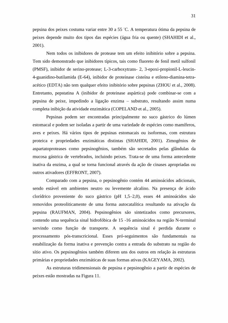
As estruturas tridimensionais de pepsina e pepsinogênio a partir de espécies de
peixes estão mostradas na Figura 11.

32
Figura 11: (A) Estrutura tridimensional da pepsina do bacalhau-do-Atlântico (Gadus
morhua) (KARLSEN, 1998) e (B) pepsinogênio do peixe mandarim-ouro
(Sinipercascherzeri) (DENG, 2010).
Enquanto pepsinas de mamíferos têm sido completamente sequenciadas,
pepsinas de peixes ainda não foram exploradas extensivamente. A primeira estrutura
tridimensional proposta para pepsina do bacalhau foi idêntica à pepsina suína quanto à
localização do domínio e sequência de aminoácidos (ANDREEVA et al., 2001). A
estrutura da pepsina do bacalhau corresponde a uma cadeia proteica simples (um
monômero) e dois domínios com dobramentos semelhantes separados por uma fenda
(KARLSEN et al., 1998). Já o sítio catalítico da pepsina é formado pela junção de
domínios e contém dois resíduos de ácido aspártico, Asp 32 e Asp 215 em cada domínio
(WORTHINGTON, 2010).
Pepsinas e pepsinogênios têm sido isolados a partir da mucosa gástrica de várias
espécies de peixes, incluindo Latimeria chalumnae (TANJI et al., 2007), Sparus latus
Houttuyn (ZHOU et al., 2007), Coryphaenoides pectoralis (KLOMKLAO et al., 2007),
Mustelus mustelus (BOUGATEF et al., 2008), Siniperca chuatsi (ZHOU et al., 2008),
Sardinella aurita (KHALED et al., 2011), Thunnus alalunga (NALINANON et al.,
2010) e Anguilla anguilla (WU et al., 2009).
2.5.1 Aplicabilidade industrial da pepsina
O emprego da pepsina no ramo industrial tem se tornado frequente nos últimos
anos. Tecnologicamente utilizada na extração de colágeno e gelatina, a enzima também
vem sendo vastamente aproveitada na terapêutica para fins de regulação da digestão,
como antisséptico dental e no tratamento de algumas doenças, incluindo dispepsia,
gastralgia, diarreia infantil e alguns tipos de câncer (GORGAS, 2009).
A B
33
Combinados com HCl, comprimidos e cápsulas contendo pepsina têm sido
desenvolvidos para melhorar a digestibilidade no trato gastrointestinal, bem como para
aumentar o apetite dos pacientes (MURADO et al., 2009). Pepsina de suínos tem sido
usada no tratamento de úlceras gástricas e na coagulação do leite para formação do
coalho, importante processo na indústria alimentícia (ALTUN; CETINUS, 2007).
A pepsina é uma das enzimas utilizadas para análise de outras proteínas devido a
sua eficiência em quebrar pontes envolvendo aminoácidos aromáticos, fenilalanina,
triptofano e tirosina. Estudos têm reforçado o emprego desta enzima na purificação de
soros antipeçonhentos aplicados à soroterapia de humanos picados por animais
venenosos (BOUSHABA et al., 2003). A enzima ainda é aproveitada na produção de
silagem de peixe, na indústria de processamento de pescado para produção de
subprodutos e na indústria de ração animal em processos de digestibilidade proteica.
Assim, a pepsina é considerada uma protease promissora, com larga aplicação
convencional e industrial, para tanto, técnicas eficazes para a sua recuperação e
purificação devem ser desenvolvidas (NALINANON et al., 2010; ZENG et al., 2012).
2.6 Colágeno
Diversos tipos de proteínas estão presentes na matriz extracelular dos tecidos
conjuntivos. As características biofísicas desta matriz são definidas pela disposição
supramolecular de elementos fibrilares, redes microfibrilares, como também de
proteínas, glicoproteínas e uma grande variedade de outras moléculas solúveis. Entre os
diferentes tipos de tecido conjuntivo podemos encontrar variações na sua composição e
estrutura (GELSE et al., 2003).
Em relação ao conteúdo proteico, o colágeno destaca-se como sendo a proteína
mais abundante do tecido conjuntivo. Esta macromolécula é encontrada em várias partes
da estrutura biológica animal, como por exemplo, na pele, nos ossos, nos tendões e nos
dentes (SENARATNE et al., 2006). O termo colágeno é derivado de palavras gregas
que significam “produzir cola” (COELHO et al., 2001), sendo essa, sua primeira
aplicação industrial reportada na literatura. Esta proteína estrutural, comum nos
vertebrados, constitui cerca de 30% do conteúdo proteico total e em humanos o
colágeno representa 6% do peso corporal (MUYONGA et al., 2004).
O colágeno apresenta como característica principal a capacidade de formação
de fibras insolúveis e elásticas, que modulam forças externas e internas exercidas dentro
do organismo. Apresenta também uma interessante capacidade de hidratação e
34
reabsorção e baixa antigenicidade. Outra importante função do colágeno é orientar
tecidos em desenvolvimento. As fibras de colágeno começam a aparecer durante o
desenvolvimento embrionário no processo inicial de diferenciação dos tecidos.
Posteriormente, tornam-se, responsáveis pela integridade dos tecidos, dos ossos,
cartilagens, pele e estrutura de vasos sanguíneos e outros órgãos (FRIESS, 1998).
2.6.1 Estrutura molecular do colágeno
Ao microscópio óptico, o colágeno apresenta-se como fibras de espessura e
orientação variadas, ocupando o compartimento extracelular, entre as células do tecido
conjuntivo. Maiores detalhes estruturais só podem ser verificados através do
microscópio eletrônico. A menor unidade é a fibrila com um padrão característico de
estrias periódicas, que se repetem a cada 64 nm. Geralmente as fibrilas encontram-se
agregadas, formando pequenos feixes, e tornam-se visíveis ao microscópio óptico
quando excedem 0,2 μm, sendo chamadas de fibras colágenas. Os agregados de 4 a 8
moléculas de colágeno são formados durante o processo de fibrilogênese. Essas fibrilas
possuem de 10 a 500 nm de diâmetro dependendo do tipo de tecido ou estágio do
desenvolvimento. As fibrilas de colágeno se auto-organizam podendo formar moléculas
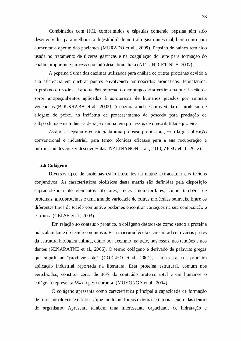
ainda maiores, como as fibras (CAMPOS, 2008). A organização estrutural do colágeno
durante a fibrilogênese, ou seja, durante o processo de agregação das microfibrilas, pode
ser vista na Figura 12.
Figura 12: Desenho esquemático da organização do colágeno na sua estrutura
supermolecular; adaptado de Ten Cate, 1994 e Yannas, 1996.
35
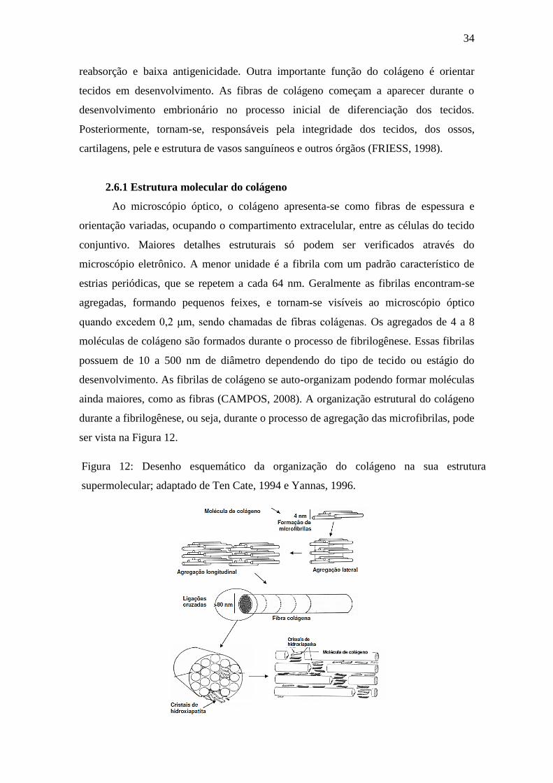
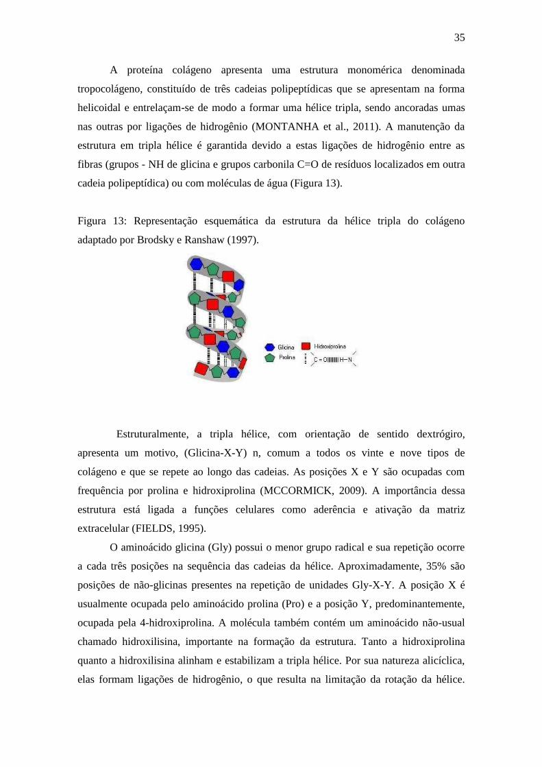
A proteína colágeno apresenta uma estrutura monomérica denominada
tropocolágeno, constituído de três cadeias polipeptídicas que se apresentam na forma
helicoidal e entrelaçam-se de modo a formar uma hélice tripla, sendo ancoradas umas
nas outras por ligações de hidrogênio (MONTANHA et al., 2011). A manutenção da
estrutura em tripla hélice é garantida devido a estas ligações de hidrogênio entre as
fibras (grupos - NH de glicina e grupos carbonila C=O de resíduos localizados em outra
cadeia polipeptídica) ou com moléculas de água (Figura 13).
Figura 13: Representação esquemática da estrutura da hélice tripla do colágeno
adaptado por Brodsky e Ranshaw (1997).
Estruturalmente, a tripla hélice, com orientação de sentido dextrógiro,
apresenta um motivo, (Glicina-X-Y) n, comum a todos os vinte e nove tipos de
colágeno e que se repete ao longo das cadeias. As posições X e Y são ocupadas com
frequência por prolina e hidroxiprolina (MCCORMICK, 2009). A importância dessa
estrutura está ligada a funções celulares como aderência e ativação da matriz
extracelular (FIELDS, 1995).
O aminoácido glicina (Gly) possui o menor grupo radical e sua repetição ocorre
a cada três posições na sequência das cadeias da hélice. Aproximadamente, 35% são
posições de não-glicinas presentes na repetição de unidades Gly-X-Y. A posição X é
usualmente ocupada pelo aminoácido prolina (Pro) e a posição Y, predominantemente,
ocupada pela 4-hidroxiprolina. A molécula também contém um aminoácido não-usual
chamado hidroxilisina, importante na formação da estrutura. Tanto a hidroxiprolina
quanto a hidroxilisina alinham e estabilizam a tripla hélice. Por sua natureza alicíclica,
elas formam ligações de hidrogênio, o que resulta na limitação da rotação da hélice.
36
Esse trio de aminoácidos, entrelaçados uns nos outros, formam uma estrutura
semelhante a uma trança de forma helicoidal, formando uma cadeia extremamente
resistente (CAMPOS, 2008).
A formação da estrutura hélice tripla estável (Figura 14) nos tecidos requer a
modificação do aminoácido prolina (Pro), pela atividade da enzima prolil-hidroxilase
(hidroxilação), para formar hidroxiprolina (Hyp) na cadeia do colágeno. Estudos
mostram que a hidroxiprolina desempenha papel extremamente importante na
estabilização da hélice tripla, pois defeitos na hidroxiprolina podem refletir na
desorganização da hélice tripla e, portanto, de todo colágeno (SENA, 2004).
O arcabouço em tripla hélice do colágeno é uma estrutura altamente
conservada que se encontra presente em todas as variações de colágeno. Em
contrapartida, a presença de domínios não colagenosos entre os diferentes tipos de
colágeno promove uma diversidade estrutural e funcional com diferentes variações
desta proteína. Essa variedade contribui gerando estruturas que exigem a ação de
enzimas específicas (colagenases) para a clivagem proteolítica. Na estrutura do
colágeno, existem ainda regiões terminais amino e carboxi constituídas de 9-26
aminoácidos nas extremidades da molécula que não formam a estrutura da hélice. Essas
regiões são denominadas telopeptídeos.
Figura 14: Estrutura do colágeno: (a) forma de tríplete presente nas matrizes colagênicas;
(b) tropocolágeno; (c) hélice tripla; (d) modelo do quarto alternado pentafibrilar proposto
por Smith (1968). (Fonte: SIONKOWSKA, 2006).
37
2.6.2 Tipos de colágeno
Diversos tipos de colágeno têm sido reportados na literatura, totalizando 29
estruturas, classificadas de I – XXIX. Esses subtipos proteicos exibem organização,
sequência e função bem distintos, sendo caracterizados por possuírem considerável
complexidade e variações na ocorrência de domínios não helicoidais. O colágeno pode
ser dividido em três grandes grupos com base na sua estrutura macromolecular: a)
colágeno fibroso estriado (I, II e III), b) colágeno não fibroso (IV), c) colágeno
miofibrilar, o qual engloba os tipos VI e VII (matriz miofibrilar), V, IX e X (colágeno
pericelular), e VIII e XI (XIONG, 1997).
Esses vários tipos de colágeno geralmente estão relacionados aos aspectos
biomecânicos, entretanto além dessa função típica, essas proteínas realizam outras
atribuições. Possuem importância fundamental no desenvolvimento de órgãos,
envolvimento em processos de reparo de tecidos e cicatrização, podendo também atuar
em eventos relacionados à sinalização celular, além de contribuir no armazenamento
local de fatores de crescimento e citocinas. Essa capacidade de se vincular a fatores de
crescimento e citocinas credencia estas moléculas como veículos de transporte com
potencial para fins terapêuticos e farmacológicos (GELSE et al., 2003).
Dentre todos os tipos de colágenos existentes que já foram descobertos, o
colágeno do tipo I tem despertado muita atenção por ser a proteína extracelular mais
abundante, sendo também apontada como responsável pela manutenção da resistência
mecânica nos ossos (SENA, 2004). Quanto à estrutura, o colágeno I trata-se de um
heteropolímero formado por dois tipos de cadeias α, uma α1 e uma α2, em que a glicina
constitui um terço do seu conteúdo de aminoácidos e possui baixos níveis de tirosina e
histidina (MCCORMICK, 2009). Segundo Xiong (1997), o colágeno do tipo I é
encontrado em todos tecidos conectivos, incluindo ossos e peles.
2.6.3 Fontes de colágeno
As mais abundantes fontes de colágeno são mamíferos, especialmente bovinos e
suínos, sendo a pele um dos subprodutos mais disponíveis para extração de colágeno
(GÓMEZ-GUILLÉN et al., 2002). A pele de animais, por exemplo, contém 30 a 35%
de proteínas e destas 90 a 95% são representadas pela fração de colágeno (COELHO et
al., 1998). Os ossos constituem outra grande fonte de colágeno, no entanto sua
composição varia consideravelmente de acordo com a espécie e o tipo do osso. Esta
variação decorre, principalmente, do fato de que os ossos contêm várias estruturas
38
teciduais distintas, dentre elas: cartilagens, tecidos ósseos esponjosos, tecidos ósseos
compactos, entre outras (COELHO et al., 1998).
O colágeno contido nos subprodutos de animais como bovinos, suínos e aves
tem sido amplamente utilizado em processos industriais por apresentar propriedades
como biocompatibilidade, biodegradabilidade e baixa antigenicidade (LIU et al., 2009).
Porém, devido aos recorrentes casos de zoonoses como: encefalopatia espongiforme
bovina (BSE), encefalopatia espongiforme transmissível (TSE), febre aftosa (FA) e
gripe aviária, a busca por fontes alternativas e mais seguras desse composto tornou-se
uma opção mais viável (ZHANG et al., 2007).
Organismos aquáticos, como os peixes, devido a grande disponibilidade, baixos
riscos de transmissão de doenças, alto rendimento nos processos de extração e ausência
de toxicidade, têm se destacado como uma alternativa frente ao colágeno de animais
terrestres. Além disso, o uso de derivados de peixes não apresenta restrições religiosas
para determinados grupos sociais (SENARATNE et al., 2006).
A indústria da pesca também tem gerado uma grande quantidade de resíduos
ricos em colágeno, pois cerca de 30% do resíduo produzido consiste em pele e ossos
com alto conteúdo de colágeno (GÓMEZ-GUILLÉN et al., 2002). Sendo assim, estudos
envolvendo extração de colágeno proveniente de espécies aquáticas têm crescido
continuamente.
Extrações bem sucedidas a partir de pele, ossos e escamas de peixes e outros
organismos marinhos foram reportadas pela literatura nesses últimos anos, a exemplo
das espécies Sepia pharaonis (AEWSIRI et al., 2009), Priacanthus tayenus e
Priacanthus macracanthus (BENJAKUL et al., 2010a), Pangasianodon hypophthalmus
(SINGH et al., 2011), Hypophthalmichthys molitrix (SAFANDOWSKA et al., 2013),
Evenchelys macrura (VEERURAJ et al., 2013) e Doryteuthis singhalensis
(VEERURAJ et al., 2014).
2.6.4 Aplicações industriais do colágeno
O colágeno, em sua forma purificada, possui várias aplicações industriais,
principalmente nos setores farmacêutico, de cosméticos e alimentos. A qualidade e
aplicação específica do colágeno extraído estão diretamente relacionadas com suas
propriedades funcionais e pureza (RUSTAD, 2003).
No que tange ao setor farmacêutico, a biomolécula do colágeno é utilizada na
fabricação de implantes vítreos, carreadores de drogas e produção de compostos

39
biologicamente ativos. Extensas pesquisas têm sido efetuadas a partir da utilização de
colágeno como biomaterial em pacientes humanos. Esses dispositivos variam desde
suturas reabsorvíveis, vasos sanguíneos sintéticos até proteção de córnea danificada,
regeneração óssea, tratamento de queimaduras na pele e muitas outras utilizações
(CAMPOS, 2008).
O uso do colágeno tem sido amplo no campo biomédico. Características
importantes como: baixa antigenicidade, biodegradabilidade e propriedades mecânicas,
hemostáticas e de matriz suporte para crescimento de células têm favorecido o emprego
do colágeno nesta área (YANNAS, 1996). Dentre os biopolímeros, o colágeno é o
material de origem animal mais abundante e fornecedor de uma ótima base para
biomateriais. Depois de extraído, o colágeno pode ser processado para obtenção de
filmes, membranas e fibras (KOKOSZKA et al., 2010).
Na área médica essa proteína pode ser aplicada no tratamento de doenças
angiogênicas, hipertensão, incontinência urinária e osteoartrite (WOO et al., 2008).
Além de todo este conjunto de opções para aplicação do colágeno, o biopolímero pode
ser de grande utilidade na indústria fotográfica, de couro e tecidos (IKOMA et al., 2003;
WOO et al., 2008; LIU et al., 2009).
Embora com diversas aplicações na indústria de alimentos, o colágeno é pouco
aproveitado tecnologicamente, em relação às suas propriedades funcionais como
extensor, umidificante, emulsionante, ligante e potencializador de textura. Diversas
fontes de colágeno são utilizadas na fabricação de produtos cárneos emulsionados,
dentre as quais destacam-se a pele suína, colágeno de ossos e do músculo esquelético. A
grande vantagem é que o colágeno trata-se de uma proteína digestível de fácil obtenção,
considerada pela maioria das legislações como um alimento e não como um aditivo
(COELHO et al., 2001).
Recentemente tem se desenvolvido materiais biológicos que combinam
diferentes biopolímeros e materiais inorgânicos ao colágeno e a outros compostos
biológicos, como a quitosana, visando gerar compostos que cumpram requisitos
funcionais específicos. Entre os produtos obtidos a partir do colágeno, a gelatina tem
apresentado um grande poder de comercialização. Isso se deve à abundância dessa
matéria prima que tem um baixo custo e possui excelentes propriedades funcionais
(KOKOSZKA et al., 2010).
40
3. OBJETIVOS
3.1 Geral
Extrair, semi-purificar e caracterizar uma protease digestiva ácida (pepsina
símile) presente no estômago do pintado (Pseudoplatystoma corruscans), bem
como avaliar o seu potencial biotecnológico para extração de colágeno, além de
isolar e caracterizar colágeno da pele do pintado.
3.2 Específicos
Caracterizar físico-quimicamente a atividade da protease ácida a partir do
extrato bruto do estômago do pintado;
Avaliar a sensibilidade da atividade proteolítica ácida presente no extrato bruto
frente a íons metálicos e inibidores específicos e inespecíficos;
Investigar o potencial uso da protease ácida para extração de colágeno da pele de
Oreochromus niloticus;
Purificar parcialmente uma protease ácida a partir do estômago do peixe P.
corruscans através de cromatografia de troca-iônica;
Avaliar a pureza e determinar a massa molecular aparente da enzima através de
SDS-PAGE e zimograma;
Caracterizar físico-quimicamente a enzima parcialmente purificada;
Avaliar a sensibilidade enzimática da fração parcialmente purificada frente a
íons metálicos e inibidores específicos e inespecíficos;
Extrair colágeno ácido solúvel (ASC) e pepsino solúvel (PSC) a partir da pele do
pintado (P. corruscans);
Determinar o rendimento da extração do colágeno ASC e PSC;
Caracterizar parâmetros de extração e solubilidade do colágeno ASC e PSC;
Determinar o peso molecular aparente das amostras de colágeno através de SDS-
PAGE.
41
REFERÊNCIAS
AEWSIRI, T., BENJAKUL, S., & VISESSANGUAN, W. Functional properties of
gelatin from cuttlefish (Sepia pharaonis) skin as affected by bleaching using hydrogen
peroxide. Food Chemistry, 115 (1), 243 – 249, 2009.
AHMAD, M., & BENJAKUL, S. Extraction and characterization of pepsin solubilized
collagen from the skin of unicorn leatherjacket (Aluterus monocerous). Food
Chemistry, 120, 817 – 824, 2010.
ALTUN, G. D.; CETINUS, S. A. Imobilization of pepsin on chitosan beads. Food
Chemistry, v. 100, p. 964 – 971, 2007.
ALVARADO, E. G. Treinamento alimentar de pintado Pseudoplatystoma corruscans
(Agassiz, 1829): Sobrevivência, crescimento e aspectos econômicos. Dissertação de
Mestrado. Universidade Estadual Paulista, Centro de Aquicultura, 2003.
ANDREEVA N. S, RUMSH L. D. Analysis of crystal structures of aspartic proteinases:
On the role of amino acid residues adjacent to the catalytic site of pepsin-like enzymes.
Protein Sci 10: 2439 - 2450, 2001.
ARRUDA, L. F. BORGHESI, R.; BRUM, A.; REGITANO D’ARCE, M.; OETTERER,
M. Nutritional aspects of nile tilapia (Oreochromis niloticus) silage. Ciência e
Tecnologia de Alimentos, v.26, n.4, p.749 - 756, 2006.
ARVANITOYANNIS, I. S.; KASSAVETI, A. Fish industry waste: treatments,
environmental impacts, current and potential uses. International Journal of Food
Science and Technology, v. 43, p. 726 - 745, 2008.
BALDISSEROTTO, B. Fisiologia de Peixes Aplicada a Piscicultura. Editora UFSM,
Santa Catarina, 33 p, 2009.
BALDISSEROTO, B; GOMES, L. C. Espécies nativas para a piscicultura no Brasil.
Santa Maria: Editora UFMS, 468 p, 2005.
42
BANCO DO NORDESTE. Manual de impactos ambientais. Orientações básicas sobre
aspectos ambientais de atividades produtivas. Fortaleza: Banco do Nordeste, 297 p,
1999.
BENÍTEZ, H. O. M.; PULIDO, J. A. R.; ZAPATA, C. R. O. Manual de reproduccion y
cultivo el bagre rayado Pseudoplatystoma fasciatum. Instituto nacional de pesca y
agricultura – INPA, 2003.
BENJAKUL, S., THIANSILAKUL, Y., VISESSANGUAN, W., ROYTRAKUL, S.,
KISHIMURA, H., PRODPRAND, T., et al. Extraction and characterization of pepsin
solubilized collagens from the skin of bigeye snapper (Priacanthus tayenus and
Priacanthus macracanthus). Journal of the Science of Food and Agriculture, 90, 132
– 138, 2010 a.
BENJAKUL S, KLOMKLAO S, SIMPSON B. K. Enzymes in fish processing. In
Enzymes in Food Technology (2ndedn), Whitehurst R. J., Van Oort M, Eds. Blackwell
Publishing, Chichester: 211 - 225, 2010 b.
BERG, J. M; STRYER, L.; TYMOCZKO, J. L. Bioquímica. 5 ed. Rio de Janeiro:
Guanabara Koogan,. 1104 p, 2004.
BEZERRA, R. S., SANTOS, J. F., LINO, M. A. S., VIEIRA, V. L. A., CARVALHO
JR., L. B. Characterization of stomach and pyloric caeca of tambaqui (Colossoma
macropomum). Journal of Food Biochemistry, 24 189 - 199, 2000.
BORGHESI, R.; ARRUDA, L. F.; OETTERER, M. A silagem de pescado na
alimentação de organismos aquáticos. Boletim CEPPA, v. 25, n. 2, p. 329 - 339, 2007.
BOUGATEF, A., BALTI, R., ZAIED, S. B., SOUISSI, N., NASRI, M. Pepsinogen and
pepsin from the stomach of smooth hound (Mustelus mustelus): Purification,
characterization and amino acid terminal sequences. Food Chemistry 107, 777 – 784,
2008.
43
BOUSHABA, R.; KUMPALUME, P.; SLATER, N. K. H. K. Kinetics of whole serum
and prepurified IgG digestion by pepsin for F (ab’)2 manufacture. Biotechnology
Progress, v. 19, p. 1176 – 1182, 2003.
BRASIL. Ministério da Pesca e Aquicultura. Boletim estatístico da pesca e aquicultura:
Brasil 2008-2009. [Brasília, DF, 2010]. 99 p. Disponível em:
<http://pt.scribd.com/doc/68775604/Anuario-Da-Pesca-Completo>. Acesso em: 15 nov.
2014.
BRODSKY, B.; RAMSHAW, J. A. M. The Collagen triple-Helix Structure Matrix
Biology, v. 15, p. 545 - 554, 1997.
BUITRAGO–SUÁREZ U. A., BURR B. M. Taxonomy of the catfish genus
Pseudoplatystoma Bleeker (Siluriformes: Pimelodidae) with recognition of eight
species. Zootaxa, v. 1512, p.1 - 38, 2007.
CAMARGO S. A. F.; PETRERE, J. R. M. Social and Financial aspects of the artisanal
fisheries of Middle São Francisco River, Minas Gerais, Brazil. Fish Manag Ecol, v. 8,
p.163 - 171, 2001.
CAMPOS, D. M. Produção e caracterização de colágeno tipo I e de compósitos
hidroxiapatita-colágeno para regeneração óssea. M.Sc. Tese, Universidade Federal do
Rio de Janeiro. Rio de Janeiro, 2008.
CASTILLO-YAÑEZ, F. J., PACHECO-AGUILAR, R., GARCÍA-CARREÑO, F. L.,
NAVARRETE-DEL TORO, M. A., LÓPEZ, M. A. Purification and biochemical
characterization of chymotrypsin from the viscera of Monterey sardine (Sardinops
sagax caeruleus). Food Chemistry [S.I.], v. 99, p. 252 - 259, 2006.
CAVALCANTE JÚNIOR, V.; ANDRADE, L. N.; BEZERRA, L. N.; GURJÃO, L. M.;
FARIAS, W. R. L. Reuso de água em um sistema integrado com peixes, sedimentação,
ostras e macroalgas. Revista Brasileira de Engenharia Agrícola e Ambiental, v. 9
(supl.), p. 118 - 122, 2005.
44
COELHO, H. S.; MORANDINI, L. M. B.; ROSA, C. S.; SANTANA, A. M.; TERRA,
N. N.; KUBOTA, E. H. Obtenção do couro suíno cozido para adição em embutidos
cárneos. In: XVI Congresso Brasileiro de Ciência e Tecnologia de Alimentos. Anais v.
2, 1998.
COELHO, H. S.; MORANDINI, L. M. B.; SANTANA, A. M.; TERRA, N. N.
Características físico-químicas do salame tipo italiano contendo couro suíno cozido.
Revista Nacional da Carne. v. 278. Ano XXIV, 2001.
COPELAND R. A. Evaluation of enzyme inhibitors in drug discovery: A guide for
medicinal chemists and pharmacologists. John Wiely and Sons Inc, Hoboken, New
Jersey, 34 – 66, 2005.
COSTA, H. M. S. Extração, purificação e caracterização de proteases digestivas e
colágeno a partir de resíduos do processamento de peixes. Tese de Doutorado.
Universidade Federal de Pernambuco, 2012.
COSTA, H. M. S., FREITAS JÚNIOR, A. C. V., AMARAL, I. P. G., HIRATA, I. Y.,
PAIVA, P. M. G., CARVALHO JR, L. B., OLIVEIRA, V., BEZERRA, R. S. Metal-
sensitive and thermostable trypsin from the crevalle jack (Caranx hippos) pyloric caeca:
purification and characterization. Chemistry Central Journal, 7:166, 2013.
CREPALDI, D. V.; CARVALHO, D. C.; SOUSA, A. B.; MELO, D. C.; SEERIG, A.
S.; OLIVIERA, D. A. A.; RIBEIRO, L. P.; TEIXEIRA, E. A.; LOPES, V. E.;
DRUMOND, M. L.; MORAES, V. E.; FARIA, P.M. C. Biologia reprodutiva do
surubim (Pseudoplatystoma corruscans). Revista Brasileira de Reprodução de
Animais, Belo Horizonte, v. 30, n. 3/4, p. 159 - 167, jul./dez., 2006 a.
CREPALDI, D. V.; FARIA, P. M. C.; TEIXEIRA,E. A. T.; RIBEIRO, L. P.; COSTA,
A. A. P.; MELO, D. C.; CINTRA, A. P. R.; PRADO, S. A.; COSTA, F. A. A.;
DRUMOND, M. L.; LOPES, V. E.; MORAIS, V. E.; Utilização de hormônios na
reprodução induzida do surubim (Pseudoplatystoma spp). Revista Brasileira de
Reprodução Animal, v.30, n.3/4, p.168 - 173, 2006 b.
45
CREPALDI, D. V.; TEIXEIRA, E.A.; FARIA, P. M. C; RIBEIRO, L. P.; MELO, D. C.;
OLIVEIRA, D. A. A.; TURRA, E. M.; QUEIROZ, B. M. Rendimento de carcaça em
surubim (Pseudoplatystoma spp.) avaliado por ultra-som. Revista Brasileira de Saúde
e Produção Animal, v. 9, n. 4, p. 813 - 824, 2008.
DENG, Y.; ZHAO, J.; LU, G.; WU, X.; TAO, Y. Cloning, characterization and
expression of the pepsinogen C from the golden mandarin fish Sinipercascherzeri
(Teleostei: Perciformes). Fish Sci 76: 819 - 826, 2010.
DOODE, M. S. Los Claro-oscuros de la Pesquería de la Sardina em
Sonora:Contradicciones y Alternativas para un Desarrollo Equilibrado. Tese de
Doutorado. El Colegio de Michoacán. Zamora, Michoacán, p. 120 – 136, 1996.
EFFRONT, J.; PRESCOTT, S. C.; VENABLE, C. S. Biochemical catalysts in life and
industry: proteolytic enzymes. Kessinger Publishing: 151 - 289, 2007.
ESPÓSITO, et al. Trypsin from the processing waste of the Lane Snapper (Lutjanus
synagris) and its compatibility with oxidants, surfactants and commercial detergents.
Journal of Agricultural and Food Chemistry, v. 58, p. 6433 – 6439, 2010.
FAO. The State of World Fisheries and Aquaculture 2014. Rome. 223 pp., 2014.
FIELDS, G. B. The collagen triple-helix: correlation of conformation with biological
activities. Connect Tissue Res. 31 (3): 235 - 243, 1995.
FREITAS-JÚNIOR, et al. Giant Amazonian fish pirarucu (Arapaima gigas): Its viscera
as a source of thermostable trypsin. Food Chemistry, v. 133, p. 1596 – 1602, 2012.
FRIESS, W. Collagen – biomaterial for drug delivery. European Journal of
Pharmaceutics and Biopharmaceutics, v. 45, n. 2, p. 113 – 136, 1998.
FUNDAÇÃO BIODIVERSITAS. Lista oficial da fauna brasileira ameaçada de
extinção, 2003. Disponível em: http://www.biodiversitas.org.br. Acesso em 13 de
novembro de 2004.
46
GELSE, K., PÖSCHL, E., & AIGNER, T.. Collagens - Structure, function, and
biosynthesis. Advanced Drug Delivery Reviews, 55, 1531 – 1546, 2003.
GEO BRASIL. Perspectivas do meio ambiente no Brasil. Santos T. C. C. e Câmara B.
D. (Org.). Brasília: Edições IBAMA. ISBN 85 – 7300 - 144-5, p.132 - 147, 440 p.:Il,
2002.
GILDBERG, A. Aspartic proteinases in fishes and aquatic invertebrates. Comparative
Biochemistry and Physiology, 91 B (3), 425 – 435, 1988.
GILDBERG, A.; STENBERG, E. A new process for advanced utilization of shrimp
waste. Process Biochemistry, v. 36, p. 809 - 812, 2001.
GILDBERG, A. Enzymes and Bioactive Peptides from Fish Waste Related to Fish
Silage, Fish Feed and Fish Sauce Production. Journal of Aquatic Food Product
Technology, 13 : 2, 3 - 11, 2004.
GÓMEZ-ESTACA, et al. Antimicrobial activity of composite edible films base don fish
gelatin and chitosan incorporated with clove essential oil. Journal of Aquatic Food
Product Technology, v. 18, p. 46 - 52, 2009.
GOMEZ-GUILLEN, M. C.; TURNAY, J.; FERNANDEZ-DIAZ, M. D.; ULMO, N.;
LIZARBE, M. A. & MONTERO, P. Structural and physical properties of gelatin
extracted from different marine species: A comparative study. Food Hydrocolloids, 16,
25 – 34, 2002.
GONZALEZ, T. & ROBERT-BADOUY, J. Bacterial aminopeptidases: Properties and
functions. Microbiology Reviews, 18, p. 319 – 344, 1996.
GORGAS, F. J. S. Dental medicine. A manual of dental material medica and
therapeutics, Nabu Press, Washington, 48 – 50, 2009.
47
HARVEY, R. A., CHAMPE, P. C., FERRIER, D. R. Bioquímica Ilustrada. 4a. ed.:
Artmed, 2009.
HIDALGO, M. C.; UREA, E.; SANZ, A. Comparative study of digestive enzymes in
fish with different nutritional habits. Proteolytic and amylase activities. Aquaculture,
170: 267 – 283, 1999.
HUANG, Y. R.; SHIAU, C. Y.; CHEN, H. H. & HUANG, B. C. Isolation and
characterization of acid and pepsin-solubilized collagens from the skin of balloon fish
(Diodon holocanthus). Food Hydrocolloids, 25 (6), 1507 – 1513, 2011.
INOUE, L. A. K. A. et al. Princípios Básicos para Produção de Alevinos de Surubins
(Pintado e Cachara). Circular Técnica. Dourados - MS, v. 15, 8 p., 2008.
IKOMA, T.; KOBAYASHI, H.; TANAKA, J.; WALSH, D. & MANN, S. Physical
properties of type I collagen extracted from fish scales of Pagrus major and
Oreochromis niloticus. International Journal of Biological Macromolecules, 32, 199
– 204, 2003.
IBAMA, Brasil. Produção Pesqueira e Aquícola. Instituto Brasileiro do Meio Ambiente
e dos Recursos Naturais Renováveis, (Estatística da pesca 2008 e 2009), 30 p., 2010.
KAGEYAMA, T. Pepsinogens, progastricsins, and prochymosins: structure, function,
evolution, and development. Cellular and molecular life Sciences. v. 59, p. 288 – 306,
2002.
KARLSEN, S.; HOUGH, E.; OLSEN, R. L. Structure and proposed amino-acid
sequence of a pepsin from Atlantic cod (Gadus morhua). Acta Crystallogr D Biol
Crystallogr 54: 32 – 46, 1998.
KATO, et al. Evaluation of ozone treated fish waste oil as a fuel for transportation.
Journal of Chemical Engineering of Japan, v. 37, p. 863 – 870, 2004.
48
KHALED, H. B.; GHORBEL-BELLAAJ, O.; HMIDET, N.; JELLOULI, K.; ALI, N.
E.; GHORBEL, S.; NASRI, M. A novel aspartic protease from the viscera of Sardinelle
(Sardinella aurita): Purification and characterization. Food Chemistry 128, 847 – 853,
2011.
KLOMKLAO, S.; KISHIMURA, H.; YABE, M.; BENJAKUL, S. Purification and
characterization of two pepsins from the stomach of pectoral rattail (Coryphaenoides
pectoralis). Comparative Biochemistry and Physiology, Part B 147, p. 682 – 689,
2007.
KOKOSZKA, S.; DEBEAUFORT, F.; HAMBLETON, A.; LENART, A.; VOILLEY,
A.; Innov. Food Sci. Emer. Technol., 11, 503, 2010.
KOMKLAO, S.; BENJAKUL, S.; VISESSANGUAN, W.; KISHIMURA, H.;
SIMPSON, B. K. Purification and characterization of trypsins from the spleen of
skipjack tuna (Katsuwonus pelanis). Food Chemistry, 100, p. 1580 – 1589, 2007.
KROGDAHL, A.; HEMREG, I.; MOMMSEN, T. P. Carbohydrates in fish nutrition:
digestion and absorption in postlarval stages. Aquaculture Nutrition, v. 11, p. 103 –
122, 2005.
KUBITZA F.; CAMPOS J. L.; BRUM, J. A. Surubim: produção intensiva no Projeto
Pacu Ltda. e Agropeixe Ltda. Panor Aquicult, v. 49, p. 25 - 32, 1998.
KUZMINA, V. V. Influence of age on digestive enzyme activity in some freshwater
teleosts. Aquaculture, 148: 25 – 37, 1996.
LAUDER, G. V.; LIEM, K. F. The evolution and interrelationships of the
Actinopterygian fishes. Bulletin of the Museum of Comparative Zoology, v. 150, n.
3, p. 95 - 197, 1983.
LIU, H. Y.; LI, D. & GUO, S. D. Studies on collagen from the skin of channel catfish
(Ictalurus punctaus). Food Chemistry, 101, 621 – 625, 2007.
49
LIU, W.; HENDREN, J.; QIN, X. J.; SHEN, J.; LIU, K. J. Normobaric hyperoxia
attenuates early blood–brain barrier disruption by inhibiting MMP-9-mediated occludin
degradation in focal cerebral ischemia. J. Neurochem. 108, 811 – 820, 2009.
LIU, D.; LIANG, L.; REGENSTEIN, J. M. & ZHOU, P.. Extraction and
characterization of pepsin-solubilised collagen from fins, scales, skins, bones and swim
bladders of bighead carp (Hypophthalmichthys nobilis). Food Chemistry, 133, 1441 –
1448, 2012.
LUZ, R. K.; ZANIBONI FILHO, E. Utilização de diferentes dietas na primeira
alimentação do mandi-amarelo (Pimelodus maculatus, Lacépède). Acta Sci. 23, 483 –
489, 2001.
MARCUSCHI, et al. Purification, characterization and substrate specificity of a trypsin
from the Amazonian fish tambaqui (Colossoma macropomum). Biochemical
Biophysical Research Communications, v. 396, p. 667 - 673, 2010.
MARINHO, S. A. M. Sobrevivência e crescimento de larvas de surubim
Pseudoplatystoma corruscans (Spix & Agassiz, 1829) sob diferentes condições
alimentares. Dissertação apresentada ao Programa de Pós- Graduação em Recursos
Pesqueiros e Aquicultura da Universidade Federal Rural de Pernambuco. Recife – PE,
2007.
MATEUS, L. A. F.; PENHA, J. M. F. Dinâmica populacional de quatro espécies de
grandes bagres na bacia do rio Cuiabá, Pantanal norte, Brasil (Siluriformes,
Pimelodidae). Revista Brasileira de Zoologia, Cuiabá, v. 24, p. 87 - 98, mar, 2007.
MCCORMICK, R. J. Collagen. In Applied muscle biology and meat science (pp. 127 -
146). Boca Raton, Florida: CRC Press, 2009.
MIRANDA, M. O. T. Surubim. Belo Horizonte: Instituto Brasileiro de Meio Ambiente
e dos Recursos Naturais Renováveis, 157 p. (Coleção Meio Ambiente. Série Estudos:
Pesca, 19), 1997.
50
MONTANHA, V. C. et al., Caracterização de Microesferas de Colágeno e Fibroína
como suporte para as células tronco. XVIII Congreso Argentino de Bioingeniería SABI
2011 - VII Jornadas de Ingeniería Clínica Mar del Plata, 28 al 30 de septiembre de
2011.
MONTE, F. T. D. Purificação e caracterização de uma protease alcalina do peixe
pirarucu (Arapaima gigas) e seu potencial uso como aditivo para detergentes
comerciais. 16 p. (Monografia de graduação em Ciências Biológicas. Universidade
Federal de Pernambuco), 2013.
MURADO M. A.; GONZÁLEZ, M. D. P.; VÁZQUEZ, J. A. Recovery of proteolytic
and collagenolytic activities from viscera by-products of rayfish (Raja clavata). Mar
Drugs 7: 803 – 815, 2009.
MURRAY, R. K.; GRANNER, D. K.; MAYES, P. A.; RODWELL, V. W. Harper:
Bioquímica. 9a Ed., São Paulo: Editora Atheneu. 919 p., 2002.
MUYONGA, J. H.; COLE, C. G. B. & DUODU, K. G. Characterization of acid soluble
collagen from skins of young and adult Nile perch (Lates niloticus). Food Chemistry,
85 (1), 81 – 89, 2004.
NALINANON, S.; BENJAKUL, S.; VISESSANGUAN, W. & KISHIMURA, H. Use
of pepsin for collagen extraction from the skin of big eye snapper (Priacanthus
tayenus). Food Chemistry, 104, 593 – 601, 2007.
NALINANON, S.; BENJAKUL, S.; KISHIMURA, H. Biochemical properties of
pepsinogen and pepsin from the stomach of albacore tuna (Thunnus alalunga). Food
Chemistry, v. 121, p. 49 - 55, 2010.
NALINANON S.; BENJAKUL, S.; KISHIMURA, H.; OSAKO, K. Type I collagen
from the skin of ornate threadfin bream (Nemipterus hexodon): Characteristics and
effect of pepsin hydrolysis. Food Chemistry 125, 500 – 507, 2011.
51
NELSON, D. L.; COX, M. M. Lenhinger: Princípios de Bioquímica. 4 ed. São Paulo:
Sarvier, 1119 p, 2004.
NELSON, D. L.; COX, M. M. Lehninger Principles of Biochemistry, New York, W.
H. Freeman and Company, 2005.
OZAWA, M.; SATAKE, K.; SUZUKI, R. Removal of aqueous chromium by fish bone
waste originated hydroxyapatite. Journal of Materials Science Letters, v. 22, p. 513 –
514, 2003.
PILECCO, J. L. et al. Desempenho zootécnico de Pseudoplatystoma spp., criados em
viveiros escavados alimentados com rações comerciais. In: Congresso Brasileiro de
Produção de Peixes Nativos de Água Doce, Encontro de Piscicultores de Mato Grosso
do Sul, 1., 2008, Anais... Mato Grosso do Sul, MS, 2008.
RAO, M. B.; TANKSALA, A. M.; GHATGE, M. S.; DESHPANDE, V. V. Molecular
and biotechnological aspects of microbial proteases. Microbiology and Molecular
Biology Reviews, v. 62, p. 597 – 635, 1998.
RAUFMAN, J. Pepsin. In Encyclopedia of Gastroenterology, Johnson L R. Ed.,
Academic Press, Amsterdam 3: 147 – 148, 2004.
ROMAGOSA E.; PAIVA P.; ANDRADE-TALMELLI E. F.; GODINHO H. M.
Biologia reprodutiva de fêmeas de cachara, Pseudoplatystoma fasciatum (teleostei,
siluriformes, pimelodidae), mantidas em cativeiro. Bol. Inst. Pesca, v. 29, p.151 - 159,
2003.
ROSAS, C.; CUZON, G.; GAXIOLA, G.; ARENA, L.; LEMAIRE, P.; SOYEZ, C.;
VAN WORMHOUDT, A. Influence of dietary carbohydrate on the metabolism of
juvenile Litopenaeus stylirostris. Journal of Experimental Marine Biology and
Ecology [S.I.], v. 249, n. 2, p. 181 – 198, 2000.
ROTTA, M. A. Aspectos gerais da fisiologia e estrutura do sistema digestivo de
teleósteos relacionados à piscicultura. Corumbá, Embrapa pantanal, 48 p, 2003.
52
RUST, M. B. Nutritional Physiology. In: HALVER, J. E.; HARDY, R. W. (Ed.). Fish
Nutrition. New York: Academic Press, p. 367 – 452, 2002.
RUSTAD, T. Utilization of marine by-products. Electronic Journal ofEnvironmental,
Agricultural and Food Chemistry, 2 (4), 2003.
SACHINDRA, N. M.; BHASKAR, N.; MAHENDRAKAR, N. S. Recovery of
carotenoids from shrimp waste in organic solvents. Waste Management, v. 26, p. 1092
– 1098, 2006.
SAFANDOWSKA, M.; PIETRUCHA, K. Effect of fish collagen modification on its
thermal and rheological properties. International Journal of Biological
Macromolecules 53, 32 – 37, 2013.
SALEH, A. M.; AFAF, S. F.; TAREK, M. M. Carbohydrases in camel (Camelus
dromedarius) pancreas. Purification and characterization of glucoamylase.
Comparative Biochemistry and Physiology Part B: Biochemistry and Molecular
Biology [S.I.], v. 140, n. 1, p. 73 -8 0, 2005.
SATO, Y.; CARDOSO, E. L.; SALLUM, W. B.; GODINHO, H. P. Indução
experimental da desova do surubim Pseudoplatystoma corruscans. In: Miranda, M. O.
T. Surubim, Belo Horizonte: IBAMA. Coleção Meio Ambiente. Série Estudos de Pesca,
n. 19, 1997.
SATO, Y.; GODINHO, H. P. Migratory fishes of the São Francisco River. In:
Carolsfeld, J.; Harvey, B.; Ross, C.; Baer, A. (Ed.). Migratory fishes of South America.
Victoria, BC, Canada: World Fisheries Trust, p. 195 – 231, 2003.
SEIBEL, N. F.; SOARES, L. A. S. Produção de silagem química com resíduos de
pescado marinho. Brazilian Journal of Food Technology, v. 6, n. 2, p.333 - 337, 2003.
53
SENA, L. A. Produção e Caracterização de Compósitos Hidroxiapatita - Colágeno para
Aplicações biomédicas. D.Sc. Tese, Universidade Federal do Rio de Janeiro, Rio de
Janeiro, 2004.
SENARATNE, L. S.; PARK, P. & KIM, S. Isolation and characterization of collagen
from brown backed toadfish (Lagocephalus gloveri) skin. Bioresource Technology, 97,
191 – 197, 2006.
SHAHIDI, F.; JANAK KAMIL, Y. V. A. Enzymes from fish and aquatic invertebrates
and their application in the food industry. Trends Food Sci Technol 12: 435 – 464,
2001.
SILVA, et al. Purification and partial characterisation of a trypsin from the processing
waste of the silver mojarra (Diapterus rhombeus). Food Chemistry, v. 129, p. 777 –
782, 2011.
SIMPSON, B. K.; HAARD, N. F. Trypsin and trypsin like enzymes from the stomach
less cunner (Tautogolabrus adspersus).Catalytic and other physical characteristics.
Journal of Agricultural and Food Chemistry, 35, 652 – 656, 1987.
SIMPSON, B. K. Digestive Proteases from Marine Animals. In: HAARD, N. F.;
SIMPSON, B. K. (eds.), Seafood Enzymes. Marcel Dekker, New York, NY, p. 191 –
213, 2000.
SINGH, P.; BENJAKUL, S.; MAQSOOD, S. & KISHIMURA, H. Isolation and
characterization of collagen extracted from the skin of striped catfish (Pangasianodon
hypophthalmus). Food Chemistry, 124 (1), 97 – 105, 2011.
SIONKOWSKA, A. The influence of UV light on collagen/poly (ethylene glycol)
blends. Polymer Degradation and Stability, v. 91, n. 2, p. 305 - 312, 2006.
TANJI, M.; YAKABE, E.; KAGEYAMA, T.; YOKOBORI, S.; ICHINOSE, M. et al.
Purification and characterization of pepsinogens from the gastric mucosa of African
54
coelacanth, Latimeria chalumnae, and properties of the major pepsins. Comp Biochem
Physiol B Biochem Mol Biol 146: 412 – 420, 2007.
TAVARES, J. F. P.; BAPTISTA, J. A. B.; MARCONE, M. F. Milk-coagulating
enzymes of tuna fish waste as a rennet substitute. Int J Food Sci Nutr 48: 169 – 176,
1997.
TEN CATE, A. R. Oral histology: development, structure and function. 4.ed. Mosby,
1994.
VEERURAJ, A.; ARUMUGAM, M. & BALASUBRAMANIAN, T. Isolation and
characterization thermostable collagen from eel-fish (Evenchelys macrura). Process
Biochemistry, 48 (10), 1592 – 1602, 2013.
VEERURAJ, A.; ARUMUGAM, M.; AJITHKUMAR, T.; BALASUBRAMANIAN, T.
Isolation and characterization of collagen from the outer skin of squid (Doryteuthis
singhalensis). Food Hydrocolloids, 2014.
VOET, D.; VOET, J. G.; PRATT, C. W. Fundamentos de Bioquímica. 3ª ed. Porto
Alegre, Editora Artes Médicas, 2006.
WOO, J-W.; YU, S-J.; CHO, S-M.; LEE, Y-B.; KIM, S-B. Extraction optimization and
properties of collagen from yellowfin tuna (Thunnus albacares) dorsal skin. Food
Hydrocolloids, v. 22, p. 879 – 887, 2008.
WORTHINGTON, V. Worthington enzyme manual: enzymes and related biochemicals;
Worthington Biochemical Corporation, U.S.A, 2010.
WU, T.; SUN, L-C.; DU, C-H.; CAI, Q-F.; ZHANG, Q-B.; SU, W-J.; CAO, M-J.
Identification of pepsinogens and pepsins from the stomach of European eel (Anguilla
anguilla). Food Chemistry, v. 115, p. 137 – 142, 2009.

55
YANNAS, I. V. Natural Materials. In: RATNER, B. D.; HOFFMAN, A. S.; SCHOEN,
F. J.; LEMONS, J. E. Biomaterials science: an introduction to materials in medicine.
San Diego: Academic Press, Chapter 2.7, p. 84 – 94, 1996.
XIONG, Y. L. Collagen. In: Food Proteins and their applications. Ed. Srinivasan
damodaram, p. 356 – 358, 1997.
ZHANG, Y.; LIU, W.; LI, G.; SHI, B.; MIAO, Y. Q. & WU, X. H. Isolation and partial
characterization of pepsin-soluble collagen from the skin of grass carp
(Ctenopharyngodon idella). Food Chemistry, 103, 906 – 912, 2007.
ZENG, S.; YIN, J.; YANG, S.; ZHANG, C.; YANG, P.; WU, W. Structure and
characteristics of acid and pepsin-solubilized collagens from the skin of cobia
(Rachycentron canadum), Food Chemistry, v. 135, n.3, p. 1975 – 1984, 2012.
ZHOU, Q.; FU, X. P.; ZHANG, L. J.; SU, W. J. & CAO, M. J. Purification and
characterization of sea bream (Sparus latus Houttuyn) pepsinogens and pepsins. Food
Chemistry, 103, 795 – 801, 2007.
ZHOU, Q.; LIU, G. M.; HUANG, Y. Y.; WENG, L.; HARA, K.; SU, W. J.; CAO, M. J.
Pepsinogens and Pepsins from Mandarin Fish (Siniperca chuatsi). J. Agric. Food
Chem., 56, 5401 – 5406, 2008.

56
CAPÍTULO 1
“Spotted sorubim (Pseudoplatystoma corruscans) viscera as a source of acid
protease for collagen extraction”
A ser submetido ao periódico
Journal LWT- Food Science and Technology
(ISSN: 0023-6438)
57
Spotted sorubim (Pseudoplatystoma corruscans) viscera as a source of acid protease 1
for collagen extraction 2
3
Andrade, D. H. H.a; Silva, J. F.a; Lino, L. H. S.a; Arcoverde, J. H. V.a; Campeche, D. F. 4
B.a; Bezerra, R. S.a 5
6
aLaboratório de Enzimologia (LABENZ), Departamento de Bioquímica, Universidade 7
Federal de Pernambuco, Cidade Universitária, 50670-420, Recife-PE, Brazil 8
9
Corresponding author: 10
Ranilson S. Bezerra 11
Laboratório de Enzimologia (LABENZ), Departamento de Bioquímica e Laboratório de 12
Imunopatologia Keizo Asami (LIKA), Universidade Federal de Pernambuco, Cidade 13
Universitária, 50670-420, Recife-PE, Brazil. 14
Tel, +55 81 21268540; Fax, +55 81 21268576 15
email: [email protected] 16
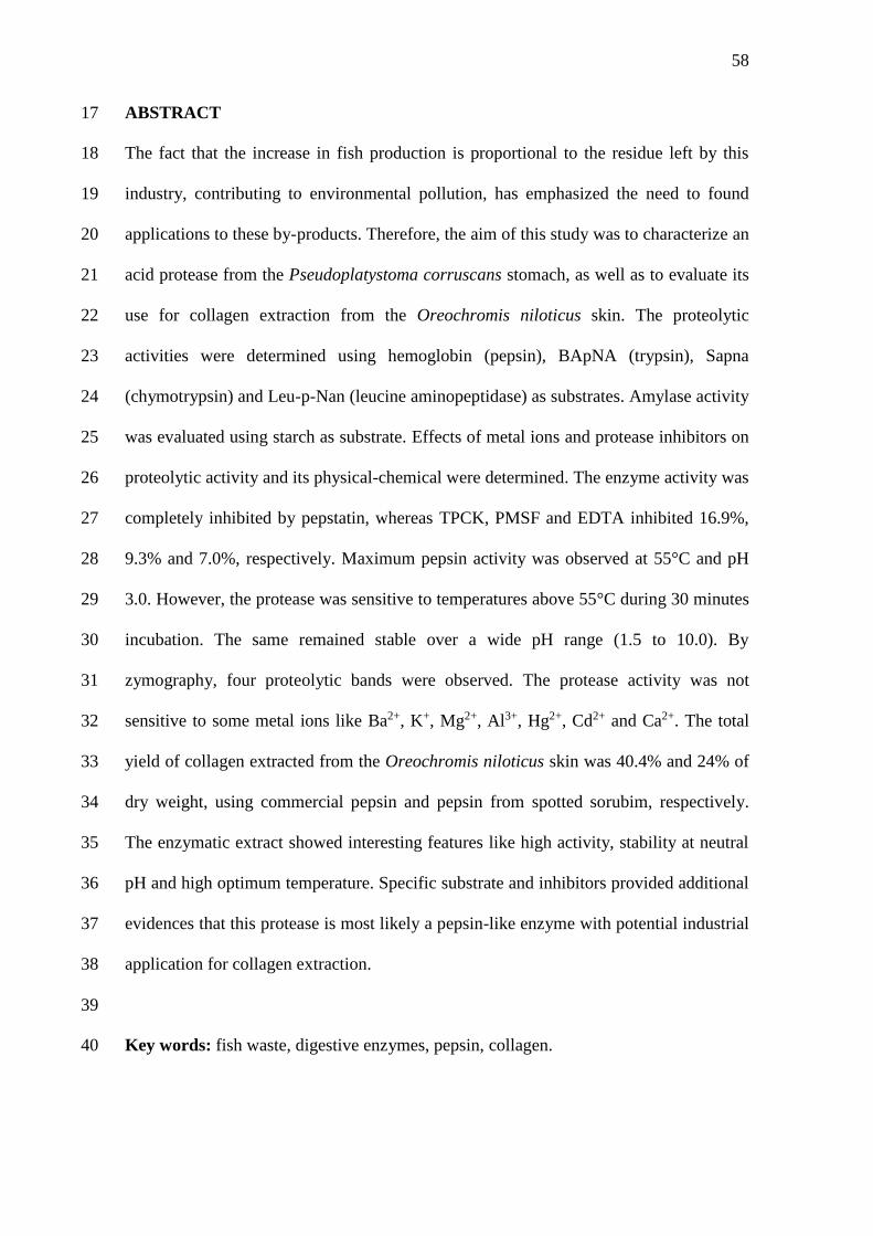
58
ABSTRACT 17
The fact that the increase in fish production is proportional to the residue left by this 18
industry, contributing to environmental pollution, has emphasized the need to found 19
applications to these by-products. Therefore, the aim of this study was to characterize an 20
acid protease from the Pseudoplatystoma corruscans stomach, as well as to evaluate its 21
use for collagen extraction from the Oreochromis niloticus skin. The proteolytic 22
activities were determined using hemoglobin (pepsin), BApNA (trypsin), Sapna 23
(chymotrypsin) and Leu-p-Nan (leucine aminopeptidase) as substrates. Amylase activity 24
was evaluated using starch as substrate. Effects of metal ions and protease inhibitors on 25
proteolytic activity and its physical-chemical were determined. The enzyme activity was 26
completely inhibited by pepstatin, whereas TPCK, PMSF and EDTA inhibited 16.9%, 27
9.3% and 7.0%, respectively. Maximum pepsin activity was observed at 55°C and pH 28
3.0. However, the protease was sensitive to temperatures above 55°C during 30 minutes 29
incubation. The same remained stable over a wide pH range (1.5 to 10.0). By 30
zymography, four proteolytic bands were observed. The protease activity was not 31
sensitive to some metal ions like Ba2+, K+, Mg2+, Al3+, Hg2+, Cd2+ and Ca2+. The total 32
yield of collagen extracted from the Oreochromis niloticus skin was 40.4% and 24% of 33
dry weight, using commercial pepsin and pepsin from spotted sorubim, respectively. 34
The enzymatic extract showed interesting features like high activity, stability at neutral 35
pH and high optimum temperature. Specific substrate and inhibitors provided additional 36
evidences that this protease is most likely a pepsin-like enzyme with potential industrial 37
application for collagen extraction. 38
39
Key words: fish waste, digestive enzymes, pepsin, collagen. 40
59
1. Introduction 41
The spotted sorubim (Pseudoplatystoma corruscans) is a carnivorous freshwater 42
fish distributed throughout the South American rivers (São Francisco, Amazonas and 43
Paraná) (Crepaldi et al., 2006). This specie comprises the largest fish of the Pimelodidae 44
family and may reach 100-120 kg. It is among the fish species that show great 45
commercial value and potential for Brazilian aquaculture (Inoue et al., 2009). 46
Increased fish production and, consequently, the volume of fish processed 47
worldwide, has generated a lot of waste and byproducts. This fact represents a great 48
challenge for entrepreneurs and scientific community interested in pursuing strategies 49
for the production of aquatic organisms, either coming from the extraction or 50
aquaculture, become a sustainable activity (Costa et al., 2013). 51
In fish processing, it is generated a large amount of liquid (wastewater) and solid 52
(skin, bones, guts and fins) wastes. These are usually discarded without treatment and 53
have no commercial value, only causing environmental pollution. From an 54
environmental and economic perspective, a better utilization of fish waste as a source of 55
biomolecules is suggested; as an example it can be highlighted the use of viscera as 56
sources of enzymes with potential industrial applications (Silva et al., 2011; Freitas-57
Júnior et al., 2012). 58
Pepsin is a major digestive enzyme present in the stomach of vertebrates and 59
belongs to the class of aspartyl proteases. It is an endopeptidase that cleaves peptide 60
bonds on the carboxyl side of aromatic amino acids residues: phenylalanine, tyrosine 61
and tryptophan. Acid proteases such as pepsin have a particular importance due to their 62
application in fish silage (Murado et al., 2009) and collagen extraction (Veeruraj et al., 63
2014). 64
60
The number of studies aiming to extract collagen from marine fish has grown 65
continuously. Several studies have reported successful extraction from skin, bones and 66
scales of fishes as striped catfish (Singh et al., 2011), marine eel-fish (Veeruraj et al., 67
2013), silver carp (Safandowska et al., 2013), amur sturgeon (Veeruraj et al., 2014), 68
grass carp, silver carp, bighead carp and black carp (Liu et al., 2014), black rockfish, sea 69
bass and red sea bream (Cho et al., 2014). 70
The use of commercial pepsin can be costly for collagen extraction, therefore, 71
the use of pepsin from spotted sorubim stomach can be a promising alternative due to its 72
abundance and unique properties. Thus the aims of this study were to characterize acid 73
protease activity from the P. corruscans stomach, as well as to use spotted sorubim 74
pepsin for collagen extraction from the Oreochromis niloticus skin. 75
76
2. Material and Methods 77
2.1. Materials 78
All reagents used in assays were of analytical grade and purchased from Sigma 79
(St. Louis, MO, USA), Merck (Darmstadt, Germany) and Bio-Rad Laboratories 80
(Ontario, Canada). 81
82
2.2. Obtaining samples and preparation of stomach extract 83
Juvenile specimens (medium length at first maturity =75 cm) of spotted sorubim 84
(n=3) with total length of 76.3 ± 1.5 cm and weighing 3.3 ± 0.1 kg were obtained from 85
the São Francisco River, in Pilão Arcado, Bahia, Brazil. Stomachs were removed, 86
washed with distilled water and stored at 4C for transport to the laboratory. Then the 87
stomachs (25.0 ± 1.3 g) were homogenized in 0.9% NaCl (w/v) at 4°C in the ratio 1:5 88
(w/v) using an electric tissue homogenizer (IKA RW 20D S32, China). The material 89
61
obtained was centrifuged (Sorvall RC-6 Superspeed Centrifuge - North Carolina, USA) 90
at 8,000 xg for 20 minutes at 4C. The supernatant (crude extract) was stored at -20°C 91
for later use. 92
93
2.3.Protein determination 94
The protein concentration in the crude extract was determined according to the 95
methodology described by Bradford (1976), using bovine serum albumin as standard. 96
97
2.4. Specific proteolytic activities 98
Enzymatic activities of trypsin, chymotrypsin and leucine aminopeptidase were 99
determined in microplates using Nα-benzoyl-DL-arginine-p-nitroanilide (BApNA) 100
succinyl proline alanine phenylalanine p-nitroanilide (SApNA) and leucine-p-101
nitroanilide (Leu-p-Nan) as specific substrates, respectively. The substrates were 102
prepared in dimethyl sulfoxide (DMSO) at a final concentration of 4 mM. For the 103
assays, 30 L of the extract were mixed with 140 L of 0.1 M Glycine-HCl buffer, pH 104
2.5 and 30 L of specific substrate for 15 minutes at 25C. The formation of p-105
nitroaniline (product) was followed at 405 nm with a microplate reader (Bio-Rad Model 106
X-MarkTM spectrophotometer, California, USA). One unit of enzyme activity was 107
defined as the amount of enzyme required to hydrolyze 1 mol of substrate per minute 108
(Bezerra et al., 2005). 109
Acid protease activity was performed according Pavlisko et al. (1997) using 110
2.0% (w/v) hemoglobin (prepared in 0.1 M Glycine-HCl, pH 2.5) as specific substrate. 111
The enzymatic reaction was prepared in triplicate using microcentrifuge tubes to which 112
were added 50 µL of sample, 350 µL of buffer 0.1 M Glycine-HCl, pH 2.5 and 100 µL 113
of substrate. After 30 minutes at 37C, 500 µL of 10% (w/v) trichloroacetic acid was 114
62
added to stop the reaction and the mixture was centrifuged at 10,000 xg for 15 minutes. 115
The absorbance at 280 nm of the supernatant was measured with a microplate reader. 116
One unit (U) of enzyme activity was defined as the amount of enzyme able to produce a 117
change of 0,001 unit of absorbance per minute. 118
119
2.5. Amylase activity 120
Amylase activity was evaluated according to the methodology of Bernfeld 121
(1955) using 2% (w/v) starch as substrate. For the assay, 60 L of sample were mixed 122
with 375 L of 10 mM phosphate buffer, pH 7.5 and 375 L of substrate. After 10 123
minute at 37ºC, 1 mL of DNSA (3,5 dinitrosalycilic acid) was added and the mixture 124
was heated at 100°C for 10 minutes. The absorbance at 570 nm was measured with a 125
microplate reader. A blank control was prepared, in which the sample was replaced by 126
distilled water. The enzymatic activity was expressed as micrograms of maltose released 127
per minute per milligram protein. 128
129
2.6. Physical-chemical properties 130
The influence of temperature on the proteolytic activity was measured by 131
incubating the enzyme extract with 2% hemoglobin in a water bath at temperatures 132
ranging from 25 to 85°C (Bezerra et al., 2005). For the assay of thermal stability, the 133
enzyme extract was incubated for 30 minutes at different temperatures (25 – 85°C) and 134
then evaluated for proteolytic activity as described in 2.3. The influence of pH on the 135
proteolytic activity was assayed by incubating the extract at different 0.2 M buffer 136
solutions (Glycine-HCl, pH 1.5-3.5; Citrate-Phosphate, pH 4.0-7.5; Tris-HCl, pH 8.0-137
9.0; and Glycine-NaOH, pH 9.5-10.0) for 30 minutes with 2% hemoglobin as substrate. 138
The absorbance at 280 nm of the supernatant was measured with a microplate reader. 139
63
The pH stability of the enzyme activity of extract was determinated after pre-incubation 140
for 30 min at the different buffers according to Klomklao (2007) modified. 141
142
2.7. Effect of protease inhibitors 143
This assay was performed using the synthetic protease inhibitors: 144
phenylmethylsulphonyl fluoride (PMSF, serine protease inhibitor), N-p-tosyl-L-lysin 145
chloromethyl ketone (TLCK, trypsin inhibitor), N-tosil-l-phenylalanine chloromethyl 146
ketone (TPCK, chymotrypsin inhibitor), at 5 mM, dissolved in DMSO, as well as 147
ethylenediamine tetraacetic acid (0.5 mM EDTA, metalloprotease inhibitor), 5 mM β-148
mercaptoethanol (reducing agent) prepared in water and 0.5 mM Pepstatin A , a pepsin 149
inhibitor, prepared in 10% methanol. The inhibitors were incubated with enzyme extract 150
at a proportion of 1:1 (v/v) for 30 minutes at 25°C. After incubation, the pepsin activity 151
assay was performed using 2% hemoglobin as substrate. The residual activity of sample 152
was estimated by comparing it to a positive control (100% activity), in which the extract 153
was incubated with DMSO, water or 10% methanol, depending on the inhibitor’s 154
solvent (Bezerra et al., 2001). 155
156
2.8. Effect of metal ions 157
Samples of the enzyme extract (30 µL) were added to a 96-well microtiter plate 158
with solutions 1.0 mM (30 µL) of MgCl2, BaCl2, KCl, CuCl2, CdCl2, CaCl2, HgCl2 or 159
AlCl3 for 30 minutes. After incubation, the pepsin activity assay was performed using 160
2% hemoglobin as substrate. The residual activity of sample was estimated by 161
comparing it to a positive control (Wu et al., 2009). 162
163
2.9. Zymogram 164
64
Protease zymogram under native conditions was carried out following the 165
procedure described by Garcia-Carreño et. al. (1993). Lyophilized extract (30 µg of 166
protein) was mixed with a solution containing 10 mM Tris-HCl (pH 8.0), 2.5% SDS, 167
10% glycerol and 0.002% bromophenol blue and applied in the polyacrylamide gel 168
(12.5% w/v). The electrophoresis proceeded at 4°C under constant electric current (11 169
A). After the migration of the proteins, the gel was incubated at 37°C and immersed in 170
2% hemoglobin at pH 2.5 for 60 minutes. Then the gel was stained with a solution 171
composed of 0.01% Coomassie Brilliant Blue, 25% methanol and 10% acetic acid for 172
24 hours. The background of the gel was destained by washing in a solution containing 173
10% (v/v) acetic acid and 25% methanol (v/v). 174
175
2.10. Use of P. corruscans acid protease for collagen extraction from the Oreochromis 176
niloticus skin 177
178
2.10.1. Obtaining and preparation of samples 179
Prior to collagen extraction, stomach extract from P. corruscans was subjected 180
to precipitation by ammonium sulfate (80% saturation). The Nile tilapia skins were 181
obtained from local fishing industry (Terra & Mar Pescados / Recife-PE, Brazil). After 182
collecting, the material were washed with cold distilled water and cut into small pieces 183
(0.5 – 0.5 cm2). 184
185
2.10.2. Pretreatment of skin 186
The collagens were prepared by the method of Nagai and Suzuki (2000) with a 187
slight modification. To remove non-collagenous proteins, the prepared fish skin was 188
mixed with 0.2 M NaOH at a skin/alkali solution ratio of 1:10 (w/v). The mixture was 189
65
continuously stirred for 3h at 4ºC and the alkali solution was changed every 30 minutes. 190
The treated skin was then washed with cold distilled water until a neutral or faintly 191
basic pH of wash water was reached. Then, it was added 10% butyl alcohol in the ratio 192
of 1:10 (w/v) to remove fats. The mixture was continuously stirred for 6h at 4ºC and the 193
skin was washed as previously described. Then, it was added 3% hydrogen peroxide at 194
a ratio of 1:10 (w/v) for whitening the skin and was carried out in the wash. 195
196
2.10.3. Extraction of acid soluble collagen (ASC) 197
The residue was extracted with 0.5 M acetic acid at sample/acid ratio of 1:20 198
(w/v) for 3 days. The resulting viscous solution was centrifuged at 20,000g for 30 min 199
at 4°C. The supernatants of the extract were combined and salted-out by adding NaCl to 200
give a final concentration of 0.9 M, followed by precipitation of the collagen by the 201
addition of NaCl to the final concentration of 2.5 M in 1.5 M Tris–HCl (pH 8.8). After 202
standing overnight, the resulting precipitate was collected by centrifugation at 20,000g 203
for 60 min and then dissolved in 10 volumes of 0.5 M acetic acid. The solution obtained 204
was dialyzed against 0.1 M acetic acid and subsequently against distilled water. The 205
dialysate was freeze-dried and referred to as acid soluble collagen (ASC). 206
207
2.10.4. Extraction of pepsin soluble collagen (PSC) 208
The pepsin soluble collagen (PSC) was obtained through the incubation of the 209
insoluble material obtained in the previous steps with commercial porcine pepsin (EC 210
3.4.23.1; crystallized and lyophilized, Sigma, MO) or fraction 80% saturation fraction 211
(ammonium sulfate) from stomach of P. corruscans. Porcine pepsin and fraction were 212
tested at the same level (5000 U/mL) for the comparative study. Incubation occurred 213
with constant homogenization for 24 hours at 4°C. The pepsin-solubilized collagen 214
66
(PSC) was obtained by the same method as the ASC. The yield of ASC or PSC was 215
calculated as: Yield (%) = (M/M0) x 100, where M is the weight of lyophilized collagen 216
(g), and M0 is the weight of lyophilized skin used (g). 217
218
2.11. Statistical analysis 219
Data of enzyme activity were analyzed using one-way analysis of variance 220
(ANOVA) complemented with Tukey’s test. Differences were reported as statistically 221
significant when P<0.05, using the program MicrocalTM OriginTM version 8.0 222
(Software, Inc, U.S.). 223
224
3. Results and Discussion 225
3.1. Enzymatic assays 226
In vitro assays were performed to determine the action of enzymes present in the 227
P. corruscans stomach extract. It was observed high activity of pepsin (23.24 ± 0.1 228
U/mg) by using 2% hemoglobin as substrate. The high proteolytic activity in the 229
stomach is certainly associated with the carnivorous habit of the species and highlights 230
the role of that structure in the digestive process. Acid proteolytic activities have been 231
previously reported in carnivorous fish (Zhao et al., 2011). 232
Specific proteolytic activities showed discrete values (0.08 ± 0.0 U/mg and 0.15 233
± 0.0 U/mg) with BApNA and Leu-p-Nan as substrates, respectively. The use of 234
SApNA, specific chromogen substrate, was not effective to determine the 235
chymotrypsin-like activity (Table 1). However, this is not the first report of such 236
enzymes in fish stomach. Lundstedt et al. (2004) showed activity for trypsin and 237
chymotrypsin in the stomach of P. corruscans. Deguara et al. (2003) discuss the 238
presence of alkaline proteases in the stomach of Sparus aurata and refer to other 239
67
authors concerning this fact. The pattern observed in proteolytic activities is probably 240
explained as a function of the pH of the reaction medium, since this factor is critical for 241
stability of the polypeptide chain of the enzyme. The distribution of proteases varies 242
depending on the species and morphology of the digestive tract. 243
The total amylase activity present in P. corruscans stomach was detected using 244
the long chain substrate, 2% starch (1.26 ± 0.3 U/mg) (Table 1). Similar result was 245
reported by Lundstedt et al. (2004), where juveniles of P. corruscans underwent 246
experimental diets. Because it is a carnivorous fish, carbohydrate digestion in spotted 247
sorubim is not considered essential in the digestive process. However, digestive 248
carbohydrases such as amylase has been reported in gastric juice and Clupea harengus 249
and Dorosoma cepidianun (Fange et al., 1979). Compared with omnivorous species, the 250
amylase in carnivorous fish has been less active (Horn et al., 2006; Al-Tameemi et al., 251
2010). 252
Studies by Seixa Filho et al. (1999) reported the amylolytic activity of 253
omnivorous freshwater fish, as piracanjuba (Brycon orbignyanus) and piau (Leporinus. 254
friderici) compared with spotted sorubim. These authors found higher amylolytic 255
activity in spotted sorubim. Regarding amylase, several factors may influence its 256
activity in fish, among them, eating habits and the structural complexity of carbohydrate 257
(Al-Tameemi et al., 2010). The influence of hormones, metabolites and temperature on 258
amylase activity in aquatic organisms have been reported (Van Wormhoudt, 1980), a 259
fact that makes it even more difficult to compare quantitative results in the literature. 260
261
3.2. Effect of temperature on acid protease activity and stability 262
Physico-chemical parameters were analyzed for acid protease activity present in 263
the stomach extract of P. corruscans. The effect of temperature on the activity of acid 264
68
protease is illustrated in Figure 1A. Enzymatic assays have shown that protease 265
exhibited maximum activity at 55°C. The activity of the enzyme at 25 – 35°C 266
corresponded to more than 50% of the activity at optimum temperature. In the 267
temperature range between 40 and 50°C, acid protease showed more than 90% of its 268
activity (Fig. 1A). 269
Results on optimum temperature of pepsin in some species of fish have been 270
reported in the literature, such as 37°C for Parona signata (Pavlisko et al., 1997), 45 to 271
50°C for S. Houttuyn latus (Zhou et al., 2007), 40°C for Mustelus mustelus (Bougatef et 272
al., 2008), 40 to 45 °C for Siniperca chuatsi (Zhou et al., 2008), 35 to 40°C for Anguilla 273
anguilla (Wu et al., 2009). Komklao et al. (2007) obtained optimum temperature of 274
45°C for pepsin in the stomach of Coryphaenoides pectoralis. The optimal temperature 275
of Thunnus alalunga pepsin was 50°C (Nalinanon et al., 2010) and 40°C for Sardinella 276
aurita aspartic protease (Khaled et al., 2011). 277
It is not known why the digestive enzymes of fish and other aquatic organisms 278
present high activity at temperatures above the physiological temperature. Probably, the 279
answer to this question is in the adaptations of their ancestors to climate change through 280
evolution (Freitas-Júnior et al., 2012). The decrease of acid protease activity from P. 281
corruscans at 60°C is presumed to be due to enzyme denaturation. Determination of the 282
optimum temperature for maximum activity of an enzyme is interesting for future 283
comparative studies, providing information on their expected activity under various 284
physiological conditions (Fernandez et al., 2001). Furthermore, pepsins may possibly be 285
used in industrial applications that require low processing temperatures, as collagen 286
extraction. 287
The resistance of acid protease from P. corruscans to heating treatment is shown 288
in Figure 1B. The enzyme remained practically stable at temperatures 25 - 45°C for 30 289
69
minutes, losing more than half of its activity after heating above 55°C. According to the 290
literature, the acid protease of Oreochromis niloticus retained more than 50% of its 291
activity after heating between 50 and 60°C for 30 minutes (El-Belgaty et al., 2004). 292
Pepsins from Pectoralis coryphaenoides were stable up to 40°C (Klomklao et al., 2007). 293
Acid proteins of Sardinops sagax caerulea were not stable above 45°C (Castillo-Yanez 294
et al., 2004). Thunus alalunga pepsin was stable up to 50°C (Nalinanon et al., 2010) 295
whereas the enzymatic activity from the viscera of Sardinella aurita was stable up to 296
40°C (Khaled et al., 2011). The enzymatic characteristics relating to thermal stability 297
tests can be useful for possible application in industrial processes (gelatin extraction, 298
milk clotting). The acid protease of P. corruscans fits well in the requirements for 299
industrial application of the enzyme, being an interesting alternative. 300
301
3.3. Effect of pH on acid protease activity and stability 302
The effect of pH on the activity of acid protease from P. corruscans was 303
determined over the pH range (1.5 to 6.5). Protease was active between pH 1.5 to 3.5, 304
with the optimum pH in the range of 3.0 (Figure 1C). Above pH 4.0, over 50% of the 305
activity of the acid protease was lost and enzymatic activity was not detected in pH 306
greater than 5.0. Optimum pH values between 2.0 and 4.0 have been reported for pepsin 307
of S. latus houttuyn (Zhou et al., 2007). Pepsins from the stomach of Coryphaenoides 308
pectoralis exhibited maximal activity at pH 3.0 and 3.5 (Klomklao et al., 2007). Similar 309
result was found for pepsins from mandarin fish (Zhou et al., 2008). The optimal pH for 310
hydrolysis of hemoglobin by pepsin was found at pH 2.0 for Mustelus mustelus 311
(Bougatef et al., 2008) and Thunnus alalunga (Nalinanon et al., 2010). Aspartic 312
proteinase isolated from the stomach of Anguilla anguilla showed optimal pH values of 313
70
2.5 and 3.5 (Wu et al., 2009). Khaled et al. (2011) reported maximum activity of pepsin 314
from the viscera of sardinelle around pH 3.0. 315
To investigate the pH stability, the enzyme was incubated in different buffers in 316
the pH range from 2.0 to 10.5 and enzymatic activity is shown in Figure 1D. The acid 317
protease retained more than 80% of its original activity in all the pH range tested. At pH 318
2.0 the enzyme retained 100% of its activity. The stability of acid protease from the P. 319
corruscans stomach at neutral pH suggests some interesting information about the 320
storage of the enzyme. According Klomklao et al., (2006), stability of pepsins to 321
particular pH can be determined by the net charge of the enzyme. Castillo-Yanez et al., 322
(2004) also reported that acidic enzymes from Sardinops sagax caerulea were stable 323
between pH 3.0 to 6.0. Thunus alalunga pepsin was stable in the pH range of 2 – 5 324
(Nalinanon et al., 2010). Pepsins were stable in the pH range of 2.0 – 6.0 (Klomklao et 325
al., 2007) and 2.0 – 5.0 (Khaled et al., 2011). At pH 1.0 and 4.0, the acid protease 326
retained 85.3% and 61.5% of its initial activity, respectively (Bougatef et al., 2008). 327
Differences related to pH stability between various species of fish indicate that different 328
molecular properties may be involved, such as enzyme conformation and stability of 329
structural links (Klomklao et al., 2007). 330
331
3.4. Effect of protease inhibitors 332
In order to better understand the class of proteases in the stomach from spotted 333
sorubim, its sensitivity to various inhibitors was tested. The proteolytic activity (with 334
the substrate hemoglobin) was completely inhibited by pepstatin A (100%), classic 335
inhibitor of aspartate proteases. The enzymatic activity was weakly affected by serine 336
protease inhibitors: TPCK (17.0%), PMSF (9.4%) and TLCK (2.7%) (Table 2). The low 337
inhibition in the presence of EDTA (7.1%) (metalloenzyme inhibitor) indicates that 338
71
these proteases activities are not fully ion-dependent. Likewise, β-Mercaptoethanol 339
(3.6%) did not show high inhibitory effect on the proteases, suggesting that the 340
reduction of disulfide bonds little affect their activity. These results show that the 341
sample is composed mainly of aspartate proteases, especially pepsin. 342
Khaled et al. (2011) reported that protease derived from the stomach of 343
Sardinella aurita was almost completely inhibited by pepstatin A (2 mM). Activity of 344
pepsin from the stomach of albacore tuna was strongly inhibited by pepstatin A with the 345
concentration range of 0.1– 10 µM (Nalinanon et al., 2010). Pepsins from the stomach 346
of Anguilla anguilla and Siniperca chuatsi also were almost completely inhibited by 347
pepstatin A, but specific inhibitors of serine proteinases (PMSF) and metalloproteinases 348
(EDTA) had no inhibitory effect on the activity of pepsin (Wu et al., 2009; Zhou et al., 349
2008, Bougatef et al., 2008). However, information regarding the inhibition of proteases 350
in vitro does not adequately reflect what occurs in vivo, where other factors may modify 351
the physiological response of fish (El-Sayed et al., 2000). 352
353
3.5. Effect of metal ions 354
Some enzymes require additional chemical component (cofactor) such as 355
inorganic ions for its activity. On the other hand, due to their bio-accumulative and non-356
biodegradable properties, some metals constitute one of the main groups of aquatic 357
pollutants (Ramesh, 2006). The effect of metallic ions on the activity of acid digestive 358
protease from spotted sorubim stomach was evaluated and is presented in Table 3. 359
Compared with the control, highest inhibition of the pepsin activity was found in the 360
presence of Ba2+ (23.7%), Mg2+ (20.7%) and K+ (16.9%) at 1 mM. Ions Al3+, Ca2+, 361
Hg2+, Cd2+ and Cu2+ inhibited 10.9%, 9.2%, 7.4%, 6.6% and 5.0%, respectively. 362
72
The aspartic protease activity from S. aurita was slightly affected by Mn2+, Ba2+, 363
and Zn2+. Ions Ca2+, Na+ and K+ no affected on the activity. However, the addition of 364
Mg2+, Cu2+ and Hg2+ decreased the activity by 50%, 50% and 60%, respectively 365
(Khaled et al., 2011). According Klomklao et al., (2007), the activity of pepsins was 366
increased in the presence of divalent cations (CaCl2, MgCl2 and CoCl2), especially 367
when the concentration used increased. The influence of metal ions on the activity of 368
pepsin was also verified by Krejpcio et al. (2002), where there was an increase of 369
enzyme activity with increasing concentrations of Al3+. The different effects of trace 370
elements on the activity of pepsin could perhaps be explained in terms of the stability 371
constants for the complexes of the various ions with pepsin. It is known that sub-lethal 372
concentrations of heavy metals can disturb the digestive enzymatic activity of the 373
exposed species (De Coen et al., 1998). 374
375
3.6. Zymogram 376
The acid proteolytic activity of the stomach from P. corruscans was evaluated 377
by zymogram. It was observed the presence of four protein bands, possibly isoforms of 378
pepsin (Figure 2). Several authors have reported the use of zymogram for confirmation 379
of activity of acid protease (Nalinanon et al., 2010; Khaled et al., 2011). Zymography 380
analysis indicated that pepsins of mandarin fish revealed degrading activity against 381
acid-denatured bovine hemoglobin (Zhou et al., 2008). Through the zymogram can 382
identify the number of active forms of certain enzyme in different species, thereby 383
providing a source of data for the knowledge and comparison of their digestive 384
physiology. 385
386
3.7. Collagen extraction 387
73
In relation to the collagen extraction, the total yield of collagen obtained from 388
the skin of Nile tilapia was 40.4% and 24% of dry weight, using commercial pepsin and 389
pepsin from spotted sorubim, respectively. ASC, PSC (commercial pepsin) and PSC 390
(stomach from spotted sorubim) showed yields of 19.3%, 21.1% and 4.7%, respectively 391
(Table 4). This result shows that the collagen present in the skin Nile tilapia is not 392
completely soluble in acetic acid 0.5 M. The fact that the PSC (commercial pepsin) 393
showed a greater extraction yield when compared to ASC, can be due to possibility of 394
the collagen molecules in the O. niloticus skin are linked through crossed covalent 395
bonds. These crosslinks happen through the condensation of groups aldehydes in the 396
telopeptides areas and intermolecular of the collagen fact that causes a decrease in 397
solubility of the protein in acetic acid (Foegeding et al., 1996, Zhang et al., 2007). The 398
low yield of PSC (stomach from spotted sorubim) can be explained by the purification 399
step used, resulting in an extract with various interfering substances. 400
By submitting the residue extraction with acetic acid to the action of pepsin, is 401
obtained as a result of the cleavage telopeptide region and hydrolysis of crosslinks 402
(Balian et al., 1977). Thus pepsin could be used as an aid for increasing the extraction 403
yield of collagen from the skin of Nile tilapia. The yield of ASC from the skin of O. 404
niloticus was higher than that from Brownstripe red snapper skin (9%) and lower than 405
that from the skins of Japanese sea-bass (51.4 %), chub mackerel (49.8%), and bullhead 406
shark (50.1%) (Nagai et al., 2000). 407
Others works reported in the literature showed the following yield for the 408
extraction of collagen from skin: Aluterus monóceros (7.6%) (Ahmad et al., 2010), 409
Priacanthus tayenus e Priancanthus macracanthus (7.7% e 7.1%) (Benjakul et al., 410
2010). These variations in yields are related both to different biological conditions, 411
which each species is subject, as well as the conditions and methods of extraction 412
74
(McCormick, 2009). Also worth pointing out that structural differences in collagen is 413
directly related to the performance of the extraction, because if the molecules in 414
telopeptide region are highly cross-linked the collagen solubility in acid tends to 415
decrease (Foegeding et al., 1996). 416
417
4. Conclusion 418
The characterization with specific substrate and inhibitors generates evidence 419
that this protease is probably a pepsin-like enzyme. It was also observed that this 420
protease showed low sensitivity to various metal ions. The enzyme showed interesting 421
features like high activity, stability at neutral pH and high optimum temperature. Acid 422
soluble and pepsin soluble collagen were extracted from the skin of O. niloticus. Thus, 423
P. corruscans pepsin could be used as an aid for increasing the extraction yield of 424
collagen from the skin of Nile tilapia. These features indicate that the stomach of P. 425
corruscans can be used as a source of acid proteases with a potential for industrial 426
application (collagen extraction). 427
428
Acknowledgements 429
This study was financially supported by the following Brazilian agencies: 430
Conselho Nacional de Pesquisa e Desenvolvimento Científico (CNPq), and the 431
Coordenação de Aperfeiçoamento de Pessoal de Nível Superior (CAPES). 432
433
References 434
Ahmad, M., & Benjakul, S. (2010). Extraction and characterisation of pepsin solubilised 435
collagen from the skin of unicorn leatherjacket (Aluterus monocerous). Food Chemistry, 436
120, 817–824. 437
75
438
Al-Tameemi, R., Aldubaikul, A., Salman, N. A. (2010). Comparative study of α-439
amylase activity in three Cyprinid species of different feeding habits from Southern 440
Iraq. Turkish Journal of Fisheries and Aquatic Sciences 10: 411-414. 441
442
Balian, G., & Bowes, J. H. (1977). The structure and properties of collagen. In A. G. 443
London: Academic Press. 444
445
Benjakul, S., Thiansilakul, Y., Visessanguan, W., Roytrakul, S., Kishimura, H., 446
Prodprand, T., et al. (2010). Extraction and characterization of pepsin solubilised 447
collagens from the skin of bigeye snapper (Priacanthus tayenus and Priacanthus 448
macracanthus). Journal of the Science of Food and Agriculture, 90, 132–138. 449
450
Bernfeld, P., (1955). Amylases, and ß. IN Colowick, S.P. & Kaplan, N.O. (Eds.) 451
Methods in Enzymology. New York, Academic Press. 452
453
Bezerra, R. S., Vieira, V. L. A., Santos, A. J. G. (2001). Ciclo reprodutivo da carapeba 454
prateada Diapterus rhombeus (Cuvier, 1829), no litoral de Pernambuco. Tropical 455
Oceanography, 1, 67-78. 456
457
Bezerra, R. S., Lins, E. J. F., Alencar, R. B., Paiva, P. M. G., Chaves, M. E. C., Coelho, 458
L. C. B. B., Jr Carvalho, L. B. (2005). Alkaline proteinase from intestine of Nile tilapia 459
(Oreochromis niloticus). Process Biochemistry, v. 40, p. 1829-1834. 460
461
76
Bradford, M. M., (1976). A rapid and sensitive method for the quantification of 462
microgram quantities of protein utilizing the principle of protein binding. Anal. 463
Biochem. 72, 248-254. 464
465
Bougatef, A., Balti, R., Zaied, S. B., Souissi, N., Nasri, M. (2008). Pepsinogen and 466
pepsin from the stomach of smooth hound (Mustelus mustelus): Purification, 467
characterization and amino acid terminal sequences. Food Chemistry 107, 777–784. 468
469
Castillo-Yanez, F. J., Pacheco-Aguilar, R., Garcia-Carreno, F. L., & Toro, M. A. N. D. 470
(2004). Characterization of acidic proteolytic enzymes from Monterey sardine 471
(Sardinops sagax caerulea) viscera. Food Chemistry, 85 (3), 343–350. 472
473
Cho, J. K., Jin, Y. G., Rha, S. J., Kim, S. J., Hwang, J. H. (2014). Biochemical 474
characteristics of four marine fish skins in Korea. Food Chemistry 159, 200 – 207. 475
476
Costa, H. M. S., Freitas Júnior, A. C. V., Amaral, I. P. G., Hirata, I. Y., Paiva, P. M. G., 477
Carvalho Jr, L. B., Oliveira, V., Bezerra, R. S. (2013). Metal-sensitive and thermostable 478
trypsin from the crevalle jack (Caranx hippos) pyloric caeca: purification and 479
characterization. Chemistry Central Journal, 7:166. 480
481
Crepaldi, D. V., Faria, P. M. C., Teixeira, E. A., Ribeiro, L. P., Costa, A. A. P., Melo, 482
D. C., Cintra, A. P. R., Prado, S. A., Costa, F. A. A., Drumond, M. L., Lopes, V. E., 483
Moraes, V. E., (2006). O surubim na aquacultura do Brasil. Rev. Bras. Reprod. Animal 484
30, 150 – 158. 485
486

77
De Coen, W. M., Vangheluwe, M. L., Janssen, C. R. (1998).The use of biomarkers in 487
Daphnia magna toxicity testing— III. Rapid toxicity testing of pure chemicals and 488
sediment pore waters using ingestion and digestive enzyme activity. Chemosphere. 37, 489
2677-2694. 490
491
Deguara, S., Jauncey, K., Agius, C. (2003). Enzyme activities and pH variations in the 492
digestive tract of gilthead sea bream. J. Fish. Biol. 62, 1033–1043. 493
494
El-Beltagy, A. E., El-Adawy, T. A., Rahma, E. H., El-Bedawey, A. A. (2004). 495
Purification and characterization of an acidic protease from the viscera of bolti fish 496
(Tilapia nilotica). Food Chem. 86, 33–39. 497
498
El-Sayed, A. M., Martinez, I. N., & Moyano, F. J. (2000). Assessment of the effect of 499
plant inhibitors on digestive proteases of Nile Tilapia Nilotica using in vitro assay. 500
Aquaculture International, 8, 403–415. 501
502
Fänge, R., Grove, D., (1979). Digestion. In: Hoar, W. S., Randall, D. J., Brett, J. T. 503
(Eds.), Fish Physiology, Bioenergetics and Growth, Vol. 8. Academic Press, New York, 504
pp. 161–260. 505
506
Fernández, I.; Moyano, F. J.; Díaz, M.; Martínez, T. (2001). Characterization of α-507
amylase activity in five species of Mediterranean sparid fishes (Sparidae, Teleostei). 508
Journal of Experimental Marine Biology and Ecology, v. 262, N. 1, P. 1-12. 509
510
78
Foegeding, E. A., Lanier, T. C., Hultin, H. O., (1996). Characteristics of edible muscle 511
tissues. In: Fennema, O. R. (Ed.), Food Chemistry. Marcel Dekker, New York, pp. 512
879–942. 513
514
Freitas-Júnior, A. C. V., Costa, H. M. S., Icimoto, M. Y., Hirata, I. Y., Marcondes, M., 515
Carvalho Jr, L. B., Oliveira, V., Bezerra, R. S. (2012). Giant Amazonian fish pirarucu 516
(Arapaima gigas): Its viscera as a source of thermostable trypsin. Food Chemistry, v. 517
133, p. 1596- 1602. 518
519
Garcia-Carreño, F. L., Dimes, L. E., Haard, N. F. (1993). Substrate-gel electrophoresis 520
for composition and molecular weight of proteinases or proteinaceous proteinase 521
inhibitors. Anal. Biochem. 214, 65-69. 522
523
Horn, M. H., Gawlicka, A. K., German, D. P., Logothetis, E. A., Cavanagh, J.W., 524
Boyle, K. S. (2006). Structure and function of the stomachless digestive system in three 525
related species of New World silverside fishes (Atherinopsidae) representing herbivory, 526
omnivory, and carnivory. Mar. Biol., 149: 1237-1245. 527
528
Inoue, L. A. K. A., Hisano, H., Ishikawa, M. M., Rotta, M. A., Senhorini, J. A. (2009). 529
Princípios básicos para produção de alevinos de surubins (pintado e cachara). Dourados: 530
Embrapa Agropecuária Oeste; Manaus: Embrapa Amazônia Ocidental; Corumbá: 531
Embrapa Pantanal. 26 p. Documentos (Embrapa Agropecuária Oeste. Documentos, 99; 532
Embrapa Amazônia Ocidental. Documentos, 68; Embrapa Pantanal. Documentos, 100. 533
Impresso, v. 15, p. 1-26. 534
535
79
Khaled, H. B., Ghorbel-Bellaaj, O., Hmidet, N., Jellouli, K., Ali, N. E., Ghorbel, S., 536
Nasri, M. (2011). A novel aspartic protease from the viscera of Sardinelle (Sardinella 537
aurita): Purification and characterization. Food Chemistry 128, 847–853. 538
539
Klomklao, S., Benjakul, S., Visessanguan, W., Kishimura, H., Simpson, B.K., (2006). 540
Purification and characterization of trypsin from the spleen of tongol tuna (Thunnus 541
tonggol). J. Agric. Food Chem. 54, 5617–5622. 542
543
Klomklao, S., Kishimura, H., Yabe, M., Benjakul, S. (2007). Purification and 544
characterization of two pepsins from the stomach of pectoral rattail (Coryphaenoides 545
pectoralis). Comparative Biochemistry and Physiology, Part B 147, P. 682–689. 546
547
Krejpcio, Z., Wójciak, R. W. (2002). The Influence of Al3+ Ions on Pepsin and Trypsin 548
Activity in Vitro. Polish Journal of Environmental Studies Vol. 11, No. 3, 251-254. 549
550
Liu, D., Zhou, P., Li, T., Regenstein, J. M. (2014). Comparison of acid-soluble 551
collagens from the skins and scales of four carp species. Food Hydrocolloids 41, 290 – 552
297. 553
554
Lundstedt, L. M.; Melo, J. F. B.; Moraes, G. (2004). Digestive enzymes and metabolic 555
profile of Pseudoplatystoma corruscans (Teleostei: Siluriformes) in response to diet 556
composition. Comparative Biochemistry and Physiology Part B 137. 557
558
McCormick, R. J. (2009). Collagen. In Applied muscle biology and meat science (pp. 559
127 - 146). Boca Raton, Florida: CRC Press. 560
80
561
Murado, M. A., González, M. D. P., Vázquez, J. A. (2009). Recovery of proteolytic and 562
collagenolytic activities from viscera by-products of rayfish (Raja clavata). Mar Drugs 563
7: 803 - 815. 564
565
Nagai, T., & Suzuki, N. (2000). Isolation of collagen from fish waste material-skin, 566
bone and fins. Food Chemistry, 68, 277–281. 567
568
Nalinanon, S., Benjakul, S., Kishimura, H. (2010). Biochemical properties of 569
pepsinogen and pepsin from the stomach of albacore tuna (Thunnus alalunga). Food 570
Chemistry 121 49–55. 571
572
Pavlisko, A., Rial, A., Vecchi, S. D., & Coppes, Z. (1997). Properties of pepsin and 573
trypsin isolated from the digestive tract of Parona signata ‘‘Palometa”. Journal of Food 574
Biochemistry, 21, 289–308. 575
576
Ramesh, M. (2006). Studies on the impact of heavy metal cadmium on certain enzymes 577
in a freshwater teleost fish, Cyprinus carpio. In 43rd Congress of the European Society 578
of Toxicology and 6th Congress of Toxicology in Developing Countries. Toxicology 579
Letters. 164 (S), 157. 580
581
Safandowska, M., Pietrucha, K. (2013). Effect of fish collagen modification on its 582
thermal and rheological properties. International Journal of Biological Macromolecules 583
53, 32 – 37. 584
585
81
Seixa filho, J. T., Oliveira, M. G. A., Donzele, J. L., Gomide, A. T. M., Menin, E., 586
(1999). Atividade de amilase em quimo de três espécies de peixes Teleostei de água 587
doce. Rev. Bras. Zootec. 28, 907–913. 588
589
Silva, J. F., Esposito, T. S., Marcuschi, M., Ribeiro, R., Cavalli, R. O., Oliveira, V., 590
Bezerra, R. S. (2011). Purification and partial characterisation of a trypsin from the 591
processing waste of the silver mojarra (Diapterus rhombeus). Food Chemistry, v. 129, 592
p. 777-782. 593
594
Singh, P., Benjakul, S., Maqsood, S., & Kishimura, H. (2011). Isolation and 595
characterization of collagen extracted from the skin of striped catfish (Pangasianodon 596
hypophthalmus). Food Chemistry, 124 (1), 97 - 105. 597
598
Van Wormhoudt, A. (1980). Regulation d'activité de l'α-Amylase à différentes 599
températures d'adaptation et en fonction de l'ablation des pédoncules oculaires et du 600
stade de mue chez Palaemon serratus. Biochemical Systematics and Ecology, v. 8, n. 2, 601
p. 193-203. 602
603
Veeruraj, A., Arumugam, M., & Balasubramanian, T. (2013). Isolation and 604
characterization thermostable collagen from eel-fish (Evenchelys macrura). Process 605
Biochemistry, 48 (10), 1592 - 1602. 606
607
Veeruraj, A., Arumugam M., Ajithkumar T., Balasubramanian, T. (2014). Isolation and 608
characterization of collagen from the outer skin of squid (Doryteuthis singhalensis). 609
Food Hydrocolloids, http://dx.doi.org/10.1016/j.foodhyd.2014.07.025. 610
82
611
Wu, T.; Sun, L-C.; Du, C-H.; Cai, Q-F.; Zhang, Q-B.; Su, W-J.; Cao, M-J. (2009). 612
Identification of pepsinogens and pepsins from the stomach of European eel (Anguilla 613
anguilla). Food Chemistry, v. 115, p.137–142. 614
615
Zhao, L., Budge, S. M., Ghaly, A. E., Brooks, M. S., Dave, D. (2011). Extraction, 616
Purification and Characterization of Fish Pepsin: A Critical Review. J Food Process 617
Technol, 2:6. 618
619
Zhou, Q., Fu, X. P., Zhang, L. J., Su, W. J., & Cao, M. J. (2007). Purification and 620
characterization of sea bream (Sparus latus Houttuyn) pepsinogens and pepsins. Food 621
Chemistry, 103, 795–801. 622
623
Zhou, Q., Liu, G. M., Huang, Y. Y., Weng, L., Hara, K., Su, W. J., Cao, M. J. (2008). 624
Pepsinogens and Pepsins from Mandarin Fish (Siniperca chuatsi) J. Agric. Food Chem., 625
56, 5401–5406. 626
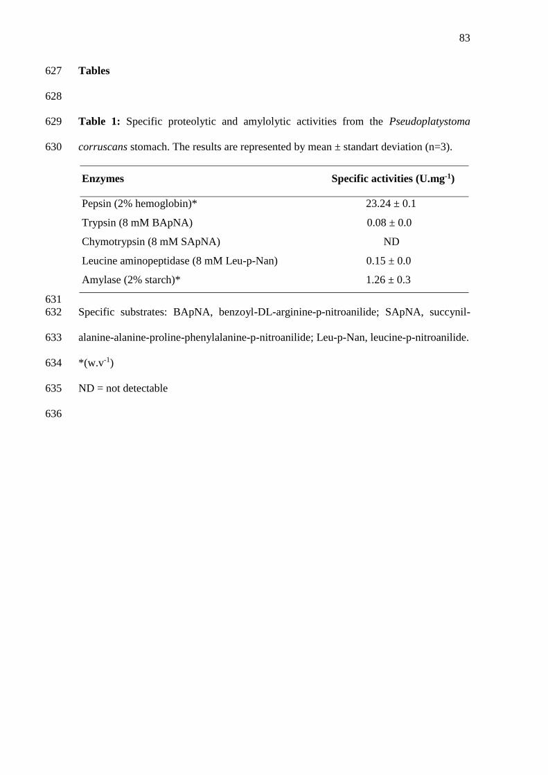
83
Tables 627
628
Table 1: Specific proteolytic and amylolytic activities from the Pseudoplatystoma 629
corruscans stomach. The results are represented by mean ± standart deviation (n=3). 630
Enzymes Specific activities (U.mg-1)
Pepsin (2% hemoglobin)* 23.24 ± 0.1
Trypsin (8 mM BApNA) 0.08 ± 0.0
Chymotrypsin (8 mM SApNA) ND
Leucine aminopeptidase (8 mM Leu-p-Nan) 0.15 ± 0.0
Amylase (2% starch)* 1.26 ± 0.3
631
Specific substrates: BApNA, benzoyl-DL-arginine-p-nitroanilide; SApNA, succynil-632
alanine-alanine-proline-phenylalanine-p-nitroanilide; Leu-p-Nan, leucine-p-nitroanilide. 633
*(w.v-1) 634
ND = not detectable 635
636
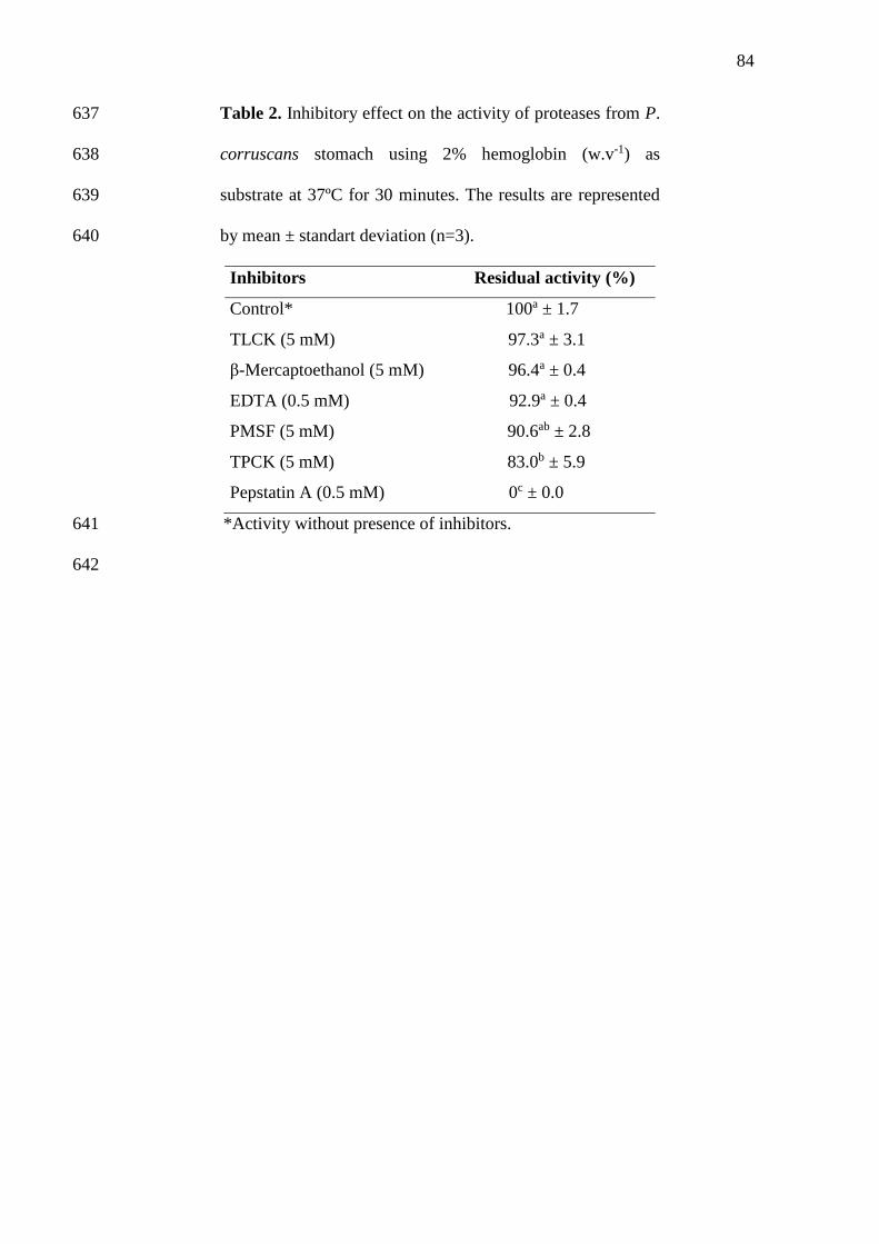
84
Table 2. Inhibitory effect on the activity of proteases from P. 637
corruscans stomach using 2% hemoglobin (w.v-1) as 638
substrate at 37ºC for 30 minutes. The results are represented 639
by mean ± standart deviation (n=3). 640
Inhibitors Residual activity (%)
Control* 100a ± 1.7
TLCK (5 mM) 97.3a ± 3.1
β-Mercaptoethanol (5 mM) 96.4a ± 0.4
EDTA (0.5 mM) 92.9a ± 0.4
PMSF (5 mM) 90.6ab ± 2.8
TPCK (5 mM) 83.0b ± 5.9
Pepstatin A (0.5 mM) 0c ± 0.0
*Activity without presence of inhibitors. 641
642
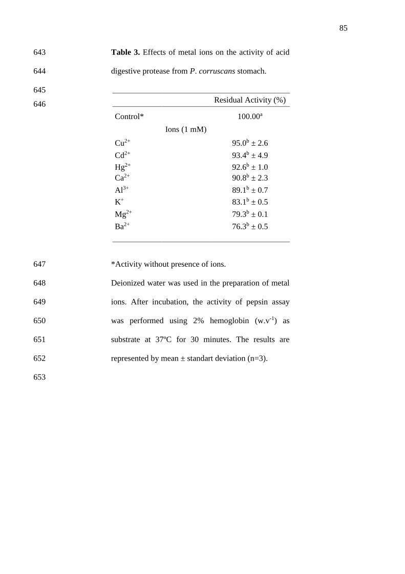
85
Table 3. Effects of metal ions on the activity of acid 643
digestive protease from P. corruscans stomach. 644
645
646
*Activity without presence of ions. 647
Deionized water was used in the preparation of metal 648
ions. After incubation, the activity of pepsin assay 649
was performed using 2% hemoglobin (w.v-1) as 650
substrate at 37ºC for 30 minutes. The results are 651
represented by mean ± standart deviation (n=3). 652
653
Residual Activity (%)
Control*
100.00a
Ions (1 mM)
Cu2+
95.0b ± 2.6
Cd2+
93.4b ± 4.9
Hg2+
92.6b ± 1.0
Ca2+
90.8b ± 2.3
Al3+
89.1b ± 0.7
K+
83.1b ± 0.5
Mg2+
79.3b ± 0.1
Ba2+
76.3b ± 0.5
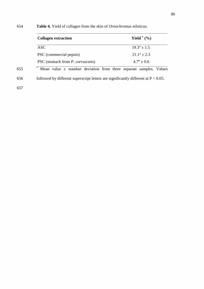
86
Table 4. Yield of collagen from the skin of Oreochromus niloticus. 654
Collagen extraction Yield * (%)
ASC 19.3a ± 1.5
PSC (commercial pepsin) 21.1a ± 2.3
PSC (stomach from P. corruscans) 4.7b ± 0.6
* Mean value ± standart deviation from three separate samples. Values 655
followed by different superscript letters are significantly different at P < 0.05. 656
657
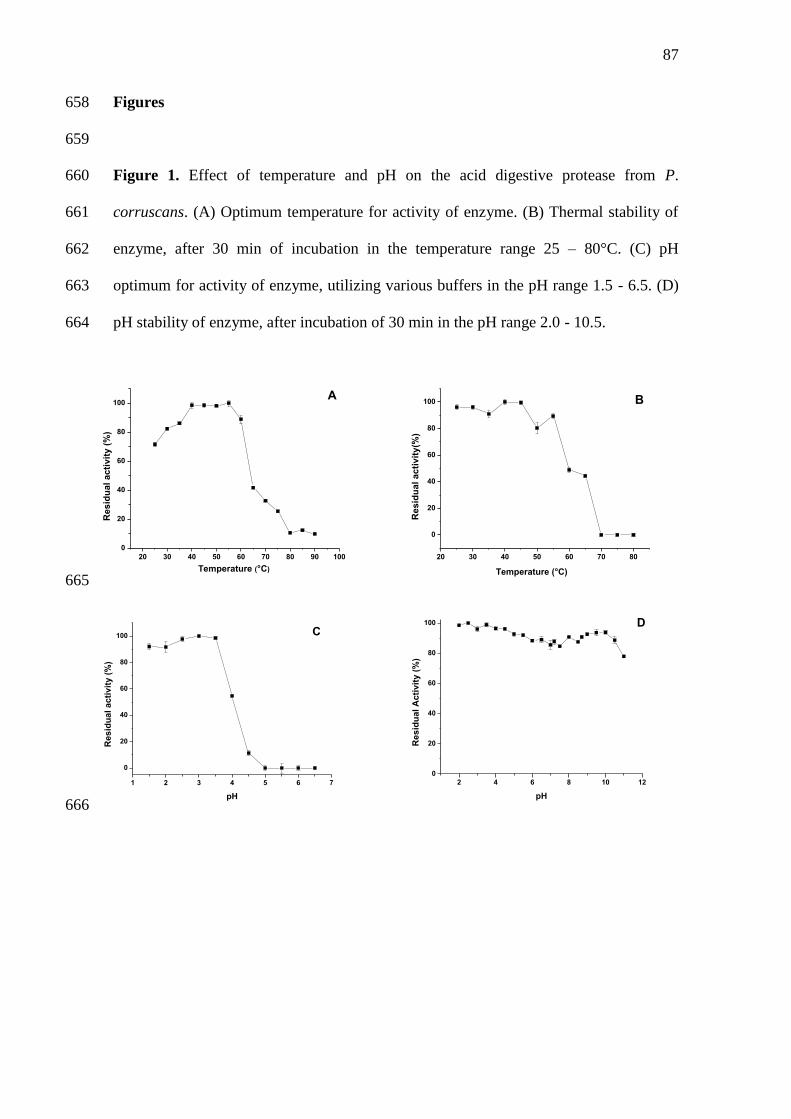
87
Figures 658
659
Figure 1. Effect of temperature and pH on the acid digestive protease from P. 660
corruscans. (A) Optimum temperature for activity of enzyme. (B) Thermal stability of 661
enzyme, after 30 min of incubation in the temperature range 25 – 80°C. (C) pH 662
optimum for activity of enzyme, utilizing various buffers in the pH range 1.5 - 6.5. (D) 663
pH stability of enzyme, after incubation of 30 min in the pH range 2.0 - 10.5. 664
20 30 40 50 60 70 80 90 100
0
20
40
60
80
100
Re
sid
ua
l a
cti
vit
y (
%)
Temperature (°C)
A
20 30 40 50 60 70 80
0
20
40
60
80
100
Temperature (°C)
Re
sid
ua
l a
cti
vit
y(%
)
B
665
1 2 3 4 5 6 7
0
20
40
60
80
100 C
Re
sid
ua
l a
cti
vit
y (
%)
pH
2 4 6 8 10 12
0
20
40
60
80
100 D
Re
sid
ua
l A
cti
vit
y (
%)
pH
666
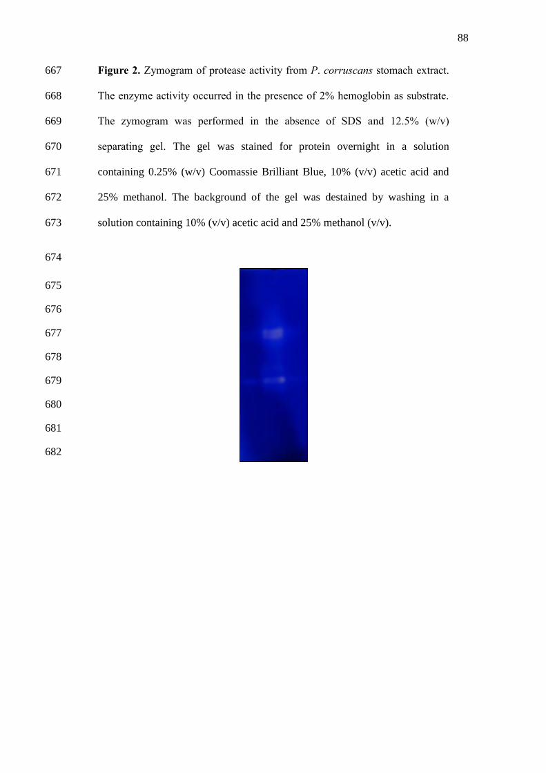
88
Figure 2. Zymogram of protease activity from P. corruscans stomach extract. 667
The enzyme activity occurred in the presence of 2% hemoglobin as substrate. 668
The zymogram was performed in the absence of SDS and 12.5% (w/v) 669
separating gel. The gel was stained for protein overnight in a solution 670
containing 0.25% (w/v) Coomassie Brilliant Blue, 10% (v/v) acetic acid and 671
25% methanol. The background of the gel was destained by washing in a 672
solution containing 10% (v/v) acetic acid and 25% methanol (v/v). 673
674
675
676
677
678
679
680
681
682
89
CAPÍTULO 2
“Extraction, partial purification and characterization of acid protease from
viscera of spotted sorubim (Pseudoplatystoma corruscans)”
A ser submetido ao periódico
FOOD CHEMISTRY
90
Extraction, partial purification and characterization of acid protease from viscera 1
of spotted sorubim (Pseudoplatystoma corruscans) 2
3
Andrade, D. H. H.a; Silva, J. F.a; Freitas Jr., A. C. V.a; Monte, F. T. D.a; Santana, W. 4
M.a; Campeche, D. F. B.a; Bezerra, R. S.a 5
6
aLaboratório de Enzimologia (LABENZ), Departamento de Bioquímica, Universidade 7
Federal de Pernambuco, Cidade Universitária, 50670-420, Recife-PE, Brazil 8
9
Corresponding author: 10
Ranilson S. Bezerra 11
Laboratório de Enzimologia (LABENZ), Departamento de Bioquímica e Laboratório de 12
Imunopatologia Keizo Asami (LIKA), Universidade Federal de Pernambuco, Cidade 13
Universitária, 50670-420, Recife-PE, Brazil. 14
Tel, +55 81 21268540; Fax, +55 81 21268576 15
email: [email protected] 16
91
ABSTRACT 17
This work aimed to extract and partially purify pepsin-like present in the spotted 18
sorubim stomach and determine the physical and chemical parameters of this protease. 19
The crude extract was subjected to three purification steps: heat treatment, fractionation 20
with ammonium sulfate and ion exchange chromatography on DEAE-cellulose column. 21
The fractions were checked for the presence of protein and pepsin activity. All steps 22
were monitored by SDS-PAGE. Effects of metal ions and protease inhibitors on its 23
activity and its physical-chemical were determined. A purification factor of 32 times 24
was obtained after application of fraction 0-60% in chromatography column. The SDS-25
PAGE showed polypeptide bands between 30.7 and 94 kDa. Maximum acidic protease 26
activity was observed at 55°C and pH 3.0. The enzyme was stable at 25 and 30°C for 30 27
minutes The acidic protease remained stable at acid and neutral pH. The proteolytic 28
activity was strongly inhibited by pepstatin A (87.3%), classic inhibitor of aspartate 29
proteases. These features indicate that the stomach of P. corruscans can be used as a 30
source of acidic proteases with potential for industrial application. 31
32
Key words: acidic protease, fish waste, partial purification. 33
92
1. Introduction 34
The global aquaculture production has experienced a steady growth due to an 35
increase in demand for fish products. It is estimated that fishing and aquaculture 36
activities in 2013 reached a new world record with 160 million tons. However, with the 37
stagnation of the amount of fish from capture, aquaculture has taken in recent years, the 38
responsibility to meet the demand for aquaculture products, by increasing the use of 39
appropriate species and technologies (FAO, 2014). 40
Increased fish production and, consequently, the volume of fish processed 41
worldwide, has generated a lot of waste and byproducts. These are usually discarded 42
without treatment and have no commercial value causing environmental pollution. From 43
environmental and economic perspective, a better utilization of fish waste as a source of 44
biomolecules is suggested; as an example can highlight the use of visceras as sources of 45
enzymes with potential industrial applications (Silva et al., 2011; Freitas-Jr et al., 2012; 46
Costa et al., 2013). 47
Protease enzymes are responsible for the hydrolysis of peptide bonds present in 48
proteins or peptides and correspond to 60% of all marketed worldwide enzyme. These 49
biomolecules have different applications in a wide variety of industries such as 50
detergents, foods, agrochemicals and pharmaceuticals (Gupta et al., 2002). Among the 51
digestive protease, pepsin is the major enzyme present in the stomach of vertebrates. It 52
is an endopeptidase (aspartyl protease) that cleaves peptide bonds on the carboxyl side 53
of residues of aromatic amino acids: phenylalanine, tyrosine and tryptophan (Pavlisko et 54
al., 1997). 55
Acid proteases such as pepsin, have a particular importance due to their 56
application in cheese making (Aehle, 2007), fish silage (Murado et al., 2009) and 57
collagen extraction (Veeruraj et al., 2014). Pepsins have been extracted, purified and 58
93
characterized from fish viscera as Sparus latus Houttuyn (Zhou et al., 2007); 59
Coryphaenoides pectoralis (Klomklao et al., 2007); Mustelus mustelus (Bougatef et al., 60
2008); Anguilla anguilla (Wu et al., 2009); Thunnus alalunga (Nalinanon et al., 2010); 61
Sardinella aurita (Khaled et al., 2011). 62
The spotted sorubim (Pseudoplatystoma corruscans) is a carnivorous freshwater 63
fish distributed throughout the South American rivers (São Francisco, Amazonas and 64
Paraná) (Crepaldi et al., 2006). This specie comprises the largest fish of the Pimelodidae 65
family and may reach 100-120 kg. It is among the fish species that show great 66
commercial value and potential for Brazilian aquaculture (Inoue et al., 2009). Hence, 67
this paper aims to extract and partially purify pepsin-like present in the spotted sorubim 68
stomach and determine the physical and chemical parameters of this protease for use in 69
biotechnological applications. 70
71
2. Material and Methods 72
73
2.1. Materials 74
All reagents used in assays were of analytical grade and purchased from Sigma 75
(St. Louis, MO, USA), Merck (Darmstadt, Germany) and Bio-Rad Laboratories 76
(Ontario, Canada). 77
78
2.2. Obtaining, collecting samples and preparation of stomach extract 79
Juvenile specimens (medium length at first maturity =75 cm) of spotted sorubim 80
(n=3) with total length of 76.3 ± 1.5 cm and weighing 3.3 ± 0.1 kg were obtained from 81
the São Francisco River, in Pilão Arcado, Bahia, Brazil. Stomachs were removed, 82
washed with distilled water and stored at 4C for transport to the laboratory. Then the 83

94
stomachs (25.0 ± 1.3g) were homogenized in 0.9% NaCl (w/v) at 4°C in the ratio 1:5 84
(w/v) using an electric tissue homogenizer (IKA RW 20D S32, China). The material 85
obtained was centrifuged (Sorvall RC-6 Superspeed Centrifuge - North Carolina, USA) 86
at 8,000 xg for 20 minutes at 4C. The supernatant (crude extract) was stored at -20°C 87
for later use. 88
89
2.3. Enzyme partial purification 90
For the partial purification of the enzymes, the stomach crude extract was first 91
submitted to a heat treatment at 30°C for 30 minutes and centrifuged at 8,000 xg for 20 92
minutes at 4C. The supernatant was collected and fractioned with ammonium sulfate at 93
a final salt concentration of 60%. After two hours a 4C the sample was centrifuged at 94
8,000 xg for 20 minutes at 4 C. The precipitate (fraction 0-60%) was resuspended in 10 95
mL of 0.9% NaCl (w/v) and dialyzed against this same buffer for 24 hours at 4C. Then, 96
the fraction was subjected to ion exchange chromatography on DEAE-cellulose column 97
at a flow rate of 0.3 mL.min-1. The chromatography fractions were checked for the 98
presence of protein (absorbance at 280 nm) and pepsin activity. Fractions with higher 99
pepsin activity were pooled and stored at -20°C for subsequent experiments. All steps 100
were monitored by SDS-PAGE. 101
102
2.4. Pepsin activity 103
Pepsin activity was performed according Pavlisko et al. (1997) using 2.0% (w/v) 104
hemoglobin (prepared in 0.1 M Glycine-HCl, pH 2.5) as specific substrate. The 105
enzymatic reaction was prepared in triplicate using microcentrifuge tubes to which were 106
added 50 µL of sample, 350 µL of buffer and 100 µL of substrate. After 30 minutes at 107
37C, 500 µL of 10% (w/v) trichloroacetic acid was added to stop the reaction and the 108
95
mixture was centrifuged at 10,000 xg for 15 minutes. The absorbance of the supernatant 109
was measured with a microplate read (Bio-Rad Model X-MarkTM spectrophotometer, 110
California, USA) at 280 nm. One unit (U) of enzyme activity was defined as the amount 111
of enzyme able to hydrolyze hemoglobin to produce a change of 0,001 unit of 112
absorbance per minute. 113
114
2.5. Protein determination 115
The protein concentration in the crude extract was determined according to the 116
methodology described by Bradford et al. (1976), using bovine serum albumin as 117
standard. 118
119
2.6. SDS-PAGE and zymogram 120
Sodium dodecyl sulfate polyacrylamide gel electrophoresis (SDS-PAGE) was 121
carried out following the procedure described by Laemmli (1970), using a 4% (w.v-1) 122
stacking gel and a 12.5% (w.v-1) separation gel. Protease zymogram under native 123
conditions was carried out following the procedure described by Garcia-Carreño et al. 124
(1993). Liofilizated sample (30 µg of protein) was mixed with a solution containing 10 125
mM Tris-HCl (pH 8.0), 2.5% SDS, 10% glycerol and 0.002% bromophenol blue and 126
applied in the electrophoresis gel (14% w/v). The electrophoresis proceded at 4°C under 127
constant electric current (11A). After the migration of the protein bands, the gel was 128
incubated at 37°C, immersed in 2% hemoglobin at pH 2.5 for 60 minutes. Then the gel 129
was treated with a solution composed of 0.01% Coomassie Brilliant Blue, 25% 130
methanol and 10% acetic acid for 24 hours. The background of the gel was destained by 131
washing in a solution containing 10% (v/v) acetic acid and 25% methanol (v/v). 132
133
96
2.7. Physical-chemical properties 134
The assays of temperature were performed in a water bath at temperatures 135
ranging from 25°C to 85°C. The influence of temperature on the proteolytic activity was 136
measured by incubating the enzyme extract with 2% hemoglobin for 30 minutes at each 137
temperature (Bezerra et al., 2005). For the assay of thermal stability, the enzyme extract 138
was incubated for 30 minutes at different temperatures and then incubated with 2% 139
hemoglobin for 30 minutes at 37°C. The influence of pH on the proteolytic activity was 140
assayed with different 0.2 M buffer solutions (Glycine-HCl, pH 1.5-3.5; Citrate-141
Phosphate, pH 4.0-7.5; Tris-HCl, pH 8.0-9.0) for 30 minutes to each buffer using 2% 142
hemoglobin as substrate. The pH stability of the enzyme extract was determinate after 143
pre-incubation for 30 min at different buffers according to Klomklao (2007) modified. 144
145
2.8. Effect of protease inhibitors 146
This assay was performed using the synthetic protease inhibitors: 147
ethylenediamine tetraacetic acid (4 mM EDTA, metalloprotease inhibitor), 4 mM β-148
mercaptoethanol (reducing agent) prepared in water and 0.5 mM Pepstatin A , a pepsin 149
inhibitor, prepared in 10% methanol. The inhibitors were incubated with enzyme extract 150
at a proportion of 1:1 (v/v) for 30 minutes at 25°C. After incubation, the pepsin activity 151
assay was performed according Pavlisko et al. (1997) using 2% hemoglobin as 152
substrate. The residual activity of sample was estimated by comparing it to a positive 153
control (100% activity), in which the sample was incubated with water or methanol, 154
depending on the inhibitor’s solvent (Bezerra et al., 2001). 155
156
2.9. Statistical analysis 157
97
Data of enzyme activity were analyzed using one-way analysis of variance 158
(ANOVA) complemented with Tukey’s test. Differences were reported as statistically 159
significant when P<0.05, using the program MicrocalTM OriginTM version 8.0 160
(Software, Inc, U.S.). 161
162
3. Results and Discussion 163
The three steps partial purification of the acid protease from the spotted sorubim 164
stomach are summarized in Table 1. After the first step (heat treatment) the specific 165
activity was higher than that in initial crude extract activity and the yield was 92.6%. 166
The second (ammonium sulfate fractionation) and third (ion exchange chromatography 167
on DEAE-cellulose column) steps presented yields of 41.1 and 30%, respectively. The 168
final supernatant (after chromatography) showed no enzymatic activity, demonstrating 169
efficiency in this stage of the partial purification process. A purification factor of 32 170
fold was obtained after application of fraction 0-60% in chromatography column. 171
According to the literature, usually, two saturations of (NH4)2SO4 solution are applied 172
to achieve purification. Wu et al. (2009) used 20-60% saturation to achieve high 173
purification of European eel pepsinogens (PGs) obtaining purification factor of 28.3, 174
36.3 and 64.2 fold. Bougatef et al. (2008) used 20-70% saturation to purification smooth 175
hound PGs (38.3% recovery after chromatography). Klomklao et al. (2007) used 30-176
70% saturation to purify pepsins from pectoral rattail (purification fator of 7.1 and 13.0 177
fold after chromatography). 178
The chromatogram of the elution of proteins (ion exchange chromatography on 179
DEAE-cellulose column) is shown in Figure 1. In this step, proteins (including pepsin) 180
are absorbed to stationary phase with oppositely charged molecules with different 181
absorption abilities. As the concentration gradient of eluant is gradually increased, 182
98
different types of proteins can be eluted and separated (Roe et al., 2001). Higher 183
enzyme activity was found in the second peak. Tanji et al. (2007) used a DEAE–184
cellulose column eluted by a gradient of 0-0.5 M NaCl in a sodium phosphate buffer at 185
0.5 ml/min of a flow rate for the purification of different types of PGs from African 186
coelacanth and North Pacific bluefin tuna. Bougatef et al. (2008) used a DEAE–187
cellulose column equilibrated by a Tris–HCl buffer with 0-0.5M NaCl to elute the 188
column at a flow rate of 1.3 ml/ min for the purification of one type of PG from smooth 189
hound. 190
After to the chromatography, the poll of the peak (higher enzyme activity) was 191
applied to a polyacrylamide gel for performing an SDS-PAGE. The polyacrylamide gel 192
electrophoresis (SDS-PAGE) (Figure 2A) shows proteins between 30.7 and 94 KDa. 193
According to the literature, pepsins from fish have molecular masses between 28 and 42 194
kDa. Bougatef et al. (2008) found the molecular weight of one type of PG and its 195
corresponding pepsin from smooth hound to be 40 and 35 kDa, respectively. Zhou et al. 196
(2008) used a similar method for molecular weight determination and obtained 36, 35, 197
38 and 35 kDa for four purified PGs and 31, 30, 32 and 30 kDa for the four 198
corresponding pepsins from mandarin fish, respectively. 199
The acid proteolytic activity of sample after chromatography process was 200
evaluated by zymogram and it was observed the presence of one protein band only 201
(Figure 2B). Several authors have reported the use of zymogram for confirmation of 202
activity of acid protease (Nalinanon et al., 2010; Khaled et al., 2011). Zymography 203
analysis indicated that pepsins of mandarin fish revealed degrading activity against 204
acid-denatured bovine hemoglobin (Zhou et al., 2008). Through the zymogram can 205
identify the number of active forms of certain enzyme in different species, thereby 206
99
providing a source of data for the knowledge and comparison of their digestive 207
physiology. 208
Physico-chemical parameters were analyzed for acid protease activity of partial 209
purified enzyme. The effect of pH on the activity of pepsin is illustrated in Figure 3A. 210
Enzymatic assays have shown that acid protease was active between pH 1.5 to 4.0 and 211
exhibited maximum activity at pH 3.0. Pepsin activity usually decreases at different pH 212
from optimum pH, presumably due to conformational changes of the enzyme because of 213
the influence of pH on the amino acid residues that compose it. 214
Optimum pH values between 2.0 and 4.0 have been reported for pepsin of S. 215
latus Houttuyn (Zhou et al., 2007). Similar results to the optimum pH of purified 216
pepsins from the stomach of other fish species can be found in the literature, such as 217
Anguilla anguilla (pH 3.5, 2.5 and 2.5 for three pepsins separate this specie) (Wu et al., 218
2009), Coryphaenoides pectoralis (pH 3.0 to 3.5 for two pepsins isolated this specie) 219
(Klomklao et al., 2007). Khaled et al. (2011) reported maximum activity of pepsin from 220
the viscera of sardinelle around pH 3.0. Different results were found for Thunnus 221
alalunga (Nalinanon et al., 2010) and Mustelus mustelus (Bougatef et al., 2008), where 222
the purified pepsin had optimum pH 2.0. 223
To investigate the pH stability, the enzyme was incubated in different buffers in 224
the pH range from 1.5 to 9.0 and residual activity shown in Figure 3B. The acid 225
protease remained stable at acid and neutral pH. Castillo-Yanez et al. (2004) also 226
reported that acid enzymes from Sardinops sagax caerulea were stable between pH 3.0 227
to 6.0. Albacore tuna pepsin was stable in the pH range of 2-5 (Nalinanon et al., 2010). 228
Pepsins were stable in the pH range of 2.0-6.0 (Klomklao et al., 2007) and 2.0-5.0 229
(Khaled et al., 2011). 230
100
According Klomklao et al. (2006) the stability of pepsins to particular pH 231
appears to be determined by the net charge of the enzyme. Pepsins are irreversibly 232
inactivated at alkaline pH (Gildberg and Raa, 1983), therefore, the irreversible 233
inactivation in alkaline condition indicates that the acid protease obtained from P. 234
corruscans may be a pepsin. Furthermore, this characteristic of being stable at neutral 235
pH is very interesting with regard to the storage of the material, since the inactive 236
enzyme can be stored at a neutral pH solution, and again be subjected to an acid pH, it 237
returns the its enzymatic activity without major losses. Differences related to pH 238
stability between various species of fish indicate that different molecular properties may 239
be involved, such as enzyme conformation, stability of structural links, in addition to 240
anatomical location (Klomklao et al., 2007). 241
The effect of temperature (25-80ºC) on the activity of partial purified enzyme 242
was evaluated and is presented in Figure 4A. The acid protease showed maximal 243
activity at 55°C, but remained more than 50% of its maximum activity in the 244
temperature range from 25 to 65ºC. Results on optimum temperature of pepsin in some 245
species of fish have been reported in the literature, such as 37°C for Parona signata 246
(Pavlisko et al., 1997), 45 to 50°C for S. Houttuyn latus (Zhou et al., 2007), 40°C for 247
Mustelus mustelus (Bougatef et al., 2008), 40 to 45°C for Siniperca chuatsi (Zhou et al., 248
2008), 35 to 40°C for Anguilla Anguilla (Wu et al., 2009). Komklao et al. (2007) 249
obtained optimum temperature of 45°C for pepsin in the stomach Coryphaenoides 250
pectoralis. The optimal temperature of Thunnus alalunga pepsin was 50°C (Nalinanon 251
et al., 2010) and 40°C for Sardinella aurita aspartic protease (Khaled et al., 2011). 252
The resistance of acid protease from P. corruscans to heat treatment is shown in 253
Figure 4B. The enzyme was stable at 25 and 30°C for 30 minutes, losing about 37 to 254
55% of its activity at temperatures of 35 to 40°C, respectively. According Klomklao et 255
101
al. (2005) digestive proteases of many aquatic organisms are active in adverse 256
conditions, as temperatures above the physiological. Pepsins from Pectoralis 257
coryphaenoides were stable up to 40°C (Komklao et al., 2007). Acid proteins of 258
Sardinops sagax caerulea were not stable above 45°C (Castillo-Yanez et al., 2004). 259
Albacore tuna pepsin was stable up to 50°C (Nalinanon et al., 2010) whereas the 260
enzymatic activity from the viscera of sardinelle was stable up to 40°C (Khaled et al., 261
2011). 262
Assays of thermal stability of the enzyme are important because provides data 263
from its relationship with possible industrial processes. Most processes where pepsins 264
can be used is realized at low temperatures or ambient temperature due to the 265
degradability of the material (collagen extraction, milk coagulation) or decrease the 266
economic burden caused by the energy consumption for heating which indicates the 267
possibility of using pepsin obtained from P. corruscans for industrial applications. 268
In order to better understand the class of proteases in the stomach from spotted 269
sorubim, its sensitivity to various inhibitors was tested. The effect of inhibitors on the 270
activity of partial purified protease are shown in Table 2. The proteolytic activity (with 271
the susbstrate hemoglobin) was strongly inhibited by pepstatin A (87.3%), classic 272
inhibitor of aspartate proteases. Can be seen in the results after 30 minutes of 273
incubation, that the partial purified enzyme retained 100% of its activity in the presence 274
of EDTA, metalloenzyme inhibitor, and about 76.8% of its activity in the presence of β-275
Mercaptoethanol, inhibitor of cysteine proteases. These results lead one to believe the 276
sample is composed mainly of aspartate proteases, especially pepsin. 277
Pepsins from the stomach of Anguilla anguilla and Siniperca chuatsi also were 278
almost completely inhibited by pepstatin A, but specific inhibitors of serine proteinases 279
(PMSF) and metalloproteinases (EDTA) had no inhibitory effect on the activity of 280
102
pepsin (Wu et al., 2009; Zhou et al., 2008, Bougatef et al., 2008). Activity of pepsin 281
from the stomach of albacore tuna was strongly inhibited by pepstatin A with the 282
concentration range of 0.1-10 µM (Nalinanon et al., 2010). Khaled et al. (2011) reported 283
that protease derived from the stomach of Sardinella aurita was almost completely 284
inhibited by pepstatin A (2 mM). 285
286
4. Conclusion 287
The study of enzymes from visceras of spotted sorubim (P. corruscans) can 288
contribute to sustainable development through the use of waste fish. This study, an acid 289
protease from spotted sorubim stomach was partial purified. Physical chemical 290
parameters were characterized and showed interesting features like high activity, 291
stability at neutral pH and high temperature optimum. The use of a specific inhibitor 292
produced evidence that this partial purified fraction is probably a pepsin-like enzyme. 293
These features indicate that the stomach of P. corruscans can be used as a source of 294
acid proteases with potential for industrial application. 295
296
Acknowledgements 297
This study was financially supported by the following Brazilian agencies: 298
Conselho Nacional de Pesquisa e Desenvolvimento Científico (CNPq), and the 299
Coordenação de Aperfeiçoamento de Pessoal de Nível Superior (CAPES). 300
301
References 302
Aehle W (2007). Enzymes in industry: production and applications. (3rdedn), Wiley–303
VCH, Weinheim, Germany, 136 - 137. 304
305
103
Bezerra, R. S.; Vieira, V. L. A.; Santos, A. J. G. (2001). Ciclo reprodutivo da carapeba 306
prateada Diapterus rhombeus (Cuvier, 1829), no litoral de Pernambuco. Tropical 307
Oceanography, 1, 67-78. 308
309
Bezerra, R. S., Lins, E. J. F., Alencar, R. B., Paiva, P. M. G., Chaves, M. E. C., Coelho, 310
L. C. B. B., Jr Carvalho, L. B. (2005). Alkaline proteinase from intestine of Nile tilapia 311
(Oreochromis niloticus). Process Biochemistry, v. 40, p. 1829-1834. 312
313
Bougatef, A., Balti, R., Zaied, S. B., Souissi, N., Nasri, M. (2008). Pepsinogen and 314
pepsin from the stomach of smooth hound (Mustelus mustelus): Purification, 315
characterization and amino acid terminal sequences. Food Chemistry 107, 777 – 784. 316
317
Bradford, M. M. (1976). A rapid and sensitive method for the quantification of 318
microgram quantities of protein utilizing the principle of protein binding. Anal. 319
Biochem. 72, 248-254. 320
321
Castillo-Yanez, F. J.; Pacheco-Aguilar, R.; Garcia-Carreno, F. L.; & Toro, M. A. N. D. 322
(2004). Characterization of acidic proteolytic enzymes from Monterey sardine 323
(Sardinops sagax caerulea) viscera. Food Chemistry, 85 (3), 343 – 350. 324
325
Costa, H. M. S., Freitas Júnior, A. C. V., Amaral, I. P. G., Hirata, I. Y., Paiva, P. M. G., 326
Carvalho Jr, L. B., Oliveira, V., Bezerra, R. S. (2013). Metal-sensitive and thermostable 327
trypsin from the crevalle jack (Caranx hippos) pyloric caeca: purification and 328
characterization. Chemistry Central Journal, 7:166. 329
330
104
Crepaldi, D. V.; Faria, P. M. C.; Teixeira, E. A.; Ribeiro, L. P.; Costa, A. A. P.; Melo, 331
D. C.; Cintra, A. P. R.; Prado, S. A.; Costa, F. A. A.; Drumond, M. L.; Lopes, V. E.; 332
Moraes, V. E. (2006). O surubim na aquacultura do Brasil. Rev. Bras. Reprod. Animal 333
30, 150 – 158. 334
335
FAO. The State of World Fisheries and Aquaculture 2014. Rome. 223 pp., 2014. 336
337
Freitas-Júnior, A. C. V., Costa, H. M. S., Icimoto, M. Y., Hirata, I. Y., Marcondes, M., 338
Carvalho Jr, L. B., Oliveira, V., Bezerra, R. S. (2012). Giant Amazonian fish pirarucu 339
(Arapaima gigas): Its viscera as a source of thermostable trypsin. Food Chemistry, v. 340
133, p. 1596- 1602. 341
342
Gildberg, A.; Raa, J. (1983). Purification and characterization of pepsins from the 343
Arctic fish capelin (Mallotus villosus). Comparative Biochemistry and Physiology (A), 344
v. 75, p. 337 – 342. 345
346
Gupta, R.; Beg, Q. K.; Larenz, P. (2002). Bacterial alkaline proteases: molecular 347
approaches and industrial applications. Applied Microbiology and Biotechnology, v. 348
59, p. 15 – 32. 349
350
Inoue, L. A. K. A.; Hisano, H.; Ishikawa, M. M.; Rotta, M. A.; Senhorini, J. A. (2009). 351
Princípios básicos para produção de alevinos de surubins (pintado e cachara). Dourados: 352
Embrapa Agropecuária Oeste; Manaus: Embrapa Amazônia Ocidental; Corumbá: 353
Embrapa Pantanal. 26 p. Documentos (Embrapa Agropecuária Oeste. Documentos, 99; 354
Embrapa Amazônia Ocidental. Documentos, 68; Embrapa Pantanal. Documentos, 100. 355
Impresso, v. 15, p. 1-26. 356
105
357
Khaled, H. B., Ghorbel-Bellaaj, O., Hmidet, N., Jellouli, K., Ali, N. E., Ghorbel, S., 358
Nasri, M. (2011). A novel aspartic protease from the viscera of Sardinelle (Sardinella 359
aurita): Purification and characterization. Food Chemistry 128, 847–853. 360
361
Klomklao, S.; Benjakul, S.; Visessanguan, W.; Simpson, B. K.; Kishimura, H. (2005) 362
Partitioning and recovery of proteinase from tuna spleen by aqueous twophase systems. 363
Process Biochem 40: 3061-3067. 364
365
Klomklao, S.; Benjakul, S.; Visessanguan, W.; Kishimura, H.; Simpson, B. K. (2006). 366
Purification and characterization of trypsin from the spleen of tongol tuna (Thunnus 367
tonggol). Journal of Agricultural Food Chemistry, v. 54, p. 5617 – 5622. 368
369
Klomklao, S.; Benjakul, S.; Visessanguan, W.; Kishimura, H.; Simpson, B.K. (2007). 370
Purification and characterization of trypsins from the spleen of skipjack tuna 371
(Katsuwonus pelamis) spleen. Food Chemistry. v. 100, p. 1580–1589. 372
373
Laemmli, U. K. (1970). Cleavage of structural proteins during the assembly of the head 374
of Bacteriophage T4. Nature, 227, 680-685. 375
376
Murado, M. A., González, M. D. P., Vázquez, J. A. (2009). Recovery of proteolytic and 377
collagenolytic activities from viscera by-products of rayfish (Raja clavata). Mar Drugs 378
7: 803 - 815. 379
380
106
Nalinanon, S.; Benjakul, S.; Kishimura, H. (2010). Biochemical properties of 381
pepsinogen and pepsin from the stomach of albacore tuna (Thunnus alalunga). Food 382
Chemistry, v. 121, p. 49-55. 383
384
Pavlisko, A.; Rial, A.; De Vecchi, S.; Coppes, Z. (1997). Properties of pepsin and 385
trypsin isolated from the digestive tract of Parona signata ‘‘Palometa”. Journal of Food 386
Biochemistry, v. 21, p. 289–308. 387
388
Roe, S. D. (2001) Purification strategy. In Protein purification techniques (2ndedn), Roe 389
S, Eds, Oxford University Press Inc, New York, 1 - 10. 390
391
Silva, J. F., Esposito, T. S., Marcuschi, M., Ribeiro, R., Cavalli, R. O., Oliveira, V., 392
Bezerra, R. S. (2011). Purification and partial characterisation of a trypsin from the 393
processing waste of the silver mojarra (Diapterus rhombeus). Food Chemistry, v. 129, 394
p. 777-782. 395
396
Tanji, M.; Yakabe, E.; Kageyama, T.; Yokobori, S.; Ichinose, M. et al. (2007). 397
Purification and characterization of pepsinogens from the gastric mucosa of African 398
coelacanth, Latimeria chalumnae, and properties of the major pepsins. Comp Biochem 399
Physiol B Biochem Mol Biol 146: 412 – 420. 400
401
Veeruraj, A., Arumugam M., Ajithkumar T., Balasubramanian, T. (2014). Isolation and 402
characterization of collagen from the outer skin of squid (Doryteuthis singhalensis). 403
Food Hydrocolloids, http://dx.doi.org/10.1016/j.foodhyd.2014.07.025. 404
405
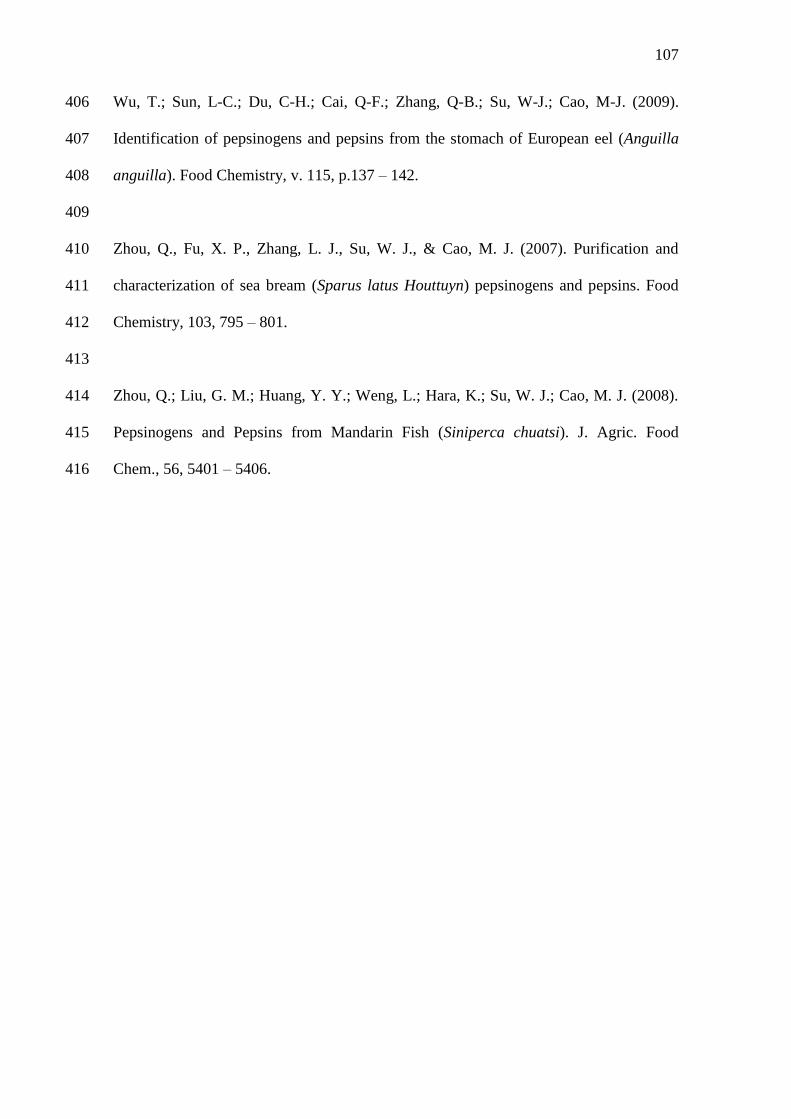
107
Wu, T.; Sun, L-C.; Du, C-H.; Cai, Q-F.; Zhang, Q-B.; Su, W-J.; Cao, M-J. (2009). 406
Identification of pepsinogens and pepsins from the stomach of European eel (Anguilla 407
anguilla). Food Chemistry, v. 115, p.137 – 142. 408
409
Zhou, Q., Fu, X. P., Zhang, L. J., Su, W. J., & Cao, M. J. (2007). Purification and 410
characterization of sea bream (Sparus latus Houttuyn) pepsinogens and pepsins. Food 411
Chemistry, 103, 795 – 801. 412
413
Zhou, Q.; Liu, G. M.; Huang, Y. Y.; Weng, L.; Hara, K.; Su, W. J.; Cao, M. J. (2008). 414
Pepsinogens and Pepsins from Mandarin Fish (Siniperca chuatsi). J. Agric. Food 415
Chem., 56, 5401 – 5406. 416
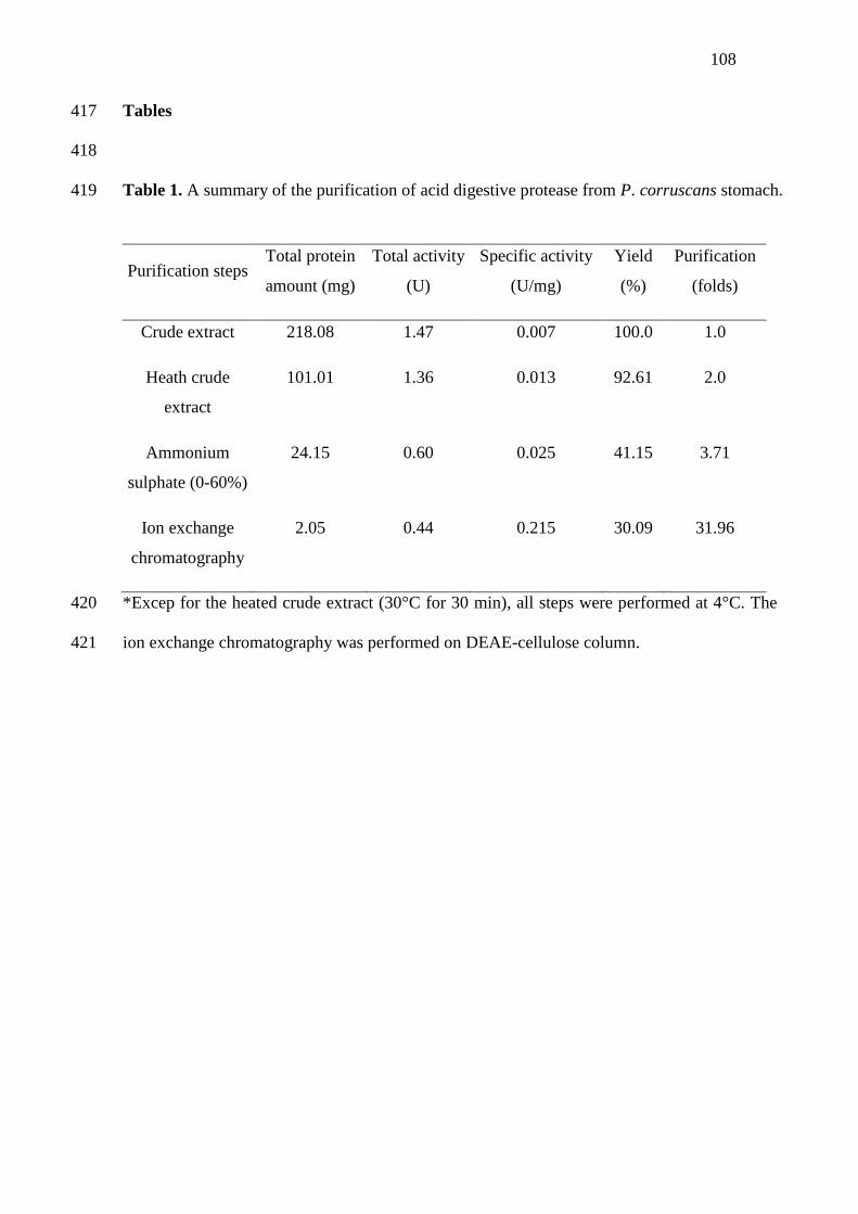
108
Tables 417
418
Table 1. A summary of the purification of acid digestive protease from P. corruscans stomach. 419
*Excep for the heated crude extract (30°C for 30 min), all steps were performed at 4°C. The 420
ion exchange chromatography was performed on DEAE-cellulose column. 421
Purification steps Total protein
amount (mg)
Total activity
(U)
Specific activity
(U/mg)
Yield
(%)
Purification
(folds)
Crude extract 218.08 1.47 0.007 100.0 1.0
Heath crude
extract
101.01 1.36 0.013 92.61 2.0
Ammonium
sulphate (0-60%)
24.15 0.60 0.025 41.15 3.71
Ion exchange
chromatography
2.05 0.44 0.215 30.09 31.96
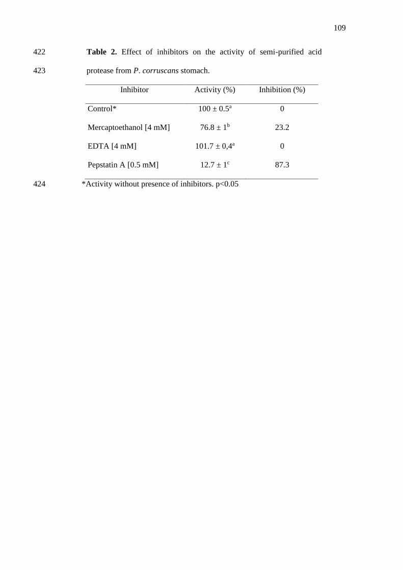
109
Table 2. Effect of inhibitors on the activity of semi-purified acid 422
protease from P. corruscans stomach. 423
Inhibitor Activity (%) Inhibition (%)
Control* 100 ± 0.5a 0
Mercaptoethanol [4 mM] 76.8 ± 1b 23.2
EDTA [4 mM] 101.7 ± 0,4a 0
Pepstatin A [0.5 mM] 12.7 ± 1c 87.3
*Activity without presence of inhibitors. p<0.05 424
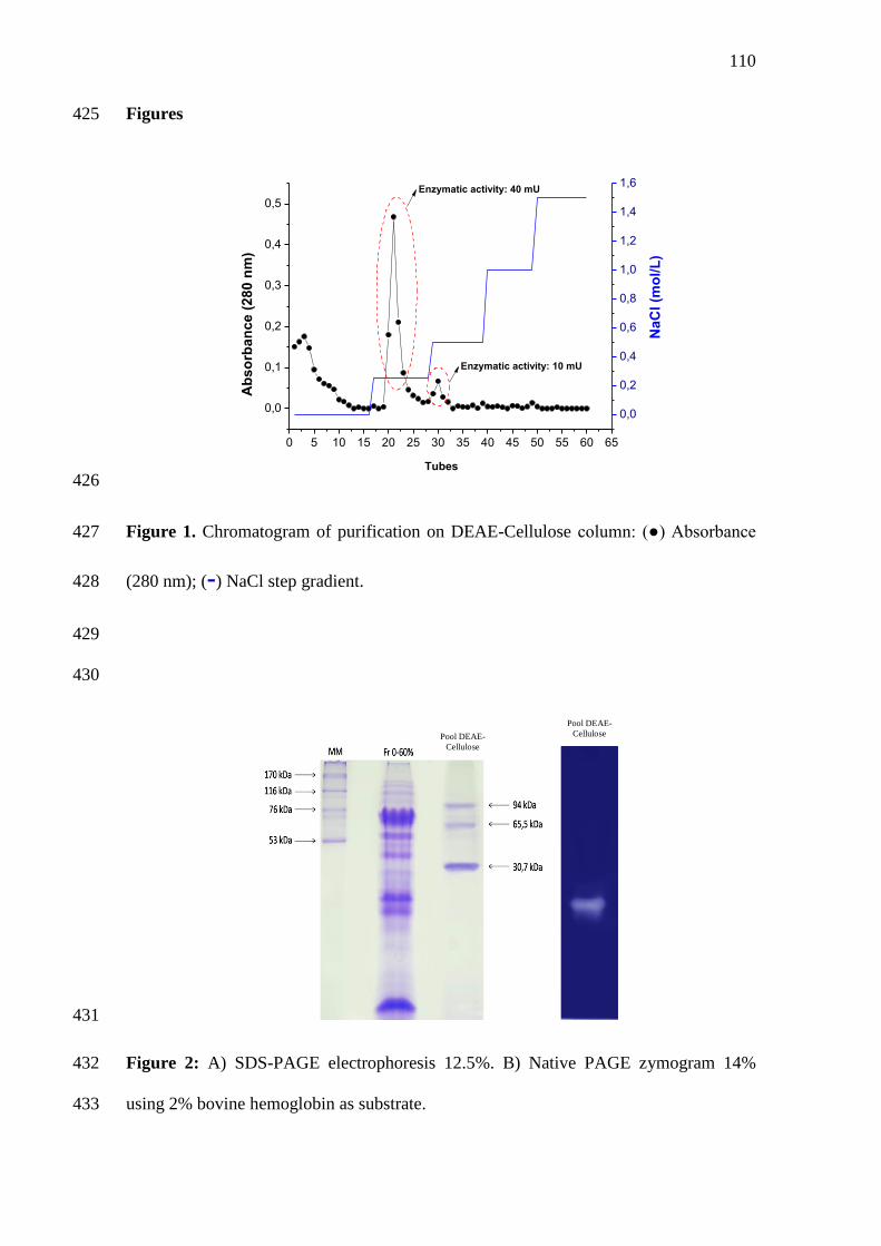
110
Figures 425
0 5 10 15 20 25 30 35 40 45 50 55 60 65
0,0
0,1
0,2
0,3
0,4
0,5
Enzymatic activity: 10 mU
Enzymatic activity: 40 mU
NaC
l (m
ol/
L)
Ab
so
rban
ce (
280 n
m)
Tubes
0,0
0,2
0,4
0,6
0,8
1,0
1,2
1,4
1,6
426
Figure 1. Chromatogram of purification on DEAE-Cellulose column: (●) Absorbance 427
(280 nm); (-) NaCl step gradient. 428
429
430
431
Figure 2: A) SDS-PAGE electrophoresis 12.5%. B) Native PAGE zymogram 14% 432
using 2% bovine hemoglobin as substrate. 433
Pool DEAE-
Cellulose
Pool DEAE-
Cellulose
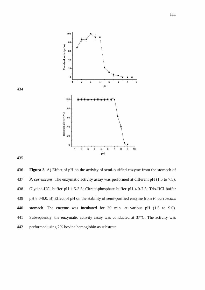
111
1 2 3 4 5 6 7 8
0
20
40
60
80
100
Resid
ual
acti
vit
y (
%)
pH
434
435
Figura 3. A) Effect of pH on the activity of semi-purified enzyme from the stomach of 436
P. corruscans. The enzymatic activity assay was performed at different pH (1.5 to 7.5). 437
Glycine-HCl buffer pH 1.5-3.5; Citrate-phosphate buffer pH 4.0-7.5; Tris-HCl buffer 438
pH 8.0-9.0. B) Effect of pH on the stability of semi-purified enzyme from P. corruscans 439
stomach. The enzyme was incubated for 30 min. at various pH (1.5 to 9.0). 440
Subsequently, the enzymatic activity assay was conducted at 37°C. The activity was 441
performed using 2% bovine hemoglobin as substrate. 442
Res
idu
al a
ctiv
ity (
%)
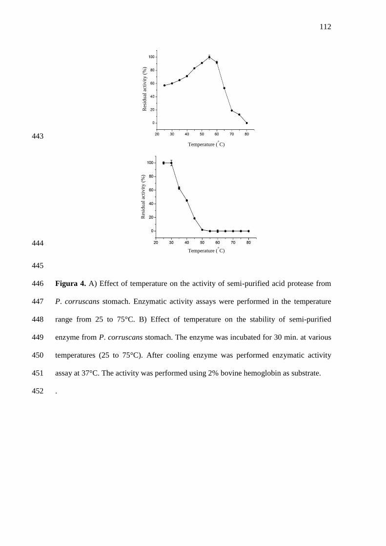
112
443
444
445
Figura 4. A) Effect of temperature on the activity of semi-purified acid protease from 446
P. corruscans stomach. Enzymatic activity assays were performed in the temperature 447
range from 25 to 75°C. B) Effect of temperature on the stability of semi-purified 448
enzyme from P. corruscans stomach. The enzyme was incubated for 30 min. at various 449
temperatures (25 to 75°C). After cooling enzyme was performed enzymatic activity 450
assay at 37°C. The activity was performed using 2% bovine hemoglobin as substrate. 451
.452
Res
idu
al a
ctiv
ity (
%)
Res
idu
al a
ctiv
ity (
%)
Temperature (◦C)
Temperature (◦C)
113
CAPÍTULO 3
Isolation and characterization of skin collagen of spotted sorubim
(Pseudoplatystoma corruscans)
A ser submetido ao periódico
FOOD HYDROCOLLOIDS
(ISSN: 0268-005X)
114
Isolation and characterization of skin collagen of spotted sorubim 1
(Pseudoplatystoma corruscans) 2
3
Andrade, D. H. H.a; Neri, R. C. A.a; Freitas Jr., A. C. V.a; Costa, H. M. S.a; Bezerra, R. 4
S. a 5
6
aLaboratório de Enzimologia (LABENZ), Departamento de Bioquímica, Universidade 7
Federal de Pernambuco, Cidade Universitária, 50670-420, Recife-PE, Brazil 8
9
Corresponding author: 10
Ranilson S. Bezerra 11
Laboratório de Enzimologia (LABENZ), Departamento de Bioquímica e Laboratório de 12
Imunopatologia Keizo Asami (LIKA), Universidade Federal de Pernambuco, Cidade 13
Universitária, 50670-420, Recife-PE, Brazil. 14
Tel, +55 81 21268540; Fax, +55 81 21268576 15
email: [email protected] 16
17
115
Abstract 18
19
Acid soluble collagen (ASC) and pepsin soluble collagen (PSC) from the skin of spotted 20
sorubim (Pseudoplatystoma corruscans) were isolated and characterized. The total yield 21
of collagen extraciton was 10.6% (dry weight). Both the ASC and PSC consisted of two 22
different α chains (α 1 and α 2), their dimers (β chain) and trimmers (γ chain), and were 23
characterized to be type I collagen. The ASC and PSC ultraviolet (UV) absorption 24
spectrum showed a maximum absorption at 247 and 246 nm, respectively. The 25
solubility of ASC and PSC reached maximum at pH 1 and 3, respectively. ASC was 26
soluble in the range of NaCl concentration 0 – 2% (w/v) whereas PSC was soluble until 27
3% (w/v). The results obtained in this study indicate the possibility of using spotted 28
sorubim skin as a source of biomolecules with great potential for biotechnological and 29
industrial application. 30
31
Keywords: acid soluble collagen, pepsin soluble collagen, fisheries by-products.32
116
1. Introduction 33
34
Collagen is the most abundant protein of animal origin and constitutes about 35
30% of total proteins (Muyonga et al., 2004). The structural protein of fibrous 36
characteristic is a unique in its ability to form insoluble fibers with high resistance the 37
traction (Gelse et al., 2003). It is main structural element of bones, cartilages, skin, 38
tendons, ligaments, blood vessels, teeth and other present structures in the vertebrates 39
(Kittiphattanabawon et al., 2010). At present, at least 29 variants of collagen have been 40
identified, and each differs considerably in amino acid sequence, structure and function, 41
more likely associated with specific genetic variants (Liu et al., 2012). All members of 42
the collagen family are characterized by domains with repetitions of the proline-rich 43
tripeptides, Gly-X-Y, involved in the formation of the triple helix (Muyonga et al., 44
2004). Among them, type I collagen is the most abundant, found in all connective 45
tissue, such as skins and bones (Nagai et al., 2008; Duan et al., 2009). 46
Collagen has a wide range of applications as food, cosmetics, biomedical 47
materials, pharmaceuticals and film industries due mainly properties as 48
biocompatibility, biodegradability and low antigenicity (Liu et al., 2009). Commonly 49
isolated from by-products of land-based animals, such as cows, pigs and poultry, the 50
uses of collagen and collagen derived products of land animal origin have become of 51
more concern due recurrent cases of zoonosis such as avian and bovine sponge 52
encephalopathy (BSE), transmissible spongiform encephalopathy (TSE), foot-and-53
mouth disease (FMD) and avian influenza (Binsi et al., 2009). As consequence, the 54
search for alternative and safer sources this compound has become an attractive 55
investment (Zhang et al., 2007; Duan et al., 2009). 56
Therefore, many scientists have been focusing their experiments to aquatic 57
animals to produce collagen including freshwater and marine fish and mollusks (Pati et 58
al., 2010; Jayathilakan et al., 2012; Veerurajat et al., 2014). Skin collagen from several 59
fish species has been extracted and characterized such as silver carp (Safandowska et 60
al., 2013), amur sturgeon (Veeruraj et al., 2014), grass carp, silver carp, bighead carp 61
and black carp (Liu et al., 2014), black rockfish, sea bass and red sea bream (Cho et al., 62
2014). The number of studies with the objective of extracting collagen from sea fish has 63
been growing continually due to advantages as great readiness; low risks of transmitting 64
diseases; high yield in the extraction processes; toxicity absence (Senaratne et al., 65
117
2006). Due to the large evolutionary distance between fish and humans, the risk of 66
disease transmission by this route becomes very low (Song et al., 2006). 67
The observation of the fact that the increase in fish production is proportional to 68
the residue left by this industry, and that it contribute to environmental pollution, have 69
emphasized the need to use these products (eg as a source of biomolecules for industrial 70
applications). Accordance to MPA (2010), Brazil produced approximately 1,2 millions 71
tons, being among the twenty larger producing of fish of the world. 72
The spotted sorubim (Pseudoplatystoma corruscans) is a carnivorous freshwater 73
fish distributed throughout the South American rivers (São Francisco, Amazonas and 74
Paraná) (Crepaldi et al., 2006). This specie comprises the largest fish of the Pimelodidae 75
family and may reach 100-120 kg. It is among the fish species that show great 76
commercial value and potential for Brazilian aquaculture (Inoue et al., 2009). Thus the 77
aim of this study was to isolate and extract the acid soluble (ASC) and pepsin soluble 78
(PSC) collagen from the skin of P. corruscans as well as characterize and suggest its 79
use as an alternative source of collagen to land animals. 80
81
2. Material and Methods 82
83
2.1 Collection and storage of samples 84
85
Juvenile specimens (medium length at first maturity =75 cm) of spotted sorubim 86
(n=3) with total length of 76.3 ± 1.5 cm and weighing 3.3 ± 0.1 kg were obtained from 87
the São Francisco River, in Pilão Arcado, Bahia, Brazil. Fish were transported to the 88
laboratory and its skin removed. The skin was washed with cold water (5-8ºC) and cut 89
into small pieces (0.5 cm2). The prepared skin samples were packed in polyethylene 90
bags and kept at -20ºC until used. 91
92
2.2 Chemicals reagents 93
94
All reagents were of analytical grade. Tipe I collagen from calf skin was 95
purchased from Sigma chemical company (CAS Number 9007-34-5) (St Louis, MO, 96
USA). 97
98
2.3 Pretreatment of skin 99
118
100
To remove non-collagenous proteins, the prepared fish skin was mixed with 101
0.2M NaOH at a skin/alkali solution ratio of 1:10 (w/v). The mixture was continuously 102
stirred for 3h at 4ºC and the alkali solution was changed every 30 minutes. The treated 103
skin was then washed with cold distilled water until a neutral or faintly basic pH of 104
wash water was reached. The pH of wash water was determined using a digital pH 105
meter (Sartorious North America, Edgewood, NY, USA). To remove fats 10% butyl 106
alcohol in the ratio of 1:10 (w/v) was used and the mixture was continuously stirred for 107
6h at 4ºC and the skin was washed as previously described. Then was added 3% 108
hydrogen peroxide at a ratio of 1:10 (w/v) for bleached the skin and was carried out in 109
the wash (Nagai and Suzuki, 2000). 110
111
2.4 Extraction of acid soluble collagen (ASC) 112
113
ASC was prepared by the method of Nagai and Suzuki (2008) with a slight 114
modification. The residue was extracted with 0.5 M acetic acid at sample/acid ratio of 115
1:20 (w/v) for 3 days. The resulting viscous solution was centrifuged at 20,000 g for 30 116
min at 4 °C. The supernatants of the extract were combined and salted-out by adding 117
NaCl to give a final concentration of 0.9 M, followed by precipitation of the collagen by 118
the addition of NaCl to the final concentration of 2.5 M in 0.75 M Tris–HCl (pH 8.8). 119
After standing overnight, the resulting precipitate was collected by centrifuging at 120
20,000 g for 60 min and then dissolved in 10 volumes of 0.5 M acetic acid. The solution 121
obtained was dialyzed against 0.1M acetic acid and subsequently against distilled water. 122
The dialysate was freeze-dried and referred to as acid soluble collagen (ASC). 123
124
2.5 Extraction of pepsin soluble collagen (PSC) 125
126
The pepsin soluble collagen (PSC) was obtained through the incubation of the 127
insoluble material obtained in the previous steps with commercial pepsin (EC 3.4.23.1; 128
crystallized and lyophilized, Sigma, MO) at enzyme/skin ratio of 1:20 (w/w) with 129
constant homogenization for 24 hours at 4 °C. The pepsin-solubilized collagen (PSC) 130
was obtained by the same method as the ASC. The yield of ASC or PSC was calculated 131
as: Yield (%) = (M/M0) x 100, where M is the weight of lyophilized collagen (g), and 132
M0 is the weight of drought skin used (g). 133
119
134
2.6 SDS–polyacrylamide gel electrophoresis (SDS–PAGE) 135
136
SDS–PAGE was performed following the method of Laemmli (1970). 137
Solubilized samples of ASC and PSC (25 μg of protein) were mixed at a ratio 1:1 (v/v) 138
with the sample buffer (containing 0.5 M Tris HCl, pH 6.8, 4% SDS, 20% glycerol and 139
10% b-ME), heated in a bath (IKA® Works Inc., China) at 85°C for 5 minutes and were 140
loaded on to polyacrylamide gels comprising a 7.5% running gel and a 4% stacking gel 141
and subjected to electrophoresis at a constant current of 15 mA/gel for 1 h and 30 min 142
using a Mini Protein II unit (Bio-Rad Laboratories, Inc., Richmond, CA, USA). After 143
electrophoresis, the gel was stained with 0.05% (w/v) Coomassie blue R-250 in 15% 144
(v/v) methanol and 5% (v/v) acetic acid and destained with 30% (v/v) methanol and 145
10% (v/v) acetic acid. Type I collagen from calf skin (Sigma-Aldrich Co., St. Louis, 146
MO) was also prepared following similar procedure and 10 μl were loaded as standard 147
collagen. High-molecular-weight protein markers (GE Healthcare UK Limited, 148
Buckinghamshire, UK) were used to estimate the molecular weight of proteins. 149
150
2.7 UV absorption spectrum 151
152
UV absorption spectra of ASC and PSC from spotted sorubim (P. corruscans) 153
skin was carried out according to Nalinanon et al. (2011), using a SmartSpec Plus 154
spectrophotometer Bio-Rad. The collagen samples were dissolved in 0.5 M acetic acid 155
solution with a sample/solution ratio of 1:1000 (w/v). The solutions were then placed 156
into a quartz cell with a path length of 1 mm. UV spectra were measured at wavelength 157
200 – 600 nm. 158
159
2.8 Solubility 160
161
The solubility of ASC and PSC was determined by the method of Montero et al. 162
(1991) with a slight modification. Collagens were dissolved in 0.5 M acetic acid to 163
obtain a final concentration of 3 mg/ml and the mixture was stirred at 4°C for 24h. 164
Thereafter, the mixture was centrifuged at 20,000 g for 60 min at 4°C. The supernatant 165
was used for solubility study. 166
167
120
2.8.1 Effect of pH on solubility 168
169
Sample solution (0.8 ml) was transferred to a 1.5 ml centrifuge tube and the pH 170
was adjusted with either 6 M NaOH or 6 M HCl to obtain the final pH ranging from 1 to 171
12. The volume of solution was made up to 1 ml by deionized water previously adjusted 172
to the same pH as the sample solution. The solution was centrifuged at 20,000 g for 60 173
min at 4°C. For all the samples, protein content in the supernatant was measured. Then 174
the relative solubility was calculated in the comparison with that of be obtained at the 175
pH giving the highest solubility. 176
177
2.8.2 Effect of NaCl on solubility 178
179
Collagen solution (0.5 ml) was mixed with 0.5 ml of NaCl in 0.5 M acetic acid 180
at various concentrations to give the final concentrations of 0%, 1%, 2%, 3%, 4%, 5% 181
and 6% (w/v). The mixture was stirred continuously at 4°C for 60 min, followed by 182
centrifuging at 20,000 g for 60 min at 4°C. Protein content in the supernatant was 183
measured and the relative solubility was calculated as previously described. 184
185
2.9. Statistical analyses 186
187
All experiments were performed in triplicate and expressed in means ± standard 188
deviation and a probability value of < 0.05 was considered significant. The mean values 189
were evaluated by analysis of variance (ANOVA) followed by Tukey test. The analyzes 190
were performed using the statistical program MicroCal Origin Version 8.0 191
(MicroCal, Northampton, MA, USA). 192
193
3. Results and Discussion 194
195
3.1 Yield of ASC and PSC from the skin of spotted sorubim 196
197
ASC e PSC were isolated from the skin of spotted sorubim with yields of 3.1% 198
and 7.5% (dry weight), respectively (Table 1). The skin was not completely solubilized 199
by 0.5 M acetic acid, as shown by the low yield of ASC. Pepsin-solubilized collagen 200
was readily solubilized from the residue from the acetic acid extraction. This result was 201

121
in agreement with Jongjareonrak et al. (2005a) who reported the incomplete 202
solubilisation of bigeye snapper skin in 0.5 M acetic acid. The fact that the PSC showed 203
a greater extraction yield, when compared to ASC, can be due to possibility of the 204
collagen molecules in the P. corruscans skin are linked through crossed covalent bonds 205
that they happen through the condensation of groups aldehydes in the telopeptides areas 206
and intermolecular of the collagen fact that causes a decrease in solubility of the protein 207
in acetic acid (Zhang et al., 2007). When submitting the residue of the acid acetic 208
extraction to the pepsin action, the material obtained is a result of cleavage of the 209
telopeptide regions and crossed covalent bonds (Foegeding et al., 1996). 210
The total yield of collagen from the skin of spotted sorubim (10.6%) was higher 211
than that from brown stripe red snapper skin (9%) and lower than that from the skins of 212
japanese sea-bass (51.4%), chub mackerel (49.8%), and bullhead shark (50.1%) (Nagai 213
et al., 2000). The pepsin was able to cleave specifically at the telopeptide region of 214
collagen from the skin of P. corruscans. Thus pepsin could be used as an aid for 215
increasing the extraction yield of collagen from the skin of spotted sorubim. 216
Others works reported in the literature showed the following yield for the 217
extraction of collagen from skin (wet weight basis): Lutjanus lutjanus (10.9%) 218
(Kittiphattanabawon et al., 2005), Aluterus monoceros (7.6%) (Ahmad et al., 2010), 219
Priacanthus tayenus and Priancanthus macracanthus (7.7% and 7.1%) (Benjakul et al., 220
2010), Doryteuthis singhalensis (81.4%) (Veeruraj et al., 2014). The yields of ASC and 221
PSC from bigeye snapper skin were 6.4% and 1.1%, respectively (Jongjareonrak et al., 222
2005a) and 9.3% and 8.8%, respectively, for ASC and PSC from brownbanded bamboo 223
shark (Chiloscyllium punctatum) (Kittiphattanabawon et al., 2010). Whereas for 224
brownstripe red snapper skin, the yields of ASC and PSC were 9% and 4.7% 225
respectively (Jongjareonrak, et. al., 2005b). The yield of ASC (dry weight) from the 226
skin of ornate threadfin bream (Nemipterus hexodon) was 24.9% (Nalinanon et al., 227
2011). ASC and PSC from skin of Diodon holocanthus (dry weight) showed yields of 228
4% and 19.5% (Huang et al., 2011) and 80% and 7.1% from skin of Evenchelys 229
macrura (Veeruraj et al., 2013), respectively. 230
These variations in yields are related both to different biological conditions, 231
which each species is subject, as well as the conditions and methods of extraction 232
(Regenstein et al., 2007; McCormick, 2009). Also worth pointing out that structural 233
differences in collagen is directly related to the performance of the extraction, because if 234
the molecules in telopeptide region are highly cross-linked the collagen solubility in 235
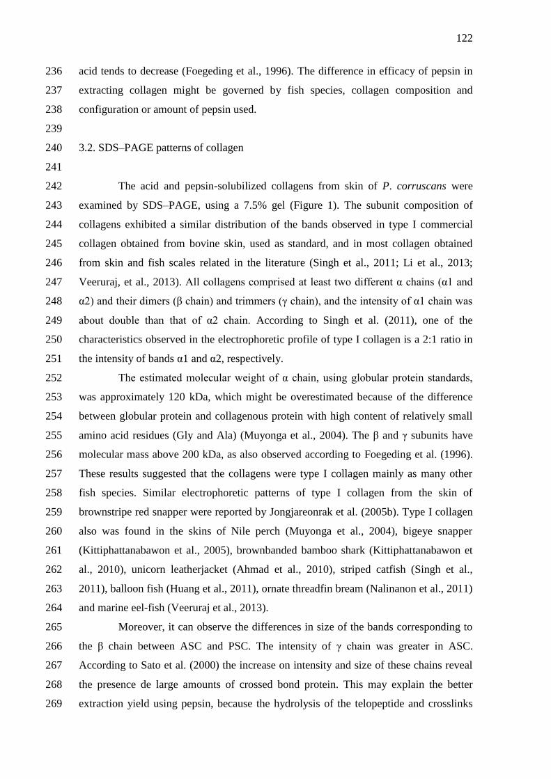
122
acid tends to decrease (Foegeding et al., 1996). The difference in efficacy of pepsin in 236
extracting collagen might be governed by fish species, collagen composition and 237
configuration or amount of pepsin used. 238
239
3.2. SDS–PAGE patterns of collagen 240
241
The acid and pepsin-solubilized collagens from skin of P. corruscans were 242
examined by SDS–PAGE, using a 7.5% gel (Figure 1). The subunit composition of 243
collagens exhibited a similar distribution of the bands observed in type I commercial 244
collagen obtained from bovine skin, used as standard, and in most collagen obtained 245
from skin and fish scales related in the literature (Singh et al., 2011; Li et al., 2013; 246
Veeruraj, et al., 2013). All collagens comprised at least two different α chains (α1 and 247
α2) and their dimers (β chain) and trimmers (γ chain), and the intensity of α1 chain was 248
about double than that of α2 chain. According to Singh et al. (2011), one of the 249
characteristics observed in the electrophoretic profile of type I collagen is a 2:1 ratio in 250
the intensity of bands α1 and α2, respectively. 251
The estimated molecular weight of α chain, using globular protein standards, 252
was approximately 120 kDa, which might be overestimated because of the difference 253
between globular protein and collagenous protein with high content of relatively small 254
amino acid residues (Gly and Ala) (Muyonga et al., 2004). The β and γ subunits have 255
molecular mass above 200 kDa, as also observed according to Foegeding et al. (1996). 256
These results suggested that the collagens were type I collagen mainly as many other 257
fish species. Similar electrophoretic patterns of type I collagen from the skin of 258
brownstripe red snapper were reported by Jongjareonrak et al. (2005b). Type I collagen 259
also was found in the skins of Nile perch (Muyonga et al., 2004), bigeye snapper 260
(Kittiphattanabawon et al., 2005), brownbanded bamboo shark (Kittiphattanabawon et 261
al., 2010), unicorn leatherjacket (Ahmad et al., 2010), striped catfish (Singh et al., 262
2011), balloon fish (Huang et al., 2011), ornate threadfin bream (Nalinanon et al., 2011) 263
and marine eel-fish (Veeruraj et al., 2013). 264
Moreover, it can observe the differences in size of the bands corresponding to 265
the β chain between ASC and PSC. The intensity of γ chain was greater in ASC. 266
According to Sato et al. (2000) the increase on intensity and size of these chains reveal 267
the presence de large amounts of crossed bond protein. This may explain the better 268
extraction yield using pepsin, because the hydrolysis of the telopeptide and crosslinks 269
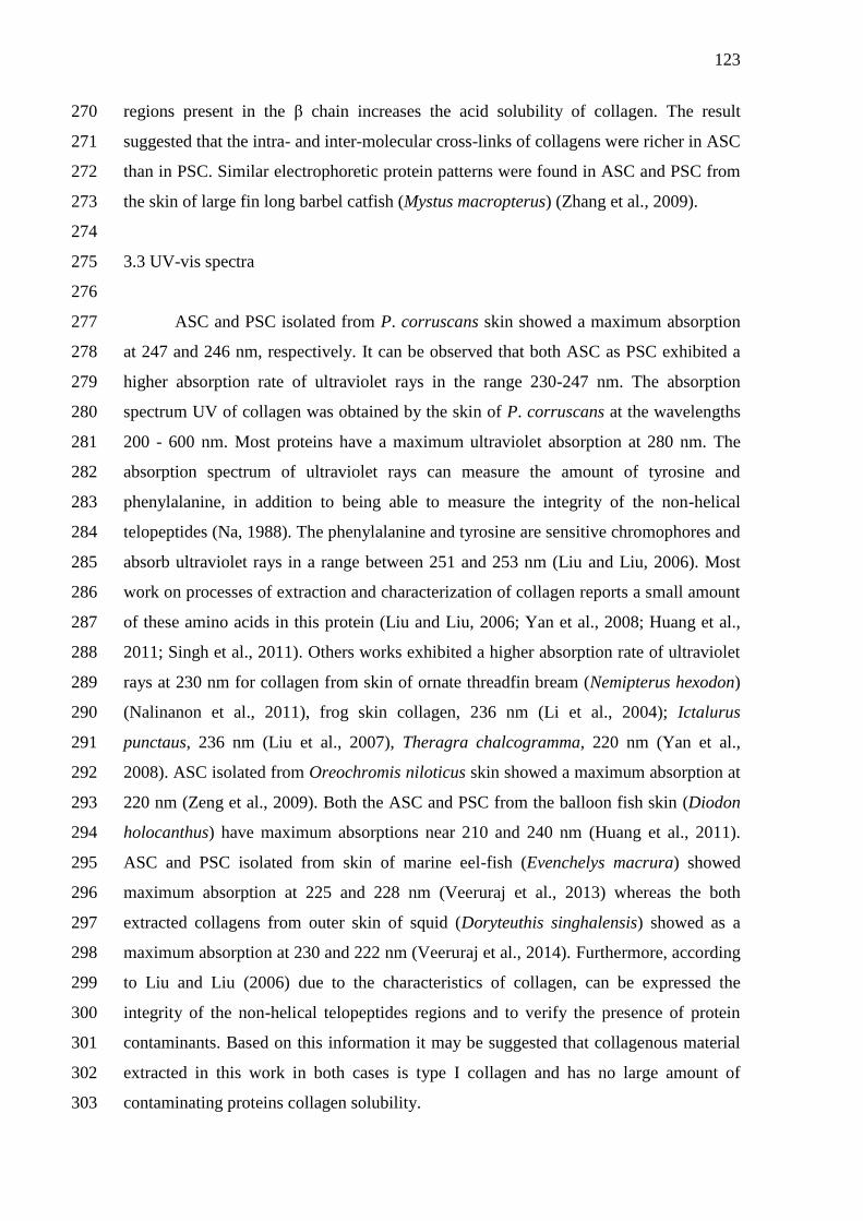
123
regions present in the β chain increases the acid solubility of collagen. The result 270
suggested that the intra- and inter-molecular cross-links of collagens were richer in ASC 271
than in PSC. Similar electrophoretic protein patterns were found in ASC and PSC from 272
the skin of large fin long barbel catfish (Mystus macropterus) (Zhang et al., 2009). 273
274
3.3 UV-vis spectra 275
276
ASC and PSC isolated from P. corruscans skin showed a maximum absorption 277
at 247 and 246 nm, respectively. It can be observed that both ASC as PSC exhibited a 278
higher absorption rate of ultraviolet rays in the range 230-247 nm. The absorption 279
spectrum UV of collagen was obtained by the skin of P. corruscans at the wavelengths 280
200 - 600 nm. Most proteins have a maximum ultraviolet absorption at 280 nm. The 281
absorption spectrum of ultraviolet rays can measure the amount of tyrosine and 282
phenylalanine, in addition to being able to measure the integrity of the non-helical 283
telopeptides (Na, 1988). The phenylalanine and tyrosine are sensitive chromophores and 284
absorb ultraviolet rays in a range between 251 and 253 nm (Liu and Liu, 2006). Most 285
work on processes of extraction and characterization of collagen reports a small amount 286
of these amino acids in this protein (Liu and Liu, 2006; Yan et al., 2008; Huang et al., 287
2011; Singh et al., 2011). Others works exhibited a higher absorption rate of ultraviolet 288
rays at 230 nm for collagen from skin of ornate threadfin bream (Nemipterus hexodon) 289
(Nalinanon et al., 2011), frog skin collagen, 236 nm (Li et al., 2004); Ictalurus 290
punctaus, 236 nm (Liu et al., 2007), Theragra chalcogramma, 220 nm (Yan et al., 291
2008). ASC isolated from Oreochromis niloticus skin showed a maximum absorption at 292
220 nm (Zeng et al., 2009). Both the ASC and PSC from the balloon fish skin (Diodon 293
holocanthus) have maximum absorptions near 210 and 240 nm (Huang et al., 2011). 294
ASC and PSC isolated from skin of marine eel-fish (Evenchelys macrura) showed 295
maximum absorption at 225 and 228 nm (Veeruraj et al., 2013) whereas the both 296
extracted collagens from outer skin of squid (Doryteuthis singhalensis) showed as a 297
maximum absorption at 230 and 222 nm (Veeruraj et al., 2014). Furthermore, according 298
to Liu and Liu (2006) due to the characteristics of collagen, can be expressed the 299
integrity of the non-helical telopeptides regions and to verify the presence of protein 300
contaminants. Based on this information it may be suggested that collagenous material 301
extracted in this work in both cases is type I collagen and has no large amount of 302
contaminating proteins collagen solubility. 303
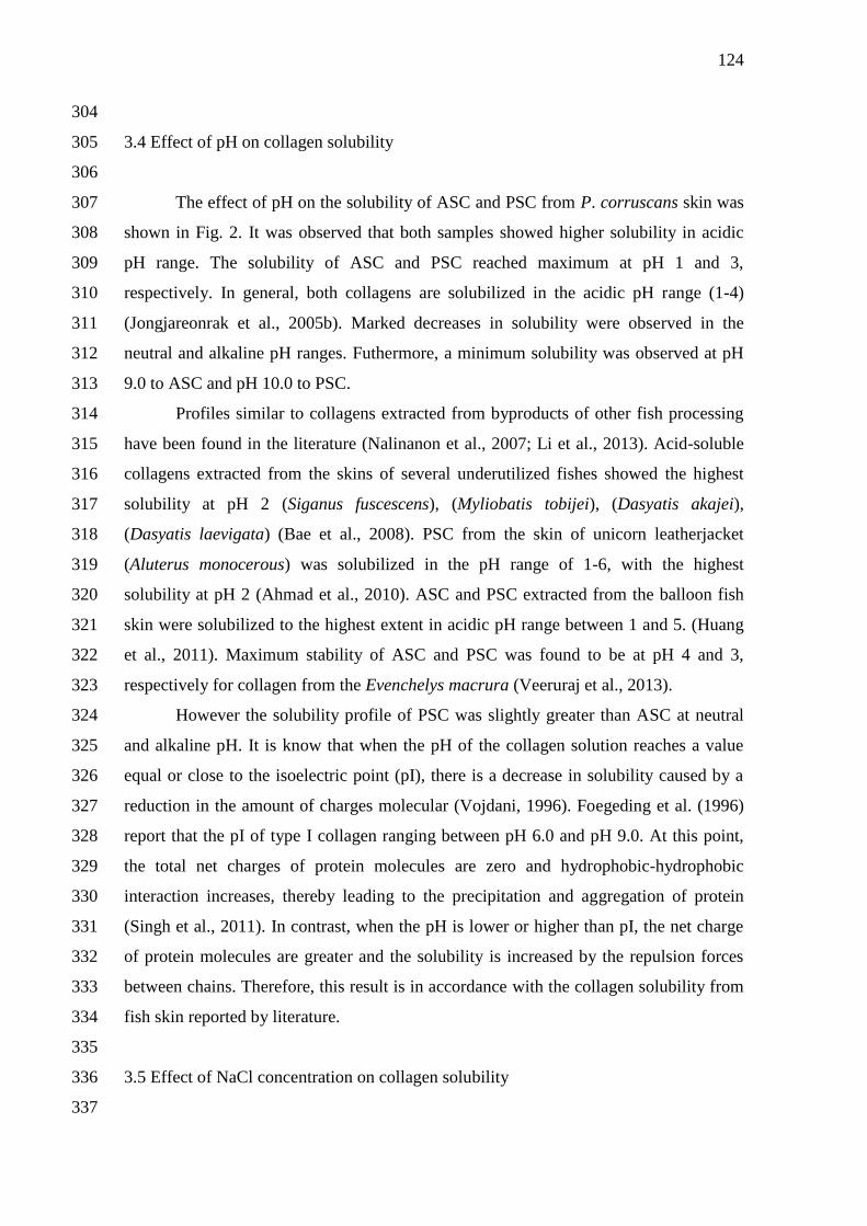
124
304
3.4 Effect of pH on collagen solubility 305
306
The effect of pH on the solubility of ASC and PSC from P. corruscans skin was 307
shown in Fig. 2. It was observed that both samples showed higher solubility in acidic 308
pH range. The solubility of ASC and PSC reached maximum at pH 1 and 3, 309
respectively. In general, both collagens are solubilized in the acidic pH range (1-4) 310
(Jongjareonrak et al., 2005b). Marked decreases in solubility were observed in the 311
neutral and alkaline pH ranges. Futhermore, a minimum solubility was observed at pH 312
9.0 to ASC and pH 10.0 to PSC. 313
Profiles similar to collagens extracted from byproducts of other fish processing 314
have been found in the literature (Nalinanon et al., 2007; Li et al., 2013). Acid-soluble 315
collagens extracted from the skins of several underutilized fishes showed the highest 316
solubility at pH 2 (Siganus fuscescens), (Myliobatis tobijei), (Dasyatis akajei), 317
(Dasyatis laevigata) (Bae et al., 2008). PSC from the skin of unicorn leatherjacket 318
(Aluterus monocerous) was solubilized in the pH range of 1-6, with the highest 319
solubility at pH 2 (Ahmad et al., 2010). ASC and PSC extracted from the balloon fish 320
skin were solubilized to the highest extent in acidic pH range between 1 and 5. (Huang 321
et al., 2011). Maximum stability of ASC and PSC was found to be at pH 4 and 3, 322
respectively for collagen from the Evenchelys macrura (Veeruraj et al., 2013). 323
However the solubility profile of PSC was slightly greater than ASC at neutral 324
and alkaline pH. It is know that when the pH of the collagen solution reaches a value 325
equal or close to the isoelectric point (pI), there is a decrease in solubility caused by a 326
reduction in the amount of charges molecular (Vojdani, 1996). Foegeding et al. (1996) 327
report that the pI of type I collagen ranging between pH 6.0 and pH 9.0. At this point, 328
the total net charges of protein molecules are zero and hydrophobic-hydrophobic 329
interaction increases, thereby leading to the precipitation and aggregation of protein 330
(Singh et al., 2011). In contrast, when the pH is lower or higher than pI, the net charge 331
of protein molecules are greater and the solubility is increased by the repulsion forces 332
between chains. Therefore, this result is in accordance with the collagen solubility from 333
fish skin reported by literature. 334
335
3.5 Effect of NaCl concentration on collagen solubility 336
337
125
The effect of NaCl on the solubility of ASC and PSC extracted from skin of 338
spotted sorubim is shown in Fig. 3. For ASC diluted in 0.5 M acetic acid, the protein 339
starts to precipitate at a concentration of 1% (w/v) and has a large percentage precipitate 340
when the NaCl concentration reaches 3% (w/v). The PSC remained more than 80% 341
solubility under large part of the variations in NaCl concentration (0-3%), having 342
precipitation of most of the its content when the salt concentration achieves 4% in the 343
solution. 344
The solubility of collagens from the skin of brownstripe red snapper (Lutjanus 345
vitta) and bigeye snapper (Priacanthus tayenus) in acetic acid solution decreased with 346
increasing NaCl concentration (Jongjareonrak et al., 2005a; Kittiphattanabawon et al., 347
2005). Similar result was reported by Zeng et al. (2009) in which a solubility of ASC 348
decreased sharply when NaCl content reached 2% and remained at constant low level at 349
4% NaCl or above. PSC from the skin of unicorn leatherjacket (Aluterus monocerous) 350
had drastic decrease in solubility at NaCl concentration higher than 2% (Ahmad et al., 351
2010). Considerable decrease also was noticeable when the concentration was increased 352
to more than 4% for acid and pepsin-solubilized collagens from the skin of balloon fish 353
(Diodon holocanthus) (Huang et al., 2011). Expressive decrease in ASC and PSC 354
solubility was observed with 3% NaCl or above for collagen extracted from the skin of 355
Evenchelys macrura (Veeruraj et al., 2013). 356
The decrease in solubility of collagens could be described as being due to a 357
“salting out” effect, which occurred at relatively high NaCl concentrations 358
(Jongjareonrak et al., 2005a). An increase in ionic strength causes a reduction in protein 359
solubility by enhancing hydrophobic–hydrophobic interactions between protein chains, 360
and increasing the competition for water with the ionic salts, leading to the induced 361
protein precipitation (Bae et al., 2008). Similar behaviours were found for ASC and 362
PSC extracted from spotted sorubim skin. 363
However, PSC showed a greater solubility than ASC at NaCl concentrations 364
used. Trend curves following a pattern where the ASC is more susceptible variations in 365
the concentration of NaCl of what the PSC are commonly reported in the literature, 366
brownstriped snapper fish (Jongjareonrak et al., 2005b); striped catfish (Singh et al., 367
2011); fish balloon (Huang et al., 2011); mackerel (Li et al., 2013). A greater solubility 368
of PSC could be due to the partial hydrolysis of high MW cross-linked molecules by 369
pepsin (Singh et al., 2011). In addition, the differences in amino acid compositions and 370
structure between ASC and PSC might result in such differences. 371
126
372
4. Conclusion 373
374
Both ASC and PSC were successfully isolated from the skin of 375
Pseudoplatystoma corruscans. Pepsin could be used as an aid for increasing the 376
extraction yield of collagen. Based on SDS–PAGE patterns and absorption spectrum 377
UV of collagen it may be suggested that collagenous material extracted in this work in 378
both cases is type I, most commercially important. It was observed that both ASC and 379
PSC showed higher solubility in acidic range of pH. However the solubility profile of 380
PSC was slightly greater than ASC at neutral and alkaline pH. The solubility decreased 381
in the presence of NaCl at concentrations above 3% although PSC showed a greater 382
solubility than ASC at NaCl concentrations used. The use of pepsin showed useful due 383
to the yield achieved and also to degree of preservation of structures, proving it caused 384
little changes in collagen. Therefore, the skin of this piscivorous catfish could be an 385
alternative source of collagen for further applications. 386
387
Acknowledgements 388
389
This study was financially supported by the following Brazilian agencies: 390
Conselho Nacional de Pesquisa e Desenvolvimento Científico (CNPq), and the 391
Coordenação de Aperfeiçoamento de Pessoal de Nível Superior (CAPES). 392
393
References 394
Ahmad, M., & Benjakul, S. (2010). Extraction and characterization of pepsin 395
solubilized collagen from the skin of unicorn leatherjacket (Aluterus monocerous). Food 396
Chemistry, 120, 817 – 824. 397
398
Bae, I., Osatomi, K., Yoshida, A., Osako, K., Yamaguchi, A., & Hara, K. (2008). 399
Biochemical properties of acid-soluble collagens extracted from the skins of under 400
utilised fishes. Food Chemistry, 108, 49 - 54. 401
402
Benjakul, S., Thiansilakul, Y., Visessanguan, W., Roytrakul, S., Kishimura, H., 403
Prodprand, T., et al. (2010). Extraction and characterization of pepsin solubilized 404

127
collagens from the skin of bigeye snapper (Priacanthus tayenus and Priacanthus 405
macracanthus). Journal of the Science of Food and Agriculture, 90, 132 – 138. 406
407
Binsi, P. K., Shamasundar, B. A., Dileep, A. O., Badii, F., & Howell, N. K. (2009). 408
Rheological and functional properties of gelatin from the skin of Bigeye snapper 409
(Priacanthus hamrur) fish: influence of gelatin on the gel-forming ability of fish mince. 410
Food Hydrocolloids, 23, 132 - 145. 411
412
Cho, J. K., Jin, Y. G., Rha, S. J., Kim, S. J., Hwang, J. H. (2014). Biochemical 413
characteristics of four marine fish skins in Korea. Food Chemistry 159, 200 – 207. 414
415
Crepaldi, D. V., Faria, P. M. C., Teixeira, E. A., Ribeiro, L. P., Costa, A. A. P., Melo, 416
D. C., Cintra, A. P. R., Prado, S. A., Costa, F. A. A., Drumond, M. L., Lopes, V. E., 417
Moraes, V. E., (2006). O surubim na aquacultura do Brasil. Rev. Bras. Reprod. Animal 418
30, 150 – 158. 419
420
Duan, R., Zhang, J., Du, X., Yao, X., & Konno, K. (2009). Properties of collagen from 421
skin, scale and bone of carp (Cyprinus carpio). Food Chemistry, 112, 702 – 706. 422
423
Foegeding, E. A., Lanier, T. C., Hultin, H.O., (1996). Characteristics of edible muscle 424
tissues. In: Fennema, O.R. (Ed.), Food Chemistry. Marcel Dekker, New York, pp. 879 425
– 942. 426
427
Gelse, K., Pöschl, E., & Aigner, T. (2003). Collagens — Structure, function, and 428
biosynthesis. Advanced Drug Delivery Reviews, 55, 1531−1546. 429
430
Huang, Y. R., Shiau, C. Y., Chen, H. H., & Huang, B. C. (2011). Isolation and 431
characterization of acid and pepsin-solubilized collagens from the skin of balloon fish 432
(Diodon holocanthus). Food Hydrocolloids, 25 (6), 1507 - 1513. 433
434
Inoue, L. A. K. A., Hisano, H., Ishikawa, M. M., Rotta, M. A., Senhorini, J. A. (2009). 435
Princípios básicos para produção de alevinos de surubins (pintado e cachara). Dourados: 436
Embrapa Agropecuária Oeste; Manaus: Embrapa Amazônia Ocidental; Corumbá: 437
Embrapa Pantanal. 26 p. Documentos (Embrapa Agropecuária Oeste. Documentos, 99; 438
128
Embrapa Amazônia Ocidental. Documentos, 68; Embrapa Pantanal. Documentos, 100. 439
Impresso, v. 15, p. 1-26. 440
441
Jayathilakan, K., Sultana, K., Radhakrishna, K., & Bawa, A. S. (2012). Utilization of 442
byproducts and waste materials from meat, poultry and fish processing industries: A 443
review. Journal of Food Science and Technology, 49, 278 – 293. 444
445
Jongjareonrak, A.; Benjakul, S.; Visessanguan, W.; Tanaka, M. (2005a). Isolation and 446
characterization of collagen from bigeye snapper (Priacanthus marcracanthus) skin. 447
Journal of the Science of Food and Agriculture, v. 85, p.1203 – 1210. 448
449
Jongjareonrak, A., Benjakul, S., Visessanguan, W., Nagai, T., & Tanaka, M. (2005b). 450
Isolation and characterisation of acid and pepsin-solubilised collagens from the skin of 451
brownstripe red snapper (Lutjanus vitta). Food Chemistry, 93, 475 – 484. 452
453
Kittiphattanabawon, P., Benjakul, S., Visessanguan, W., Nagai, T., & Tanaka, M. 454
(2005). Characterization of acid-soluble collagen from skin and bone of bigeye snapper 455
(Priacanthus tayenus). Food Chemistry, 89, 363 – 372. 456
457
Kittiphattanabawon, P., Benjakul, S., Visessanguan,W., & Shahidi, F. (2010). Isolation 458
and characterization of collagen from the cartilages of brownbanded bamboo shark 459
(Chiloscyllium punctatum) and blacktip shark (Carcharhinus limbatus). LWT and Food 460
Science and Technology, 43 (5), 792 - 800. 461
462
Laemmli, U. K. (1970). Cleavage of structural proteins during the assembly of the head 463
of bacteriophage T4. Nature, v. 227, p. 680–685. 464
465
Li, H., Liu, B. L., Gao, L. Z., & Chen, H. L. (2004). Studies on bullfrog skin collagen. 466
Food Chemistry, 84, 65 – 69. 467
468
Li, Z. R., Wang, B., Chi, C. F., Zhang, Q. H., Gong, Y. D., Tang, J. J., et al. (2013). 469
Isolation and characterization of acid soluble collagens and pepsin soluble collagens 470
from the skin and bone of Spanish mackerel (Scomberomorous niphonius). Food 471
Hydrocolloids, 31 (1), 103 - 113. 472
129
473
Liu, Y. K., & Liu, D. C. (2006). Comparison of physical-chemical properties of type I 474
collagen from different species. Food Chemistry, 99, 244 – 251. 475
476
Liu, H. Y., Li, D., & Guo, S. D. (2007). Studies on collagen from the skin of channel 477
catfish (Ictalurus punctaus). Food Chemistry, 101, 621 – 625. 478
479
Liu, W., Hendren, J., Qin, X.J., Shen, J., Liu, K.J. (2009). Normobaric hyperoxia 480
attenuates early blood–brain barrier disruption by inhibiting MMP-9-mediated occludin 481
degradation in focal cerebral ischemia. J. Neurochem. 108, 811 – 820. 482
483
Liu, D., Liang, L., Regenstein, J. M., & Zhou, P. (2012). Extraction and characterization 484
of pepsin-solubilised collagen from fins, scales, skins, bones and swim bladders of 485
bighead carp (Hypophthalmichthys nobilis). Food Chemistry, 133, 1441 - 1448. 486
487
Liu, D., Zhou, P., Li, T., Regenstein, J. M. (2014). Comparison of acid-soluble 488
collagens from the skins and scales of four carp species. Food Hydrocolloids 41, 290 – 489
297. 490
491
McCormick, R. J. (2009). Collagen. In Applied muscle biology and meat science (pp. 492
127 - 146). Boca Raton, Florida: CRC Press. 493
494
MPA 2010 - Boletim Estatístico da Pesca e Aquicultura- Ministério da Pesca e 495
Aquicultura. 496
497
Montero, P., Jimennez-Colmenero, F., & Borderias, J. (1991). Effect of pH and the 498
presence of NaCl on some hydration properties of collagenous material from trout 499
(Salmo irideus Gibb) muscle and skin. Journal of the Science of Food and Agriculture, 500
54, 137 – 146. 501
502
Muyonga, J. H., Cole, C. G. B., & Duodu, K. G. (2004). Characterization of acid 503
soluble collagen from skins of young and adult Nile perch (Lates niloticus). Food 504
Chemistry, 85 (1), 81 - 89. 505
506
130
Na, G. C. (1988). UV spectroscopic characterization of type I collagen. Collagen and 507
Related Research, 8, 315 – 330. 508
509
Nagai, T., & Suzuki, N. (2000). Isolation of collagen from fish waste material - skin, 510
bone and fins. Food Chemistry, 68, 277–281. 511
512
Nagai, T., Suzuki, N., & Nagashima, T. (2008). Collagen from common minke whale 513
(Balaenoptera acutorostrata) unesu. Food Chemistry, 111, 296 – 301. 514
515
Nalinanon, S., Benjakul, S., Visessanguan, W., & Kishimura, H. (2007). Use of pepsin 516
for collagen extraction from the skin of big eye snapper (Priacanthus tayenus). Food 517
Chemistry, 104, 593 – 601. 518
519
Nalinanon S., Benjakul, S., Kishimura, H., Osako, K. (2011). Type I collagen from the 520
skin of ornate threadfin bream (Nemipterus hexodon): Characteristics and effect of 521
pepsin hydrolysis. Food Chemistry 125, 500 – 507. 522
523
Pati, F., Adhikari, B., & Dhara, S. (2010). Isolation and characterization of fish scale 524
collagen of higher thermal stability. Bioresource Technology, 101, 3737 - 3742. 525
526
Regenstein, J. M., & Zhou, P. (2007). Collagen and gelatin from marine by-product. In 527
F. Shahidi (Ed.), Maximising the value of marine by-products (pp. 279 – 303). Boca 528
Raton, Florida: CRC Press. 529
530
Safandowska, M., Pietrucha, K. (2013). Effect of fish collagen modification on its 531
thermal and rheological properties. International Journal of Biological Macromolecules 532
53, 32 – 37. 533
534
Sato, K., Ebihara, T., Adachi, E., Kawashima, S., Hattori, S., & Irie, S. (2000). Possible 535
involvement of aminotelopeptide in self-assembly and thermal stability of collagen I as 536
revealed by its removal with protease. Journal of Biological Chemistry, 275, 25870 – 537
25875. 538
539
131
Senaratne, L. S., Park, P., & Kim, S. (2006). Isolation and characterization of collagen 540
from brown backed toadfish (Lagocephalus gloveri) skin. Bioresource Technology, 97, 541
191 – 197. 542
543
Singh, P., Benjakul, S., Maqsood, S., & Kishimura, H. (2011). Isolation and 544
characterization of collagen extracted from the skin of striped catfish (Pangasianodon 545
hypophthalmus). Food Chemistry, 124 (1), 97 - 105. 546
547
Song, E., Yeon Kim, S., Chun, T., Byun, H. J., Lee, Y. M. (2006). Biomaterials, v. 27, 548
p. 2951. doi:10.1016/j.biomaterials.2006.01.015. 549
550
Veeruraj, A., Arumugam, M., & Balasubramanian, T. (2013). Isolation and 551
characterization thermostable collagen from eel-fish (Evenchelys macrura). Process 552
Biochemistry, 48 (10), 1592 - 1602. 553
554
Veeruraj, A., Arumugam M., Ajithkumar T., Balasubramanian, T. (2014). Isolation and 555
characterization of collagen from the outer skin of squid (Doryteuthis singhalensis). 556
Food Hydrocolloids, http://dx.doi.org/10.1016/j.foodhyd.2014.07.025. 557
558
Vojdani, F. (1996). Solubility. In G. M. Hall (Ed.), Methods of testing protein 559
functionality (pp. 11 – 60). Great Britain: St. Edmundsbury Press. 560
561
Yan, M., Li, B., Zhao, X., Ren, G., Zhuang, Y., Hou, H., et al. (2008). Characterization 562
of acid-soluble collagen from the skin of walleye pollack (Theragra chalcogramma). 563
Food Chemistry, 107, 1581 – 1586. 564
565
Zeng, S. K., Zhang, C. H., Lin, H., Yang, P., Hong, P. Z., & Jiang, Z. (2009). Isolation 566
and characterization of acid-solubilized collagen from the skin of Nile tilapia 567
(Oreochromis niloticus). Food Chemistry, 116, 879 and 883. 568
569
Zhang, Y., Liu, W., Li, G., Shi, B., Miao, Y. Q., & Wu, X. H. (2007). Isolation and 570
partial characterization of pepsin-soluble collagen from the skin of grass carp 571
(Ctenopharyngodon idella). Food Chemistry, 103, 906 – 912. 572
573
132
Zhang, M., Liu, W., & Li, G. (2009). Isolation and characterization of collagens from 574
the skin of largefin longbarbel catfish (Mystus macropterus). Food Chemistry, 115, 826 575
– 831. 576
577
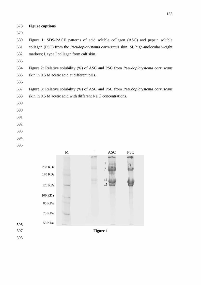
133
Figure captions 578
579
Figure 1: SDS-PAGE patterns of acid soluble collagen (ASC) and pepsin soluble 580
collagen (PSC) from the Pseudoplatystoma corruscans skin. M, high-molecular weight 581
markers; I, type I collagen from calf skin. 582
583
Figure 2: Relative solubility (%) of ASC and PSC from Pseudoplatystoma corruscans 584
skin in 0.5 M acetic acid at different pHs. 585
586
Figure 3: Relative solubility (%) of ASC and PSC from Pseudoplatystoma corruscans 587
skin in 0.5 M acetic acid with different NaCl concentrations. 588
589
590
591
592
593
594
595
596
Figure 1 597
598
M I ASC PSC
200 KDa
100 KDa
170 KDa
120 KDa α2
α1
β
γ
70 KDa
85 KDa
53 KDa

134
0 2 4 6 8 10 12
0
20
40
60
80
100
Re
lati
ve
Co
lla
ge
n S
olu
bilit
y (
%)
pH
599
Figure 2 600
601
602
0 1 2 3 4 5 6
0
20
40
60
80
100
Re
lati
ve
co
lla
ge
n s
olu
bilit
y(%
)
NaCl Concentration (% (w/v))
603
Figure 3 604
605
□ ASC
○ PSC
□ ASC
○ PSC
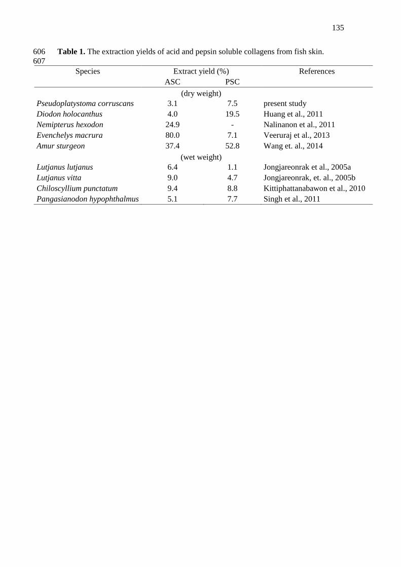
135
Table 1. The extraction yields of acid and pepsin soluble collagens from fish skin. 606
607
Species Extract yield (%) References
ASC PSC
(dry weight)
Pseudoplatystoma corruscans 3.1 7.5 present study
Diodon holocanthus 4.0 19.5 Huang et al., 2011
Nemipterus hexodon 24.9 - Nalinanon et al., 2011
Evenchelys macrura 80.0 7.1 Veeruraj et al., 2013
Amur sturgeon 37.4 52.8 Wang et. al., 2014
(wet weight)
Lutjanus lutjanus 6.4 1.1 Jongjareonrak et al., 2005a
Lutjanus vitta 9.0 4.7 Jongjareonrak, et. al., 2005b
Chiloscyllium punctatum 9.4 8.8 Kittiphattanabawon et al., 2010
Pangasianodon hypophthalmus 5.1 7.7 Singh et al., 2011
136
CONSIDERAÇÕES FINAIS
A presente tese reportou a purificação parcial e caracterização de uma protease
ácida (pepsina símile) do estômago do pintado (P. corruscans), bem como o
aproveitamento de resíduos do peixe (viscera e pele) para aplicação na extração de
colágeno. O processo de purificação parcial, juntamente com as etapas de caracterização
enzimática evidenciaram características interessantes da protease ácida estudada como
acentuada atividade enzimática, estabilidade em pH neutro e elevada temperatura ótima,
reforçando o grande potencial de utilização das vísceras do pintado como fonte de
obtenção de proteases ácidas para aplicação industrial.
Em relação à aplicação biotecnológica dos resíduos do peixe, foi possível
realizar a extração de colágeno proveniente da pele, bem como proceder com a
utilização de protease ácida para aumentar o rendimento da extração. Colágeno ácido
solúvel e pepsino solúvel tipo I foram isolados e caracterizados com rendimentos de
3,1% e 7,5% respectivamente. O biomaterial obtido apresentou propriedades físico-
químicas similares a colágenos extraídos de pele de outros peixes citados na literatura.
Através das análises de caracterização dos colágenos obtidos (ASC e PSC)
também se verificou a importância da utilização da pepsina no processo de obtenção de
colágeno, pois além do aumento no rendimento, a estrutura da proteína manteve-se
preservada, com pequenas mudanças. Assim, é possível afirmar que proteases ácidas
provenientes do pintado possuem um grande potencial para aplicação em processos
industriais, como o de aditivo no processo de extração de colágeno.
137
ANEXOS
ANEXO 1:
Guide for authors
LWT- Food Science and Technology
INTRODUCTION
LWT - Food Science and Technology is an official journal of the Swiss Society of Food Science and
Technology (SGLWT/SOSSTA) and the International Union of Food Science and Technology (IUFoST).
LWT - Food Science and Technology is an international journal that publishes innovative papers in the
fields of food chemistry, biochemistry, microbiology, technology and nutrition. The work described
should be innovative either in the approach or in the methods used. The significance of the results either
for the science community or for the food industry must also be specified. Contributions that do not fulfil
these requirements will not be considered for review and publication. Submission of a paper will be held
to imply that it presents original research, that it has not been published previously, and that it is not under
consideration for publication elsewhere.
Papers featuring animal trials are outside the scope of the journal and will not be considered for
publication.
Types of paper
Three types of peer-reviewed papers will be published:
Timely Reviews. These concise reviews should present a focused aspect on a topic of current interest or
an emerging field. They are not intended as comprehensive literature surveys covering all aspects of the
topic, but should include all major findings and bring together reports from a number of sources. They
should aim to give balanced, objective assessments by giving due reference to relevant published work,
and not merely present the prejudices of individual authors or summarise only work carried out by the
authors or by those with whom the authors agree. Undue speculation should also be avoided. These
reviews will receive priority in publication.
The reviews may address pertinent issues in food science, technology, processing, nutritional aspects of
raw and processed foods and may include nutraceuticals, functional foods, use of "omics" in food quality,
food processing and preservation, and food production. Topics to be covered should be at the cutting edge
of science, well thought out, succinct, focused and clear. Ideally, the review should provide a view of the
state of the art and suggest possible future needs and trends. All articles will be subjected to peer review
process.
138
Submit an abstract of the proposed review to the Reviews Editor (Professor Shridhar Sathe,
[email protected] for consideration prior to preparing the full length manuscript. Abstract of the proposed
work should include the following: a. The abstract should identify the need for the proposed article, the
intended audience, and five key words.
b. Title (120 characters or less)
c. Short abstract (≤ 300 words).
d. Identify the address and contact information for the contact author. The contact information should
include author name, postal address, telephone number, fax number, and email.
e. Anticipated time needed to complete the proposed work once the initial abstract has been approved
Manuscript Preparation
a. All lines and pages must be continuously numbered.
b. All text should be double-spaced.
c. Total manuscript length ≤ 3,000 words (text portion).
d. Total number of Tables ≤ 5.
e. Total number of figures ≤ 5.
f. Maximum number of references (including those cited in tables and figures) not to exceed 50.
g. In the reference list identify five (5) key references (indicated by an * in front of the reference in the
reference section). In two to three sentences explain why this reference is a key reference.
Research Papers. Reports of complete, scientifically sound, original research which contributes new
knowledge to its field. The paper must be organised as described in Article Structure below. Papers
should not exceed 5000 words (approximately 18 typed pages).
Research Notes. Brief reports of scientifically sound, original research of limited scope of new findings.
Research Notes have the formal organisation of a full paper. Such notes will receive priority of
publication. Research notes should not exceed 2500 words (approximately 9 typed pages).
Contact details for submission
Submission for all types of manuscripts to LWT - Food Science and Technology proceeds totally online.
Via the Elsevier Editorial System (EES) website for this journal, http://ees.elsevier.com/lwt, you will be
guided step-by-step through the creation and uploading of the various files.
Page charges
This journal has no page charges.
Revised papers received more than three months after reviewers' comments were sent may be treated as
new submissions, at the discretion of the Editor. If the author has not replied to reminders/enquiries about
revisions within 6 months, the paper will be considered to have lapsed, and any subsequent submission
139
will be treated as a new submission and must be submitted to the journal using the above process,
addressed to the Editor-in-Chief, with an explanation that it had previously been submitted to the journal.
BEFORE YOU BEGIN
Ethics in publishing
For information on Ethics in publishing and Ethical guidelines for journal publication see
http://www.elsevier.com/publishingethics and http://www.elsevier.com/ethicalguidelines.
By submitting this manuscript, the authors agree that text, equations, or figures from previously published
articles or books have been clearly identified in full and their origin clearly explained in the adjacent text,
with appropriate references given at the end of the paper. Duplication of text is rarely justified, even with
diligent referencing. Exceptions may be made for descriptions of standard experimental techniques, or
other standard methods used by the author in the investigation; but an appropriate citation is preferable.
Authors who duplicate material from their own published work in a new article, without clearly
identifying the repeated material and its source as outlined above, are self-plagiarising.
Conflict of interest
All authors are requested to disclose any actual or potential conflict of interest including any financial,
personal or other relationships with other people or organizations within three years of beginning the
submitted work that could inappropriately influence, or be perceived to influence, their work. See also
http://www.elsevier.com/conflictsofinterest.
Submission declaration
Submission of an article implies that the work described has not been published previously (except in the
form of an abstract or as part of a published lecture or academic thesis), that it is not under consideration
for publication elsewhere, that its publication is approved by all authors and tacitly or explicitly by the
responsible authorities where the work was carried out, and that, if accepted, it will not be published
elsewhere including electronically in the same form, in English or in any other language, without the
written consent of the copyright-holder.
Authorship
All authors should have made substantial contributions to all of the following: (1) the conception and
design of the study, or acquisition of data, or analysis and interpretation of data, (2) drafting the article or
revising it critically for important intellectual content, (3) final approval of the version to be submitted.
Changes to authorship
This policy concerns the addition, deletion, or rearrangement of author names in the authorship of
accepted manuscripts: Before the accepted manuscript is published in an online issue: Requests to add or
remove an author, or to rearrange the author names, must be sent to the Journal Manager from the
corresponding author of the accepted manuscript and must include: (a) the reason the name should be
140
added or removed, or the author names rearranged and (b) written confirmation (e-mail, fax, letter) from
all authors that they agree with the addition, removal or rearrangement. In the case of addition or removal
of authors, this includes confirmation from the author being added or removed. Requests that are not sent
by the corresponding author will be forwarded by the Journal Manager to the corresponding author, who
must follow the procedure as described above. Note that: (1) Journal Managers will inform the Journal
Editors of any such requests and (2) publication of the accepted manuscript in an online issue is
suspended until authorship has been agreed. After the accepted manuscript is published in an online issue:
Any requests to add, delete, or rearrange author names in an article published in an online issue will
follow the same policies as noted above and result in a corrigendum.
Copyright
Upon acceptance of an article, authors will be asked to complete a 'Journal Publishing Agreement' (for
more information on this and copyright see http://www.elsevier.com/copyright). Acceptance of the
agreement will ensure the widest possible dissemination of information. An e-mail will be sent to the
corresponding author confirming receipt of the manuscript together with a 'Journal Publishing Agreement'
form or a link to the online version of this agreement. Subscribers may reproduce tables of contents or
prepare lists of articles including abstracts for internal circulation within their institutions. Permission of
the Publisher is required for resale or distribution outside the institution and for all other derivative works,
including compilations and translations (please consult http://www.elsevier.com/permissions). If excerpts
from other copyrighted works are included, the author(s) must obtain written permission from the
copyright owners and credit the source(s) in the article. Elsevier has preprinted forms for use by authors
in these cases: please consult http://www.elsevier.com/permissions.
Retained author rights
As an author you (or your employer or institution) retain certain rights; for details you are referred to:
http://www.elsevier.com/authorsrights.
Role of the funding source
You are requested to identify who provided financial support for the conduct of the research and/or
preparation of the article and to briefly describe the role of the sponsor(s), if any, in study design; in the
collection, analysis and interpretation of data; in the writing of the report; and in the decision to submit
the article for publication. If the funding source(s) had no such involvement then this should be stated.
Please see http://www.elsevier.com/funding.
Funding body agreements and policies
Elsevier has established agreements and developed policies to allow authors whose articles appear in
journals published by Elsevier, to comply with potential manuscript archiving requirements as specified
as conditions of their grant awards. To learn more about existing agreements and policies please visit
http://www.elsevier.com/fundingbodies.

141
Open access
This journal offers you the option of making your article freely available to all via the ScienceDirect
platform. To prevent any conflict of interest, you can only make this choice after receiving notification
that your article has been accepted for publication. The fee of $3,000 excludes taxes and other potential
author fees such as color charges. In some cases, institutions and funding bodies have entered into
agreement with Elsevier to meet these fees on behalf of their authors. Details of these agreements are
available at http://www.elsevier.com/fundingbodies. Authors of accepted articles, who wish to take
advantage of this option, should complete and submit the order form (available at
http://www.elsevier.com/locate/openaccessform.pdf). Whatever access option you choose, you retain
many rights as an author, including the right to post a revised personal version of your article on your
own website. More information can be found here: http://www.elsevier.com/authorsrights.
Language and language services
Please write your text in good English (American or British usage is accepted, but not a mixture ofthese).
Authors who equire information about language editing and copyediting services pre- and post-
submission please visit http://webshop.elsevier.com/languageservices or our customer support site at
http://support.elsevier.com for more information.
Submission
Submission to this journal proceeds totally online and you will be guided stepwise through the creation
and uploading of your files. The system automatically converts source files to a single PDF file of the
article, which is used in the peer-review process. Please note that even though manuscript source files are
converted to PDF files at submission for the review process, these source files are needed for further
processing after acceptance. All correspondence, including notification of the Editor's decision and
requests for revision, takes place by e-mail removing the need for a paper trail. Authors must provide and
use an email address unique to themselves and not shared with another author registered in EES, or a
department.
Review Process
A peer review system involving two or three reviewers is used to ensure high quality of manuscripts
accepted for publication. The Editor-in-Chief and Editors have the right to decline formal review of the
manuscript when it is deemed that the manuscript is 1) on a topic outside the scope of the Journal, 2)
lacking technical merit, 3) focused on foods or processes that are of narrow regional scope and
significance, 4) fragmentary and provides marginally incremental results, or 5) is poorly written.
Referees
Please submit, with the manuscript, the names, addresses and e-mail addresses of three potential referees.
Note that the editor retains the sole right to decide whether or not the suggested reviewers are used.
Peer Reviews
142
It is the journal policy to keep the peer reviewing anonymous. Names of reviewers are only revealed if
they are in agreement with the request of the author. When submitting a manuscript, authors may indicate
names of experts who are not suitable/appropriate for reviewing the paper.
PREPARATION
Use of wordprocessing software
It is important that the file be saved in the native format of the wordprocessor used. The text should be in
single-column format. Keep the layout of the text as simple as possible. Most formatting codes will be
removed and replaced on processing the article. In particular, do not use the wordprocessor's options to
justify text or to hyphenate words. However, do use bold face, italics, subscripts, superscripts etc. When
preparing tables, if you are using a table grid, use only one grid for each individual table and not a grid for
each row. If no grid is used, use tabs, not spaces, to align columns. The electronic text should be prepared
in a way very similar to that of conventional manuscripts (see also the Guide to Publishing with Elsevier:
http://www.elsevier.com/guidepublication). Note that source files of figures, tables and text graphics will
be required whether or not you embed your figures in the text. See also the section on Electronic artwork.
To avoid unnecessary errors you are strongly advised to use the 'spell-check' and 'grammar-check'
functions of your wordprocessor.
All lines must be consecutively numbered throughout the manuscript.
Article structure
Subdivision - numbered sections
Divide your article into clearly defined and numbered sections. Subsections should be numbered 1.1 (then
1.1.1, 1.1.2, ...), 1.2, etc. (the abstract is not included in section numbering). Use this numbering also for
internal cross-referencing: do not just refer to 'the text'. Any subsection may be given a brief heading.
Each heading should appear on its own separate line.
Introduction
State the objectives of the work and provide an adequate background, avoiding a detailed literature survey
or a summary of the results.
Material and methods
Provide sufficient detail to allow the work to be reproduced. Methods already published should be
indicated by a reference: only relevant modifications should be described.
Results
Results should be clear and concise.
Discussion
143
This should explore the significance of the results of the work, not repeat them. A combined Results and
Discussion section is often appropriate. Avoid extensive citations and discussion of published literature.
Conclusions
The main conclusions of the study may be presented in a short Conclusions section, which may stand
alone or form a subsection of a Discussion or Results and Discussion section.
Appendices
If there is more than one appendix, they should be identified as A, B, etc. Formulae and equations in
appendices should be given separate numbering: Eq. (A.1), Eq. (A.2), etc.; in a subsequent appendix, Eq.
(B.1) and so on. Similarly for tables and figures: Table A.1; Fig. A.1, etc.
Essential title page information
• Title. Concise and informative. Titles are often used in information-retrieval systems. Avoid
abbreviations and formulae where possible.
• Author names and affiliations. Where the family name may be ambiguous (e.g., a double name), please
indicate this clearly. Present the authors' affiliation addresses (where the actual work was done) below the
names. Indicate all affiliations with a lower-case superscript letter immediately after the author's name
and in front of the appropriate address. Provide the full postal address of each affiliation, including the
country name and, if available, the e-mail address of each author.
• Corresponding author. Clearly indicate who will handle correspondence at all stages of refereeing and
publication, also post-publication. Ensure that telephone and fax numbers (with country and area
code) are provided in addition to the e-mail address and the complete postal address. Contact
details must be kept up to date by the corresponding author.
• Present/permanent address. If an author has moved since the work described in the article was done, or
was visiting at the time, a 'Present address' (or 'Permanent address') may be indicated as a footnote to that
author's name. The address at which the author actually did the work must be retained as the main,
affiliation address. Superscript Arabic numerals are used for such footnotes.
Abstract
A concise and factual abstract is required. The abstract should state briefly the purpose of the research,
the principal results and major conclusions. An abstract is often presented separately from the article, so it
must be able to stand alone. For this reason, References should be avoided, but if essential, then cite the
author(s) and year(s). Also, non-standard or uncommon abbreviations should be avoided, but if essential
they must be defined at their first mention in the abstract itself.
Abstracts should not exceed 200 words for research papers and notes, or 300 words for reviews.
Highlights
Highlights are mandatory for this journal. They consist of a short collection of bullet points that convey
the core findings of the article and should be submitted in a separate file in the online submission system.
144
Please use 'Highlights' in the file name and include 3 to 5 bullet points (maximum 85 characters,
including spaces, per bullet point). See http://www.elsevier.com/highlights for examples.
Keywords
Immediately after the abstract, provide a maximum of 6 keywords, using British spelling and avoiding
general and plural terms and multiple concepts (avoid, for example, 'and', 'of'). Be sparing with
abbreviations: only abbreviations firmly established in the field may be eligible. These keywords will be
used for indexing purposes.
If possible the Food Science and Technology Abstracts (FSTA) Thesaurus should be used (IFIS
Publ.,Shinfield, Reading RG2 9BB, UK http://www.foodScienceCentral.com).
Abbreviations
Define abbreviations that are not standard in this field in a footnote to be placed on the first page of the
article. Such abbreviations that are unavoidable in the abstract must be defined at their first mention there,
as well as in the footnote. Ensure consistency of abbreviations throughout the article.
Acknowledgements
Collate acknowledgements in a separate section at the end of the article before the references and do not,
therefore, include them on the title page, as a footnote to the title or otherwise. List here those individuals
who provided help during the research (e.g., providing language help, writing assistance or proof reading
the article, etc.).
Units
Follow internationally accepted rules and conventions: use the international system of units (SI). If other
units are mentioned, please give their equivalent in SI.
Do not use %, ppm, M, N, etc. as units for concentrations. If analytical data are reported, replicate
analyses must have been carried out and the number of replications must be stated.
Database linking and Accession numbers
Elsevier aims at connecting online articles with external databases which are useful in their respective
research communities. If your article contains relevant unique identifiers or accession numbers
(bioinformatics) linking to information on entities (genes, proteins, diseases, etc.) or structures deposited
in public databases, then please indicate those entities according to the standard explained below.
Authors should explicitly mention the database abbreviation (as mentioned below) together with the
actual database number, bearing in mind that an error in a letter or number can result in a dead link in the
online version of the article. Please use the following format: Database ID: xxxx Links can be provided
in your online article to the following databases (examples of citations are given in parentheses):
• GenBank: Genetic sequence database at the National Center for Biotechnical Information (NCBI)
(GenBank ID: BA123456)
145
• PDB: Worldwide Protein Data Bank (PDB ID: 1TUP)
• CCDC: Cambridge Crystallographic Data Centre (CCDC ID: AI631510)
• TAIR: The Arabidopsis Information Resource database (TAIR ID: AT1G01020)
• NCT: ClinicalTrials.gov (NCT ID: NCT00222573)
• OMIM: Online Mendelian Inheritance in Man (OMIM ID: 601240)
• MINT: Molecular INTeractions database (MINT ID: 6166710)
• MI: EMBL-EBI OLS Molecular Interaction Ontology (MI ID: 0218)
• UniProt: Universal Protein Resource Knowledgebase (UniProt ID: Q9H0H5)
Math formulae
Present simple formulae in the line of normal text where possible and use the solidus (/) instead of a
horizontal line for small fractional terms, e.g., X/Y. In principle, variables are to be presented in italics.
Powers of e are often more conveniently denoted by exp. Number consecutively any equations that have
to be displayed separately from the text (if referred to explicitly in the text).
Footnotes
Footnotes should be used sparingly. Number them consecutively throughout the article, using superscript
Arabic numbers. Many wordprocessors build footnotes into the text, and this feature may be used. Should
this not be the case, indicate the position of footnotes in the text and present the footnotes themselves
separately at the end of the article. Do not include footnotes in the Reference list.
Table footnotes
Indicate each footnote in a table with a superscript lowercase letter.
Artwork
Electronic artwork
General points
• Make sure you use uniform lettering and sizing of your original artwork.
• Save text in illustrations as 'graphics' or enclose the font.
• Only use the following fonts in your illustrations: Arial, Courier, Times, Symbol.
• Number the illustrations according to their sequence in the text.
• Use a logical naming convention for your artwork files.
• Provide captions to illustrations separately.
• Produce images near to the desired size of the printed version.
• Submit each figure as a separate file.
A detailed guide on electronic artwork is available on our website:
http://www.elsevier.com/artworkinstructions
You are urged to visit this site; some excerpts from the detailed information are given here.
Formats

146
Regardless of the application used, when your electronic artwork is finalised, please 'save as' or convert
the images to one of the following formats (note the resolution requirements for line drawings, halftones,
and line/halftone combinations given below):
EPS: Vector drawings. Embed the font or save the text as 'graphics'.
TIFF: Color or grayscale photographs (halftones): always use a minimum of 300 dpi.
TIFF: Bitmapped line drawings: use a minimum of 1000 dpi.
TIFF: Combinations bitmapped line/half-tone (color or grayscale): a minimum of 500 dpi is required.
If your electronic artwork is created in a Microsoft Office application (Word, PowerPoint, Excel) then
please supply 'as is'.
Please do not:
• Supply files that are optimised for screen use (e.g., GIF, BMP, PICT, WPG); the resolution is too low;
• Supply files that are too low in resolution;
• Submit graphics that are disproportionately large for the content.
Color artwork
Please make sure that artwork files are in an acceptable format (TIFF, EPS or MS Office files) and with
the correct resolution. If, together with your accepted article, you submit usable color figures then
Elsevier will ensure, at no additional charge, that these figures will appear in color on the Web (e.g.,
ScienceDirect and other sites) regardless of whether or not these illustrations are reproduced in color in
the printed version. For color reproduction in print, you will receive information regarding the costs
from Elsevier after receipt of your accepted article. Please indicate your preference for color: in print
or on the Web only. For further information on the preparation of electronic artwork, please see
http://www.elsevier.com/artworkinstructions.
Please note: Because of technical complications which can arise by converting color figures to 'gray scale'
(for the printed version should you not opt for color in print) please submit in addition usable black and
white versions of all the color illustrations.
Figure captions
Figures must be comprehensible without reference to the text. Ensure that each illustration has a caption.
Supply captions separately, not attached to the figure. A caption should comprise a brief title (not on the
figure itself) and a description of the illustration. Keep text in the illustrations themselves to a minimum
but explain all symbols and abbreviations used in the caption. If analytical data are reported, replicate
analyses must have been carries out. State the number of replications and provide standard error or other
evidence of reliability of the data.
Tables
Number tables consecutively in accordance with their appearance in the text. Include a short but
informative title. Provide the experimental conditions, as far as they are necessary for understanding. The
147
reader should not have to refer to the text in order to understand the tables. Place footnotes to tables
below the table body and indicate them with superscript lowercase letters. Avoid vertical rules. Be
sparing in the use of tables and ensure that the data presented in tables do not duplicate results described
elsewhere in the article. If analytical data are reported, replicate analyses must have been carried out.
State the number of replications and give standard error or other evidence of reliability of data.
Probabilities may be indicated by * P < 0.05, ** P < 0.01 and *** P < 0.001.
References
Citation in text
Please ensure that every reference cited in the text is also present in the reference list (and vice versa).
Any references cited in the abstract must be given in full. Unpublished results and personal
communications are not recommended in the reference list, but may be mentioned in the text. If these
references are included in the reference list they should follow the standard reference style of the journal
and should include a substitution of the publication date with either 'Unpublished results' or 'Personal
communication'. Citation of a reference as 'in press' implies that the item has been accepted for
publication.
Web references
As a minimum, the full URL should be given and the date when the reference was last accessed. Any
further information, if known (DOI, author names, dates, reference to a source publication, etc.), should
also be given. Web references can be listed separately (e.g., after the reference list) under a different
heading if desired, or can be included in the reference list.
References in a special issue
Please ensure that the words 'this issue' are added to any references in the list (and any citations in the
text) to other articles in the same Special Issue.
Reference management software
This journal has standard templates available in key reference management
packages EndNote (http://www.endnote.com/support/enstyles.asp) and Reference Manager
(http://refman.com/support/rmstyles.asp). Using plug-ins to wordprocessing packages, authors only need
to select the appropriate journal template when preparing their article and the list of references and
citations to these will be formatted according to the journal style which is described below.
Reference style
Text: Citations in the text should follow the referencing style used by the American Psychological
Association. You are referred to the Publication Manual of the American Psychological Association,
Sixth Edition, ISBN 978-1-4338-0561-5, copies of which may be ordered from
http://books.apa.org/books.cfm?id=4200067 or APA Order Dept., P.O.B. 2710, Hyattsville, MD 20784,
USA or APA, 3 Henrietta Street, London, WC3E 8LU, UK. Details concerning this referencing style can
also be found at http://linguistics.byu.edu/faculty/henrichsenl/apa/apa01.html. List: references should be
148
arranged first alphabetically and then further sorted chronologically if necessary. More than one reference
from the same author(s) in the same year must be identified by the letters 'a', 'b', 'c', etc., placed after the
year of publication.
Examples:
Reference to a journal publication:
Van der Geer, J., Hanraads, J. A. J., & Lupton, R. A. (2010). The art of writing a scientific article.
Journal of Scientific Communications, 163, 51–59.
Reference to a book:
Strunk, W., Jr., & White, E. B. (1979). The elements of style. (4th ed.). New York: Longman, (Chapter4).
Reference to a chapter in an edited book:
Mettam, G. R., & Adams, L. B. (2009). How to prepare an electronic version of your article. In B.
S.Jones, & R. Z. Smith (Eds.), Introduction to the electronic age (pp. 281–304). New York: E-Publishing
Inc.
Video data
Elsevier accepts video material and animation sequences to support and enhance your scientific research.
Authors who have video or animation files that they wish to submit with their article are strongly
encouraged to include these within the body of the article. This can be done in the same way as a figure or
table by referring to the video or animation content and noting in the body text where it should be placed.
All submitted files should be properly labeled so that they directly relate to the video file's content. In
order to ensure that your video or animation material is directly usable, please provide the files in one of
our recommended file formats with a preferred maximum size of 50 MB. Video and animation files
supplied will be published online in the electronic version of your article in Elsevier Web products,
including ScienceDirect: http://www.sciencedirect.com. Please supply 'stills' with your files: you can
choose any frame from the video or animation or make a separate image. These will be used instead of
standard icons and will personalize the link to your video data. For more detailed instructions please visit
our video instruction pages at http://www.elsevier.com/artworkinstructions. Note: since video and
animation cannot be embedded in the print version of the journal, please provide text for both the
electronic and the print version for the portions of the article that refer to this content.
Supplementary data
Elsevier accepts electronic supplementary material to support and enhance your scientific research.
Supplementary files offer the author additional possibilities to publish supporting applications,
highresolution images, background datasets, sound clips and more. Supplementary files supplied will be
published online alongside the electronic version of your article in Elsevier Web products, including
ScienceDirect: http://www.sciencedirect.com. In order to ensure that your submitted material is directly
usable, please provide the data in one of our recommended file formats. Authors should submit the

149
material in electronic format together with the article and supply a concise and descriptive caption for
each file. For more detailed instructions please visit our artwork instruction pages at
http://www.elsevier.com/artworkinstructions.
Submission checklist
The following list will be useful during the final checking of an article prior to sending it to the journal
for review. Please consult this Guide for Authors for further details of any item. Ensure that the
following items are present: One author has been designated as the corresponding author with contact
details:
E-mail address
• Full postal address
• Telephone and fax numbers
All necessary files have been uploaded, and contain:
• Keywords
• All figure captions
• All tables (including title, description, footnotes)
Further considerations
• Manuscript has been 'spell-checked' and 'grammar-checked'
• References are in the correct format for this journal
• All references mentioned in the Reference list are cited in the text, and vice versa
• Permission has been obtained for use of copyrighted material from other sources (including the Web)
• Color figures are clearly marked as being intended for color reproduction on the Web (free of charge)
and in print, or to be reproduced in color on the Web (free of charge) and in black-and-white in print
• If only color on the Web is required, black-and-white versions of the figures are also supplied for
printing purposes
For any further information please visit our customer support site at http://support.elsevier.com.
AFTER ACCEPTANCE
Use of the Digital Object Identifier
The Digital Object Identifier (DOI) may be used to cite and link to electronic documents. The DOI
consists of a unique alpha-numeric character string which is assigned to a document by the publisher
upon the initial electronic publication. The assigned DOI never changes. Therefore, it is an ideal medium
for citing a document, particularly 'Articles in press' because they have not yet received their full
bibliographic information. The correct format for citing a DOI is shown as follows (example taken from a
document in the journal Physics Letters B): doi:10.1016/j.physletb.2010.09.059 When you use the DOI to
create URL hyperlinks to documents on the web, the DOIs are guaranteed never to change.
Proofs
One set of page proofs (as PDF files) will be sent by e-mail to the corresponding author (if we do not
have an e-mail address then paper proofs will be sent by post) or, a link will be provided in the e-mail so

150
that authors can download the files themselves. Elsevier now provides authors with PDF proofs which
can be annotated; for this you will need to download Adobe Reader version 7 (or higher) available free
from http://get.adobe.com/reader. Instructions on how to annotate PDF files will accompany the proofs
(also given online). The exact system requirements are given at the Adobe site:
http://www.adobe.com/products/reader/tech-specs.html. If you do not wish to use the PDF annotations
function, you may list the corrections (including replies to the Query Form) and return them to Elsevier in
an e-mail. Please list your corrections quoting line number. If, for any reason, this is not possible, then
mark the corrections and any other comments (including replies to the Query Form) on a printout of your
proof and return by fax, or scan the pages and e-mail, or by post. Please use this proof only for checking
the typesetting, editing, completeness and correctness of the text, tables and figures. Significant changes
to the article as accepted for publication will only be considered at this stage with permission from the
Editor. We will do everything possible to get your article published quickly and accurately – please let us
have all your corrections within 48 hours. It is important to ensure that all corrections are sent back to us
in one communication: please check carefully before replying, as inclusion of any subsequent corrections
cannot be guaranteed. Proofreading is solely your responsibility. Note that Elsevier may proceed with the
publication of your article if no response is received.
Offprints
The corresponding author, at no cost, will be provided with a PDF file of the article via e-mail. For an
extra charge, paper offprints can be ordered via the offprint order form which is sent once the article is
accepted for publication. The PDF file is a watermarked version of the published article and includes a
cover sheet with the journal cover image and a disclaimer outlining the terms and conditions of use.
AUTHOR INQUIRIES
For inquiries relating to the submission of articles (including electronic submission) please visit this
journal's homepage. Contact details for questions arising after acceptance of an article, especially those
relating to proofs, will be provided by the publisher. You can track accepted articles at
http://www.elsevier.com/trackarticle. You can also check our Author FAQs
(http://www.elsevier.com/authorFAQ) and/or contact Customer Support via http://support.elsevier.com.
151
ANEXO 2
GUIDE FOR AUTHORS
FOOD CHEMISTRY
INTRODUCTION
Ten essential rules to ensure your manuscript is handled promptly The manuscript fits the Aims and
Scope of the journal (http://www.journals.elsevier.com/food-chemistry ) Manuscript is in accordance with
ARTICLE TYPE – GUIDELINES (http://www.elsevier.com/journals/food-chemistry/0308-8146/guide-
for-authors#14000) The text is written in good English. Authors who feel their manuscript may require
editing to conform to correct scientific English may wish to use an English Language Editing service such
as the one available from Elsevier's WebShop (http://webshop.elsevier.com/languageediting/ ).
Manuscript text is divided into numbered sections; line and page numbers are added and text is double
spaced An ethical statement is required for experiments involving humans or animals Conflict of interest
statement is included at the end of the manuscript The number of figures and tables combined does not
exceed a total of 6; additional tables and figures can be submitted as supplementary material. All relevant
references should be provided in the Reference list. Cover letter is prepared, introducing your article and
explaining the novelty of the research Highlights are prepared (a birds' eye view of your article in 3-5
points, 85 characters each).
Types of paper
Original research papers; review articles; rapid communications; short communications; viewpoints;
letters to the Editor; book reviews.
1.Research papers - original full-length research papers which have not been published previously, except
in a preliminary form, and should not exceed 7,500 words (including allowance for no more than 6 tables
and illustrations). Research papers should not contain more than 40 references.
2.Review articles - will be accepted in areas of topical interest, will normally focus on literature published
over the previous five years, and should not exceed 10,000 words (including allowance for no more than
6 tables and illustrations). Review articles should not contain more than 80 references.) If it is felt
absolutely necessary to exceed this number, please contact the editorial office for advice before
submission.
3.Rapid communications - an original research paper reporting a major scientific result or finding with
significant implications for the research community, designated by the Editor.
4.Short communications - Short communications of up to 3000 words, describing work that may be of a
preliminary nature but which merits immediate publication. These papers should not contain more than 30
references.
5.Viewpoints - Authors may submit viewpoints of about 1200 words on any subject covered by the Aims
and Scope.
6.Letters to the Editor - Letters are published from time to time on matters of topical interest.
152
7.Book reviews
BEFORE YOU BEGIN
Ethics in publishing
For information on Ethics in publishing and Ethical guidelines for journal publication see
http://www.elsevier.com/publishingethics and http://www.elsevier.com/journal-authors/ethics. The work
described in your article must have been carried out in accordance with The Code of Ethics of the World
Medical Association (Declaration of Helsinki) for experiments involving humans
http://www.wma.net/en/30publications/10policies/b3/index.html ; EU Directive 2010/63/EU for animal
experiments http://ec.europa.eu/environment/chemicals/lab_animals/legislation_en.htm.
Guidelines in the US and Canada, Europe and Australia specifically state that hypothermia (use of ice
slurries) is not an acceptable method for killing fish in the research environment. We are aware that in the
past papers using the same or similar methods have been accepted in Food Chemistry. However, the
journal reserves the right to change/enforce submission criteria especially in the relation to publication of
ethical research.AUTHOR INFORMATION PACK 29 Nov 2014 www.elsevier.com/locate/foodchem 5
Conflict of interest
All authors are requested to disclose any actual or potential conflict of interest including any financial,
personal or other relationships with other people or organizations within three years of beginning the
submitted work that could inappropriately influence, or be perceived to influence, their work. See also
http://www.elsevier.com/conflictsofinterest. Further information and an example of a Conflict of Interest
form can be found at: http://help.elsevier.com/app/answers/detail/a_id/286/p/7923.
Submission declaration and verification
Submission of an article implies that the work described has not been published previously (except in the
form of an abstract or as part of a published lecture or academic thesis or as an electronic preprint, see
http://www.elsevier.com/postingpolicy), that it is not under consideration for publication elsewhere, that
its publication is approved by all authors and tacitly or explicitly by the responsible authorities where the
work was carried out, and that, if accepted, it will not be published elsewhere in the same form, in English
or in any other language, including electronically without the written consent of the copyright-holder. To
verify originality, your article may be checked by the originality detection service CrossCheck
http://www.elsevier.com/editors/plagdetect.
Changes to authorship
This policy concerns the addition, deletion, or rearrangement of author names in the authorship of
accepted manuscripts: Before the accepted manuscript is published in an online issue: Requests to add or
remove an author, or to rearrange the author names, must be sent to the Journal Manager from the
corresponding author of the accepted manuscript and must include: (a) the reason the name should be
added or removed, or the author names rearranged and (b) written confirmation (e-mail, fax, letter) from
all authors that they agree with the addition, removal or rearrangement. In the case of addition or removal
153
of authors, this includes confirmation from the author being added or removed. Requests that are not sent
by the corresponding author will be forwarded by the Journal Manager to the corresponding author, who
must follow the procedure as described above. Note that: (1) Journal Managers will inform the Journal
Editors of any such requests and (2) publication of the accepted manuscript in an online issue is
suspended until authorship has been agreed. After the accepted manuscript is published in an online issue:
Any requests to add, delete, or rearrange author names in an article published in an online issue will
follow the same policies as noted above and result in a corrigendum.
Copyright
This journal offers authors a choice in publishing their research: Open access and Subscription.
For subscription articles
Upon acceptance of an article, authors will be asked to complete a 'Journal Publishing Agreement' (for
more information on this and copyright, see http://www.elsevier.com/copyright). An e-mail will be sent to
the corresponding author confirming receipt of the manuscript together with a 'Journal Publishing
Agreement' form or a link to the online version of this agreement. Subscribers may reproduce tables of
contents or prepare lists of articles including abstracts for internal circulation within their institutions.
Permission of the Publisher is required for resale or distribution outside the institution and for all other
derivative works, including compilations and translations (please consult
http://www.elsevier.com/permissions). If excerpts from other copyrighted works are included, the
author(s) must obtain written permission from the copyright owners and credit the source(s) in the article.
Elsevier has preprinted forms for use by authors in these cases: please consult
http://www.elsevier.com/permissions.
For open access articles
Upon acceptance of an article, authors will be asked to complete an 'Exclusive License
Agreement' (for more information see http://www.elsevier.com/OAauthoragreement). Permitted reuse of
open access articles is determined by the author's choice of user license (see
http://www.elsevier.com/openaccesslicenses).
Retained author rights
As an author you (or your employer or institution) retain certain rights. For more information on author
rights for: Subscription articles please see http://www.elsevier.com/journal-authors/author-rights-and-
responsibilities.AUTHOR INFORMATION PACK 29 Nov 2014 www.elsevier.com/locate/foodchem 6
Open access articles please see http://www.elsevier.com/OAauthoragreement.
Role of the funding source
You are requested to identify who provided financial support for the conduct of the research and/or
preparation of the article and to briefly describe the role of the sponsor(s), if any, in study design; in the
collection, analysis and interpretation of data; in the writing of the report; and in the decision to submit
the article for publication. If the funding source(s) had no such involvement then this should be stated.
Funding body agreements and policies
Elsevier has established agreements and developed policies to allow authors whose articles appear in
journals published by Elsevier, to comply with potential manuscript archiving requirements as specified
154
as conditions of their grant awards. To learn more about existing agreements and policies please visit
http://www.elsevier.com/fundingbodies.
Open access
This journal offers authors a choice in publishing their research:
Open access
• Articles are freely available to both subscribers and the wider public with permitted reuse
• An open access publication fee is payable by authors or their research funder
Subscription
• Articles are made available to subscribers as well as developing countries and patient groups through
our access programs (http://www.elsevier.com/access)
• No open access publication fee
All articles published open access will be immediately and permanently free for everyone to read and
download. Permitted reuse is defined by your choice of one of the following Creative Commons user
licenses:
Creative Commons Attribution (CC BY): lets others distribute and copy the article, to create extracts,
abstracts, and other revised versions, adaptations or derivative works of or from an article (such as a
translation), to include in a collective work (such as an anthology), to text or data mine the article, even
for commercial purposes, as long as they credit the author(s), do not represent the author as endorsing
their adaptation of the article, and do not modify the article in such a way as to damage the author's honor
or reputation.
Creative Commons Attribution-NonCommercial-ShareAlike (CC BY-NC-SA): for noncommercial
purposes, lets others distribute and copy the article, to create extracts, abstracts and other revised versions,
adaptations or derivative works of or from an article (such as a translation), to include in a collective work
(such as an anthology), to text and data mine the article, as long as they credit the author(s), do not
represent the author as endorsing their adaptation of the article, do not modify the article in such a way as
to damage the author's honor or reputation, and license their new adaptations or creations under identical
terms (CC BY-NC-SA).
Creative Commons Attribution-NonCommercial-NoDerivs (CC BY-NC-ND): for noncommercial
purposes, lets others distribute and copy the article, and to include in a collective work (such as an
anthology), as long as they credit the author(s) and provided they do not alter or modify the article.
To provide open access, this journal has a publication fee which needs to be met by the authors or their
research funders for each article published open access.
Your publication choice will have no effect on the peer review process or acceptance of submitted
articles.
The open access publication fee for this journal is $2,200, excluding taxes. Learn more about Elsevier's
pricing policy: http://www.elsevier.com/openaccesspricing. Language (usage and editing services) Please
write your text in good English (American or British usage is accepted, but not a mixture of these).
Authors who feel their English language manuscript may require editing to eliminate possible
grammatical or spelling errors and to conform to correct scientific English may wish to use the English
Language Editing service available from Elsevier's WebShop
155
(http://webshop.elsevier.com/languageediting/) or visit our customer support site
(http://support.elsevier.com) for more information.AUTHOR INFORMATION PACK 29 Nov 2014
www.elsevier.com/locate/foodchem 7
Submission
Submission to this journal proceeds totally online and you will be guided stepwise through the creation
and uploading of your files. The system automatically converts source files to a single PDF file of the
article, which is used in the peer-review process. Please note that even though manuscript source files are
converted to PDF files at submission for the review process, these source files are needed for further
processing after acceptance. All correspondence, including notification of the Editor's decision and
requests for revision, takes place by e-mail removing the need for a paper trail.
Authors must provide and use an email address unique to themselves and not shared with another author
registered in EES, or a department.
Referees
Authors are required to submit, with the manuscript, the names, addresses and e-mail addresses of 3
potential referees. Note that the editor retains the sole right to decide whether or not the suggested
reviewers are used.
Review Policy
A peer review system involving two or three reviewers is used to ensure high quality of manuscripts
accepted for publication. The Managing Editor and Editors have the right to decline formal review of a
manuscript when it is deemed that the manuscript is
1) on a topic outside the scope of the Journal;
2) lacking technical merit;
3) focused on foods or processes that are of narrow regional scope and significance;
4) fragmentary and providing marginally incremental results; or
5) is poorly written.
PREPARATION
Use of wordprocessing software
General: Manuscripts must be typewritten, double-spaced with wide margins on one side of white paper.
Each page must be numbered, and lines must be consecutively numbered from the start to the end of the
manuscript. Good quality printouts with a font size of 12 or 10 pt are required. The corresponding author
should be identified (include a Fax number and E-mail address). Full postal addresses must be given for
all co-authors. Authors should consult a recent issue of the journal for style if possible. An electronic
copy of the paper should accompany the final version. The Editors reserve the right to adjust style to
certain standards of uniformity. Authors should retain a copy of their manuscript since we cannot accept
responsibility for damage or loss of papers. Original manuscripts are discarded one month after
publication unless the Publisher is asked to return original material after use.
Article structure
Follow this order when typing manuscripts: Title, Authors, Affiliations, Abstract, Keywords, Main text,
Acknowledgements, Appendix, References, Vitae, Figure Captions and then Tables. Do not import the
Figures or Tables into your text. The corresponding author should be identified with an asterisk and
156
footnote. All other footnotes (except for table footnotes) should be identified with superscript Arabic
numbers. The title of the paper should unambiguously reflect its contents. Where the title exceeds 70
characters a suggestion for an abbreviated running title should be given.
Subdivision - numbered sections
Divide your article into clearly defined and numbered sections. Subsections should be numbered 1.1 (then
1.1.1, 1.1.2, ...), 1.2, etc. (the abstract is not included in section numbering). Use this numbering also for
internal cross-referencing: do not just refer to 'the text'. Any subsection may be given a brief heading.
Each heading should appear on its own separate line.
Essential title page information
• Title. Concise and informative. Titles are often used in information-retrieval systems. Avoid
abbreviations and formulae where possible.
• Author names and affiliations. Where the family name may be ambiguous (e.g., a double name), please
indicate this clearly. Present the authors' affiliation addresses (where the actual work was done) below the
names. Indicate all affiliations with a lower-case superscript letter immediately after the author's name
and in front of the appropriate address. Provide the full postal address of each affiliation, including the
country name and, if available, the e-mail address of each author.AUTHOR INFORMATION PACK 29
Nov 2014 www.elsevier.com/locate/foodchem 8
• Corresponding author. Clearly indicate who will handle correspondence at all stages of refereeing and
publication, also post-publication. Ensure that phone numbers (with country and area code) are provided
in addition to the e-mail address and the complete postal address. Contact details must be kept up to date
by the corresponding author.
• Present/permanent address. If an author has moved since the work described in the article was done, or
was visiting at the time, a 'Present address' (or 'Permanent address') may be indicated as a footnote to that
author's name. The address at which the author actually did the work must be retained as the main,
affiliation address. Superscript Arabic numerals are used for such footnotes.
Abstract
A concise and factual abstract is required. The abstract should state briefly the purpose of the research,
the principal results and major conclusions. An abstract is often presented separately from the article, so it
must be able to stand alone. For this reason, References should be avoided, but if essential, then cite the
author(s) and year(s). Also, non-standard or uncommon abbreviations should be avoided, but if essential
they must be defined at their first mention in the abstract itself.
The abstract should not exceed 150 words.
Highlights
Highlights are mandatory for this journal. They consist of a short collection of bullet points that convey
the core findings of the article and should be submitted in a separate editable file in the online submission
system. Please use 'Highlights' in the file name and include 3 to 5 bullet points (maximum 85 characters,
including spaces, per bullet point). See http://www.elsevier.com/highlights for examples.
Chemical compounds
You can enrich your article by providing a list of chemical compounds studied in the article. The list of
compounds will be used to extract relevant information from the NCBI PubChem Compound database
157
and display it next to the online version of the article on ScienceDirect. You can include up to 10 names
of chemical compounds in the article. For each compound, please provide the PubChem CID of the most
relevant record as in the following example: Glutamic acid (PubChem CID:611). The PubChem CIDs can
be found via http://www.ncbi.nlm.nih.gov/pccompound. Please position the list of compounds
immediately below the 'Keywords' section. It is strongly recommended to follow the exact text formatting
as in the example below: Chemical compounds studied in this article Ethylene glycol (PubChem CID:
174); Plitidepsin (PubChem CID: 44152164); Benzalkonium chloride (PubChem CID: 15865) More
information is available at: http://www.elsevier.com/PubChem.
Units
Follow internationally accepted rules and conventions: use the international system of units (SI). If other
units are mentioned, please give their equivalent in SI.
Temperatures should be given in degrees Celsius. The unit 'billion' is ambiguous and should not be used.
Database linking
Elsevier encourages authors to connect articles with external databases, giving their readers oneclick
access to relevant databases that help to build a better understanding of the described research. Please
refer to relevant database identifiers using the following format in your article: Database: xxxx (e.g.,
TAIR: AT1G01020; CCDC: 734053; PDB: 1XFN). See http://www.elsevier.com/databaselinking for
more information and a full list of supported databases.
Artwork
Electronic artwork
General points
• Make sure you use uniform lettering and sizing of your original artwork.
• Embed the used fonts if the application provides that option.
• Aim to use the following fonts in your illustrations: Arial, Courier, Times New Roman, Symbol, or
use fonts that look similar.
• Number the illustrations according to their sequence in the text.
• Use a logical naming convention for your artwork files.
• Provide captions to illustrations separately.
• Size the illustrations close to the desired dimensions of the printed version.AUTHOR INFORMATION
PACK 29 Nov 2014 www.elsevier.com/locate/foodchem 9
• Submit each illustration as a separate file.
A detailed guide on electronic artwork is available on our website:
http://www.elsevier.com/artworkinstructions
You are urged to visit this site; some excerpts from the detailed information are given here.
Formats
If your electronic artwork is created in a Microsoft Office application (Word, PowerPoint, Excel) then
please supply 'as is' in the native document format.
Regardless of the application used other than Microsoft Office, when your electronic artwork is
finalized, please 'Save as' or convert the images to one of the following formats (note the resolution
requirements for line drawings, halftones, and line/halftone combinations given below):
158
EPS (or PDF): Vector drawings, embed all used fonts.
TIFF (or JPEG): Color or grayscale photographs (halftones), keep to a minimum of 300 dpi.
TIFF (or JPEG): Bitmapped (pure black & white pixels) line drawings, keep to a minimum of 1000 dpi.
TIFF (or JPEG): Combinations bitmapped line/half-tone (color or grayscale), keep to a minimum of
500 dpi.
Please do not:
• Supply files that are optimized for screen use (e.g., GIF, BMP, PICT, WPG); these typically have a
low number of pixels and limited set of colors;
• Supply files that are too low in resolution;
• Submit graphics that are disproportionately large for the content.
Please insert the following text before the standard text - Photographs, charts and diagrams are all to be
referred to as "Figure(s)" and should be numbered consecutively in the order to which they are referred.
They should accompany the manuscript, but should not be included within the text. All illustrations
should be clearly marked with the figure number and the author's name. All figures are to have a caption.
Captions should be supplied on a separate sheet.
Color artwork
Please make sure that artwork files are in an acceptable format (TIFF (or JPEG), EPS (or PDF), or MS
Office files) and with the correct resolution. If, together with your accepted article, you submit usable
color figures then Elsevier will ensure, at no additional charge, that these figures will appear in color on
the Web (e.g., ScienceDirect and other sites) regardless of whether or not these illustrations are
reproduced in color in the printed version. For color reproduction in print, you will receive information
regarding the costs from Elsevier after receipt of your accepted article. Please indicate your preference for
color: in print or on the Web only. For further information on the preparation of electronic artwork, please
see http://www.elsevier.com/artworkinstructions. Please note: Because of technical complications that
can arise by converting color figures to 'gray scale' (for the printed version should you not opt for color in
print) please submit in addition usable black and white versions of all the color illustrations.
Figure captions
Ensure that each illustration has a caption. Supply captions separately, not attached to the figure. A
caption should comprise a brief title (not on the figure itself) and a description of the illustration. Keep
text in the illustrations themselves to a minimum but explain all symbols and abbreviations used.
Tables
Number tables consecutively in accordance with their appearance in the text. Place footnotes to tables
below the table body and indicate them with superscript lowercase letters. Avoid vertical rules. Be
sparing in the use of tables and ensure that the data presented in tables do not duplicate results described
elsewhere in the article.
References
Citation in text
Please ensure that every reference cited in the text is also present in the reference list (and vice versa).
Any references cited in the abstract must be given in full. Unpublished results and personal
communications are not recommended in the reference list, but may be mentioned in the text. If these
159
references are included in the reference list they should follow the standard reference style of the journal
and should include a substitution of the publication date with either 'Unpublished results' or 'Personal
communication'. Citation of a reference as 'in press' implies that the item has been accepted for
publication.AUTHOR INFORMATION PACK 29 Nov 2014 www.elsevier.com/locate/foodchem 10
Web references
As a minimum, the full URL should be given and the date when the reference was last accessed. Any
further information, if known (DOI, author names, dates, reference to a source publication, etc.), should
also be given. Web references can be listed separately (e.g., after the reference list) under a different
heading if desired, or can be included in the reference list.
Example: CTAHR (College of Tropical Agriculture and Human Resources, University of Hawaii). Tea
(Camellia sinensis) a New Crop for Hawaii, 2007. URL
http://www.ctahr.hawaii.edu/oc/freepubs/pdf/tea_04_07.pdf . Accessed 14.02.11.
All publications cited in the text should be presented in a list of references following the text of the
manuscript. See Types of Paper for reference number limits. In the text refer to the author's name
(without initials) and year of publication (e.g. "Steventon, Donald and Gladden (1994) studied the
effects..." or "...similar to values reported by others (Anderson, Douglas, Morrison & Weiping, 1990)...").
For 2-6 authors all authors are to be listed at first citation. At subsequent citations use first author et al..
When there are more than 6 authors, first author et al. should be used throughout the text. The list of
references should be arranged alphabetically by authors' names and should be as full as possible, listing
all authors, the full title of articles and journals, publisher and year. The manuscript should be carefully
checked to ensure that the spelling of authors' names and dates are exactly the same in the text as in the
reference list.
Reference style
Text: Citations in the text should follow the referencing style used by the American Psychological
Association. You are referred to the Publication Manual of the American Psychological Association,
Sixth Edition, ISBN 978-1-4338-0561-5, copies of which may be ordered from
http://books.apa.org/books.cfm?id=4200067 or APA Order Dept., P.O.B. 2710, Hyattsville, MD 20784,
USA or APA, 3 Henrietta Street, London, WC3E 8LU, UK. List: references should be arranged first
alphabetically and then further sorted chronologically if necessary. More than one reference from the
same author(s) in the same year must be identified by the letters 'a', 'b', 'c', etc., placed after the year of
publication.
Examples:
Reference to a journal publication:
Van der Geer, J., Hanraads, J. A. J., & Lupton, R. A. (2010). The art of writing a scientific article.
Journal of Scientific Communications, 163, 51–59.
Reference to a book:
Strunk, W., Jr., & White, E. B. (2000). The elements of style. (4th ed.). New York: Longman, (Chapter
4).
Reference to a chapter in an edited book:
Mettam, G. R., & Adams, L. B. (2009). How to prepare an electronic version of your article. In B. S.
160
Jones, & R. Z. Smith (Eds.), Introduction to the electronic age (pp. 281–304). New York: E-Publishing
Inc.
AudioSlides
The journal encourages authors to create an AudioSlides presentation with their published article.
AudioSlides are brief, webinar-style presentations that are shown next to the online article on
ScienceDirect. This gives authors the opportunity to summarize their research in their own words and to
help readers understand what the paper is about. More information and examples are available at
http://www.elsevier.com/audioslides. Authors of this journal will automatically receive an invitation e-
mail to create an AudioSlides presentation after acceptance of their paper.
Supplementary data
Elsevier accepts electronic supplementary material to support and enhance your scientific research.
Supplementary files offer the author additional possibilities to publish supporting applications,
highresolution images, background datasets, sound clips and more. Supplementary files supplied will be
published online alongside the electronic version of your article in Elsevier Web products, including
ScienceDirect: http://www.sciencedirect.com. In order to ensure that your submitted material is directly
usable, please provide the data in one of our recommended file formats. Authors should submit the
material in electronic format together with the article and supply a concise and descriptive caption for
each file. For more detailed instructions please visit our artwork instruction pages at
http://www.elsevier.com/artworkinstructions.AUTHOR INFORMATION PACK 29 Nov 2014
www.elsevier.com/locate/foodchem 11
Submission checklist
The following list will be useful during the final checking of an article prior to sending it to the journal
for review. Please consult this Guide for Authors for further details of any item.
Ensure that the following items are present:
One author has been designated as the corresponding author with contact details:
• E-mail address
• Full postal address
• Phone numbers
All necessary files have been uploaded, and contain:
• Keywords
• All figure captions
• All tables (including title, description, footnotes)
Further considerations
• Manuscript has been 'spell-checked' and 'grammar-checked'
• References are in the correct format for this journal
• All references mentioned in the Reference list are cited in the text, and vice versa
• Permission has been obtained for use of copyrighted material from other sources (including the Web)
• Color figures are clearly marked as being intended for color reproduction on the Web (free of charge)
and in print, or to be reproduced in color on the Web (free of charge) and in black-and-white in print
161
• If only color on the Web is required, black-and-white versions of the figures are also supplied for
printing purposes
For any further information please visit our customer support site at http://support.elsevier.com.
Additional information
Abbreviations for units should follow the suggestions of the British Standards publication BS 1991. The
full stop should not be included in abbreviations, e.g. m (not m.), ppm (not p.p.m.), % and '/' should be
used in preference to 'per cent' and 'per'. Where abbreviations are likely to cause ambiguity or may not be
readily understood by an international readership, units should be put in full. Current recognised (IUPAC)
chemical nomenclature should be used, although commonly accepted trivial names may be used where
there is no risk of ambiguity.
The use of proprietary names should be avoided. Papers essentially of an advertising nature will not be
accepted.
AFTER ACCEPTANCE
Use of the Digital Object Identifier
The Digital Object Identifier (DOI) may be used to cite and link to electronic documents. The DOI
consists of a unique alpha-numeric character string which is assigned to a document by the publisher
upon the initial electronic publication. The assigned DOI never changes. Therefore, it is an ideal medium
for citing a document, particularly 'Articles in press' because they have not yet received their full
bibliographic information. Example of a correctly given DOI (in URL format; here an article in the
journal Physics Letters B):
http://dx.doi.org/10.1016/j.physletb.2010.09.059 . When you use a DOI to create links to documents on
the web, the DOIs are guaranteed never to change.
Online proof correction
Corresponding authors will receive an e-mail with a link to our online proofing system, allowing
annotation and correction of proofs online. The environment is similar to MS Word: in addition to editing
text, you can also comment on figures/tables and answer questions from the Copy Editor. Web-based
proofing provides a faster and less error-prone process by allowing you to directly type your corrections,
eliminating the potential introduction of errors. If preferred, you can still choose to annotate and upload
your edits on the PDF version. All instructions for proofing will be given in the e-mail we send to
authors, including alternative methods to the online version and PDF.
We will do everything possible to get your article published quickly and accurately. Please use this proof
only for checking the typesetting, editing, completeness and correctness of the text, tables and figures.
Significant changes to the article as accepted for publication will only be considered at this stage with
permission from the Editor.It is important to ensure that all corrections are sent back to us in one
communication. Please check carefully before replying, as inclusion of any subsequent corrections cannot
be guaranteed. Proofreading is solely your responsibility.AUTHOR INFORMATION PACK 29 Nov
2014 www.elsevier.com/locate/foodchem 12
Offprints
The corresponding author, at no cost, will be provided with a personalized link providing 50 days free
access to the final published version of the article on ScienceDirect. This link can also be used for sharing
162
via email and social networks. For an extra charge, paper offprints can be ordered via the offprint order
form which is sent once the article is accepted for publication. Both corresponding and co-authors may
order offprints at any time via Elsevier's WebShop
(http://webshop.elsevier.com/myarticleservices/offprints). Authors requiring printed copies of multiple
articles may use Elsevier WebShop's 'Create Your Own Book' service to collate multiple articles within a
single cover (http://webshop.elsevier.com/myarticleservices/booklets).
AUTHOR INQUIRIES
You can track your submitted article at http://help.elsevier.com/app/answers/detail/a_id/89/p/8045/. You
can track your accepted article at http://www.elsevier.com/trackarticle. You are also welcome to contact
Customer Support via http://support.elsevier.com.
163
ANEXO 3:
Normas da revista: FOOD HYDROCOLLOIDS
AUTHOR INFORMATION PACK
TABLE OF CONTENTS.
• Description
• Audience
• Impact Factor
• Abstracting and Indexing
• Editorial Board
• Guide for Authors
DESCRIPTION.
Food Hydrocolloids only publishes original and novel research that is of high scientific quality. Research
areas include basic and applied aspects of the characteristics, properties, functionality and use of
macromolecules in food systems. Hydrocolloids in this context include polysaccharides, modified
polysaccharides and proteins acting alone, or in mixture with other food components, as thickening
agents, gelling agents, film formers or surface-active agents. Included within the scope of the journal are
studies of real and model food colloids - dispersions, emulsions and foams – and the associated
physicochemical stability phenomena - creaming, sedimentation, flocculation and coalescence.
In particular, Food Hydrocolloids covers: the full scope of hydrocolloid behaviour, including isolation
procedures, chemical and physicochemical characterization, through to end use and analysis in finished
food products; structural characterization of established food hydrocolloids and new ones ultimately
seeking food approval; gelling mechanisms, syneresis and polymer synergism in the gelation process;
rheological investigations where these can be correlated with hydrocolloids functionality, colloid stability
or organoleptic properties; theoretical, computational or simulation approaches to the study of colloidal
stability, provided that they have a clear relationship to food systems; surface properties of absorbed
films, and their relationship to foaming and emulsifying behaviour; phase behaviour of low-molecular-
weight surfactants or soluble polymers, and their relationship to food colloid stability; droplet and bubble
growth, bubble nucleation, thin-film drainage and rupture processes; fat and water crystallization and the
influence of hydrocolloids on these phenomena, with respect to stability and texture; direct applications of
hydrocolloids in finished food products in all branches of the food industry, including their interactions
with other food components;and toxicological, physiological and metabolic studies of hydrocolloids.
Benefits to authors
We also provide many author benefits, such as free PDFs, a liberal copyright policy, special discounts on
Elsevier publications and much more. Please click here for more information on our author services .
Please see our Guide for Authors for information on article submission. If you require any further
information or help, please visit our support pages: http://support.elsevier.com
AUDIENCE.
Food scientists and technologists, R&D managers, concerned with the application of science in the use,
development and manufacture of food hydrocolloids.
164
IMPACT FACTOR.
2011: 3.473 © Thomson Reuters Journal Citation Reports 2012
ABSTRACTING AND INDEXING.
AGRICOLA
BIOSIS
Biodeterioration Abstracts
Chemical Abstracts
Crop Physiology Abstracts
Current Contents/Agriculture, Biology & Environmental Sciences
Dairy Science Abstracts
EMBiology
Elsevier BIOBASE
Field Crop Abstracts
Food Science and Technology Abstracts
Foods Adlibra
Grasslands and Forage Abstracs
Horticultural Abstracts
Nutrition Abstracts
Research Alert
Review of Medical and Veterinary Mycology
Science Citation Index
Scopus
Sugar Industry Abstracts
EDITORIAL BOARD
.Editor-in-Chief
P.A. Williams, Materials and Analytical Science, Glyndwr University, Plas Coch, Mold Road, Wrexham,
LL11 2AW,
UK, Email: [email protected]
Founding and Executive Editor
G.O. Phillips, Phillips Hydrocolloid Research Ltd, 2 Plymouth Drive, Radyr, Cardiff, CF15 8BL, UK,
Email: [email protected]
Associate Editors
E. Dickinson, Dept. of Food Science, University of Leeds, Woodhouse Lane, Leeds, LS2 9JT, UK,
Email: [email protected]
E.A. Foegeding, Dept. of Food Science, College of Agriculture and Life Sciences, North Carolina State
University,
Box 7624, Raleigh, NC 27695-7624, USA, Email: [email protected]
S. Kasapis, School of Applied Sciences, RMIT University, LaTrobe Street, Melbourne, VIC 3000,
Australia, Email: [email protected]

165
K. Nishinari, Fac. of Human Life Sciences, Sumiyoshi, Osaka University, 558 Osaka, Japan, Email:
Editorial Board Members
T. Bialopiotrowicz, The John Paul II Catholic University of Lublin Off-Campus Faculty of Social
Sciences in
Stalowa Wola, Stalowa Wola, Poland
M. Britten, Agriculture and Agri-Food Canada (AAFC), St.-Hyacinthe, QC, Canada
M. Corredig, University of Guelph, Guelph, ON, Canada
J. Coupland, Pennsylvania State University, Pittsburgh, PA, USA
S.W. Cui, Agriculture and Agri-Food Canada (AAFC), Guelph, ON, Canada
R. Cunha, Universidade Estadual de Campinas (UNICAMP), Campinas, Brazil
L. D'Angelo, Glyndwr University, Wrexham, UK
C.G. de Kruif, NIZO Food Research, Ede, Netherlands
K.I. Draget, Norwegian Biopolymer Laboratory, Trondheim, Norway
D.E. Dunstan, University of Melbourne, Parkville, Australia
U. Einhorn-Stoll, Technische Universität Berlin (TUB), Berlin, Germany
Y. Fang, Hubei University of Technology, Wuhan, China
S. Fiszman, Consejo Superior de Investigaciones Científicas (CSIC), Valencia, Spain
T.J. Foster, University of Nottingham, Loughborough, Leicestershire, England, UK
T. Funami, San-Gi Gen FFI Inc, Osaka, Japan
L.N. Gerchenson, Ciudad Universitaria, Buenos Aires, Argentina
H.D. Goff, University of Guelph, Guelph, ON, Canada
C. Gomez-Guillen, Consejo Superior de Investigaciones Científicas (CSIC), Madrid, Spain
F.M. Goycoolea, Westfälische Wilhelms-Universität Münster, Münster, Germany
Y. Hemar, University of Auckland, Auckland, New Zealand
A.A. Karim, Industrial Technology University, Penang, Malaysia
J.F. Kennedy, Chembiotech Laboratories, Worcester, UK
V. Kontogiorgos, University of Huddersfield, Huddersfield, UK
R.D. Ludescher, Rutgers University, New Brunswick, NJ, USA
D.J. McClements, University of Massachusetts, Amherst, MA, USA
P.D. Meyer, Sensus, Roosendaal, Netherlands
C. Michon, AgroParisTech, Massy Cedex, France
E.R. Morris, University College Cork, Cork, Ireland
T. Nicolai, Université du Maine, Le Mans, France
J.M.R. Patino, Universidad de Sevilla, Seville, Spain
A. Pilosof, Universidad de Buenos Aires, Buenos Aires, Argentina
P. Rao, Fuzhou University, Fuzhou, Fujian, China
M. Rinaudo, CERMAV, St Martin d'Heres, France
C. Schmitt, Nestlé Research Center, Lausanne 26, Switzerland
F. Sedlmeyer, University of Applied Sciences, Mönchengladbach, Germany

166
M. Semenova, Russian Academy of Sciences, Moscow, Russian Federation
S. Simsek, North Dakota State University, Fargo, ND, USA
H. Singh, Massey University, Palmerston North, New Zealand
R. Tester, Glasgow Caledonian University, Glasgow, UK
S. Turgeon, Université Laval, Quebec, QC, Canada
T. Vasiljevic, Victoria University, Melbourne, VIC, Australia
M. Vazquez, Universidade de Santiago de Compostela, Lugo, Spain
P. Wilde, Institute of Food Research, Norwich, UK
M.A.K. Williams, Massey University, Palmerston North, New Zealand
M Yadav, U.S. Department of Agriculture (USDA), Agricultural Research Service (ARS), Wyndmoor,
PA, USA
N. Young, DuPont Nutrition and Biosciences ApS, Brabrand, Denmark
H. Zhang, Shanghai Jiao Tong University, Shanghai, China
GUIDE FOR AUTHORS
.INTRODUCTION
Food Hydrocolloids only publishes original and novel research that is of high scientific quality. Research
areas include basic and applied aspects of the characteristics, properties, functionality and use of
macromolecules in food systems. Hydrocolloids in this context include polysaccharides, modified
polysaccharides and proteins acting alone, or in mixture with other food components, as thickening
agents, gelling agents, film formers or surface-active agents. Included within the scope of the journal are
studies of real and model food colloids - dispersions, emulsions and foams - and the associated
physicochemical stability phenomena - creaming, sedimentation, flocculation and coalescence.
In particular, Food Hydrocolloids covers: the full scope of hydrocolloid behaviour, including isolation
procedures, chemical and physicochemical characterization, through to end use and analysis in finished
food products; structural characterization of established food hydrocolloids and new ones ultimately
seeking food approval; gelling mechanisms, syneresis and polymer synergism in the gelation process;
rheological investigations where these can be correlated with hydrocolloids functionality, colloid stability
or organoleptic properties; theoretical, computational or simulation approaches to the study of colloidal
stability, provided that they have a clear relationship to food systems; surface properties of absorbed
films, and their relationship to foaming and emulsifying behaviour; phase behaviour of low-molecular-
weight surfactants or soluble polymers, and their relationship to food colloid stability; droplet and bubble
growth, bubble nucleation, thin-film drainage and rupture processes; fat and water crystallization and the
influence of hydrocolloids on these phenomena, with respect to stability and texture; direct applications of
hydrocolloids in finished food products in all branches of the food industry, including their interactions
with other food components;and toxicological, physiological and metabolic studies of hydrocolloids.
Types of paper
Original research papers (Regular papers) Review papers Short communications Book reviews
Original research papers should report the results of original research. The material should not have been
previously published elsewhere, except in a preliminary form.
167
Review papers will be accepted in areas of topical interest and will normally focus on literature published
over the previous five years.
Short Communications are concise but complete descriptions of a limited investigation, which will not
be included in a later paper. Short Communications should be as completely documented, both by
reference to literature, and description of the experimental procedures employed, as a regular paper. They
should not occupy more than 4 printed pages (about 8 manuscript pages, including figures, tables and
references).
Contact details for submission
Submission for all types of manuscripts to Food Hydrocolloids proceeds totally online. Via the Elsevier
Editorial System (EES) website for this journal, http://ees.elsevier.com/foodhyd, you will be guided step-
by-step through the creation and uploading of the various files.
Page charges
This journal has no page charges.
BEFORE YOU BEGIN
Ethics in publishing
For information on Ethics in publishing and Ethical guidelines for journal publication see
http://www.elsevier.com/publishingethics and http://www.elsevier.com/ethicalguidelines.
Conflict of interest
All authors are requested to disclose any actual or potential conflict of interest including any financial,
personal or other relationships with other people or organizations within three years of beginning the
submitted work that could inappropriately influence, or be perceived to influence, their work. See also
http://www.elsevier.com/conflictsofinterest.
Submission declaration
Submission of an article implies that the work described has not been published previously (except in the
form of an abstract or as part of a published lecture or academic thesis or as an electronic preprint, see
http://www.elsevier.com/postingpolicy), that it is not under consideration for publication elsewhere, that
its publication is approved by all authors and tacitly or explicitly by the responsible authorities where the
work was carried out, and that, if accepted, it will not be published elsewhere including electronically in
the same form, in English or in any other language, without the written consent of the copyright-holder.
Changes to authorship
This policy concerns the addition, deletion, or rearrangement of author names in the authorship of
accepted manuscripts:
Before the accepted manuscript is published in an online issue: Requests to add or remove an author, or
to rearrange the author names, must be sent to the Journal Manager from the corresponding author of the
accepted manuscript and must include: (a) the reason the name should be added or removed, or the author
names rearranged and (b) written confirmation (e-mail, fax, letter) from all authors that they agree with
the addition, removal or rearrangement. In the case of addition or removal of authors, this includes
confirmation from the author being added or removed. Requests that are not sent by the corresponding
author will be forwarded by the Journal Manager to the corresponding author, who must follow the
procedure as described above. Note that: (1) Journal Managers will inform the Journal Editors of any such
168
requests and (2) publication of the accepted manuscript in an online issue is suspended until authorship
has been agreed. After the accepted manuscript is published in an online issue: Any requests to add,
delete, or rearrange author names in an article published in an online issue will follow the same policies
as noted above and result in a corrigendum.
Copyright
Upon acceptance of an article, authors will be asked to complete a 'Journal Publishing Agreement' (for
more information on this and copyright see http://www.elsevier.com/copyright). Acceptance of the
agreement will ensure the widest possible dissemination of information. An e-mail will be sent to the
corresponding author confirming receipt of the manuscript together with a 'Journal Publishing Agreement'
form or a link to the online version of this agreement. Subscribers may reproduce tables of contents or
prepare lists of articles including abstracts for internal circulation within their institutions. Permission of
the Publisher is required for resale or distribution outside the institution and for all other derivative works,
including compilations and translations (please consult http://www.elsevier.com/permissions). If excerpts
from other copyrighted works are included, the author(s) must obtain written permission from the
copyright owners and credit the source(s) in the article. Elsevier has preprinted forms for use by authors
in these cases: please consult http://www.elsevier.com/permissions.
Retained author rights
As an author you (or your employer or institution) retain certain rights; for details you are referred to:
http://www.elsevier.com/authorsrights.
Role of the funding source
You are requested to identify who provided financial support for the conduct of the research and/or
preparation of the article and to briefly describe the role of the sponsor(s), if any, in study design; in the
collection, analysis and interpretation of data; in the writing of the report; and in the decision to submit
the article for publication. If the funding source(s) had no such involvement then this should be stated.
Please see http://www.elsevier.com/funding.
Funding body agreements and policies
Elsevier has established agreements and developed policies to allow authors whose articles appear in
journals published by Elsevier, to comply with potential manuscript archiving requirements as specified
as conditions of their grant awards. To learn more about existing agreements and policies please visit
http://www.elsevier.com/fundingbodies.
Open access
This journal does not ordinarily have publication charges; however, authors can now opt to make their
articles available to all (including non-subscribers) via the ScienceDirect platform, for which a fee of
$3000 applies (for further information on open access see http://www.elsevier.com/about/open-
access/open-access-options). Please note that you can only make this choice after receiving notification
that your article has been accepted for publication, to avoid any perception of conflict of interest. The fee
excludes taxes and other potential costs such as color charges. In some cases, institutions and funding
bodies have entered into agreement with Elsevier to meet these fees on behalf of their authors. Details of
these agreements are available at http://www.elsevier.com/fundingbodies. Authors of accepted articles,
who wish to take advantage of this option, should complete and submit the order form (available at

169
http://www.elsevier.com/locate/openaccessform.pdf). Whatever access option you choose, you retain
many rights as an author, including the right to post a revised personal version of your article on your
own website. More information can be found here: http://www.elsevier.com/authorsrights.
Language (usage and editing services)
Please write your text in good English (American or British usage is accepted, but not a mixture of these).
Authors who feel their English language manuscript may require editing to eliminate possible
grammatical or spelling errors and to conform to correct scientific English may wish to use the English
Language Editing service available from Elsevier's WebShop
http://webshop.elsevier.com/languageediting/ or visit our customer support site
http://support.elsevier.com for more information.
Submission
Submission to this journal proceeds totally online and you will be guided stepwise through the creation
and uploading of your files. The system automatically converts source files to a single PDF file of the
article, which is used in the peer-review process. Please note that even though manuscript source files are
converted to PDF files at submission for the review process, these source files are needed for further
processing after acceptance. All correspondence, including notification of the Editor's decision and
requests for revision, takes place by e-mail removing the need for a paper trail.
Authors must provide and use an email address unique to themselves and not shared with another author
registered in EES, or a department.
Referees
Please submit, with the manuscript, the names, addresses and e-mail addresses of 4 potential referees.
Note that the editor retains the sole right to decide whether or not the suggested reviewers are used.
Papers Prepared as Part of a Series
Authors are advised that closely related papers, or papers prepared as part of a series (Part I, Part II, etc.)
should be submitted in strict chronological sequence, to allow for the first paper to pass through the
reviewing process before the second one is considered. Papers in a series will all be handled by the same
editor.
PREPARATION
Use of wordprocessing software
It is important that the file be saved in the native format of the wordprocessor used. The text should be in
single-column format. Keep the layout of the text as simple as possible. Most formatting codes will be
removed and replaced on processing the article. In particular, do not use the wordprocessor's options to
justify text or to hyphenate words. However, do use bold face, italics, subscripts, superscripts etc. When
preparing tables, if you are using a table grid, use only one grid for each individual table and not a grid for
each row. If no grid is used, use tabs, not spaces, to align columns.
The electronic text should be prepared in a way very similar to that of conventional manuscripts (see also
the Guide to Publishing with Elsevier: http://www.elsevier.com/guidepublication). Note that source files
of figures, tables and text graphics will be required whether or not you embed your figures in the text. See
also the section on Electronic artwork.
170
To avoid unnecessary errors you are strongly advised to use the 'spell-check' and 'grammar-check'
functions of your wordprocessor.
Lines must be numbered consecutively throughout the manuscript, and all pages must be numbered.
Article structure
Subdivision - numbered sections
Divide your article into clearly defined and numbered sections. Subsections should be numbered 1.1 (then
1.1.1, 1.1.2, ...), 1.2, etc. (the abstract is not included in section numbering). Use this numbering also for
internal cross-referencing: do not just refer to 'the text'. Any subsection may be given a brief heading.
Each heading should appear on its own separate line.
Introduction
State the objectives of the work and provide an adequate background, avoiding a detailed literature survey
or a summary of the results.
Material and methods
Provide sufficient detail to allow the work to be reproduced. Methods already published should be
indicated by a reference: only relevant modifications should be described.
Results
Results should be clear and concise.
Discussion
This should explore the significance of the results of the work, not repeat them. A combined Results and
Discussion section is often appropriate. Avoid extensive citations and discussion of published literature.
Conclusions
The main conclusions of the study may be presented in a short Conclusions section, which may stand
alone or form a subsection of a Discussion or Results and Discussion section.
Appendices
If there is more than one appendix, they should be identified as A, B, etc. Formulae and equations in
appendices should be given separate numbering: Eq. (A.1), Eq. (A.2), etc.; in a subsequent appendix, Eq.
(B.1) and so on. Similarly for tables and figures: Table A.1; Fig. A.1, etc.
Essential title page information
• Title. Concise and informative. Titles are often used in information-retrieval systems. Avoid
abbreviations and formulae where possible.
• Author names and affiliations. Where the family name may be ambiguous (e.g., a double name), please
indicate this clearly. Present the authors' affiliation addresses (where the actual work was done) below the
names. Indicate all affiliations with a lower-case superscript letter immediately after the author's name
and in front of the appropriate address. Provide the full postal address of each affiliation, including the
country name and, if available, the e-mail address of each author.
• Corresponding author. Clearly indicate who will handle correspondence at all stages of refereeing and
publication, also post-publication. Ensure that phone numbers (with country and area code) are
provided in addition to the e-mail address and the complete postal address.
Contact details must be kept up to date by the corresponding author.
171
• Present/permanent address. If an author has moved since the work described in the article was done, or
was visiting at the time, a 'Present address' (or 'Permanent address') may be indicated as a footnote to that
author's name. The address at which the author actually did the work must be retained as the main,
affiliation address. Superscript Arabic numerals are used for such footnotes.
Abstract
A concise and factual abstract is required. The abstract should state briefly the purpose of the research,
the principal results and major conclusions. An abstract is often presented separately from the article, so it
must be able to stand alone. For this reason, References should be avoided, but if essential, then cite the
author(s) and year(s). Also, non-standard or uncommon abbreviations should be avoided, but if essential
they must be defined at their first mention in the abstract itself.
The abstract should not exceed 250 words.
Graphical abstract
A Graphical abstract is mandatory for this journal. It should summarize the contents of the article in a
concise, pictorial form designed to capture the attention of a wide readership online. Authors must
provide images that clearly represent the work described in the article. Graphical abstracts should be
submitted as a separate file in the online submission system. Image size: please provide an image with a
minimum of 531 × 1328 pixels (h × w) or proportionally more. The image should be readable at a size of
5 × 13 cm using a regular screen resolution of 96 dpi. Preferred file types: TIFF, EPS, PDF or MS Office
files. See http://www.elsevier.com/graphicalabstracts for examples.
Authors can make use of Elsevier's Illustration and Enhancement service to ensure the best presentation
of their images also in accordance with all technical requirements: Illustration Service.
Highlights
Highlights are mandatory for this journal. They consist of a short collection of bullet points that convey
the core findings of the article and should be submitted in a separate file in the online submission system.
Please use 'Highlights' in the file name and include 3 to 5 bullet points (maximum 85 characters,
including spaces, per bullet point). See http://www.elsevier.com/highlights for examples.
Keywords
Immediately after the abstract, provide a maximum of 6 keywords, using American spelling and avoiding
general and plural terms and multiple concepts (avoid, for example, 'and', 'of'). Be sparing with
abbreviations: only abbreviations firmly established in the field may be eligible. These keywords will be
used for indexing purposes.
Acknowledgements
Collate acknowledgements in a separate section at the end of the article before the references and do not,
therefore, include them on the title page, as a footnote to the title or otherwise. List here those individuals
who provided help during the research (e.g., providing language help, writing assistance or proof reading
the article, etc.).
Database linking
Elsevier encourages authors to connect articles with external databases, giving their readers oneclick
access to relevant databases that help to build a better understanding of the described research.
172
Please refer to relevant database identifiers using the following format in your article: Database: xxxx
(e.g., TAIR: AT1G01020; CCDC: 734053; PDB: 1XFN). See http://www.elsevier.com/databaselinking
for more information and a full list of supported databases.
Footnotes
Footnotes should be used sparingly. Number them consecutively throughout the article, using superscript
Arabic numbers. Many wordprocessors build footnotes into the text, and this feature may be used. Should
this not be the case, indicate the position of footnotes in the text and present the footnotes themselves
separately at the end of the article. Do not include footnotes in the Reference list.
Table footnotes
Indicate each footnote in a table with a superscript lowercase letter.
Formulae
1. Formulae should be typewritten. Leave ample space around the formulae.
2. Subscripts and superscripts should be clear.
3. Greek letters and other non-Latin or handwritten symbols should be explained where they are first
used. Take special care to show clearly the difference between zero (0) and the letter O, and between one
(1) and the letter l.
4. Give the meaning of all symbols immediately after the equation in which they are first used.
5. For simple fractions use the solidus (/) instead of a horizontal line.
6. Equations should be numbered serially at the right-hand side in parentheses. In general only equations
explicitly referred to in the text need be numbered.
7. The use of fractional powers instead of root signs is recommended. Also powers of e are often more
conveniently denoted by exp.
8. Levels of statistical significance which can be mentioned without further explanation are *p<0.05,
**p<0.01 and ***p<0.001.
9. In chemical formulae, valence of ions should be given as, e.g. Ca2+ and CO2-3, not as Ca++
or CO--3.
10. Isotope numbers should precede the symbols, e.g. 18O.
11. The repeated writing of chemical formulae in the text is to be avoided where reasonably possible;
instead, the name of the compound should be given in full. Exceptions may be made in the case of a very
long name occurring very frequently or in the case of a compound being described as the end product of a
gravimetric determination (e.g. phosphate as P2O5).
Conventions
In general, the journal follows the conventions of the CBE Style Manual (Council of Biology
Editors, Bethesda, MD, 1983, 5th edn). Follow Chemical Abstracts and its indexes for chemical names.
For guidance in the use of biochemical terminology follow the recommendations issued by the IUPAC-
IUB Commission on Biochemical Nomenclature, as given in Biochemica Nomenclature and Related
Documents, published by the Biochemical Society, UK. (see the website
http://www.iupac.org/publications/compendium/index/html) .For enzymes use the recommended name
assigned by the IUPAC-IUB Commission on Biochemical Nomenclature, 1978, as given in Enzyme
173
Nomenclature, published by Academic Press, New York, 1980. Where possible, use the recommended SI
(Systeme International) units.
Artwork
Electronic artwork
General points
• Make sure you use uniform lettering and sizing of your original artwork.
• Embed the used fonts if the application provides that option.
• Aim to use the following fonts in your illustrations: Arial, Courier, Times New Roman, Symbol, or use
fonts that look similar.
• Number the illustrations according to their sequence in the text.
• Use a logical naming convention for your artwork files.
• Provide captions to illustrations separately.
• Size the illustrations close to the desired dimensions of the printed version.
• Submit each illustration as a separate file.
A detailed guide on electronic artwork is available on our website:
http://www.elsevier.com/artworkinstructions
You are urged to visit this site; some excerpts from the detailed information are given here.
Formats
If your electronic artwork is created in a Microsoft Office application (Word, PowerPoint, Excel) then
please supply 'as is' in the native document format.
Regardless of the application used other than Microsoft Office, when your electronic artwork is finalized,
please 'Save as' or convert the images to one of the following formats (note the resolution requirements
for line drawings, halftones, and line/halftone combinations given below): EPS (or PDF): Vector
drawings, embed all used fonts.
TIFF (or JPEG): Color or grayscale photographs (halftones), keep to a minimum of 300 dpi.
TIFF (or JPEG): Bitmapped (pure black & white pixels) line drawings, keep to a minimum of 1000 dpi.
TIFF (or JPEG): Combinations bitmapped line/half-tone (color or grayscale), keep to a minimum of 500
dpi.
Please do not:
• Supply files that are optimized for screen use (e.g., GIF, BMP, PICT, WPG); these typically have a low
number of pixels and limited set of colors;
• Supply files that are too low in resolution;
• Submit graphics that are disproportionately large for the content.
Color artwork
Please make sure that artwork files are in an acceptable format (TIFF, EPS or MS Office files) and with
the correct resolution. If, together with your accepted article, you submit usable color figures then
Elsevier will ensure, at no additional charge, that these figures will appear in color on the Web (e.g.,
ScienceDirect and other sites) regardless of whether or not these illustrations are reproduced in color in
the printed version. For color reproduction in print, you will receive information regarding the costs
from Elsevier after receipt of your accepted article. Please indicate your preference for color: in print
174
or on the Web only. For further information on the preparation of electronic artwork, please see
http://www.elsevier.com/artworkinstructions.
Please note: Because of technical complications which can arise by converting color figures to 'gray scale'
(for the printed version should you not opt for color in print) please submit in addition usable black and
white versions of all the color illustrations.
Figure captions
Ensure that each illustration has a caption. Supply captions separately, not attached to the figure. A
caption should comprise a brief title (not on the figure itself) and a description of the illustration. Keep
text in the illustrations themselves to a minimum but explain all symbols and abbreviations used.
Tables
Number tables consecutively in accordance with their appearance in the text. Place footnotes to tables
below the table body and indicate them with superscript lowercase letters. Avoid vertical rules. Be
sparing in the use of tables and ensure that the data presented in tables do not duplicate results described
elsewhere in the article.
References
Citation in text
Please ensure that every reference cited in the text is also present in the reference list (and vice versa).
Any references cited in the abstract must be given in full. Unpublished results and personal
communications are not recommended in the reference list, but may be mentioned in the text. If these
references are included in the reference list they should follow the standard reference style of the journal
and should include a substitution of the publication date with either 'Unpublished results' or 'Personal
communication'. Citation of a reference as 'in press' implies that the item has been accepted for
publication and a copy of the title page of the relevant article must be submitted.
Web references
As a minimum, the full URL should be given and the date when the reference was last accessed. Any
further information, if known (DOI, author names, dates, reference to a source publication, etc.), should
also be given. Web references can be listed separately (e.g., after the reference list) under a different
heading if desired, or can be included in the reference list.
References in a special issue
Please ensure that the words 'this issue' are added to any references in the list (and any citations in the
text) to other articles in the same Special Issue.
Reference management software
This journal has standard templates available in key reference management
packages EndNote (http://www.endnote.com/support/enstyles.asp) and Reference Manager
(http://refman.com/support/rmstyles.asp). Using plug-ins to wordprocessing packages, authors only need
to select the appropriate journal template when preparing their article and the list of references and
citations to these will be formatted according to the journal style which is described below.
Reference style
Text: Citations in the text should follow the referencing style used by the American Psychological
Association. You are referred to the Publication Manual of the American Psychological Association,

175
Sixth Edition, ISBN 978-1-4338-0561-5, copies of which may be ordered from
http://books.apa.org/books.cfm?id=4200067 or APA Order Dept., P.O.B. 2710, Hyattsville, MD 20784,
USA or APA, 3 Henrietta Street, London, WC3E 8LU, UK.
List: references should be arranged first alphabetically and then further sorted chronologically if
necessary. More than one reference from the same author(s) in the same year must be identified by the
letters 'a', 'b', 'c', etc., placed after the year of publication.
Examples:
Reference to a journal publication:
Van der Geer, J., Hanraads, J. A. J., & Lupton, R. A. (2010). The art of writing a scientific article.
Journal of Scientific Communications, 163, 51–59.
Reference to a book:
Strunk, W., Jr., & White, E. B. (2000). The elements of style. (4th ed.). New York: Longman, (Chapter 4).
Reference to a chapter in an edited book:
Mettam, G. R., & Adams, L. B. (2009). How to prepare an electronic version of your article. In B. S.
Jones, & R. Z. Smith (Eds.), Introduction to the electronic age (pp. 281–304). New York: E-Publishing
Inc.
Journal abbreviations source
Journal names should be abbreviated according to
Index Medicus journal abbreviations: http://www.nlm.nih.gov/tsd/serials/lji.html;
List of title word abbreviations: http://www.issn.org/2-22661-LTWA-online.php;
CAS (Chemical Abstracts Service): http://www.cas.org/content/references/corejournals.
Video data
Elsevier accepts video material and animation sequences to support and enhance your scientific research.
Authors who have video or animation files that they wish to submit with their article are strongly
encouraged to include links to these within the body of the article. This can be done in the same way as a
figure or table by referring to the video or animation content and noting in the body text where it should
be placed. All submitted files should be properly labeled so that they directly relate to the video file's
content. In order to ensure that your video or animation material is directly usable, please provide the files
in one of our recommended file formats with a preferred maximum size of 50 MB. Video and animation
files supplied will be published online in the electronic version
of your article in Elsevier Web products, including ScienceDirect: http://www.sciencedirect.com.
Please supply 'stills' with your files: you can choose any frame from the video or animation or make a
separate image. These will be used instead of standard icons and will personalize the link to your video
data. For more detailed instructions please visit our video instruction pages at
http://www.elsevier.com/artworkinstructions. Note: since video and animation cannot be embedded in the
print version of the journal, please provide text for both the electronic and the print version for the
portions of the article that refer to this content.
Supplementary data
Elsevier accepts electronic supplementary material to support and enhance your scientific research.
Supplementary files offer the author additional possibilities to publish supporting applications,
176
highresolution images, background datasets, sound clips and more. Supplementary files supplied will be
published online alongside the electronic version of your article in Elsevier Web products, including
ScienceDirect: http://www.sciencedirect.com. In order to ensure that your submitted material is directly
usable, please provide the data in one of our recommended file formats. Authors should
submit the material in electronic format together with the article and supply a concise and descriptive
caption for each file. For more detailed instructions please visit our artwork instruction pages at
http://www.elsevier.com/artworkinstructions.
Submission checklist
The following list will be useful during the final checking of an article prior to sending it to the journal
for review. Please consult this Guide for Authors for further details of any item.
Ensure that the following items are present:
One author has been designated as the corresponding author with contact details:
• E-mail address
• Full postal address
• Phone numbers All necessary files have been uploaded, and contain:
• Keywords
• All figure captions
• All tables (including title, description, footnotes) Further considerations
• Manuscript has been 'spell-checked' and 'grammar-checked'
• References are in the correct format for this journal
• All references mentioned in the Reference list are cited in the text, and vice versa
• Permission has been obtained for use of copyrighted material from other sources (including the Web)
• Color figures are clearly marked as being intended for color reproduction on the Web (free of charge)
and in print, or to be reproduced in color on the Web (free of charge) and in black-and-white in print
• If only color on the Web is required, black-and-white versions of the figures are also supplied for
printing purposes
For any further information please visit our customer support site at http://support.elsevier.com.
AFTER ACCEPTANCE
Use of the Digital Object Identifier
The Digital Object Identifier (DOI) may be used to cite and link to electronic documents. The DOI
consists of a unique alpha-numeric character string which is assigned to a document by the publisher
upon the initial electronic publication. The assigned DOI never changes. Therefore, it is an ideal medium
for citing a document, particularly 'Articles in press' because they have not yet received their full
bibliographic information. Example of a correctly given DOI (in URL format; here an article in the
journal Physics Letters B): http://dx.doi.org/10.1016/j.physletb.2010.09.059
When you use a DOI to create links to documents on the web, the DOIs are guaranteed never to change.
Proofs
One set of page proofs (as PDF files) will be sent by e-mail to the corresponding author (if we do not
have an e-mail address then paper proofs will be sent by post) or, a link will be provided in the e-mail so
that authors can download the files themselves. Elsevier now provides authors with PDF proofs which
177
can be annotated; for this you will need to download Adobe Reader version 7 (or higher) available free
from http://get.adobe.com/reader. Instructions on how to annotate PDF files will accompany the proofs
(also given online). The exact system requirements are given at the Adobe site:
http://www.adobe.com/products/reader/tech-specs.html.
If you do not wish to use the PDF annotations function, you may list the corrections (including replies to
the Query Form) and return them to Elsevier in an e-mail. Please list your corrections quoting line
number. If, for any reason, this is not possible, then mark the corrections and any other comments
(including replies to the Query Form) on a printout of your proof and return by fax, or scan the pages and
e-mail, or by post. Please use this proof only for checking the typesetting, editing, completeness and
correctness of the text, tables and figures. Significant changes to the article as accepted for publication
will only be considered at this stage with permission from the Editor. We will do everything possible to
get your article published quickly and accurately – please let us have all your corrections within 48 hours.
It is important to ensure that all corrections are sent back to us in one communication: please check
carefully before replying, as inclusion of any subsequent corrections cannot be guaranteed. Proofreading
is solely your responsibility. Note that Elsevier may proceed with the publication of your article if no
response is received.
Offprints
The corresponding author, at no cost, will be provided with a PDF file of the article via email (the PDF
file is a watermarked version of the published article and includes a cover sheet with the journal cover
image and a disclaimer outlining the terms and conditions of use). For an extra charge, paper offprints can
be ordered via the offprint order form which is sent once the article is accepted for publication. Both
corresponding and co-authors may order offprints at any time via Elsevier's WebShop
(http://webshop.elsevier.com/myarticleservices/offprints). Authors requiring printed copies of multiple
articles may use Elsevier WebShop's 'Create Your Own Book' service to collate multiple articles within a
single cover (http://webshop.elsevier.com/myarticleservices/offprints/myarticlesservices/booklets).
AUTHOR INQUIRIES
For inquiries relating to the submission of articles (including electronic submission) please visit this
journal's homepage. For detailed instructions on the preparation of electronic artwork, please visit
http://www.elsevier.com/artworkinstructions. Contact details for questions arising after acceptance of an
article, especially those relating to proofs, will be provided by the publisher. You can track accepted
articles at http://www.elsevier.com/trackarticle. You can also check our Author FAQs at
http://www.elsevier.com/authorFAQ and/or contact Customer Support via http://support.elsevier.com.
Recommended