Embed Size (px)
Citation preview
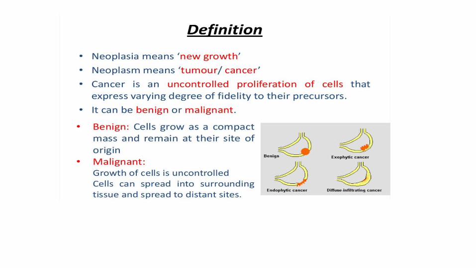
TUMOR MARKERS
Prakash Pokhrel
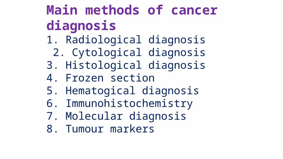
Main methods of cancer diagnosis1. Radiological diagnosis 2. Cytological diagnosis3. Histological diagnosis4. Frozen section5. Hematogical diagnosis6. Immunohistochemistry7. Molecular diagnosis8. Tumour markers
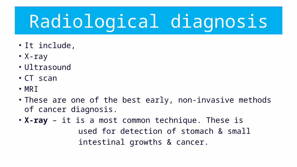
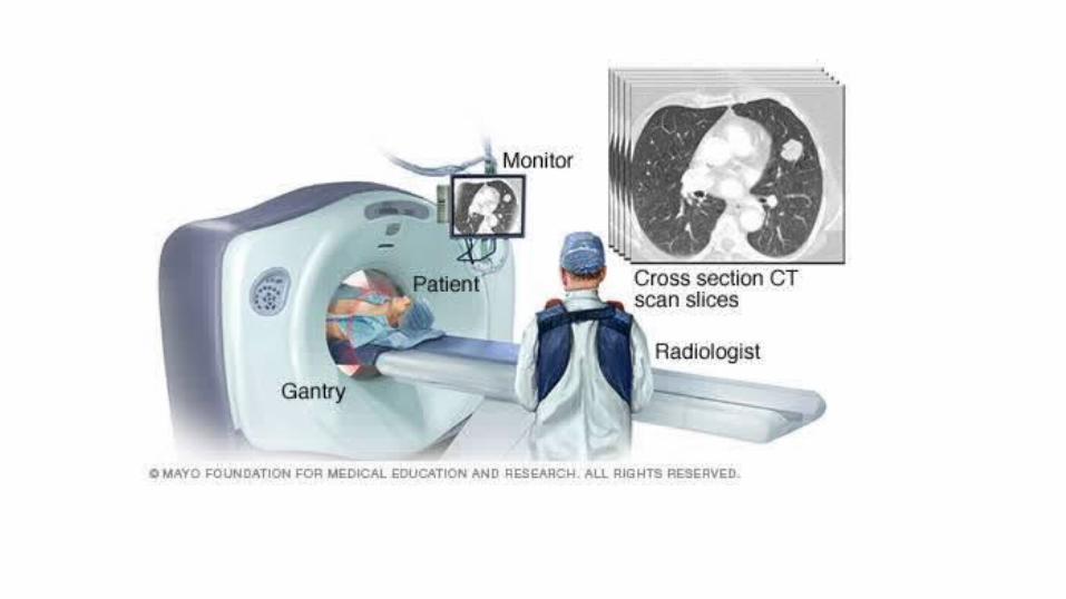
• It include,• X-ray• Ultrasound• CT scan• MRI• These are one of the best early, non-invasive methods of cancer diagnosis. • X-ray – it is a most common technique. These is used for detection of stomach & small intestinal growths & cancer.
Radiological diagnosis

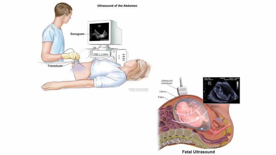
• Ultrasound- an exam in which the sound waves are bounced off tissue and echoes are Converted into picture• CT scan – (computerized tomography) It uses radiographic beams to create detail Computerized picture. It is more precise than a standard X-ray . • MRI- (Magnetic resonance imaging) It uses powerful magnetic field to create Detail computerized images of the body’s Soft tissue, large blood vessels & major organs.
2.CYTOLOGICAL DIAGNOSIS
• 1. Fine needle aspiration cytology (FNAC)• Fine needle aspiration cytology is a popular method
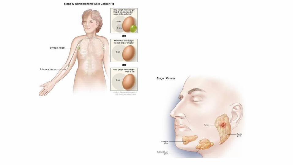
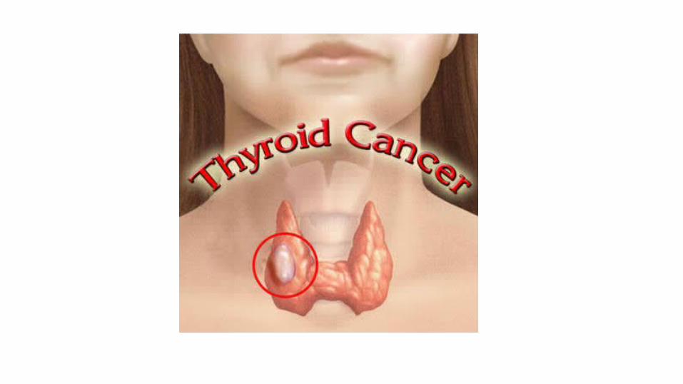
of tumor diagnosis particularly for palpable tumors• Lymph nodal tumors • Breast tumors• Salivary gland tumors• Thyroid tumors
• Procedure : the skin over the area is cleaned with antiseptic solution like sprit. Tumor is fixed by holding it. A 24 g needle is pushed inside the tumor and The material is sucked by a syringe is prepared from the. aspirated material. It is increased by putting the needle under CT
or Ultrasound guidance. The smear is stained
Giemsa Stain.
3.HISTOLOGICAL DIAGNOSIS
• For histological diagnosis the following methods of sampling is done:• Biopsy- biopsy is a surgical removal of small piece of tissue For
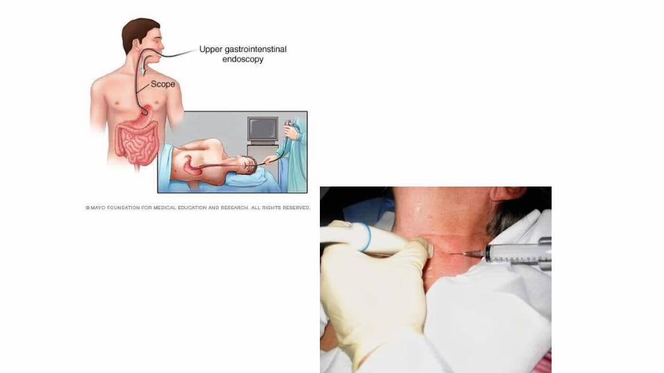
microscopic examination for the presence of cancer cell. There are three ways tissues can be removed for • Biopsy:- • Endoscopy• Needle biopsy• Surgical biopsy
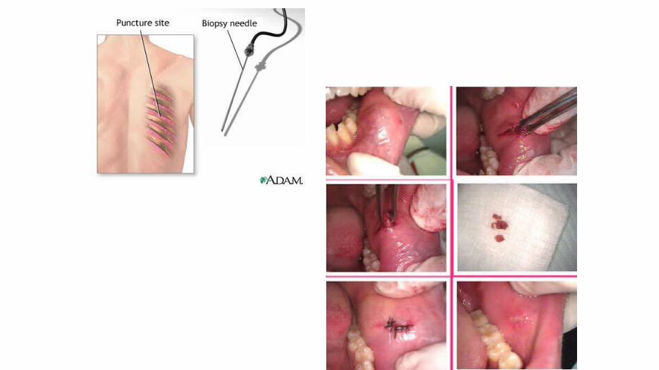
• Endoscopy- in this process , A thin, flexible tube with a tiny camera on the end is inserted into the body cavity. This allows the doctor to view the abnormal area.• Needle biopsy - the doctor takes a small tissue sample by Inserting
a needle into abnormal area. Different types of needles are used, EX:Vim Silverman needle for liver biopsy
• Renal biopsy needle for renal tissue• True cut biopsy needle for prostatic Tissue or breast tissue Surgical Biopsy:-• There are two types of surgical biopsies.• An excisional biopsy :it is performed when the doctor removes the
entire tumor, often with some surrounding normal tissue.
• An incisional biopsy: it is performed when the doctor removes just a portion of the tumor. If cancer is found to be present, the entire tumor may be removed immediately or during another operation.
The processing of tissue and its diagnosis takes a two or three days.
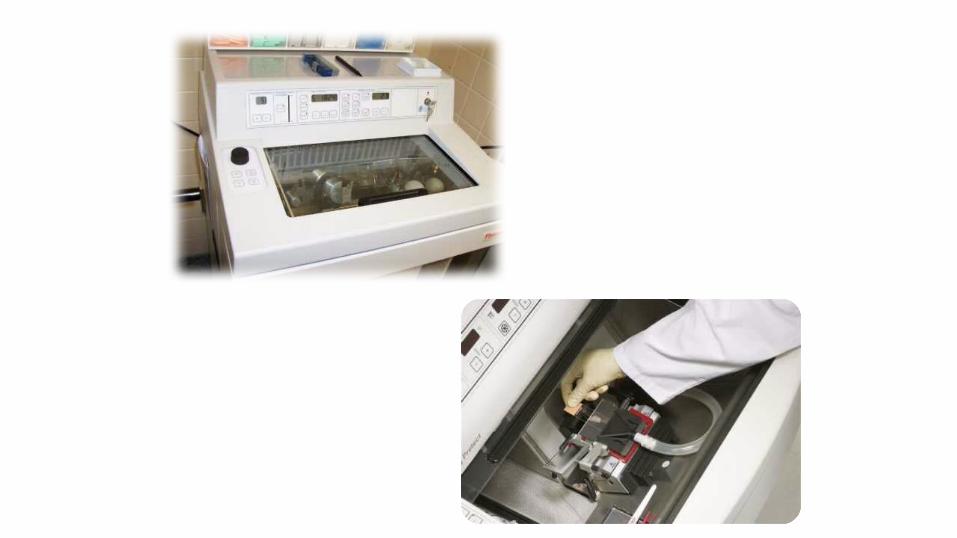
4. FROZEN SECTION• Frozen section is quick diagnosis method.
The tissue is quickly frozen at around -20’ c in frozen section
• cryostat which makes the tissue hard. -tissue is immediately sectioned & stained -the whole process from receiving, staining
to diagnosis can be completed within 10 to 15 mints.

5. HAEMATOLOGICAL DIAGNOSIS
• -Marrow is aspirated by bone marrow aspiration needle biopsied by trephine needle. It is useful in the diagnosis of Leukemia
• Metastasis from lymphoma or solid tumors. This is needed for staging
• Leukemia
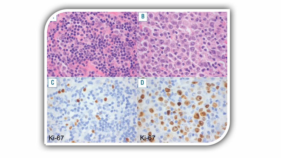
6.IMMUNOHISTOCHEMISTRY
Large number of monoclonal antibodies are available which are useful for:• typing of a malignant tumor. Poorly differentiated tumors are difficult to
morphologically type but if it shows positivity for cytokeratin antibody then it can
be typed as carcinoma. • T cell or B cell monoclonal antibody positivity in the T cell or B cell lymphoma.• classification of leukemia and lymphomas. Determination of site of primary in metastatic tumor.
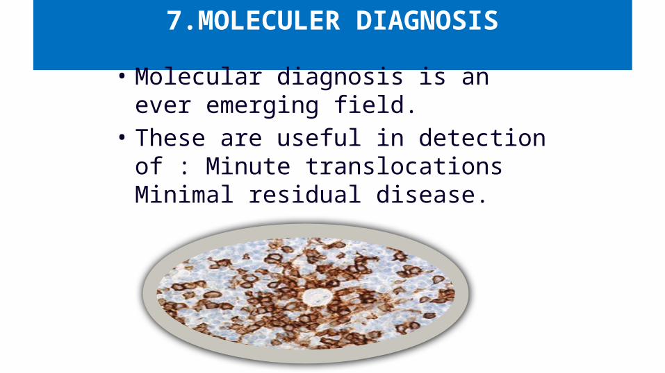
7.MOLECULER DIAGNOSIS
• Molecular diagnosis is an ever emerging field.
• These are useful in detection of : Minute translocations Minimal residual disease.
OUTLINE
Definition Clinical uses
Classification Examples
TUMOR MARKERS
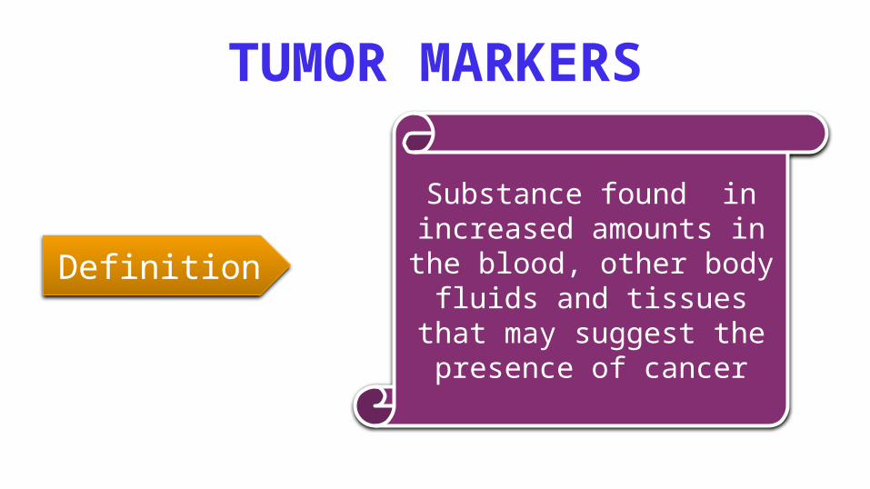
Substance found in increased amounts in the blood, other body fluids and tissues that
may suggest the presence of cancer
Definition
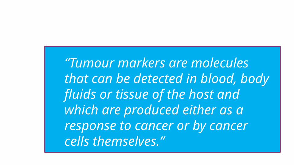
“Tumour markers are molecules that can be detected in blood, body fluids or tissue of the host and which are produced either as a response to cancer or by cancer cells themselves.”
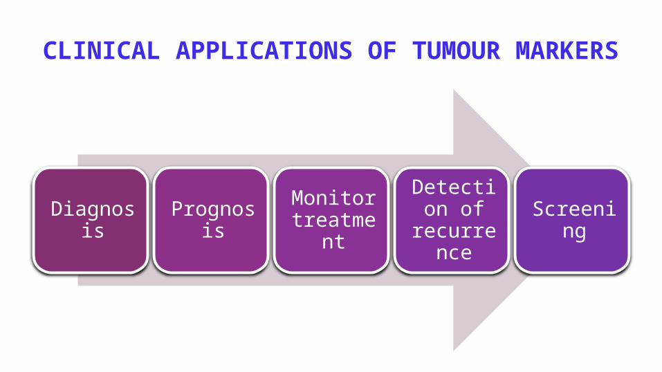
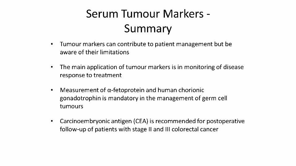
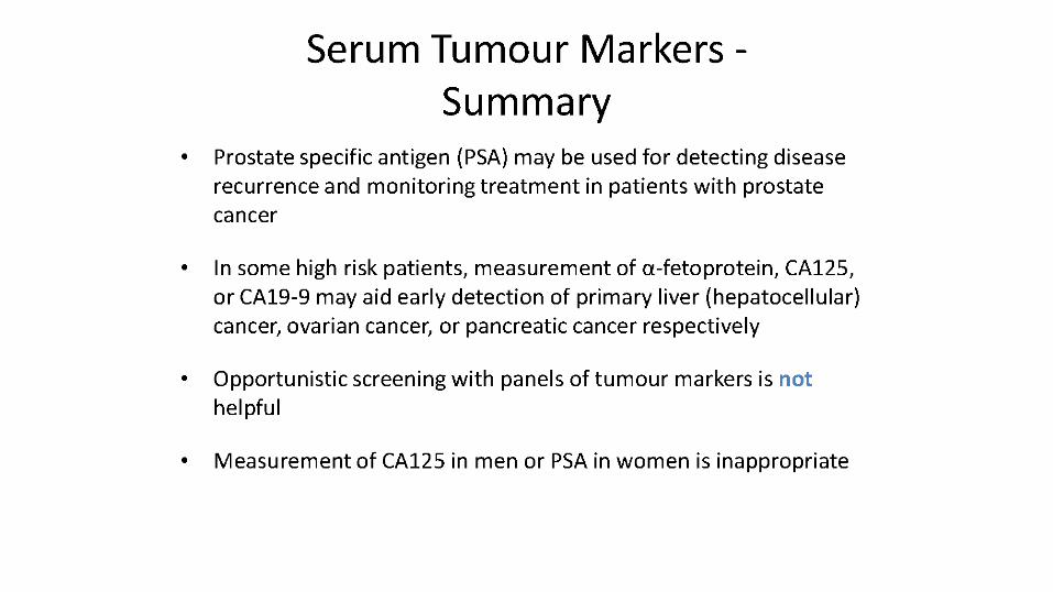
CLINICAL APPLICATIONS OF TUMOUR MARKERS
Diagnosis Prognosis Monitor treatment
Detection of
recurrence
Screening
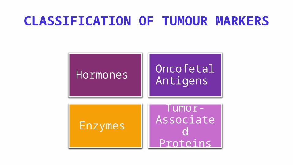
CLASSIFICATION OF TUMOUR MARKERS
Hormones Oncofetal Antigens
Enzymes Tumor-
Associated Proteins
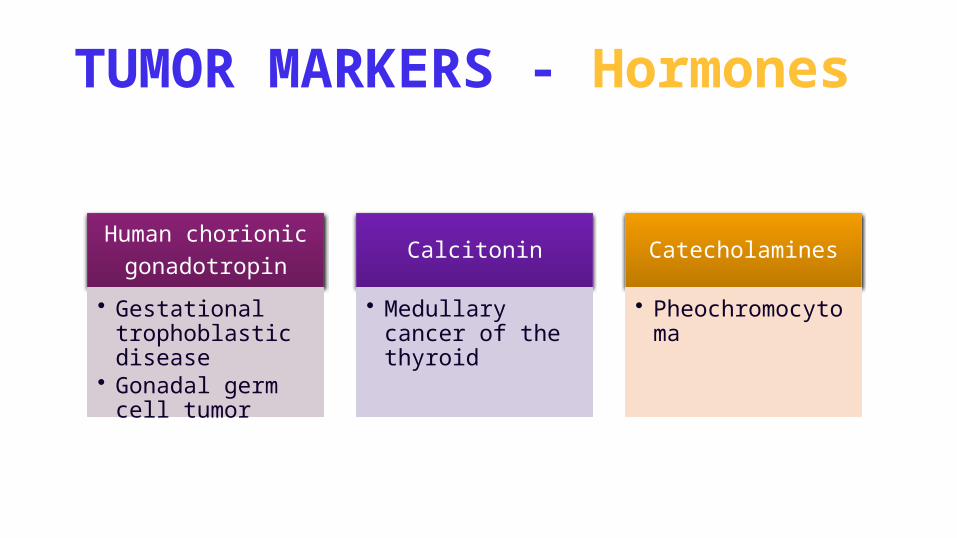
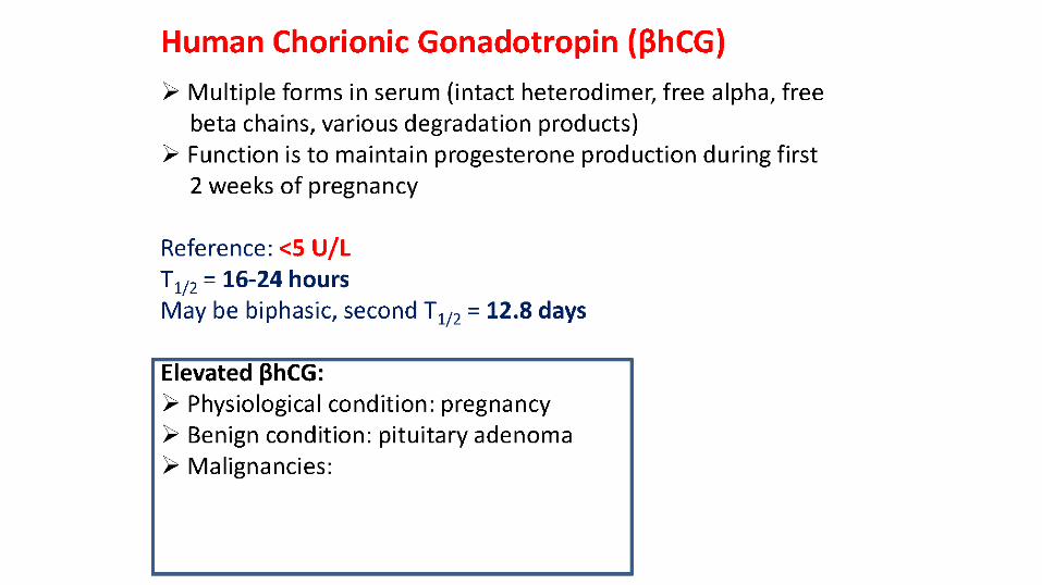
TUMOR MARKERS - Hormones
Human chorionicgonadotropin
• Gestational trophoblastic disease
• Gonadal germ cell tumor
Calcitonin
• Medullary cancer of the thyroid
Catecholamines
• Pheochromocytoma
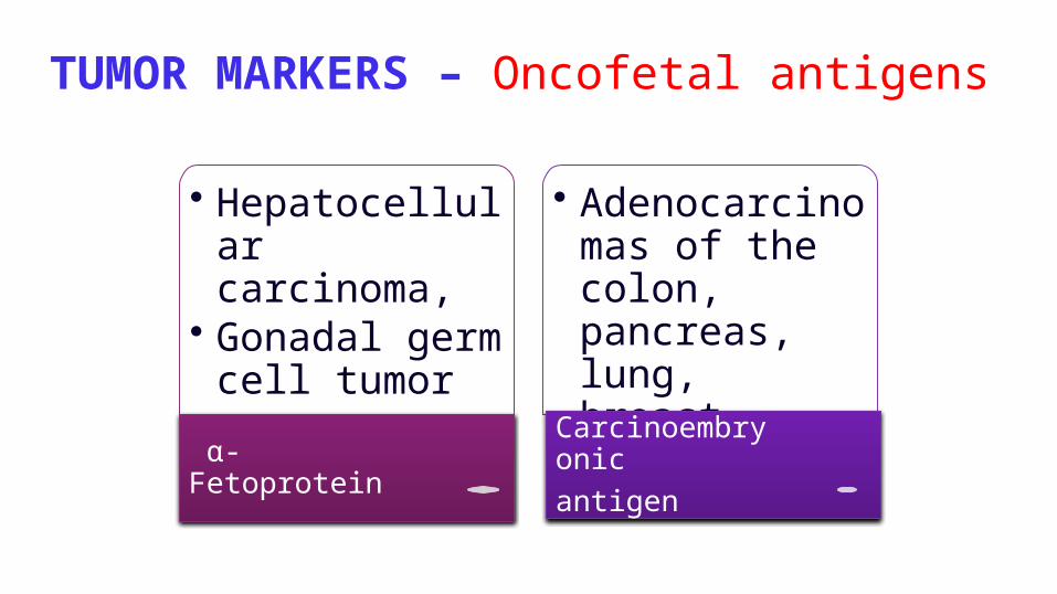
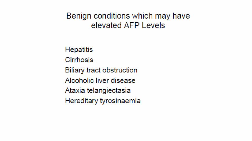
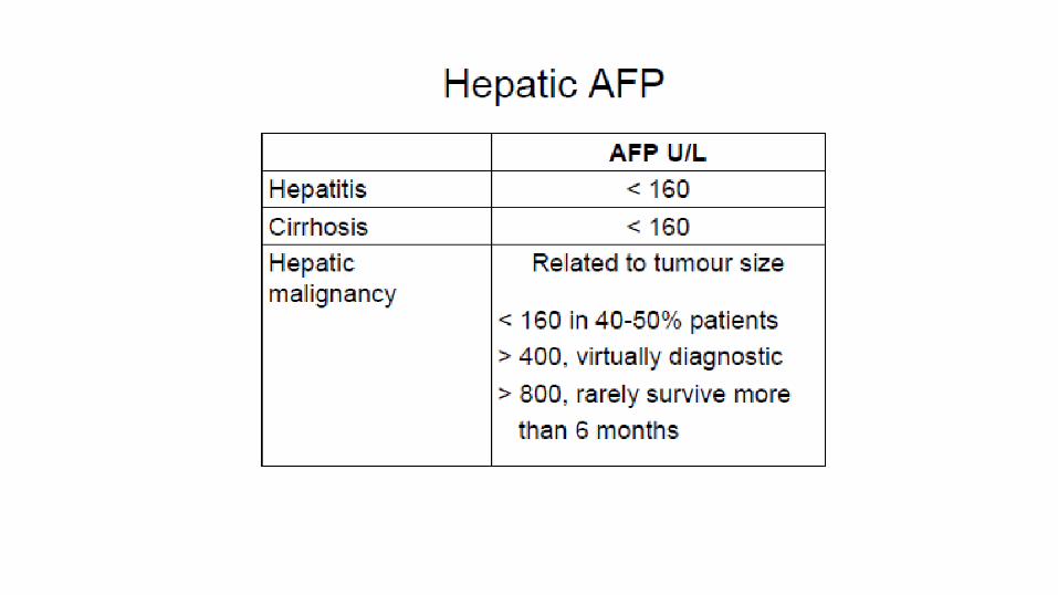
TUMOR MARKERS – Oncofetal antigens
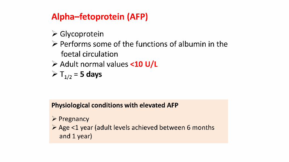
• Hepatocellular carcinoma,
• Gonadal germ cell tumor
α-Fetoprotein
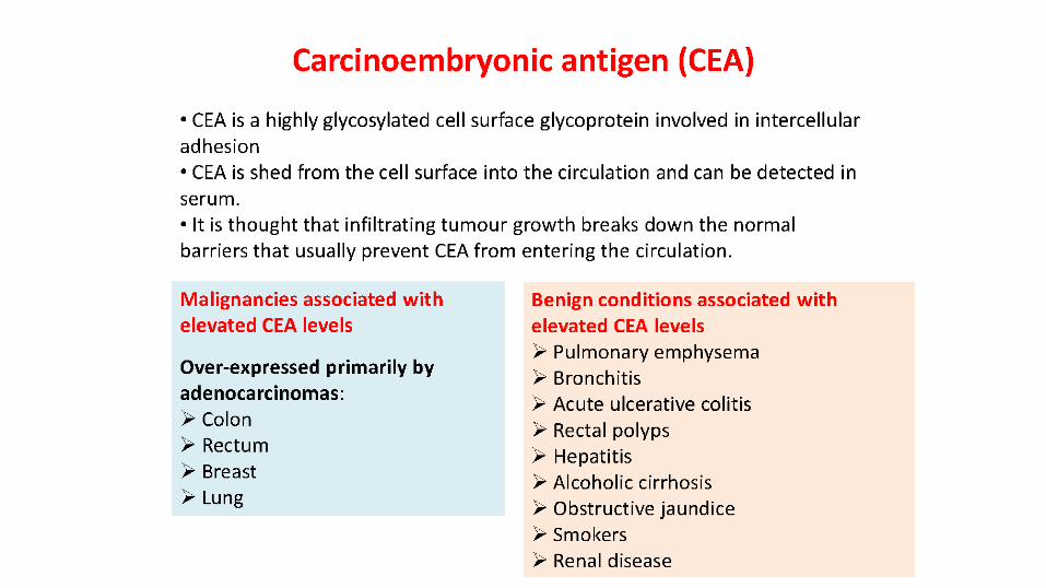
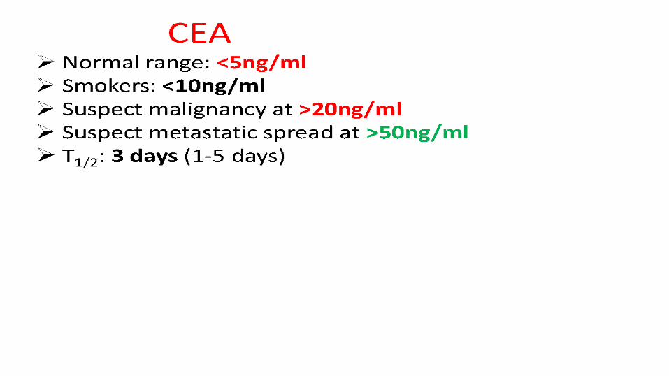
• Adenocarcinomas of the colon, pancreas, lung, breast, ovary
Carcinoembryonicantigen
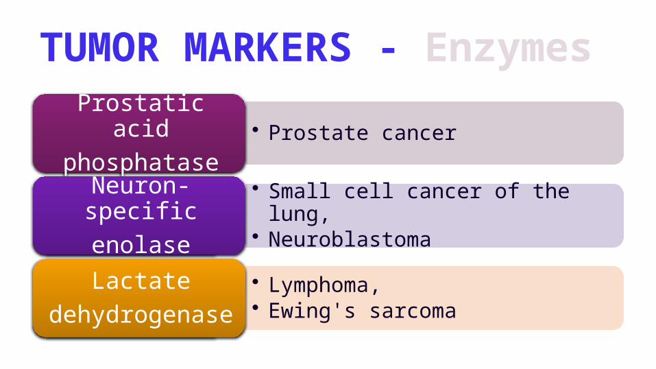
TUMOR MARKERS - Enzymes
• Prostate cancerProstatic acidphosphatase
• Small cell cancer of the lung,• Neuroblastoma
Neuron-specificenolase
• Lymphoma, • Ewing's sarcoma
Lactatedehydrogenase
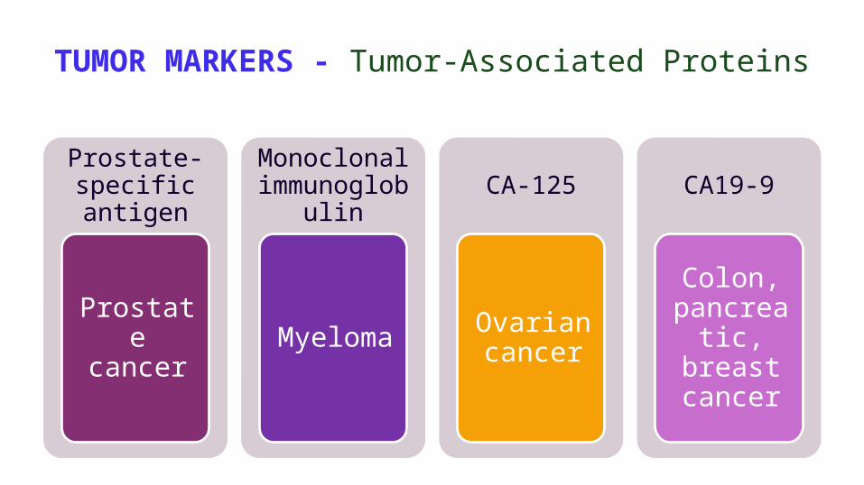
TUMOR MARKERS - Tumor-Associated Proteins
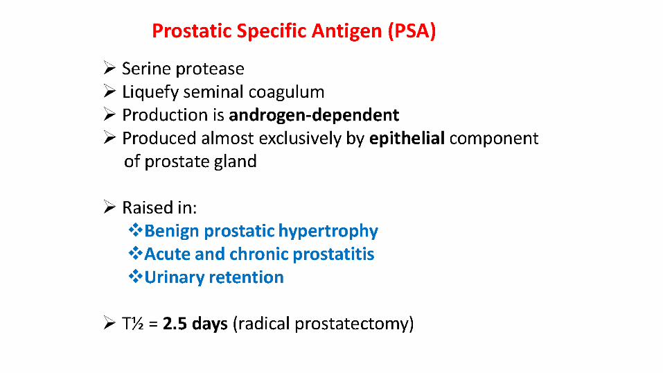
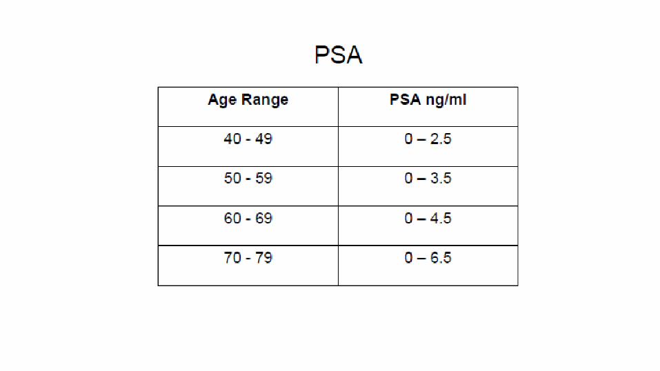
Prostate-specific antigen
Prostate cancer
Monoclonal immunoglobuli
n
Myeloma
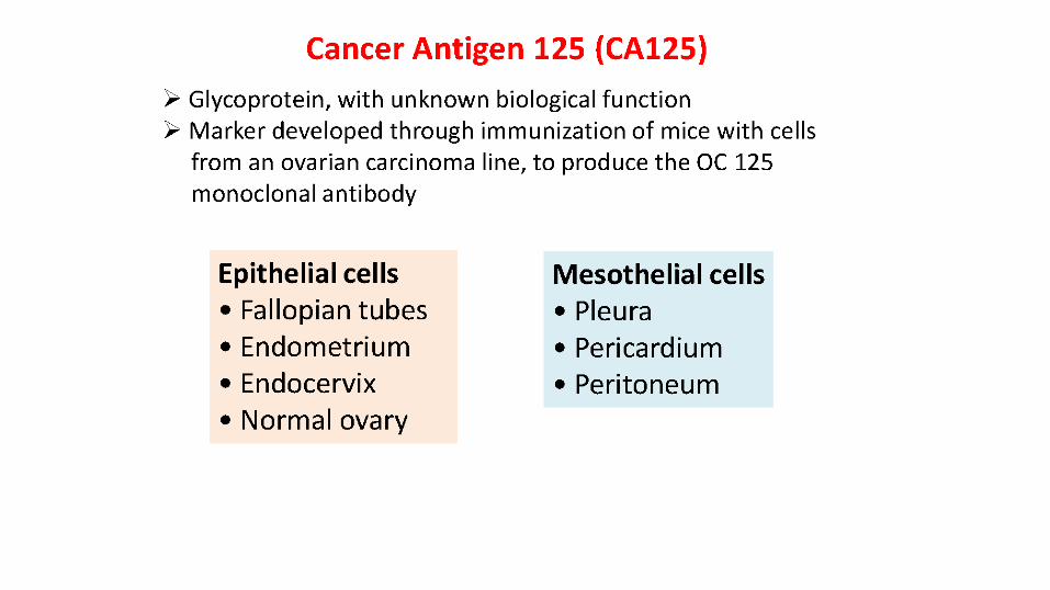
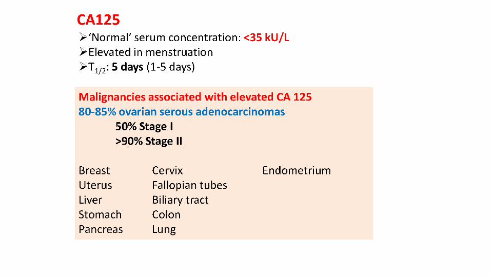
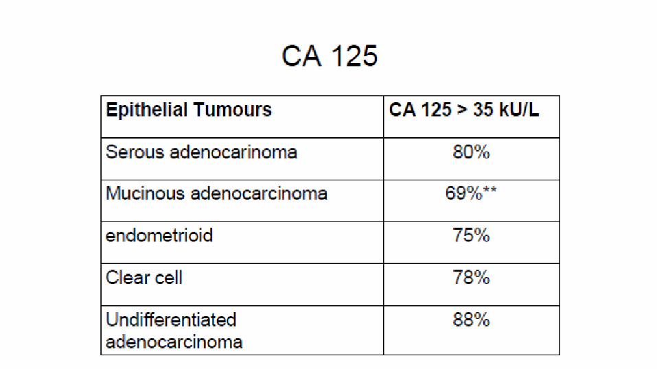
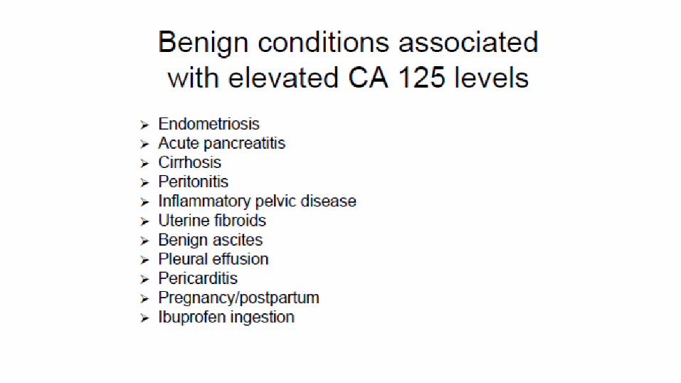
CA-125
Ovarian cancer
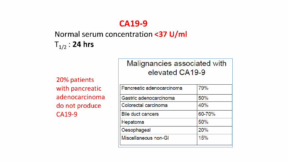
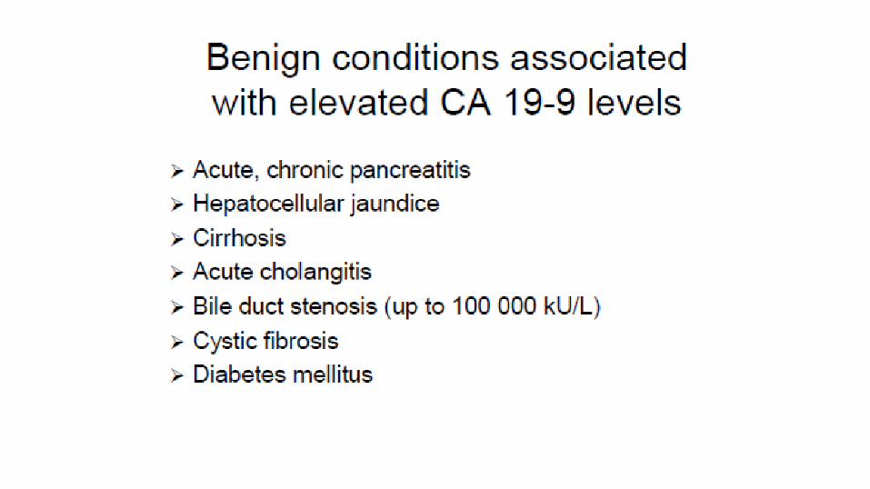
CA19-9
Colon, pancreatic
, breast cancer
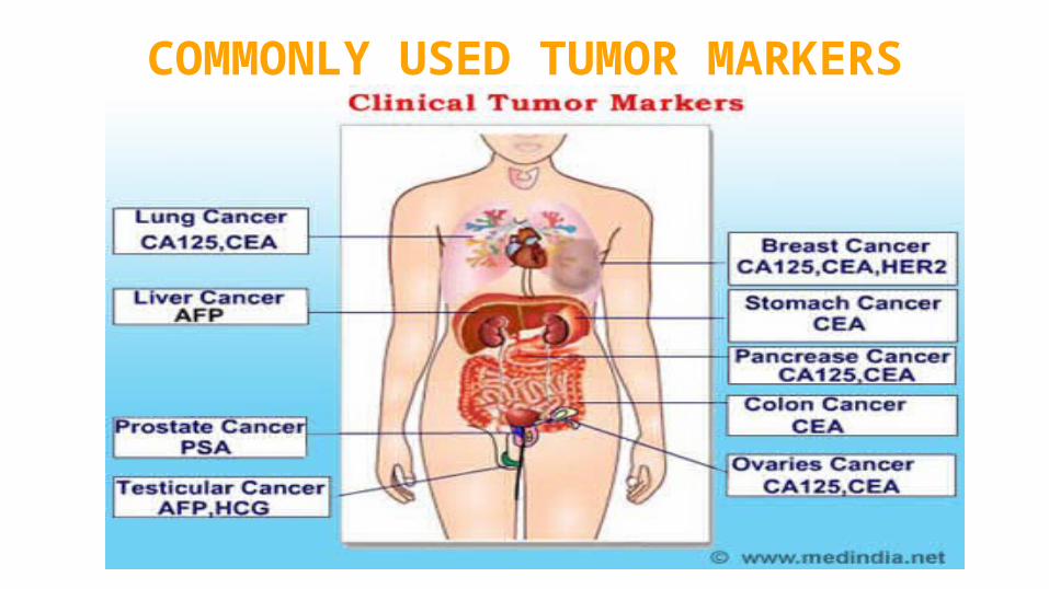
COMMONLY USED TUMOR MARKERS
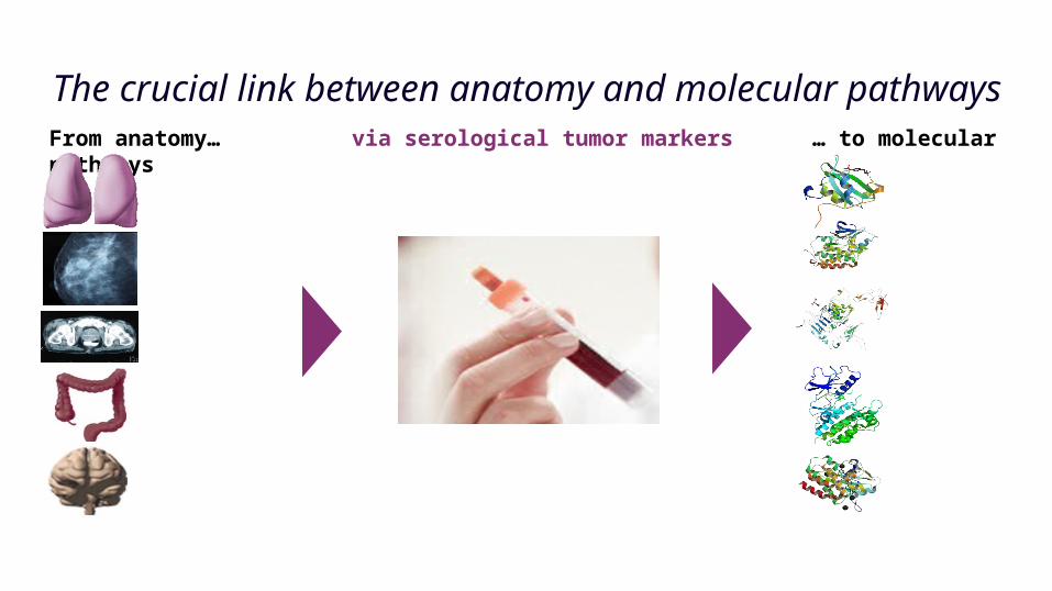
The crucial link between anatomy and molecular pathways
From anatomy… via serological tumor markers … to molecular pathways


![GI Tumor Markers[1]](https://img.pdfslide.tips/doc/110x75/55cf9d2d550346d033ac8c81/gi-tumor-markers1.jpg)