Embed Size (px)
Citation preview
Ernesto García Ureta
http://www.garciaureta.com/29th European Congress of Cytology Praga
A PROPOSITO DE UN CASO
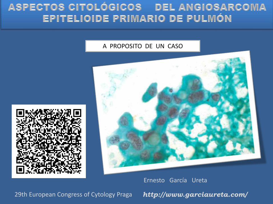
Varón 45 años
Esputos hemoptoicos
Disnea
Perdida de peso
Sospecha Clínica de Tuberculosis


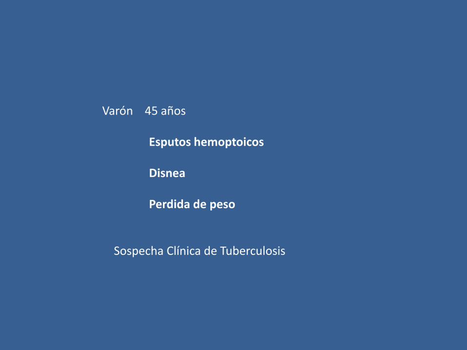
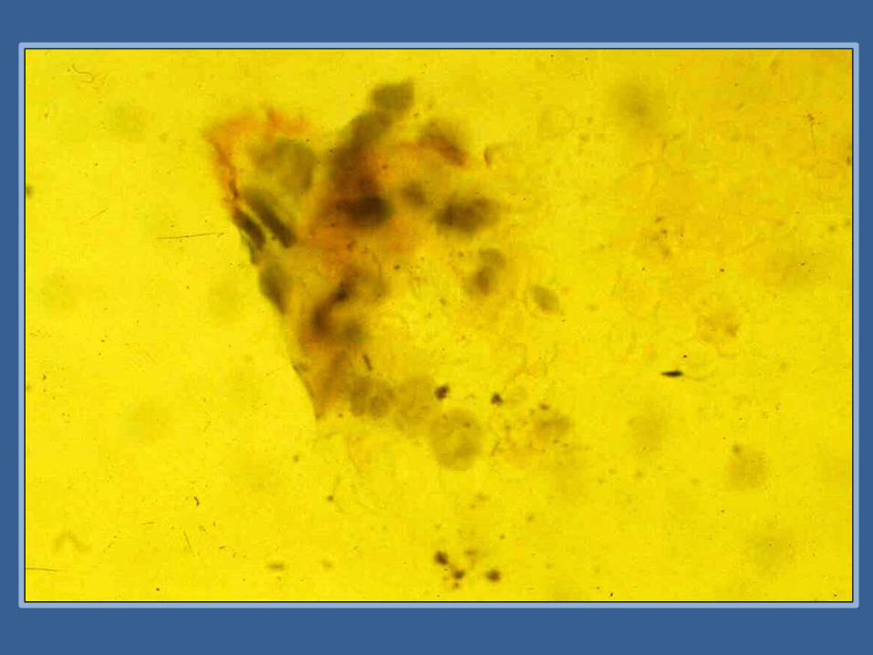
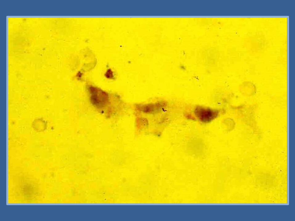
RECUENTO DIFERENCIAL PORCENTUAL
20%
2%
67%
11%
- 5%
432
10594
Polinucleares Neutrófilos
Eosinófilos
Histiocitos
Linfocitos
Células Ciliadas
Numero Células mmc
Numero Hematies mmc
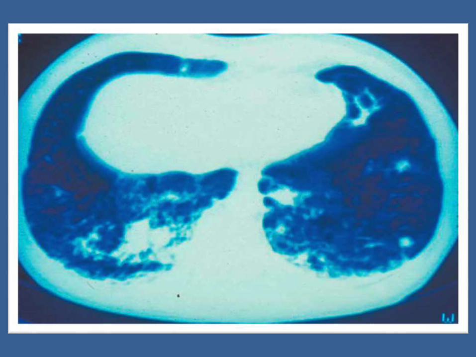
ENFERMEDAD ALVEOLO INTERSTICIAL
ENFERMEDAD METASTASICA
CARCINOMA BRONQUIOLOALVEOLAR
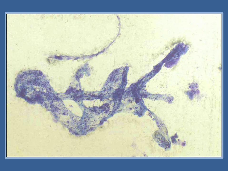
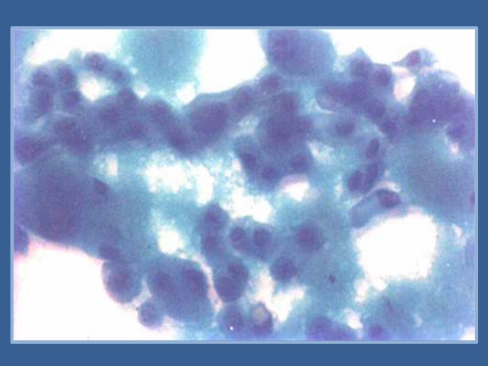

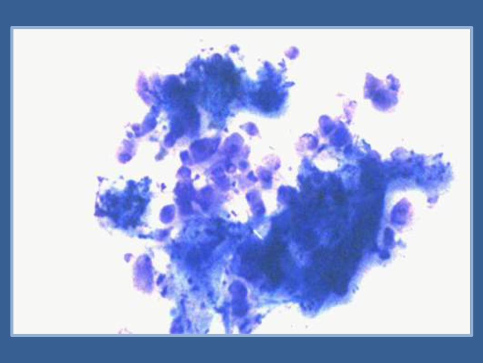
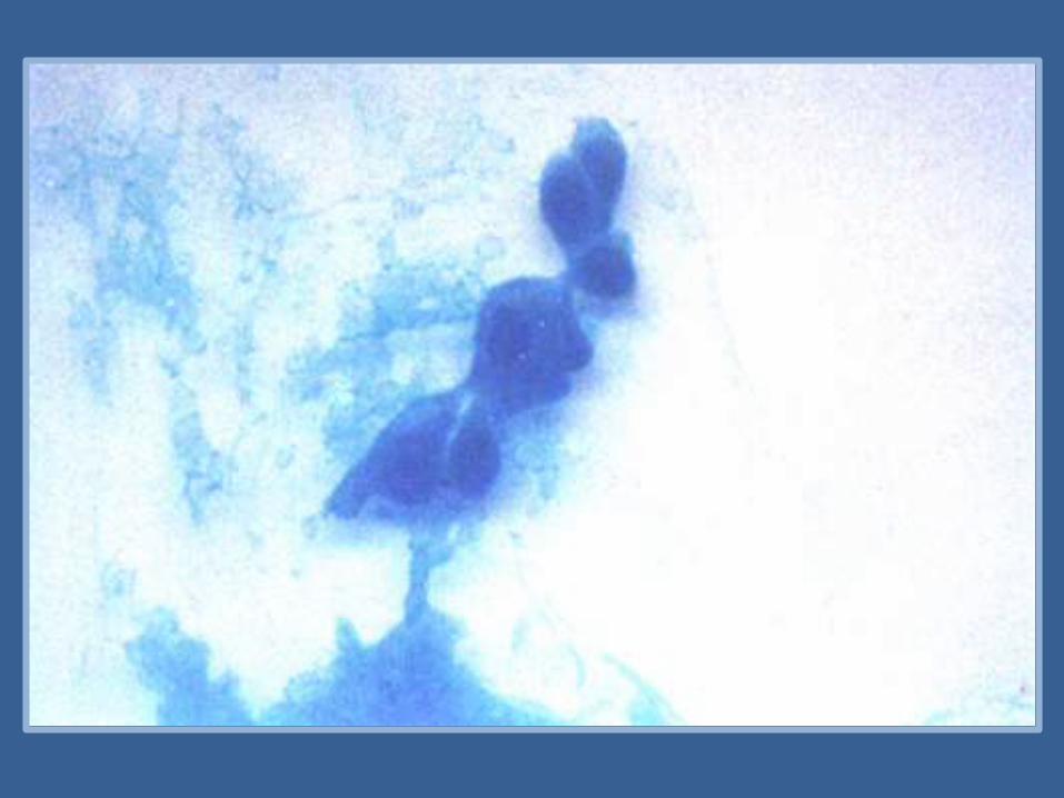
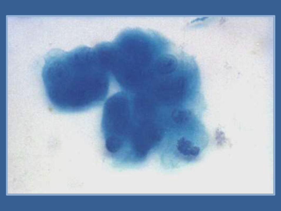
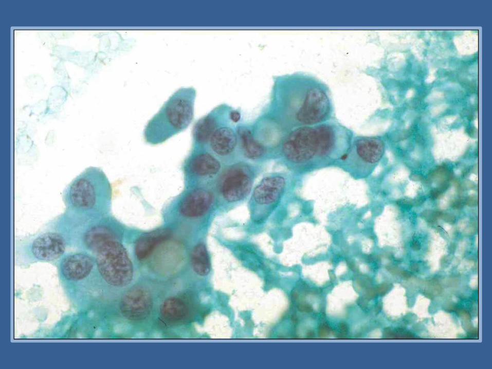

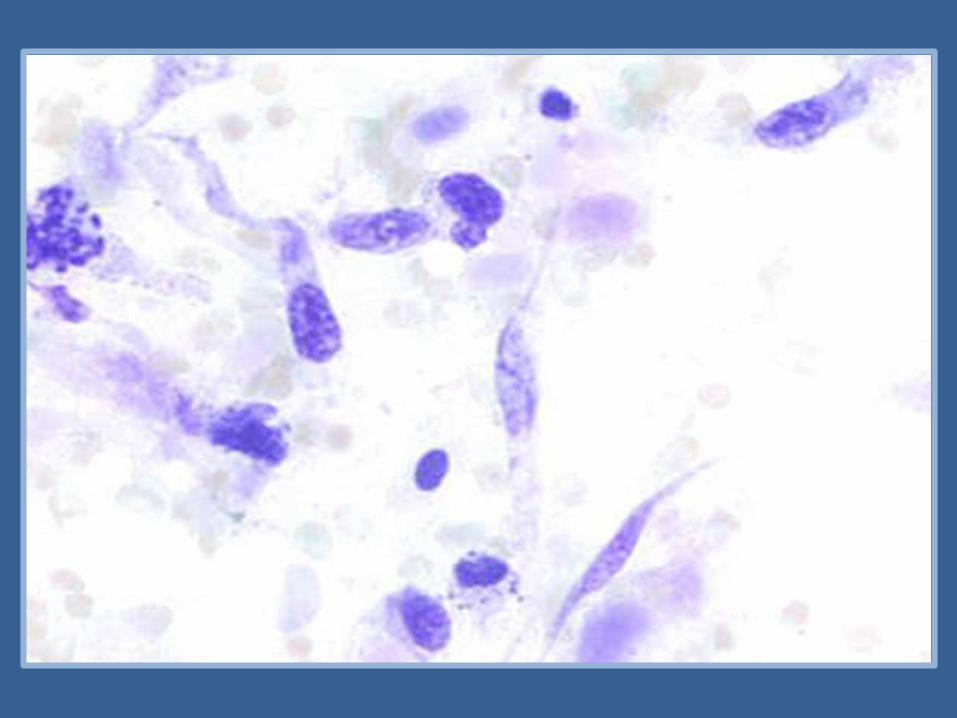
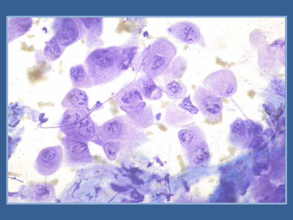
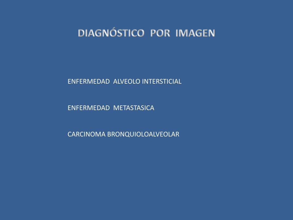
CARCINOMA ADENOIDE ESCAMOSOCON PATRON SEUDOVASCULAR
CARCINOMA ADENO ESCAMOSO
CARCINOMA BRONQUIOLOALVEOLAR
CARCINOIDE VARIANTE ONCOCITICA

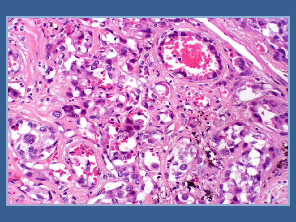

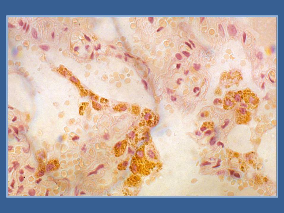

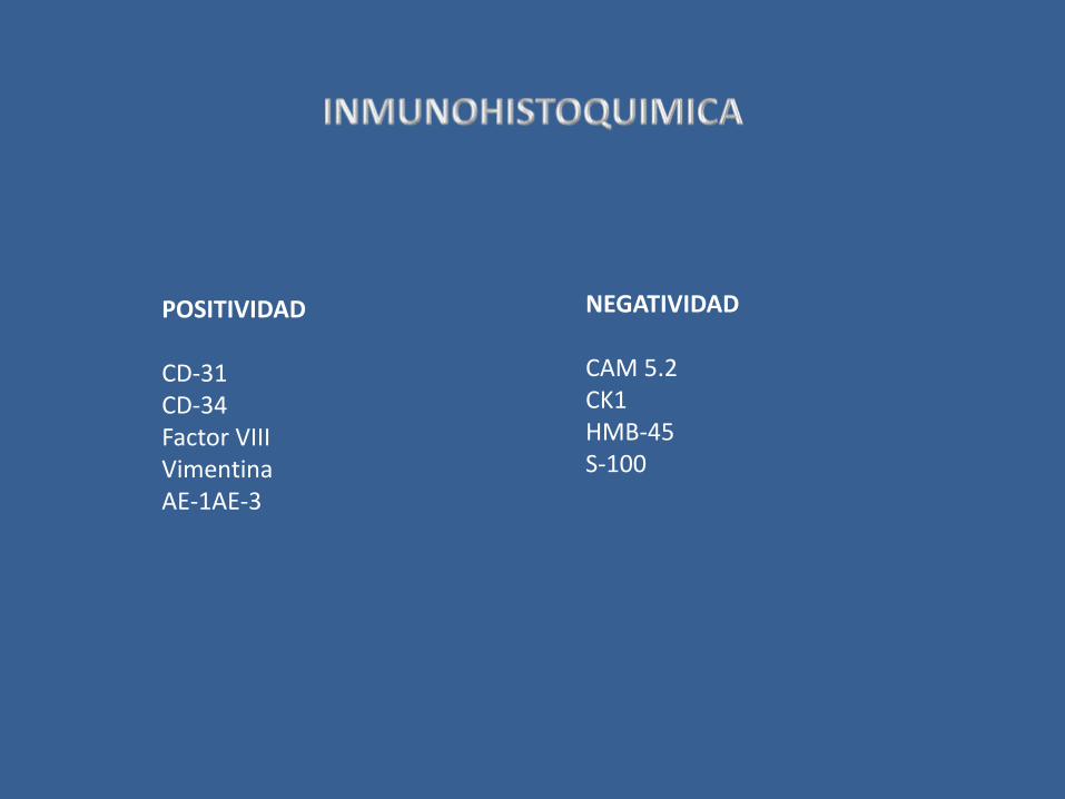
POSITIVIDAD
CD-31CD-34Factor VIIIVimentinaAE-1AE-3
NEGATIVIDAD
CAM 5.2CK1HMB-45S-100
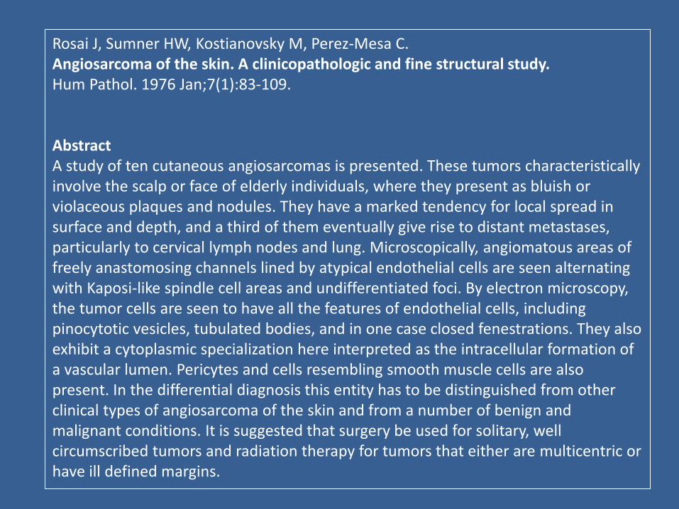
Rosai J, Sumner HW, Kostianovsky M, Perez-Mesa C.Angiosarcoma of the skin. A clinicopathologic and fine structural study.Hum Pathol. 1976 Jan;7(1):83-109.
AbstractA study of ten cutaneous angiosarcomas is presented. These tumors characteristically involve the scalp or face of elderly individuals, where they present as bluish or violaceous plaques and nodules. They have a marked tendency for local spread in surface and depth, and a third of them eventually give rise to distant metastases, particularly to cervical lymph nodes and lung. Microscopically, angiomatous areas of freely anastomosing channels lined by atypical endothelial cells are seen alternating with Kaposi-like spindle cell areas and undifferentiated foci. By electron microscopy, the tumor cells are seen to have all the features of endothelial cells, including pinocytotic vesicles, tubulated bodies, and in one case closed fenestrations. They also exhibit a cytoplasmic specialization here interpreted as the intracellular formation of a vascular lumen. Pericytes and cells resembling smooth muscle cells are also present. In the differential diagnosis this entity has to be distinguished from other clinical types of angiosarcoma of the skin and from a number of benign and malignant conditions. It is suggested that surgery be used for solitary, well circumscribed tumors and radiation therapy for tumors that either are multicentric or have ill defined margins.
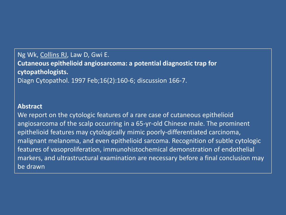
Ng Wk, Collins RJ, Law D, Gwi E.Cutaneous epithelioid angiosarcoma: a potential diagnostic trap forcytopathologists.Diagn Cytopathol. 1997 Feb;16(2):160-6; discussion 166-7.
AbstractWe report on the cytologic features of a rare case of cutaneous epithelioidangiosarcoma of the scalp occurring in a 65-yr-old Chinese male. The prominentepithelioid features may cytologically mimic poorly-differentiated carcinoma, malignant melanoma, and even epithelioid sarcoma. Recognition of subtle cytologicfeatures of vasoproliferation, immunohistochemical demonstration of endothelialmarkers, and ultrastructural examination are necessary before a final conclusion maybe drawn

Mullick SS, et alActa Cytol. 1997;41(3):839-44
Leslie DAm J Clin Pathol 2000;114:210-219
Jeon YK et alActa Cytol. 2004 ;48(2):223-8
Siddaraju N, et alActa Cytol. 2008 ;52(1):109-13.
Ryu HS et al.Diagn_Cytopathol. 2011 ;39(11):801-7

http://www.garciaureta.com/


![Hepatic angiosarcoma with an associated focal nodular ... · vascular channels [1,2]. Focal nodular hyperplasia (FNH), on the other hand, is a benign hepatic lesion displaying hepatocytic](https://img.pdfslide.tips/doc/110x75/5f05ab797e708231d4141d25/hepatic-angiosarcoma-with-an-associated-focal-nodular-vascular-channels-12.jpg)