Embed Size (px)
Citation preview

ACTUALIDAD Y AVANCES
ARTICULOS

Imaging of
salivary gland
tumours
Department of Radiology,
Neuroradiology and Nuclear Medicine, University Hospital
of Bern
Autor: Harriet C. Thoeny
Se presenta una breve reseña sobre la
anatomía de las glándulas salivales y
epidemiologia de los tumores de las mismas
Presentación de casos clínicos y
diferenciación de tumores
ARTÍCULO DEREFERENCIA
http://www.ncbi.nlm.nih.gov/pmc/articles/PMC1866314/
Y también de las diferentes modalidades de imagen que se emplean para realizar diagnósticos y la importancia de estos.

Objetivo: Caracterizar las lesiones hepáticas focales detectadas y evaluadas con tomografía computada multidetector.
Caracterización de lesiones hepáticas focales con tomografía computada multidetector
Material y método: Se realizó un estudio retrospectivo, descriptivo y doble ciego donde se revisaron 64 estudios de tomografíacomputada multidetector (TCMD) de abdomen realizados en el periodo de febrero 2010 a febrero de 2011.
Resultados: Se analizaron 27 (58%) hombres y 37 (42%) mujeres con edad promedio de 51.2años. La lesión hipervascular benigna más frecuente fue el hemangioma, demostrado en 13pacientes (20%) y la lesión maligna más frecuente fue el hepatocarcinoma en 12 pacientes(18.7%). La lesión hipovascular benigna más frecuente fue el quiste hepático simple ( 6pacientes, 9.3%) y la maligna fue la lesión metastásica ( 6 pacientes 9.3%). La lesiónseudotumoral más frecuente fue el absceso hepático (11 pacientes, 17.1%).
http://www.medigraphic.com/pdfs/anaradmex/arm-2012/arm121g.pdf
imágenes Articulo.
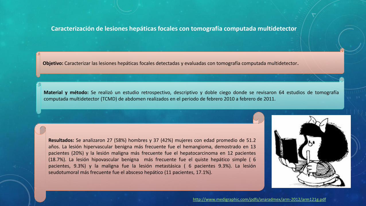
lesiones Focales hepáticas mediante tomografía computada multidector
LESION FOCAL HEPATICA Hipovascular e hipodensa en todo el estudio dinámico condensidad de líquido en relación con un quiste simple delHígado
Lesión focal menor a 1.0 cm.
Lesión subcapsular isodensa alparénquima con signo delhematocrito

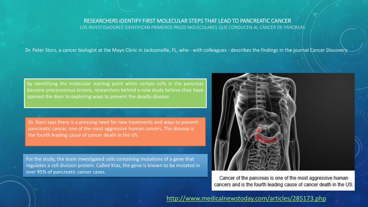
RESEARCHERS IDENTIFY FIRST MOLECULAR STEPS THAT LEAD TO PANCREATIC CANCERLOS INVESTIGADORES IDENTIFICAN PRIMEROS PASOS MOLECULARES QUE CONDUCEN AL CÁNCER DE PÁNCREAS
Dr. Peter Storz, a cancer biologist at the Mayo Clinic in Jacksonville, FL, who - with colleagues - describes the findings in the journal Cancer Discovery
by identifying the molecular starting point when certain cells in the pancreasbecome precancerous lesions, researchers behind a new study believe they haveopened the door to exploring ways to prevent the deadly disease
Dr. Storz says there is a pressing need for new treatments and ways to prevent pancreatic cancer, one of the most aggressive human cancers. The disease is the fourth leading cause of cancer death in the US.
For the study, the team investigated cells containing mutations of a gene that regulates a cell division protein. Called Kras, the gene is known to be mutated in over 95% of pancreatic cancer cases.
http://www.medicalnewstoday.com/articles/285173.php

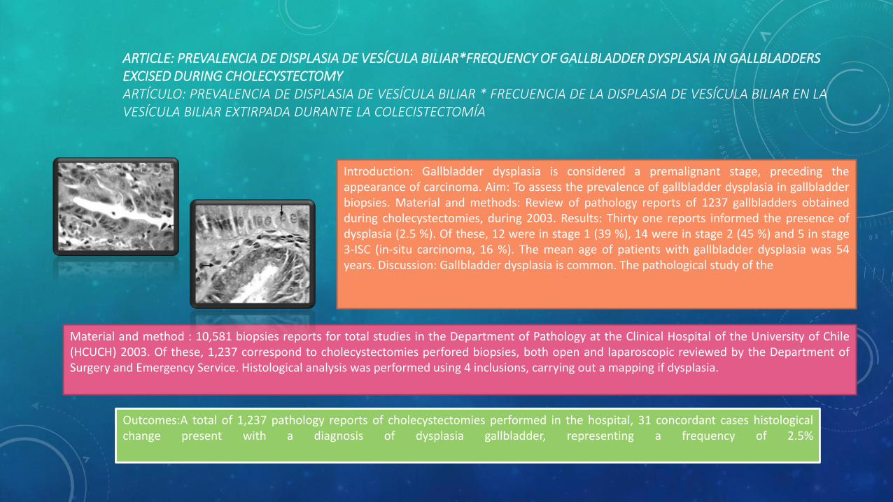
ARTICLE: PREVALENCIA DE DISPLASIA DE VESÍCULA BILIAR*FREQUENCY OF GALLBLADDER DYSPLASIA IN GALLBLADDERS EXCISED DURING CHOLECYSTECTOMYARTÍCULO: PREVALENCIA DE DISPLASIA DE VESÍCULA BILIAR * FRECUENCIA DE LA DISPLASIA DE VESÍCULA BILIAR EN LA VESÍCULA BILIAR EXTIRPADA DURANTE LA COLECISTECTOMÍA
Introduction: Gallbladder dysplasia is considered a premalignant stage, preceding theappearance of carcinoma. Aim: To assess the prevalence of gallbladder dysplasia in gallbladderbiopsies. Material and methods: Review of pathology reports of 1237 gallbladders obtainedduring cholecystectomies, during 2003. Results: Thirty one reports informed the presence ofdysplasia (2.5 %). Of these, 12 were in stage 1 (39 %), 14 were in stage 2 (45 %) and 5 in stage3-ISC (in-situ carcinoma, 16 %). The mean age of patients with gallbladder dysplasia was 54years. Discussion: Gallbladder dysplasia is common. The pathological study of the
Material and method : 10,581 biopsies reports for total studies in the Department of Pathology at the Clinical Hospital of the University of Chile(HCUCH) 2003. Of these, 1,237 correspond to cholecystectomies perfored biopsies, both open and laparoscopic reviewed by the Department ofSurgery and Emergency Service. Histological analysis was performed using 4 inclusions, carrying out a mapping if dysplasia.
Outcomes:A total of 1,237 pathology reports of cholecystectomies performed in the hospital, 31 concordant cases histologicalchange present with a diagnosis of dysplasia gallbladder, representing a frequency of 2.5%