Embed Size (px)
Citation preview
Developmental Dysplasia of the Hip
Dr.NAVEEN RATHOR(RESIDENT DOCTOR)
DEPT. OF ORTHOPEDICSRNT MEDICAL COLLEGE
UDAIPUR
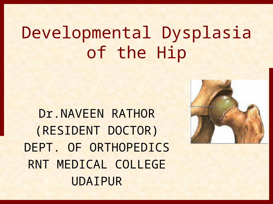
• Definition• Dysplasia of the hip that develop
during fetal life or in infancy.• It ranges from dysplasia of the
acetabulum (shallow acetabulum) to subluxation of the joint to complete dislocation.
• The old name was ‘‘congenital dysplasia of the hip (CDH).’’ The name has changed to indicate that not all cases are present at birth and that some cases can develop later on during infancy and childhood
Developmental Dysplasia of the Hip
1. Complete hip dislocation.2. Partial hip subluxation.3. Hip dysplasia (incomplete
development).4.Dislocatable hip
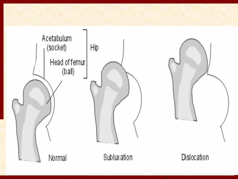
Dysplasia: radiographic finding of increased obliquity and loss of concavity of the acetabulum, with an intact Shenton's line(deficient development of acetabulum)
Subluxation: femoral head is in partial contact with the acetabulum
Dislocation:femoral head is not in contact with the acetabulum
Incidence• Most newborn screening studies suggest
that some degree of hip instability can be detected in 1/100 to 1/250 babies, actual dislocated or dislocatable hips are much less common, being found in 1-1.5 of 1000 live births.
• There is marked geographic and racial variation in the incidence of DDH.
• More inidence of DDH IN Sweden,Yugoslavia and Canada.
Etiology• A positive family history for DDH is found in 12-
33% of affected patients. • DDH is more common among female patients
(80%). This is thought to be due to the greater susceptibility of female fetuses to maternal hormones such as relaxin, which increases ligamentous laxity
• Primigravida.• Breech presentation(2-3%).• Oligohydramnios ,primi gravida and large baby
( crowding phenomenon ).• Adduction and Extension postnatally.
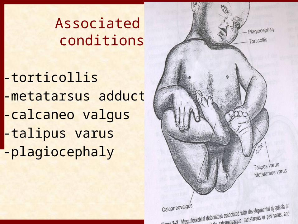
Associated conditions
-torticollis-metatarsus adducts-calcaneo valgus-talipus varus-plagiocephaly
• The left hip is the most commonly affected hip
• In the most common fetal position, this is the hip that is usually forced into adduction against the mother’s sacrum.
• Girls are affected 5 times more than boys.
• Types:• DDH is classified into two major groups :• Typical and teratologic . • Typical DDH occurs in otherwise normal
patients or those without defined syndromes or genetic conditions.
• Teratologic hip dislocations usually have identifiable causes such as arthrogyposis or a genetic syndrome and occur before birth.
Teratological DDH
Irreducible False acetabulum Defective anterior acetabulum
“anteverted” Increased femoral neck
anteversion
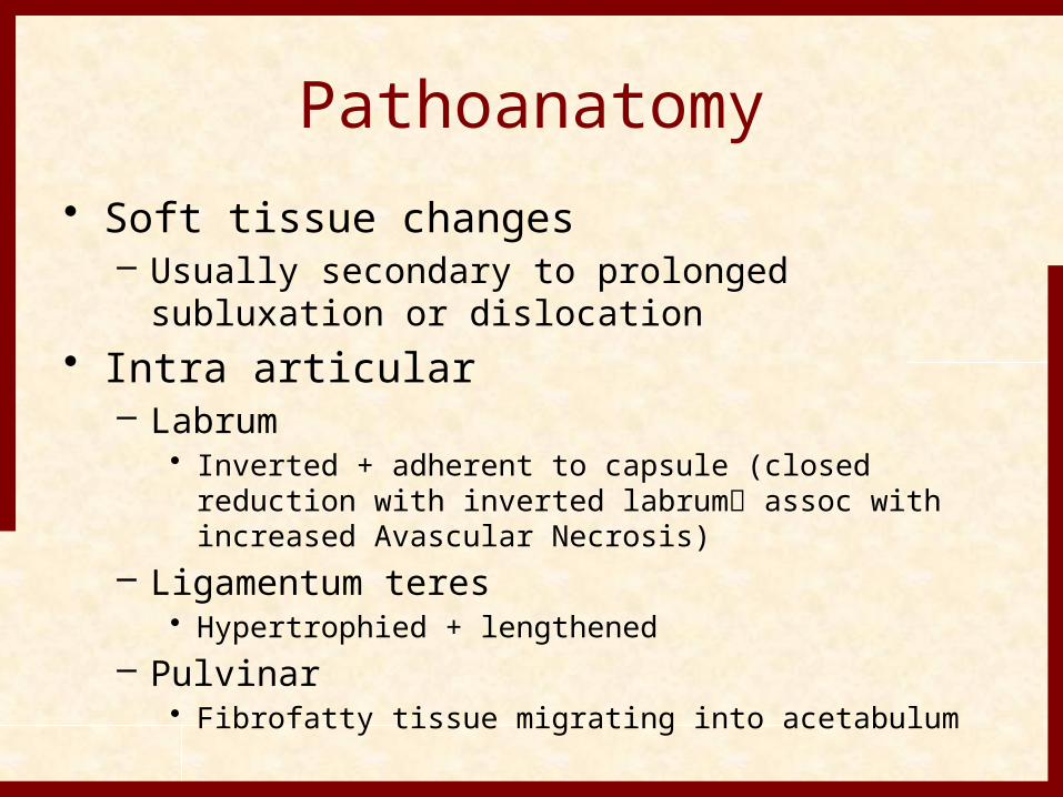
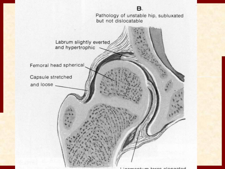
Pathoanatomy• Soft tissue changes
– Usually secondary to prolonged subluxation or dislocation
• Intra articular– Labrum
• Inverted + adherent to capsule (closed reduction with inverted labrum assoc with increased Avascular Necrosis)
– Ligamentum teres• Hypertrophied + lengthened
– Pulvinar• Fibrofatty tissue migrating into acetabulum
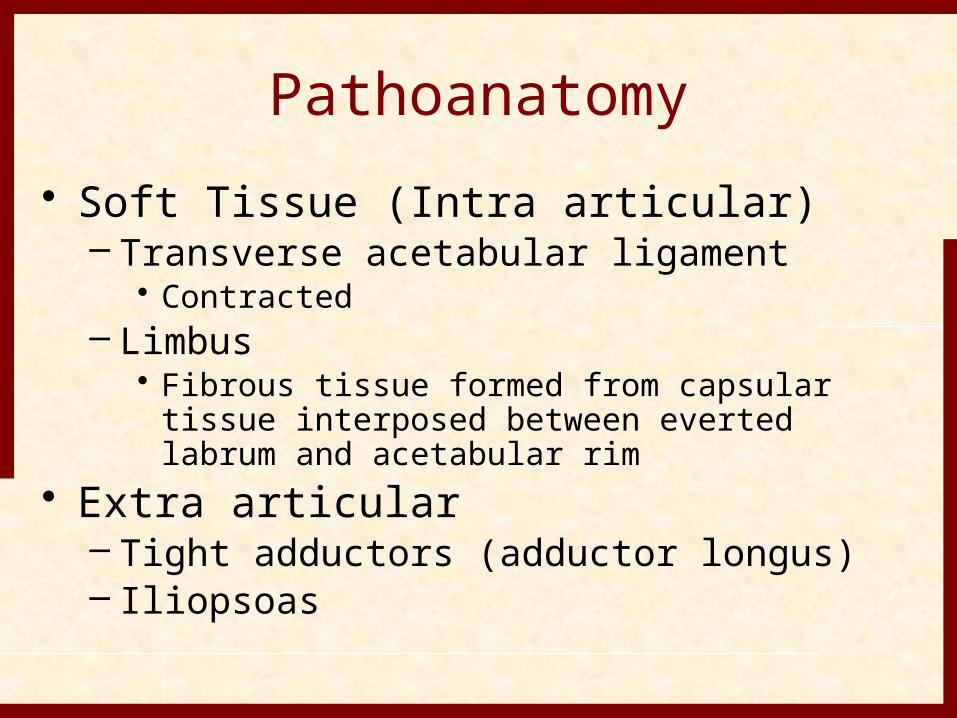
Pathoanatomy• Soft Tissue (Intra articular)
– Transverse acetabular ligament• Contracted
– Limbus• Fibrous tissue formed from capsular tissue
interposed between everted labrum and acetabular rim
• Extra articular– Tight adductors (adductor longus)– Iliopsoas
CLINACAL PRESENTATION
Neonatal PresentationExam one hip at a timeBaby must be quietBarlow’s sign: provocative maneuverOrtolani’s sign: reduces hipOther signs not helpful in newborn
CLINICAL FINDINGS• IN NEWBORNS• Usually asymptomatic and must be
screened by special maneuvers• 1) Barlow test.It is a provocative test that attempts to
dislocate an unstable hip.- Flexion ,adduction, posteriorly.- “Clunk”

The Barlow test for developmental dislocation of the hip in a neonate.A, With the infant supine, the examiner holds both of the child's knees and gently adducts one hip and pushes posteriorly.B, When the examination is positive, the examiner will feel the femoral head make a small jump (arrow) out of the acetabulum (Barlow's
sign). When the pressure is released, the head is felt to slip back into place.
• 2) Ortolani test It is a maneuver to reduce a recently
dislocated hip.• Flexion, abduction, anteriorly. • We can`t use X-rays because the
acetabulum and proximal femur are cartilaginous and wont be shown on X-ray.
• US is the best method to Dx.
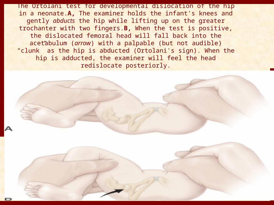
The Ortolani test for developmental dislocation of the hip in a neonate.A, The examiner holds the infant's knees and gently abducts the hip while lifting up on the greater
trochanter with two fingers.B, When the test is positive, the dislocated femoral head will fall back into the acetabulum
(arrow) with a palpable (but not audible) “clunk” as the hip is abducted (Ortolani's sign). When the hip is adducted, the
examiner will feel the head redislocate posteriorly.
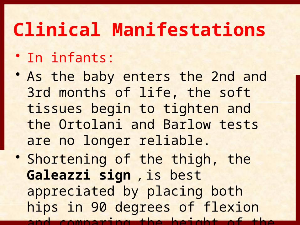
Clinical Manifestations• In infants:• As the baby enters the 2nd and 3rd
months of life, the soft tissues begin to tighten and the Ortolani and Barlow tests are no longer reliable.
• Shortening of the thigh, the Galeazzi sign , is best appreciated by placing both hips in 90 degrees of flexion and comparing the height of the knees, looking for asymmetry
• Asymmetry of thigh and gluteal skin folds.
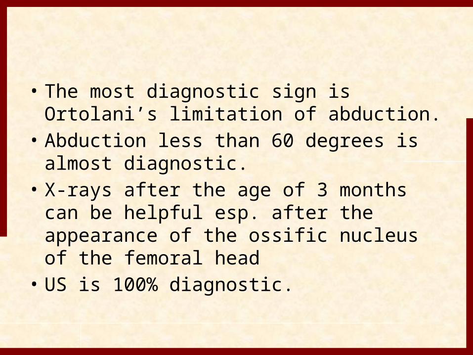
• The most diagnostic sign is Ortolani’s limitation of abduction.
• Abduction less than 60 degrees is almost diagnostic.
• X-rays after the age of 3 months can be helpful esp. after the appearance of the ossific nucleus of the femoral head
• US is 100% diagnostic.
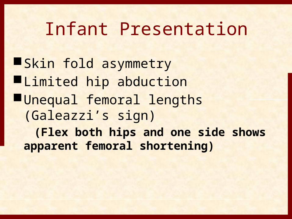
Infant PresentationSkin fold asymmetryLimited hip abductionUnequal femoral lengths (Galeazzi’s
sign) (Flex both hips and one side shows
apparent femoral shortening)
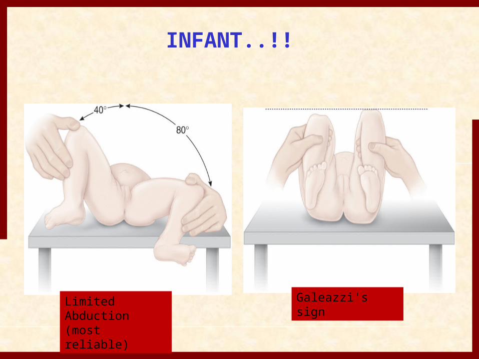
INFANT..!!
Limited Abduction(most reliable)
Galeazzi's sign
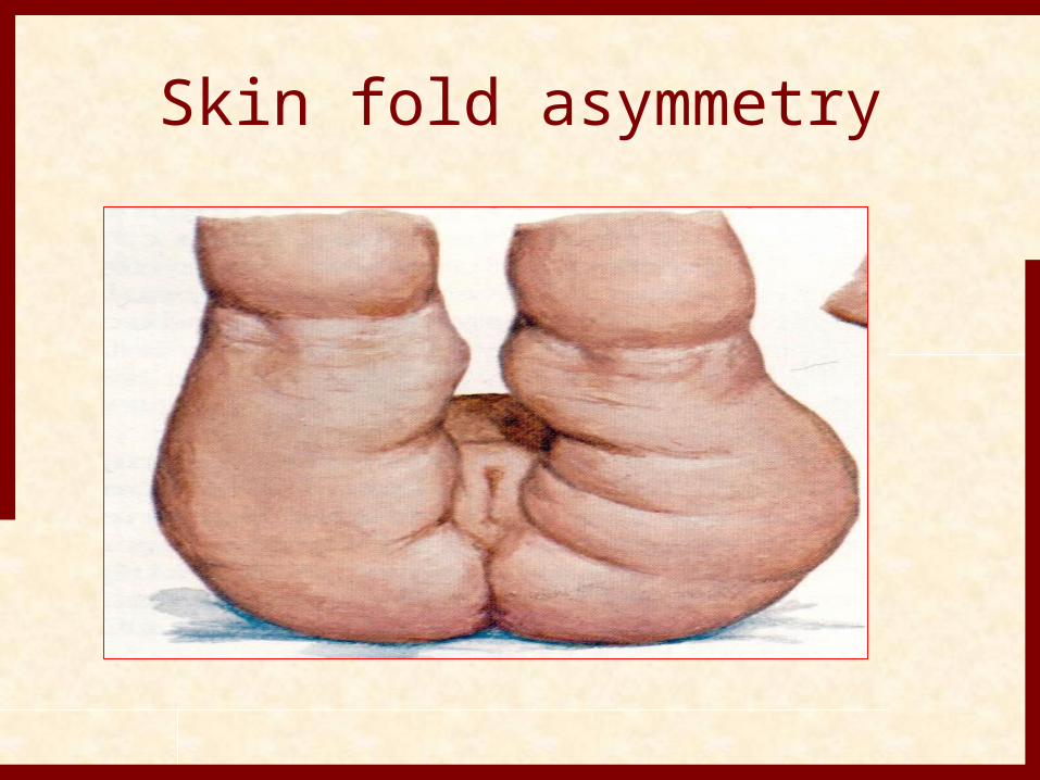
Skin fold asymmetry
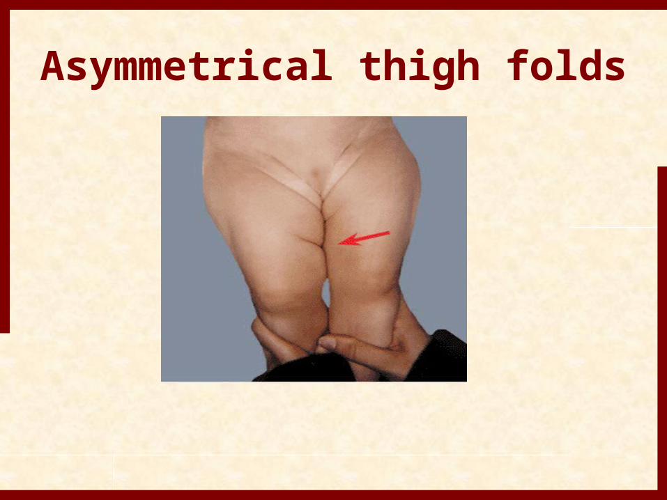
Asymmetrical thigh folds
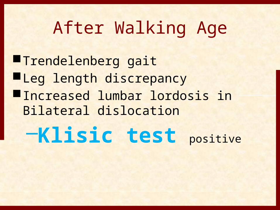
After Walking AgeTrendelenberg gaitLeg length discrepancyIncreased lumbar lordosis in Bilateral
dislocation –Klisic test positive

The examiner places the middle finger over the greater trochanter, and the index finger on the anterior superior iliac spine.A, With a
normal hip, an imaginary line drawn between the two fingers points to the umbilicus.B, When the hip is dislocated, the trochanter is elevated
and the line projects halfway between the umbilicus and the pubis.
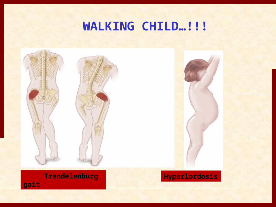
WALKING CHILD…!!!
Trendelenburg gait
Hyperlordosis
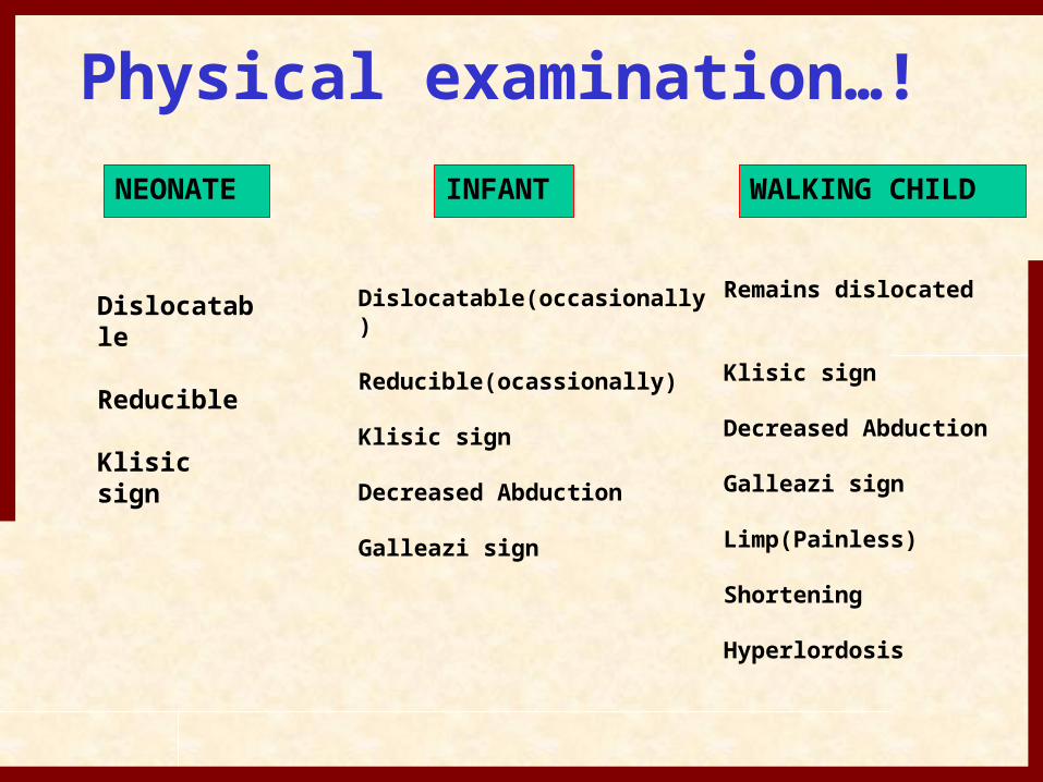
Physical examination…!NEONATE INFANT WALKING CHILD
Dislocatable
Reducible
Klisic sign
Dislocatable(occasionally)
Reducible(ocassionally)
Klisic sign
Decreased Abduction
Galleazi sign
Remains dislocated
Klisic sign
Decreased Abduction
Galleazi sign
Limp(Painless)
Shortening
Hyperlordosis
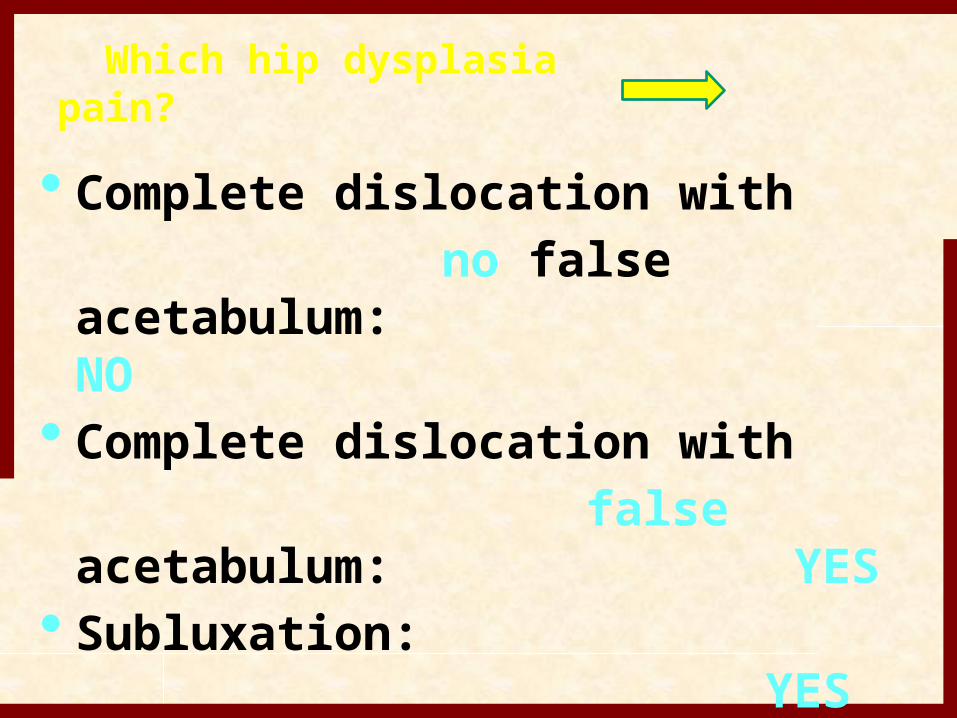
Which hip dysplasia pain?
• Complete dislocation with no false acetabulum:
NO• Complete dislocation with false acetabulum:
YES• Subluxation:
YES
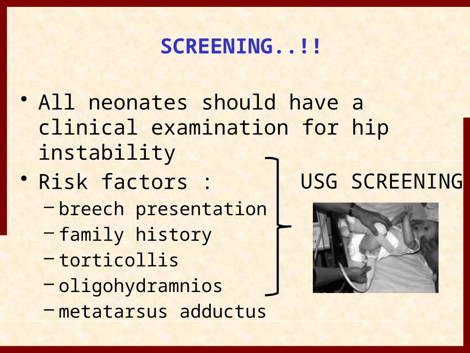
• All neonates should have a clinical examination for hip instability
• Risk factors :– breech presentation– family history– torticollis– oligohydramnios– metatarsus adductus
USG SCREENING
SCREENING..!!
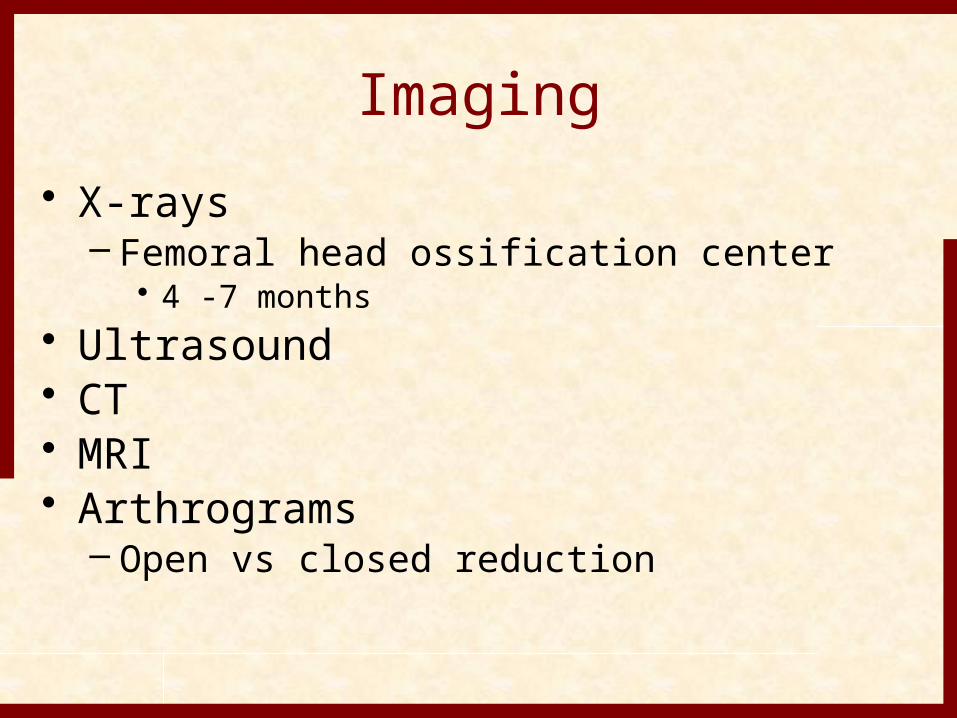
Imaging• X-rays
– Femoral head ossification center• 4 -7 months
• Ultrasound• CT• MRI• Arthrograms
– Open vs closed reduction
Radiograph• It is not reliable in early stages of DDH but new
born screening may reveal severe acetabular dysplasia or teratological dislocation.
• as child grows soft tissue become contracted and radiographs become more helpful in diagnosis.
• Most common used lines of reference are vertical line of Perkins and horizontal line of Hilgenreiner, both used to assess the position of femoral head.
Von Rosen view• In this view both hips are Abducted,
Internally Rotated and Extended.• Line is drawn along femoral shaft,
which intersect acetabulum.• In dislocated hip, it crosses above the
acetabulum.
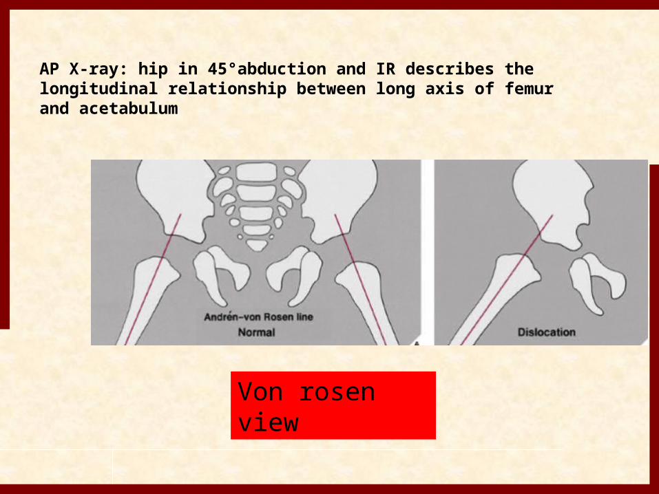
Von rosen view
AP X-ray: hip in 45°abduction and IR describes the longitudinal relationship between long axis of femur and acetabulum
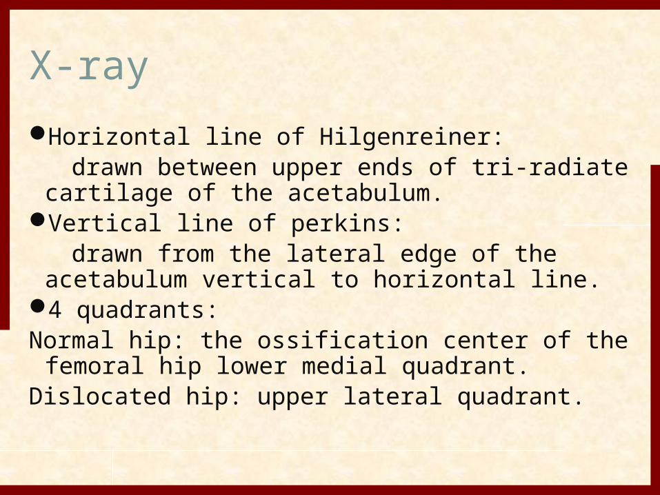
X-rayHorizontal line of Hilgenreiner: drawn between upper ends of tri-radiate
cartilage of the acetabulum.Vertical line of perkins: drawn from the lateral edge of the acetabulum
vertical to horizontal line.4 quadrants:Normal hip: the ossification center of the femoral
hip lower medial quadrant.Dislocated hip: upper lateral quadrant.
Perkin line is through lateral margin of acetabulum
• While hilgenreiner line is through triradiate cartilage.
• Shenton line is curved line that begins at lesser trochanter, goes upto femoral neck, and connect with line along inner margin of pubis.
• In normal hip, medial beak of femoral metaphysis lies in lower inner quadrant produced by junction of Perkin and hilgenreiner lines.
RADIOGRAPHY…!!
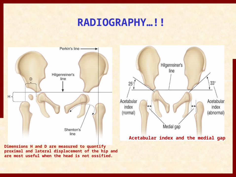
Dimensions H and D are measured to quantify proximal and lateral displacement of the hip and are most useful when the head is not ossified.
Acetabular index and the medial gap
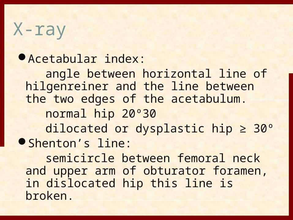
X-rayAcetabular index: angle between horizontal line of
hilgenreiner and the line between the two edges of the acetabulum.
normal hip 20º30 dilocated or dysplastic hip ≥ 30ºShenton’s line: semicircle between femoral neck
and upper arm of obturator foramen, in dislocated hip this line is broken.
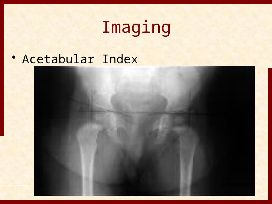
Imaging• Acetabular Index
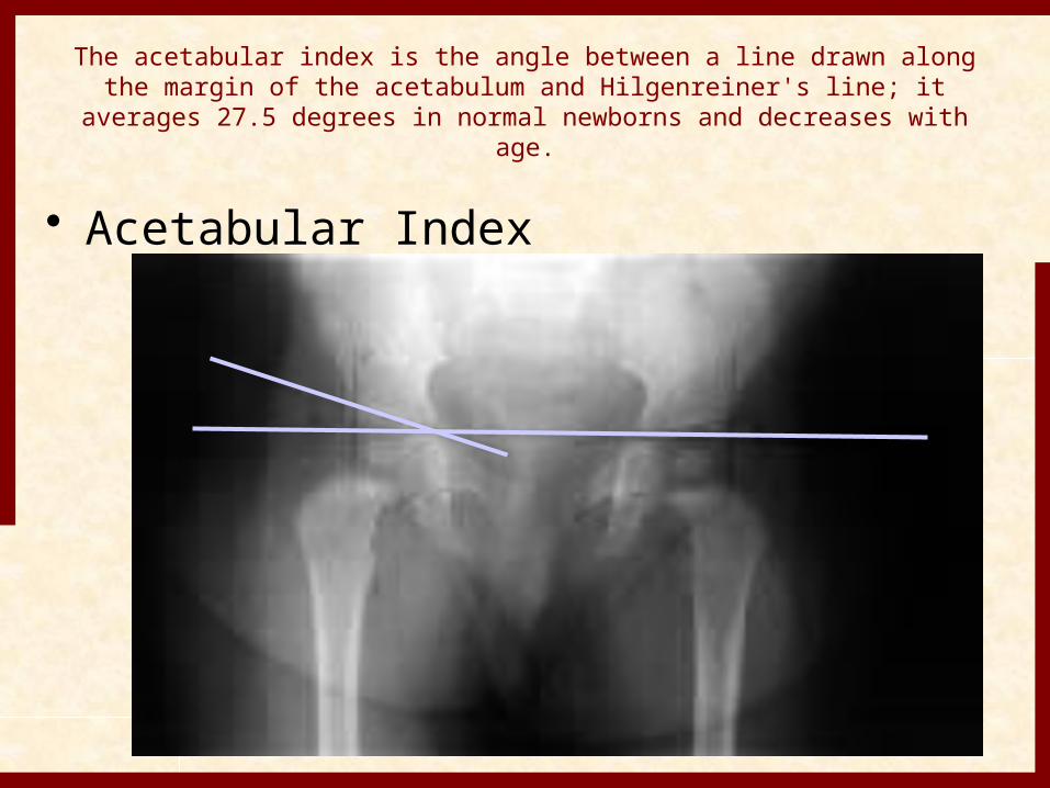
The acetabular index is the angle between a line drawn along the margin of the acetabulum and Hilgenreiner's line; it averages 27.5
degrees in normal newborns and decreases with age.
• Acetabular Index
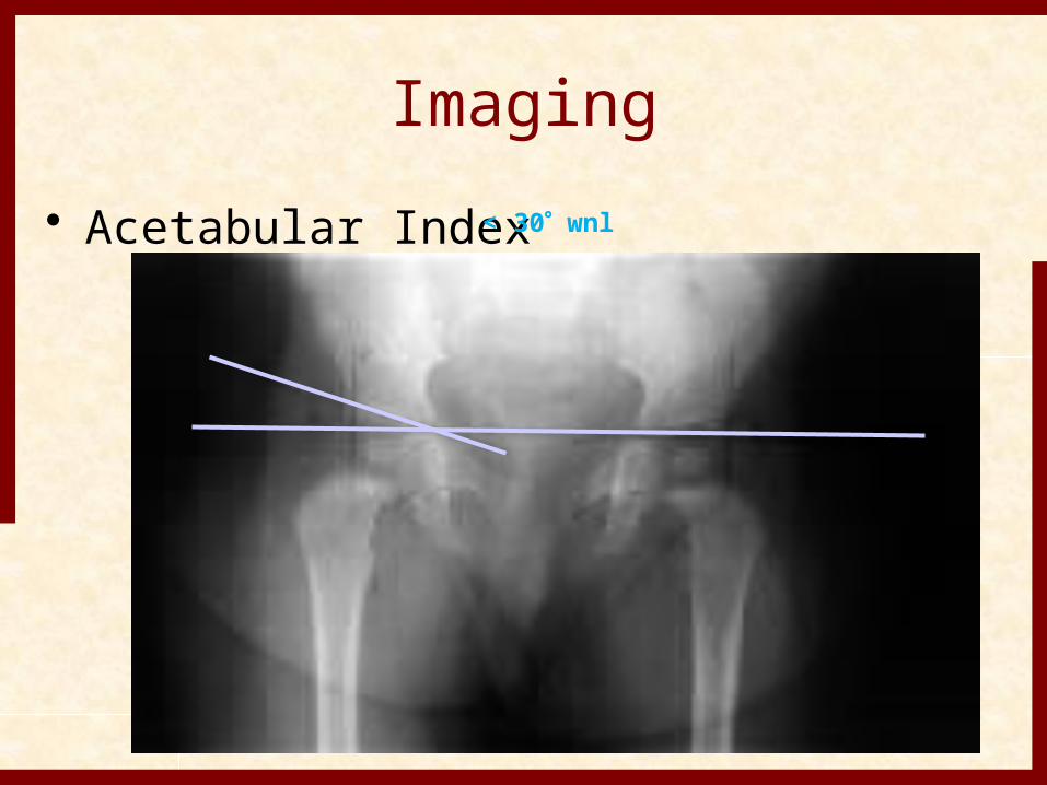
Imaging• Acetabular Index< 30 wnl
Centre –Edge angle• It is useful to measure hip position.• It is formed at the junction of Perkin
line with line that connects lateral margin of acetabulum to the center of femoral head.
• In children 6-13 yr. old, >19 degree is considered normal.
• In children >=14 yr. old, >25 degree is considered normal.
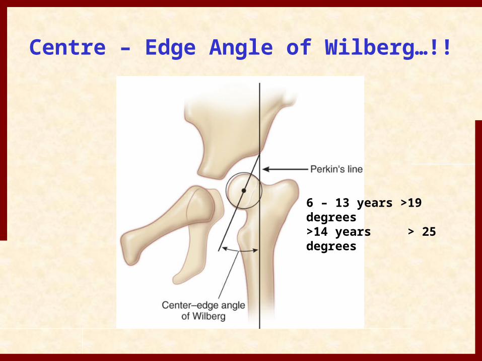
Centre – Edge Angle of Wilberg…!!
6 – 13 years >19 degrees>14 years > 25 degrees
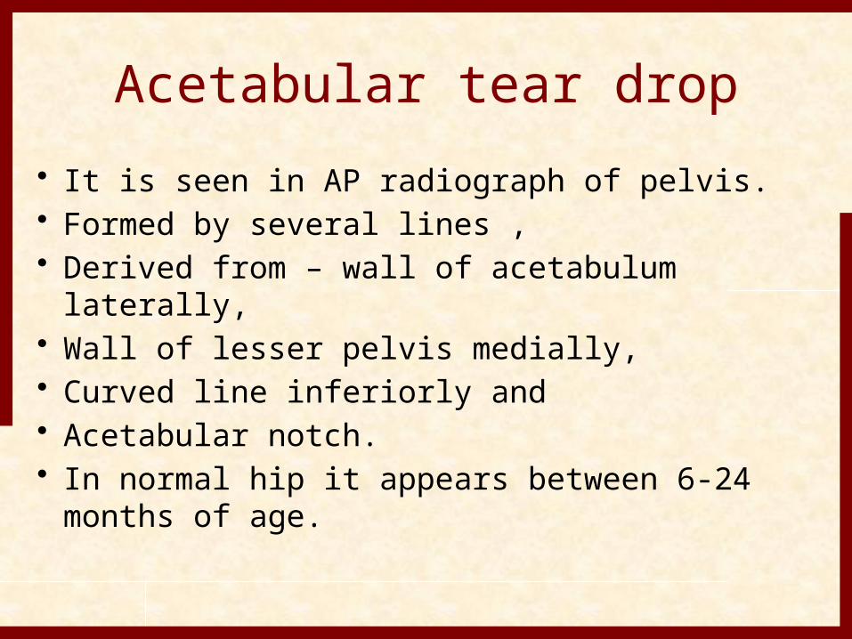
Acetabular tear drop• It is seen in AP radiograph of pelvis.• Formed by several lines ,• Derived from – wall of acetabulum laterally,• Wall of lesser pelvis medially,• Curved line inferiorly and• Acetabular notch.• In normal hip it appears between 6-24
months of age.
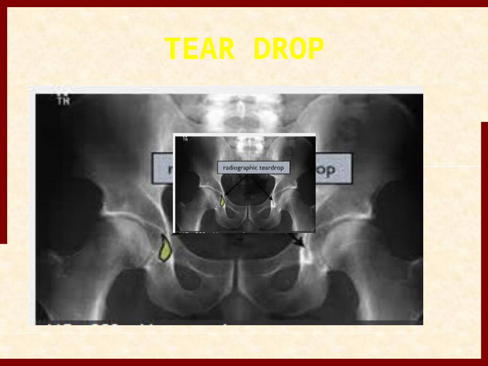
TEAR DROP
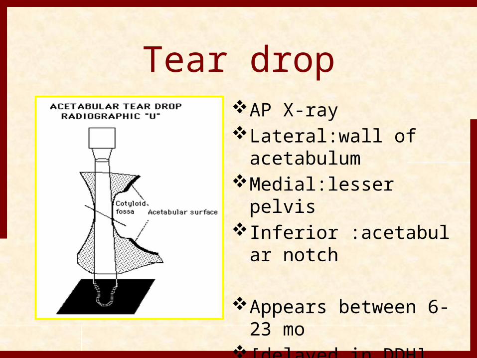
Tear dropAP X-rayLateral:wall of
acetabulumMedial:lesser pelvisInferior :acetabular
notch
Appears between 6-23 mo
[delayed in DDH]V-shaped in DDH
It significans is in the pronosis.
• Hips in which teardrop appears within 6 months of reduction have better outcome than in which it appears late.
• 4 types have been noted:-• Open , closed , crossed and reversed.• Also be describe as U- or V- shaped.• V- shaped associated with poor
outcome.
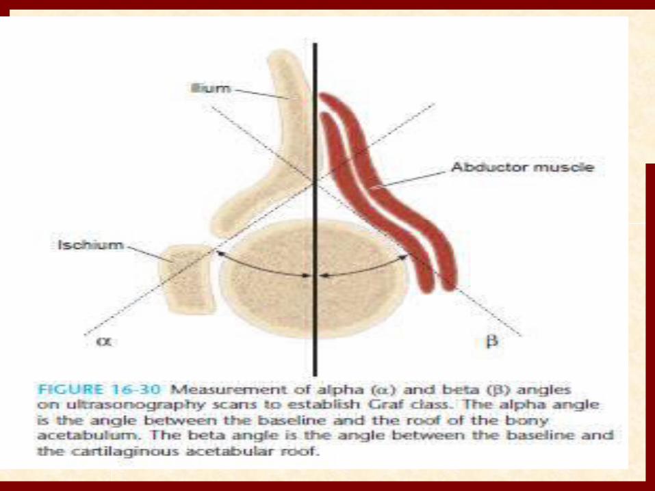
DIAGNOSIS• 1. ULTRA SOUND• In the Graf technique, the transducer is
placed over the greater trochanter, which allows visualization of the ilium, the bony acetabulum, the labrum, and the femoral epiphysis
• The angle formed by the line of the ilium and a line tangential to the boney roof of the acetabulum is termed the α angle and represents the depth of the acetabulum.
• Values > 60 degrees are considered normal, and those < 60 degrees imply acetabular dysplasia.
• The β angle is formed by a line drawn tangential to the labrum and the line of the ilium; this represents the cartilaginous roof of the acetabulum.
• A normal β angle is < 55 degrees, as the femoral head subluxates, the β angle increases.
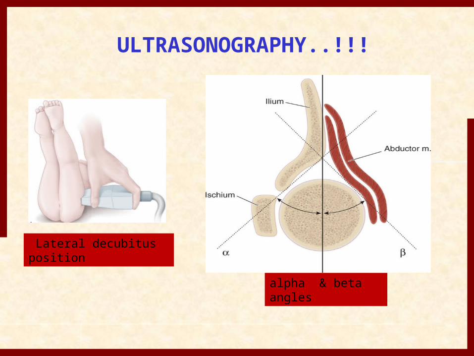
ULTRASONOGRAPHY..!!!
Lateral decubitus position
alpha & beta angles
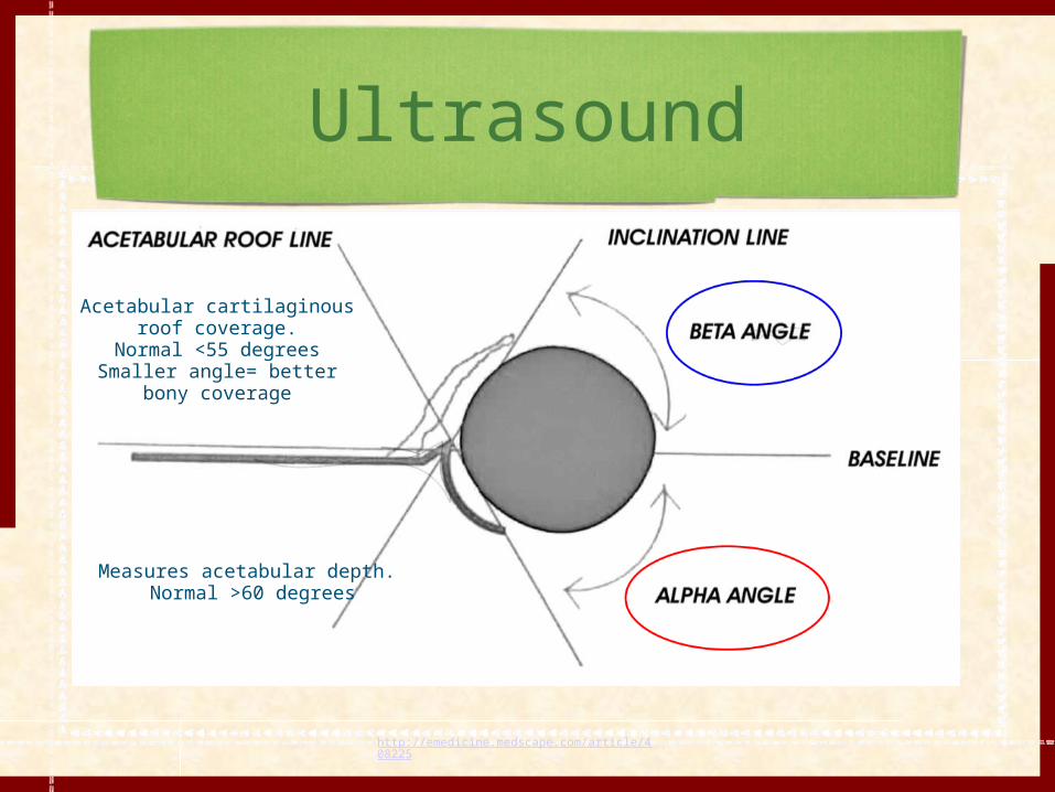
Ultrasound
http://emedicine.medscape.com/article/408225
Measures acetabular depth. Normal >60 degrees
Acetabular cartilaginous roof coverage.
Normal <55 degreesSmaller angle= better bony
coverage
• In DDH , alpha angle decreases and beta angle increases, depending upon femoral head subluxation.
• Depending upon alpha angle measurment he proposed a classification system
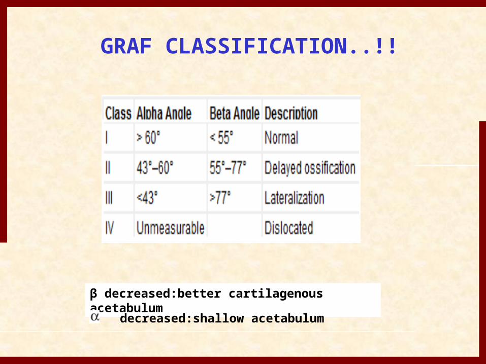
GRAF CLASSIFICATION..!!
β decreased:better cartilagenous acetabulum decreased:shallow acetabulum
MRI• It gives excellent anatomical visualization of
infant hip.• Kashiwagi and associates proposed
classification of hips with DDH.• Group 1 hips had sharp acetabular rim, all
were reducible with Pavlik hareness.• Group 2 hips had a rounded acetabular rim
and almost all are reducible with Pavlik hareness.
Group 3 hips have inverted acetabular rim, and none was reducible with hareness.
• MRI findings includes :- • Widening of iliac bone, • Lateral drift of superior and posterior
portions of acetabular floor, • Overgrowth of acetabular cartilage,• Convexity of posterior portion of
cartilage.
Treatment• Is divided in 5 age – related groups• 1) newborn ( birth to 6 months old )• 2) infant ( 6 to 18 months old )• 3) toddler ( 18 to 36 months old )• 4) child ( 3 to 8 yrs. Old )• 5) adolescent and young adult ( > 8
yrs. Old )
Treatment Options• Age of patient at presentation• Family factors• Reducibility of hip• Stability after reduction• Amount of acetabular dysplasia
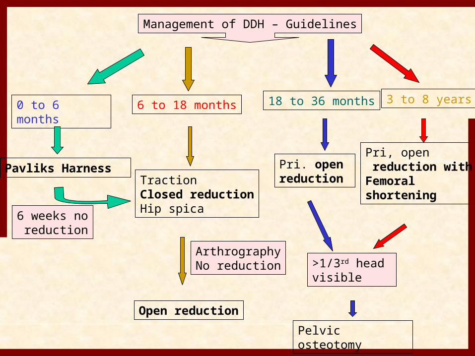
Management of DDH – Guidelines
0 to 6 months
Pavliks Harness
6 to 18 months 18 to 36 months 3 to 8 years
Traction Closed reductionHip spica
Open reduction
Pri. open reduction
Pelvic osteotomy
Pri, open reduction withFemoral shortening
6 weeks no reduction
ArthrographyNo reduction >1/3rd head
visible
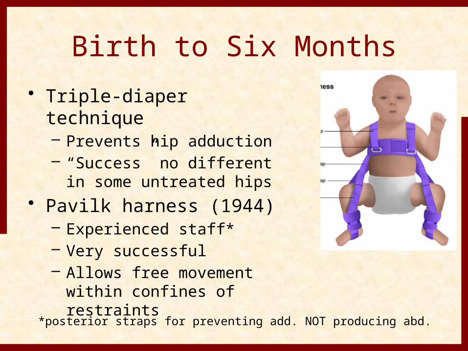
Birth to Six Months• Triple-diaper technique
– Prevents hip adduction– “Success” no different in
some untreated hips• Pavilk harness (1944)
– Experienced staff*– Very successful– Allows free movement
within confines of restraints*posterior straps for preventing add. NOT producing abd.
• Pavlik harness :- is used in first 6 months , shows excellent result in t/t of DDH.
• It is dynamic flexion-abduction orthosis.• c/I in children who are crawling or fixed soft
tissue contracture, or teratological dislocation present.
• After application, radiograph is taken and confirm the reduction. Hip is placed in flexion of 110 and abduction to occur by gravity itself .
Birth to Six Months• Pavlik harness
– Indications• Fully reducible hip*• Child not attempting to stand• Family
• Close regular follow-up (every 1-2 weeks)• For imaging and adjustments
• Duration• Childs age at hip stability + 3 months
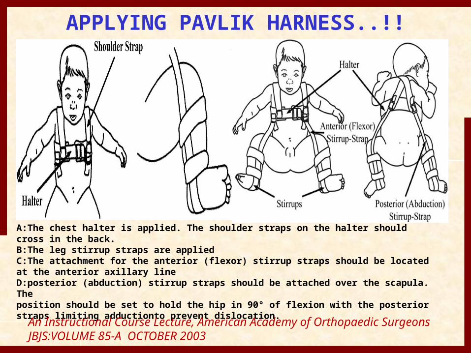
APPLYING PAVLIK HARNESS..!!
A:The chest halter is applied. The shoulder straps on the halter should cross in the back.B:The leg stirrup straps are appliedC:The attachment for the anterior (flexor) stirrup straps should be located at the anterior axillary lineD:posterior (abduction) stirrup straps should be attached over the scapula. Theposition should be set to hold the hip in 90° of flexion with the posterior straps limiting adductionto prevent dislocation.
An Instructional Course Lecture, American Academy of Orthopaedic SurgeonsJBJS:VOLUME 85-A OCTOBER 2003
How long pavlik harness should be continued
• After closed reduction and application of pavlik hareness. Patient is follow up in every 1-2 weeks.
• At this time, hip stability is checked.• Pavlik hareness is discontinued 6
weeks after clinically hip stability is obtained.
• To weaning of up to 2 hrs. per week until brace is worn at night time.
Persistent dislocation of hip• May be present after application of pavlik
hareness , 4 basic pattern is observed• Superior, inferior , lateral and posterior.• If present following manuvre should be done• Superior – additional flexion is required,• Inferior – flexion should be decreased,• Lateral – closed observation to see for direction
of femoral neck towards triradiate cartilage.
Head may be gradually reduce and dock into the acetabulum.
Persistent posterior dislocation is difficult to treat. As tight hip adductor muscle are present.If any of this persistent dislocation present for more than 3 to 6 weeks, pavlik hareness should be discontinued.t/t includes closed or open reduction and casting.
Pavlik Harness• Failures
– Poor parent compliance– Improper use by the physician
• Inadequate initial reduction• Failure to recognize persistent dislocation
– Viere et al 1990• Bilateral dislocation• Absent Ortolani’s sign• > 7weeks of age
Pavlik Harness• Complications
– Avascular necrosis• Forced hip abduction• Safe zone (abd/adduction and
flexion/extension)– Femoral nerve palsy
• Hyperflexion
*Be aware of Pavlik Harness Disease*Follow until skeletal maturity
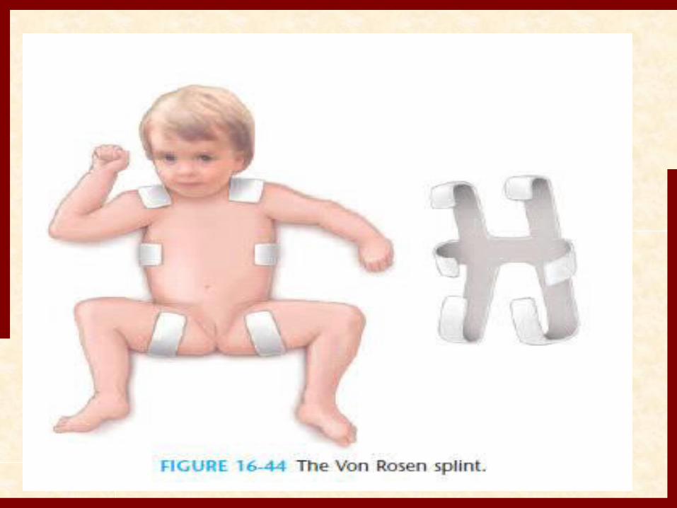
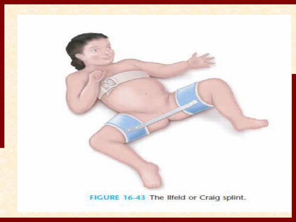
Other splint• Ilfeld and von rosen splint have high
rate of success with fewer complication but not superior to pavlik hareness.
• Frejka pillow and triple diaper are not used because of high rate of AVN.
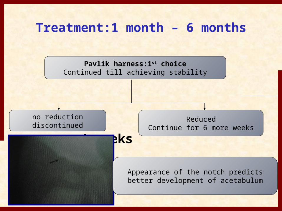
Treatment:1 month – 6 months
4 weeks
Pavlik harness:1st choiceContinued till achieving stability
no reductiondiscontinued
ReducedContinue for 6 more weeks
Appearance of the notch predictsbetter development of acetabulum
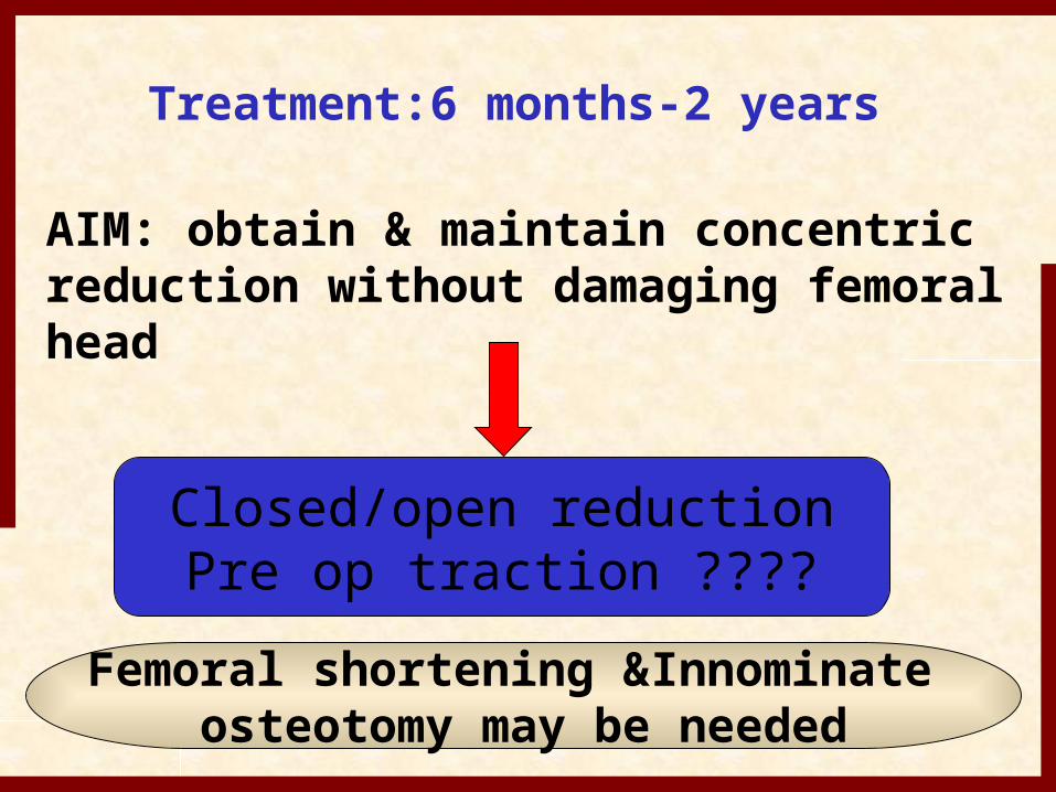
Treatment:6 months-2 years
AIM: obtain & maintain concentric reduction without damaging femoral head
Closed/open reductionPre op traction ????
Femoral shortening &Innominate osteotomy may be needed
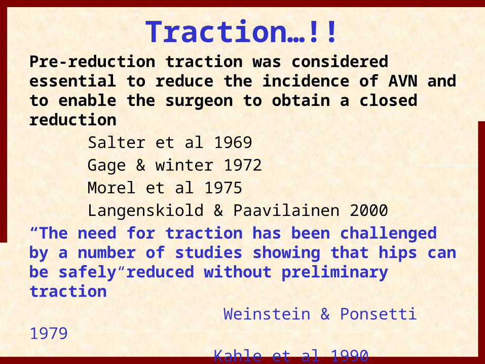
Traction…!!Pre-reduction traction was considered essential to reduce the incidence of AVN and to enable the surgeon to obtain a closed reduction Salter et al 1969 Gage & winter 1972 Morel et al 1975 Langenskiold & Paavilainen 2000“The need for traction has been challenged by a number of studies showing that hips can be safely reduced without preliminary traction” Weinstein & Ponsetti 1979 Kahle et al 1990 Quinn et al 1994 Current reccomendation: No traction
Closed Reduction…!!
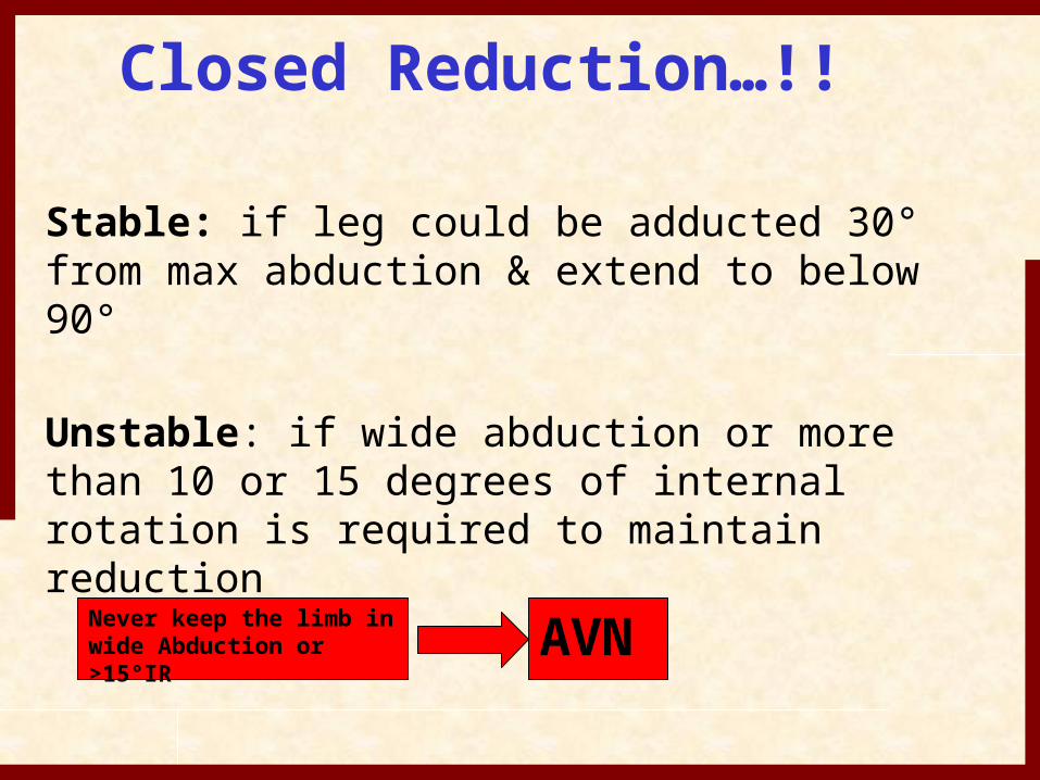
Stable: if leg could be adducted 30° from max abduction & extend to below 90°
Unstable: if wide abduction or more than 10 or 15 degrees of internal rotation is required to maintain reduction
Never keep the limb inwide Abduction or >15°IR
AVN
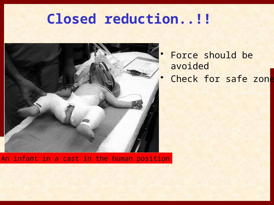
Closed reduction..!!
An infant in a cast in the human position
• Force should be avoided
• Check for safe zone
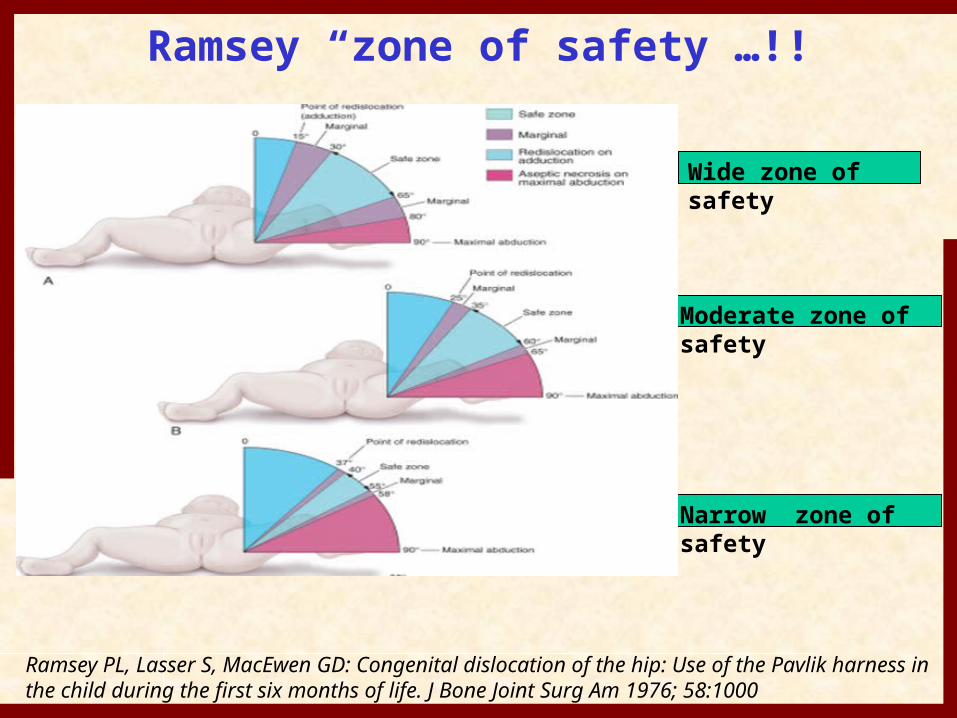
Ramsey “zone of safety”…!!
Wide zone of safety
Moderate zone of safety
Narrow zone of safety
Ramsey PL, Lasser S, MacEwen GD: Congenital dislocation of the hip: Use of the Pavlik harness in the child during the first six months of life. J Bone Joint Surg Am 1976; 58:1000
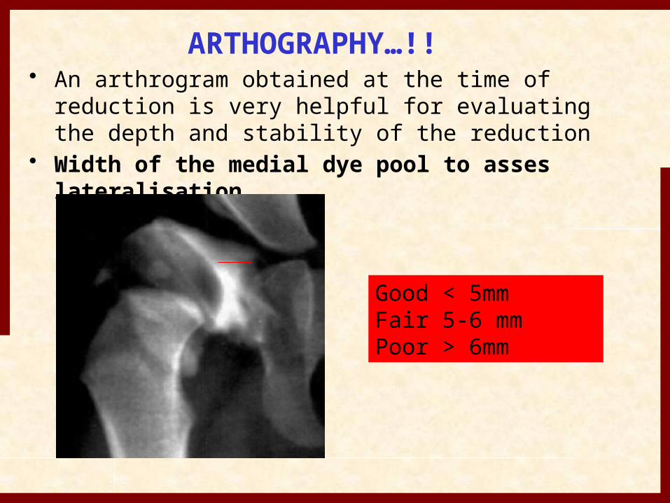
ARTHOGRAPHY…!!• An arthrogram obtained at the time of reduction is
very helpful for evaluating the depth and stability of the reduction
• Width of the medial dye pool to asses lateralisation
Good < 5mmFair 5-6 mmPoor > 6mm
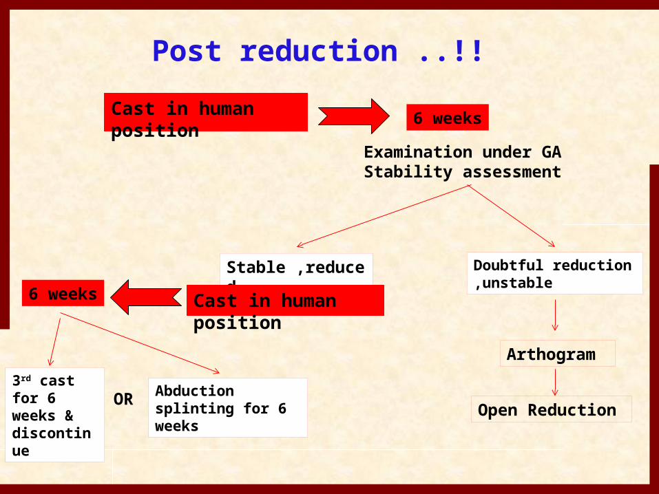
Post reduction ..!!Cast in human position 6 weeks
Examination under GA Stability assessment
Stable ,reduced
Doubtful reduction,unstable
Arthogram
Cast in human position6 weeks
3rd cast for 6 weeks & discontinue
Abduction splinting for 6 weeks
OR Open Reduction
Open Reduction…!!
• Unable to achieve closed reduction
• Widening of the joint space
• Unstable reductions• Loss of reduction on
follow up• Advanced age
Open reduction can be performed by
• Anterior • Anteromedial • Medial approach• Anterior approach :- pathology in the
anterior and lateral aspect of hip can be easily reached and pelvic osteotomy can be easily performed.
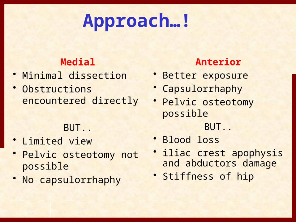
Approach…!
Medial• Minimal dissection• Obstructions
encountered directly
BUT..• Limited view• Pelvic osteotomy not
possible• No capsulorrhaphy
Anterior• Better exposure• Capsulorrhaphy• Pelvic osteotomy
possibleBUT..
• Blood loss• iliac crest apophysis
and abductors damage• Stiffness of hip
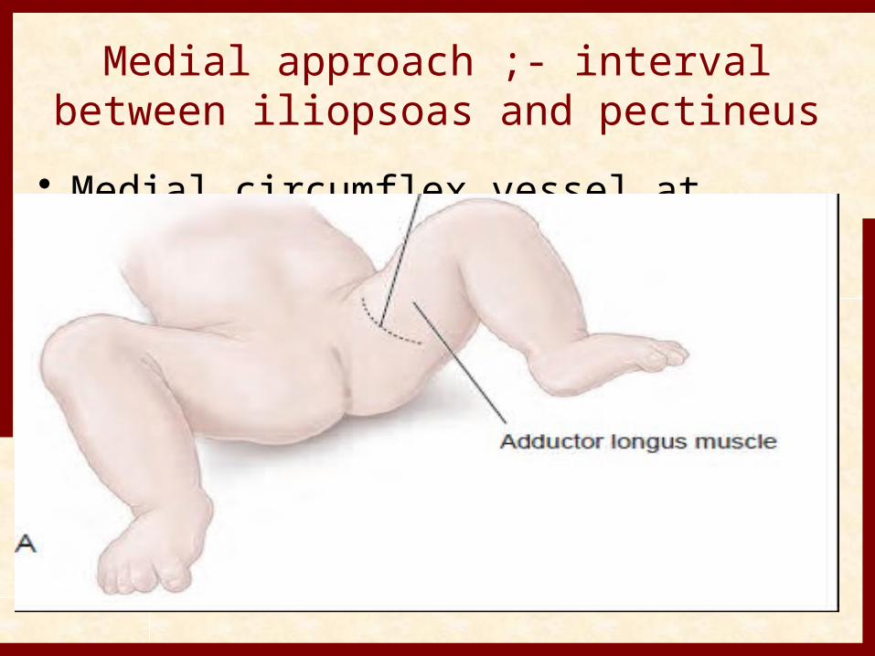
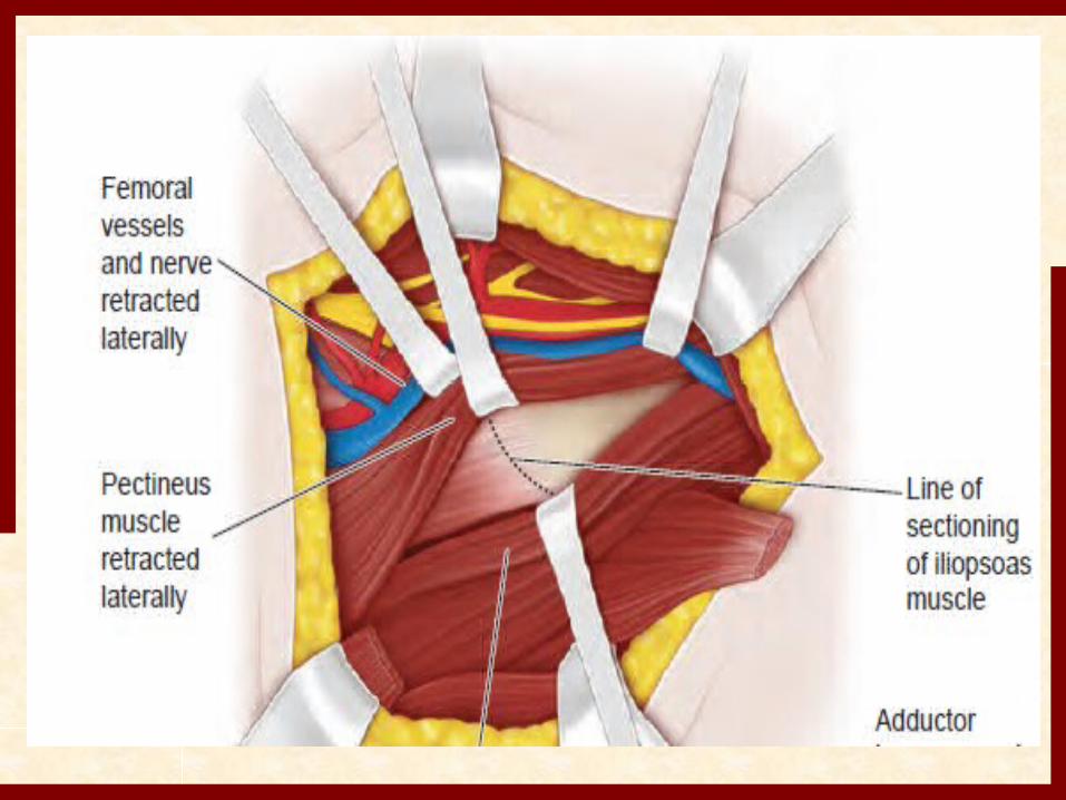
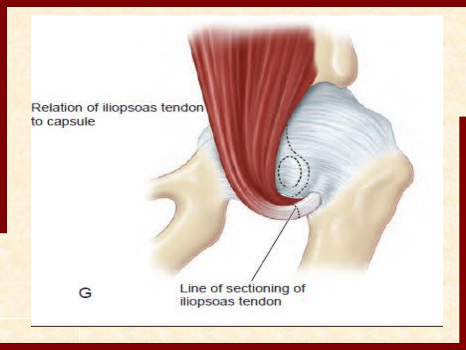
Medial approach ;- interval between iliopsoas and pectineus
• Medial circumflex vessel at higher risk.
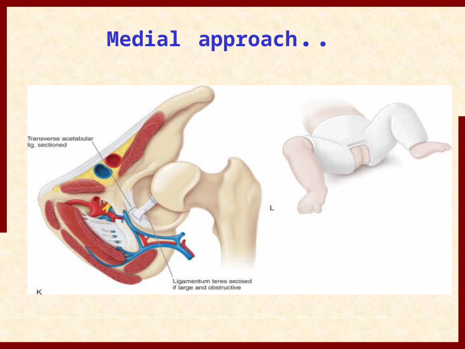
Medial approach..

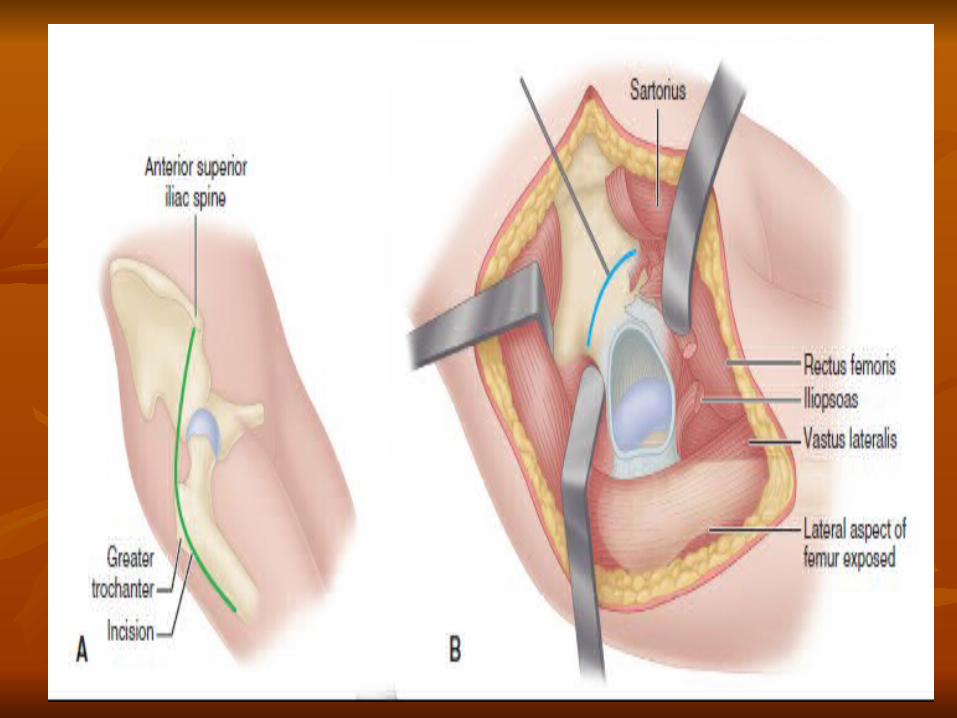
Anterior approach…!!
• Smith-Peterson anterior approach• Stood the test of time• More commonly used • Bikini incision better cosmetic results
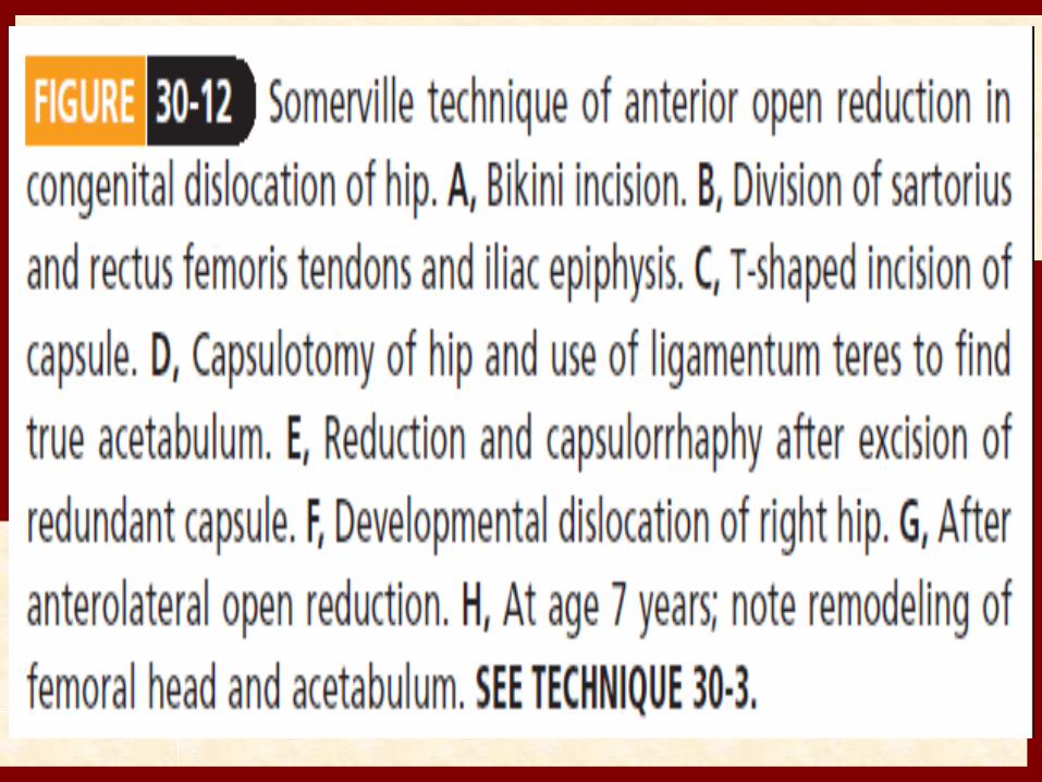
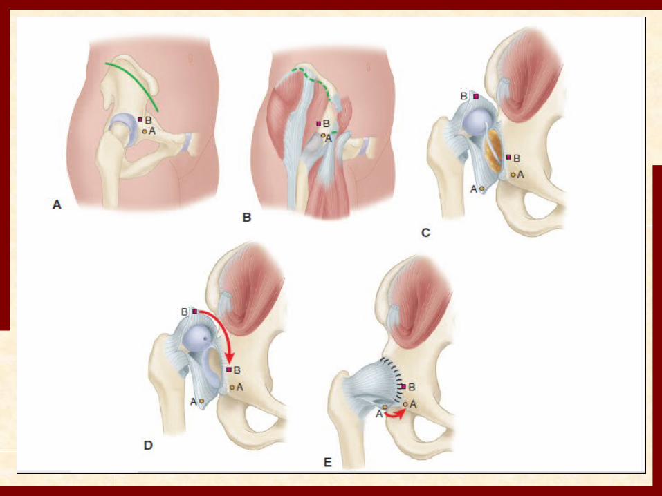
T-capsulotomy..!!
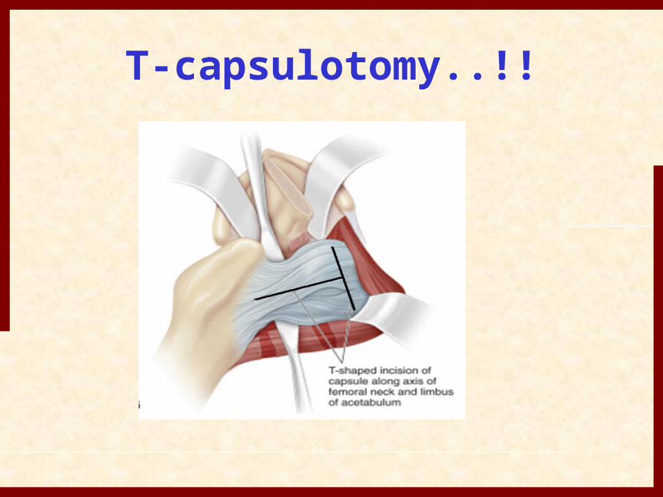
Ligamentum teres
use of ligamentum teres to find true acetabulum
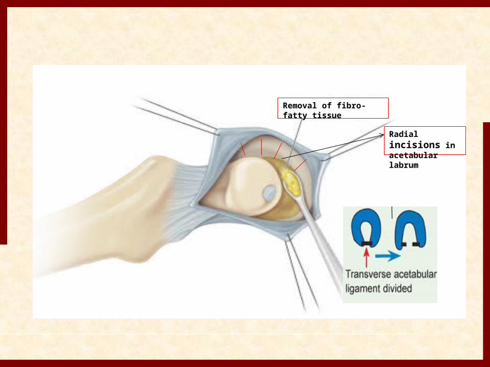
Radial incisions in acetabular labrum
Removal of fibro-fatty tissue
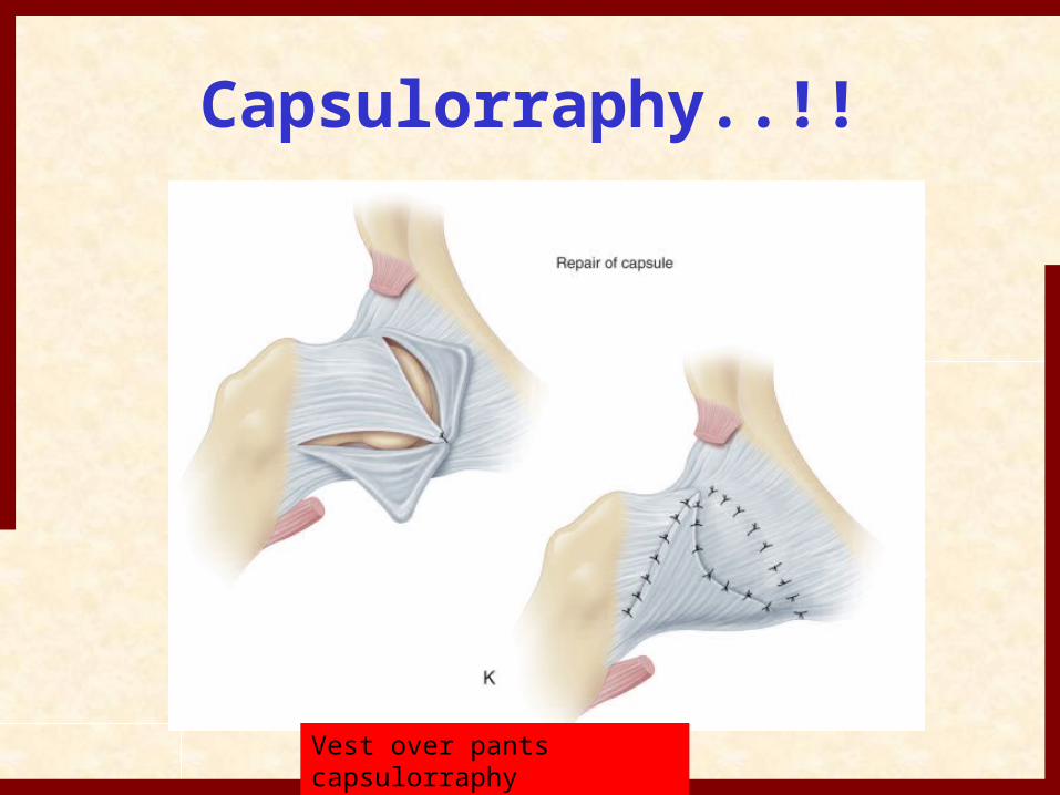
Capsulorraphy..!!
Vest over pants capsulorraphy
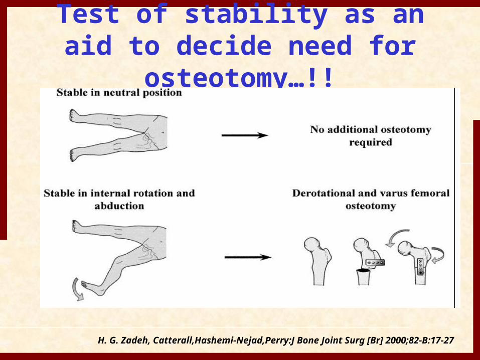
Test of stability as an aid to decide need for osteotomy…!!
H. G. Zadeh, Catterall,Hashemi-Nejad,Perry:J Bone Joint Surg [Br] 2000;82-B:17-27
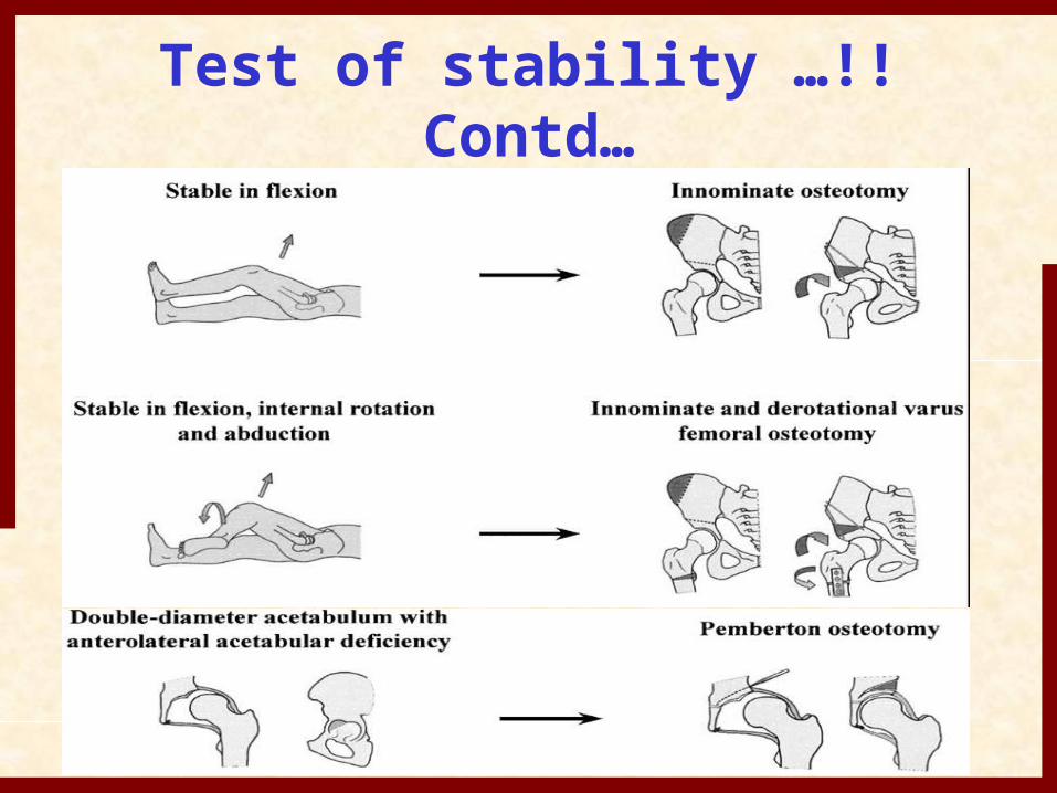
Test of stability …!! Contd…
2 Years of Age and Older
• For child 2 -3 years of age, during open reduction acetabular coverage if insufficient warrants reorientation osteotomy
• If coxa valga with excessive anteversion, VDRO may be done.
• Children > 3 years usually need an acetabular procedureFemoral shortening is essential part of it’s management. In past , child is put on skeletal traction but result of shortening are better and morbidity is less.
Open Reduction with Femoral Shortening..!
• Pressure leads to risk of AVN• Better results than preoperative traction in older
children with less morbidityWhen to do??
• Anticipated increased pressure on reduced femur head
• Recommended in child > 2yrs.• distract the joint few millimeter per operatively• Judge the tightness of soft tissues after reduction• irreducible dislocation
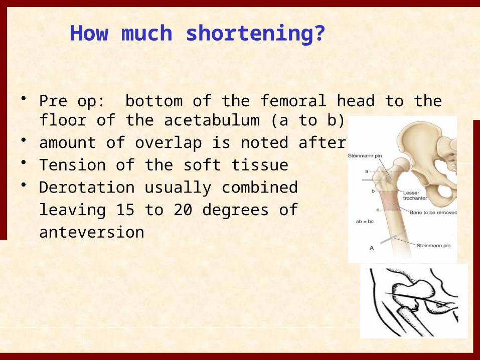
How much shortening?
• Pre op: bottom of the femoral head to the floor of the acetabulum (a to b)
• amount of overlap is noted after osteotomy• Tension of the soft tissue• Derotation usually combined
leaving 15 to 20 degrees of anteversion

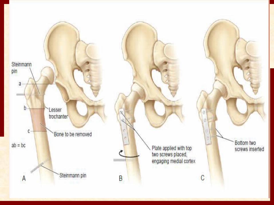
Open Reduction with Femoral Shortening..!
Subtrochanteric cut
Overlap method to determine theamount to shorten the femur.
Internal fixation with an appropriate blade-plate
Primary femoral shortening
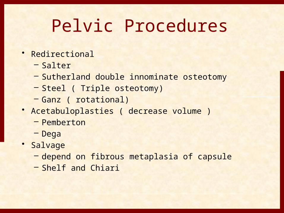
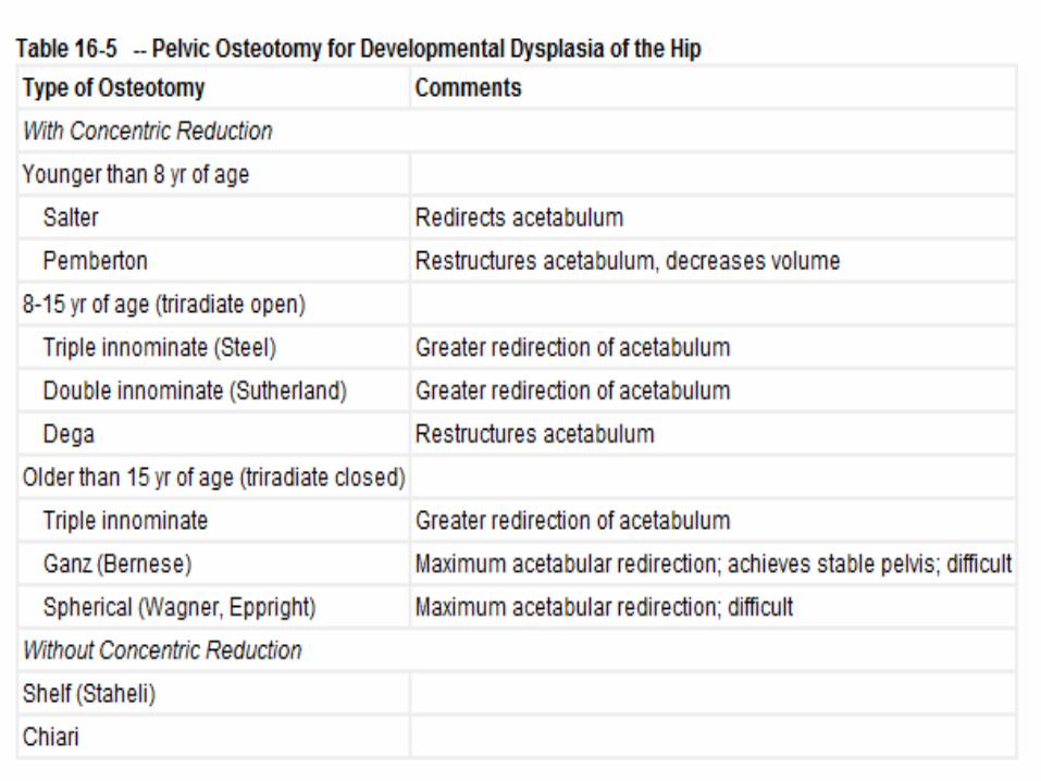
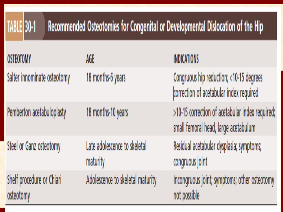
Pelvic Procedures• Redirectional
– Salter – Sutherland double innominate osteotomy – Steel ( Triple osteotomy)– Ganz ( rotational)
• Acetabuloplasties ( decrease volume )– Pemberton– Dega
• Salvage – depend on fibrous metaplasia of capsule– Shelf and Chiari
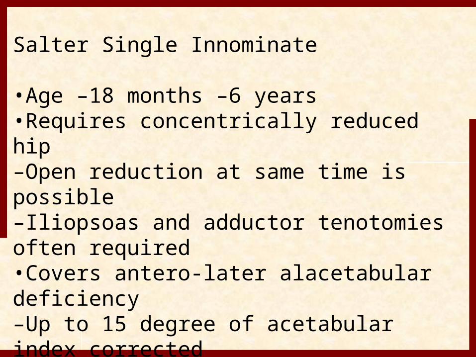
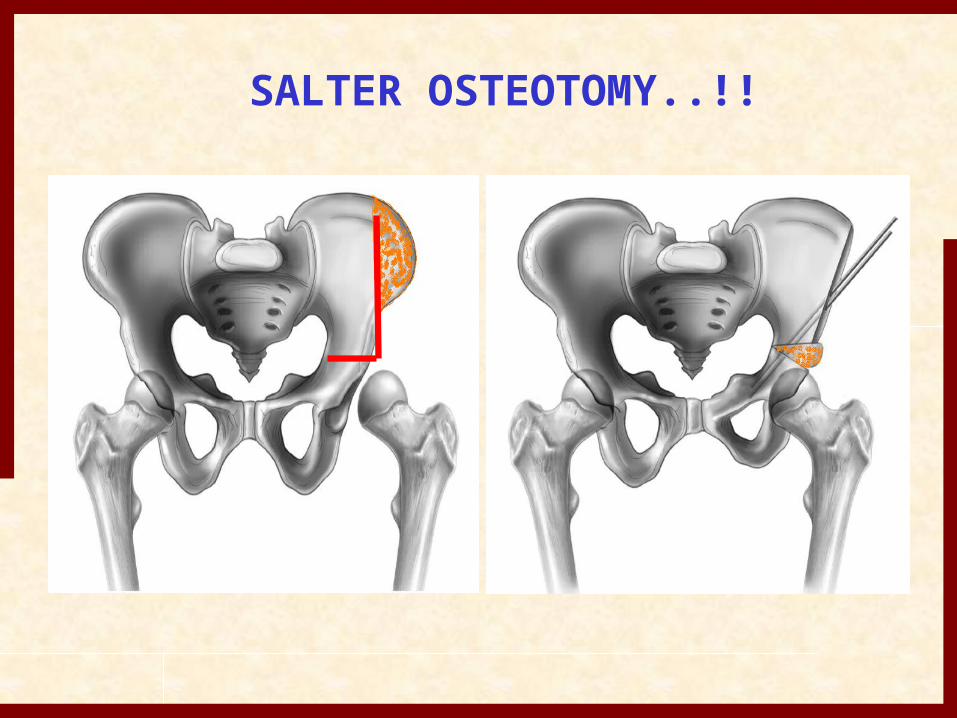
Salter Single Innominate
•Age –18 months –6 years•Requires concentrically reduced hip–Open reduction at same time is possible–Iliopsoas and adductor tenotomies often required•Covers antero-later alacetabular deficiency–Up to 15 degree of acetabular index corrected
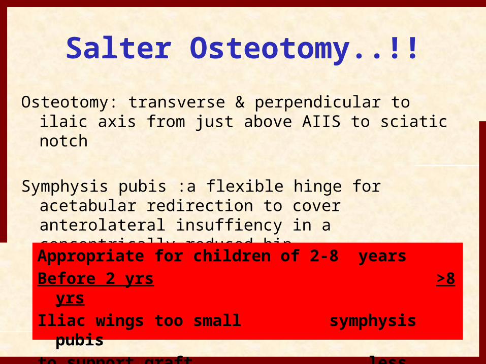
Salter Osteotomy..!!Osteotomy: transverse & perpendicular to ilaic axis
from just above AIIS to sciatic notch
Symphysis pubis :a flexible hinge for acetabular redirection to cover anterolateral insuffiency in a concentrically reduced hip
Appropriate for children of 2-8 yearsBefore 2 yrs >8 yrsIliac wings too small symphysis pubisto support graft less mobile
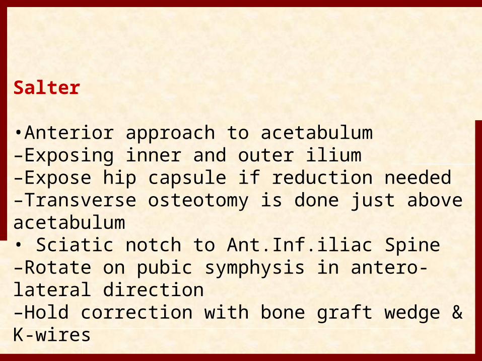
Salter
•Anterior approach to acetabulum–Exposing inner and outer ilium–Expose hip capsule if reduction needed–Transverse osteotomy is done just above acetabulum• Sciatic notch to Ant.Inf.iliac Spine–Rotate on pubic symphysis in antero-lateral direction–Hold correction with bone graft wedge & K-wires

SALTER OSTEOTOMY..!!
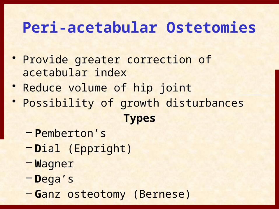
Peri-acetabular Ostetomies• Provide greater correction of acetabular
index• Reduce volume of hip joint• Possibility of growth disturbances
Types– Pemberton’s – Dial (Eppright) – Wagner – Dega’s– Ganz osteotomy (Bernese)
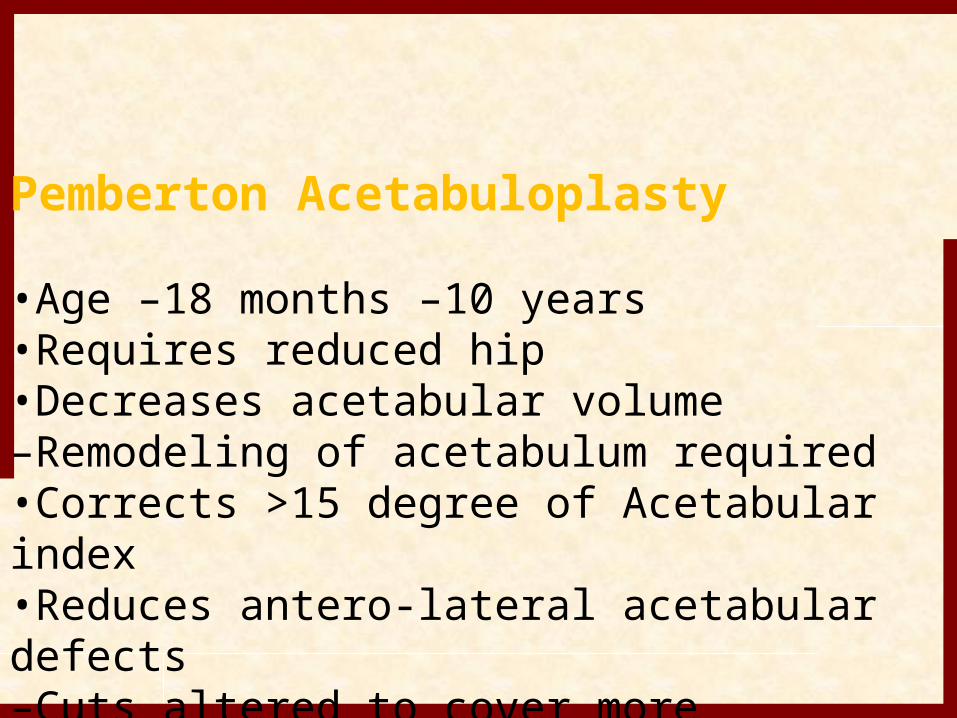
Pemberton Acetabuloplasty
•Age –18 months –10 years•Requires reduced hip•Decreases acetabular volume–Remodeling of acetabulum required •Corrects >15 degree of Acetabular index•Reduces antero-lateral acetabular defects–Cuts altered to cover more anteriorly or laterally
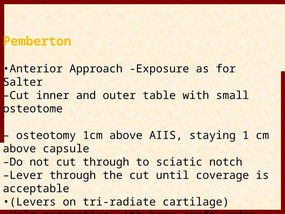
Pemberton
•Anterior Approach -Exposure as for Salter–Cut inner and outer table with small osteotome
– osteotomy 1cm above AIIS, staying 1 cm above capsule–Do not cut through to sciatic notch–Lever through the cut until coverage is acceptable•(Levers on tri-radiate cartilage)–Hold correction with bone graft wedge
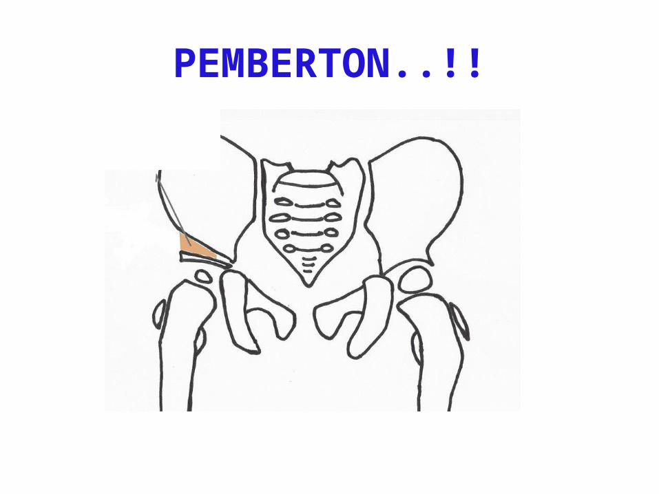
PEMBERTON..!!
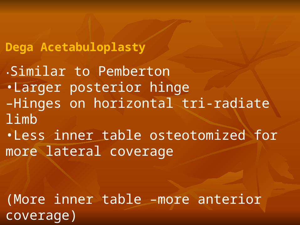
Dega Acetabuloplasty
•Similar to Pemberton•Larger posterior hinge–Hinges on horizontal tri-radiate limb•Less inner table osteotomized for more lateral coverage
(More inner table –more anterior coverage)
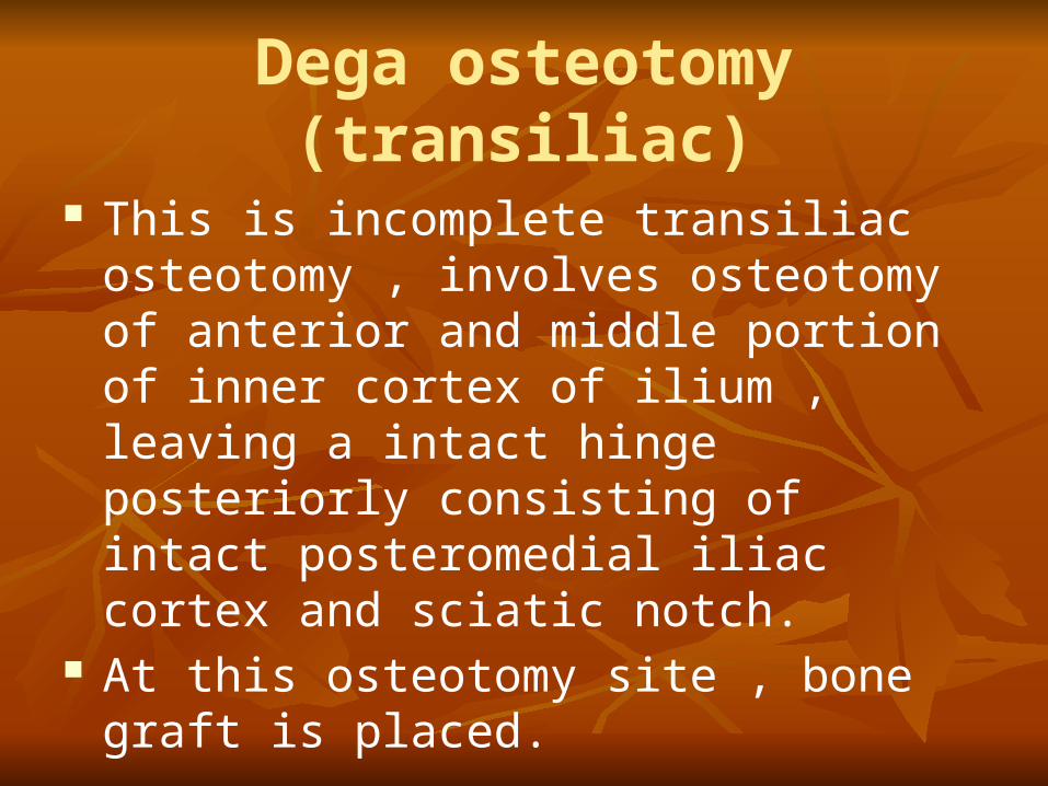
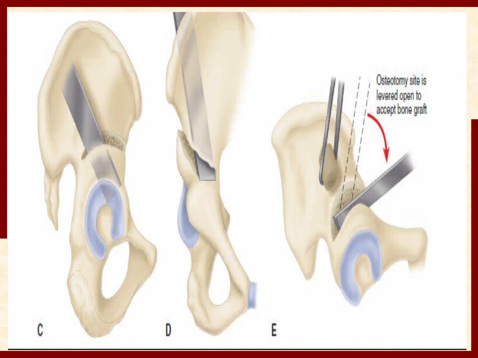
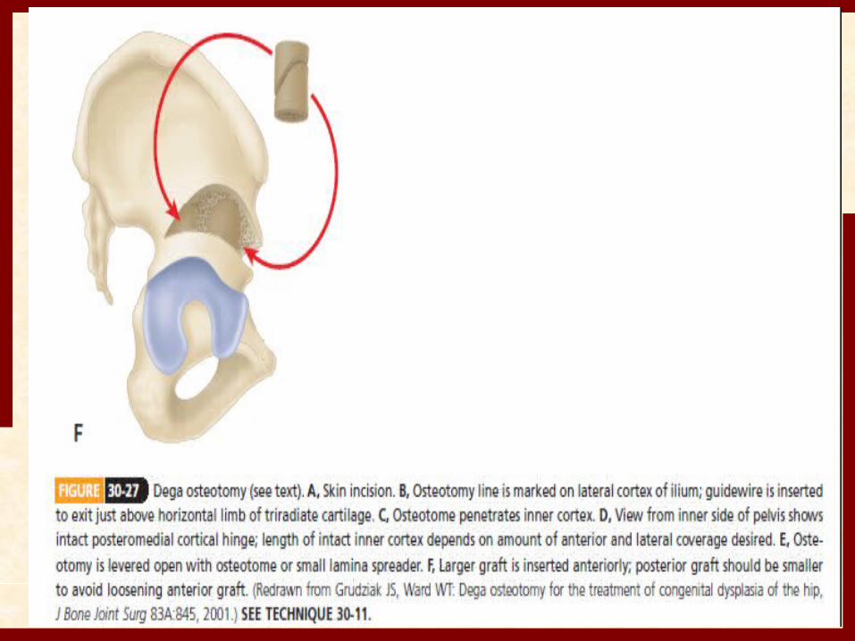
Dega osteotomy (transiliac) This is incomplete transiliac osteotomy ,
involves osteotomy of anterior and middle portion of inner cortex of ilium , leaving a intact hinge posteriorly consisting of intact posteromedial iliac cortex and sciatic notch.
At this osteotomy site , bone graft is placed.
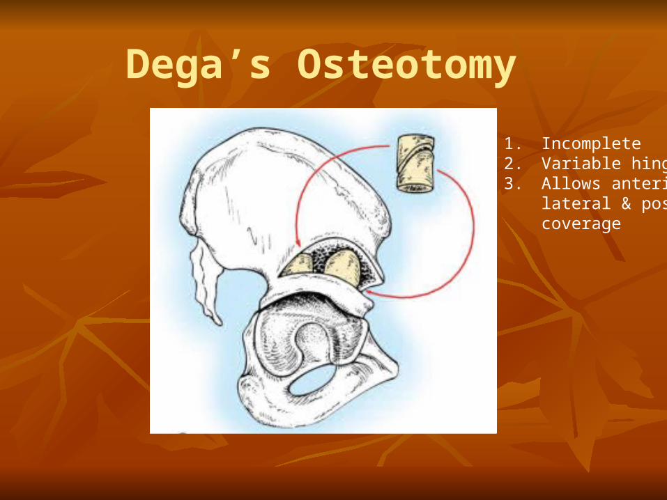
Dega’s Osteotomy
1. Incomplete2. Variable hinge3. Allows anterior,
lateral & posteriorcoverage
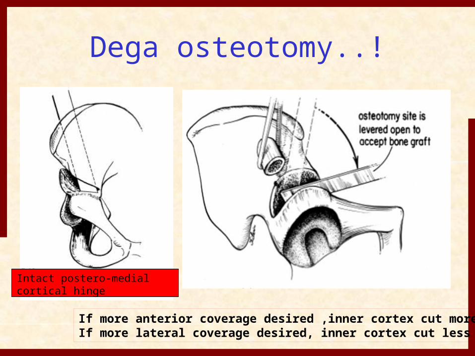
Dega osteotomy..!
Intact postero-medial cortical hinge
If more anterior coverage desired ,inner cortex cut moreIf more lateral coverage desired, inner cortex cut less
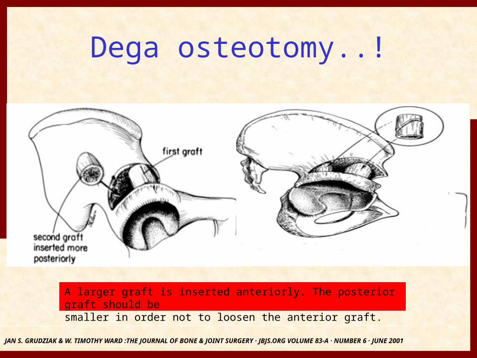
Dega osteotomy..!
JAN S. GRUDZIAK & W. TIMOTHY WARD :THE JOURNAL OF BONE & JOINT SURGERY · JBJS.ORG VOLUME 83-A · NUMBER 6 · JUNE 2001
A larger graft is inserted anteriorly. The posterior graft should besmaller in order not to loosen the anterior graft.
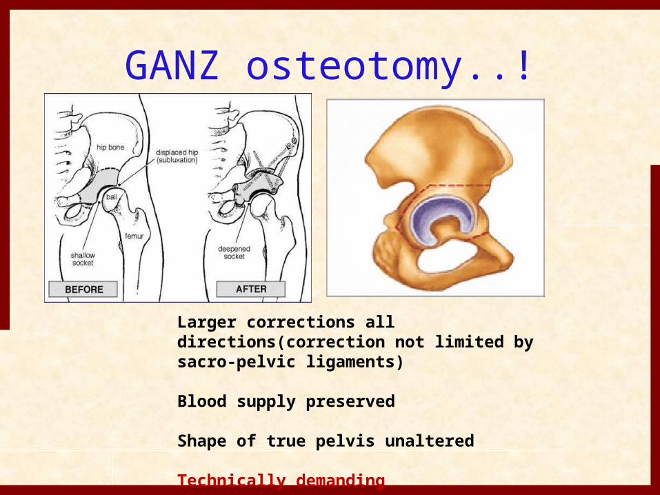
GANZ osteotomy..!
Larger corrections all directions(correction not limited by sacro-pelvic ligaments)
Blood supply preserved
Shape of true pelvis unaltered
Technically demanding
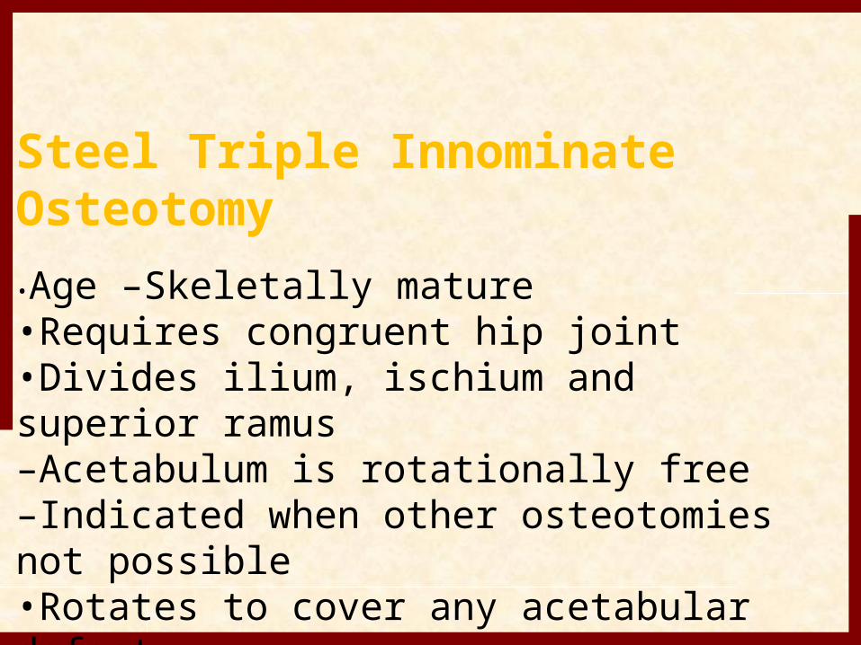
Steel Triple Innominate Osteotomy•Age –Skeletally mature•Requires congruent hip joint•Divides ilium, ischium and superior ramus–Acetabulum is rotationally free–Indicated when other osteotomies not possible•Rotates to cover any acetabular defect
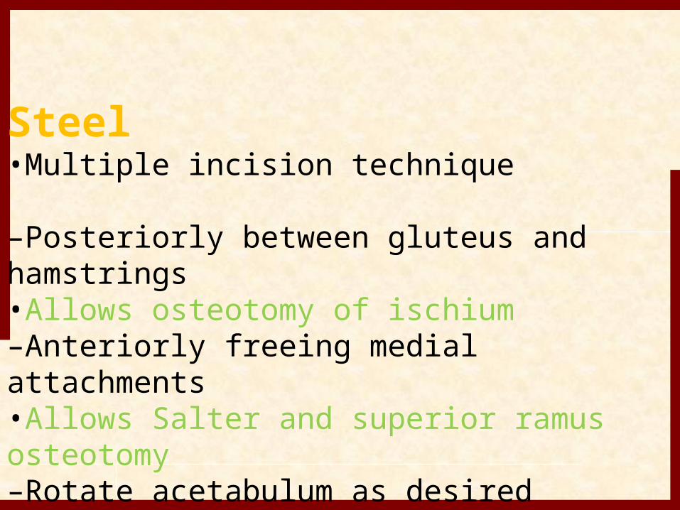
Steel•Multiple incision technique
–Posteriorly between gluteus and hamstrings•Allows osteotomy of ischium–Anteriorly freeing medial attachments•Allows Salter and superior ramus osteotomy–Rotate acetabulum as desired•Avoid externally rotating–Bone graft wedge is fixed as per Salter type
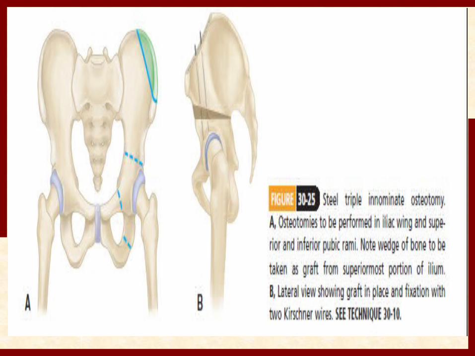
Steel triple innominate osteotomy
• Ischium, superior pubic ramus and ilium superior to acetabulum all are divided and acetabulum is repositioned and stabilized by bone graft and pins.
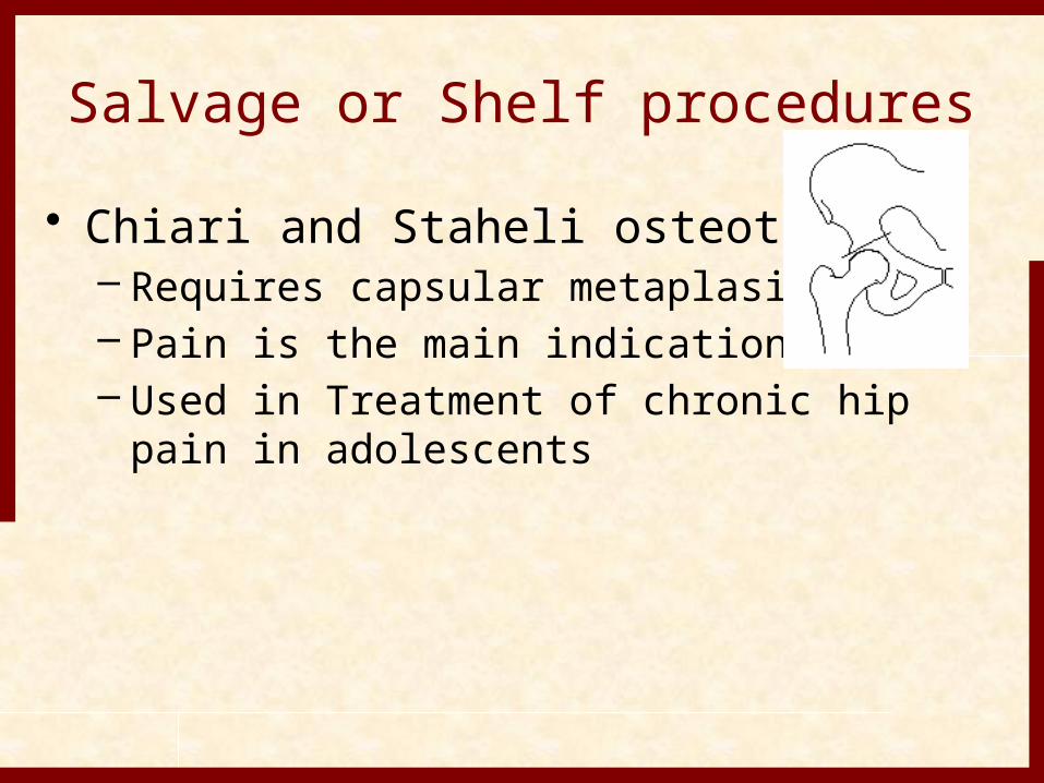
Salvage or Shelf procedures• Chiari and Staheli osteotomies
– Requires capsular metaplasia– Pain is the main indication– Used in Treatment of chronic hip pain in
adolescents
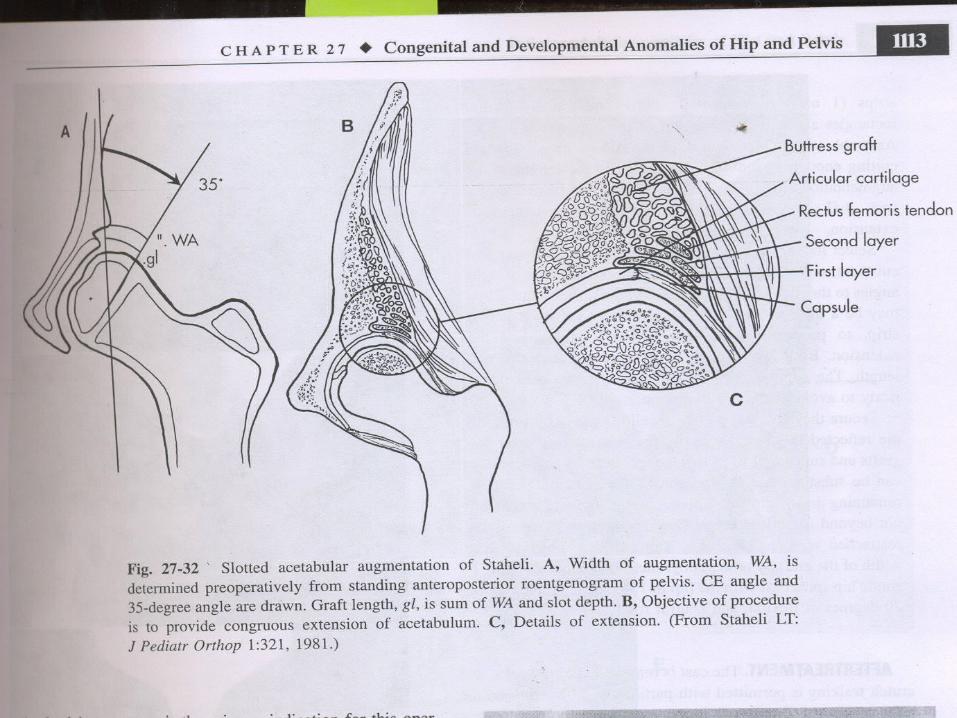
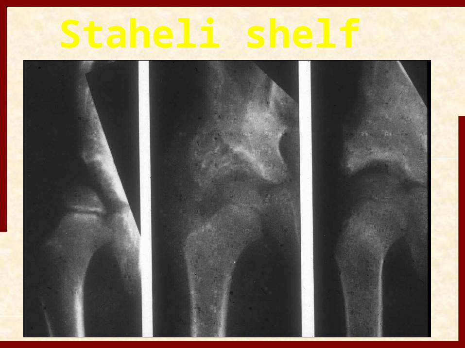
Staheli Shelf Procedure•Age –older child to skeletal maturity•Salvage operation•Indicated for non-concentric hips•Augments supero-lateral deficency–Slotted bone graft placed over capsule deepening the acetablum
Staheli•Anterior approach is used with outer wall exposure only–Identify superior acetabular edge–Create slot 1cm deep along edge in cephlad angle–Remove 1 cm cortical strips from outer table
•Insert into slot, cutting at desired lateral overhang
•2nd layer inserted lengthwise
•Use remaining to fill in above slot edge–Hold in place with reflected fascia and adductors
Staheli shelf
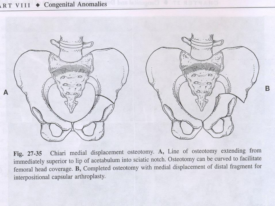
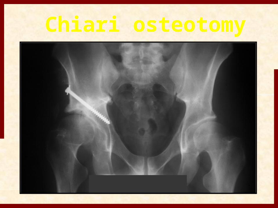
Chiari Medial Displacement•Age –skeletally mature•Salvage operation only–Used when no other osteotomy possible–Possible with subluxed hip•Covers well laterally–Anterior and posterior augmentation may be necessary•May be useful in other conditions–Coxamagna, OA in dysplasichips
•Anterior approach –as per Salter–Identify superior extent of capsule–Cut from AIIS to notch following capsule curve•Angle osteotome10-20ocephlad–Displace distal fragment medially 50-100%•Ensure complete head coverage•Leg abduction, hinges on pubic symphysis
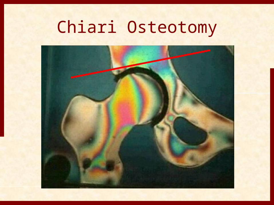
Chiari Osteotomy
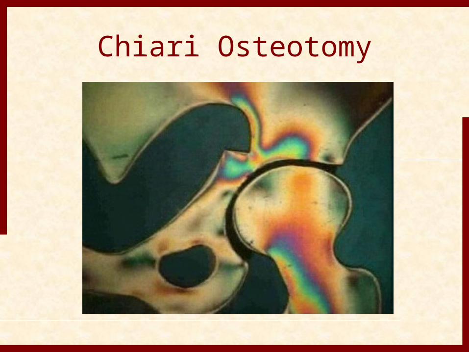
Chiari Osteotomy
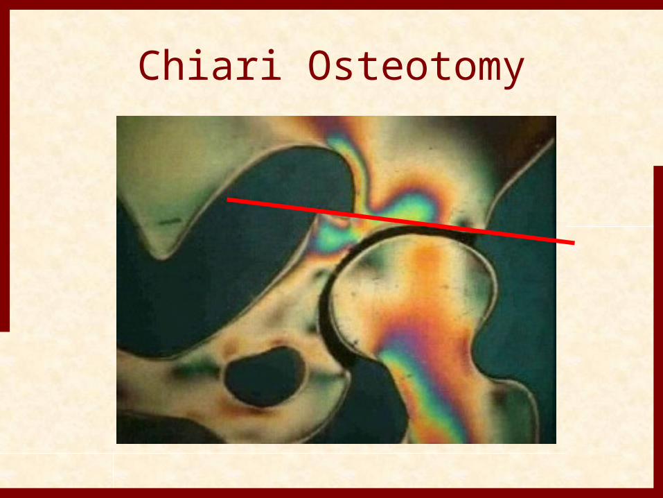
Chiari Osteotomy
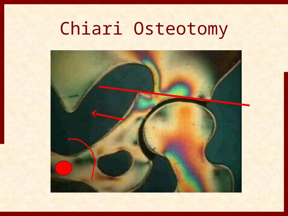
Chiari Osteotomy
Chiari osteotomy
Complications of Treatment
• Worst complication is disturbance of growth in proximal femur including the epiphysis and physeal plate
• commonly referred to as AVN however, no pathology to confirm this
• may be due to vascular insults to epiphysis or physeal plate or pressure injury
• occurrs only in patients that have been treated and may be seen in opposite normal hip
Necrosis of Femoral Head• Extremes of position in abduction ( greater 60
degrees ) and abduction with internal rotation
• compression on medial circumflex artery as passes the iliopsoas tendon and compression of the terminal branch between lateral neck and acetabulum
• “ frog leg position “ uniformly results in proximal growth disturbance
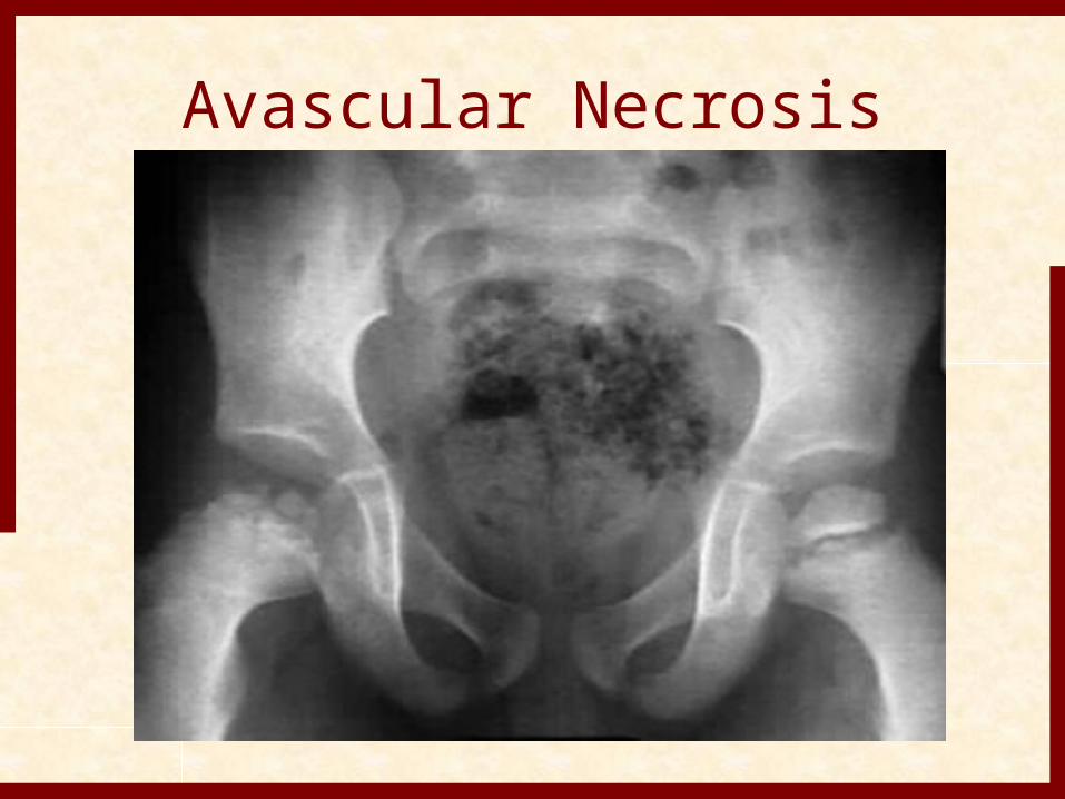
Avascular Necrosis
• extreme position can also cause pressure necrosis onf epiphyseal cartilage and physeal plate
• severin method can obtain reduction but very high incidence of necrosis
• multiple classification systems with Salter most popular
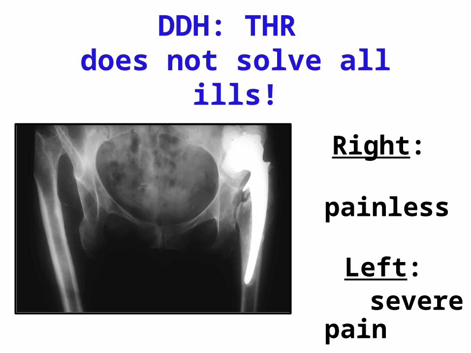
DDH: THR does not solve all ills!
Right: painless
Left: severe pain
THANK YOU


![INNgIs / ]DDh + 0 / ÂÇÊÄ /7 P](https://img.pdfslide.tips/doc/110x75/5affb8617f8b9a84338b9657/inngis-ddh-0-7-pgyig-pi-ji-ig-h-jpqh-egqjq-djpigig-sk-dingi.jpg)