Embed Size (px)
Citation preview

ENDOSCOPY
Edited by Somchai Amornyotin
Endoscopyhttp://dx.doi.org/10.5772/50355Edited by Somchai Amornyotin
ContributorsNurten Savas, George Pados, Pongsakorn Tantilipikorn, Geboes, Petr Lukeš, Michal Zabrodsky, Jan Plzak, MartinChovanec, Jaroslav Betka, Eva Foltynova, Jan Betka, Norman Miner, Joachim Aerts, Robert Peric, Mark De Mol, NicoVan Walree, Lee-Ching Zhu, Verena Grieco, Qinghua Feng, Somchai Amornyotin, Liliana Streba, Paul Mitrut, AncaOana Docea, Cornelia-Daniela Calina, Kris R. Jatana, Charles Elmaraghy, Timothy McEvoy, Rickul Varshney, Marc A.Tewfik, Saul Frenkiel, Faisal Zawawi, John Calleary, Puru Naidu, Jessica Packer, Maheshi Samaraweera, Matthaeus Ch.Grasl, Alexander Aloy, Costin Teodor Streba, Costin Teodor Teodor Streba, Mihai Olteanu, Bogdan Oprea, RalucaMarinas, Mimi Floarea Nitu, Emilia Crisan, Tudorel Ciurea, Johan Van Den Bogaerde, Dario Sorrentino
Published by InTechJaneza Trdine 9, 51000 Rijeka, Croatia
Copyright © 2013 InTechAll chapters are Open Access distributed under the Creative Commons Attribution 3.0 license, which allows users todownload, copy and build upon published articles even for commercial purposes, as long as the author and publisherare properly credited, which ensures maximum dissemination and a wider impact of our publications. However, userswho aim to disseminate and distribute copies of this book as a whole must not seek monetary compensation for suchservice (excluded InTech representatives and agreed collaborations). After this work has been published by InTech,authors have the right to republish it, in whole or part, in any publication of which they are the author, and to makeother personal use of the work. Any republication, referencing or personal use of the work must explicitly identify theoriginal source.
NoticeStatements and opinions expressed in the chapters are these of the individual contributors and not necessarily thoseof the editors or publisher. No responsibility is accepted for the accuracy of information contained in the publishedchapters. The publisher assumes no responsibility for any damage or injury to persons or property arising out of theuse of any materials, instructions, methods or ideas contained in the book.
Publishing Process Manager Iva SimcicTechnical Editor InTech DTP teamCover InTech Design team
First published April, 2013Printed in Croatia
A free online edition of this book is available at www.intechopen.comAdditional hard copies can be obtained from [email protected]
Endoscopy , Edited by Somchai Amornyotinp. cm.ISBN 978-953-51-1071-2

Contents
Preface VII
Section 1 General Aspects 1
Chapter 1 Endoscopy and Histopathology 3Karel Geboes, Karen Geboes and Anne Jouret-Mourin
Chapter 2 Cleaning, Disinfection and Sterilization of Heat-SensitiveEndoscopes 33Norman Miner
Chapter 3 Anesthetic Management for LaparoscopicCholecystectomy 39Somchai Amornyotin
Chapter 4 Adhesion Prevention Strategies in Laparoscopic Surgery 49George Pados, Anastasios Makedos and Basil Tarlatzis
Section 2 Head and Neck 73
Chapter 5 Narrow Band Imaging (NBI) — Endoscopic Method forDetection of Head and Neck Cancer 75Petr Lukes, Michal Zabrodsky, Jan Plzak, Martin Chovanec, JaroslavBetka, Eva Foltynova and Jan Betka
Chapter 6 Therapeutic and Diagnostic Approaches in Rhinologyand Allergy 89Pongsakorn Tantilipikorn
Chapter 7 Role of Endoscopic Sinus Surgery in Pediatric AcuteComplicated Sinusitis 103Timothy P. McEvoy, Charles A. Elmaraghy and Kris R. Jatana
Chapter 8 Endoscopy - An Advancement in Sinus and SkullBase Surgery 121Rickul Varshney, Faisal Zawawi, Marc A Tewfik and Saul Frenkiel
Section 3 Respiratory Tract 139
Chapter 9 Endoscopy of Larynx and Trachea with Rigid Laryngo-Tracheoscopes Under Superimposed High-Frequency JetVentilation (SHFJV) 141Alexander Aloy and Matthaeus Grasl
Chapter 10 Bronchology – A Well Branched Tree 201Mihai Olteanu, Costin Teodor Streba, Bogdan Oprea, RalucaMarinas, Mimi Floarea Nitu, Emilia Crisan and Tudorel Ciurea
Chapter 11 Diagnostic and Therapeutic Approaches in RespiratoryEndoscopy 255R. Peric, M. de Mol, N. van Walree and J.G. Aerts
Section 4 Miscellaneous 267
Chapter 12 Innovative Uses and Emerging Technologies inEndoscopy 269J. Van Den Bogaerde and D. Sorrentino
Chapter 13 The Use of Endoscopic Ultrasound-Guided Fine NeedleAspiration (EUS-FNA) in Cytopathology Diagnosis 287Lee-Ching Zhu, Qinghua Feng and Verena S. Grieco
Chapter 14 Endoscopy in Pregnant Patients 321Nurten Akyurek Savas
Chapter 15 Endoscopy in Pregnancy 349Paul Mitrut, Anca Oana Docea, Cornelia - Daniela Calina and LilianaStreba
Chapter 16 Modern Upper Urinary Tract Endoscopy 363Puru Naidu, Jessica Packer, Maheshi Samaraweera and John G.Calleary
ContentsVI
Preface
Endoscopy is a fast moving field, and new techniques are constantly emerging. Flexible endos‐copy became the principal investigational tool of the pathological abnormalities with great im‐pact on modern medicine. In recent decades, endoscopy has evolved and branched out from avisual diagnostic modality, to enhanced video and computer assisted imaging, with impressiveinterventional capabilities. Some new endoscopic techniques will be too complex or expensiveto make the leap into general practice, others already show major progress in the managementof diseases. Modern endoscopy has seen advances not only in the types of endoscopes availa‐ble, but also the types of interventions amenable to the endoscopic approach. As in any field,demands of service delivery by conventional equipment and newer, more glamorous, and usu‐ally more expensive technologies are often in competition.
Modern endoscopic equipment provides us with the benefit of many technical advances. Newvideo-endoscopes, magnification endoscopes and confocal of narrow band imaging endoscopesemerged. An increased knowledge of normal and pathologic endoscopic patterns has been in‐creasing in the last decades. Endoscopy is an effective and safe procedure even in special popu‐lations including pediatric patients, pregnant patients and liver transplant patients. It served asthe tool for diagnosis and therapeutic interventions of many organs including gastrointestinaltract, head and neck, respiratory tract and others. In this book the authors will discuss some ofthe emerging techniques and technologies used to increase the diagnostic and therapeutic yieldin the various organs.
The contributions in this book are very valuable. InTech Open Access Publisher selected severalknown names from many countries with different levels of development. Multiple specificpoints of view were presented together with various topics regarding diagnostic or therapeuticendoscopy. The readers can take into consideration of practical knowledge in the endoscopicfield. This book actually represents a valuable tool for formation and continuous medical edu‐cation in the endoscopy considering the performances or technical possibilities in differentparts of the world.
I very much appreciate and thank to all authors of this book. Many thanks to InTech Open Ac‐cess Publisher which offered me the possibility of editing this attractive book. It was a realpleasure to read such interesting works by so many experts from all over the world. Finally, Ialso thank Ms. Iva Simcic for her perfect, prompt and efficient co-operation.
Assoc. Prof. Somchai Amornyotin MD, FRCATDepartment of Anesthesiology and Siriraj GI Endoscopy Center
Faculty of Medicine Siriraj HospitalMahidol University, Bangkok, Thailand


Section 1
General Aspects

Chapter 1
Endoscopy and Histopathology
Karel Geboes, Karen Geboes andAnne Jouret-Mourin
Additional information is available at the end of the chapter
http://dx.doi.org/10.5772/52739
1. Introduction
Endoscopy and histopathology are two morphological diagnostic procedures which allowdirect examination of organs with optical methods. They can detect abnormalities of thenormal anatomy and histology and provide a precise diagnosis. Based on the informationderived from these investigations an adequate treatment, either medical or surgical can beproposed. The optical resolution of both methods is different. Classical endoscopy is usingessentially the naked eye observation of the tissue which allows a diagnosis of an ulcer or araised lesion for instance, while histopathology is reaching the cellular and sub-cellular level.The new endoscopic techniques however do increase the optical resolution. The majorcontributions of histopathology to endoscopy are situated in inflammatory and neoplasticdiseases. Histopathology allows a more precise diagnosis of the type of inflammation and abetter classification of tumours. This has again an impact upon treatment. For the diagnosis,histopathology can be an essential element, as illustrated by gluten sensitive enteropathy(although serology is also an essential element) or by identification of specific pathogens suchas Giardia lamblia, Mycobacterium avium, cryptosporidia.... Histopathology can further beimportant for the confirmation of a diagnosis but very often it will provide a more precisediagnosis by determining the aetiology of inflammation as illustrated by autoimmune gastritis,or by typing a tumour (adenocarcinoma or lymphoma). In addition, histopathology canprovide essential elements for further therapy strategy by demonstrating the presence orabsence of risk factors for residual tumour in polypectomy or endoscopic mucosal resection.Indirectly, it offers the possibility of using additional techniques such as biomarkers fordysplasia and cancer or the demonstration of mutations such as KRAS in colorectal cancer orHER2 amplification in oesophageal and gastric cancer.[1, 2].These applications can haveimportant therapeutic consequences. It has been shown for instance that activating mutationsof the KRAS gene are associated with poor response to anti-EGFR therapies and that patients
with tumors that had high levels of HER2 protein expression derived the greatest benefit fromtreatment with trastuzumab..
2. What is the influence of endoscopy on the diagnostic yield ofhistopathology?
2.1. General requirements for the endoscopist and the pathologist
A close collaboration between the endoscopist and the pathologist is essential for an accuratediagnosis. This imposes on each of the partners some constraints.
Overall the endoscopist should provide the pathologists with a copy of the endoscopy reportmentioning the sites of the biopsies, a macroscopic description of the lesions if present and theadjacent mucosa and essential clinical information such as the age of the patient, the immunestatus of the patient, duration of symptoms and treatment if any.
The pathologist should provide information of the quality of the biopsies (number and sizeand depth of the samples) in order to avoid false conclusions, a degree of probability of hisinitial diagnosis and if needed suggest particular conditions for further sampling or ancillarytechniques such as immune histochemistry. Contentious cases should be selected for clinicpathological discussion.[3]
2.2. Sampling of biopsies
The diagnostic yield of histopathology depends upon the experience of the pathologist butalso upon the quality of the biopsy samples and sampling error. The quality of the samples isinfluenced by a variety of elements such as the size and shape of the biopsy forceps, the natureand location of the disease, the experience of the endoscopist and the number of samples.During endoscopy samples can be obtained by way of different techniques. These includepinch biopsy, suction biopsy with a multipurpose tube (which provides larger samples), brushcytology, endoscopic fine needle aspiration (offering material from deep areas in the lesion)and snare excision or strip biopsy.
Pinch biopsy is the most common technique. Several types of biopsy forceps are available. Adistinction can be made between those with elliptical and those with round cups. Generallythe samples obtained with elliptical cups are larger. A forceps with round cups may be moreappropriate for children in order to avoid complications. The size of the biopsy forcepsdetermines partly the size (surface and depth) of the samples. The small forceps has a widthof 1.8 mm when opened. The average forceps has a 2.4 mm diameter and allows to obtainsamples containing the muscularis mucosae (and upper submucosa) in 60% of the cases. Thelarger Jumbo forceps has a 3.4 mm diameter. Samples obtained with this forceps are larger,but, they usually contain not more submucosa and the risk of complications (perforation andbleeding) may be more important, whereas it is minimal with the smaller forceps (if the patienthas normal coagulation). A forceps can have a central spike so that it stays in position in the
Endoscopy4
mucosa, during the procedure. The spike can induce artefacts which should not be confusedwith erosions.
The anatomic location or certain types of lesions may be the reason why samples are of lessgood quality or superficial in nature. This is often so in areas immediately distal to a stricture,and at the papilla of Vater in the duodenum. The extrahepatic bile ducts and the pancreaticduct are other areas where biopsies are more difficult to obtain and hence usually smaller. Ifthe biopsies of the papilla are taken following sphincterotomy, coagulation artefacts are likelyto be present.
In order to obtain samples of appropriate depth air insufflation during the endoscopicexamination should be limited. When over-insufflation occurs the mucosa is stretched andpushed towards the underlying submucosa and the samples are likely to be more superficial.
The samples obtained with a forceps are usually limited to the mucosa. Normally they are notsuitable for the assessment of submucosal or deeper lesions. This means that they are not goodfor instance for a diagnosis of “vasculitis”, except for small vessel disease. By the use of a"burrowing technique" whereby several biopsies are taken in the same area information ofdeeply situated lesions can eventually be obtained. An alternative are samples obtained withendoscopic ultrasound guided fine needle aspiration. They are usually smaller but they permitboth morphologic and cytologic analysis of lesions within or adjacent to the gastrointestinal(GI) tract. They can be used for the assessment of neoplastic lesions, but because of the smallsize, they are not good for conditions such as vasculitis.[4]
2.3. Larger samples
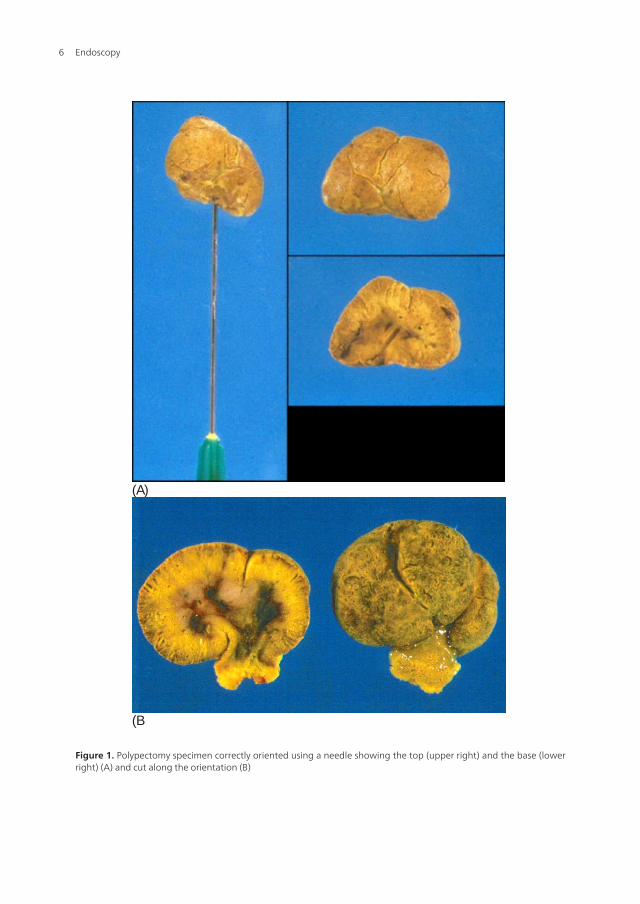
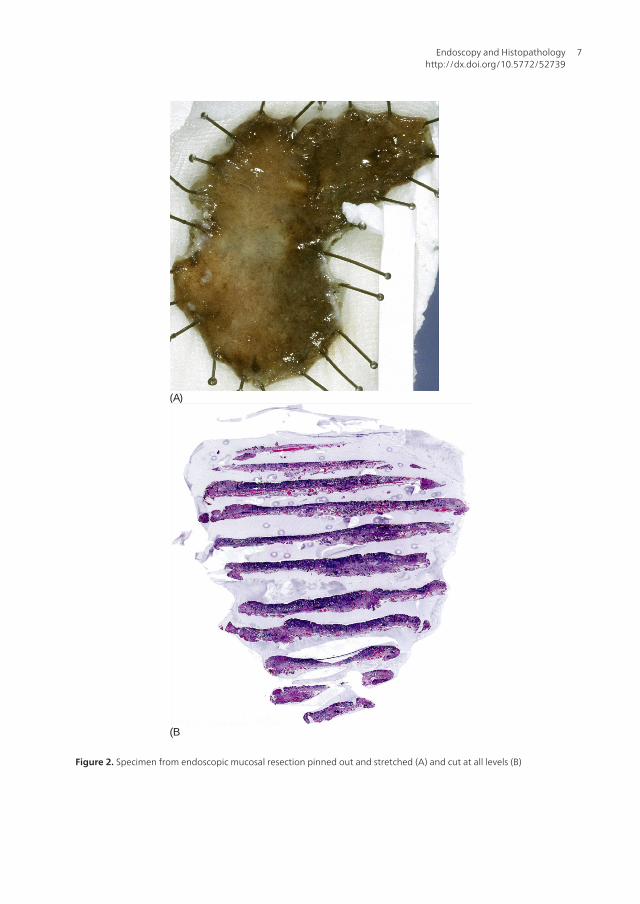
Larger samples are obtained with endoscopic mucosal resection (EMR) or endoscopicalsubmucosal resection (ESD) and snare polypectomy. These samples must be handledadequately by the endoscopist or/and in the pathology lab. The histopathological interpreta‐tion of these samples provides important information for subsequent management andassessment of the risk for residual cancer. A correct diagnostic process involves, tumourdifferentiation, precise determination of deep infiltration, lymphatic permeation and adequatedetermination of the section margin. Identification of this area is easy if the lesion is adequatelyoriented. In the case of polypectomy, the endoscopist could identify the section margin withIndia ink or with a pin if the lesion is removed in one piece. The specimen will be cut alongthe marker. (Fig. 1) In the case of EMR /ESD, ideally, the specimen should be oriented, pinnedand stretched on card board in the endoscopy unit.(Fig 2) If the specimen is not removed inone piece, reconstruction of the specimens should be attempted. Painting of the base andmargins is useful, as tumour extension to the deep margin implies surgery and remnants ofthe neoplastic epithelium at the lateral margins indicate re-excision or postoperativedestruction.[5, 6] Good communication between the pathologist and the clinicians is importantfor the assessment of the efficacy of the treatment and for the design of the strategy of theadditional treatment which is based upon the depth of invasion of the lesion. If the resectionhas been performed in piecemeal fashion and the specimen is received in two or morefragments, it may be impossible to determine the true margin of resection, if the endoscopistdid not attempt to identify the true margin or placed the true margin in a separate container.[7]
Endoscopy and Histopathologyhttp://dx.doi.org/10.5772/52739
5
(A)
(B
Figure 1. Polypectomy specimen correctly oriented using a needle showing the top (upper right) and the base (lowerright) (A) and cut along the orientation (B)
Endoscopy6
(A)
(B
Figure 2. Specimen from endoscopic mucosal resection pinned out and stretched (A) and cut at all levels (B)
Endoscopy and Histopathologyhttp://dx.doi.org/10.5772/52739
7
2.4. Endoscopic ultrasound-guided fine-needle aspiration biopsy
Endoscopic ultrasound guided fine needle aspiration has become the most accurate modalityfor characterization of pancreatic cystic and solid lesions, differential diagnosis of indetermi‐nate masses and locoregional staging of some digestive cancers (gastric, oesophagus, pancreas,biliary tract…). It should be performed in the primary mass but also in distant lymph nodes,or metastatic locations. EUS-FNA has a high sensitivity, specificity, positive predictive valueand accuracy in the assessment of biliopancreatic tumours. The performance of this techniqueis dependent on the endoscopist and cytopathologist experience. It is well known that accuracyof FNA increases when the technique is performed by an experienced clinician and when theslides are reviewed by an experienced cytopathologist the collaboration between these twophysicians is also very important.[8]
2.5. Number of samples
Several studies have shown that the diagnostic yield of histopathology is increased andsampling error is decreased by increasing the number of biopsies. This has been demonstratedfor inflammatory diseases such as chronic idiopathic inflammatory bowel diseases (IBD) andfor neoplastic diseases.[9] Therefore different guidelines for endoscopic sampling in variousdiseases have been developed.[10-12] ECCO guidelines propose to obtain “multiple” biopsiesfrom five sites around the colon (including the rectum) and the ileum for a reliable diagnosisof Crohn’s disease. Multiple biopsies imply a minimum of two samples from each site (Table)This is also true for a diagnosis of collagenous or lymphocytic colitis. Thickening of thesubepithelial collagen table in collagenous colitis is indeed not homogeneous. Such guidelinesare very important in clinical practice. They limit sampling error and compensate for the smallsize of the samples. However, the introduction of new technologies and modern endoscopesincluding zoom endoscopy, high magnification endoscopy and more sophisticated techniquessuch as laser-scanning confocal endoscopy and endo-cytoscopy (microscope incorporated inthe endoscope) will change practice in the future by offering the possibility of targeted biopsies.In a recent study at our institute, chromo endoscopy (CE) and narrow band imaging (NBI)were used to detect dysplasia in ulcerative colitis. A total of 268 raised lesions were detectedin 83 patients (156 lesions in 45 patients with CE and 112 lesions in 38 patients with NBI). Onhistology, 44 were shown to be neoplastic (26 lesions in 10 patients with CE and 18 lesions in12 patients with NBI): 1 adenocarcinoma, 1 high grade dysplasia, 2 dysplasia associated lesionor mass, and 17 adenoma like mass. The new endoscopic techniques are also narrowing thegap between endoscopy and pathology. Laser scanning endoscopy provides a microscopy-level image without obtaining a biopsy specimen. Endo-cytoscopy is based on the technologyof light contact microscopy. The tip of an endoscope is placed in direct contact with a dye-stained surface and then the surface is scanned with condensed normal white light, producingcellular-level imaging. Laser endoscopy increases the real time diagnostic yield and can beused to confirm dysplasia with high accuracy. Bio-endoscopy is another technique underconsideration. It involves the use of monoclonal antibodies labelled with a fluorescent tag ofreporter probes (molecules that enter cells) or fluorescent DNA probes for FISH in order todetect in situ molecular changes or chromosomal instability.[13-16]
Endoscopy8
Table 1. Recommendations for biopsy strategies in inflammatory conditions of the gastrointestinal tract
While these new techniques can offer real time images and diagnosis, the interpretation of theimages still depends on the morphological features of the lesions, as observed with microscopyand some lesions like sessile serrated adenomas are still beyond the reach of real timediagnosis. The endoscopist must therefore have a thorough knowledge of pathology.
3. Specimen handling
Specimen handling should be done carefully in order to allow optimal diagnostic work up. Itimplies proper identification of the patient, including the age, specification of the site of origin,fixation and in some instances, orientation. Adequate fixation by an appropriate fixative is
Endoscopy and Histopathologyhttp://dx.doi.org/10.5772/52739
9
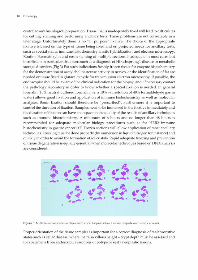
central to any histological preparation. Tissue that is inadequately fixed will lead to difficultiesfor cutting, staining and performing ancillary tests. These problems are not correctable in alater stage. Unfortunately there is no "all purpose" fixative. The choice of the appropriatefixative is based on the type of tissue being fixed and on projected needs for ancillary tests,such as special stains, immune histochemistry, in situ hybridization, and electron microscopy.Routine Haematoxylin and eosin staining of multiple sections is adequate in most cases butinsufficient in particular situations such as a diagnosis of Hirschsprung's disease or metabolicstorage disorders.(Fig 3) For such indications freshly frozen tissue for enzyme histochemistryfor the demonstration of acetylcholinesterase activity in nerves, or the identification of fat areneeded or tissue fixed in glutaraldehyde for transmission electron microscopy. If possible, theendoscopist should be aware of the clinical indication for the biopsy, and, if necessary contactthe pathology laboratory in order to know whether a special fixation is needed. In generalformalin (10% neutral buffered formalin, i.e. a 10% v/v solution of 40% formaldehyde gas inwater) allows good fixation and application of immune histochemistry as well as molecularanalyses. Bouin fixation should therefore be “proscribed”. Furthermore it is important tocontrol the duration of fixation. Samples need to be immersed in the fixative immediately andthe duration of fixation can have an impact on the quality of the results of ancillary techniquessuch as immune histochemistry. A minimum of 6 hours and no longer than 48 hours isrecommended for adequate molecular biology procedures such as for HER2 immunehistochemistry in gastric cancer.[17] Frozen sections will allow application of most ancillarytechniques. Freezing must be done properly (by immersion in liquid nitrogen for instance) andquickly in order to avoid the formation of ice cristals. Rapid adequate freezing and preventionof tissue degeneration is equally essential when molecular techniques based on DNA analysisare considered.
Figure 3. Multiple sections from multiple endoscopic biopsies allow a more complete microscopic analysis
Proper orientation of the tissue samples is important for a correct diagnosis of malabsorptivestates such as celiac disease, where the ratio villous height – crypt depth must be assessed andfor specimens from endoscopic resections of polyps or early neoplastic lesions.
Endoscopy10
4. Immune histochemistry and other ancillary techniques
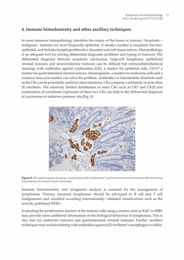
In most instances histopathology identifies the nature of the lesion or tumour. Neoplastic –malignant - tumours are most frequently epithelial. A smaller number is neoplastic but non-epithelial, and includes lymphoproliferative disorders and soft tissue tumors. Histopathologyis an adequate tool for solving differential diagnostic problems and typing of tumours. Thedifferential diagnosis between anaplastic carcinomas, large-cell lymphoma, epithelioidstromal tumours and neuroendocrine tumours can be difficult but immunohistochemicalstainings with antibodies against cytokeratins (CK), a marker for epithelial cells, CD117 amarker for gastrointestinal stromal tumors, chromogranin, a marker for endocrine cells and acommon leucocyte marker can solve the problem. Antibodies to intermediate filaments suchas the CKs can be potentially useful in other situations. CKs comprise a subfamily of more than20 members. The relatively limited distribution of some CKs such as CK7 and CK20 andexamination of coordinate expression of these two CKs can help in the differential diagnosisof carcinomas of unknown primary site.(Fig. 4)
Figure 4. Microphotograph showing a rectal biopsy with Cytokeratin 7 positive immunohistochemistry demonstratingthe presence of a breast cancer metastasis.
Immune histochemistry and cytogenetic analysis is essential for the management oflymphomas. Primary intestinal lymphomas should be sub-typed in B cell and T cellmalignancies and classified according internationally validated classifications such as therecently published WHO.
Evaluating the proliferation fraction of the tumour cells using a marker such as Ki67 or MIB1may provide some additional information on the biological behaviour of lymphomas. This isalso true for endocrine tumours and gastrointestinal stromal tumours. Further ancillarytechniques may include staining with antibodies against p53 for Barret’s oesophagus or colitis-
Endoscopy and Histopathologyhttp://dx.doi.org/10.5772/52739
11
associated dysplasia. Currently a number of markers are under investigation for a moreaccurate identification of early neoplasia.
Histochemistry (histological special stains) searching for mucins or other substances, andoccasionally electron microscopy and genetic markers can also be applied on biopsy samples.Many stainings can be performed on routinely formalin fixed material. Increasingly there issome overlap, between immune histochemistry and molecular techniques since geneticmarkers can be demonstrated also by immune histochemistry. This is for instance true forlarge-bowel cancers with microsatellite instability (MS), where the products of the DNA repairgenes hMLH1, hMSH2 and MSH6, or the lack of them, can be demonstrated immunehistochemically. These products do not however cover the whole range of MS. DNA or RNAextraction and genetic analysis remains important and there may even be a growing need.
5. The oesophagus
5.1. Inflammatory conditions
At present, there is no ideal scenario for a biopsy series for the diagnosis of gastro-oesophageal reflux disease (GORD). In general, it is accepted that changes in the squamousmucosa are usually found in the distal oesophagus close to the squamo-columnar junction.Biopsies from the squamous mucosa should be completed with biopsies from the cardia.Histological changes indicative of gastro-oesophageal reflux are indeed found at both sidesof the squamo-columnar junction.[18-21] The diagnosis of this condition, called carditis,which occurs in the absence of signs of gastritis in the antrum and corpus due toHelicobacter pylori or other causes of gastritis implies also biopsies of antrum and corpusin order to exclude the presence of these causes. A biopsy run for GORD should thereforeideally include samples from the distal oesophagus, particularly from the Z-line and at 2cm above, from the cardia distal to the Z-line and from the stomach.[22, 23] However, inmost cases, peptic oesophagitis due to GORD - the most common inflammatory conditionof the oesophagus - does not require biopsy diagnosis for those patients presenting withtypical symptoms and macroscopic endoscopic alterations.[24]
Biopsies are mainly useful in patients presenting with normal endoscopy and abnormal acidexposure (non-erosive reflux disease – NERD), in patients with typical symptoms and normalendoscopy and pH-metry or in patients with atypical symptoms. The presence of “dilatedintercellular spaces (DIS)” or of a combination of DIS with other microscopic features such asbasal zone hyperplasia observed in GORD may confirm the suspected diagnosis of reflux.[21]There are however several other types of oesophagitis. The presence of an intense eosinophilinfiltration must orient towards a diagnosis of eosinophilic oesophagitis. Eosinophilicoesophagitis can present a typical endoscopic pattern known as “ringed oesophagus” but theoesophagus can appear normal in up to 20% of the patients. It is important to recognise thatthe eosinophilic infiltration may have a heterogeneous distribution within the oesophagus.Therefore, when considering eosinophilic oesophagitis, it is critical to have biopsies frommultiple areas, including the distal, mid, and proximal oesophagus.[25] Biopsies of the
Endoscopy12
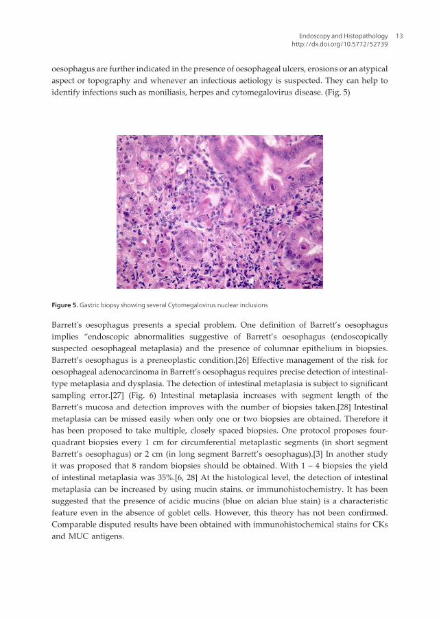
oesophagus are further indicated in the presence of oesophageal ulcers, erosions or an atypicalaspect or topography and whenever an infectious aetiology is suspected. They can help toidentify infections such as moniliasis, herpes and cytomegalovirus disease. (Fig. 5)
Figure 5. Gastric biopsy showing several Cytomegalovirus nuclear inclusions
Barrett's oesophagus presents a special problem. One definition of Barrett’s oesophagusimplies “endoscopic abnormalities suggestive of Barrett’s oesophagus (endoscopicallysuspected oesophageal metaplasia) and the presence of columnar epithelium in biopsies.Barrett’s oesophagus is a preneoplastic condition.[26] Effective management of the risk foroesophageal adenocarcinoma in Barrett’s oesophagus requires precise detection of intestinal-type metaplasia and dysplasia. The detection of intestinal metaplasia is subject to significantsampling error.[27] (Fig. 6) Intestinal metaplasia increases with segment length of theBarrett’s mucosa and detection improves with the number of biopsies taken.[28] Intestinalmetaplasia can be missed easily when only one or two biopsies are obtained. Therefore ithas been proposed to take multiple, closely spaced biopsies. One protocol proposes four-quadrant biopsies every 1 cm for circumferential metaplastic segments (in short segmentBarrett’s oesophagus) or 2 cm (in long segment Barrett’s oesophagus).[3] In another studyit was proposed that 8 random biopsies should be obtained. With 1 – 4 biopsies the yieldof intestinal metaplasia was 35%.[6, 28] At the histological level, the detection of intestinalmetaplasia can be increased by using mucin stains. or immunohistochemistry. It has beensuggested that the presence of acidic mucins (blue on alcian blue stain) is a characteristicfeature even in the absence of goblet cells. However, this theory has not been confirmed.Comparable disputed results have been obtained with immunohistochemical stains for CKsand MUC antigens.
Endoscopy and Histopathologyhttp://dx.doi.org/10.5772/52739
13
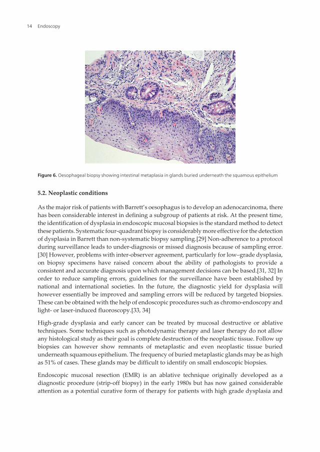
Figure 6. Oesophageal biopsy showing intestinal metaplasia in glands buried underneath the squamous epithelium
5.2. Neoplastic conditions
As the major risk of patients with Barrett’s oesophagus is to develop an adenocarcinoma, therehas been considerable interest in defining a subgroup of patients at risk. At the present time,the identification of dysplasia in endoscopic mucosal biopsies is the standard method to detectthese patients. Systematic four-quadrant biopsy is considerably more effective for the detectionof dysplasia in Barrett than non-systematic biopsy sampling.[29] Non-adherence to a protocolduring surveillance leads to under-diagnosis or missed diagnosis because of sampling error.[30] However, problems with inter-observer agreement, particularly for low–grade dysplasia,on biopsy specimens have raised concern about the ability of pathologists to provide aconsistent and accurate diagnosis upon which management decisions can be based.[31, 32] Inorder to reduce sampling errors, guidelines for the surveillance have been established bynational and international societies. In the future, the diagnostic yield for dysplasia willhowever essentially be improved and sampling errors will be reduced by targeted biopsies.These can be obtained with the help of endoscopic procedures such as chromo-endoscopy andlight- or laser-induced fluoroscopy.[33, 34]
High-grade dysplasia and early cancer can be treated by mucosal destructive or ablativetechniques. Some techniques such as photodynamic therapy and laser therapy do not allowany histological study as their goal is complete destruction of the neoplastic tissue. Follow upbiopsies can however show remnants of metaplastic and even neoplastic tissue buriedunderneath squamous epithelium. The frequency of buried metaplastic glands may be as highas 51% of cases. These glands may be difficult to identify on small endoscopic biopsies.
Endoscopic mucosal resection (EMR) is an ablative technique originally developed as adiagnostic procedure (strip-off biopsy) in the early 1980s but has now gained considerableattention as a potential curative form of therapy for patients with high grade dysplasia and
Endoscopy14
superficial cancers. It is also a good tool for histological staging because the procedure allowsto remove intact mucosa and submucosa enabling complete evaluation of mucosal andsubmucosal invasion. EMR as a diagnostic tool has been shown to be superior to mucosalbiopsy and inter-observer agreement of Barrett’s oesophagus related dysplasia is significantlybetter compared with biopsy specimens.[35] The presence of a double layer of muscularismucosae, which is a hallmark of Barrett’s oesophagus, is an important landmark. Only wheninvasion extends beyond the deeper layer (the genuine muscularis mucosae), a diagnosis ofsubmucosal invasion is justified.[36]
Endoscopic biopsies are also commonly used for the diagnosis of cancer of the oesophagusand the distinction between squamous cell carcinoma and adenocarcinoma. Two samples canprovide a positive diagnosis in 95.8% of cases. The addition of four samples increases thepositive yield to 100%. There is no statistically significant difference in the yield according tothe site and type of growth.[37] However, in strictures the diagnosis can be difficult. In thissituation, the additional use of brush cytology may increase the diagnostic yield. Soft tissuetumours and lymphomas are less common in the oesophagus. The so-called Abrikosoff tumouror granular cell tumour, a relatively rare lesion, may present a problem as the overlyingsquamous epithelium can show hyperplasia which might be confused with neoplastic changes.The tumour itself is composed of aggregates of round cells with a characteristic granularcytoplasm showing S100 positivity with immunohistochemical stains. If the biopsy samplesare too superficial, the diagnosis can however be difficult. Fine needle aspiration biopsy couldbe used for the former, although most soft tissue tumours of the oesophagus are not malignant.Brush cytology can be helpful for the diagnosis of infections.
6. The stomach
6.1. Inflammatory conditions
Throughout the GI tract, mucosal features such as redness, oedema, swelling, bleeding,erosions and ulcers can be observed. They reflect inflammation and tissue damage but mayalso be due to mucosal atrophy and epithelial metaplasia. Metaplasia is most readily detectedendoscopically in the distal oesophagus but it is also common in the stomach. In the latter itmay appear as small red depressions simulating erosions or aphthoid ulcers, as an irregularnodular area or as larger geographic red areas. The red colour and a depressed or nodularappearance can be explained by thinning of the mucosa due to atrophy and increased visibilityof the vessels. Pathology is useful to confirm the endoscopic abnormality and probablediagnosis, or to exclude such abnormalities or give another explanation. A depressed red spotcan indeed be a genuine erosion but it may also represent a vascular ectasia or a small area ofmucosal atrophy. Inflammatory conditions in the stomach include gastritis and reactivegastropathy (chemical gastropathy, bile reflux). The latter is characterized by epithelialdamage and a minimal inflammatory cell reaction. Several types of gastritis can bedistinguished and histopathology plays a major role in this distinction. An aetiology-basedclassification was proposed in the Sydney system at the World Congress of Gastroenterology
Endoscopy and Histopathologyhttp://dx.doi.org/10.5772/52739
15
in 1990 and updated in 1994.[38, 39] The Sydney system also established the need for takingdifferent biopsies of the gastric mucosa.[38] The guidelines include a) two biopsies of thecorpus and two of the antrum for an overall assessment of the distribution of the gastritis andthe distinction between antral gastritis, corpus gastritis and pan gastritis; b) one biopsy of theangulus because atrophic gastritis and intestinal metaplasia are related with the developmentof gastric cancer and occur most commonly at the angulus; c) the same area is the mostappropriate area to look for the presence of dysplasia. In small children, this approach mayhowever not be appropriate. Two samples from the stomach may be sufficient. Biopsydiagnosis should include the morphological site or sites, the morphological lesions present,and any potential cause. The sensitivity and specificity for the diagnosis of Helicobacter pylorigastritis are high, varying between 88 and 99% for the former and 90 and 100% for thespecificity. The negative predictive value is near 100% for antral biopsies. Active gastritis, orgastritis with neutrophils is often Helicobacter pylori positive and will imply treatment,whether activity is mild, moderate or severe. Grading atrophy and intestinal metaplasia is lessreproducible.[40] Staging of gastritis has been proposed among others by the so-called OLGAsystem but may be difficult to apply in routine practice.[41] Grading and staging couldhowever be useful for the identification of patients at risk for cancer. In addition to the gastricbiopsies ii seems reasonable to obtain, during the first diagnostic examination, also duodenalbiopsies to look for the presence of mucous surface (gastric) metaplasia, a requirement forHelicobacter pylori colonisation of the duodenum which can induce duodenal ulcers, or forepithelial lymphocytosis. If the stomach biopsies are normal and duodenitis is found onhistopathology, a Helicobacter pylori-induced duodenitis is highly unlikely. If the patient haslymphocytic gastritis of the antrum and epithelial lymphocytosis in the duodenum, a diagnosisof celiac disease should be suspected. Follow-up biopsies for gastritis can be considered whena treatment for HP has been given in order to assess eradication or when intestinal metaplasiaand atrophy are very extensive.
Whenever special forms of gastritis are suspected multiple biopsies are needed. Histopathol‐ogy can identify a variety of pathogens in infectious gastritis. Many of the special types lackendoscopic abnormalities. Lymphocytic gastritis can present as a hypertrophic variant witherosions and thickening of the gastric wall suggestive of Menetrier’s disease. It can be diffuseor corporeal and correspond in these forms to varioliform gastritis. It can also be limited to theantrum and in this case includes various conditions ( reflux gastritis, HP gastritis or coeliacdisease) must be considered. [42] The histopathology of gastroduodenal Crohn’s diseaseincludes a wide spectrum of changes, including the presence of granulomas as well as focallyenhanced (active) gastritis.[43] A correct diagnosis of Crohn's disease of the stomach can bereached more accurately when multiple samples of the suspected sites (n=5) and of normalsites are available. Granulomas can be detected in biopsies from macroscopically abnormalmucosa as well as in biopsies from normal mucosa. The frequency of detecting granulomasvaries between 4.6% and 26% depending upon the presence of endoscopic lesions, the numberof biopsies and the number of sections examined. Multiple biopsies will increase the diagnosticyield. Focally enhanced or focally active gastritis is typified by small collections of lymphocytesand histiocytes surrounding a small group of foveolae or gastric glands, often with infiltratesof neutrophils. Several studies have found that focally enhanced gastritis is common in adult
Endoscopy16
Crohn’s disease patients. However, studies that used control groups have reported aprevalence of focally enhanced gastritis in non-IBD patients in up to 19.4%. Therefore, this typeof gastritis may not be a good marker for the diagnosis of IBD or IBD-related gastritis in adults.[44, 45] It may still be a good marker in children although it may not reliably distinguishbetween Crohn’s disease and ulcerative colitis. Some studies have found that focally enhancedgastritis is present in up to 20% of paediatric ulcerative colitis patients, suggesting that thistype of gastritis is a marker of IBD in general in children.
Biopsies are less indicated for the diagnosis of vascular abnormalities. They can howeverbe useful for the diagnosis of “gastric antral vascular ectasia” (GAVE). GAVE is a rarecondition (prevalence approximately 3/10000 upper endoscopies), characterised by red spotsin linear array in the antrum of the stomach. Based on the striped features from the antrumat endoscopy, the disorder has been called the “watermelon” stomach. The histologicallesion consists of numerous dilated vessels in the mucosa, often with microthrombi, withfibromuscular hyperplasia and fibrohyalinosis of the perivascular lamina propria. Themucosa shows no or mild chronic inflammation or atrophy with intestinal metaplasia.[46]GAVE must be distinguished from “portal hypertensive gastropathy” and from “gastricvascular ectasia”.[47]
6.2. Neoplastic conditions
In patients with marked atrophic gastritis or pernicious anaemia, the possibility of endocrinecell hyperplasia and dysplasia needs to be considered, and immunostains can readily answerthis question. In patients with endocrine tumours (carcinoïds), the issue is whether these aresporadic, associated with atrophic gastritis, or even multiple endocrine neoplasia (MEN) andZollinger-Ellison syndrome. Biopsies of adjacent gastric body mucosa will show whether thereis hyperplasia of parietal cells without atrophy as in Zollinger Ellison and MEN, atrophy asseen in pernicious anemia, or normal mucosa as seen in sporadic endocrine tumours.
The macroscopic differential diagnosis between benign and malignant ulcers of the stomachis correct, on average, in only 75% of cases (52% to 94% of cases depending on the seriesreported in the literature).[3] Hence, the differential diagnosis can depend upon histology.Chromo-endoscopy with targeted biopsies will change the guidelines in the future.
In the series reported in the literature, the proportion of cancer-positive biopsies variesbetween 49% and 56% and about 25% of the biopsies are considered inadequate. A method ofbiopsy by quadrants with a technique that avoids the lesion to be covered by the bleeding fromearlier biopsies reduces the number of unusable biopsies to 5.7% and increases the proportionof cancer-positive biopsies to 67%. An average of 7 - 10 biopsies is required to reach enoughsensitivity and in order to avoid false negative results.[48, 49] When gastric lymphoma issuspected multiple biopsies are also required. If the lesion presents as an ulcer, biopsies fromthe edge (as for carcinoma) and the ulcer base should be obtained. Proper fixation (in order toallow additional tests such as immuno-histochemistry and Polymerase Chain Reaction) isabsolutely indicated.
Endoscopy and Histopathologyhttp://dx.doi.org/10.5772/52739
17
Histopathology is also very useful for the identification of metastases or secondary malignantinvolvement of the GI tract a problem which is becoming more common. Breast and melanomaare the most frequently found. Approximately 1 metastasis is observed per 3847 upper GIendoscopies and 1 lower metastasis per 1871 colonoscopies. The stomach and duodenum arethe most common locations. Immune histochemistry for cytokeratin patterns and othermarkers can help to identify the primary origin if needed.
Overall a microscopic diagnosis of polyps (elevated lesions) depends on the type of the lesionand the size and number of biopsies. Polyps of epithelial origin can be diagnosed with classicalpinch biopsies. They include benign lesions such as fundic gland polyps and neoplastic lesionssuch as adenomas or neuro-endocrine dysplasia. A complete evaluation may need larger snarebiopsies and implies orientation. This is also needed for EMR specimens from early –superficial gastric cancer and adenomas. As in Barrett’s oesophagus, a good orientation isessential for the assessment of the risk factors for residual tumour and the need for additionalsurgery. In contrast with the oesophagus, soft tissue tumours are more common in the stomach.These are usually gastrointestinal stromal tumours (GIST). These tumours show a positivestaining with antibodies directed against CD117, DOG1 and often also for CD34 (87% positivecases in the stomach). They produce polypoid lesions with a smooth or ulcerated surface as aresult of a submucosal process. Such a process can be inflammatory or tumoral and will oftennot be diagnosed adequately when the surface is intact and only mucosal biopsies are available(because of the superficial nature of these biopsies).
7. The duodenum
7.1. Inflammatory conditions
In the duodenum, inflammatory lesions include Helicobacter-associated disease, and otherinfections, malabsorption, drug-associated disease and the pathology of the papilla of Vater.Many GI diseases or systemic diseases (Helicobacter pylori, Crohn’s disease, vasculitis,eosinophilic infiltrates) affect both the stomach and duodenum. Therefore, if duodenalbiopsies are taken for any reason it is good to include biopsies of the antrum, in addition. Anyduodenitis, inevitably raises the question of whether the condition may be associated withHelicobacter or drugs and biopsies of the antrum can solve this issue readily. Histopathologyof the duodenum alone is indeed less useful for the diagnosis of Helicobacter pylori. Cytologyis superior with a sensitivity which varies between 56% and 100% and a specificity between58% and 93% depending on the coloration (modified Giemsa seems superior).[50]
Histopathology is certainly adequate for the diagnosis of other infections such as Giardialamblia and strongyloides stercoralis.
A subtle increase of eosinophils in the duodenum may be associated with allergy andfunctional dyspepsia.[51]
Biopsy of the small intestine remains superior for the diagnosis of Whipple’s disease and it isthe gold standard for the diagnosis of celiac disease. Biopsies of the descending duodenum,
Endoscopy18
rather than the more distal intestine seem sufficient for the diagnosis of celiac disease. Jumboforceps have no marked advantage over standard size biopsies.[52] Due to the patchy natureof villous changes, multiple biopsies are necessary. It has been suggested that at least fourendoscopic biopsies must be taken.[53, 54] Ideally, the specimens are oriented properly inorder to allow adequate assessment of villous height and crypt depth. The specimens cantherefore be immersed in the fixative after being placed on a Millipore filter paper, luminalside upwards.
The recognition of the spectrum of histological changes in celiac disease as classified by Marshor modifications of this classification has provided a major advantage in the diagnosis. Theearliest lesions have still a normal villous architecture but show intraepithelial lymphocytosis( >30-40 per 100 epithelial cells).[55] An intraepithelial lymphocytosis is not, however specificfor celiac disease and may be seen in infective enteropathies, Crohn’s disease, non steroidalanti-inflammatory drug usage, giardiasis and other conditions. Furthermore, celiac disease isnot the only possible cause of subtotal or total villous atrophy. Other possibilities such asautoimmune enteropathy must be considered, especially in neonates, but also in adults.Serology remains therefore and important diagnostic tool. Histopathology is also essential forthe diagnosis of rare congenital disorders such as microvillous inclusion disease and “tuftingenteropathy” (also called intestinal epithelial dysplasia, with the term dysplasia used in itsethymological meaning of “malformation” ; the pathology is due to defects in cell adhesiondue to defects in the EpCam gene).
7.2. Neoplastic conditions
Refractory sprue is a condition that appears to consist of several diseases, includingcollagenous sprue and enteropathy-type T-cell lymphoma (ETL). Histology can help identifythese.[56]
Duodenal biopsies are also indicated in patients presenting with duodenal polyps. Many ofthese, especially in the first duodenum, are benign lesions and represent inflammatory polypsor ectopic gastric tissue.
Malignant small bowel tumours constitute less than 5% of GI malignancies. Four majordifferent histological types of malignant small bowel tumours can be distinguished :adenocarcinomas, endocrine tumours, lymphomas and soft tissue tumours. Adenocarcino‐ma is the most common type. As in the large bowel, most adenocarcinomas arise from pre-existing adenomas that occur sporadically or in the context of familial adenomatouspolyposis (FAP), hereditary nonpolyposis colorectal cancer (HNPCC) or variant syndromes.In patients with FAP "adenomas" are most commonly found in the duodenum. In aprospective study of 100 patients upper GI endoscopy revealed adenomatous polyps in theduodenum in 33. They occur mainly in the second part of the duodenum but may involvealso the first and third part. A special staging system for duodenal polyposis has beendesigned whereby the lesions were subdivided in different stages according to the polypnumber, size and histological type. The histological part of this system distinguishes thevarious types of polyps and grades of dysplasia. The types are : tubular/ hyperplastic/inflammatory polyp = 1 point; tubulo-villous = 2 points; villous = 3 points; dysplasia is
Endoscopy and Histopathologyhttp://dx.doi.org/10.5772/52739
19
graded into mild = 1 point, moderate = 2, severe = 3. [57] Other polyps that may occur inthe duodenum or sporadic hamartomas or Peutz-Jeghers polyps and polyps observed inother non-adenomatous polyposis syndromes.
Endocrine tumours of the small intestine include well differentiated neuro-endocrine tumoursand malignant large cell neuro-endocrine carcinomas. In the GI tract, most endocrine tumoursoccur in the small bowel (29% of total) with the highest frequency in the ileum. Endoscopicbiopsies are often negative because of the superficial nature of the samples.
Lymphomatous infiltrates in the GI tract are frequently found as part of a disseminated disease.Primary GI lymphoma defined as an extra-nodal lymphoma arising in the GI tract with bulkof the lesion in this site, is a rare disorder. These lymphomas represent 5 to 10% of all NonHodgkin lymphomas. Despite the fact that the small intestine is the preferential part of the gutwhere the mucosa associated lymphoid tissue (MALT) is localized, less than 25% of the GIlymphomas affect the small intestine.
The duodenum is also the site of the papilla of Vater where the extra-hepatic bile andpancreatic ducts end. Tissue histopathology may be obtained during endoscopic retrogradecholangiopancreatography (ERCP) by brushing, biopsy, bile aspiration or a combination ofthese. Biopsies of the bile ducts have a specificity between 90% and 100% with a sensitivitybetween 43% and 81% for the diagnosis of cholangiocarcinoma. Brush cytology has a similarhigh specificity of nearly 100% but sensitivity is lower ranging from 18% - 60%. The lowsensitivity is linked to low cellularity of many of these tumours. Repeated brushing mayincrease the yield. During ERCP, miniature cholangioscopes can be used and with theseendoscopes, directed tissue biopsies can be obtained. The biopsies are usually smaller thanstandard forceps biopsies of the GI tract and may be inadequate in up to 28% of the samples.[58] However, with more modern equipment adequate tissue for examination can beobtained.[59]
8. The terminal ileum and colon
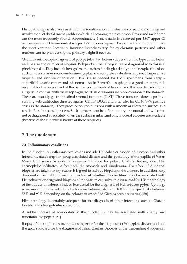
8.1. Inflammatory conditions
Ileocolonoscopy is an important tool for the diagnosis of diarrhoea and colitis. Several studiesshow that colonoscopy and biopsy is useful in the investigation of patients with chronicdiarrhoea yielding a histological diagnosis in 22 –31% of patients who had a macroscopicallynormal colon at colonoscopy.[60-63] Histological diagnosis includes a variety of conditionssuch as spirochetosis, pseudomelanosis coli, collagenous colitis and lymphocytic colitis andvariant forms.(Fig 7) The correct diagnosis of collagenous colitis implies multiple biopsies fromdifferent segments because thickening of the collagen layer can be discontinuous.[64]Histopathology can also help to identify amyloidosis and rare metabolic lysosomal or storagedisorders such as Tangier disease and systemic diseases such as mastocytosis.[65]
Endoscopy20
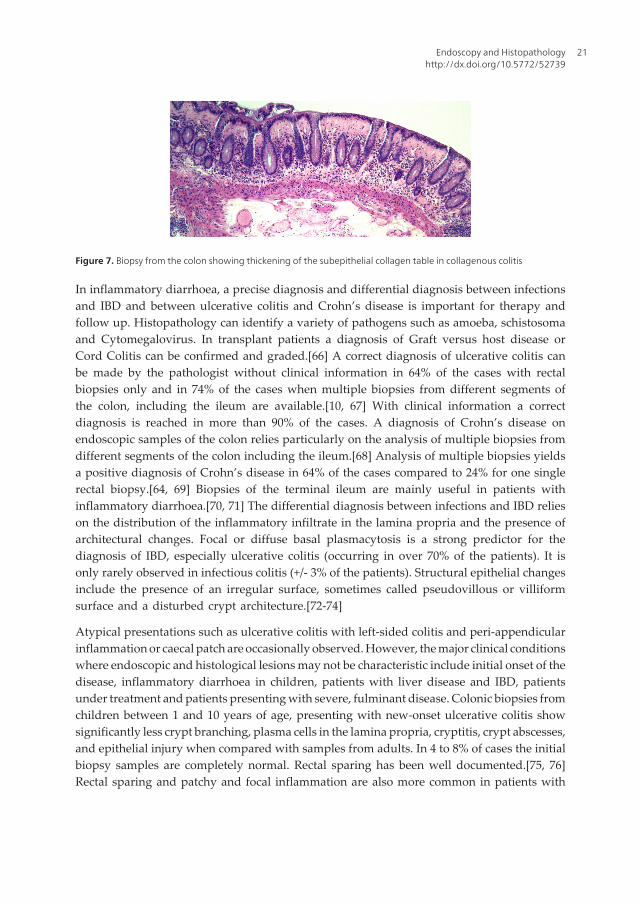
Figure 7. Biopsy from the colon showing thickening of the subepithelial collagen table in collagenous colitis
In inflammatory diarrhoea, a precise diagnosis and differential diagnosis between infectionsand IBD and between ulcerative colitis and Crohn’s disease is important for therapy andfollow up. Histopathology can identify a variety of pathogens such as amoeba, schistosomaand Cytomegalovirus. In transplant patients a diagnosis of Graft versus host disease orCord Colitis can be confirmed and graded.[66] A correct diagnosis of ulcerative colitis canbe made by the pathologist without clinical information in 64% of the cases with rectalbiopsies only and in 74% of the cases when multiple biopsies from different segments ofthe colon, including the ileum are available.[10, 67] With clinical information a correctdiagnosis is reached in more than 90% of the cases. A diagnosis of Crohn’s disease onendoscopic samples of the colon relies particularly on the analysis of multiple biopsies fromdifferent segments of the colon including the ileum.[68] Analysis of multiple biopsies yieldsa positive diagnosis of Crohn’s disease in 64% of the cases compared to 24% for one singlerectal biopsy.[64, 69] Biopsies of the terminal ileum are mainly useful in patients withinflammatory diarrhoea.[70, 71] The differential diagnosis between infections and IBD relieson the distribution of the inflammatory infiltrate in the lamina propria and the presence ofarchitectural changes. Focal or diffuse basal plasmacytosis is a strong predictor for thediagnosis of IBD, especially ulcerative colitis (occurring in over 70% of the patients). It isonly rarely observed in infectious colitis (+/- 3% of the patients). Structural epithelial changesinclude the presence of an irregular surface, sometimes called pseudovillous or villiformsurface and a disturbed crypt architecture.[72-74]
Atypical presentations such as ulcerative colitis with left-sided colitis and peri-appendicularinflammation or caecal patch are occasionally observed. However, the major clinical conditionswhere endoscopic and histological lesions may not be characteristic include initial onset of thedisease, inflammatory diarrhoea in children, patients with liver disease and IBD, patientsunder treatment and patients presenting with severe, fulminant disease. Colonic biopsies fromchildren between 1 and 10 years of age, presenting with new-onset ulcerative colitis showsignificantly less crypt branching, plasma cells in the lamina propria, cryptitis, crypt abscesses,and epithelial injury when compared with samples from adults. In 4 to 8% of cases the initialbiopsy samples are completely normal. Rectal sparing has been well documented.[75, 76]Rectal sparing and patchy and focal inflammation are also more common in patients with
Endoscopy and Histopathologyhttp://dx.doi.org/10.5772/52739
21
primary sclerosing cholangitis (PSC) without clinically overt colitis, when compared topatients with ulcerative colitis without PSC.[77, 78]
When the differential diagnosis between ulcerative colitis and Crohn’s disease can not besolved with endoscopic biopsies the patient should be categorized as “IBD unclassified”.[79]Clinical and histo-pathological follow up will eventually solve the diagnosis in most cases.
During follow up of IBD, histopathology can identify persistent active inflammation inulcerative colitis more reliably than endoscopy.[80] Persistent microscopic inflammation maybe important in the pathogenesis of dysplasia in IBD.
A complication of Kock pouch and ileal pouch anal anastomosis (IPAA) is the developmentof a primary inflammation within the pouch which is associated with a clinical syndrometermed “pouchitis”. This condition is common after surgery for ulcerative colitis, but can occuralso after surgery for other indications. Pouch biopsy specimens from well functioningpouches can show mild villous shortening and chronic inflammation. The most consistentfinding in pouchitis is ulceration. Grading of pouchitis depends on clinical features, endoscopicfindings and histology. The degree of polymorphonuclear infiltration and the proportion ofulcerated area are items of the score. There are no guidelines for the number and location ofbiopsies from a pouch but there is some evidence that a biopsy, taken 5 cm above the ileoanalanastomosis from the posterior and anterior wall may be the most sensitive for a diagnosis ofpouchitis. Pouchitis must be distinguished from “cuffitis” or “short-strip pouchitis”, which isinflammation in the columnar cuff mucosa distal to the pouch. The top end of the anal canalis lined by columnar mucosa like that of the rectum. In a hand sewn pouch-anal anastomosis,this mucosa is stripped, albeit often incompletely since the junction between columnarepithelium and squamous or transitional epithelium is difficult to distinguish. Islands ofcolumnar mucosa may be left behind.
Histology is also important for the differential diagnosis of eosinophilic disorders of thegastrointestinal tract. Eosinophils are constitutively present in the gastrointestinal mucosaoutside the oesophagus and the precise normal numbers have not been defined. In thecolon geographical and seasonal differences in numbers have been observed. In humans,appendix, caecum and ascending colon contain the highest numbers Therefore a diagnosisof eosinophilic (gastro-)enteritis is difficult. An intraepithelial position of eosinophils maybe the most reliable marker of disease. Eosinophilic disorders can be separated into primary(idiopathic) and secondary diseases, primary having no known cause, and secondary dueto other illnesses associated with eosinophilia such a infections, celiac disease, IBD and drugrelated pathology. A third situation is observed in the hypereosinophilic syndrome, aheterogeneous group of rare diseases defined by persistent blood eosinophilia for morethan 6 months with evidence of organ involvement (blood eosinophilia > 1500/mm3).
Primary eosinophilic enteritis has been called allergic gastro-enteropathy, because a subset ofpatients have an associated allergic component. Although considered idiopathic, an allergicmechanism may be involved as most patients exhibit increased food-specific IgE levels.
Endoscopy22
8.2. Neoplastic conditions
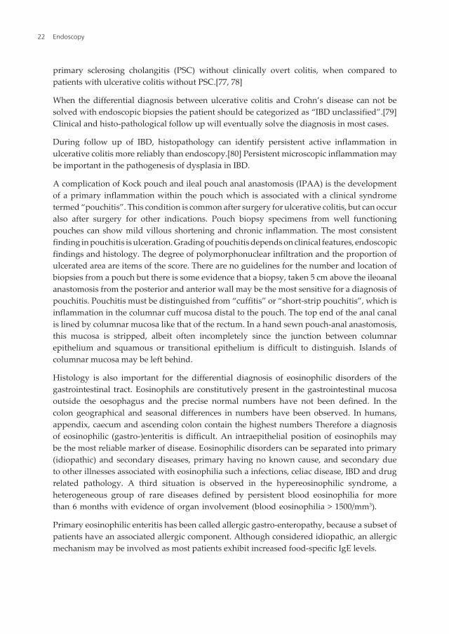
Crohn’s disease and ulcerative colitis carry an increased cancer risk. A pathway of “colitis –dysplasia – cancer” has been identified and this allows surveillance of patients with anincreased risk (longstanding disease; extensive colitis; ulcerative colitis with primarysclerosing cholangitis…).(Fig. 8) It has been estimated that 33 to 64 biopsies are required todetect dysplasia with 90% and 95% probabilities respectively. Yet, with 20-40 biopsies less than0.1% of the colorectal mucosa is covered.[81, 82] Current practice guidelines recommend that4 biopsy specimens be taken from every 10 cm (0.05 % of the entire area of the colon) of diseasedbowel in addition to macroscopically atypical lesions.[83] However, the detection rate of IBD-related dysplasia can substantially be improved with targeted biopsies obtained with thenewly developed endoscopic techniques and this procedure should replace the random biopsyguidelines in the future.[84] Dysplasia in IBD can appear as polypoid lesions or as flat lesions.Polypoïd lesions can occur in a mucosa with signs of colitis, or in a mucosa with flat dysplasia.Therefore, biopsies should be obtained from the elevated lesion and from the surroundingtissue. The microscopic diagnosis of “dysplasia” is based on the presence of cytological andarchitectural abnormalities showing “unequivocal, non-invasive (confined within thebasement membrane), neoplastic transformation of the epithelium excluding all reactivechanges”.[85] Biopsies positive for dysplasia can be subdivided into low-grade and high-grade. The grade of dysplasia is determined by the features of the most dysplastic portion. Thetwo grade classification appears to be reproducible, although in general the agreement is betterfor high-grade dysplasia. Because of the diagnostic problems related to dysplasia ancillarytechniques such as staining for p53 and AMACR can be applied on the tissue samples in orderto improve the diagnosis. P53/AMACR coexpression seems to be of potential value forpredicting neoplastic progression in ulcerative colitis patients with flat low grade dysplasia orindefinite lesions.[86]
Figure 8. Raised polypoid lesion in a biopsy from a patient with ulcerative colitis showing microscopic features of dys‐plasia : DALM
Endoscopy and Histopathologyhttp://dx.doi.org/10.5772/52739
23
Sporadic adenomas and polypoid “dysplasia” in IBD can be managed with endoscopictechniques and complete local excision appears to be adequate. Endoscopic resectionspecimens of IBD-related neoplasia should be handled properly, like all polypectomyspecimens. They should be removed entirely if possible. Sporadic small polyps can be handledwith a cold or hot biopsy forceps. While the latter can induce coagulation artefacts, the damageusually does not prevent adequate histological interpretation. Larger polyps should beoriented. The pathologist will identify the origin of the lesion, epithelial or not and the nature :neoplastic or not.
In recent years it has become clear that hyperplastic polyps are a heterogeneous group oflesions, now reported as “serrated lesions”. They include benign polyps, so called (traditional)hyperplastic polyps which can be subdivided in several types (microvesicular type, goblet-cell-rich type and mucin-poor type) and lesions with a neoplastic potential. The distinctionbetween the hyperplastic subtypes has a high inter-observer variation and therefore routinedistinction of these subtypes is not necessary.[87] Among the lesions with a neoplasticpotential, traditional serrated adenomas, with cytological dysplasia and sessile serratedadenomas or polyps have been identified. Both these lesions have a neoplastic potentialthrough the serrated neoplastic pathway. In sessile serrated polyps, the epithelial cells showhowever some atypia or features of dysplasia.[88] Therefore a distinction is made betweensessile serrated adenomas with and without dysplasia. A proper diagnosis of sessile serratedadenomas implies orientation of the endoscopic biopsy samples. The lesion is indeedcharacterized by dilatation of the crypts from top to bottom. Epithelial serration and dilatationare usually more prominent in the basal part of the crypts and this can not be evaluatedproperly on tangentially sectioned samples.
Histopathology allows grading of dysplasia in polyps and determination of the tubular orvillous nature of the lesion. Tubular adenomas are by definition dysplastic and hence atleast low-grade dysplastic lesions. Identification of high-grade dysplasia and intramucosalcarcinoma is important. Endoscopic surveillance of patients with so-called “advancedadenoma” may need to be different from that performed in patients without advancedadenomas. In polyps, the occurrence of invasive cancer, must be differentiated from high-grade dysplasia, intramucosal cancer and entrapped (pseudo-invasive) mucosa. Only whencancer invades the submucosa, it is considered to have the potential to metastasize, althoughlymphangiogenesis can occur in the mucosa as shown in ulcerative colitis.[89] Theestablished histopathological criteria that determine the treatment options of polypectomyversus subsequent surgical resection because of the risk of residual tumour are the statusof the resection margin, the histological grade, lympho-vascular invasion, budding of cellsand invasion into the submucosa below the stalk of the polyps but above the muscularispropria. Various staging systems have been proposed for this purpose.[7, 90]
As in the stomach and the small intestine, lymphomas and mesenchymal tumours can alsooccur in the colon and biopsies are suitable for a correct diagnosis.
Endoscopy24
9. Conclusions
Histopathology plays a critical role in GI practice. Endoscopic biopsies are important in orderto establish, confirm or exclude a diagnosis suspected clinically or endoscopically, both in theabsence and presence of endoscopic abnormalities. Biopsy diagnosis is greatly facilitated whenthe endoscopist provides adequate samples and understands the criteria used for histologicaldiagnosis. Histopathology plays also a major role in the design of therapeutic strategy. A closecollaboration between the endoscopist and the pathologist is therefore highly useful.
Author details
Karel Geboes1*, Karen Geboes2 and Anne Jouret-Mourin3
*Address all correspondence to: [email protected]
1 Department of Pathology, University of Leuven KUL, Leuven, Belgium
2 Department of Gastroenterology, Digestive Oncology, University of Gent, Gent, Belgium
3 Department of Pathology, Saint Luc Hospital, UCL, Brussels, Belgium
References
[1] Amado, R. G, Wolf, M, Peeters, M, et al. (2008). Wild-type KRAS is required for pni‐tumumab efficacy in patients with metastatic colorectal cancer. J Clin Oncol. , 26,1626-1634.
[2] Rüschoff, J, Dietel, M, Baretton, G, Arbogast, S, et al. (2010). HER2 diagnostics in gas‐tric cancer guideline validation and development of standardized immunohisto‐chemical testing. Virchows Arch. , 457, 299-307.
[3] Mainguet, P, & Jouret, A. The collaboration between the endoscopist and the pathol‐ogist. ((1996). Acta Endoscop. , 26, 67-77.
[4] Geboes, K. La collaboration entre l’endoscopiste et le pathologiste. ((2005). Acta En‐doscop. , 36, 245-56.
[5] Lauwers, G, Forcione, D. G, Nishioka, N. S, et al. (2009). Novel endoscopic therapeu‐tic modalities for superficial neoplasms arising in Barrett’s esophagus : a primer forsurgical pathologists. Mod Pathol. , 22, 488-498.
[6] Flejou, J. F. Histological assessment of oesophageal columnar mucosa. ((2008). BestPract Res Clin Gastroenterol. , 22, 671-686.
Endoscopy and Histopathologyhttp://dx.doi.org/10.5772/52739
25
[7] Cooper, H. S. Pathology of the endoscopically removed malignant colorectal polyp.((2007). Curr Diagn Pathol. , 13, 423-427.
[8] Weynand, B, Borbath, I, Galant, C, Piessevaux, H, & Deprez, P. H. (2011). Optimizingspecimen collection and laboratory procedures reduces the non-diagnostic rate forendoscopic ultrasound-guided fine-needle aspiration of solid lesions of the pancreas.Cytopathology.
[9] Dejaco, C, Osterreicher, C, Angelberger, S, et al. (2003). Diagnosing colitis : a prospec‐tive study on essential parameters for reaching a diagnosis. Endoscopy. , 35,1004-1008.
[10] Stange, E. F. Travis SPL, Vermeire S, et al. ((2006). European evidence-based consen‐sus on the diagnosis and management of Crohn’s disease : definitions and diagnosis.Gut. 55 Suppl I: ii15., 1.
[11] Faller, G, Berndt, R, Borchard, F, et al. (2003). Histopathological diagnosis of Barrett’smucosa and associated neoplasias. Results of a consensus conference of the WorkingGroup for “Gastrointestinal Pathology of the German Society for pathology” on 22September 2001. Pathology , 24, 9-14.
[12] Stein, H. J. (1996). Esophageal cancer: screening and surveillance. Results of a consen‐sus conference held at the VIth world congress of the International Society for Dis‐eases of the Esophagus. Dis Esophagus. 9: SS19., 3.
[13] Kiesslich, R, Fritsch, J, Holtmann, M, et al. (2003). Methylene blue-aided chromoen‐doscopy for the detection of intraepithelial neoplasia and colon cancer in ulcerativecolitis. Gastroenterology , 124, 880-888.
[14] Kiesslich, R, & Neurath, M. F. (2004). Review : Potential of new endoscopic techni‐ques : intravital staining and in vivo confocal endomicroscopy for the detection ofpremalignant lesions and early cancer in patients with ulcerative colitis. Acta Endo‐scopica , 34, 189-197.
[15] Kiesslich, R, Burg, J, Vieth, M, et al. (2004). Confocal laser endoscopy for diagnosingintraepithelial neoplasias and colorectal cancer in vivo. Gastroenterology , 127,706-713.
[16] Inoue, H, Kudo, S, & Shiokawa, A. (2005). Technology Insight : laser-scanning confo‐cal microscopy and endocytoscopy for cellular observation of the gastrointestinaltract. Nature clinical practice gasthep. , 2, 31-37.
[17] Jouret-mourin, A, Hoorens, A, Kockx, M, et al. (2011). Belgian guidelines for HER2testing in gastric cancer. Belg J Med Oncol. , 5, 14-22.
[18] Riddell, R. H. (1996). The biopsy diagnosis of gastroesophageal reflux disease, “car‐ditis,” and Barrett’s esophagus, and sequelae of therapy. Am J Surg Pathol. 20 Suppl1: S, 31-50.
Endoscopy26
[19] Glickman, J. N, Fox, V, Antonioli, D. A, Wang, H. H, & Odze, R. D. (2002). Morpholo‐gy of the cardia and significance of carditis in pediatric patients. Am J Surg Pathol. ,26, 1032-1039.
[20] Dent, J. (2007). Microscopic esophageal mucosal injury in nonerosive reflux disease.Clin Gastroenterol Hepatol. , 5, 4-16.
[21] Tytgat, G. (2008). The value of esophageal histology in the diagnosis of gastroesopha‐geal reflux disease in patients with heartburn and normal endoscopy. Cur Gastroen‐terol Rep. , 10, 231-234.
[22] Vieth, M. (2008). Contribution of histology to the diagnosis of reflux disease. BestPract Res Clin Gastroenterol., 22, 625-638.
[23] Takubo, K, Honma, N, Aryal, G, et al. (2005). Is there a set of histologic changes thatare invariably reflux associated? Arch Pathol Lab Med. , 129, 159-163.
[24] Dent, J, Brun, J, Fendrick, A. M, et al. (1999). An evidence-based appraisal of refluxdisease management- the Genval workshop report. Gut 44: SS16., 1.
[25] Chang, F, & Anderson, S. (2008). Clinical and pathological features of eosinophilicoesophagitis : a review. Pathology , 40, 3-8.
[26] Vakil, N, Van Zanten, S. V, Kahrilas, P, Dent, J, & Jones, R. (2006). The Montreal defi‐nition and classification of Gastroesophageal Reflux Disease : a global evidence-based consensus. Am J Gastroenterol. , 101, 1900-1920.
[27] Weinstein, W. M, & Ippoliti, A. F. (1996). The diagnosis of Barrett’s esophagus: gob‐lets, goblets, goblets. Gastrointest Endosc. , 44, 91-95.
[28] Gatenby, P. A, Ramus, J. R, Caygill, C. P, Shepherd, N. A, & Watson, A. (2008). Rele‐vance of the detection of intestinal metaplasia in non-dysplastic columnar-linedesophagus. Scand J Gastroenterol. , 43, 524-530.
[29] Abela, J, Going, J. J, Mackenzie, J. F, Mckernan, M, Mahoney, O, & Stuart, S. RC.((2008). Systematic four-quadrant biopsy detects Barrett’s dysplasia in more patientsthan non-systematic biopsy. Am J Gastroenterol. , 103, 850-855.
[30] Peters, F. P, Curvers, W. L, Rosmolen, W. D, et al. (2008). Surveillance history of en‐doscopically treated patients with early Barrett’s neoplasia : nonadherence to the Se‐attle biopsy protocol leads to sampling error. Dis Esophagus , 21, 475-479.
[31] Reid, B. J, Haggitt, R. C, Rubin, C. E, et al. (1985). Criteria for dysplasia in Barrett’sesophagus: a cooperative consensus study. Gastroenterology 88: 1552 (abstract).
[32] Sagan, C, Fléjou, J. F, Diebold, M. D, & Potet, F. Le Bodic MF. ((1994). Reproductibi‐lité des critères histologiques de dysplasie sur muqueuse de Barrett. GastroenterolClin Biol. , 18, 31-34.
[33] Gossner, L, Pech, O, May, A, Vieth, M, Stolte, M, & Ell, C. (2006). Comparison ofmethylene blue-directed biopsies and four-quadrant biopsies in the detection of
Endoscopy and Histopathologyhttp://dx.doi.org/10.5772/52739
27
high-grade intraepithelial neoplasia and early cancer in Barrett’s esophagus. Dig Liv‐er Dis. , 38, 724-729.
[34] Curvers, W. L, & Kiesslich, R. Bergman JJGHM. ((2008). Novel imaging techniques inthe detection of oesophageal neoplasia. Best Pract Res Clin Gastroenterol. , 22,687-720.
[35] Mino-kenudson, M, Hull, M. J, Brown, I, et al. (2007). EMR for Barrett’s esophagus-related superficial neoplasms offers better diagnostic reproducibility than mucosalbiopsy. Gastrointest Endosc. , 66, 667-669.
[36] Geboes, K, Ectors, N, Geboes, K. P, & Lambert, R. (2005). Intraepithelial neoplasia,dysplasia and early cancer of the digestive tract : Modifications in terminology. Cur‐rent Cancer Therapy Reviews , 1, 145-155.
[37] Lal, N, Bhasin, D. K, Malik, A. K, Gupta, N. M, Singh, K, & Mehta, S. K. (1992). Opti‐mal number of biopsy specimens in the diagnosis of carcinoma of the oesophagus.Gut , 33, 724-726.
[38] Price, A. B. (1991). The Sydney System : Histological division. J Gastroenterol andHepatol. , 6, 209-222.
[39] Dixon, M. F, Genta, R. M, Yardley, J. H, & Correa, P. (1996). Classification and grad‐ing of gastritis : the updated Sydney System. Am J Surg Pathol. , 20, 1161-1181.
[40] Nichols, L, Sughayer, M, De Girolami, P. C, et al. (1991). Evaluation of diagnosticmethods for Helicobacter pylori gastritis. Am J Clin Pathol. , 95, 769-773.
[41] Rugge, M, & Correa, P. DiMario F, et al. ((2008). The Olga staging of gastritis: a tuto‐rial. Dig & Liver disease , 40, 650-658.
[42] Haot, J, Jouret, A, Willette, M, Gossuin, A, & Mainguet, P. B. (1990). Lymphocyticgastritis : prospective study of its relationship with varioliform gastitis. Gut, 31; ,282-285.
[43] Oberhuber, G, Puspok, A, Oesterreicher, C, et al. (1997). Focally enhanced gastritis : afrequent type of gastritis in patients with Crohn’s disease. Gastroenterology , 112,698-706.
[44] Xin, W, & Greenson, J. K. (2004). The clinical significance of focally enhanced gastri‐tis. Am J Surg Pathol, , 28, 1347-1351.
[45] Yao, K, Yao, T, Iwashita, A, et al. (2000). Microaggregate of immunostained macro‐phages in noninflamed gastroduodenal mucosa: a new useful histological marker fordifferentiating Crohn’s colitis from ulcerative colitis. Am J Gastroenterol. , 95,1967-1973.
[46] Gilliam, J. H, Geisinger, K. R, Wu, W. C, et al. (1989). Endoscopic biopsy is diagnosticin gastric antral vascular ectasia. The “Watermelon stomach”. Dig Dis Sci. , 34,885-888.
Endoscopy28
[47] Misra, V, Misra, S. P, Dwivedi, M, et al. (1997). Histomorphometric study of portalhypertensive enteropathy. Am J Clin Pathol. , 108, 652-657.
[48] Vyberg, M, Hougen, H. P, & Tonnesen, K. (1983). Diagnostic accuracy of endoscopicgastrobiopsy in carcinoma of the stomach. Acta Path Microbiol Immunol Scand (A). ,91, 483-487.
[49] Misiewicz, J. J. Tytgat GNJ, Goodwin CS, Price AB, Sipponen P, Strickland RG((1990). The Sydney system : a new classification of gastritis. Proceedings of the 9thWorld Congress of Gastroenterology. Sydney, Australia, , 1-10.
[50] Debongnie, J. C, Delmee, M, Mainguet, P, Beyaert, C, Haot, J, & Legros, G. (1992).Cytology : a simple, rapid, sensitive method in the diagnosis of Helicobacter pylori.Am J Gastroenterol. , 87, 20-23.
[51] Walker, M. M, Salehian, S. S, Murray, C. E, Rajendran, A, Hoare, J. M, Negus, R, Po‐well, N, & Talley, N. J. (2010). Implications of eosinophilia in the normal duodenalbiopsy- an association with allergy and functional dyspepsia. Aliment PharmacolTher. 31; , 1129-1136.
[52] Mee, A. S, Burke, M, Vallon, A. G, Newman, J, & Cotton, P. B. (1985). Small bowelbiopsy for malabsorption : comparison of diagnostic adequacy of endoscopic forcepsand capsule biopsy specimens. Br Med J. , 291, 769-772.
[53] Green PHRRostami K, Marsh MN. ((2005). Diagnosis of coeliac disease. Best PractRes Clin Gastroenterol. , 19, 389-400.
[54] Dickson, B. C, Streutker, C. J, & Chetty, R. (2006). Coeliac disease : an update forpathologists. J Clin Pathol. , 59, 1008-1016.
[55] Ensari, A. (2010). Gluten sensitive enteropathy (celiac disease) : controversies in diag‐nosis and classification. Arch Pathol Lab Med. , 134, 826-836.
[56] Brousse, N. Meijer JWR. ((2005). Malignant complications of coeliac disease. BestPract Res Clin Gastroenterol. , 19, 401-412.
[57] Spigelman, A. D, Williams, C. B, Talbot, I. C, & Domizio, P. Phillips RKS. ((1989). Up‐per gastrointestinal cancer in patients with familial adenomatous polyposis. Lancetii: , 783-785.
[58] Van Caillie, M. A, Geboes, K, Van Eyken, P, & Van Steenbergen, W. (2006). The diag‐nostic value of intraductal biopsy of the extrahepatic bile ducts. Tijdschr Geneesk. ,62, 1035-1043.
[59] Nguyen, K. Sing Jr JT. ((2008). Review of endocopic techniques in the diagnosis andmanagement of cholangiocarcinoma. World J Gastroenterol. , 14, 2995-2999.
[60] Prior, A, Lessels, A. M, & Whorwell, P. J. (1987). Is biopsy necessary if colonoscopy isnormal? Dig Dis Sci. , 32, 673-676.
Endoscopy and Histopathologyhttp://dx.doi.org/10.5772/52739
29
[61] Whitehead, R. (1990). Colitis : Problems in definition and diagnosis. Virchows ArchivPathol Anat. , 417, 187-190.
[62] Marshall, J. B, Singh, R, & Diaz-arias, A. A. (1995). Chronic, unexplained diarrhea :are biopsies necessary if colonoscopy is normal? Am J Gastroenterol. , 90, 372-376.
[63] Shah, R. J, Fenoglio-preiser, C, Bleau, B. L, & Giannella, R. A. (2001). Usefulness ofcolonoscopy with biopsy in the evaluation of patients with chronic diarrhea. Am JGastroenterol. , 96, 1091-1095.
[64] Geboes, K. (2008). Lymphocytic, collagenous and other microscopic colitides : pathol‐ogy and the relationship with idiopathic inflammatory bowel diseases. GastroenterolClin Biol. , 32, 689-694.
[65] Kirsch, R, Geboes, K, Shepherd, N. A, et al. (2008). Systemic mastocytosis involvingthe gastrointestinal tract : Clinicopathologic and molecular study of five cases. ModPathol. , 21, 1508-1516.
[66] Herrera, A. F, Soriano, G, Bellizzi, A. M, Hornick, J. L, Ho, V. T, Ballen, K. K, Baden,L. R, Cutler, C. S, Antin, J. H, Soiffer, R. J, & Marty, F. M. (2011). Cord colitis syn‐drome in cord-blood stem-cell transplantation. N Engl J Med. , 365, 815-854.
[67] Bentley, E, Jenkins, D, Campbell, F, & Warren, B. F. (2002). How could pathologistsimprove the initial diagnosis of colitis? Evidence from an international workshop. JClin Pathol. , 55, 955-960.
[68] Dejaco, C, Osterreicher, C, Angelberger, S, et al. (2003). Diagnosing colitis : a prospec‐tive study on essential parameters for reaching a diagnosis. Endoscopy , 35,1004-1008.
[69] Stange, E. F. Travis SPL, Vermeire S, et al. ((2008). European evidence-based consen‐sus on the diagnosis and management of ulcerative colitis : Definitions and diagno‐sis. J Crohn’s & Colitis , 2, 1-23.
[70] Mchugh, J. B, Appelman, H. D, & Mckenna, B. J. (2007). The diagnostic value of en‐doscopic terminal ileum biopsies. Am J Gastroenterol. , 102, 1084-1089.
[71] Geboes, K. (2007). The strategy for biopsies of the terminal ileum should be evidencebased. Am J Gastroenterol. , 102, 1090-1092.
[72] Schumacher, G, Kollberg, B, & Sandstedt, B. (1994). A prospective study of first at‐tacks of inflammatory bowel disease and infectious colitis. Histologic course duringthe 1st year after presentation. Scand J Gastroenterol. , 29, 318-332.
[73] Jenkins, D, Balsitis, M, Gallivan, S, et al. (1997). Guidelines for the initial biopsy diag‐nosis of suspected chronic idiopathic inflammatory bowel disease. The British Soci‐ety of Gastroenterology Initiative. J Clin Pathol. , 50, 93-105.
Endoscopy30
[74] Seldenrijk, C. A, Morson, B. C, et al. (1991). Histopathological evaluation of colonicmucosal biopsy specimens in chronic inflammatory bowel disease : diagnostic impli‐cations. Gut , 32, 1514-15.
[75] Markowitz, J, Kahn, E, Grancher, K, et al. (1993). Atypical rectosigmoid histology inchildren with newly diagnosed ulcerative colitis. Am J Gastroenterol. , 88, 2034-2037.
[76] Robert, M. E, Tang, L, Hao, M, et al. (2004). Patterns of inflammation in mucosal bi‐opsies of ulcerative colitis. Perceived differences in pediatric populations are limitedto children younger than 10 years. Am J Surg Pathol. , 28, 183-189.
[77] Loftus EV JrHarewood GC, Loftus CG, et al. ((2005). PSC-IBD : a unique form of in‐flammatory bowel disease associated with primary sclerosing cholangitis. Gut , 54,91-96.
[78] Perdigoto, R, & Wiesner, R. H. LaRusso NF, et al. ((1991). Inflammatory bowel dis‐ease associated with primary sclerosing cholangitis: Incidence, severity and relation‐ship to liver disease. Gastroenterology 100:A238.
[79] Geboes, K, Colombel, J. F, Greenstein, A, et al. (2008). Indeterminate colitis : A reviewof the concept- What’s in a name? Inflamm Bowel Dis. , 14, 860-867.
[80] Geboes, K, Riddell, R, Öst, Ä, Jensfelt, B, Persson, T, & Löfberg, R. (2000). A reprodu‐cible grading scale for histological assessment of inflammation in ulcerative colitis.Gut , 47, 404-409.
[81] Rosenstock, E, Farmer, R. G, Petras, R, et al. (1985). Surveillance for colonic carcino‐ma in ulcerative colitis. Gastroenterology. , 89, 1342-1346.
[82] Rubin, C. E, Haggitt, R. C, et al. (1992). DNA-aneuploidy in colonic biopsies predictsfuture development of dysplasia in ulcerative colitis. Gastroenterology. , 103,1611-1620.
[83] Kornbluth, A, & Sachar, D. B. (1997). Ulcerative colitis practice guidelines in adults.American College of Gastroenterology, Practice Parameters Committee. Am J Gastro‐enterol. , 92, 204-211.
[84] Kiesslich, R, Fritsch, J, Holtmann, M, et al. (2003). Methylene blue-aided chromoen‐doscopy for the detection of intraepithelial neoplasia and colon cancer in ulcerativecolitis. Gastroenterology. , 124, 880-888.
[85] Riddell, R. H, Goldman, H, Ransohoff, D. F, et al. (1983). Dysplasia in inflammatorybowel disease : Standardized classification with provisional clinical applications.Hum Pathol, , 14, 931-968.
[86] Van Schaik FDMOldenburg B, Offerhaus JA, Schipper MEI, Vleggaar FP, SiersemaPD et al. ((2012). Role of immunohistochemical markers in predicting progression ofdysplasia to advanced neoplasia in patients with ulcerative colitis. Inflamm BowelDis. , 18, 480-488.
Endoscopy and Histopathologyhttp://dx.doi.org/10.5772/52739
31

[87] Yantiss, R. K. (2007). Serrated colorectal polyps and the serrated neoplastic pathway :emerging concepts in colorectal carcinogenesis. Curr Diagn Pathol. , 13, 456-466.
[88] Jouret-mourin, A, & Geboes, K. Serrated lesions of the colorectum : a new entity :What should an endoscopist know about it? ((2012). Acta Gastroenterol Belg. , 76,197-202.
[89] Kaiserling, E, Kröber, S, & Geleff, S. (2003). Lymphatic vessels in the colonic mucosain ulcerative colitis. Lymphology. , 36, 52-61.
[90] Ueno, H, Mochizuki, H, Hashiguchi, Y, et al. (2004). Risk factors for an adverse out‐come in early invasive colorectal carcinoma. Gastroenterology , 127, 385-394.
Endoscopy32
Chapter 2
Cleaning, Disinfection and Sterilizationof Heat-Sensitive Endoscopes
Norman Miner
Additional information is available at the end of the chapter
http://dx.doi.org/10.5772/50355
1. Introduction
High level disinfection is a process that kills high numbers of all types of vegetative non-spore forming bacteria (Gram-positive and Gram-negative), fungi, all types of viruses (hy‐drophylic and lipophylic), and mycobacteria (TB), but not necessarily high numbers ofbacterial spores in the relatively brief exposure time for disinfection. Sterilization is a proc‐ess that kills all types of microbes including highly resistant bacterial spores. Sterilizationusually requires a much longer exposure time than disinfection. Gastroscopes, colono‐scopes, urethroscopes, and cystoscopes normally do not break the barrier between non-ster‐ile areas of the body and sterile areas, and therefore high level disinfection is the commonlyaccepted practice for these endoscopes. Bronchoscopes are a possible exception as they mayenter into the sterile bronchioles, although they make that entrance through the non-sterilenasal passages where they likely become contaminated with the flora of the oral-nasal bodycavities. Endoscopists, and certainly patients, all agree that endoscopes and their accessoriessuch as biopsy forceps should be thoroughly cleaned of all body fluids and any possible mi‐crobes between patients, even if not technically sterilized.
2. Methods
Unless endoscopes can be completely dried after disinfection, creating an environment wheremicrobes cannot survive or multiply, or there is laboratory culturing evidence that endoscopesstill contain very few or no microbes, they should be disinfected and rinsed again in the morn‐ing before first use. Any microbes that might remain in the endoscopes after disinfection, or beintroduced into the endoscopes by means of non-sterile rinse water, could multiply overnight
© 2013 Miner; licensee InTech. This is an open access article distributed under the terms of the CreativeCommons Attribution License (http://creativecommons.org/licenses/by/3.0), which permits unrestricted use,distribution, and reproduction in any medium, provided the original work is properly cited.
or over a weekend or holiday to unsafe numbers, or begin to form a biofilm within the chan‐nels of the endoscope. Certain microbes such as Staphylococcus aureus, Pseudomonas aerugi‐nosa, or Escherichia coli can divide every 60 minutes or faster in dark, moist, and warmconditions. Just 10 or 20 microbes remaining in an endoscope at 4:00 PM can multiply to aboutone million colony forming units by the time of the 1st case at about 8:00 AM the next day, andmany more if the starting number is higher than “just 10 or 20” if there is enough substrate tosustain a population that size. The hospital laboratory or a contract laboratory/microbiologistcan be a valuable ally to know by testing how many microbes are in the channels of an endo‐scope after a cleaning/disinfecting procedure, or after a storage interval. It takes only a few mi‐nutes for a microbiologist to flush recovery solutions through an endoscope, and then assaythose fluids to validate the numbers, if any, of microbes within the channels of an endoscope.At intervals of about three or four months, or after newly trained personnel begin to processand disinfect endoscopes, or the disinfecting procedure is modified, a sampling of the endo‐scopes should be cultured to validate that the endoscopes are indeed disinfected, and anynumbers of bacteria in the endoscopes are very low. B.T. Petersen, et. al. is another publicationregarding methods for reprocessing flexible endoscopes [1].
3. Cleaning endoscopes
The initial cleaning step happens in the Procedure Room when the insertion tube is removedfrom the patient and wiped with a gauze pad saturated with a disinfectant, and the internalair, suction, and water channels are flushed with water. This initial cleaning step removesmuch of the organic soil and probably removes and kills at least 99% of any microbes on theendoscope. The endoscope is then transported in a plastic bin to the reprocessing room andtested for possible leaks. The control valves are removed, and the endoscope and all parts(valves) are submerged in a deep stainless steel sink (or plastic basin) with about two orthree gallons of warm tap water with a disinfectant-detergent solution. The endoscopeshould be cleaned within about 30.0 min of leaving the procedure room, and not allowed todry. Organic soil and microbes are much more difficult to remove and to kill after they be‐come dry than when they are wet. A disinfectant-detergent cleaning solution is a concen‐trate of a low-foaming surfactant (detergent) and disinfectant. These disinfectant-detergentcleaning solutions are concentrates intended and labeled to be diluted in the range of 1:16 to1:32. The label will give the exact dilution to be used, and also identify that the solution willkill Gram-positive and Gram-negative bacteria, fungi, and hydrophylic and lipophylic vi‐ruses, (and possibly even mycobacteria and some spores or spore-forming bacteria, depend‐ing on the particular disinfectant-detergent chosen). Labels that use the scientific names ofmicrobes can be difficult for non-microbiologists to understand. The hospital laboratory, orany microbiologist, can help to translate the label into common terms such as Gram positiveor Gram negative bacteria, fungi, and viruses. The disassembled valves should be flexed andlightly brushed with the disinfectant-detergent cleaning solution (cleaning solution), the ex‐terior of the endoscope should be wiped with a soft cloth saturated with the cleaning solu‐tion, and the biopsy or working channel of the endoscope should be filled with the cleaningsolution and brushed the entire length several times with a tight-fitting brush. Insert the
Endoscopy34
brush into the channel until the brush emerges from the channel. Brush the channel repeat‐edly about three or four times. Allow the brush to emerge from the channels submergedwithin the cleaning solution to avoid a splattering of microbes in the air. The air-water chan‐nel is too small to brush, and care should be taken to ensure that this small channel is notblocked and is filled with the cleaning solution. This wiping and brushing of all parts of theendoscope with the cleaning solution should continue for about 5.0 min which allows thedisinfectant to kill many of any contaminating microbes. This cleaning procedure is com‐pleted by triple-rinsing all parts and sections of the endoscope with fresh warm tap water.Some cleaning solutions contain enzymes, and enzymes are not antimicrobial, and are notcompatible with disinfectants. Enzymes require at least body temperature (35 °C) and manyminutes to function. Enzymes are proteins, and it is proteins such as blood and tissues thatare being removed from the endoscopes. For those reasons, especially the fact that enzyme-detergent combinations are not antimicrobial, and are not compatible with disinfectants,these enzyme-detergent combinations are not recommended by this author for cleaning en‐doscopes. Enzyme products are useful as a soak to dissolve clogged air/water channels thatare too narrow to be brushed. The purpose of using a disinfectant-detergent cleaning solu‐tion during this cleaning stage is to remove organic soil (blood, mucous, fecal matter), andlower the number of microbes on the endoscope in advance of the final stage of disinfectingor sterilizing, and to protect the technician and the environment from a splattering of washwater containing potentially infectious microbes.
4. High level disinfection of the endoscope
After the endoscope and all of its parts have been cleaned as described above in a disinfec‐tant-detergent cleaning solution, and thoroughly triple-rinsed with tap water to remove thedisinfectant-detergent, the rinsed endoscope should then be soaked in a high level disinfec‐tant at the labeled exposure time and temperature for any particular high level disinfectant.A high level disinfectant is a disinfectant that is labeled to kill all Gram-Positive and Gram-Negative vegetative bacteria, all fungi, all mycobacteria (TB), and all types of viruses, hydro‐phylic and lipophylic, within the labeled exposure time and temperature. High leveldisinfectants are also able to kill bacterial spores and spore-forming bacteria, although ittakes a longer time to kill high numbers of bacterial spores than to kill vegetative microbes.After the initial disinfection and rinse in the procedure room, and the procedure of cleaningthe endoscope with the disinfectant-detergent cleaning solution, the number of spore-form‐ing bacteria on the endoscope should be very low, and thus this disinfecting procedure willalso remove and kill many bacterial spores.
5. Sterilization of an endoscope
The procedure to sterilize an endoscope contains all the steps for cleaning and high leveldisinfection of an endoscope as described above, but the exposure time for sterilization is
Cleaning, Disinfection and Sterilization of Heat-Sensitive Endoscopeshttp://dx.doi.org/10.5772/50355
35
longer than for disinfection in order to kill spores and spore-forming bacteria [2]. The labelof the high level disinfectant will identify the sterilization soak time, which can be manyhours. Also, the final rinse should be with sterilized water rather than ordinary tap water.Air should be forced through the internal channels with a syringe to dry them, and the en‐doscope should be stored in some sterile manner such as covering the endoscope with asterile wrap or cloth. Hang the endoscope so it can further drip dry.
6. Protective clothing
Although care is taken to not splash contaminated water from an endoscope during re‐processing, there will be some splash and splatter as brushes emerge from the internalchannels, and otherwise. Personnel should wear a plastic face shield covering the eyes,nose, and mouth; hair covering; elbow-length rubber gloves; and long-sleeved, water-proof gowns or coats.
7. Examples of disinfectant-detergent cleaning solutions
A disinfectant-detergent cleaning solution will be a concentrate intended to be diluted withtap water, usually 16- or 32-fold. The detergent should be anionic or non-ionic, low foaming,and easily rinsed from the endoscope. Use the lowest concentration (highest dilution) rec‐ommended to facilitate rinsing and removal of the disinfectant-detergent solution. There aredozens of these disinfectant-detergents listed on the US Environmental Protection Agency(EPA) web site. Local Sales Representatives will also be able to provide written descriptionsand access to disinfectant-detergent concentrates. They contain modified phenolic chemicals(phenylphenol, amylphenol, etc); quaternary ammonium chemicals (dimethyl benzyl am‐monium chloride); sodium hypochlorite/bleach; iso- or ethyl alcohol; or other common dis‐infectants. These disinfectant-detergents are being used in the cleaning stage to cleanorganic material from the endoscopes by way of the detergent, to lower the bioburden ofmicrobes on the endoscope by way of the disinfectant, and to provide some respite to theenvironment and to the operator from the microbes inevitably splashed with the cleaningwater. Several endoscopes can be cleaned in a basin filled with the diluted disinfectant-de‐tergent at one time. Discard/drain the disinfectant-detergent cleaning solution after each en‐doscope cleaning procedure. The basin could be repeatedly drained and filled with tapwater to triple rinse the endoscopes, or additional basins could be used for rinse water.
8. Examples of high level disinfectants
High level disinfectants are able to kill high numbers of all vegetative bacteria, fungi, vi‐ruses, and mycobacteria within a relatively brief exposure time such as about 10.0 min to
Endoscopy36
20.0 min at temperatures of about 20 °C to about 25 °C. The label of the high level disin‐fectant will have an exposure time and temperature. However, if the cleaning procedureused a disinfectant-detergent which would have killed large numbers of microbes, thehigh level disinfectant exposure time can be shortened to 20.0 min. With a longer expo‐sure time such as hours, a high level disinfectant can also kill highly resistant forms ofbacterial spores. Alkaline glutaraldehyde solutions (Cidex Solution), solutions of glutaral‐dehyde enhanced with isopropanol (Aldahol High Level Disinfectant), and peracetic acidsolutions are high level disinfectants. The sterilization time for Ortho-Phthalaldehyde(OPA) is 32 hours, which is not practical, and OPA is not labelled as a sterilant. Alkalineglutaraldehyde solutions with and without isopropanol, and peracetic acid solutions arehighly soluble in water, and thus are easily rinsed from endoscopes. OPA has a low solu‐bility in water, and is difficult to rinse away from endoscopes with any practical numberof rinses [3]. Trade names of some high level disinfectants are Cidex Activated Dialde‐hyde Solution, Aldahol High Level Disinfectant [4], Rapicide High Level Disinfectant, Re‐sert XL HLD, and Acecide High Level Disinfectant.
9. Automatic endoscope reprocessing machines
Automatic endoscope reprocessing (AER) machines are available. Some of these AER ma‐chines also claim to be able to clean the endoscopes as well as to disinfect them. The clean‐ing action of the cleaning solution for these AER machines is by force rather than by brush,and this author is skeptical that such forceful rinsing is able to clean the channels of endo‐scopes as well as a brush. If an AER machine is used, the endoscope should be manuallybrushed before it is placed into the machine. Duodenoscopes have an elevator wire channelthat must be manually cleaned regardless of a final clean in a machine.
10. Protection from irritating disinfectant vapors
Glutaraldehyde solutions, glutaraldehyde-isopropanol solutions, and peracetic acid solu‐tions all have irritating and sensitizing chemical vapors. These solutions should be con‐tained and used in a manner to protect workers from the irritating and sensitizing chemicalvapors. This can be done by soaking the cleaned endoscope in a covered container of thedisinfectant, and/or working in a hood that ventilates the chemical vapors through a filter toremove the vapors, or ventilates the vapors to outside air. If a hood is not available, a smallfan can be positioned to blow the chemical vapors away from the operator. Any spilled highlevel disinfectant should be immediately cleaned up. Serious and effective efforts must bemade to ventilate and eliminate and protect personnel from the vapors of glutaraldehyde,OPA, and peracetic acid.
Cleaning, Disinfection and Sterilization of Heat-Sensitive Endoscopeshttp://dx.doi.org/10.5772/50355
37

11. Biofilms
The interior channels of endoscopes can be convoluted, and the air-water channel is too nar‐row to brush. Therefore it is possible over time for the channels of an endoscope to developa film of microbes, called a biofilm. Endoscope channels that contain a biofilm cannot be dis‐infected [5]. Detection of a biofilm is one reason why endoscopes should be periodically cul‐tured. Biofilms can be removed by soaking the endoscope and all of its channels in anenzyme detergent for a prolonged period of time such as one or two hours, followed by vig‐orous brushing of the channels. After this prolonged soaking, brushing and rinsing proce‐dure, the endoscope should again be cultured to determine that the biofilm has beenremoved. The hospital laboratory or a contract microbiologist can be the Endoscopists bestfriend, and the only way to know for certain that procedures lead to a disinfected endoscopeis to culture the endoscope, at least periodically.
Author details
Norman Miner
MicroChem Laboratory, Inc.Euless, Texas, USA
References
[1] Petersen, B T, Chennat, J, Cohen, J, Cotton, P B, Greenwald, D A, et.al. Multisocietyguideline on reprocessing flexible gastrointestinal endoscopes. Infect. Control andHosp. Epidem. 2011, 33: pp 527 – 537.
[2] Miner N, Harris V, Ebron T, Cao T. Sporicidal activity of disinfectants as one possiblecause for bacteria in patient-ready endoscopes. Gastroenterol Nurs.2009; 30: 285-90.
[3] Miner N, Harris V, Lukomski N, Ebron T. Rinsability of ortho-phthalaldehyde fromendoscopes. Diagnost and TherapEndosc. 2012; 2012: Article 853781.
[4] Miner N, Harris V, Cao T D, Ebron T, and Lukomski N. Aldahol High Level Disin‐fectant. Amer J Infect Control. 2010; 38: No.3, 205 – 211.
[5] Pajkos A, Vickery K, Cossart Y. Is biofilm accumulation on endoscope tubing a con‐tributor to the failure of cleaning and decontamination? J Hosp Infect. 2004; 58: 224 –9.
Endoscopy38
Chapter 3
Anesthetic Management for LaparoscopicCholecystectomy
Somchai Amornyotin
Additional information is available at the end of the chapter
http://dx.doi.org/10.5772/52742
1. Introduction
Laparoscopic surgery aims to minimize trauma of the interventional process but still ach‐ieve a satisfactory therapeutic result. It is commonly performed because of various advan‐tages such as reduced postoperative pain, faster recovery and more rapid return to normalactivities, shorter hospital stay, and reduced postoperative pulmonary complications. Theoperative technique requires inflating gas into the abdominal cavity to provide a surgicalprocedure. An intra-abdominal pressure (IAP) of 10-15 mmHg is used. Carbon dioxide(CO2) is commonly used because it does not support combustion, is cleared more rapidlythan other gases, and is highly soluble in blood. However, the disadvantage of CO2 is thatthe absorption of CO2 can cause hypercapnia and respiratory acidosis [1].
Laparoscopic cholecystectomy (LC) procedure offers several advantages such as a reductionin stress response, postoperative pain, postoperative wound infection rate, intraoperativebleeding, impairment of respiratory function and pulmonary complications, short recoverytime, and cosmetic appearance [1,2]. LC reduces hospital stay but has no overall effect onpostoperative mortality [3]. The risk factors for perioperative complications in patients un‐dergoing LC can be estimated based on patient characteristics, clinical findings and the sur‐geon’s experience [4]. The advantages should to be balanced with potential adverse effectscaused by CO2 pneumoperitoneum.
The physiological effects of intra-abdominal CO2 insufflation combined with the variationsin patient positioning can have a major impact on cardiorespiratory function. In addition,the sequential effects of anesthesia combine to produce a characteristic hemodynamic re‐sponse. A thorough understanding of these physiological changes is fundamental for opti‐mal anesthetic care. Several anesthetic techniques can be performed for LC. Generalanesthesia using balanced anesthetic technique including intravenous drugs, inhalation
© 2013 Amornyotin; licensee InTech. This is an open access article distributed under the terms of the CreativeCommons Attribution License (http://creativecommons.org/licenses/by/3.0), which permits unrestricted use,distribution, and reproduction in any medium, provided the original work is properly cited.
agents and muscle relaxants is usually used. Short acting drugs such as propofol, atracur‐irm, vecuronium, sevoflurane or desflurane represent the maintenance drugs of choice. Pre‐procedure assessment and preparation, appropriate monitoring and a high index ofsuspicion can result in early diagnosis and treatment of complications.
2. Pathophysiological effects during laparoscopic cholecystectomy
2.1. Physiological effects of pneumoperitoneum
Carbon dioxide was shown to be affected by raising the intra-abdominal pressure (IAP)above the venous pressure which prevents CO2 resorption leading to hypercapnia. Hyper‐capnia activates the sympathetic nervous system leading to an increase in blood pressure,heart rate, arrhythmias and myocardial contractility as well as it also sensitizes myocardiumto catecholamines [5]. Increased IAP may compress venous vessels causing an initial in‐crease in preload, followed by a sustained decrease in preload.
2.2. Respiratory effects
The changes in pulmonary function during LC include reduction in lung volumes, decreasein pulmonary compliance, and increase in peak airway pressure [6]. Increased IAP shifts thediaphragm cephalad and reduces diaphragmatic excursion, resulting in early closure ofsmaller airways leading to intraoperative atelectasis with a decrease in functional residualcapacity. Additionally, the upward displacement of diaphragm leads to preferential ventila‐tion of nondependent parts of lung, which results in ventilation-perfusion (V/Q) mismatchwith a higher degree of intrapulmonary shunting. Oxygenation is minimally affected withno significant change in alveolar arterial oxygen gradient [7]. Higher IAP reduces the thora‐cic compliance and may cause pneumothorax and pneumomediastinum due to the in‐creased in alveolar pressures [6].
2.3. Cardiovascular effects
Hemodynamic changes include the alterations in arterial blood pressure, arrhythmias andcardiac arrest. These cardiovascular changes depend on the interaction of several factors in‐cluding patient positioning, neurohumoral response and the patient factors such as cardior‐espiratory status and intravascular volume. The principal responses are an increase insystemic vascular resistance, mean arterial blood pressure and myocardial filling pressures,with little change in heart rate [2]. CO2 pneumoperitoneum is associated with increased pre‐load and afterload in patients undergoing LC. It also decreased heart performance (fraction‐al shortening), but does not affect cardiac output [8]. The patients with normalcardiovascular function are able to well tolerate these hemodynamic changes. At IAP levelsgreater than 15 mmHg, venous return decreases leading to decreased cardiac output and hy‐potension [9]. However, these changes are short lived and have no statistical significance at10 minutes from the time that the patient undergoes pneumoperitoneum [10].
Endoscopy40
Bradyarrhythmias are attributed to vagal stimulation caused by insertion of the needle orthe trocar, peritoneal stretch, stimulation of the fallopian tube during bipolar electrocauteri‐zation, or carbon dioxide embolization [11]. These may induce cardiovascular collapse dur‐ing laparoscopy even in the healthy patients. Increased concentrations of CO2 andcatecholamines can create tachyarrhythmias. Paroxysmal tachycardia and hypertension, fol‐lowed by ventricular fibrillation, have been reported [12].
2.4. Effects of other systems
Increases in IAP, cardiovascular responses to peritoneal insufflations, changes in patient po‐sition and alterations in CO2 concentration can alter intracranial pressure (ICP) and cerebralperfusion. ICP shows a significant further increase. Cerebral blood flow has been shown toincrease significantly during CO2 insufflation.
Pneumoperitoneum reduces renal cortical and medullary blood flow with an associated re‐duction in glomerular filtration rate (GFR), urinary output and creatinine clearance [2]. Thereduction of renal blood flow may be due to a direct pressure effect on renal cortical bloodflow and renal vascular compression as well as an increase in antidiuretic hormone (ADH),aldosterone and renin. Pretreatment with an ADH antagonist improves urine output andurea excretion despite an unaltered GFR.
Increased in IAP reduces femoral venous blood flow. This is due to increased pressure onthe inferior vena cava and iliac veins, which reduces venous blood flow in the lower ex‐tremetries. It also has been shown to reduce the portal blood flow, which may lead to transi‐ent elevation of liver enzymes.
The C-reactive protein and interleukin-6 levels are less elevated after laparoscopy comparedto the open surgery, suggesting an attenuation of the surgical inflammatory response [13].
Patient positions can further compromise cardiac and respiratory functions, can increasethe risk of regurgitation and can result in peripheral nerve injuries. Head-up position re‐duces venous return, cardiac output, cardiac index and mean arterial blood pressure aswell as an increase in peripheral and pulmonary vascular resistance [5,14]. Head-downposition increases volume and cardiac output back towards normal. Respiratory functionis impaired because of the cephalad shifting of diaphragm is exaggerated. Intracranialpressure is increased.
3. Anesthetic management
3.1. Preoperative assessment
The general health status of each patient must be evaluated. History and physical examina‐tions are generally sufficient techniques. The patients with cardiorespiratory diseases re‐quire additional investigation. To aid in assessment risk, the American Society ofAnesthesiologists (ASA) has developed a classification system for patients, which categoriz‐
Anesthetic Management for Laparoscopic Cholecystectomyhttp://dx.doi.org/10.5772/52742
41
es individuals on a general health basis. In this preoperative assessment, there are no differ‐ences in a routine practice between the laparoscopy and the open surgery.
3.2. Patient monitoring
Appropriate patient selection with proper monitoring to detect and reduce complicationsmust be used to ensure optimal anesthesia care during LC. Standard intraoperative monitor‐ing including noninvasive blood pressure, electrocardiogram, pulse oximeter, airway pres‐sure, end tidal carbon dioxide (ETCO2), body temperature and peripheral nerve stimulationis routinely used. Invasive hemodynamic monitoring may be appropriate in the patientswith hemodynamic unstable or those with compromised cardiopulmonary function [1].
ETCO2 is most commonly used as a noninvasive indicator of PaCO2 in evaluating the ade‐quacy of ventilation. Careful consideration should be taken for the gradient between PaCO2
and the tension of CO2 in expired gas (PECO2) because of V/Q mismatch. However, in thepatients with compromised cardiopulmonary function, the gradient between PaCO2 and PE‐CO2 increases to become unpredictable. Direct arterial blood gas analysis may be consideredto detect hypercarbia. Generally, the airway pressure monitor is routinely used during inter‐mittent positive pressure ventilation. The high airway pressure can help detection of exces‐sive elevation in IAP.
3.3. Anesthetic techniques
Various anesthetic techniques can be performed for LC. However, general anesthesia withendotracheal intubation for controlled ventilation is the most common anesthetic technique.In short procedures and in certain patients, ventilation using supraglottic airway device canbe used as an alternative. General anesthesia without endotracheal intubation can be usedsafely and effectively with a ProSeal laryngeal mask airway in non-obese patients [15]. Theuse of laryngeal mask airway results in less sore throat and provide smoother emergencewith less post-extubation coughing compared with endotracheal intubation [16].
3.3.1. General anesthesia
General anesthesia using balanced anesthesia technique including inhalation agents, intra‐venous drugs and muscle relaxant drugs is usually used. The uses of rapid and short actingvolatile anesthetics such as sevoflurane and desflurane as well as rapid and short acting in‐travenous drugs such as propofol, etomidate, remifentanil, fentanyl, atracurium, vecuroni‐um and rocuronium are commonly used and have allowed anesthesiologists to moreconsistently achieve a recovery profile. Propofol is effective and safe even in children andelderly patients [17-21].
Ventilation should be adjusted to keep ETCO2 of around 35 mmHg by adjusting the minuteventilation [1]. In patients with chronic obstructive pulmonary disease and in patients witha history of spontaneous pneumothorax or bullous emphysema, an increase in respiratoryrate rather than tidal volume is preferable to avoid increased alveolar inflation and reducethe risk of pneumothorax [22].
Endoscopy42
Furthermore, the use of an auditory evoked potential or Bispectral index monitor to titratethe volatile anesthetics leads to a significant reduction in the anesthetic requirement, result‐ing in a shorter postanesthesia care stay and an improved quality of recovery from the pa‐tient’s perspective [23].
Combination of local anesthetic wound infiltration, intraperitoneum spray of local anesthet‐ic, paracetamol and non-steroidal anti-inflammatory drugs or cyclooxygenase 2 inhibitorsprovides the most effective pain relief, which can be supplemented with small doses ofopioids.
3.3.2. Regional anesthesia
Several advantages of regional anesthesia technique are quicker recovery, decreased postop‐erative nausea and vomiting, fewer hemodynamic changes, less postoperative pain, shorterhospital stay, early diagnosis of complications, improved patient satisfaction and cost effec‐tiveness [24]. This anesthetic technique requires a cooperative patient, low IAP to reducepain and ventilation disturbances, gentle surgical technique and a supportive operatingroom staff. However, regional anesthesia technique is not commonly used for LC. This tech‐nique should be performed in combination with other anesthetic techniques. Local anesthet‐ic infiltration at the trocar site combined with general anesthesia significantly reducespostoperative pain and decreases medication usage costs [25]. Additionally, subcostal trans‐versusabdominis block provides superior postoperative analgesia, improves theater efficien‐cy by reducing time to discharge from the recovery unit and reduces opioid requirementfollowing LC [26]. Bilateral paravertebral blockade at T5-6 level combined with general an‐esthesia can be used for LC [27].
Mehta and college had been conducted a prospective, randomized, controlled trial to com‐pare spinal anesthesia with the gold standard general anesthesia for elective LC in thehealthy patients. Their study demonstrated that spinal anesthesia was adequate and safe forLC in otherwise healthy patients and offered better postoperative pain control than generalanesthesia without limiting the recovery [28]. The interim analysis of a controlled random‐ized trial is also confirmed [29]. Thoracic epidural anesthesia with 0.75% ropivacaine andfentanyl for elective LC is also efficacious and has preserved ventilation and hemodynamicchanges within physiological limits during pneumoperitoneum with minimal treatable sideeffects [30]. In addition, epidural anesthesia might be applicable for LC. However, the inci‐dence rate of intraoperative referred pain is high, and so careful patient recruitment andmanagement of shoulder pain should be considered [31].
4. Intraoperative complications
Misplacement of the needle can lead to intravascular, subcutaneous tissue, preperitonealspace, bowel, and omentum. Inadvertent insufflation of gas into intravascular vessels, tearof abdominal wall or peritoneal vessels, can produce to gas embolism. Although, it is rarebut it is a potentially lethal complication and can result in severe hypotension, cyanosis, ar‐
Anesthetic Management for Laparoscopic Cholecystectomyhttp://dx.doi.org/10.5772/52742
43

rhythmias and asystole. Subcutaneous emphysema may occur after direct subcutaneous gasinsufflations. The majority of subcutaneous emphysema has no specific intervention. It canresolve soon after the abdomen is deflated and nitrous oxide is discontinued to ovoid expan‐sion of closed space.
Pneumothorax can occur when the airway pressure is high. The gas traverses into the thoraxthrough the tear of visceral peritoneum, parietal pleura during dissection, or spontaneousrupture of pre-existing emphysematous bulla [1]. Pneumothorax can be asymptomatic orcan increase the peak airway pressure, decrease oxygen saturation, hypotension, and evencardiac arrest in severe cases. The treatment is according to the severity of cardiopulmonarycompromise [32].
Extension of subcutaneous emphysema into thorax and mediastinum can lead to pneumo‐mediastinum. Pneumopericardium can occur when the gas is forced through the inferiorvena cava into the mediastinum and pericardium. Their managements depend on the se‐verity of the cardiovascular dysfunction.
The other complications can be presented. Accidental insertion of the trocar or needle in‐to the major or minor vessels, gastrointestinal tract injuries and urinary tract injuries canoccur [32].
5. Postoperative period
The efficacy of post-anesthesia care units is therefore important to facilitate return to normalfunctions. In the early postoperative period, respiratory rate and ETC02 of laparoscopic pa‐tients breathing spontaneously are higher as compared with open surgery. So, the ventila‐tion requirement is increased. The patients with respiratory dysfunction can have problemsexcreting excessive CO2 load, which results in more hypercapnia. Additionally, the patientswith cardiovascular diseases are more prone to hemodynamic changes and instabilities.
Although LC results in less discomfort compared with the open surgery, postoperative painstill can be considerable. Several medications used intraoperatively for prevention and treat‐ment of postoperative pain are the uses of local anesthesia, opioids, nonsteroidal anti-in‐flammatory drugs, and multimodal analgesia techniques. Additionally, preprocedureadministration of parecoxib is clinically effective [33].
Postoperative nausea and vomiting (PONV) is a common and distressing symptom follow‐ing LC. The use of multimodal analgesia regimens and the reduction of opioid doses arelikely to reduce the incidence of PONV. Propofol-based anesthesia has been associated withreduced PONV [34]. Ondansetron has been found to provide effective prophylaxis againstPONV [35]. Administration of ondansetron at the end of surgery produces a significantlygreater anti-emetic effect compared to pre-induction dosing. Reduced preoperative anxietyby providing more information should also relieve postoperative adverse effects in order topromote faster and better postoperative recovery period.
Endoscopy44
6. Summary
Laparoscopic cholecystectomy has proven to be a major advance in the treatment of patientswith symptomatic gall bladder diseases. Several advantages from this procedure are mini‐mal tissue trauma, reduction of postoperative pain, quicker recovery, shortening the hospi‐tal stay. Pneumoperitoneum induces intraoperative cardiorespiratory changes. Arterial CO2
increases because of CO2 absorption from the pneumoperitoneum. Improved knowledge ofpathophysiological changes in the patients allows for successful anesthetic management.Proper patient selection and preparation as well as adequate monitoring should be per‐formed. General anesthesia and controlled ventilation comprise the accepted anesthetictechnique. Balanced anesthesia technique including inhalation agent, intravenous drug andmuscle relaxant is commonly used. Intraoperative complications may arise due to physio‐logic changes associated with patient positioning and pneumoperitoneum. Multimodal an‐algesic regimen combining opioids, non-steroidal anti-inflammatory drugs, and localanesthetic infiltration is the most effective regimen for postoperative pain management.
Author details
Somchai Amornyotin
Department of Anesthesiology and Siriraj GI Endoscopy Center, Faculty of Medicine, SirirajHospital, Mahidol University, Bangkok, Thailand
References
[1] Gerges FJ, Kanazi GE, Jabbour-Khoury SI. (2006). Anesthesia for laparoscopy: a re‐view. Journal of Clinical Anesthesia 2001; 18(1): 67-78.
[2] Leonard IE, Cunningham AJ. Anesthetic consideration for laparoscopic cholecystec‐tomy. Best Practice & Research Clinical Anesthesiology 2002; 16(1): 1-20.
[3] McMahon AJ, Fischbacher CM, Frame SH, MacLeod MC. Impact of laparoscopiccholecystectomy: a population-based study. Lancet 2000; 356(11): 1632-1637.
[4] Giger UF, Michel JM, Opitz I, et al. Risk factors for perioperative complications in pa‐tients undergoing laparoscopic cholecystectomy: analysis of 22,953 consecutive casesfrom the Swiss Association of laparoscopic and thoracoscopic surgery database. Jour‐nal of American College of Surgeons 2006; 203(5): 723-728.
[5] Gutt CN, Oniu T, Mehrabi A, et al. Circulatory and respiratory complications of car‐bon dioxide insufflations. Digestive Surgery 2004; 21(2): 95-105.
Anesthetic Management for Laparoscopic Cholecystectomyhttp://dx.doi.org/10.5772/52742
45
[6] Rauh R, Hemmerling TM, Rist M, Jacobi KE. Influence of pneumoperitoneum andpatient positioning on respiratory system compliance. Journal of Clinical Anesthesia2001; 13(5): 361-365.
[7] Sharma KC, Brandstetter RD, Brensilver JM, Jung LD. Cardiopulmonary physiologyand pathophysiology as a consequence of laparoscopic surgery. Chest 1996; 110(3):810-815.
[8] Larsen JF, Svendsen FM, Pedersen V. Randomized clinical trial of the effect of pneu‐moperitoneum on cardiac function and hemodynamics during laparoscopic chole‐cystectomy. British Journal of Surgery 2004; 91(7): 848-854.
[9] Odeberg S, Ljungqvist O, Svenberg T, et al. Hemodynamic effects of pneumoperito‐neum and the influence of posture during anesthesia for laparoscopic surgery. Ac‐taAnaesthesiologicaScandinavica 1994; 38(3): 276-283.
[10] Zuckerman RS, Heneghan S. The duration of hemodynamic depression during lapa‐roscopic cholecystectomy. Surgical Endoscopy 2002; 16(8): 1233-1236.
[11] Sprung J. Abdelmalak B, Schoenwald PK. Recurrent complete heart block in ahealthy patient during laparoscopic electrocauterization of the fallopian tube. Anes‐thesiology 1998; 88(5): 1401-1403.
[12] Cheong MA, Kim YC, Park HK, et al. Paroxysmal tachycardia and hypertension withor without ventricular fibrillation during laparoscopic adrenalectomy: two case re‐ports in patients with noncatecholamine-secreting adrenocortical adenomas. Journalof Laparoendoscopic& Advanced Surgical Techniques A 1999; 9(3): 277- 281.
[13] Grabowski JE, Talamini MA. Physiological effects of pneumoperitoneum. Journal ofGastrointestinal Surgery 2009; 13(5): 1009-1016.
[14] Hirvonen EA, Poikolainen EO, Paakkonen ME, Nuutinen LS. The adverse hemody‐namic effects of anesthesia, head-up tilt, and carbon dioxide pneumoperitoneumduring laparoscopic cholecystectomy. Surgical Endoscopy 2000; 14(3): 272-277.
[15] Maltby JR, Beriault MT, Watson NC, Liepert D, Fick GH. The LMA-ProSeal is an ef‐fective alternative to tracheal intubation for laparoscopic cholecystectomy. CanadianJournal of Anesthesia, 2002; 49(8): 857-862
[16] Cook TM, Lee G, Nolan JP. The ProSeal laryngeal mask airway: a review of the litera‐ture. Canadian Journal of Anesthesia 2005; 52(7): 739-760.
[17] Amornyotin S. Chalayonnavin W, Kongphlay S. Assisted sedation for percutaneousendoscopic gastrostomy in sick patients in a developing country. GastroenterologyInsights 2010; 2(e5): 17-20.
[18] Amornyotin S. Prakanrattana U, Chalayonnavin W, Kongphlay S, Kachintorn U.(2010). Propofol based sedation does not increase perforation rate during colonoscop‐ic procedure. Gastroenterology Insights 2010; 2(e4): 13-16.
Endoscopy46
[19] Amornyotin S. Chalayonnawin W, Kongphlay S. Propofol-based sedation does notincrease rate of complication during percutaneous endosopic gastrostomy proce‐dure. Gastroenterology Research and Practice 2011 Article ID 134819; 6 pages, doi:10.1155/2011/134819.
[20] Amornyotin S, Srikureja W, Pausawasdi N, Prakanrattana U, Kachintorn U. Intrave‐nous sedation for gastrointestinal endoscopy in very elderly patients of Thailand.Asian Biomedicine 2011; 5(4): 485-491.
[21] Amornyotin S. Kachintorn U, Chalayonnawin W, Kongphlay S. Propofol-based deepsedation for endoscopic retrograde cholangiopancreatography procedure in sick eld‐erly patients in a developing country. Therapeutics and Clinical Risk Management2011; 7: 251-255.
[22] Salihoglu Z, Demiroluk S, Dikmen Y. Respiratory mechanics in morbid obese pa‐tients with chronic obstructive pulmonary disease and hypertension during pneumo‐peritoneum. European Journal of Anaesthesiology 2003; 20(8): 658-661.
[23] Recart A, Gasanova I, White PF, et al. (2003). The effect of cerebral monitoring on re‐covery after general anesthesia: a comparison of the auditory evoked potential andBispectral index devices with standard clinical practice. Anesthesia Analgesia 2003;97(6): 1667-1674.
[24] Collins LM, Vaghadia H. Regional anesthesia for laparoscopy. Anesthesiology Clinicof North America 2001; 19(1): 43-55.
[25] Hasaniya NW, Zayed FF, Faiz H, Severino R. Preinsertion local anesthesia at the tro‐car site improves perioperative pain and decreases costs of laparoscopic cholecystec‐tomy. Surgical Endoscopy 2001; 15(9): 962-964.
[26] Tolchard S, Davies R, Martindale S. Efficacy of the subcostal transversusabdominisplane block in laparoscopic cholecystectomy: comparison with conventional port-siteinfiltration. Journal of Anaesthesiology Clinical Pharmacology 2012; 28(3): 339-343.
[27] Naja MZ, Ziade MF, Lonnqvist PA. General anesthesia combined with bilateral para‐vertebral blockade (T5-6) vs. general anesthesia for laparoscopic cholecystectomy: aprospective, randomized clinical trial. European Journal of Anaesthesiology 2004;21(6): 489-495.
[28] Mehta PJ, Chavda HR, Wadhwana AP, Porecha MM. Comparative analysis of spinalversus general anesthesia for laparoscopic cholecystectomy: a controlled, prospec‐tive, randomized trial. Anesthesia: Essays and Researches 2010; 4(2): 91-95.
[29] Tzovaras G, Fafoulakis F, Pratsas K, et al. Spinal vs general anesthesia for laparo‐scopic cholecystectomy: an interim analysis of a controlled randomized trial. Ar‐chives of Surgery 2008; 143(5): 497-501.
[30] Gupta A, Gupta K, Gupta PK, Agarwal N, Rastogi B. Efficacy of thoracic epidural an‐esthesia for laparoscopic cholecystectomy. Anesthesia: Essays and Researches 2011;5(2): 138-141.
Anesthetic Management for Laparoscopic Cholecystectomyhttp://dx.doi.org/10.5772/52742
47

[31] Lee JH, Huh J, Kim DK, et al. Laparoscopic cholecystectomy under epidural anesthe‐sia: a clinical feasibility study. Korean Journal of Anesthesiology 2010; 59(6): 383-388.
[32] Joshi GP. Complications of laparoscopy. Anesthesiology Clinic of North America2001; 19(1): 89-105.[33]
[33] Amornyotin S, Chalayonnawin W, Kongphlay S. A randomized controlled trial ofpreprocedure administration of parecoxib for therapeutic endoscopic retrogradecholangiopancreatography. Journal of Pain Research 2012; 5: 251-256.
[34] Fujii Y. Management of postoperative nausea and vomiting in patients undergoinglaparoscopic cholecystectomy. Surgical Endoscopy 2011; 25(3): 691-695.
[35] Wu SJ, Xiong XZ, Cheng TY, Lin YX, Cheng NS. Efficacy of ondansetronvs metoclo‐pramide in prophylaxis of postoperative nausea and vomiting after laparoscopiccholecystectomy: a systematic review and meta-analysis. Hepatogastroenterology2012; 59(119), Doi: 10.5754/hge11811 [Epub ahead of print]
Endoscopy48
Chapter 4
Adhesion Prevention Strategies inLaparoscopic Surgery
George Pados, Anastasios Makedos andBasil Tarlatzis
Additional information is available at the end of the chapter
http://dx.doi.org/10.5772/52694
1. Introduction
Adhesions are defined as abnormal attachments between tissues and organs [1]. Intra-ab‐dominal adhesions may be classified as congenital or acquired [2]. Congenital adhesions area consequence of embryological anomaly in the development of the peritoneal cavity. Ac‐quired adhesions result from the inflammatory response of the peritoneum that arises afterintra-abdominal inflammatory processes (e.g. acute appendicitis, pelvic inflammatory dis‐ease, exposure to intestinal contents and previous use of intrauterine contraceptive devices),radiation and surgical trauma [3]. It has been reported that the majority of acquired adhe‐sions (about 90%) are post-surgical [2].
Factors associated with the formation of post-surgical adhesions include tissue trauma, in‐fection, ischaemia,reaction to foreign bodies (sutures, powder from gloves, gauze particlesetc.), haemorrhage, tissue overheating or desiccation and exposure to irrigation fluids [4].The incidence of intra-abdominal adhesions ranges from 67% to 93% after general surgicalabdominal operations and from 60% to 90% after gynecological procedures. Not unexpect‐edly, adhesion formation is considered one of the most common post-operative complica‐tions [2,5]. Post-surgically, many adhesions may be asymptomatic or can lead to a broadspectrum of clinical problems, including intestinal obstruction, chronic pelvic or abdominalpain and female infertility, requiring re-admission and often additional surgery, while at thesame time they can complicate future surgical procedures [6]. Adhesion-related re-opera‐tions are a common consequence of gynecological procedures and adhesiolysis is followedby a high incidence of adhesion reformation and de-novo adhesion formation [7].
The major strategies for adhesion prevention in gynecological surgery aim at the optimiza‐tion of surgical technique and use of adhesion-prevention agents. Laparoscopic surgery in
© 2013 Pados et al.; licensee InTech. This is an open access article distributed under the terms of the CreativeCommons Attribution License (http://creativecommons.org/licenses/by/3.0), which permits unrestricted use,distribution, and reproduction in any medium, provided the original work is properly cited.
gynecology represents the most innovative surgical approach, compared with laparotomysince it has been shown from a large number of clinical, but also experimental studies, thatis associated with less development of de novo adhesions. Without any doubt, the most im‐portant factor is the operating surgeon, whose attention to proper surgical technique willserve as a mainstay for adhesion formation.
2. Mechanism of adhesion development
The mechanism of adhesion formation represents a variation of the physiological healingprocess [8]. The process of peritoneal healing differs from that of other tissues. Peritonealdefects heal by a process of metaplasia from the underlying mesenchyme, partly from mi‐gration of epithelial cells from the free peritoneal fluid and minimally through proliferationof epithelial cells from the defect’s edges. Consequently, peritoneal wounds need the sametime to heal regardless of their size, in contrast with other tissues, such as the skin, wherelarge injuries take longer to heal than do small injuries [9]. There is no difference betweenperitoneal healing and adhesion formation for the first 3 days after peritoneal injury. Injuriesto the peritoneum cause a disruption of stromal mast cells, resulting in the release of hista‐mine and vasoactive cinins. Also as a response to trauma, various cytokines, such as inter‐leukin (IL)-1, IL-6 and tumor necrosis factor-a (TNF-a) are locally released. The cytokinesattract and activate macrophages to secrete vasodilating substances, which in turn, cause anincrease in capillary permeability, leading to the formation of fibrous exudate [10]. Plateletsare an important component of the inflammatory exudate and have the ability to adhere it tothe traumatized surfaces. The platelet degranulation releases adrenaline, transforminggrowth factor b and serotonin and contribute to the production of prostaglandins and leuko‐trienes. The chemokines direct the migration of cells to the injury site, while platelets con‐tribute to the initial fibrin clot and to the initiation of the coagulation process [11]. Theactivation of the coagulation cascade leads to the transformation of the inactive prothrombininto thrombin that triggers the conversion of fibrinogen into monomers of fibrin, which in‐teract and polymerize. The initially soluble polymer becomes insoluble by coagulation fac‐tors such as factor XIIIa [12]. The exudate coagulates within 3 h and forms a fibrinousmaterial that plugs the defective area and generates attachments between adjacent tissuesurfaces. The presence of blood and post-operative bleeding increases the fibrin deposition.Most of the fibrin depositions will disperse by fibrinolysis. The fibrinous mass that will re‐main, results in the organization and formation of adhesions [8]. Polymorphonuclear cells,macrophages, fibroblasts and mesothelial cells migrate and proliferate into the fibrinousexudate. Macrophages increase in number, change function and secrete a variety of substan‐ces that recruit mesothelial cells onto the injured surfaces. Mesothelial cells form islands,proliferate and cover the injured area. All these cells release a variety of substances such asplasminogen activator, plasminogen activator inhibitor, arachidonic acid metabolites,reac‐tive oxygen species, cytokines, IL-1, IL-6, tumor necrosis factor-a, prostaglandin E2, collage‐nase, elastase and the transforming growth factors leukotriene B4 a and b(TGFa and TGFb).These factors modulate the process of peritoneal healing and adhesion formation [12-14].
Endoscopy50
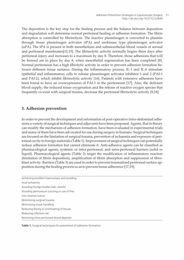
The deposition is the key step for the healing process and the balance between depositionand degradation will determine normal peritoneal healing or adhesion formation. The fibrinabsorption is controlled by fibrinolysis. The inactive plasminogen is converted to plasminthrough tissue plasminogen activator (tPA) and urokinase type plasminogen activator(uPA). The tPA is present in both mesothelium and submesothelial blood vessels of serosaland peritoneal membranes[12,15]. The fibrinolytic activity normally begins three days afterperitoneal injury and increases to a maximum by day 8. Therefore, those adhesions that willbe formed are in place by day 8, when mesothelial regeneration has been completed [8].Normal peritoneum has a high fibrolytic activity in order to prevent adhesion formation be‐tween different tissue surfaces. During the inflammatory process, IL-1 and IL-6 stimulateepithelial and inflammatory cells to release plasminogen activator inhibitor 1 and 2 (PAI-1and PAI-2), which inhibit fibrinolytic activity [16]. Patients with extensive adhesions havebeen found to have an overexpression of PAI-1 in the peritoneum [17]. Also, the deficientblood supply, the reduced tissue oxygenation and the release of reactive oxygen species thatfrequently co-exist with surgical trauma, decrease the peritoneal fibrinolytic activity [9,18].
3. Adhesion prevention
In order to prevent the development and reformation of post-operative intra-abdominal adhe‐sions a variety of surgical techniques and adjuvants have been proposed. Agents, that in theorycan modify the mechanism of adhesion formation, have been evaluated in experimental trialsand many of them have been advocated for use during surgery in humans. Surgical techniquesare focused on the limitation of surgical trauma, prevention of ischaemia and exposure of peri‐toneal cavity to foreign materials (Table 1). Improvement of surgical techniques can potentiallyreduce adhesion formation but cannot eliminate it. Anti-adhesive agents can be classified aspharmacological agents, systemic or intra-peritoneal, and intra-peritoneal barriers (solid orliquid). Pharmacological agents (Table 2) target the modification of inflammatory reaction(limitation of fibrin deposition), amplification of fibrin absorption and suppression of fibro‐blast activity. Barriers (Table 3) are used in order to prevent traumatized peritoneal surface ap‐position during the healing process so as to prevent tissue adherence [17,19].
Achieving excellent haemostasis and avoiding
local ischaemia
Avoiding foreign bodies (talc, starch)
Avoiding peritoneum suturing or use of fine
non-reactive suture
Minimizing surgical trauma
Minimizing tissue handling
Reducing drying or overheating of tissues
Reducing infection risk
Removing intra-peritoneal blood deposits
Table 1. Surgical techniques for prevention of adhesion formation
Adhesion Prevention Strategies in Laparoscopic Surgeryhttp://dx.doi.org/10.5772/52694
51
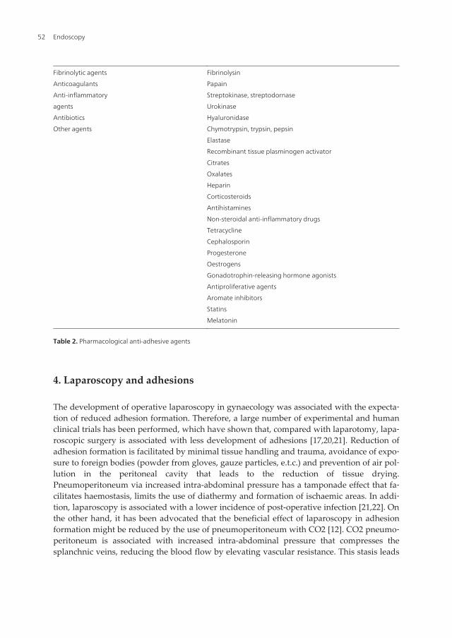
Fibrinolytic agents
Anticoagulants
Anti-inflammatory
agents
Antibiotics
Other agents
Fibrinolysin
Papain
Streptokinase, streptodornase
Urokinase
Hyaluronidase
Chymotrypsin, trypsin, pepsin
Elastase
Recombinant tissue plasminogen activator
Citrates
Oxalates
Heparin
Corticosteroids
Antihistamines
Non-steroidal anti-inflammatory drugs
Tetracycline
Cephalosporin
Progesterone
Oestrogens
Gonadotrophin-releasing hormone agonists
Antiproliferative agents
Aromate inhibitors
Statins
Melatonin
Table 2. Pharmacological anti-adhesive agents
4. Laparoscopy and adhesions
The development of operative laparoscopy in gynaecology was associated with the expecta‐tion of reduced adhesion formation. Therefore, a large number of experimental and humanclinical trials has been performed, which have shown that, compared with laparotomy, lapa‐roscopic surgery is associated with less development of adhesions [17,20,21]. Reduction ofadhesion formation is facilitated by minimal tissue handling and trauma, avoidance of expo‐sure to foreign bodies (powder from gloves, gauze particles, e.t.c.) and prevention of air pol‐lution in the peritoneal cavity that leads to the reduction of tissue drying.Pneumoperitoneum via increased intra-abdominal pressure has a tamponade effect that fa‐cilitates haemostasis, limits the use of diathermy and formation of ischaemic areas. In addi‐tion, laparoscopy is associated with a lower incidence of post-operative infection [21,22]. Onthe other hand, it has been advocated that the beneficial effect of laparoscopy in adhesionformation might be reduced by the use of pneumoperitoneum with CO2 [12]. CO2 pneumo‐peritoneum is associated with increased intra-abdominal pressure that compresses thesplanchnic veins, reducing the blood flow by elevating vascular resistance. This stasis leads
Endoscopy52
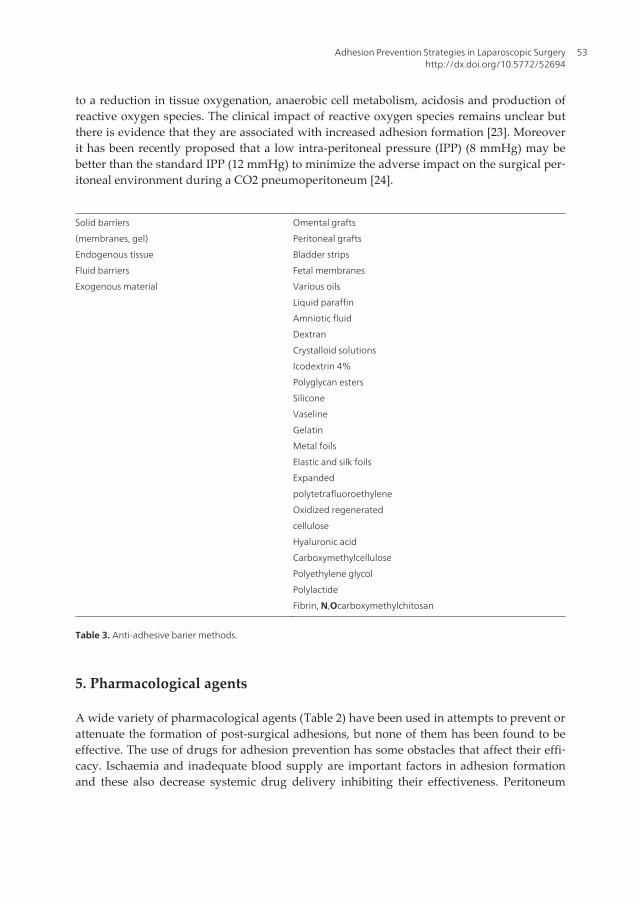
to a reduction in tissue oxygenation, anaerobic cell metabolism, acidosis and production ofreactive oxygen species. The clinical impact of reactive oxygen species remains unclear butthere is evidence that they are associated with increased adhesion formation [23]. Moreoverit has been recently proposed that a low intra-peritoneal pressure (IPP) (8 mmHg) may bebetter than the standard IPP (12 mmHg) to minimize the adverse impact on the surgical per‐itoneal environment during a CO2 pneumoperitoneum [24].
Solid barriers
(membranes, gel)
Endogenous tissue
Fluid barriers
Exogenous material
Omental grafts
Peritoneal grafts
Bladder strips
Fetal membranes
Various oils
Liquid paraffin
Amniotic fluid
Dextran
Crystalloid solutions
Icodextrin 4%
Polyglycan esters
Silicone
Vaseline
Gelatin
Metal foils
Elastic and silk foils
Expanded
polytetrafluoroethylene
Oxidized regenerated
cellulose
Hyaluronic acid
Carboxymethylcellulose
Polyethylene glycol
Polylactide
Fibrin, N,Ocarboxymethylchitosan
Table 3. Anti-adhesive barier methods.
5. Pharmacological agents
A wide variety of pharmacological agents (Table 2) have been used in attempts to prevent orattenuate the formation of post-surgical adhesions, but none of them has been found to beeffective. The use of drugs for adhesion prevention has some obstacles that affect their effi‐cacy. Ischaemia and inadequate blood supply are important factors in adhesion formationand these also decrease systemic drug delivery inhibiting their effectiveness. Peritoneum
Adhesion Prevention Strategies in Laparoscopic Surgeryhttp://dx.doi.org/10.5772/52694
53
has an extremely rapid absorption mechanism, that limits the half life and efficacy of manyintra-peritoneally administered agents. Anti-adhesion agents must not affect normal woundhealing, which has steps in common with adhesion formation (fibrinous exudate, fibrin dep‐osition, fibroblast activity and proliferation) [19,25]. The clinical effectiveness of these agentshas been evaluated in a systematic review and meta-analysis that analysed data from rele‐vant randomized controlled trials (RCT) published up to 2005 [26].
6. Non-steroidal anti-inflammatory drugs
Non-steroidal anti-inflammatory drugs (NSAID) affect adhesion formation by several mech‐anisms. They act by modifying arachidonic acid metabolism and altering cyclooxygenase ac‐tivities. This results in decreased vascular permeability, platelet aggregation, andcoagulation and enhanced macrophage function. A number of locally and systemically ad‐ministered NSAID have been used in experimental trials.
No relevant clinical trials assessing the effectiveness of NSAID in adhesion prevention havebeen published to date in patients undergoing gynaecological surgery. Their clinical efficacyis questionable probably because of inadequate concentrations at the sites of surgical traumaor by rapid absorption from the peritoneal membrane [3,19,27].
7. Corticosteroids and antihistamines
Corticosteroids alter the inflammatory response by reducing vascular permeability and de‐creasing cytokine and chemotactic factor secretion. Antihistamines inhibit fibroblast prolif‐eration and stabilize lysosomal membranes and histamine secretion. Corticosteroids havebeen used alone or plus antihistamines by intra-peritoneal or systemic administration orby flushing through Fallopian tubes post-operatively and were effective in many, but notall, experimental models. In the limited data from RCT [28,29,30], no significant beneficialeffect was detected with the use of corticosteroids (systemic, intra-peritoneal or Fallopiantube flushing) in the deterioration of adhesion score at second-look laparoscopy or on theprobability of clinical pregnancy. However, limited data suggest that the addition of post-operative steroids to systemic intra-operative steroids might be associated with a favoura‐ble outcome both in terms of adhesion score deterioration (increase) or improvement(reduction) [31]. On the other hand, adverse events such as suppression of the pituitary–adrenal axis, immunosuppression and delayed wound healing have been reported withthe use of corticosteroids [19,32,33]. Regarding antihistamines, only one RCT has evaluat‐ed the role of oral promethazine in the prevention of adhesion formation after pelvic sur‐gery. In that study, no significant difference was detected either in deterioration orimprovement of adhesion score in patients who received promethazine as compared withthose who did not [31].
Endoscopy54
8. Progesterone and oestrogens
Progesterone has been used for the prevention of post-operative adhesions. Administrationof progesterone resulted in less adhesion formation in animal models, but does not appearto be effective in humans. Oestrogens have been associated with increased adhesion forma‐tion in animal models. It was demonstrated that a hypo-oestrogenic state, produced by go‐nadotrophin-releasing hormone agonists or aromatase inhibitors such as tamoxifen andanastrazole, decrease development of post-operative adhesions in experimental models[3,34,35]. This hypothesis, however, has never been tested in humans.
9. Anticoagulants – Fibrinolytics
Anticoagulants such as heparin can reduce adhesion formation by inhibition of the coagu‐lation cascade and promotion of fibrinolysis [36]. The use of heparin for intra-peritonealirrigation in a dose that can reduce adhesion formation was associated with haemorrhageand delayed wound healing, but low-dose heparin irrigation showed no benefit in adhe‐sion reduction [3,19,37]. In the only available RCT, heparin delivery with oxidized regen‐erated cellulose failed to demonstrate a superior effect compared with oxidizedregenerated cellulose alone [38]. Furthermore, in experimental trials, the combination ofcarboxymethylcellulose or 32% dextran 70 plus heparin failed to reduce adhesion forma‐tion [38,39,40].
Fibrinolytic agents as streptokinase, elastase and tissue plasminogen activator produced byrecombinant DNA techniques (rtPA) can contribute in adhesion prevention directly by re‐ducing the fibrinous mass and indirectly by stimulating plasminogen activator activity. Sys‐temic administration of anticoagulants is impeded by lack of safety. The concentrations offibrinolytic agents required to prevent adhesion formation are too close to the anticoagulato‐ry concentrations and increase the risk for post-operative haemorrhage and delayed woundhealing. Intra-peritoneal administration is ineffective due to rapid absorption by the perito‐neal membrane [3,25,41]. The use of carboxymethylcellulose gel and oxidized regeneratedcellulose as a carrier to deliver rtPA intra-peritonealy was not associated with a reduction ofadhesion formation in animal models [42,43].
A recent study has investigated the impact of gonadotropin-releasing hormone analogue(GnRH-a) on coagulation and fibrinolytic activities and its effectiveness in the prevention ofpelvic adhesion after myomectomy in thirty-two infertile women. Patients treated withGnRH-a showed significant decrease in plasminogen activator inhibitorPAI, thrombin acti‐vatable fibrinolysis inhibitor (TAFI), factors V, and VIII and increased protein C (PC), but nosignificant change in plasminogen and α2-antiplasmin levels compared with control group,suggesting a possible critical role of the GnRH-a therapy in preventing postoperative adhe‐sion development [44].
Adhesion Prevention Strategies in Laparoscopic Surgeryhttp://dx.doi.org/10.5772/52694
55
10. Antibiotics
Antibiotics are commonly used for prophylaxis against post-operative infections and hencethe inflammatory response that leads to adhesion formation. Peritoneal irrigation with anti‐biotic solutions does not reduce adhesion formation, while it has been shown that in somecases it may promote them [45].
11. Other pharmacological regimens
Many other agents, such as apoprotin, noxytioline, growth factor inhibitors and modulators,phosphatidylcholine, thiazolidinediones, colchicine and calcium channel blockers, havebeen utilized in experimental trials. The intra-peritoneal administration of noxytioline is theonly one of these interventions that has been tested in the context of a RCT and no signifi‐cant difference was identified in terms of reduction of adhesions and clinical pregnancyrates in patients who were administered intra-peritoneal noxytioline and the control group[29]. There is no data from RCT to support the conclusion that any of the other agents is effi‐cacious in preventing the development of post-operative adhesions [19,46,47].
12. Anti-adhesive barriers
The failure of pharmacological regimens to prevent adhesion formation has led to the reviv‐al of the barrier technique. With the barrier technique, traumatized peritoneal surfaces arekept separated, during mesothelial regeneration, thus precluding adherence of adjacent or‐gans and tissues and reducing the development of adhesions. The separation can be ach‐ieved by the use of solid (films or gels) or fluid barriers [19,25]. Anti-adhesive barriers arecurrently the most useful adjuvant for prevention of post-operative adhesion formation. Nu‐merous substances (Table 3) have been used as mechanical barriers to separate tissue surfa‐ces. Most of these materials are of historical interest only and had no effect or evenaggravated adhesion formation [48,49]. An anti-adhesive agent should be effective, safe, eco‐nomical and easy to use in both open and laparoscopic surgery [50]. The clinical effective‐ness of several of these agents has been evaluated in two recent Cochrane reviews [26,51].
13. Solid barriers
Solid barriers are placed over one or between two traumatized surfaces providing a separa‐tion that averts tissue apposition during the critical period of fibrin formation and mesothe‐lial regeneration following surgical trauma. It should be noted though, that solid barriershave some significant drawbacks. They are often ineffective in the presence of blood, have acomplex preparation and application, do not conform easily to the shape of pelvic organs,
Endoscopy56
need suturing and are difficult to use via laparoscopic surgery. Their benefits are also limit‐ed to the site of application and do not prevent the development of adhesions at sites of in‐direct trauma. So the surgeon has to surmise where adhesions will be formed in order tochoose the placement sites and optimize barrier efficacy [52]. Not infrequently, this provesto be a challenging task, since, for example, midline laparotomy initiates a generalized peri‐toneal response that can lead to adhesion formation distant from surgical trauma [53].
14. Expanded polytetrafluoroethylene
Expanded polytetrafluoroethylene (Preclude, Gore-Tex Surgical Membrane; Johnson andJohnson, Arlington, TX) is a non-absorbable, non-reactive, synthetic material that
inhibits cellular migration and tissue adherence. In the only available RCT, it has beenshown to be associated with fewer post-operative de-novo adhesions after myomectomywhen compared with no treatment [54]. Moreover, when compared with oxidized regener‐ated cellulose, Preclude was found to be more effective in terms of adhesion reformation af‐ter adhesiolysis [55]. However, in another RCT, no evidence of a beneficial effect of Precludewas demonstrated in the de-novo formation of adhesions after laparoscopic myomectomywhen compared to oxidized regenerated cellulose [56]. Expanded polytetrafluoroethylenehas the disadvantages that it must be sutured in place, is difficult to use in laparoscopic sur‐gery and, ideally, requires a subsequent surgical procedure for removal after the injury hashealed [57]. The use of Preclude in Europe is limited and it has been withdrawn from themarket in USA after the development of the absorbable barriers [25].
15. Oxidized regenerated cellulose
Oxidized regenerated cellulose (Interceed (TC7); Johnson and Johnson) is the first degrada‐ble barrier that was used in clinical practice and represents a modification of its precursorSurgicel, which has been used as a haemostatic agent for a long time. It is a mesh designedto be placed over or between traumatized surfaces. About 8 h after the application in theperitoneal cavity, it becomes a viscous gel and finally it is degraded to monosaccharides andcompletely absorbed in about 2 weeks [3,22]. Oxidized regenerated cellulose use in laparo‐scopic surgery is feasible [8]. In order to evaluate the efficacy of oxidized regenerated cellu‐lose in the prevention of the development of post-surgical adhesions, many studies havebeen carried out. A meta-analysis of 11 relevant RCT [51] has shown that the barrier is safeand reduces significantly the incidence of de-novo adhesions, as well as the reformation ofadhesions as compared with no treatment in laparoscopy [58-62]. In laparotomy, the availa‐ble RCT [63-68], when meta-analysed [51], demonstrated that a significant reduction in thereformation (or mixture) of adhesions can be expected with the use of oxidized regeneratedcellulose as compared with the no treatment group. The product is site specific, thus the effi‐cacy is limited to surgical situations where raw surfaces can be completely covered with the
Adhesion Prevention Strategies in Laparoscopic Surgeryhttp://dx.doi.org/10.5772/52694
57

mesh and its benefit is limited to the site of barrier placement. The fundamental disadvant‐age is that it becomes ineffective when the entire area is not completely haemostatic. Thepresence of small amounts of blood in the peritoneal cavity or post-operative bleeding re‐sults in blood permeating the mesh, fibrin deposition and, finally, adhesion formation[8,69,70]. In addition, as reported previously, the combination of oxidized regenerated cellu‐lose plus heparin resulted in a significant reduction of adhesion formation and reformationin experimental models. This improvement in efficacy was not confirmed in clinical trials[36,38,40]. Oxidized regenerated cellulose has been approved by the US Food and Drink Ad‐ministration (FDA) for use in open surgery in the USA [69].
16. Hyaluronic acid
Hyaluronic acid (HA) is a linear polysaccharide with repeating disaccharide units that arecomposed of sodium D-glucuronate and N-acetyl-D-glucosamine. It is a naturally occurringcomponent of many body tissues and fluids, where it provides mechanically protective andphysically supportive roles [69]. Various combinations of HA have been used for the pre‐vention of adhesion formation. HA and carboxymethylcellulose (Seprafilm; Genzyme, Cam‐bridge, Massachusetts, USA) is an absorbable membrane that dissolves and forms ahydrophilic gel approximately 24 h after placement. It is a site-specific barrier and acts byseparating mechanically opposite tissue surfaces and lasts for 7 days. The HA is completelycleared from the body within 4 weeks, but the absorption of carboxymethylcellulose is notwell known. It does not conform to the shape of pelvic organs as well as oxidized regenerat‐ed cellulose and is usually used to prevent adhesions between the incision of anterior ab‐dominal wall and bowel or omentum [71,72]. Its use in laparoscopic procedures is difficult.In a blind prospective, randomized, multicentre study, the treatment of patients after myo‐mectomy with Seprafilm significantly reduced the extent and area of post-operative uterineadhesions [72]. Potential side effects include induced foreign body reaction, higher incidenceof pulmonary emboli and intra-peritoneal abscess formation, but these findings were notstatistically significant in the relevant trials [3,73]. High cost is another limitation because,for an effective protection from intestinal obstruction, a mean of 4.5 sheets per patient is re‐quired [25]. Seprafilm has been approved by the FDA for use in open surgery in the USA[22]. Ferric hyaluronate 0.5% gel (Intergel; Gynecare, Sommerville, New Jersey, USA) is aviscous gel that provides a broader coverage than previous site-specific agents. It wasshown to be easy to use in open and laparoscopic surgery. In relevant prospective random‐ized trials, ferric hyaluronate was associated with a significant reduction of severity and ex‐tent of post-operative adhesions and statistically significant improvement of the AmericanFertility Society (AFS) and modified AFS scores at second-look laparoscopy [69,74,75]. It waswithdrawn from the market in 2003 because of problems with late onset post-operative painand rare reports of sclerosing peritonitis [25,76]. Low-viscosity 0.04% HA combined withphosphate-buffered saline (Sepracoat; Genzyme) is a bioabsorbable macromolecular dilutesolution of HA that is cleared from the body in less than 5 days. The solution is applied inthe peritoneal cavity before any tissue manipulation in order to protect peritoneal surfaces
Endoscopy58
from indirect trauma and finally before the end of the procedures [3,77,78]. In a blind, pro‐spective, randomized, placebo-controlled multicentre study, where patients had undergoneopen gynecological procedures, low-viscosity 0.04% HA resulted in a statistically significantreduction of adhesions, as well as of the mean adhesion score, at second-look laparoscopy.However, it was not effective in reducing post-operative adhesion formation at sites of di‐rect surgical trauma [78]. It has been approved by the FDA for use in open surgery in theUSA [22]. HA cross-linked to HA (Hyalobarrier Gel; Baxter, Bracknell, UK) is a site-specifichighly viscous gel is considered as easy to use in laparoscopic and open surgery. In a pro‐spective, randomized, controlled study where the rate of post-surgical adhesions after lapa‐roscopic myomectomy was examined, cross-linked HA resulted in significantly moreadhesion-free patients [79]. Pregnancy rates at 6 and 12 months after laparoscopic myomec‐tomy were significantly higher in patients treated with cross-linked HA [80]. In anotherrandomized trial, adhesion-free patients after laparoscopic myomectomy were greater in thetreatment group but the difference was not statistically significant. The incidence and severi‐ty of adhesions was similar in both groups, but a significant reduction of uterine adhesionswas found in the treatment group [81]. When the data from the two aforementioned studieswere combined, a statistically significant reduction of adhesions during second-look laparo‐scopy was detected in the group of patients treated with HA-cross-linked HA as comparedwith the control group. Auto-cross-linked internal ester form of HA (ACP gel; Fidia Ad‐vanced Biopolymers, AbanoTerme, Italy) has the biocompatibility of the original polymerbut higher viscosity and extended residence. It is a gel that has been shown to be efficaciousin reducing abdominal adhesions in experimental models [82,83]. Two prospective random‐ized controlled trials have been published so far by the same group regarding the use ofACP gel for the prevention of intrauterine adhesions after hysteroscopic surgery. In thesestudies, ACP gel has been associated with a significant reduction in the incidence and theseverity of subsequent intrauterine adhesions [84,85]. A stratified analysis of these two stud‐ies confirmed this finding by demonstrating a significant reduction in the proportion of pa‐tients with adhesions at second-look hysteroscopy. Cross-linked thiol-modified HA with 4%polyethyleneglycoldiacrylate (Carbylan-S and Carbylan-SX; CarbylanBioSurgery, Palo Alto,CA, USA) is a bioabsorbable solution of HA. Carbylan-S is a hydrogel and Carbylan-SX hastwo formats, a sprayable gel and a hydrogel film. In animal models, Carbylan- S containingmitomycin C and Carbylan-SX were effective in prevention of post-operative intra-abdomi‐nal adhesions [52,86].
17. Polylactide
Polylactide (copolymer of 70:30 poly(L-lactide-CO-D, L-lactide; SurgiWrap, MacroporeBio‐surgeryinc., San Diego, USA) is a bioabsorbable film with a long absorption period (up to 6months). It is metabolized to lactic acid and finally to CO2 and exhaled through the respira‐tory system. It requires suturing in order to avoid its loss from the site. In preclinical studies,polylactide appears to be effective in the reduction of adhesion formation, but there are nodata currently for safety and efficacy in humans [87,88].
Adhesion Prevention Strategies in Laparoscopic Surgeryhttp://dx.doi.org/10.5772/52694
59
18. Polyethylene glycol
Polyethylene glycol (SprayGel; Confluent Surgical, Waltham, Massachusetts, USA). It is a syn‐thetic hydrogel formed when two polyethylene glycol-based liquids are sprayed together withan air assisted sprayer at the target tissue, where they cross-link and form a hydrogel barrier.One liquid is clear and one is coloured with methylene blue in order that facilitate its application.The gel remains intact for approximately 5–7 days and then gradually breaks down by hydroly‐sis and is cleared through the kidneys [89]. Drawbacks of the product are the intricacy of prepa‐ration and application, the time required to cover the target tissue and the high cost. In aprospective randomized controlled phase-III trial, in 40 patients undergoing myomectomy, pol‐ylactide resulted in a significant decrease in the mean tenacity score. The extent of adhesions wasincreased in the control group but the difference was not significant. Also, the proportion of ad‐hesion-free patients at second-look laparoscopy was increased in the treatment group but thedifference was not statistically significant [89]. It has been approved for use in laparoscopic andopen surgery in Europe, but by the FDA only for use in open surgery in USA [22,25].
19. Carboxymethylcellulose
Carboxymethylcellulose is a high-molecular-weight polysaccharide, derivative of cellulose.The mechanism of its absorption is not well known. It has been used in combination with rtPAand with HA. A composite gel of carboxymethylcellulose and polyethylene oxide (Oxiplex;FzioMed, San Luis Obispo, CA, USA) is a viscoelastic gel, which acts as a barrier between tissuesthat inhibits protein deposition and thrombus formation [74]. The gel is absorbed by 6 weeks,but in cases where large amounts of gel were applied in multiple layers to the surgically treatedsites or in cases of stage-IV endometriosis, small collections of gelatinous material were noted inareas of gel application or in areas deep in the cul-de-sac [90]. In two blind randomized control‐led trials, where patients underwent adnexal surgery, carboxymethylcellulose and polyethy‐lene oxide showed a significant improvement of AFS score in the treated group, but not in allclinical situations. It did not appear to provide this benefit to patients with grade-IV endometrio‐sis [74,91]. Another double-blind prospective randomized controlled trial has shown that themean AFS score for patients in the treatment group was unchanged, while in control patients anincreased AFS score was noted [91]. No statistical pooling was feasible for these three studies,since the data were not analysed and presented per randomization unit (they were analysed peradnexa and not per patient). It is easy to use in laparoscopic surgery and it has been approved inEurope for use in abdominal and pelvic surgery [25].
20. Fibrin glue
Fibrin glue (Tissucol; Baxter International, Deerfield, IL, USA) is a biological product. Fibringlue is made by mixing human fibrinogen with bovine thrombin, calcium and factor XIII
Endoscopy60
[92]. Obviously, the use of human blood products raises a theoretical risk for transmission ofinfectious diseases. According to the pathogenesis of adhesions, application of fibrin glue atthe traumatized peritoneal surfaces should increase adhesion formation. Possibly, fibringlue application confines fibrin deposition and averts the development of attachments be‐tween opposing tissue surfaces. In animal studies, the use of fibrin glue has been shown todecrease adhesion formation and reformation but clinical data are limited. Fibrin glue hasnot been approved by the FDA for use in USA [22,47]. So far, no relevant data from trials inhumans have been published.
21. Carboxymethylchitosan
N,O-carboxymethylchitosan (Adhes-X, Chitogenics, New Jersey, USA) is a purified deriva‐tive of chitin obtained from the exoskeleton of shrimp and has similar structure to hyaluron‐ic acid and carboxymethylcellulose [93]. The product comprises both a clear gel and asolution. The gel is placed initially at the sites of surgical trauma where it is tamped with alaparoscopic instrument and subsequently the solution is placed at the same places. Its effi‐cacy and safety have been confirmed in some animal models. A prospective randomizedcontrolled study, performed on 34 patients undergoing laparoscopy for various gynaecolog‐ical indications, demonstrated a decrease in the recurrence, extent and severity of adhesionsand a decrease of de-novo adhesion formation at second-look laparoscopy, but none of thesefindings were statistically significant [94].
22. Fluid barriers
Fluids constitute an ideal barrier agent because their action is not limited to the site of applica‐tion. Their function is provided by hydrofloration of intra-peritoneal structures in the liquidthat is infused into the peritoneal cavity at the end of the surgical procedure. Hydroflorationprovides a temporary separation between raw peritoneal surfaces allowing independent heal‐ing without the formation of adhesions. Possibly, fluid circulation in the peritoneal cavity con‐tributes to the prevention of adhesion formation by diluting fibrinous exudates released fromtraumatized surfaces. Fluid barriers may prevent adhesion formation both at the traumatizedarea and elsewhere in the pelvis. The instillation of fluids in the peritoneal cavity may be associ‐ated with some undesirable side effects, such as leakage from the incision, labial oedema, feel‐ing of fluid moving around, abdominal discomfort, abdominal distension and complicationssuch as pulmonary and peripheral oedema. Large volumes of intra-peritoneal fluids may de‐crease the peritoneum ability to confront bacterial infections [3,17,76].
23. Crystalloid solutions
Crystalloid solutions (Ringer’s lactate, NaCl 0.9%) are rapidly absorbed by the peritonealcavity, at a rate of 30–50 ml/h. Consequently, 24 h after the surgery, minimal or no crystal‐
Adhesion Prevention Strategies in Laparoscopic Surgeryhttp://dx.doi.org/10.5772/52694
61
loid solution would be left in the peritoneal cavity. The instillation of crystalloids does notseem to result in decreased adhesion formation. They are commonly used but they are notapproved for use as anti-adhesive agents [25,69,95]. Crystalloids have also been used in vari‐ous combinations with heparin, steroids, antihistamines and other pharmacological agentsin randomized controlled trials, but none of them has been found effective in decreasingpost-operative adhesion formation or improving pregnancy rates [96].
24. Dextran
Dextran (32% dextran 70; Hyskon; Pharmacia, Piscataway, New Jersey, USA) is a 1–6-linkeddextrose polymer. A summary [26] of the available data from relevant RCT [37,97-99] dem‐onstrated a decreased proportion of patients with adhesions at second-look laparoscopy inthe group that received 32% dextran 70, as compared with the group that did not. However,despite the fact that the patients with improvement and deterioration in the adhesion scoreat second-look laparoscopy were increased and decreased, respectively, in the treatmentgroup, when compared with the control group, this difference was not statistically signifi‐cant. In addition, its use was associated with significant side effects as pulmonary and pe‐ripheral oedema caused by its osmotic properties, liver function abnormalities, pleuraleffusion and, rarely, allergic reactions or anaphylactic shock and disseminated intra-vascu‐lar coagulation. It has not been approved for use as an anti-adhesive agent [3,17,100,101].
25. Polyglycan esters
Polyglycanesters (Adcon-P; Gliatech, Cleveland, Ohio) is a viscous bioabsorbable solution.Its prototypes Adcon-L and Adcon-T/N were found effective for adhesion prevention in spi‐nal and neurosurgical procedures. Experimental studies have shown that application of Ad‐con-P effectively reduces development of post-operative intra-abdominal adhesions. Thereare no data for the safety and the efficacy of this product in humans [102].
26. Icodextrin
Icodextrin 4% (Adept; Shire Pharmaceuticals, Basingstoke, Hampshire, UK) is a 1–4-linkedglucose polymer. Icodextrin 4% is a clear isomolar solution and does not predispose to infec‐tion. It is absorbed gradually via the lymphatic system into the systemic circulation, where itis digested to oligosaccharides by amylase. Amylase is absent from the human peritonealcavity. Preclinical studies had shown significant reduction of post-operative adhesions andconfirmed the safety of icodextrin 4%. It was indicated that the agent was more effective inadhesion reduction when used as both an irrigant and post-operative instillate [103]. In asmall double-blind prospective randomized multicentre study, icodextrin 4% resulted in the
Endoscopy62
reduction of incidence, severity and extent of adhesions but these results were not statistical‐ly significant [104]. However, recently, in the largest prospective randomized double-blindmulticentre study for an anti-adhesive agent, icodextrin 4% has been shown to result in asignificant reduction of incidence, severity and extent of adhesions and a significant im‐provement of AFS score. Also the study showed that icodextrin 4% prevents the deteriora‐tion of pre-existing adhesions, considering that patients with the higher number of adhesionlysed at initial surgery had the greater reduction in adhesion incidence [95]. A stratifiedanalysis of these two studies revealed a statistically significant effect oficodextrin 4% use onthe de-novo formation of adhesions, as well as on the proportion of patients with an im‐provement of the adhesion score at second-look laparoscopy [105]. Simultaneously withclinical trials, a European patients registry (ARIEL) was created allowing surgeons to recordand report the experiences of the use of icodextrin 4% in open and laparoscopic gynaecolog‐ical and general surgery. The registry provides feedback on routine use in 4620 patients(2882 that underwent gynaecological and 1738 general surgery). The general consensus isthat it is easy to use in both open and laparoscopic surgery, it is well tolerated by patientsand the incidence of adverse events is considered similar to the control group [76,106]. Lowcost is another advantage of icodextrin 4%. Adept (1.5 litre bags) are about half the price ofeach sheet of Interceed or Seprafilm and it is about four times cheaper than one SprayGelpackage [50]. Icodextrin 4% has been approved for use in open and laparoscopic surgery inEurope and it was the first anti-adhesive agent that has been approved by the FDA for usein laparoscopic surgery in the USA [25,76,91,95].
27. Conclusions
Development of post-operative adhesion is a widespread consequence of surgical traumaand healing following open or laparoscopic gynecological surgery and is associated withsignificant complications. At present, the main strategy to avoid formation and reformationof adhesions is focused on the use of careful surgical techniques and anti-adhesive agents.Reduction of adhesion formation after laparoscopic surgery in comparison to the more con‐ventional approach by laparotomy can be attributed to less tissue manipulation, less tissuedrying, avoidance of insertion of foreign bodies such as talc from the surgical gloves, fibersfrom the gauzes e.t.c. On the other hand pneumoperitoneum during laparoscopy exerts atamponade effect that facilitates hemostasis, so minimizing the use of electrocautery, whichis known that leads to the formation of ischaemic areas and therefore predisposing to adhe‐sion formation. The use of CO2 for pnemoperitoneum may lead to adhesion formation, sinceits use is associated with reduction in tissue oxygenation, acidosis and release ofreactiveoxygen species, which are considered adhesiogenic. Therefore, the addition of oxygen, theheating of the insufflatedCO2 or the alternative use of other gases (i.e. helium) may be bene‐ficial in terms of reduction of de novo adhesion formation. At the end of each operation an«underwater» examination should be used in order to document complete intra-peritonealhemostasis, since it has been clearly demonstrated that incomplete hemostasis is associatedwith adhesion formation.
Adhesion Prevention Strategies in Laparoscopic Surgeryhttp://dx.doi.org/10.5772/52694
63
Finally, several anti-adhesive agents are used during laparoscopic intrventions, in order tominimizepostoperative adhesions. These fall into two main categories, which include phar‐macological agents and barrier methods. Limited data support the use of the former, eitherlocally or systemically. Barriers, which mechanically separate the opposed serosal surfacesand exert their beneficial action, at least partly, because they remain in place beyond the crit‐ical 3-day point, at which competition of fibrinolytic activityandfibrosis will lead to adhe‐sion formation. The tissue separation can be achieved either by the use of solid (films or gel)or fluid barriers. The clinical effectiveness of several of these agents has been thoroughlyevaluated in two recent Cochrane reviews by Metwally et al [26] and Ahmad et al [51].
Author details
George Pados, Anastasios Makedos and Basil Tarlatzis
1st Dept. of OB-GYN, School of Medicine, Aristotle University of Thessaloniki, «Papageor‐giou» General Hospital, N. Efkarpia, Greece
References
[1] Holmdahl, L., Risberg, B., Beck, D.E., et al. Adhesions: pathogenesis and prevention-panel discussion and summary. Eur. J. Surg. Suppl. 1997; 56: 62.
[2] Menzies, D., Ellis, H. Intestinal obstruction from adhesions – how big is the problem?Ann. R. Coll. Surg. Engl. 1992; 72: 60–63.
[3] Liakakos, T., Thomakos, N., Fine, P.M., Dervenis, C., Young, R.L. Peritoneal adhe‐sions: etiology, pathophysiology, and clinical significance. Recent advances in pre‐vention and management.Dig. Surg. 2001; 18: 260–273.
[4] Menzies, D. Peritoneal adhesions. Incidence, cause, and prevention. Surg. Annu.1992; 24: 27–45.
[5] Operative Laparoscopy Study Group. Postoperative adhesion development after op‐erative laparoscopy: evaluation at early second-look procedures. Fertil. Steril. 1991;55: 700–704.
[6] Lower, A.M., Hawthorn, R.J., Clark, D., et al. Adhesion- related readmissions follow‐ing gynaecological laparoscopy or laparotomy in Scotland: an epidemiological studyof 24 046 patients. Hum. Reprod. 2004;19: 1877–1885.
[7] Diamond, M.P., Freeman, M.L. Clinical implications of postsurgical adhesions. Hum.Reprod. Update. 2001; 7: 567–576.
[8] Pados, G.A., Devroey, P. Adhesions. Curr. Opin. Obstet. Gynecol. 1992; 4: 412–418.
Endoscopy64
[9] DiZerega, G.S. Biochemical events in peritoneal tissue repair. Eur. J. Surg. Suppl.1997; 10: 16.
[10] Badia, J.M., Whawell, S.A., Scott-Coombes, D.M., Abel, P.D., Williamson, R.C.,Thompson, J.N. Peritoneal and systemic cytokine response to laparotomy. Br. J. Surg.1996;83: 347–348.
[11] Boland, G.M., Weigel, R.J. Formation and prevention of postoperative abdominal ad‐hesions. J. Surg. Res. 2006; 132: 3–12.
[12] Binda, M.M., Molinas, C.R., Koninckx, P.R. Reactive oxygen species and adhesionformation: clinical implications in adhesion prevention. Hum. Reprod. 2003; 18:2503–2507.
[13] Drollette, C.M., Badawy, S.Z. Pathophysiology of pelvic adhesions. Modern trends inpreventing infertility. J. Reprod. Med. 1992; 37: 107–121.
[14] Rodgers, K.E., diZerega, G.S. Function of peritoneal exudate cells after abdominalsurgery. J. Invest. Surg. 1993; 6: 9–23.
[15] Dano, K., Andreasen, P.A., Grondahl-Hansen, J., Kristensen, P., Nielsen, L.S., Skriver,L. Plasminogen activators, tissue degradation, and cancer. Adv. Cancer Res. 1985; 44:139–266.
[16] Buckman, R.F., Woods, M., Sargent, L., Gervin, A.S. A unifying pathogenetic mecha‐nism in the etiology of intraperitoneal adhesions. J. Surg. Res. 1976; 20: 1–5.
[17] Sutton, C. Adhesions and their prevention. Obstet. Gynaecol. 2005; 7: 168–176.
[18] Raftery, A.T. Effect of peritoneal trauma on peritoneal fibrinolytic activity and intra‐peritoneal adhesion formation. An experimental study in the rat. Eur. Surg. Res.1981;13:397–401.
[19] Risberg, B. Adhesions: preventive strategies. Eur. J. Surg. Suppl. 1997;32: 39.
[20] Lundorff, P., Hahlin, M., Kallfelt, B., Thorburn, J., Lindblom, B. Adhesion formationafter laparoscopic surgery in tubal pregnancy: a randomized trial versus laparotomy.Fertil. Steril. 1991;55: 911–915.
[21] Schafer, M., Krahenb hl, L., Buchler, M.W. Comparison of adhesion formation inopen and laparoscopic surgery. Dig. Surg. 1998; 15: 148–152.
[22] The Practice Committee of the ASRM. Control and prevention of peritoneal adhe‐sions in gynecologic surgery. Fertil. Steril. 2006;86: S1–S5.
[23] de Souza, A.M., Wang, C.C., Chu, C.Y., Lam, P.M., Rogers, M.S. The effect of intra-abdominal pressure on the generation of 8-iso prostaglandin F2alpha during laparo‐scopy in rabbits. Hum. Reprod. 2003;18: 2181–2188.
[24] Matsuzaki S, Jardon K, Maleysson E, D’Arpiani F, Canis M, Botchorishvili R. Impactof intraperitoneal pressure of a CO2 pneumoperitoneum on the surgical peritonealenvironment. Hum Reprod. 2012;27(6): 1613-23.
Adhesion Prevention Strategies in Laparoscopic Surgeryhttp://dx.doi.org/10.5772/52694
65
[25] Trew, G: Postoperative adhesions and their prevention. Rev. Gynaecol. Perinat.Pract. 2006;6: 47–56.
[26] Metwally, M., Watson, A., Lilford, R., Vandekerckhove, P. Fluid and pharmacologi‐cal agents for adhesion prevention after gynaecological surgery. Cochrane DatabaseSyst Rev, CD001298, 2006.
[27] Luciano, A.A., Hauser, K.S., Benda, J. Evaluation of commonly used adjuvants in theprevention of postoperative adhesions. Am. J. Obstet. Gynecol. 1983;146: 88–92.
[28] Jansen, R.P. Failure of intraperitoneal adjuncts to improve the outcome of pelvic op‐erations in young women. Am. J. Obstet. Gynecol. 1985;153: 363–371.
[29] Querleu, D., Vankeerberghen-Deffense, F., Boutteville, C. The effect of noxytiolineand systemic corticosteroids in infertility surgery. A prospective randomized study.(Traitement adjuvant des plastiestubaires. Etude prospective randomisee des cortico‐ides par voiegeneraleet de la noxytioline). J. Gynecol. Obstet. Biol. Reprod. 1989;18:935–940.
[30] Rock, J.A., Siegler, A.M., Meisel, M.B. The efficacy of postoperative hydrotubation: arandomized prospective multicenter clinical trial. Fertil. Steril. 1984;42(2): 373–376.
[31] Jansen, R.P. Controlled clinical approaches to investigating the prevention of perito‐neal adhesions. Prog. Clin. Biol. Res. 1990;358: 177–192.
[32] Granat, M., Schenker, J.G., Mor-Yosef, S., Rosenkovitch, E., Castellanos, R.C., Galili,U. Effects of dexamethasone on proliferation of autologous fibroblasts and on the im‐mune profile in women undergoing pelvic surgery for infertility. Fertil. Steril.1983;39: 180–186.
[33] Harris, W.J., Daniell, J.F: Use of corticosteroids as an adjuvant in terminal salpingos‐tomy. Fertil. Steril. 1983;40: 785–789.
[34] Kaya, U., Oktem, M., Zeyneloglu, H.B., Ozen, O., Kuscu, E. Impact of aromatase in‐hibitors on adhesion formation in a rat model. Fertil. Steril. 2007;87: 934–939.
[35] Montanino-Oliva, M., Metzger, D.A., Luciano, A.A. Use of medroxyprogesteroneacetate in the prevention of postoperative adhesions. Fertil. Steril. 1996;65: 650–654.
[36] Diamond, M.P., Linsky, C.B., Cunningham, T. Adhesion reformation: reduction bythe use of Interceed (TC7) plus heparin. J. Gynecol. Surg. 1991;7(a): 1–6.
[37] Jansen, R.P.Failure of peritoneal irrigation with heparin during pelvic operationsupon young women to reduce adhesions. Surg. Gynecol. Obstet. 1988;166: 154–160.
[38] Reid, R.L., Hahn, P.M., Spence, J.E., Tulandi, T., Yuzpe, A.A., Wiseman, D.M. Arandomized clinical trial of oxidized regenerated cellulose adhesion barrier (Inter‐ceed, TC7) alone or in combination with heparin. Fertil. Steril.1997; 67: 23–29.
Endoscopy66
[39] Diamond, M.P., Linsky, C.B., Cunningham, T., et al. Synergistic effects of INTER‐CEED(TC7) and heparin in reducing adhesion formation in the rabbit uterine hornmodel. Fertil. Steril. 1991;55(b): 389–394.
[40] Reid, R.L., Lie, K., Spence, J.E., Tulandi, T., Yuzpe, A. Clinical evaluation of the effi‐cacy of heparin-saturated Interceed for prevention of adhesion reformation in thepelvic sidewall of the human. Prog. Clin. Biol. Res. 1993;381: 261–264.
[41] Doody, K.J., Dunn, R.C., Buttram Jr., V.C. Recombinant tissue plasminogen activatorreduces adhesion formation in a rabbit uterine horn model. Fertil. Steril. 1989;51:509–512.
[42] Bothin, C. Counteracting postsurgical adhesions – the effect of combining oxidizedregenerated cellulose and tissue plasminogen activator. Int. J. Fertil. MenopausalStud. 1995;40: 102–105.
[43] Gehlbach, D.L., O’Hair, K.C., Parks, A.L., Rosa, C. Combined effects of tissue plasmi‐nogen activator and carboxymethylcellulose on adhesion reformation in rabbits. Int.J. Fertil. Menopausal Stud. 1994;39: 172–176.
[44] Di Nardo MA, Annunziata ML, Ammirabile M, Di Minno MN, Ruocco AL, De FalcoM, Di Lieto A.Pelvic adhesion and gonadotropin-releasing hormone analogue: effectsof triptorelin acetate depot on coagulation and fibrinolytic activities. Reprod Sci.2012;19(6):615-22.
[45] Rappaport, W.D., Holcomb, M., Valente, J., Chvapil, M. Antibiotic irrigation and theformation of intraabdominal adhesions. Am. J. Surg. 1989;158: 435–437.
[46] Demirturk, F., Aytan, H., Caliskan, A., et al. The effect of rosiglitazone in the preven‐tion of intra-abdominal adhesion formation in a rat uterine horn model. Hum. Re‐prod. 2006;21: 3008–3013,.
[47] Hellebrekers, B.W., Trimbos-Kemper, G.C., van Blitterswijk, C.A., Bakkum, E.A.,Trimbos, J.B. Effects of five different barrier materials on postsurgical adhesion for‐mation in the rat. Hum. Reprod. 2000;15: 1358–1363.
[48] Arora, M., Jaroudi, K.A., Hamilton, C.J., Dayel, F. Controlled comparison of intercedeand amniotic membrane graft in the prevention of postoperative adhesions in therabbit uterine horn model. Eur. J. Obstet. Gynecol. Reprod. Biol. 1994;55: 179–182.
[49] Seifer, D.B., Diamond, M.P., DeCherney, A.H. An appraisal of barrier agents in thereduction of adhesion formation following surgery. J. Gynecol. Surg. 1990; 6: 3–10.
[50] Trew, G., Lower, A. Consensus in adhesion reduction management. Obstetr. Gynae‐col. 2004; 6: 1–2.
[51] Ahmad, G., Duffy, J.M., Farquhar, C., et al. Barrier agentsfor adhesion prevention af‐ter gynaecological surgery. Cochrane Database Syst Rev, CD000475, 2008.
Adhesion Prevention Strategies in Laparoscopic Surgeryhttp://dx.doi.org/10.5772/52694
67
[52] Liu, Y., Li, H., Shu, X.Z., Gray, S.D., Prestwich, G.D. Crosslinkedhyaluronan hydro‐gels containing mitomycin C reduce postoperative abdominal adhesions. Fertil. Ster‐il. (Suppl. 1). 2005; 83:1275–1283.
[53] Becker, J.M., Stucchi, A.F.Intra-abdominal adhesion prevention: are we getting anycloser? Ann. Surg. 2004;240: 202–204.
[54] Myomectomy ASG. An expanded polytetrafluoroethylene barrier (Gore-Tex SurgicalMembrane) reduces post-myomectomy adhesion formation. The Myomectomy Ad‐hesion Multicenter Study Group. Fertil. Steril. 1995;63: 491–493.
[55] Haney, A.F., Hesla, J., Hurst, B.S., et al. Expanded polytetrafluoroethylene (Gore-TexSurgical Membrane) is superior to oxidized regenerated cellulose (Interceed TC7+) inpreventing adhesions. Fertil. Steril. 1995;63: 1021–1026.
[56] Korell, M. Reduction of adhesion by INTERCEED Barrier and Gortex Surgical Mem‐brane after laparoscopic myomectomy, Moglichkeiten der Adhasionprophylaxe, Mu‐nich, 1994.
[57] Farquhar, C., Vandekerckhove, P., Watson, A., Vail, A., Wiseman, D: Barrier agentsfor preventing adhesions after surgery for subfertility. Cochrane Database Syst Rev,CD000475, 2000.
[58] Keckstein, J., Ulrich, U., Sasse, V., Roth, A., Tuttlies, F., Karageorgieva, E. Reductionof postoperative adhesion formation after laparoscopic ovarian cystectomy. Hum.Reprod. 1996;11: 579–582.
[59] Mais, V., Ajossa, S., Marongiu, D., Peiretti, R.F., Guerriero, S., Melis, G.B. Reductionof adhesion reformation after laparoscopic endometriosis surgery: a randomized trialwith an oxidized regenerated cellulose absorbable barrier. Obstet. Gynecol.1995;86(a): 512–515.
[60] Mais, V., Ajossa, S., Piras, B., Guerriero, S., Marongiu, D., Melis, G.B. Prevention ofde-novo adhesion formation after laparoscopic myomectomy: a randomized trial toevaluate the effectiveness of an oxidized regenerated cellulose absorbable barrier.Hum. Reprod. 1995;10(b): 3133–3135.
[61] Saravelos, H., Li, T.C. Post-operative adhesions after laparoscopic electrosurgicaltreatment for polycystic ovarian syndrome with the application of Interceed to oneovary: a prospective randomized controlled study. Hum. Reprod. 1996;11: 992–997.
[62] Wallwiener, D., Meyer, A., Bastert, G. Adhesion formation of the parietal and viscer‐al peritoneum: an explanation for the controversy on the use of autologous and allo‐plastic barriers? Fertil. Steril. 1998;69: 132–137.
[63] Azziz, R. Microsurgery alone or with INTERCEED Absorbable Adhesion Barrier forpelvic sidewall adhesion re-formation. The INTERCEED (TC7) Adhesion BarrierStudy Group II. Surg. Gynecol. Obstet. 1993;177: 135–139.
Endoscopy68
[64] Franklin, R.R. Reduction of ovarian adhesions by the use of Interceed. Ovarian Adhe‐sion Study Group. Obstet. Gynecol. 1995;86: 335–340.
[65] Li, T.C., Cooke, I.D. The value of an absorbable adhesion barrier, Interceed, in theprevention of adhesion reformation following microsurgical adhesiolysis. Br. J. Ob‐stet. Gynaecol. 1994;101: 335–339.
[66] Nordic APSG. The efficacy of Interceed(TC7)* for prevention of reformation of post‐operative adhesions on ovaries, fallopian tubes, and fimbriae in microsurgical opera‐tions for fertility: a multicenter study. Nordic Adhesion Prevention Study Group.Fertil. Steril. 1995;63: 709–714.
[67] Sekiba, K. Use of Interceed(TC7) absorbable adhesion barrier to reduce postoperativeadhesion reformation in infertility and endometriosis surgery. The Obstetrics andGynecology Adhesion Prevention Committee. Obstet. Gynecol. 1992;79: 518–522.
[68] Van Geldorp, H. Interceed absorbable adhesion barrier reduces the formation ofpostsurgical adhesions after ovarian surgery. Fertil. Steril. 1994;273: 213–214.
[69] Johns, D.B., Keyport, G.M., Hoehler, F., diZerega, G.S. Reduction of postsurgical ad‐hesions with Intergel adhesion prevention solution: a multicenter study of safety andefficacy after conservative gynecologic surgery. Fertil. Steril. 2001;76: 595–604.
[70] Larsson, B. Efficacy of Interceed in adhesion prevention in gynecologic surgery: a re‐view of 13 clinical studies. J. Reprod. Med. 1996;41: 27–34.
[71] Beck, D.E. The role of Seprafilmbioresorbable membrane in adhesion prevention.Eur. J. Surg. Suppl. 1997;49: 55.
[72] Diamond, M.P.Reduction of adhesions after uterine myomectomy by Seprafilmmembrane (HAL-F): a blinded, prospective, randomized, multicenter clinical study.Seprafilm Adhesion Study Group. Fertil. Steril. 1996;66: 904–910.
[73] Klingler, P.J., Floch, N.R., Seelig, M.H., Branton, S.A., Wolfe, J.T., Metzger, P.P. Se‐prafilm-induced peritoneal inflammation: a previously unknown complication. Re‐port of a case. Dis. Colon Rectum 1999;42: 1639–1643.
[74] Lundorff, P., Donnez, J., Korell, M., Audebert, A.J., Block, K., diZerega, G.S. Clinicalevaluation of a viscoelastic gel for reduction of adhesions following gynaecologicalsurgery by laparoscopy in Europe. Hum. Reprod. 2005;20: 514–520.
[75] Thornton, M.H., Johns, D.B., Campeau, J.D., Hoehler, F., DiZerega, G.S. Clinical eval‐uation of 0.5% ferric hyaluronate adhesion prevention gel for the reduction of adhe‐sions following peritoneal cavity surgery: open-label pilot study. Hum. Reprod.1998;13: 1480–1485.
[76] Sutton, C., Minelli, L., Garcıa, E., et al.Use of icodextrin 4% solution in the reductionof adhesion formation after gynaecological surgery. Gynecol. Surg. 2005;2: 287–296.
Adhesion Prevention Strategies in Laparoscopic Surgeryhttp://dx.doi.org/10.5772/52694
69
[77] Burns, J.W., Skinner, K., Colt, J., et al. Prevention of tissue injury and postsurgical ad‐hesions by precoating tissues with hyaluronic acid solutions. J. Surg. Res. 1995;59:644–652.
[78] Diamond, M.P. Reduction of de novo postsurgical adhesions by intraoperative pre‐coating with Sepracoat (HAL-C) solution: a prospective, randomized, blinded, place‐bo-controlled multicenter study. The Sepracoat Adhesion Study Group. Fertil. Steril.1998;69: 1067–1074.
[79] Pellicano, M., Bramante, S., Cirillo, D., et al. Effectiveness of autocrosslinked hyalur‐onic acid gel after laparoscopic myomectomy in infertile patients: a prospective,randomized, controlled study. Fertil. Steril. 2003;80: 441–444.
[80] Pellicano, M., Guida, M., Bramante, S., et al: Reproductive outcome after autocros‐slinked hyaluronic acid gel application in infertile patients who underwent laparo‐scopic myomectomy. Fertil. Steril. 2005;83: 498–500.
[81] Mais, V., Bracco, G.L., Litta, P., Gargiulo, T., Melis, G.B. Reduction of postoperativeadhesions with an auto-crosslinkedhyaluronan gel in gynaecological laparoscopicsurgery: a blinded, controlled, randomized, multicentre study. Hum. Reprod.2006;21: 1248–1254.
[82] Belluco, C., Meggiolaro, F., Pressato, D., et al. Prevention of postsurgical adhesionswith an autocrosslinkedhyaluronan derivative gel. J. Surg. Res. 2001;100: 217- 221.
[83] De Iaco, P.A., Stefanetti, M., Pressato, D., et al. A novel hyaluronan-based gel in lapa‐roscopic adhesion prevention: preclinical evaluation in an animal model. Fertil. Ster‐il. 1998;69: 318– 323.
[84] Acunzo, G., Guida, M., Pellicano, M., et al. Effectiveness of auto-cross-linked hyalur‐onic acid gel in the prevention of intrauterine adhesions after hysteroscopicadhesiol‐ysis: a prospective, randomized, controlled study. Hum. Reprod. 2003;18: 1918–1921.
[85] Guida, M., Acunzo, G., Di SpiezioSardo, A., et al. Effectiveness of auto-crosslinkedhyaluronic acid gel in the prevention of intrauterine adhesions after hysteroscopicsurgery: a prospective, randomized, controlled study. Hum. Reprod. 2004;19:1461–1464.
[86] Liu, Y., Shu, X.Z., Prestwich, G.D. Reduced postoperative intra-abdominal adhesionsusing Carbylan-SX, a semisynthetic glycosaminoglycan hydrogel. Fertil. Steril.2007;87: 940–948.
[87] Zong, X., Li, S., Chen, E., et al. Prevention of postsurgery- induced abdominal adhe‐sions by electrospunbioabsorbablenanofibrouspoly(lactide-co-glycolide)-based mem‐branes. Ann. Surg. 2004;240: 910–915.
[88] Avital, S., Bollinger, T.J., Wilkinson, J.D., Marchetti, F., Hellinger, M.D., Sands, L.R.Preventing intra-abdominal adhesions with polylactic acid film: an animal study.Dis. Colon Rectum 2005;48: 153–157.
Endoscopy70
[89] Mettler, L., Audebert, A., Lehmann-Willenbrock, E., Schive-Peterhansl, K., Jacobs,V.R. A randomized, prospective, controlled, multicenter clinical trial of a sprayable,site-specific adhesion barrier system in patients undergoing myomectomy. Fertil.Steril. 2004;82: 398–404.
[90] Young, P., Johns, A., Templeman, C., et al. Reduction of postoperative adhesions af‐ter laparoscopic gynecological surgery with Oxiplex/AP Gel: a pilot study. Fertil.Steril. 2005; 84: 1450–1456.
[91] diZerega, G.S., Verco, S.J., Young, P., et al. A randomized, controlled pilot study ofthe safety and efficacy of 4% icodextrin solution in the reduction of adhesions follow‐ing laparoscopic gynaecological surgery. Hum. Reprod. 2002;17: 1031–1038.
[92] Evrard, V.A., De Bellis, A., Boeckx, W., Brosens, I.A. Peritoneal healing after fibringlue application: a comparative study in a rat model. Hum. Reprod. 1996;11: 1877–1880.
[93] Krause, T.J., Zazanis, G.A., McKinnon, R.D. Prevention of postoperative adhesionswith the chitin derivative N–O–carboxymethylchitosan. Wound Repair Regen.1996;4: 53–57.
[94] Diamond, M.P., Luciano, A., Johns, D.A., Dunn, R., Young, P., Bieber, E. Reduction ofpostoperative adhesions by N, O-carboxymethylchitosan: a pilot study. Fertil. Steril.2003;80: 631–636.
[95] Brown, C.B., Luciano, A.A., Martin, D., Peers, E., Scrimgeour, A., diZerega, G.S.Adept (icodextrin 4% solution) reduces adhesions after laparoscopic surgery for ad‐hesiolysis: a double- blind, randomized, controlled study. Fertil. Steril. 2007;88: 1413–1426.
[96] Wiseman, D.M., Trout, J.R., Franklin, R.R., Diamond, M.P. Meta-analysis of the safetyand efficacy of an adhesion barrier (Interceed TC7) in laparotomy. J. Reprod. Med.1999;44: 325–331.
[97] Adhesion Study Group. Reduction of postoperative pelvic adhesions with intraperi‐toneal 32% dextran 70: a prospective, randomized clinical trial. Fertil. Steril. 1983;40:612–619.
[98] Larsson, B., Lalos, O., Marsk, L., et al.Effect of intraperitoneal instillation of 32% dex‐tran 70 on postoperative adhesion formation after tubal surgery. Acta Obstet. Gyne‐col. Scand. 1985;64: 437–441.
[99] Rosenberg, S.M., Board, J.A. High-molecular weight dextran in human infertility sur‐gery. Am. J. Obstet. Gynecol. 1984;148: 380–385.
[100] Ricaurte, E., Hilgers, T.W. Safety of intraperitoneal 32% dextran 70 as an antiadhe‐sion adjuvant. J. Reprod. Med. 1989;34: 535–539.
Adhesion Prevention Strategies in Laparoscopic Surgeryhttp://dx.doi.org/10.5772/52694
71
[101] Watson, A., Vandekerckhove, P., Lilford, R. Pharmacological adjuvants during infer‐tility surgery: a systematic review of evidence derived from randomized controlledtrials. Hum. Fertil. 1999; 2: 149–157.
[102] Oncel, M., Remzi, F.H., Senagore, A.J., Connor, J.T., Fazio, V.W. Application of Ad‐con-P or Seprafilm in consecutive laparotomies using a murine model. Am. J. Surg.2004;187: 304–308.
[103] Verco, S.J., Peers, E.M., Brown, C.B., Rodgers, K.E., Roda, N., diZerega, G. Develop‐ment of a novel glucose polymer solution (icodextrin) for adhesion prevention: pre-clinical studies. Hum. Reprod. 2000;15: 1764–1772.
[104] diZerega, G.S., Coad, J., Donnez, J. Clinical evaluation of endometriosis and differen‐tial response to surgical therapy with and without application of Oxiplex/AP* adhe‐sion barrier gel. Fertil. Steril. 2007;87: 485–489.
[105] Pados G, Venetis CA, Almaloglou K, Tarlatzis BC. Prevention of intra-peritoneal ad‐hesions in gynaecological surgery: theory and evidence. Reproductive BioMedicineOnline. 2010;21: 290– 303.
[106] Menzies, D., Pascual, M.H., Walz, M.K., et al. Use of icodextrin 4% solution in theprevention of adhesion formation following general surgery: from the multicentreARIEL Registry. Ann. R. Coll. Surg. Engl. 2006;88: 375–382.
Endoscopy72

Section 2
Head and Neck

Chapter 5
Narrow Band Imaging (NBI) — Endoscopic Method forDetection of Head and Neck Cancer
Petr Lukes, Michal Zabrodsky, Jan Plzak,Martin Chovanec, Jaroslav Betka, Eva Foltynova andJan Betka
Additional information is available at the end of the chapter
http://dx.doi.org/10.5772/52738
1. Introduction
The most common head and neck malignancy is squamous cell carcinoma (SCC). It is wellrecognized that these tumours may arise in multiple sites, either synchronously or metachro‐nously [1]. The surgical and oncological treatment of large tumours leads to a significantmutilation of the affected individual. The results are not only cosmetic, but also functionaldefects, such as swallowing, chewing, breathing disorders and voice production deterioration.Early detection of these tumours is therefore one of the most important factors of treatmentsuccess [2].
Efforts to achieve the earliest detection of malignant disease have led to the development ofnew endoscopic examination methods. Lesions few millimetres in diameter are in most casesimpossible to detect using conventional white light endoscopy. This led to the introduction ofspecial endoscopic methods that allow detection of lesions of millimetre dimensions. Newtechniques such as autofluorescence [3], contact endoscopy and optical coherence tomography(OCT) [4] are increasingly used in ENT practice. In the last few years, NBI (Narrow BandImaging) method has been introduced [5]. This diagnostic tool was already proved as a usefulscreening method in other endoscopic fields (e.g. gastroenterology) [6]. Superficial mucosallesions that would be missed by regular white light endoscopy, can be identified, in view oftheir neoangiogenetic pattern of vasculature, using the narrow-band imaging in head and neckmucosa.
2. The principles of NBI endoscopy
The best case scenario for early detection of mucosal SCC would be at the stage of dysplasiaor carcinoma in situ (CIS). These early lesions are difficult to detect in white light endoscopy,if they are less then 1 cm in diameter [7]. In the oesophagus, these lesions can be easily detectedby Lugol chromoendoscopy, where squamous dysplasia and CIS appear as Lugol voidinglesions [8]. This is not suitable for ivestigation of upper aerodigestive tract due to severemucosal irritation caused by Lugol solution [7]. Chromoendoscopy is able to show differencesin epithelium quality. NBI shows differences in epithelium quality and changes of mucosalvascularization. NBI is an optical image enhancement technology that enhances vessels in themucosal surface and patterns of the mucosa using the characteristics of light spectrum [6]. Itis a non-invasive technique that can be carried out in the outpatient clinic without need ofgeneral anaesthesia. NBI system consists of the same components as conventional videoen‐doscopic systems - light source, camera unit and camera head or chip equipped videoendo‐scope. In addition, NBI system contains a special image processor and a lighting unit withspecial filters that narrow frequency range of emitted light to 400-430 nm (centered at 415 nm)and 525-555 nm (centered at 540 nm) bands. It relies on the principle of depth of light pene‐tration. In contrast to red light, 415 nm wavelenght light has less penetration and less scatteringthus enhancing image resolution. The blue filter is designed to correspond to the peakabsorption spectrum of haemoglobin to enhance the image of capillary vessels (IPCL -Intraepithelial Papillary Capillary Loops) on mucosal surface. 540 nm wavelenght lightpenetrates deeper and highlights the submucosal vascular plexus. The reflection is capturedby a charge coupled device chip (CCD), and an image processor creates a composite pseudo‐colour image, which is displayed on a monitor, enabling NBI to enhance mucosal contrastwithout the use of dyes [9] (Figure 1). In the resulting image the mucosal microvascularizationis displayed brown and submucosal vessels cyan (Figure 2). Vascular structures are displayedwith greater contrast to the epithelium than in white light illumination [10]. In detection ofsurface mucosal changes characteristic for neoplastic lesions (e.g. dysplasia, ca in-situ,carcinoma) epithelial abnormalities (thickening, changes in the surface layer) and vascularchanges can be better observed in NBI. In developing neoangiogenesis IPCL changes occur(expansion, extension and changes of course). These changed IPCL are noticeable in the NBIas brown dots irregularly distributed in the demarcated area of altered epithelium [6]. It ispossible to detect lesions measuring a few millimetres in diameter.
3. The use of NBI endoscopy in ENT
Typical suspect finding in NBI image is defined as well demarcated brownish area, exhibitingscattered brown dots within this area on close view [11] (Figure 3). Brown dots are caused byexpansion of IPCL, which is due to neoangiogenesis in tumour growth. The finding of browndots, spread freely in the mucosa of e.g. post-irradiation oedema, without boundary line ofaltered epithelium, must be carefully distinguished. Such image can not be judged as suspectfor neoplasia (Figure 4).
Endoscopy76
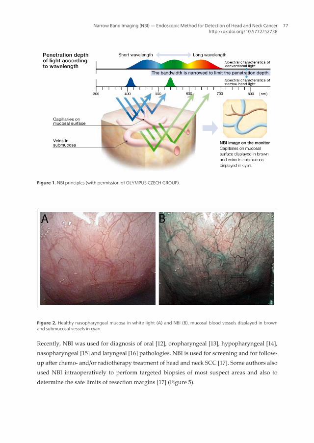
Figure 1. NBI principles (with permission of OLYMPUS CZECH GROUP).
Figure 2. Healthy nasopharyngeal mucosa in white light (A) and NBI (B), mucosal blood vessels displayed in brownand submucosal vessels in cyan.
Recently, NBI was used for diagnosis of oral [12], oropharyngeal [13], hypopharyngeal [14],nasopharyngeal [15] and laryngeal [16] pathologies. NBI is used for screening and for follow-up after chemo- and/or radiotherapy treatment of head and neck SCC [17]. Some authors alsoused NBI intraoperatively to perform targeted biopsies of most suspect areas and also todetermine the safe limits of resection margins [17] (Figure 5).
Narrow Band Imaging (NBI) — Endoscopic Method for Detection of Head and Neck Cancerhttp://dx.doi.org/10.5772/52738
77
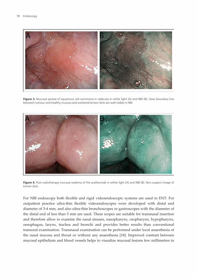
For NBI endoscopy both flexible and rigid videoendoscopic systems are used in ENT. Foroutpatient practice ultra-thin flexible videoendoscopes were developed with distal enddiameter of 3-4 mm, and also ultra-thin bronchoscopes or gastroscopes with the diameter ofthe distal end of less than 5 mm are used. These scopes are suitable for transnasal insertionand therefore allow to examine the nasal sinuses, nasopharynx, oropharynx, hypopharynx,oesophagus, larynx, trachea and bronchi and provides better results than conventionaltransoral examination. Transnasal examination can be performed under local anaesthesia ofthe nasal mucosa and throat or without any anaesthesia [18]. Improved contrast betweenmucosal epithelium and blood vessels helps to visualize mucosal lesions few millimetres in
Figure 3. Mucosal spread of squamous cell carcinoma in vallecula in white light (A) and NBI (B). Clear boundary linebetween tumour and healthy mucosa and scattered brown dots are well visible in NBI.
Figure 4. Post-radiotherapy mucosal oedema of the arythenoids in white light (A) and NBI (B). Non-suspect image ofbrown dots.
Endoscopy78
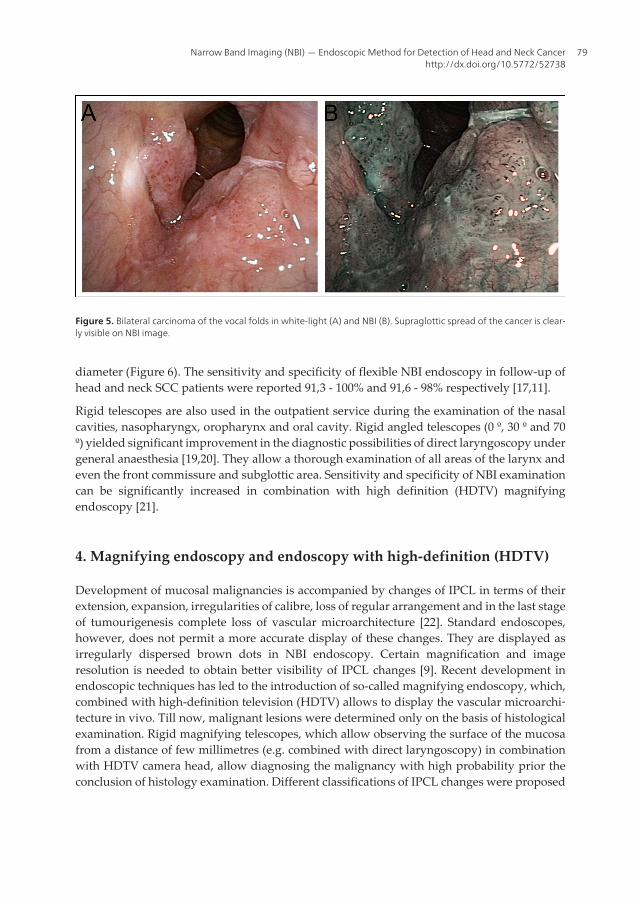
diameter (Figure 6). The sensitivity and specificity of flexible NBI endoscopy in follow-up ofhead and neck SCC patients were reported 91,3 - 100% and 91,6 - 98% respectively [17,11].
Rigid telescopes are also used in the outpatient service during the examination of the nasalcavities, nasopharyngx, oropharynx and oral cavity. Rigid angled telescopes (0 º, 30 º and 70º) yielded significant improvement in the diagnostic possibilities of direct laryngoscopy undergeneral anaesthesia [19,20]. They allow a thorough examination of all areas of the larynx andeven the front commissure and subglottic area. Sensitivity and specificity of NBI examinationcan be significantly increased in combination with high definition (HDTV) magnifyingendoscopy [21].
4. Magnifying endoscopy and endoscopy with high-definition (HDTV)
Development of mucosal malignancies is accompanied by changes of IPCL in terms of theirextension, expansion, irregularities of calibre, loss of regular arrangement and in the last stageof tumourigenesis complete loss of vascular microarchitecture [22]. Standard endoscopes,however, does not permit a more accurate display of these changes. They are displayed asirregularly dispersed brown dots in NBI endoscopy. Certain magnification and imageresolution is needed to obtain better visibility of IPCL changes [9]. Recent development inendoscopic techniques has led to the introduction of so-called magnifying endoscopy, which,combined with high-definition television (HDTV) allows to display the vascular microarchi‐tecture in vivo. Till now, malignant lesions were determined only on the basis of histologicalexamination. Rigid magnifying telescopes, which allow observing the surface of the mucosafrom a distance of few millimetres (e.g. combined with direct laryngoscopy) in combinationwith HDTV camera head, allow diagnosing the malignancy with high probability prior theconclusion of histology examination. Different classifications of IPCL changes were proposed
Figure 5. Bilateral carcinoma of the vocal folds in white-light (A) and NBI (B). Supraglottic spread of the cancer is clear‐ly visible on NBI image.
Narrow Band Imaging (NBI) — Endoscopic Method for Detection of Head and Neck Cancerhttp://dx.doi.org/10.5772/52738
79

for oesophagus and pharynx [23], oral cavity [12] and larynx [16]. In oesophagus and pharynxthe IPCL changes are graded as Type I (normal IPCL) to Type V (cancer IPCL). Type V is furthersubdivided into 4 sublevels. According to these levels, the depth of cancer invasion into themucosa and submucosal tissue can be evaluated [23]. For oral cavity just Type I (normal IPCL)to Type IV (cancer IPCL) grades are proposed [12] (Figure 7, 8).
5. Limits of NBI endoscopy
In some cases even NBI endoscopy examination does not bring the expected results. Since NBIis an optical method based on observation of the mucosal surface, conditions that prevent adirect view of the clear mucous membrane may limit or completely baffle the examination.Most often this is due to stagnant saliva or sticky mucus, especially in patients with a historyof oncology treatment. Also, lesions that are characterized by a high layer of hyperkeratosisprevent visualization of mucosal vascularization – eg. verrucous carcinomas [24].
Figure 6. Larynx - the left vocal cord dysplasia in white light (A) and NBI (B), arrows point demarcated area of alteredepithelium with the presence of brown dots. Another dysplastic lesion in the same patient in the posterior part of thevocal cord in white light (C) and NBI (D).
Endoscopy80
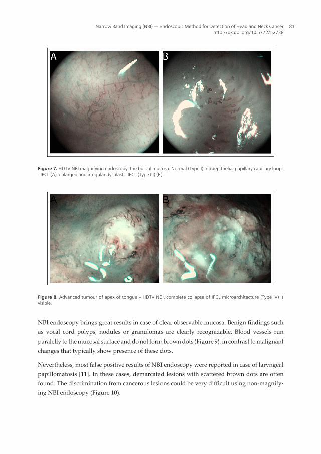
NBI endoscopy brings great results in case of clear observable mucosa. Benign findings suchas vocal cord polyps, nodules or granulomas are clearly recognizable. Blood vessels runparalelly to the mucosal surface and do not form brown dots (Figure 9), in contrast to malignantchanges that typically show presence of these dots.
Nevertheless, most false positive results of NBI endoscopy were reported in case of laryngealpapillomatosis [11]. In these cases, demarcated lesions with scattered brown dots are oftenfound. The discrimination from cancerous lesions could be very difficult using non-magnify‐ing NBI endoscopy (Figure 10).
Figure 7. HDTV NBI magnifying endoscopy, the buccal mucosa. Normal (Type I) intraepithelial papillary capillary loops- IPCL (A), enlarged and irregular dysplastic IPCL (Type III) (B).
Figure 8. Advanced tumour of apex of tongue – HDTV NBI, complete collapse of IPCL microarchitecture (Type IV) isvisible.
Narrow Band Imaging (NBI) — Endoscopic Method for Detection of Head and Neck Cancerhttp://dx.doi.org/10.5772/52738
81
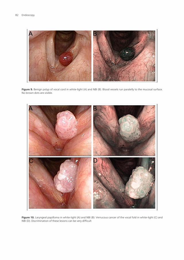
Figure 9. Benign polyp of vocal cord in white-light (A) and NBI (B). Blood vessels run paralelly to the mucosal surface.No brown dots are visible.
Figure 10. Laryngeal papilloma in white-light (A) and NBI (B). Verrucous cancer of the vocal fold in white-light (C) andNBI (D). Discrimination of these lesions can be very difficult
Endoscopy82
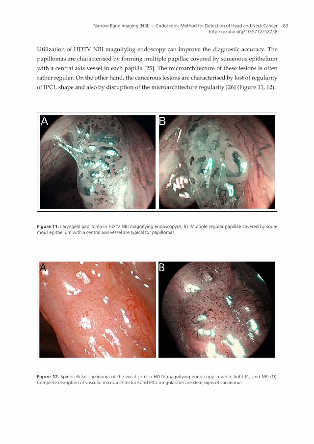
Utilization of HDTV NBI magnifying endoscopy can improve the diagnostic accuracy. Thepapillomas are characterised by forming multiple papillae covered by squamous epitheliumwith a central axis vessel in each papilla [25]. The microarchitecture of these lesions is oftenrather regular. On the other hand, the cancerous lesions are characterised by lost of regularityof IPCL shape and also by disruption of the microarchitecture regularity [26] (Figure 11, 12).
Figure 11. Laryngeal papilloma in HDTV NBI magnifying endoscopy(A, B). Multiple regular papillae covered by squa‐mous epithelium with a central axis vessel are typical for papillomas.
Figure 12. Spinocellular carcinoma of the vocal cord in HDTV magnifying endoscopy in white light (C) and NBI (D).Complete disruption of vascular microarchitecture and IPCL irregularities are clear signs of carcinoma.
Narrow Band Imaging (NBI) — Endoscopic Method for Detection of Head and Neck Cancerhttp://dx.doi.org/10.5772/52738
83
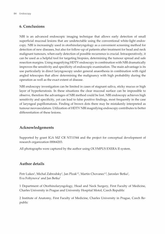
6. Conclusions
NBI is an advanced endoscopic imaging technique that allows early detection of smallsuperficial mucosal lesions that are undetectable using the conventional white-light endos‐copy. NBI is increasingly used in otorhinolaryngology as a convenient screening method fordetection of new diseases, but also for follow-up of patients after treatment for head and neckmalignant tumours, when early detection of possible recurrence is crucial. Intraoperatively, itcan be used as a helpful tool for targeting biopsies, determining the tumour spread and saferesection margins. Using magnifying HDTV endoscopy in combination with NBI dramaticallyimproves the sensitivity and specificity of endoscopic examination. The main advantage is itsuse particularly in direct laryngoscopy under general anaesthesia in combination with rigidangled telescopes that allow determining the malignancy with high probability during theoperation as well as the exact extent of disease.
NBI endoscopy investigation can be limited in cases of stagnant saliva, sticky mucus or highlayer of hyperkeratosis. In these situations the clear mucosal surface can be impossible toobserve, therefore the advantages of NBI method could be lost. NBI endoscopy achieves highsensitivity and specificity, yet can lead to false positive findings, most frequently in the caseof laryngeal papillomatosis. Finding of brown dots there may be mistakenly interpreted astumour neovasculature. Utilization of HDTV NBI magnifying endoscopy contributes to betterdifferentiation of these lesions.
Acknowledgements
Supported by grant IGA MZ CR NT11544 and the project for conceptual development ofresearch organization 00064203.
All photographs were captured by the author using OLYMPUS EXERA II system.
Author details
Petr Lukes1, Michal Zabrodsky1, Jan Plzak1,2, Martin Chovanec1,2, Jaroslav Betka1,Eva Foltynova1 and Jan Betka1
1 Department of Otorhinolaryngology, Head and Neck Surgery, First Faculty of Medicine,Charles University in Prague and University Hospital Motol, Czech Republic
2 Institute of Anatomy, First Faculty of Medicine, Charles University in Prague, Czech Re‐public
Endoscopy84
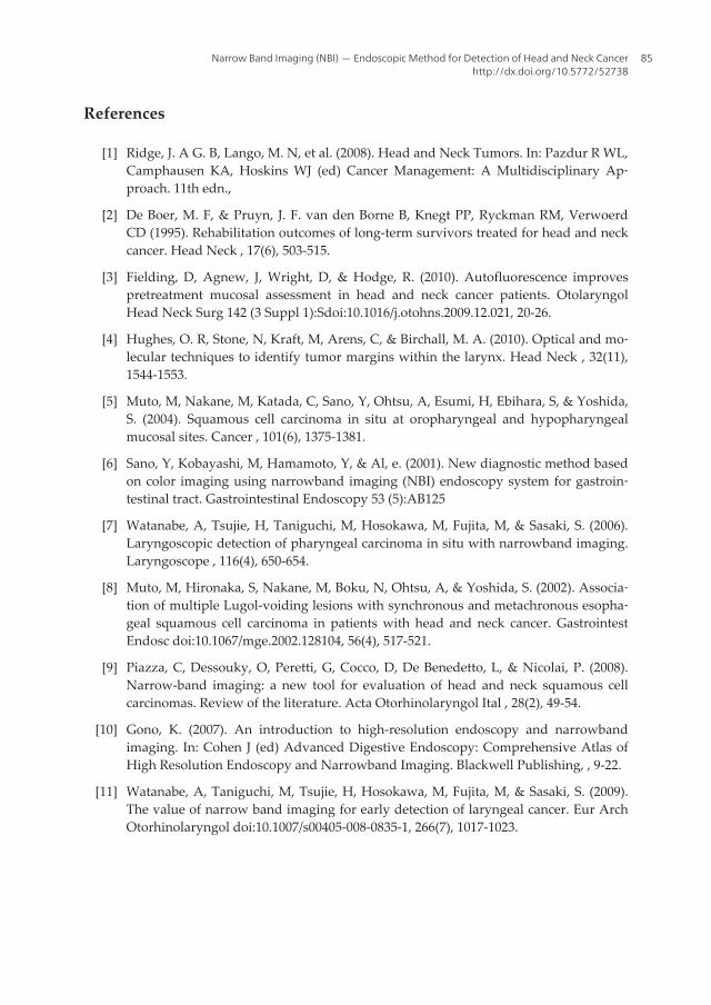
References
[1] Ridge, J. A G. B, Lango, M. N, et al. (2008). Head and Neck Tumors. In: Pazdur R WL,Camphausen KA, Hoskins WJ (ed) Cancer Management: A Multidisciplinary Ap‐proach. 11th edn.,
[2] De Boer, M. F, & Pruyn, J. F. van den Borne B, Knegt PP, Ryckman RM, VerwoerdCD (1995). Rehabilitation outcomes of long-term survivors treated for head and neckcancer. Head Neck , 17(6), 503-515.
[3] Fielding, D, Agnew, J, Wright, D, & Hodge, R. (2010). Autofluorescence improvespretreatment mucosal assessment in head and neck cancer patients. OtolaryngolHead Neck Surg 142 (3 Suppl 1):Sdoi:10.1016/j.otohns.2009.12.021, 20-26.
[4] Hughes, O. R, Stone, N, Kraft, M, Arens, C, & Birchall, M. A. (2010). Optical and mo‐lecular techniques to identify tumor margins within the larynx. Head Neck , 32(11),1544-1553.
[5] Muto, M, Nakane, M, Katada, C, Sano, Y, Ohtsu, A, Esumi, H, Ebihara, S, & Yoshida,S. (2004). Squamous cell carcinoma in situ at oropharyngeal and hypopharyngealmucosal sites. Cancer , 101(6), 1375-1381.
[6] Sano, Y, Kobayashi, M, Hamamoto, Y, & Al, e. (2001). New diagnostic method basedon color imaging using narrowband imaging (NBI) endoscopy system for gastroin‐testinal tract. Gastrointestinal Endoscopy 53 (5):AB125
[7] Watanabe, A, Tsujie, H, Taniguchi, M, Hosokawa, M, Fujita, M, & Sasaki, S. (2006).Laryngoscopic detection of pharyngeal carcinoma in situ with narrowband imaging.Laryngoscope , 116(4), 650-654.
[8] Muto, M, Hironaka, S, Nakane, M, Boku, N, Ohtsu, A, & Yoshida, S. (2002). Associa‐tion of multiple Lugol-voiding lesions with synchronous and metachronous esopha‐geal squamous cell carcinoma in patients with head and neck cancer. GastrointestEndosc doi:10.1067/mge.2002.128104, 56(4), 517-521.
[9] Piazza, C, Dessouky, O, Peretti, G, Cocco, D, De Benedetto, L, & Nicolai, P. (2008).Narrow-band imaging: a new tool for evaluation of head and neck squamous cellcarcinomas. Review of the literature. Acta Otorhinolaryngol Ital , 28(2), 49-54.
[10] Gono, K. (2007). An introduction to high-resolution endoscopy and narrowbandimaging. In: Cohen J (ed) Advanced Digestive Endoscopy: Comprehensive Atlas ofHigh Resolution Endoscopy and Narrowband Imaging. Blackwell Publishing, , 9-22.
[11] Watanabe, A, Taniguchi, M, Tsujie, H, Hosokawa, M, Fujita, M, & Sasaki, S. (2009).The value of narrow band imaging for early detection of laryngeal cancer. Eur ArchOtorhinolaryngol doi:10.1007/s00405-008-0835-1, 266(7), 1017-1023.
Narrow Band Imaging (NBI) — Endoscopic Method for Detection of Head and Neck Cancerhttp://dx.doi.org/10.5772/52738
85
[12] Takano, J. H, Yakushiji, T, Kamiyama, I, Nomura, T, Katakura, A, Takano, N, & Shi‐bahara, T. (2010). Detecting early oral cancer: narrowband imaging system observa‐tion of the oral mucosa microvasculature. Int J Oral Maxillofac Surg , 39(3), 208-213.
[13] Matsuba, H, Katada, C, Masaki, T, Nakayama, M, Okamoto, T, Hanaoka, N, Tanabe,S, Koizumi, W, Okamoto, M, & Muto, M. (2011). Diagnosis of the extent of advancedoropharyngeal and hypopharyngeal cancers by narrow band imaging with magnify‐ing endoscopy. Laryngoscope doi:10.1002/lary.21553, 121(4), 753-759.
[14] Watanabe, A, Taniguchi, M, Tsujie, H, Fujita, M, & Sasaki, S. (2007). Early detectionof recurrent hypopharyngeal cancer after radiotherapy by utilizing narrow-bandimaging--report of a case]. Nippon Jibiinkoka Gakkai Kaiho , 110(10), 680-682.
[15] Lin, Y. C, & Wang, W. H. (2011). Narrow-band imaging for detecting early recurrentnasopharyngeal carcinoma. Head Neck doi:10.1002/hed.21310, 33(4), 591-594.
[16] Ni, X. G, He, S, Xu, Z. G, Gao, L, Lu, N, Yuan, Z, Lai, S. Q, Zhang, Y. M, Yi, J. L,Wang, X. L, Zhang, L, Li, X. Y, & Wang, G. Q. (2010). Endoscopic diagnosis of laryng‐eal cancer and precancerous lesions by narrow band imaging. J Laryngol Otol ,125(3), 288-296.
[17] Piazza, C, Cocco, D, De Benedetto, L, Bon, F. D, Nicolai, P, & Peretti, G. (2010). Roleof narrow-band imaging and high-definition television in the surveillance of headand neck squamous cell cancer after chemo- and/or radiotherapy. Eur Arch Otorhi‐nolaryngol doi:10.1007/s00405-010-1236-9, 267(9), 1423-1428.
[18] Peery, A. F, Hoppo, T, Garman, K. S, Dellon, E. S, Daugherty, N, Bream, S, Sanz, A. F,Davison, J, Spacek, M, Connors, D, Faulx, A. L, Chak, A, Luketich, J. D, Shaheen, N. J,& Jobe, B. A. (2012). Feasibility, safety, acceptability, and yield of office-based,screening transnasal esophagoscopy (with video). Gastrointest Endosc e942. doi:10.1016/j.gie.2012.01.021, 75(5), 945-953.
[19] Sanli, A, Celebi, O, Eken, M, Oktay, A, Aydin, S, & Ayduran, E. (2008). Role of the 30degrees telescope in evaluation of laryngeal masses during direct laryngoscopy. Jour‐nal of voice : official journal of the Voice Foundation doi:10.1016/j.jvoice.2007.02.001,22(2), 238-244.
[20] Eryilmaz, A, Akmansu, H, Topcu, E, Acar, A, & Korkmaz, H. (2004). The role of 70-degree telescopic examination during direct laryngoscopic evaluation of laryngealcancers. Eur Arch Otorhinolaryngol doi:10.1007/s00405-003-0674-z, 261(5), 267-269.
[21] Piazza, C, Cocco, D, & De Benedetto, L. Del Bon F, Nicolai P, Peretti G (2009). Nar‐row band imaging and high definition television in the assessment of laryngeal can‐cer: a prospective study on 279 patients. Eur Arch Otorhinolaryngol , 267(3), 409-414.
[22] Fujii, S, Yamazaki, M, Muto, M, & Ochiai, A. (2010). Microvascular irregularities areassociated with composition of squamous epithelial lesions and correlate with sube‐
Endoscopy86
pithelial invasion of superficial-type pharyngeal squamous cell carcinoma. Histopa‐thology doi:10.1111/j.1365-2559.2010.03512.x, 56(4), 510-522.
[23] Inoue, H, Kaga, M, Yato, Y, Sugaya, S, & Kudo, S. (2007). Magnifying endoscopic di‐agnosis of tissue atypia and cancer invasion depth in the area of pharyngo-esopha‐geal squamous epithelium by NBI enhanced magnification image: IPCL patternclassification. In: Cohen J (ed) Advanced Digestive Endoscopy: Comprehensive Atlasof High Resolution Endoscopy and Narrowband Imaging. Blackwell Publishing,Malden, Massachusetts, , 49-66.
[24] Lukes, P. (2012). Discrimination of laryngeal papillomas and superficial laryngealcancer using NBI HDTV magnifying endoscopy. Lecture. Laryngology- Cutting EdgeLaryngology for the 21st Century, Kuala Lumpur, Malaysia
[25] Andrea, M, Dias, O, & Santos, A. (1995). Contact endoscopy during microlaryngealsurgery: a new technique for endoscopic examination of the larynx. Ann Otol RhinolLaryngol , 104(5), 333-339.
[26] Zabrodsky, M, Plzak, J, Betka, J, & Lukes, P. (2011). NBI HDTV Magnifying Endos‐copy in Laryngeal Papillomatosis and Laryngeal Spinocellular Cancer. OtolaryngolHead Neck Surg 145 (2 suppl):195
Narrow Band Imaging (NBI) — Endoscopic Method for Detection of Head and Neck Cancerhttp://dx.doi.org/10.5772/52738
87

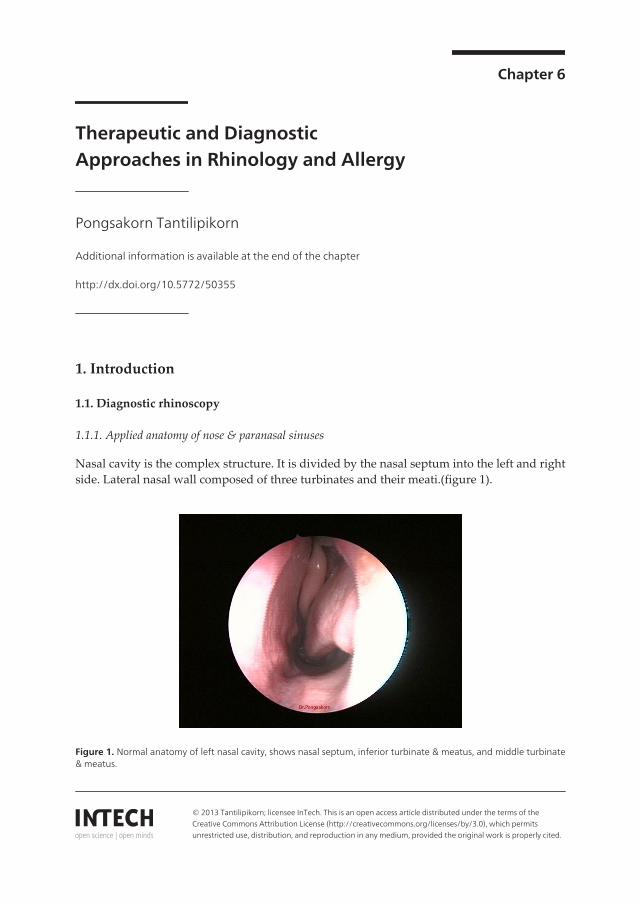
Chapter 6
Therapeutic and DiagnosticApproaches in Rhinology and Allergy
Pongsakorn Tantilipikorn
Additional information is available at the end of the chapter
http://dx.doi.org/10.5772/50355
1. Introduction
1.1. Diagnostic rhinoscopy
1.1.1. Applied anatomy of nose & paranasal sinuses
Nasal cavity is the complex structure. It is divided by the nasal septum into the left and rightside. Lateral nasal wall composed of three turbinates and their meati.(figure 1).
Figure 1. Normal anatomy of left nasal cavity, shows nasal septum, inferior turbinate & meatus, and middle turbinate& meatus.
© 2013 Tantilipikorn; licensee InTech. This is an open access article distributed under the terms of theCreative Commons Attribution License (http://creativecommons.org/licenses/by/3.0), which permitsunrestricted use, distribution, and reproduction in any medium, provided the original work is properly cited.
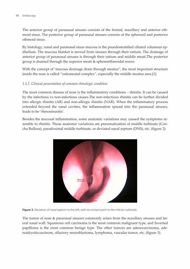
The anterior group of paranasal sinuses consists of the frontal, maxillary and anterior eth‐moid sinus. The posterior group of paranasal sinuses consists of the sphenoid and posteriorethmoid sinus.
By histology, nasal and paranasal sinus mucosa is the psuedostratified ciliated columnar ep‐ithelium. The mucous blanket is moved from sinuses through their ostium. The drainage ofanterior group of paranasal sinuses is through their ostium and middle meati.The posteriorgroup is drained through the superior meati & sphenoethmoidal recess.
With the concept of ‘mucous drainage drain through meatus”, the most important structureinside the nose is called “ostiomeatal complex”, especially the middle meatus area.[1]
1.1.2. Clinical presentation of common rhinologic condition
The most common disease of nose is the inflammatory conditions – rhinitis. It can be causedby the infectious vs non-infectious causes.The non-infectious rhinitis can be further dividedinto allergic rhinitis (AR) and non-allergic rhinitis (NAR). When the inflammatory processextended beyond the nasal cavities, the inflammation spread into the paranasal sinuses,leads to be ‘rhinosinusitis’.
Besides the mucosal inflammation, some anatomic variations may caused the symptoms re‐semble to rhinitis. Those anatomic variations are pneumatization of middle turbinate (Con‐cha Bullosa), paradoximal middle turbinate, or deviated nasal septum (DNS), etc. (figure 2).
Figure 2. Deviation of nasal septum to the left, with its contact point to the inferior turbinate.
The tumor of nose & paranasal sinuses commonly arises from the maxillary sinuses and lat‐eral nasal wall. Squamous cell carcinoma is the most common malignant type, and Invertedpapilloma is the most common benign type. The other tumors are adenocarcinoma, ade‐noidcysticcarcinom, olfactory neuroblastoma, lymphoma, vascular tumor, etc. (figure 3).
Endoscopy90
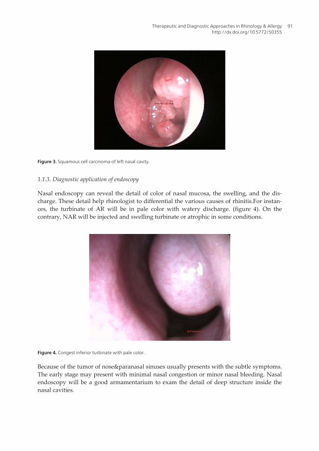
Figure 3. Squamous cell carcinoma of left nasal cavity.
1.1.3. Diagnostic application of endoscopy
Nasal endoscopy can reveal the detail of color of nasal mucosa, the swelling, and the dis‐charge. These detail help rhinologist to differential the various causes of rhinitis.For instan‐ces, the turbinate of AR will be in pale color with watery discharge. (figure 4). On thecontrary, NAR will be injected and swelling turbinate or atrophic in some conditions.
Figure 4. Congest inferior turbinate with pale color.
Because of the tumor of nose¶nasal sinuses usually presents with the subtle symptoms.The early stage may present with minimal nasal congestion or minor nasal bleeding. Nasalendoscopy will be a good armamentarium to exam the detail of deep structure inside thenasal cavities.
Therapeutic and Diagnostic Approaches in Rhinology & Allergyhttp://dx.doi.org/10.5772/50355
91
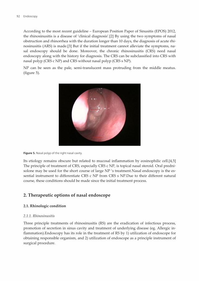
According to the most recent guideline – European Position Paper of Sinusitis (EPOS) 2012,the rhinosinusitis is a disease of ‘clinical diagnosis’.[2] By using the two symptoms of nasalobstruction and rhinorrhea with the duration longer than 10 days, the diagnosis of acute rhi‐nosinusitis (ARS) is made.[3] But if the initial treatment cannot alleviate the symptoms, na‐sal endoscopy should be done. Moreover, the chronic rhinosinusitis (CRS) need nasalendoscopy along with the history for diagnosis. The CRS can be subclassified into CRS withnasal polyp (CRS c NP) and CRS without nasal polyp (CRS s NP).
NP can be seen as the pale, semi-translucent mass protruding from the middle meatus.(figure 5).
Figure 5. Nasal polyp of the right nasal cavity.
Its etiology remains obscure but related to mucosal inflammation by eosinophilic cell.[4,5]The principle of treatment of CRS, especially CRS c NP, is topical nasal steroid. Oral predni‐solone may be used for the short course of large NP ‘s treatment.Nasal endoscopy is the es‐sential instrument to differentiate CRS c NP from CRS s NP.Due to their different naturalcourse, these conditions should be made since the initial treatment process.
2. Therapeutic options of nasal endoscope
2.1. Rhinologic condition
2.1.1. Rhinosinusitis
Three principle treatments of rhinosinusitis (RS) are the eradication of infectious process,promotion of secretion in sinus cavity and treatment of underlying disease (eg. Allergic in‐flammation).Endoscopy has its role in the treatment of RS by 1) utilization of endoscope forobtaining responsible organism, and 2) utilization of endoscope as a principle instrument ofsurgical procedure.
Endoscopy92

The selection of antibiotic for treatment of infectious RS usually follows the guideline thatdepends on the prevalence of responsible organism in each community. Three most com‐mon organisms of RS are Streptococcus pneumoniae, Haemophilusinfluenzae and Moraxella catar‐rhalis.[6,7]Most of the guidelines suggest the high dose-amoxycillin with/without clavulanicacid as the first –line drug.When there is little (minimal) response of RS patient, the physi‐cian may switch to the second-line drug or consider taking the microbiologic culture/sensi‐tivity test.
The gold standard of obtaining RS specimen for culture is the sinus puncture, usually fromthe maxillary sinus through the inferior meatus. But the so-called maxillary Antral punc‐ture&Irrigation (AI) is considered as an invasive procedure, which can cause the significantpain for RS patient. To avoid this limitation and minimal contamination of non-pathogenicorganism in the nasal cavity, endoscopic-guided culture at the ostiomeatal complex (OMC)is the alternation procedure with comparable accuracy to AI.[8] (figure 6)
Figure 6. Endoscopic-guide culture from the left middle meatus.
If the RS patient fails to the medical treatment, the surgical procedure is needed. Conven‐tionally, the open-approach sinus surgeries are the Caldwell-Luc (CWL) and the externalfrontoethmoidectomy (eg. Lynch). Prof Messerklinger proposed the breakthrough principleof mucociliary drainage through OMC in 1975.With this principle and the advancement ofsurgical instrument (especially the rigid endoscope), Prof Kennedy and Prof Stammbergerlead the concept of “Functional Endoscopic Sinus Surgery-FESS” into the acceptable sinusprocedure as a standard treatment.[9]
FESS consists of the utilization of rigid nasal endoscope and the cutting forceps to clear thepathology at OMC area. Then, obstructed secretion inside the sinus cavities can be drainedout through OMC by the function of respiratory cilia.
Over the 30 years, there are tremendous improvement of surgical instrument and the ad‐junctive procedure.For instance, the cutting&suction instrument such as the microdebrider
Therapeutic and Diagnostic Approaches in Rhinology & Allergyhttp://dx.doi.org/10.5772/50355
93
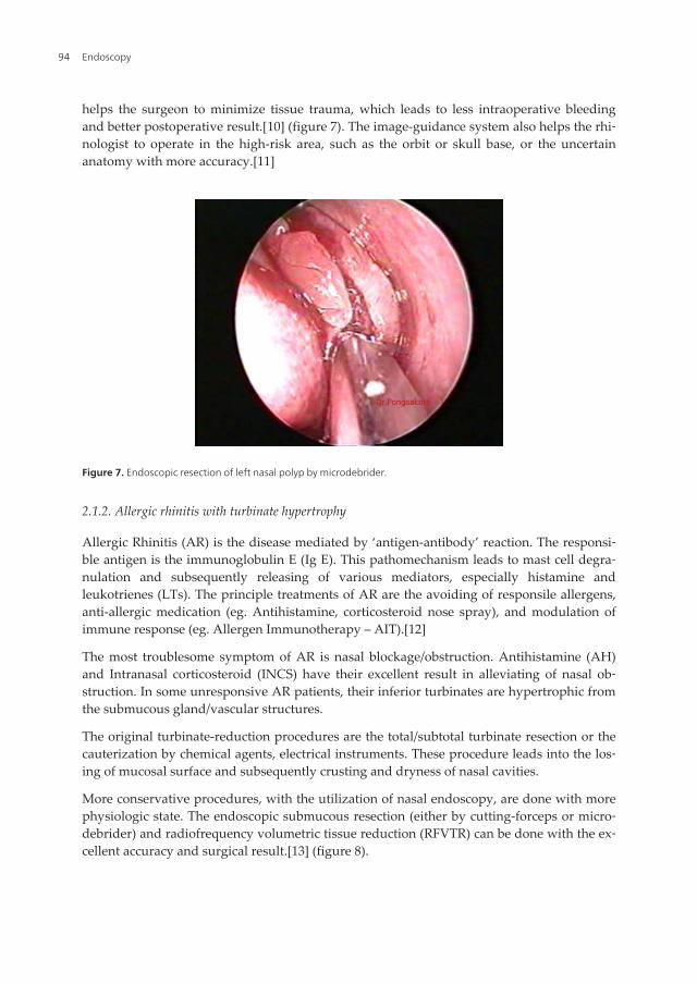
helps the surgeon to minimize tissue trauma, which leads to less intraoperative bleedingand better postoperative result.[10] (figure 7). The image-guidance system also helps the rhi‐nologist to operate in the high-risk area, such as the orbit or skull base, or the uncertainanatomy with more accuracy.[11]
Figure 7. Endoscopic resection of left nasal polyp by microdebrider.
2.1.2. Allergic rhinitis with turbinate hypertrophy
Allergic Rhinitis (AR) is the disease mediated by ‘antigen-antibody’ reaction. The responsi‐ble antigen is the immunoglobulin E (Ig E). This pathomechanism leads to mast cell degra‐nulation and subsequently releasing of various mediators, especially histamine andleukotrienes (LTs). The principle treatments of AR are the avoiding of responsile allergens,anti-allergic medication (eg. Antihistamine, corticosteroid nose spray), and modulation ofimmune response (eg. Allergen Immunotherapy – AIT).[12]
The most troublesome symptom of AR is nasal blockage/obstruction. Antihistamine (AH)and Intranasal corticosteroid (INCS) have their excellent result in alleviating of nasal ob‐struction. In some unresponsive AR patients, their inferior turbinates are hypertrophic fromthe submucous gland/vascular structures.
The original turbinate-reduction procedures are the total/subtotal turbinate resection or thecauterization by chemical agents, electrical instruments. These procedure leads into the los‐ing of mucosal surface and subsequently crusting and dryness of nasal cavities.
More conservative procedures, with the utilization of nasal endoscopy, are done with morephysiologic state. The endoscopic submucous resection (either by cutting-forceps or micro‐debrider) and radiofrequency volumetric tissue reduction (RFVTR) can be done with the ex‐cellent accuracy and surgical result.[13] (figure 8).
Endoscopy94
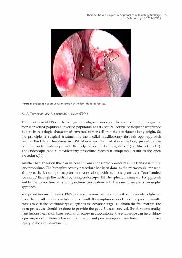
Figure 8. Endoscopic submucous resection of the left inferior turbinate.
2.1.3. Tumor of nose & paranasal sinuses (PNS)
Tumor of nose&PNS can be benign or malignant in-origin.The most common benign tu‐mor is inverted papilloma.Inverted papilloma has its natural course of frequent recurrencedue to its histologic character of ‘inverted tumor cell into the attachment bony origin. Sothe principle of surgical treatment is the medial maxillectomy through open-approachsuch as the lateral rhinotomy or CWL.Nowadays, the medial maxillectomy procedure canbe done under endoscope with the help of suction&cutting device (eg. Microdebrider).The endoscopic medial maxillectomy procedure reaches it comparable result as the openprocedure.[14]
Another benign lesion that can be benefit from endoscopic procedure is the transnasal pitui‐tary procedure. The hypophysectomy procedure has been done as the microscopic transept‐al approach. Rhinologic surgeon can work along with neurosurgeon as a ‘four-handedtechnique’ through the nostrils by using endoscope.[15] The sphenoid sinus can be approachand further procedure of hypophysectomy can be done with the same principle of transeptalapproach.
Malignant tumors of nose & PNS can be squamous cell carcinoma that commonly originatesfrom the maxillary sinus or lateral nasal wall. Its symptom is subtle and the patient usuallycomes to visit the otorhinolaryngologist as the advance stage. To obtain the free-margin, theopen procedure should be done to provide the good 5-years survival. But for some malig‐nant lesions near skull base, such as olfactory neuroblastoma, the endoscope can help rhino‐logic surgeon to delineate the surgical margin and precise surgical resection with minimizedinjury to the vital structure.[16]
Therapeutic and Diagnostic Approaches in Rhinology & Allergyhttp://dx.doi.org/10.5772/50355
95
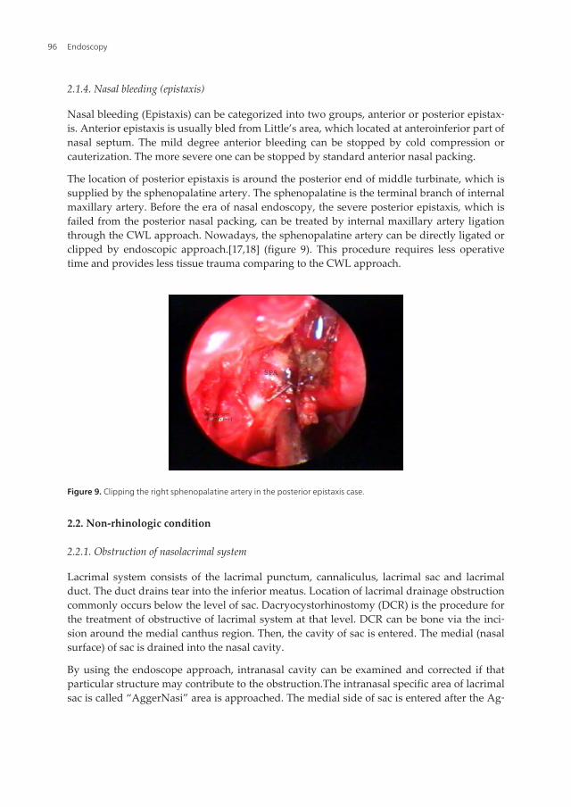
2.1.4. Nasal bleeding (epistaxis)
Nasal bleeding (Epistaxis) can be categorized into two groups, anterior or posterior epistax‐is. Anterior epistaxis is usually bled from Little’s area, which located at anteroinferior part ofnasal septum. The mild degree anterior bleeding can be stopped by cold compression orcauterization. The more severe one can be stopped by standard anterior nasal packing.
The location of posterior epistaxis is around the posterior end of middle turbinate, which issupplied by the sphenopalatine artery. The sphenopalatine is the terminal branch of internalmaxillary artery. Before the era of nasal endoscopy, the severe posterior epistaxis, which isfailed from the posterior nasal packing, can be treated by internal maxillary artery ligationthrough the CWL approach. Nowadays, the sphenopalatine artery can be directly ligated orclipped by endoscopic approach.[17,18] (figure 9). This procedure requires less operativetime and provides less tissue trauma comparing to the CWL approach.
Figure 9. Clipping the right sphenopalatine artery in the posterior epistaxis case.
2.2. Non-rhinologic condition
2.2.1. Obstruction of nasolacrimal system
Lacrimal system consists of the lacrimal punctum, cannaliculus, lacrimal sac and lacrimalduct. The duct drains tear into the inferior meatus. Location of lacrimal drainage obstructioncommonly occurs below the level of sac. Dacryocystorhinostomy (DCR) is the procedure forthe treatment of obstructive of lacrimal system at that level. DCR can be bone via the inci‐sion around the medial canthus region. Then, the cavity of sac is entered. The medial (nasalsurface) of sac is drained into the nasal cavity.
By using the endoscope approach, intranasal cavity can be examined and corrected if thatparticular structure may contribute to the obstruction.The intranasal specific area of lacrimalsac is called “AggerNasi” area is approached. The medial side of sac is entered after the Ag‐
Endoscopy96
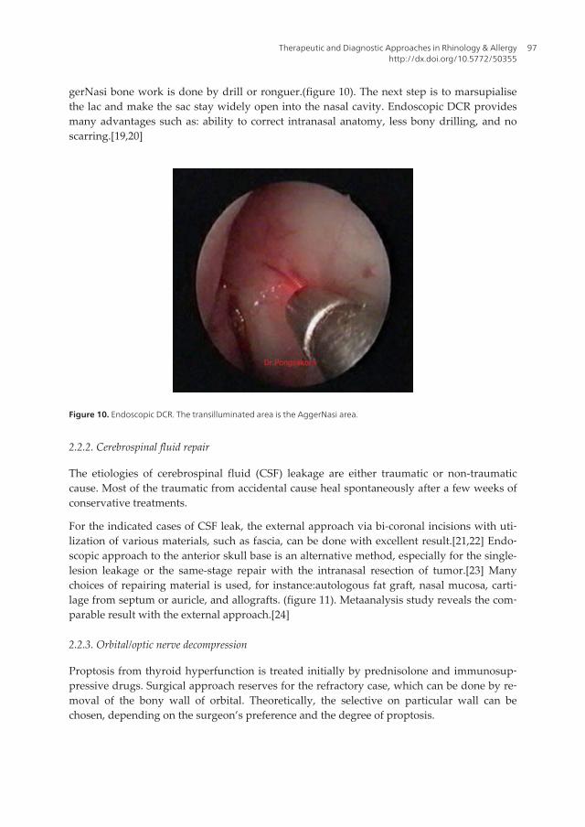
gerNasi bone work is done by drill or ronguer.(figure 10). The next step is to marsupialisethe lac and make the sac stay widely open into the nasal cavity. Endoscopic DCR providesmany advantages such as: ability to correct intranasal anatomy, less bony drilling, and noscarring.[19,20]
Figure 10. Endoscopic DCR. The transilluminated area is the AggerNasi area.
2.2.2. Cerebrospinal fluid repair
The etiologies of cerebrospinal fluid (CSF) leakage are either traumatic or non-traumaticcause. Most of the traumatic from accidental cause heal spontaneously after a few weeks ofconservative treatments.
For the indicated cases of CSF leak, the external approach via bi-coronal incisions with uti‐lization of various materials, such as fascia, can be done with excellent result.[21,22] Endo‐scopic approach to the anterior skull base is an alternative method, especially for the single-lesion leakage or the same-stage repair with the intranasal resection of tumor.[23] Manychoices of repairing material is used, for instance:autologous fat graft, nasal mucosa, carti‐lage from septum or auricle, and allografts. (figure 11). Metaanalysis study reveals the com‐parable result with the external approach.[24]
2.2.3. Orbital/optic nerve decompression
Proptosis from thyroid hyperfunction is treated initially by prednisolone and immunosup‐pressive drugs. Surgical approach reserves for the refractory case, which can be done by re‐moval of the bony wall of orbital. Theoretically, the selective on particular wall can bechosen, depending on the surgeon’s preference and the degree of proptosis.
Therapeutic and Diagnostic Approaches in Rhinology & Allergyhttp://dx.doi.org/10.5772/50355
97
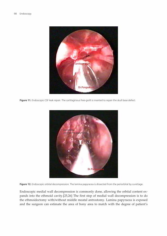
Figure 11. Endoscopic CSF leak repair. The cartilaginous free-graft is inserted to repair the skull base defect.
Figure 12. Endoscopic orbital decompression. The lamina papyracea is dissected from the periorbital by curettage.
Endoscopic medial wall decompression is commonly done, allowing the orbital content ex‐pands into the ethmoid cavity.[25,26] The first step of medial wall decompression is to dothe ethmoidectomy with/without middle meatal antrostomy. Lamina papyracea is exposedand the surgeon can estimate the area of bony area to match with the degree of patient’s
Endoscopy98
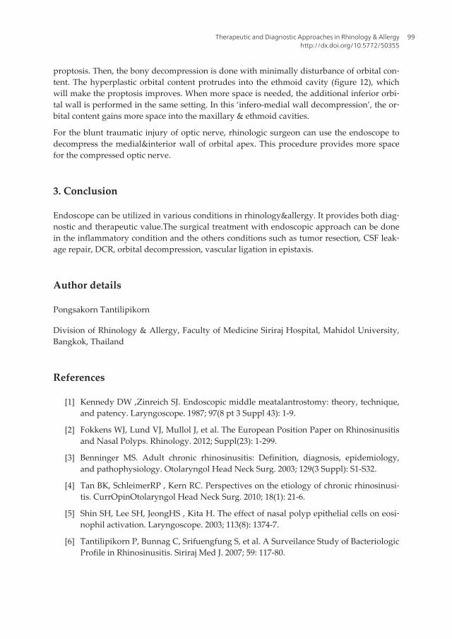
proptosis. Then, the bony decompression is done with minimally disturbance of orbital con‐tent. The hyperplastic orbital content protrudes into the ethmoid cavity (figure 12), whichwill make the proptosis improves. When more space is needed, the additional inferior orbi‐tal wall is performed in the same setting. In this ‘infero-medial wall decompression’, the or‐bital content gains more space into the maxillary & ethmoid cavities.
For the blunt traumatic injury of optic nerve, rhinologic surgeon can use the endoscope todecompress the medial&interior wall of orbital apex. This procedure provides more spacefor the compressed optic nerve.
3. Conclusion
Endoscope can be utilized in various conditions in rhinology&allergy. It provides both diag‐nostic and therapeutic value.The surgical treatment with endoscopic approach can be donein the inflammatory condition and the others conditions such as tumor resection, CSF leak‐age repair, DCR, orbital decompression, vascular ligation in epistaxis.
Author details
Pongsakorn Tantilipikorn
Division of Rhinology & Allergy, Faculty of Medicine Siriraj Hospital, Mahidol University,Bangkok, Thailand
References
[1] Kennedy DW ,Zinreich SJ. Endoscopic middle meatalantrostomy: theory, technique,and patency. Laryngoscope. 1987; 97(8 pt 3 Suppl 43): 1-9.
[2] Fokkens WJ, Lund VJ, Mullol J, et al. The European Position Paper on Rhinosinusitisand Nasal Polyps. Rhinology. 2012; Suppl(23): 1-299.
[3] Benninger MS. Adult chronic rhinosinusitis: Definition, diagnosis, epidemiology,and pathophysiology. Otolaryngol Head Neck Surg. 2003; 129(3 Suppl): S1-S32.
[4] Tan BK, SchleimerRP , Kern RC. Perspectives on the etiology of chronic rhinosinusi‐tis. CurrOpinOtolaryngol Head Neck Surg. 2010; 18(1): 21-6.
[5] Shin SH, Lee SH, JeongHS , Kita H. The effect of nasal polyp epithelial cells on eosi‐nophil activation. Laryngoscope. 2003; 113(8): 1374-7.
[6] Tantilipikorn P, Bunnag C, Srifuengfung S, et al. A Surveilance Study of BacteriologicProfile in Rhinosinusitis. Siriraj Med J. 2007; 59: 117-80.
Therapeutic and Diagnostic Approaches in Rhinology & Allergyhttp://dx.doi.org/10.5772/50355
99
[7] Brook I. Microbiology and management of sinusitis. J Otolaryngol. 1996; 25(4):249-56.
[8] Ozcan M, Unal A, Aksaray S, Yalcin F, Akdeniz T. Correlation of middle meatus andethmoid sinus microbiology in patients with chronic sinusitis. Rhinology. 2002; 40(1):24-7.
[9] Stammberger H. Functional endoscopic sinus surgery: concept, indications and re‐sults of the Messerklinger technique. Eur Arch Otorhinolaryngol. 1990; 247: 63-76.
[10] SetliffRC , Parsons DS. The "hummer": new instrumentation for functional endoscop‐ic sinus surgery. Am J Rhinol. 1994; 8: 275-8.
[11] Tantilipikorn P, Metheetrairut C, Lumyongsatien J, Bedavanija A, Assanasen P. Im‐age-guided Surgery in Rhinology. Siriraj Med J. 2010; 62(203-6).
[12] Bousquet J, Khaltaev N, Cruz AA, et al. Allergic Rhinitis and its Impact on Asthma(ARIA) 2008. Allergy. 2008; 63(Suppl 86): 8-160.
[13] Banhiran W, Tantilipikorn P, Metheetrairut C, Assanasen P, Bunnag C. Quality ofLife in Patients with Chronic Rhinitis after Radiofrequency Inferior Turbinate Reduc‐tion. J Med Assoc Thai. 2010; 93(8): 950-60.
[14] Sham CL, Woo JK , van Hasselt CA. Endoscopic resection of inverted papilloma ofthe nose and paranasal sinuses. J Laryngol Otol. 1998; 112(8): 758-64.
[15] Briner HR, Simmen D, Jones N. Endoscopic sinus surgery: advantages of the bimanu‐al technique. Am J Rhinol. 2005; 19: 269-73.
[16] Harvey RJ, Winder M, Parmar P, Lund V. Endoscopic skull base surgery for sinonas‐al malignancy. OtolaryngolClin North Am. 44(5): 1081-140.
[17] Agreda B, Urpegui A, Ignacio Alfonso J, Valles H. Ligation of the sphenopalatine ar‐tery in posterior epistaxis. Retrospective study of 50 patients]. ActaOtorrinolaringolEsp. 62(3): 194-8.
[18] Howe DJ , Skinner DW. Outcomes of endoscopic sphenopalatine artery ligation forepistaxis: a five-year series from a single institution. Ear Nose Throat J. 91(2): 70-2.
[19] Al-Qahtani AS. Primary endoscopic dacryocystorhinostomy with or without siliconetubing: A prospective randomized study. Am J Rhinol Allergy. 26(4): 332-4.
[20] Feng YF, Cai JQ, Zhang JY , Han XH. A meta-analysis of primary dacryocystorhinos‐tomy with and without silicone intubation. Can J Ophthalmol. 46(6): 521-7.
[21] McCormack B, Cooper PR ,Persky M. Extrncranial repair of cerebrospinal fluid fistu‐las: technique and results in 37 patients. Neurosurgery. 1990; 27: 412-7.
[22] Persky MS, Rothstein SG , Breda SD. Extracranial repair of cerebrospinal fluid otorhi‐norrhea. Laryngoscope. 1991; 101: 7-15.
Endoscopy100

[23] Lanza D, O'Brien D , Kennedy D. Endoscopic repair of cerebrospinal fluid fistulaeand encephaloceles. Laryngoscope. 1996; 106: 1119-25.
[24] Hegazy H, Carrau R ,Snyderman C. Transnasal endoscopic repair of cerebrospinalfluid rhinorrhea: a meta-analysis. Laryngoscope. 2000; 110: 1166-72.
[25] Sheng H, Cai C, Cheng Y, et al. Endoscopic orbital decompression for thyroid-associ‐ated ophthalmopathy. Lin Chung Er Bi Yan HouTou Jing WaiKeZaZhi. 26(1): 27-9.
[26] Boboridis KG, Bunce C. Surgical orbital decompression for thyroid eye disease. Co‐chrane Database Syst Rev. 2011; (12): CD007630.
Therapeutic and Diagnostic Approaches in Rhinology & Allergyhttp://dx.doi.org/10.5772/50355
101

Chapter 7
Role of Endoscopic Sinus Surgeryin Pediatric Acute Complicated Sinusitis
Timothy P. McEvoy, Charles A. Elmaraghy andKris R. Jatana
Additional information is available at the end of the chapter
http://dx.doi.org/10.5772/52551
1. Introduction
Sinus surgery was initially done transnasally or by a transmaxillary approach with the useof external lighting (often a headlight) and the surgeon’s direct vision. Endoscopic techni‐ques for intranasal surgery were first pioneered for adult applications and were graduallyapplied to pediatric sinus surgery as endoscope technology improved and smaller diameterendoscopes became available. Techniques that were developed in Europe were championedby Kennedy in the United States, with an emphasis on targeted removal of diseased tissueand preservation of normal mucosa [1, 2].
In 1989, Gross et al. published one of the first case series documenting functional endoscopicsinus surgery in the pediatric population. They noted that all 57 children in the series toler‐ated the procedure well and that there were no major complications [3]. Since that time, en‐doscopic intranasal surgical techniques in children have become commonplace forapplications ranging from chronic sinusitis and nasal polyposis to the treatment of compli‐cations of acute sinusitis as well as approaches to the skull base.
2. Equipment
2.1. Camera and monitors
Initially, endoscopic sinus surgery was performed with the surgeon looking directlythrough the optical telescope. As digital cameras improved, the images could be magnifiedand viewed on a monitor allowing the surgeon better visualization as well as improving
© 2013 McEvoy et al.; licensee InTech. This is an open access article distributed under the terms of theCreative Commons Attribution License (http://creativecommons.org/licenses/by/3.0), which permitsunrestricted use, distribution, and reproduction in any medium, provided the original work is properly cited.
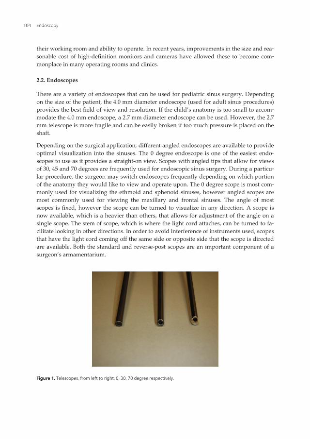
their working room and ability to operate. In recent years, improvements in the size and rea‐sonable cost of high-definition monitors and cameras have allowed these to become com‐monplace in many operating rooms and clinics.
2.2. Endoscopes
There are a variety of endoscopes that can be used for pediatric sinus surgery. Dependingon the size of the patient, the 4.0 mm diameter endoscope (used for adult sinus procedures)provides the best field of view and resolution. If the child’s anatomy is too small to accom‐modate the 4.0 mm endoscope, a 2.7 mm diameter endoscope can be used. However, the 2.7mm telescope is more fragile and can be easily broken if too much pressure is placed on theshaft.
Depending on the surgical application, different angled endoscopes are available to provideoptimal visualization into the sinuses. The 0 degree endoscope is one of the easiest endo‐scopes to use as it provides a straight-on view. Scopes with angled tips that allow for viewsof 30, 45 and 70 degrees are frequently used for endoscopic sinus surgery. During a particu‐lar procedure, the surgeon may switch endoscopes frequently depending on which portionof the anatomy they would like to view and operate upon. The 0 degree scope is most com‐monly used for visualizing the ethmoid and sphenoid sinuses, however angled scopes aremost commonly used for viewing the maxillary and frontal sinuses. The angle of mostscopes is fixed, however the scope can be turned to visualize in any direction. A scope isnow available, which is a heavier than others, that allows for adjustment of the angle on asingle scope. The stem of scope, which is where the light cord attaches, can be turned to fa‐cilitate looking in other directions. In order to avoid interference of instruments used, scopesthat have the light cord coming off the same side or opposite side that the scope is directedare available. Both the standard and reverse-post scopes are an important component of asurgeon’s armamentarium.
Figure 1. Telescopes, from left to right, 0, 30, 70 degree respectively.
Endoscopy104
2.3. Endoscopic surgical instruments
Miniaturized versions of adult sinus surgery instruments are available from several instru‐ment manufacturers. These instruments allow surgeons to open particular sinuses whileavoiding disturbing normal mucosa.
Another important instrument for endoscopic sinus surgery is the microdebrider. Severalmanufacturers have produced powered devices that can remove tissue during endoscopicsinus surgery. For example, a 4.0 and a 2.9 mm microdebrider blade can be used depend‐ing on the size of the patient. The smaller powered instruments do tend to get cloggedwith bone chips more frequently. In addition, endoscopic drills exist which can preciselyremove bone.
3. Pertinent sinonasal embryology
During fetal development the nasal placode is noted at around four and a half weeks.The nasal cavity is then formed due to the fusion of the medial and lateral nasal proc‐esses. The turbinates develop shortly thereafter at around 40 days [4]. Ethmoid buddingis noted around 11 to 12 weeks and maxillary budding is noted around 14 to 15 weeksof development [5].
The progression of sinus development was initially documented via cadaver studies, and arecent large-scale imaging study helped re-confirm these findings through non-invasiveMRI scanning to evaluate the volume of the paranasal sinuses of patients with healthy si‐nuses undergoing scans for other reasons. It was found that initial pneumatization was not‐ed for the maxillary and ethomid sinuses at birth, for the sphenoid sinus at 9 months, andfor the frontal sinus after 5 years of age. Additionally the development of the paranasal si‐nuses was noted to be ongoing until at least the late teen years [5].
The fact the sinuses are continuing to develop as children grow becomes important in con‐sidering the possible acute complications of sinusitis that will be discussed later in this chap‐ter. The peak time for development of the frontal sinus and the diploic venous system is inadolescence [6], which partially explains why adolescents are the most common age groupafflicted by intracranial complications of sinusitis.
4. Clinical presentation of pediatric acute complicated sinusitis
Due the relative rarity of cases of acute complicated sinusitis in children, the exact incidenceis unclear. The most frequent complications of sinusitis in children are extracranial and in‐clude periorbital/orbital cellulitis and subperiosteal/orbital abscess. These two complicationscomprise up to 90 percent of complications of sinusitis in children [7]. Opthalmological eval‐uation is critical in these cases to thoroughly assess the status of the eye. However, in thesubset of patients who require inpatient admission for treatment, the incidence of intracrani‐
Role of Endoscopic Sinus Surgery in Pediatric Acute Complicated Sinusitishttp://dx.doi.org/10.5772/52551
105
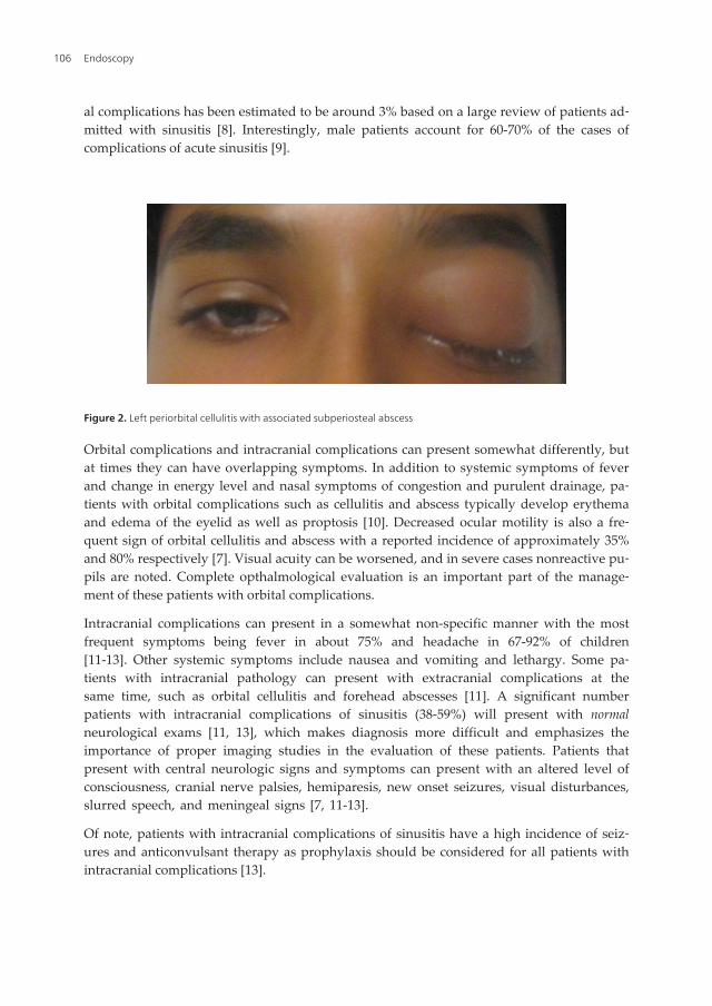
al complications has been estimated to be around 3% based on a large review of patients ad‐mitted with sinusitis [8]. Interestingly, male patients account for 60-70% of the cases ofcomplications of acute sinusitis [9].
Figure 2. Left periorbital cellulitis with associated subperiosteal abscess
Orbital complications and intracranial complications can present somewhat differently, butat times they can have overlapping symptoms. In addition to systemic symptoms of feverand change in energy level and nasal symptoms of congestion and purulent drainage, pa‐tients with orbital complications such as cellulitis and abscess typically develop erythemaand edema of the eyelid as well as proptosis [10]. Decreased ocular motility is also a fre‐quent sign of orbital cellulitis and abscess with a reported incidence of approximately 35%and 80% respectively [7]. Visual acuity can be worsened, and in severe cases nonreactive pu‐pils are noted. Complete opthalmological evaluation is an important part of the manage‐ment of these patients with orbital complications.
Intracranial complications can present in a somewhat non-specific manner with the mostfrequent symptoms being fever in about 75% and headache in 67-92% of children[11-13]. Other systemic symptoms include nausea and vomiting and lethargy. Some pa‐tients with intracranial pathology can present with extracranial complications at thesame time, such as orbital cellulitis and forehead abscesses [11]. A significant numberpatients with intracranial complications of sinusitis (38-59%) will present with normalneurological exams [11, 13], which makes diagnosis more difficult and emphasizes theimportance of proper imaging studies in the evaluation of these patients. Patients thatpresent with central neurologic signs and symptoms can present with an altered level ofconsciousness, cranial nerve palsies, hemiparesis, new onset seizures, visual disturbances,slurred speech, and meningeal signs [7, 11-13].
Of note, patients with intracranial complications of sinusitis have a high incidence of seiz‐ures and anticonvulsant therapy as prophylaxis should be considered for all patients withintracranial complications [13].
Endoscopy106
5. Role of imaging in pediatric acute complicated sinusitis
5.1. Computed Tomography (CT)
CT scanning is the gold standard for imaging of the paranasal sinuses. Unlike for patientswith chronic sinusitis for whom non-contast studies may be sufficient, children with possi‐ble complications of acute sinusitis require contrast-enhanced studies. One drawback of CTscanning is that it does lack specificity due to mucosal changes that may be considered inci‐dental and due to the slow resolution of edema after infections [10, 14]. Although there is apriority to avoid the radiation of a CT scan in children, it remains a crucial imaging modali‐ty for diagnosis and management of acute complicated sinusitis. CT can often miss intracra‐nial involvement in cases of acute complicated sinusitis up to 50% of the time. Patients 7years and older with orbital infections have been shown to have about a 10% incidence ofconcomitant intracranial involvement [15].
5.2. Magnetic Resonance Imaging (MRI)
Contrast-enhanced MRI scans are necessary for patients who are suspected of having intra‐cranial complications of sinusitis. MRI is superior to CT in that it is able to delineate earlycerebritis changes as well as provide further detail regarding the meninges, marrow spacesas well as the orbital apex and cavernous sinus [10, 14].
5.3. Intraoperative surgical guidance
Image guidance systems are used widely by both otolaryngologists and neurosurgeons foradult applications. Data regarding the utility of image guidance in children is not as abun‐dant. However, literature reviews of the indications and safety of image guidance in pedia‐tric sinus and skull base surgery supports its use for complex cases and cases with distortedanatomy which includes cases of acute complicated sinusitis. Additionally, there were nocomplications that were reported in either retrospective studies[16, 17]. Image guidance caninclude CT and/or MRI, however, for cases of acute complicated sinusitis, CT is the mostuseful for defining bony landmarks within the sinonasal cavity. MRI can certainly assistwith soft tissue landmarks when needed in complex cases.
6. Role of medical management
Medical management of children with acute complicated sinusitis is mainly centered aroundthe administration of IV antibiotics. Broad-spectrum antibiotics are chosen until culture-di‐rected therapy can be provided. Adjunctive therapy to help reduce mucosal edema includessystemic steroids and decongestants, and topical decongestants (oxymetazaline) and topicalsteroids. However, there is not much literature to support the use of these agents in the set‐ting of acute complicated sinusitis, but there is no definitive evidence that these would beharmful. Additionally, nasal saline irrigation or nasal saline spray can be helpful to clear se‐
Role of Endoscopic Sinus Surgery in Pediatric Acute Complicated Sinusitishttp://dx.doi.org/10.5772/52551
107
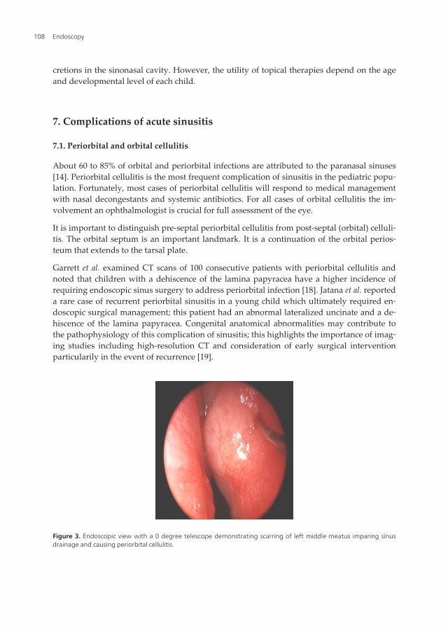
cretions in the sinonasal cavity. However, the utility of topical therapies depend on the ageand developmental level of each child.
7. Complications of acute sinusitis
7.1. Periorbital and orbital cellulitis
About 60 to 85% of orbital and periorbital infections are attributed to the paranasal sinuses[14]. Periorbital cellulitis is the most frequent complication of sinusitis in the pediatric popu‐lation. Fortunately, most cases of periorbital cellulitis will respond to medical managementwith nasal decongestants and systemic antibiotics. For all cases of orbital cellulitis the im‐volvement an ophthalmologist is crucial for full assessment of the eye.
It is important to distinguish pre-septal periorbital cellulitis from post-septal (orbital) celluli‐tis. The orbital septum is an important landmark. It is a continuation of the orbital perios‐teum that extends to the tarsal plate.
Garrett et al. examined CT scans of 100 consecutive patients with periorbital cellulitis andnoted that children with a dehiscence of the lamina papyracea have a higher incidence ofrequiring endoscopic sinus surgery to address periorbital infection [18]. Jatana et al. reporteda rare case of recurrent periorbital sinusitis in a young child which ultimately required en‐doscopic surgical management; this patient had an abnormal lateralized uncinate and a de‐hiscence of the lamina papyracea. Congenital anatomical abnormalities may contribute tothe pathophysiology of this complication of sinusitis; this highlights the importance of imag‐ing studies including high-resolution CT and consideration of early surgical interventionparticularily in the event of recurrence [19].
Figure 3. Endoscopic view with a 0 degree telescope demonstrating scarring of left middle meatus imparing sinusdrainage and causing periorbital cellulitis.
Endoscopy108
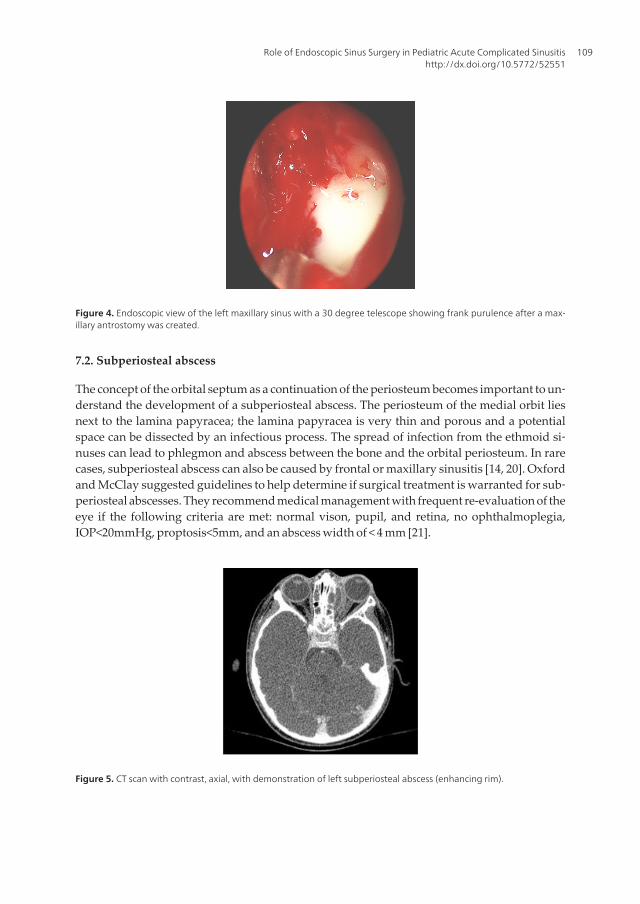
Figure 4. Endoscopic view of the left maxillary sinus with a 30 degree telescope showing frank purulence after a max‐illary antrostomy was created.
7.2. Subperiosteal abscess
The concept of the orbital septum as a continuation of the periosteum becomes important to un‐derstand the development of a subperiosteal abscess. The periosteum of the medial orbit liesnext to the lamina papyracea; the lamina papyracea is very thin and porous and a potentialspace can be dissected by an infectious process. The spread of infection from the ethmoid si‐nuses can lead to phlegmon and abscess between the bone and the orbital periosteum. In rarecases, subperiosteal abscess can also be caused by frontal or maxillary sinusitis [14, 20]. Oxfordand McClay suggested guidelines to help determine if surgical treatment is warranted for sub‐periosteal abscesses. They recommend medical management with frequent re-evaluation of theeye if the following criteria are met: normal vison, pupil, and retina, no ophthalmoplegia,IOP<20mmHg, proptosis<5mm, and an abscess width of < 4 mm [21].
Figure 5. CT scan with contrast, axial, with demonstration of left subperiosteal abscess (enhancing rim).
Role of Endoscopic Sinus Surgery in Pediatric Acute Complicated Sinusitishttp://dx.doi.org/10.5772/52551
109
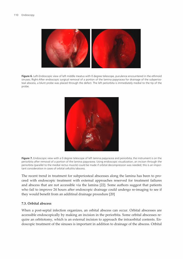
Figure 6. Left-Endoscopic view of left middle meatus with 0 degree telescope, purulence encountered in the ethmoidsinuses; Right-After endoscopic surgical removal of a portion of the lamina papyracea for drainage of the subperios‐teal abscess, a blunt probe was placed through the defect. The left periorbita is immediately medial to the tip of theprobe.
Figure 7. Endoscopic view with a 0 degree telescope of left lamina papyracea and periorbita, the instrument is on theperiorbita after removal of a portion of the lamina papyracea. Using endoscopic visualization, an incison through theperiorbita (parallel to the medial rectus muscle) could be made if orbital decompression was needed; this is an impor‐tant consideration in cases of orbital cellulitis/abscess.
The recent trend in treatment for subperiosteal abscesses along the lamina has been to pro‐ceed with endoscopic treatment with external approaches reserved for treatment failuresand abscess that are not accessible via the lamina [22]. Some authors suggest that patientswho fail to improve 24 hours after endoscopic drainage could undergo re-imaging to see ifthey would benefit from an additinal drainage procedure [20]
7.3. Orbital abscess
When a post-septal infection organizes, an orbital abscess can occur. Orbital abscesses areaccessible endoscopically by making an incision in the periorbita. Some orbital abscesses re‐quire an orbitotomy, which is an external incision to approach the intraorbital contents. En‐doscopic treatment of the sinuses is important in addition to drainage of the abscess. Orbital
Endoscopy110

abscesses are true emergencies as they are often associated with acute vision changes secon‐dary to pressure on the optic nerve or vasospasm of the retinal artery.
7.4. Meningitis
Meinigitis can be seen in conjunction with other suppurative complications of sinusitis. En‐hancement of the meninges is seen on CT or MRI if performed with contrast [14]. Meningitis asthe sole complication of sinusitis does not always need to be treated with endoscopic surgery.Patients can be started on broad-spectrum antibiotics that cross the blood brain barrier and theirclinical progress can be watched closely for 48 hours [23]. Any patient with bacterial meningitisneeds to have a close follow-up with audiological testing as significant hearing loss can occur.
7.5. Epidural abscess
Epidural abscess is a collection of purulence external to the dural layer. For children withepidural abscesses due to paranasal sinus infections, there is some controversy as to the ap‐propriate treatment. Some neurosurgeons feel that if the sinuses are addressed surgicallyand antibiotics are used, some patients may be able to avoid craniotomy, but this belief isnot shared by all in the specialty [24]. Regardless of the exact surgical treatment modality(endoscopic treatment alone or in conjunction with craniotomy), it is important to treat pa‐tients with a multidisciplinary team that includes infectious disease, otolaryngology, andneurosurgery specialists.
7.6. Subdural empyema
Subdural empyema is a collection of purulence between the dura and arachnoid layers. It isconsidered to be a surgical emergency. Coordination between the neurosurgery and otolar‐yngology teams is paramount to address the collection via a craniotomy and to address thesinus disease that was causative via endoscopic sinus surgery [6, 25].
Figure 8. T1 contrast enhanced MRI showing large subdural empyema in left temporal-parietal region in a 9 year-oldwith mental status changes and paresis.
Role of Endoscopic Sinus Surgery in Pediatric Acute Complicated Sinusitishttp://dx.doi.org/10.5772/52551
111
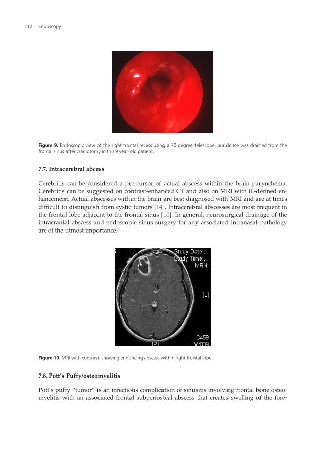
Figure 9. Endoscopic view of the right frontal recess using a 70 degree telescope, purulence was drained from thefrontal sinus after craniotomy in this 9 year-old patient.
7.7. Intracerebral abcess
Cerebritis can be considered a pre-cursor of actual abscess within the brain parynchema.Cerebritis can be suggested on contrast-enhanced CT and also on MRI with ill-defined en‐hancement. Actual abscesses within the brain are best diagnosed with MRI and are at timesdifficult to distinguish from cystic tumors [14]. Intracerebral abscesses are most frequent inthe frontal lobe adjacent to the frontal sinus [10]. In general, neurosurgical drainage of theintracranial abscess and endoscopic sinus surgery for any associated intranasal pathologyare of the utmost importance.
Figure 10. MRI with contrast, showing enhancing abscess within right frontal lobe.
7.8. Pott’s Puffy/osteomyelitis
Pott’s puffy “tumor“ is an infectious complication of sinusitis involving frontal bone osteo‐myelitis with an associated frontal subperiosteal abscess that creates swelling of the fore‐
Endoscopy112
head [26]. It can also be associated with intracranial abscesses in some cases. The etiologycan be infection from sinus disease or due to trauma to the frontal bone resulting in a con‐duit for spread of infection. Medical management of sinus disease is a component of treat‐ment, but surgical intervention is paramount. The approach to treat Pott’s puffy tumor canbe endoscopic with a frontal sinusotomy and anterior ethomidectomy, but a combined ap‐proach with an external incision may be required depending on the size of the subperiostealabscess and whether or not there are associated intracranial complications. Following surgi‐cal treatment, patients will require prolonged courses of IV antibiotics until the osteomyeli‐tis has resolved [27].
Figure 11. Axial CT showing left frontal sinus opacification and connection to scalp, consistent with Pott’s Puffy. Ap‐proximately 80 mL of purulence was drained from the scalp abscess.
Figure 12. Endoscopic view of the left frontal sinus with a 70 degree telescope.
Role of Endoscopic Sinus Surgery in Pediatric Acute Complicated Sinusitishttp://dx.doi.org/10.5772/52551
113
7.9. Cavernous sinus thrombosis
Cavernous sinus thrombosis can occur due to the spread of infection from the middle thirdof the face or from the sinuses and orbit due to the valveless venous system in the anatomi‐cal region [28]. Emergent drainage of any paranasal sinus or orbital infection is important inconjunction with intravenous antibiotics. Systemic anticoagulation is considered in all ofthese cases. Despite aggressive treatment, cavernous sinus thrombosis remains a life-threat‐ening illness (in addition to causing to cranial neuropathy and blindness) that requires mul‐tidisciplinary care in an intensive care unit setting.
8. Complications of endoscopic sinus surgery
Major complications after endoscopic sinus surgery include CSF leak, orbital injury andhemorrhage. A recent retrospective study by Ramakrishnan et al. looked at overall rates ofmajor complications after endoscopic sinus surgery. The overall rate of major complicationsacross all age groups was 1%. This was divided among patients with CSF leak (0.17%), pa‐tients with orbital injuries (0.07%), and patients with significant hemorrhage that requiredblood transfusion (0.76%). This data represents significant improvement from previous ret‐rospective analyses. When the complications were stratified by age, they found that CSFleak was less likely to occur in children, but orbital injury was more likely in the pediatricpopulation. This likely stems from smaller maxillary sinuses and lower orbital floors inyoung children.
Concerns have been raised that sinus surgery in children could affect facial growth. An ani‐mal study looking at facial growth in piglets that underwent endoscopic sinus surgery re‐vealed decreased facial growth on the operative side. This same study investigated the typesof bone that were removed from pediatric patients who underwent endoscopic sinus sur‐gery and noted that children less than 9 years old had immature bone in contrast to childrenover 9 years old and older who had more mature bone in their specimens [29].
A quantitative study in pediatric patients was performed by Senior et al. in order to evaluatefacial growth using CT scan images and volumetric analysis. Their study looked at patientswho underwent sinus surgery due to orbital cellulitis and abscess and re-evaluated the pa‐tients 4 to 10 years later. There was no statistically significant difference in sinus volumefrom the operated side to the non-operated side. However, a small difference in orbital vol‐ume was noted from the operated side to the non-operated side. Overall, they concludedthat sinus surgery in children has minimal impact on sinus and facial growth [30].
9. Post-operative management
9.1. Inpatient
Patients with acute complicated sinusitis will require close monitoring after surgical in‐tervention. Patients with intracranial involvement may require intensive care unit man‐
Endoscopy114
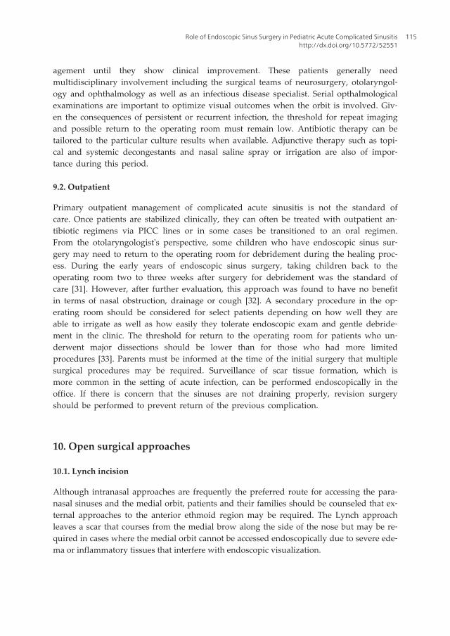
agement until they show clinical improvement. These patients generally needmultidisciplinary involvement including the surgical teams of neurosurgery, otolaryngol‐ogy and ophthalmology as well as an infectious disease specialist. Serial opthalmologicalexaminations are important to optimize visual outcomes when the orbit is involved. Giv‐en the consequences of persistent or recurrent infection, the threshold for repeat imagingand possible return to the operating room must remain low. Antibiotic therapy can betailored to the particular culture results when available. Adjunctive therapy such as topi‐cal and systemic decongestants and nasal saline spray or irrigation are also of impor‐tance during this period.
9.2. Outpatient
Primary outpatient management of complicated acute sinusitis is not the standard ofcare. Once patients are stabilized clinically, they can often be treated with outpatient an‐tibiotic regimens via PICC lines or in some cases be transitioned to an oral regimen.From the otolaryngologist's perspective, some children who have endoscopic sinus sur‐gery may need to return to the operating room for debridement during the healing proc‐ess. During the early years of endoscopic sinus surgery, taking children back to theoperating room two to three weeks after surgery for debridement was the standard ofcare [31]. However, after further evaluation, this approach was found to have no benefitin terms of nasal obstruction, drainage or cough [32]. A secondary procedure in the op‐erating room should be considered for select patients depending on how well they areable to irrigate as well as how easily they tolerate endoscopic exam and gentle debride‐ment in the clinic. The threshold for return to the operating room for patients who un‐derwent major dissections should be lower than for those who had more limitedprocedures [33]. Parents must be informed at the time of the initial surgery that multiplesurgical procedures may be required. Surveillance of scar tissue formation, which ismore common in the setting of acute infection, can be performed endoscopically in theoffice. If there is concern that the sinuses are not draining properly, revision surgeryshould be performed to prevent return of the previous complication.
10. Open surgical approaches
10.1. Lynch incision
Although intranasal approaches are frequently the preferred route for accessing the para‐nasal sinuses and the medial orbit, patients and their families should be counseled that ex‐ternal approaches to the anterior ethmoid region may be required. The Lynch approachleaves a scar that courses from the medial brow along the side of the nose but may be re‐quired in cases where the medial orbit cannot be accessed endoscopically due to severe ede‐ma or inflammatory tissues that interfere with endoscopic visualization.
Role of Endoscopic Sinus Surgery in Pediatric Acute Complicated Sinusitishttp://dx.doi.org/10.5772/52551
115
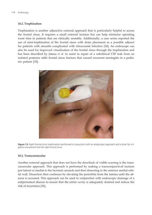
10.2. Trephination
Trephination is another adjunctive external approach that is particularly helpful to accessthe frontal sinus. It requires a small external incision but can help minimize operatingroom time in patients that are clinically unstable. Additionally, a case series reported theuse of mini-trephination of the frontal sinus with drain placement as a possible adjunctfor patients with sinusitis complicated with intracranial infection [34]. An endoscope canalso be used for improved visualization of the frontal sinus through the trephination andhas been described by Jatana et al. to assist in repair of a subclinical CSF leak from anisolated posterior table frontal sinus fracture that caused recurrent meningitis in a pedia‐tric patient [35].
Figure 13. Right frontal sinus trephination performed in conjuction with an endoscopic approach and a drain for irri‐gation was placed into the right frontal sinus.
10.3. Transcaruncular
Another external approach that does not have the drawback of visible scarring is the trans‐caruncular approach. This approach is performed by making a transconjunctival incisionjust lateral or medial to the lacrimal caruncle and then dissecting to the anterior medial orbi‐tal wall. Dissection then continues by elevating the periorbita from the lamina until the ab‐scess is accessed. This approach can be used in conjunction with endoscopic drainage of asubperiosteal abscess to ensure that the entire cavity is adequately drained and reduce therisk of recurrence [36].
Endoscopy116
11. Conclusion
Endoscopic surgical techniques are an important component of treatment for acute compli‐cated sinusitis in the pediatric population. Given the diverse nature or presenting signs andsymptoms, a high index of suspicion for complications of sinusitis must be maintained forpatients presenting with complaints previously described. Using endoscopic approaches tothe sinuses, the otolaryngologist is an important member of the multidisciplinary team thatcares for patients with complications of sinusitis.
Author details
Timothy P. McEvoy1, Charles A. Elmaraghy1,2 and Kris R. Jatana1,2
*Address all correspondence to: [email protected]
1 Department of Pediatric Otolaryngology-Head and Neck Surgery, Nationwide Children’sHospital, Columbus, Ohio, USA
2 Department of Otolaryngology-Head and Neck Surgery, Wexner Medical Center at TheOhio State University, Columbus, Ohio, USA
References
[1] Kennedy DW.Functional endoscopic sinus surgery. Technique. Arch Otolaryngol.(1985). Epub 1985/10/01., 111(10), 643-9.
[2] Kennedy DW, Zinreich SJ, Rosenbaum AE, Johns ME.Functional endoscopic sinussurgery. Theory and diagnostic evaluation. Arch Otolaryngol. (1985). Epub1985/09/01., 111(9), 576-82.
[3] Gross CW, Gurucharri MJ, Lazar RH, Long TE.Functional endonasal sinus surgery(FESS) in the pediatric age group. The Laryngoscope(1989). Epub 1989/03/01., 99(3),272-5.
[4] al, R., & , . Embryology and Anatomy of the Paranasal Sinuses. 4th ed(2003).
[5] Adibelli, Z. H., Songu, M., & Adibelli, H. Paranasal sinus development in children: Amagnetic resonance imaging analysis. American journal of rhinology & allergy.(2011). Epub 2011/06/30., 25(1), 30-5.
[6] Yucel, O. T., & Ogretmenoglu, O. Subdural empyema and blindness due to caver‐nous sinus thrombosis in acute frontal sinusitis. International journal of pediatricotorhinolaryngology(1998). Epub 1999/04/06., 121 EOF-5 EOF.
Role of Endoscopic Sinus Surgery in Pediatric Acute Complicated Sinusitishttp://dx.doi.org/10.5772/52551
117
[7] Oxford, L. E., & Mc Clay, J. Complications of acute sinusitis in children. Otolaryngol‐ogy--head and neck surgery : official journal of American Academy of Otolaryngolo‐gy-Head and Neck Surgery(2005). Epub 2005/07/19., 133(1), 32-7.
[8] Lerner DN, Choi SS, Zalzal GH, Johnson DL.Intracranial complications of sinusitis inchildhood. The Annals of otologyrhinology, and laryngology. (1995). Pt 1):Epub1995/04/01., 288 EOF-93 EOF.
[9] DeMuri GP, Wald ER.Complications of acute bacterial sinusitis in children. The Pe‐diatric infectious disease journal(2011). Epub 2011/07/14., 30(8), 701-2.
[10] Vazquez, E., Creixell, S., Carreno, J. C., Castellote, A., Figueras, C., Pumarola, F., et al.Complicated acute pediatric bacterial sinusitis: Imaging updated approach. Currentproblems in diagnostic radiology(2004). Epub 2004/06/25., 33(3), 127-45.
[11] Germiller JA, Monin DL, Sparano AM, Tom LW.Intracranial complications of sinusi‐tis in children and adolescents and their outcomes. Archives of otolaryngology--head& neck surgery(2006). Epub 2006/09/20., 132(9), 969-76.
[12] Kombogiorgas, D., Seth, R., Athwal, R., Modha, J., & Singh, J. Suppurative intracrani‐al complications of sinusitis in adolescence. Single institute experience and review ofliterature. British journal of neurosurgery(2007). Epub 2007/12/12., 21(6), 603-9.
[13] Hicks, C. W., Weber, J. G., Reid, J. R., & Moodley, M. Identifying and managing in‐tracranial complications of sinusitis in children: a retrospective series. The Pediatricinfectious disease journal(2011). Epub 2011/03/19., 30(3), 222-6.
[14] Reid JR.Complications of pediatric paranasal sinusitis. Pediatric radiology(2004).Epub 2004/07/28., 34(12), 933-42.
[15] Herrmann BW, Forsen JW, Jr.Simultaneous intracranial and orbital complications ofacute rhinosinusitis in children. International journal of pediatric otorhinolaryngolo‐gy(2004). Epub 2004/04/15., 68(5), 619-25.
[16] Parikh, S. R., Cuellar, H., Sadoughi, B., Aroniadis, O., & Fried, M. P. Indications forimage-guidance in pediatric sinonasal surgery. International journal of pediatric oto‐rhinolaryngology(2009). Epub 2009/01/23., 73(3), 351-6.
[17] Benoit, Silvera. V. M., Nichollas, R., Jones, D., Mc Gill, T., & Rahbar, R. Image guid‐ance systems for minimally invasive sinus and skull base surgery in children. Inter‐national journal of pediatric otorhinolaryngology(2009). Epub 2009/08/22., 73(10),1452-7.
[18] Garrett, M. G. J., & Elmaraghy, Jatana. K. R. Radiographic findings in pediatric peri‐orbital cellulitis. American Society of Pediatric Otolaryngology (ASPO); San Diego,California(2012).
[19] Jatana KR, Grischkan JM, Skomorowski MJ, Elmaraghy CA.Recurrent unilateral peri‐orbital cellulitis in a pediatric patient--an anatomic abnormalit. y. International jour‐nal of pediatric otorhinolaryngology(2008). Epub 2008/08/16., 72(10), 1577-80.
Endoscopy118
[20] Noordzij JP, Harrison SE, Mason JC, Hashisaki GT, Reibel JF, Gross CW.Pitfalls inthe endoscopic drainage of subperiosteal orbital abscesses secondary to sinusitis.American journal of rhinology(2002). Epub 2002/05/28., 16(2), 97-101.
[21] Oxford, L. E., & Mc Clay, J. Medical and surgical management of subperiosteal orbi‐tal abscess secondary to acute sinusitis in children. International journal of pediatricotorhinolaryngology(2006). Epub 2006/08/15., 70(11), 1853-61.
[22] Soon VT.Pediatric subperiosteal orbital abscess secondary to acute sinusitis: a 5-yearreview. American journal of otolaryngology. (2011). Epub 2009/12/25., 32(1), 62-8.
[23] Younis, R. T., Anand, V. K., & Childress, C. Sinusitis complicated by meningitis: cur‐rent management. The Laryngoscope(2001). Epub 2001/09/25., 111(8), 1338-42.
[24] Heran, N. S., Steinbok, P., & Cochrane, D. D. Conservative neurosurgical manage‐ment of intracranial epidural abscesses in children. Neurosurgery(2003). discussion7-8. Epub 2003/10/02., 53(4), 893-7.
[25] Waseem, M., Khan, S., & Bomann, S. Subdural empyema complicating sinusitis. TheJournal of emergency medicine(2008). Epub 2007/12/25., 35(3), 277-81.
[26] Blumfield, E., Misra, M., Pott’s, puffy., tumor, intracranial., orbital, complications.,the, initial., presentation, of., sinusitis, in., healthy, adolescents. a., & case, series.Emergency radiology(2011). Epub 2011/03/08., 18(3), 203-10.
[27] Parida, P. K., Surianarayanan, G., Ganeshan, S., & Saxena, S. K. Pott’s puffy tumor inpediatric age group: A retrospective study. International journal of pediatric otorhi‐nolaryngology(2012). Epub 2012/06/19.
[28] Cannon ML, Antonio BL, McCloskey JJ, Hines MH, Tobin JR, Shetty AK.Cavernoussinus thrombosis complicating sinusitis. Pediatric critical care medicine : a journal ofthe Society of Critical Care Medicine and the World Federation of Pediatric Intensiveand Critical Care Societies(2004). Epub 2003/12/31., 5(1), 86-8.
[29] Mair EA, Bolger WE, Breisch EA.Sinus and facial growth after pediatric endoscopicsinus surgery. Archives of otolaryngology--head & neck surgery(1995). Epub1995/05/01., 121(5), 547-52.
[30] Senior, B., Wirtschafter, A., Mai, C., Becker, C., & Belenky, W. Quantitative impact ofpediatric sinus surgery on facial growth. The Laryngoscope(2000). Epub 2000/11/18.,110(11), 1866-70.
[31] Mair EA.Pediatric functional endoscopic sinus surgery: postoperative care. Otolar‐yngologic clinics of North America(1996). Epub 1996/02/01., 29(1), 207-19.
[32] Mitchell RB, Pereira KD, Younis RT, Lazar RH.Pediatric functional endoscopic sinussurgery: is a second look necessary? The Laryngoscope. (1997). Epub 1997/09/18.,107(9), 1267-9.
Role of Endoscopic Sinus Surgery in Pediatric Acute Complicated Sinusitishttp://dx.doi.org/10.5772/52551
119
[33] Cable BB, Mair EA.Pediatric functional endoscopic sinus surgery: frequently askedquestions. The Annals of otologyrhinology, and laryngology. (2006). Epub2006/10/19., 115(9), 643-57.
[34] Mc Intosh, D. L., & Mahadevan, M. Frontal sinus mini-trephination for acute sinusitiscomplicated by intracranial infection. International journal of pediatric otorhinolar‐yngology(2007). Epub 2007/07/14., 71(10), 1573-7.
[35] Jatana, K. R., Ryoo, C., Skomorowski, M., Butler, N., & Kang, D. R. Minimally inva‐sive repair of an isolated posterior table frontal sinus fracture in a pediatric patient.Otolaryngology--head and neck surgery : official journal of American Academy ofOtolaryngology-Head and Neck Surgery. (2008). Epub 2008/05/28., 138(6), 809-11.
[36] Pelton RW, Smith ME, Patel BC, Kelly SM.Cosmetic considerations in surgery for or‐bital subperiosteal abscess in children: experience with a combined transcaruncularand transnasal endoscopic approach. Archives of otolaryngology--head & neck sur‐gery(2003). Epub 2003/06/18., 129(6), 652-5.
Endoscopy120
Chapter 8
Endoscopy -An Advancement in Sinusand Skull Base Surgery
Rickul Varshney, Faisal Zawawi, Marc A Tewfik andSaul Frenkiel
Additional information is available at the end of the chapter
http://dx.doi.org/10.5772/52749
1. Introduction
Skull base and sinus surgery has evolved dramatically throughout the past century. It isnot long ago that the maxillary sinus would principally be reached via an anterior ap‐proach through the gingivobuccal sulcus of the oral cavity. The earliest days of endos‐copy date back to the early 1900s, when Hirschmann used a modified cystoscope toexamine the sinuses. Thereafter, modern endoscopy has seen advances not only in thetypes of endoscopes available, but also the types of interventions amenable to the endo‐scopic approach.
However, even in this modern era of refined endoscopic instrumentation and technique,opinion remains split regarding the optimal approach to certain areas of the skull base.For example, areas such as the anterior cranial fossa and the infratemporal fossa are of‐ten approached through external transcutaneous approaches despite the development ofadequate and safe transnasal endoscopic pathway.
The current chapter aims to provide a complete comparison of endoscopic versus openapproaches of routinely performed sinus and skull base surgical procedures. It will alsoemphasize the advantages of endoscopy versus traditional approaches for sinus andskull base surgery, including management of tumors, infectious processes, congenitalanomalies and traumatic injuries.
© 2013 Varshney et al.; licensee InTech. This is an open access article distributed under the terms of theCreative Commons Attribution License (http://creativecommons.org/licenses/by/3.0), which permitsunrestricted use, distribution, and reproduction in any medium, provided the original work is properly cited.
2. Evolution of endoscopic sinus surgery
2.1. History of the emergence of endoscopes
The historical evolution of endoscopic sinus surgery is quite fascinating. Hippocrates can beconsidered one of the first practitioners of rhinology. He was one of the first to documenttreatment of nasal polyps using snares or sponges on a string.[1] Thereafter, there weremany ancient rhinology physicians that contributed to the evolution of rhinology, howeverwe will focus on the evolution of endoscopic sinus surgery in this chapter.
The first reports of endoscopic visualization of the sinuses date back to 1901 when Hirsch‐man used a small cystoscope with an electric bulb to examine the maxillary sinus throughan oroantral fistula.[2] Thereafter, in 1902, Reichert performed the first known endoscopicsinus surgery, performing maxillary sinus manipulation through an oroantral fistula.[3]Continuing this trend, in 1922, Spielberg employed antroscopes to access the maxillary sinusvia the inferior meatus.[2] The term “sinuscopy” was later popularized by Maltz in 1925.[2]
At that time, these endoscopes were not surprisingly restricted in terms of optical quality,field of view and illumination, relying on flame or electric bulbs.[2] In the 1960s, Hopkinsdeveloped the rod optic endoscope. Hopkins, also known for the development of the fiberoptic gastroscope and zoom lens for cameras, revolutionized the optical quality available tosurgeons.[3] Thereafter, Karl Storz in Germany created angled endoscopes ranging from 0 to120 degrees, thus allowing visualization of a field of view never previously imagined.[2]
In the 1970s, this new and exciting armamentarium of endoscopic tools allowed surgeonssuch as Messerklinger, Stammberger, Draf and Wigand to transition sinus surgery from aradical operation to a minimally invasive procedure.[2,3] In 1978, Messerklinger publishedthe landmark reference “Endoscopy of the nose”, due to large part from this remarkableevolution in endoscopic sinus surgery tools. He thoroughly examined the mucociliary clear‐ance pattern and endoscopic changes of the osteomeatal complex,[3] thus further under‐standing sinus disease.
At that time, Draf published his own work on the sinuses and it was his experience thatmade frontal sinus access more realistic and safe.[4] Stammberger was also enthusiastic andprominent in the field of endoscopic surgery and popularized Messerklinger’s ideas world‐wide. The immense work of these pioneers remodeled sinus surgery. The principle of treat‐ing sinus disease from a functional point of view at the site of obstruction replaced thetraditional mucosal stripping approach to treat inflammatory disease.[3] This work led tothe term “functional endoscopic sinus surgery,” which was coined by Kennedy in 1985.[2] Inthe same year, the first two established courses in North America on endoscopic sinus sur‐gery were given at the Johns Hopkins Hospital.[3]
Furthermore, more contemporary surgeons, following the work of the pioneers of rhinolo‐gy, expanded the limits to which nasal endoscopy could treat disease. Areas of the skullbase were also accessed and different types of procedures amenable to this approach revolu‐tionized the field of rhinology. The first description of trans-sphenoidal approach to the sell‐
Endoscopy122
ar region dates back to 1907, perfored by Schloffer.[4,5] Another prominent surgeon,Cushing, performed this procedure for many years, but abandoned this approach in 1927due to the high complication rate. The procedure rapidly lost popularity until Dott, a disci‐ple of Cushing, created a lighted speculum as an aid for trans-sphenoidal visualization.[6] Inthe 1960s, Guiot, a disciple of Dott, popularised this approach.[4] He was the first neurosur‐geon to perform transsphenoidal surgery.[6]
In 1967, Jules Hardy reported the use of the surgical microscope for transsphenoidal sur‐gery.[5]He is credited for developing the fundamental principles of pituitary surgery uptothis day.[5] Jankowski published the first series on endoscopic pituitary surgery proceduresin 1992.[7] The term “functional endoscopic pituitary surgery” was coined by Cappabiancaand de Divitiis.[6]
However, the endoscopic approach to the skull base didn’t end at the sella region. Weiss, in1987, was the first to publish about extending the transsphenoidal approach to access supra‐sellar lesions.[5] Thereafter, the first report of endoscopic transsphenoidal approach for re‐section of a large clivus chordoma was published in 1996 by Jho.[8] More contemporarysurgeons have continued this endoscopic advacement towards the skull base with new ap‐proaches to areas such as the infratemporal fossa and the anterior cranial fossa.
With the evolution of endoscopes, also came an evolution in surgical instruments used insinus surgery. Early endoscopic surgery was performed using grasping forceps, which oftenstripped mucosa and denuded bone. At first, this was considered ideal as the theory of re‐moving all the diseased mucosa was preached. However, with endoscopes, surgeons wereable to visualize the sinuses on post-operative follow-ups and discover the osteitis, scarringand osteoneogenesis that their surgery had caused. Thus, there was a movement to createnew endoscopic fine-cutting instruments, originally developped for orthopedic cartilagework, to perform minimally invasive “functional” surgery. Later, an evolutionary descend‐ent of the through cutting instruments, was the emergence of the microdebrider, also origi‐nally used in orthopedics. Setliff and Parsons introduced the microdebrider to sinonasalsurgery.[3]
It is important to emphasize the work of pioneers such as Messerklinger, Draf, Wigand andothers as a focal transition point in the medical community’s understanding of sinus disease.In fact, before their reports, sinus disease treatment was based on an invasive exenterativeapproach of removing all the inflammed mucosa of the sinuses. The main focus of treatmentwas to obliterate and remove all the sinonasal regions of disease. However, the work ofthese legendary surgeons allowed physicians to understand the functional aspect of sinusanatomy. They demonstrated that a large amount of sinus disease was based on an impair‐ment of adequate drainage and that resolution of the latter obstruction would allow sympto‐matic relief.
This concept of mucosal preservation still applies today and is the basis of a large portion ofrhinology practice. In fact, rhinologists are careful to avoid mucosal stripping that can po‐tentially cause impaired mucociliary clearance or neo-osteogenesis.[9] This has motivated
Endoscopy – An Advancement in Sinus and Skull Base Surgeryhttp://dx.doi.org/10.5772/52749
123
the development of precise instrumentation including sharp through-cutters and microde‐briders that mimise trauma to adjacent healthy tissue.
2.2. Endoscopes of the 21ST century
Endoscopes today have offered sinus surgeons the ability to increase the potential applica‐tions of endoscopic sinus surgery. There is a strong movement by manufacturers to improveoptical quality of their endoscopes. One such improvement is the use of the xenon light,which was an upgrade to the halogen light source, with advantages such as durability, di‐minished heat production and energy consumption. Another improvement is the diameterof the endoscopes with some available as small as 2 mm. The most common scopes current‐ly employed in sinus and skull base surgery are 4 mm in diameter by 20 cm long; 2.7 mmdiameter scopes are commonly reserved for pediatric cases. These scopes are available invarious angles ranging from 0 to 30, 45, and 70 degrees and provide high quality imaging.[9]
Furthermore, there have been improvements in the processing camera used for endocopic sur‐gery. Modern cameras have gone from 1 silicone chip to 3 silicone chip cameras in order to proc‐ess the three primary colors [red, blue and green] as a means to enhance contrast and balance ofthe projected image.[2] High definition cameras have replaced traditional cameras, workingthrough a progressive scanning mechanism instead of an interlaced scanning manner. This re‐fers to the way each frame is scanned by the camera, and results in an upgrade of 60 frames persecond being seen by the high definition cameras instead of 30 frames per second provided bythe older generation. The previously used interlaced scanning provided a significant amount offlickering, which was compensated by image blurring.
Finally, as the image viewed is dependent on the resolution of the monitor, even the latterhas evolved dramatically. With the advent of high definition monitors, 16:9 aspect ratio hasreplaced the traditional 4:3 aspect ratio provided by standard definition monitors, translat‐ing in an increase in pixel density from 640x480 to 1929x1080.[2] What this means for thesurgeon is better color, contrast, resolution and peripheral visualization of the surgical field.
3. Endoscopic surgery versus open traditional approach
Sinus surgery traditionally was performed via open approaches, be it a Caldwell-Luc proce‐dure for the maxillary sinus or an osteoplastic flap approach to the frontal sinus. With theadvent of the endoscopes, these traditional invasive extenterative methods have largelybeen replaced by functional endoscopic sinus surgery. In this section, we examine the differ‐ent sinuses and contrast these approaches.
3.1. Frontal sinus
The first report of frontal sinus surgery was by Rimge in 1750, where he used an externalapproach to obliterate the sinus.[10] In 1884, Ogston and Luck described an anterior wallperforation technique using a trephine to create a drainage pathway into the anterior eth‐
Endoscopy124
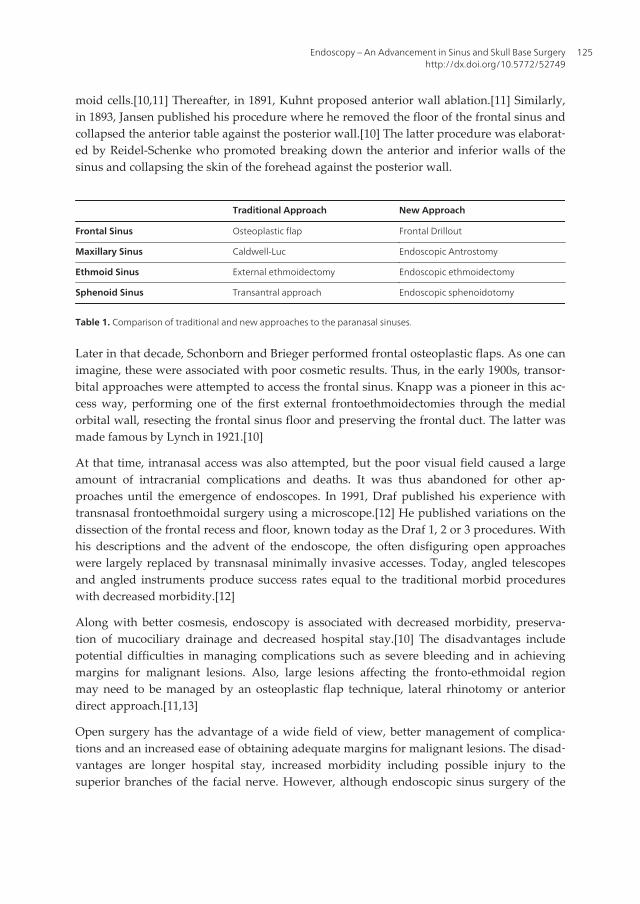
moid cells.[10,11] Thereafter, in 1891, Kuhnt proposed anterior wall ablation.[11] Similarly,in 1893, Jansen published his procedure where he removed the floor of the frontal sinus andcollapsed the anterior table against the posterior wall.[10] The latter procedure was elaborat‐ed by Reidel-Schenke who promoted breaking down the anterior and inferior walls of thesinus and collapsing the skin of the forehead against the posterior wall.
Traditional Approach New Approach
Frontal Sinus Osteoplastic flap Frontal Drillout
Maxillary Sinus Caldwell-Luc Endoscopic Antrostomy
Ethmoid Sinus External ethmoidectomy Endoscopic ethmoidectomy
Sphenoid Sinus Transantral approach Endoscopic sphenoidotomy
Table 1. Comparison of traditional and new approaches to the paranasal sinuses.
Later in that decade, Schonborn and Brieger performed frontal osteoplastic flaps. As one canimagine, these were associated with poor cosmetic results. Thus, in the early 1900s, transor‐bital approaches were attempted to access the frontal sinus. Knapp was a pioneer in this ac‐cess way, performing one of the first external frontoethmoidectomies through the medialorbital wall, resecting the frontal sinus floor and preserving the frontal duct. The latter wasmade famous by Lynch in 1921.[10]
At that time, intranasal access was also attempted, but the poor visual field caused a largeamount of intracranial complications and deaths. It was thus abandoned for other ap‐proaches until the emergence of endoscopes. In 1991, Draf published his experience withtransnasal frontoethmoidal surgery using a microscope.[12] He published variations on thedissection of the frontal recess and floor, known today as the Draf 1, 2 or 3 procedures. Withhis descriptions and the advent of the endoscope, the often disfiguring open approacheswere largely replaced by transnasal minimally invasive accesses. Today, angled telescopesand angled instruments produce success rates equal to the traditional morbid procedureswith decreased morbidity.[12]
Along with better cosmesis, endoscopy is associated with decreased morbidity, preserva‐tion of mucociliary drainage and decreased hospital stay.[10] The disadvantages includepotential difficulties in managing complications such as severe bleeding and in achievingmargins for malignant lesions. Also, large lesions affecting the fronto-ethmoidal regionmay need to be managed by an osteoplastic flap technique, lateral rhinotomy or anteriordirect approach.[11,13]
Open surgery has the advantage of a wide field of view, better management of complica‐tions and an increased ease of obtaining adequate margins for malignant lesions. The disad‐vantages are longer hospital stay, increased morbidity including possible injury to thesuperior branches of the facial nerve. However, although endoscopic sinus surgery of the
Endoscopy – An Advancement in Sinus and Skull Base Surgeryhttp://dx.doi.org/10.5772/52749
125
frontal sinus is often highly effective, certain select cases nevertheless still require open ap‐proaches with osteoplasties.
3.2. Maxillary sinus
The first descriptions of open approaches to the maxillary sinus date back to the early 1700s.Over a century later, in 1893, in the United States, Caldwell described an anterior approachvia an incision in the gingivobuccal sulcus coupled with an inferior antrostomy. This proce‐dure was almost simultaneously popularized in Europe by Luc, and was later coined theCaldwell-Luc procedure.[14] A century after that, Stammberger and then Kennedy intro‐duced the middle meatus antrostomy in the 1980s.[15] At that time, inferior and middlemeatal antrostomies were being performed by different group of surgeons. When com‐pared, middle meatal antrostomies demonstrate better resolution of symptoms and longerpatency rates.[14] The reason for this is believed to be the fact that the normal mucociliaryclearance patterns of the maxillary sinus tend to move secretions toward the natural ostiumin the midle meatus, and not toward the inferior meatal antrostomy.
With the emergence of endoscopes, especially angled telescopes to look within the maxillarysinus, as well as the development of the coronal bone view on CT scans in 1987, the ap‐proach to the maxillary sinus changed dramatically. A study performed by Penttila et al.demonstrated that patients undergoing surgery for chronic maxillary sinusitis reported im‐provement in 50.7% of the Caldwell-Luc group and in 76.7% of the endoscopic sinus surgerygroup.[16] Also, higher complication rates have been described in patients undergoing theCaldwell-Luc procedure.[17] These include pain, facial swelling and numbness, dentalnumbness, persistent oroantral fistulas, wound dehiscence, dacrocystitis, tooth decay, per‐sistent symptoms and bleeding.[15]
Although endoscopic surgery has largely replaced Caldwell-Luc procedures, there is still arole for the latter in certain cases. Cutler et al. performed the procedure in 37 patients whohad failed endoscopic sinus surgery for refractory sinusitis and reported a 92% responsebased on follow-up endoscopic examination or computed tomography scan imaging.[18]Other possible indications include the removal of dentigerous cyst and benign tumors locat‐ed within the maxillary sinus.
3.3. Ethmoid sinus
The management of ethmoid sinus disease has been the source of a lot of controversy in thepast. Many surgical methods for ethmoidectomies have been described, ranging from intra‐nasal, transantral, external approaches employing a headlight, to endoscopic approaches.
In 1912, Mosher described intranasal extirpation of the ethmoid labyrinth.[19] He promotedthe complete resection of the middle turbinate along with the ethmoid sinuses, which wasdebated by opposing surgeons, including Pratt. The latter preached the importance of themiddle turbinate as a landmark to reduce operative morbidity.
In 1929, Mosher and Smith, a well-known rhinologist, promoted a transition towards an ex‐ternal approach as means to reduce complications.[19] At the same time, other surgeons
Endoscopy126
promoted a transantral approach, originally described by Jansen in the 1800s. All these ap‐proaches were being practiced without any consensus as to the optimal technique.
The transnasal approach, which was popularized by Mosher, consisted of progressive exent‐eration of the ethmoid air calls in antero-posterior direction. As mentioned, certain surgeonsremoved the middle turbinate as well. Along with the ethmoidectomy, the nasofrontal ductwas also unroofed. In this method, the procedure was performed through a nasal speculumwith a headlight. Magnification was provided by loupes or insertion of lenses onto the head‐light. Certain surgeons, namely Dixon and Heermann, advocated the use of microscopesduring the transnasal approach to the ethmoids.[19] However, the high incidence of compli‐cations and the availability of the transantral and external approaches, caused this methodto fall in popularity over the next few decades.[19]
These procedures have all been replaced by endoscopic sinus surgery. The latter permitscontrolled removal of the diseased tissue and obstructing bony partitions in a stepwise fash‐ion, with decreased complications. Ethmoidectomies are also commonly performed in con‐junction with other procedures, thus a vast access with the endoscope is essential.
3.4. Sphenoid sinus
The history, indications and approaches to the sphenoid sinus will be discussed in details inthe skull base section. The open, microscopic approach versus the endoscopic approach tosphenoid sinus surgery and access to the skull base will be contrasted.
4. Septoplasty
Apart from the sinuses, the nasal septum is also amenable to open or endoscopic surgery. In1842, Langenbeck first described the entities of septal crests and spurs.[1] Thereafter, in theearly 1900s, Freer published about the removal of thickened portions of the septal cartilage.He invented a number of instruments to perform this procedure. Simultaneously, Killianfurther developed many of the techniques of septal surgery.[1]
Cottle first described septoplasty in 1947 to treat nasal obstruction.[20] It is only in 1991 thatthe endoscopic technique to treat septal deformities was first popularized by Lanza et al.and Stammberger.[21] Thereafter, Lanza described isolated septal spur surgery using endo‐scopic access.[21,22]
Advantages over the open technique include targeted approach to the septal deformity, lim‐ited mucosal flap dissection, superior magnification of the field and less physical distortionas there is no need of a nasal speculum. Finally, it is a superior teaching tool as trainees canvisualize the surgery on a screen in real-time.[23] Furthermore, endoscopic visualization al‐lows assessment of deformities in the nasal valve region and posterior septum.[21]
Proponents of endoscopic surgery advocate the minimally invasive aspect of this approachas only the mucosa overlying the deviated segment is elevated through a Killian incision.
Endoscopy – An Advancement in Sinus and Skull Base Surgeryhttp://dx.doi.org/10.5772/52749
127
This may explain why authors have reported higher degrees of septal tears with the opentechnique.[23] This is particularly useful in revision cases, where fibrosis adheres the septalmucosa in areas previously operated. Other advantages of the endoscopic approach includeshorter operative time, decreased bleeding, decreased pain[24,25] and decreased synechiaeformation.[20]
Rotenberg’s group recently demonstrated that there was no difference in post-operative out‐comes in terms of nasal obstruction between both groups,[23] in support of previous sur‐geons’ findings.[25] However, other authors have stated significant differences in nasalsymptoms with endoscopic groups doing better in follow-up assessments.[20,24]
The endoscopic septoplasty has gained popularity in recent years. Nevertheless, certain cas‐es still require an open approach. The relative contraindications to endoscopic septoplastyare when the deformity involves a deflection of the caudal septal cartilage, and when exter‐nal nasal deformities require a concomitant open rhinoplasty.[20,21]
5. Skull base
Through the years, the work of Messerklinger and other pioneers in nasal endoscopy helpedto develop functional endoscopic sinus surgery as a means to treat sinus disease from a me‐chanical point of view. However, contemporary surgeons have expanded the limits to whichnasal endoscopy can treat disease. In fact, the areas of the skull base accessible and the typesof procedures amenable to this approach have revolutionized the field of rhinology.
Traditional methods required external skin incisions, translocation of the cranium or maxil‐lofacial skeleton and retraction of the brain.[26] Endoscopic access is based on modular ana‐tomical approaches in the sagittal planes, for anterior cranial fossa, pituitary, and transclivalposterior cranial fossa surgery; and coronal planes, for pterygopalatine fossa and infratem‐poral fossa surgery.[27]
Endoscopic endonasal approaches have improved visualization and decreased collateraltrauma to the craniofacial tissues. They provide faster healing and recovery time, decreasedneurovascular injuries, complete oncologic resections and better endocrinologic outcomes.Potential limitations of the endoscopic approach include location, extent and nature of thedisease and importantly surgeon expertise and available equipment, including image guid‐ance.[26]
There are multiple approaches available to the skull base depending on the location of thedisease, namely the transcribriform, transsellar, transplanum [drilling the planumsphenoi‐dale and the tuberculum sellae, transclival and the transodontoid approaches.[6] The latterapply to sagittal plane. In terms of coronal plane, the skull base can be divided into medialpetrous apex, petroclival region, Meckel’s cave, cavernous sinus and infratemporal fossa.[6]
In this section, we explore endoscopic surgery as it relates to the pituitary gland, the anteriorcranial fossa, the clivus and the infratemporal fossa.
Endoscopy128
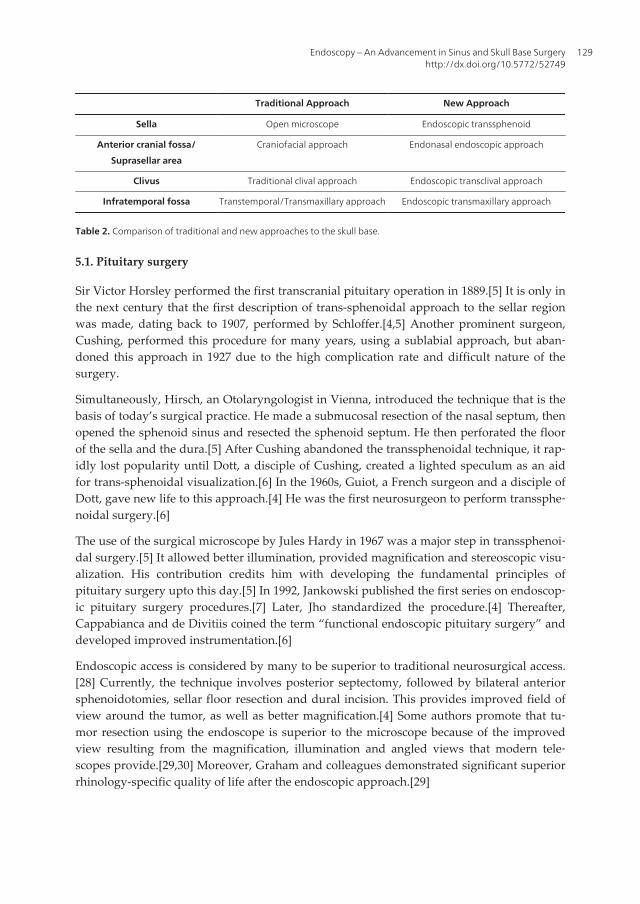
Traditional Approach New Approach
Sella Open microscope Endoscopic transsphenoid
Anterior cranial fossa/
Suprasellar area
Craniofacial approach Endonasal endoscopic approach
Clivus Traditional clival approach Endoscopic transclival approach
Infratemporal fossa Transtemporal/Transmaxillary approach Endoscopic transmaxillary approach
Table 2. Comparison of traditional and new approaches to the skull base.
5.1. Pituitary surgery
Sir Victor Horsley performed the first transcranial pituitary operation in 1889.[5] It is only inthe next century that the first description of trans-sphenoidal approach to the sellar regionwas made, dating back to 1907, performed by Schloffer.[4,5] Another prominent surgeon,Cushing, performed this procedure for many years, using a sublabial approach, but aban‐doned this approach in 1927 due to the high complication rate and difficult nature of thesurgery.
Simultaneously, Hirsch, an Otolaryngologist in Vienna, introduced the technique that is thebasis of today’s surgical practice. He made a submucosal resection of the nasal septum, thenopened the sphenoid sinus and resected the sphenoid septum. He then perforated the floorof the sella and the dura.[5] After Cushing abandoned the transsphenoidal technique, it rap‐idly lost popularity until Dott, a disciple of Cushing, created a lighted speculum as an aidfor trans-sphenoidal visualization.[6] In the 1960s, Guiot, a French surgeon and a disciple ofDott, gave new life to this approach.[4] He was the first neurosurgeon to perform transsphe‐noidal surgery.[6]
The use of the surgical microscope by Jules Hardy in 1967 was a major step in transsphenoi‐dal surgery.[5] It allowed better illumination, provided magnification and stereoscopic visu‐alization. His contribution credits him with developing the fundamental principles ofpituitary surgery upto this day.[5] In 1992, Jankowski published the first series on endoscop‐ic pituitary surgery procedures.[7] Later, Jho standardized the procedure.[4] Thereafter,Cappabianca and de Divitiis coined the term “functional endoscopic pituitary surgery” anddeveloped improved instrumentation.[6]
Endoscopic access is considered by many to be superior to traditional neurosurgical access.[28] Currently, the technique involves posterior septectomy, followed by bilateral anteriorsphenoidotomies, sellar floor resection and dural incision. This provides improved field ofview around the tumor, as well as better magnification.[4] Some authors promote that tu‐mor resection using the endoscope is superior to the microscope because of the improvedview resulting from the magnification, illumination and angled views that modern tele‐scopes provide.[29,30] Moreover, Graham and colleagues demonstrated significant superiorrhinology-specific quality of life after the endoscopic approach.[29]
Endoscopy – An Advancement in Sinus and Skull Base Surgeryhttp://dx.doi.org/10.5772/52749
129
With the increasing popularity of trans-sphenoidal sellar surgery, the concept of approach‐ing parasellar regions through this pathway gained massive enthusiasm. Weiss, in 1987, wasthe first to publish about extending the transsphenoidal approach to access suprasellar le‐sions.[5] Therefore, anterior cranial fossa lesions, for example craniopharyngiomas, can bewell managed through endoscopic routes.[31]
Compared to traditional approaches, transsphenoidal approaches for craniopharyngiomasand Rathke’s cyst of the anterior cranial fossa demonstrate lower recurrence rate and de‐creased complications.[4,32] Couldwell and colleagues published about 105 patients under‐going extended transsphenoidal approaches to the cavernous sinus, suprasellar region andclival region. They concluded this approach to be a safe alternative to cranial approaches.[33] This approach has the advantage of less operative time, less brain manipulation andthus, decreased infarction and decreased neurovascular risk.
Exclusive endoscopic transsphenoidal technique, without an accompanying microscope,was described in the 1990s.[34] Jho and Carrau, considered the pioneers of the pure endo‐scopic endonasal approach, published a series of patients in 1997.[5,34] An advantage of theimproved visualization with the endoscope over the microscope includes the decreasedneed of fluoroscopy intraoperatively, as vital structures are more easily identified.
5.2. Anterior cranial fossa
Among the most recent advances in skull base surgery is the fully endoscopic approach forlesions of the anterior cranial fossa. These include esthesioneuroblastomas, olfactory groovemeningiomas, and select sinonasal malignancies with extension to the skull base. Devaiahand colleagues published a meta-analysis of articles with patients undergoing resection ofesthesioneuroblastomas.[35] They found that there was a significantly greater survival ratefor endoscopic resections versus open surgery. However, patients undergoing open resec‐tions had higher tumor stage thus biasing results to a certain degree.[35] This, however, issecondary to the mentality of a number of surgeons who believe that larger tumors shouldbe resected via an open approach. Komotar and colleagues similarly demonstrated better re‐section of tumors in the endoscopic group.[36] They also demonstrated better results interms of post-operative CSF leaks and recurrence rates.
The concern of adequacy of piecemeal resections of neoplasms obtained via the endoscopicapproach has been a motivating factor for many surgeons to prefer an open approach forcertain tumors. However, Wellman and colleagues presented cases of malignancies of theparanasal sinuses that either underwent en-bloc resection or piecemeal resection through ananterior craniofacial approach.[37] They demonstrated less complication and improved sur‐vival in the piecemeal group with an average follow-up of over four years. Thus, given therecent experience of surgeons with these tumors, the current practice for esthesioneuroblas‐toma is to obtain negative margins, regardless of which approach is utilized.[36,38]
Cushing was one of the first to report the resection of olfactory groove meningiomasthrough a unilateral frontal craniotomy.[39] Thereafter, other approaches such as a wide bi‐
Endoscopy130
frontal craniotomy, a pterional approach and more recently the endoscopic pathway havebeen described.
The advantages of the open approaches, consisting of the bifrontal or unilateral frontal cra‐niotomy, include wide exposure for large tumor resection. Other than the risk to neurovas‐cular structures such as the optic nerves, the disadvantages are the need to retract the brain,thus a potential for cerebral infarction[40] and brain edema resulting in brain herniation intothe craniotomy window. The latter may even sometimes necessitate a partial frontal lobecto‐my.[39] Furthermore the open approach limits access to the sellar, suprasellar and retro‐chiasmal regions.[40]
The pterional approach is a more recent approach that doesn’t require frontal sinus transec‐tions and thus the risk of CSF leaks. However, it does not provide a good field of view dueto its narrow pathway and may require a lot of brain retraction.[39]
The endoscopic 2-surgeon technique has replaced the open approach to anterior cranial fos‐sa meningiomas in certain cases. In their review, Komotar and colleagues found that menin‐giomas were the most challenging in terms of isolated endoscopic approach, thusdemonstrating a need for more research and technical innovation. Wormald’s group pub‐lished a large series on endoscopic resections of anterior cranial fossa meningiomas.[40]They demonstrated complete resections in over 90% of cases. Other then the obvious cos‐metic benefit of the endoscopic approach, another advantage is the avoidance of brain re‐traction. Furthermore, this access allows the surgeon to identify the dural attachment of themeningioma early in the procedure and thus minimise bleeding.[40] Another major benefitis that the main site of recurrence, namely the anterior cranial fossa bone floor, is adequatelyresected in order to visualize the mass.[39] It is known that recurrence of these tumors isthought to be prevented by proper resection of surrounding bone and dura, which is moreeasily performed by endoscopic access. Also, similarly to other skull base resections, the an‐gled endoscopes allow superior visualization of the tumor and the surrounding vital struc‐tures.
In terms of CSF leak post-endoscopic resections, Wormald’s group demonstrated a decreasein incidence with use of the vascularized pedicled septal flap.[40] Cases where the endo‐scopic approach may not be suitable include those with major optic canal extension or en‐casement of the internal carotid or anterior communicating arteries.[40]
5.3. Clivus
Traditionally, clival lesions were treated through an anterior approach, necessitating largefacial incisions and significant brain retraction. In fact, clivus region lesions often necessitat‐ed extensive dissections such as transfacial maxillotomy, lateral transcranial skull base ap‐proaches, transoral approaches and petrosal approaches.[41,42] However, despite thesewide facelifts, the view of this region was still limited.
The use of endonasal microscopic transsphenoidal approach has also been described.[43]However, the narrow field of view doesn’t expose the petrous apex, optic canal, parasellarregion, lower clivus, and ventral craniocervical junction adequately.[44] Furthermore, the
Endoscopy – An Advancement in Sinus and Skull Base Surgeryhttp://dx.doi.org/10.5772/52749
131
close proximity of vital structures such as the carotid arteries, the basilar arteries, the brain‐stem and the cavernous sinuses make resection even more difficult and dangerous.
Despite the obvious cosmetic complications of these open approaches, there was also signifi‐cant risk of neurovascular injury, cerebral infarction, carotid artery and optic nerve injury.[44] Furthermore, the transoral approach involved splitting the palate, with the potential forvelopharyngeal insufficiency.
Considering the above, endoscopic techniques were tried with improved illumination, mag‐nification, as well as wider field of view which are essential in the narrow space of work.The first report of endoscopic transsphenoidal approach for resection of a large chordomawas published in 1996 by Jho.[8] Thereafter, this technique has been reported by several sur‐geons for the clivus.[42,45]
Another advantage of the endoscopic technique relates to the theory of surgical seeding ofchordoma tumor cells during dissection. Thus, it is not surprising that traditional methodswith extensive tissue dissection have conferred a high recurrence rate.[44] In order to avoidthe latter, dissection should be limited to the shortest distance possible. This is providedthrough a transnasal route, as the floor of the nasal cavity is at the level of the inferior bor‐der of the clivus.[42] Finally, some may feel that the endonasal route may lead to increasedintracranial infections. However, authors have shown that the incidence of meningitis didnot increase after endonasal approach with antibiotics.[46]
The experience of our institution with endoscopic endonasal approaches to this region hasbeen quite positive. This has been echoed by other authors who have demonstrated that re‐gions such as the clivus[27] and petrous apex[27,47] are well accessible endoscopically.However, in our experience, certain cases may require a combined approach with a craniot‐omy, such as tumors with a large intradural component. Thus, careful pre-operative plan‐ning with imaging is essential in these cases with the two key components of decisionmaking being safety and adequate resection of the tumor.
5.4. Infratemporal fossa
Authors have described the Caldwell-Luc procedure[48], a trans-facial access[49,50] andtrans-oral approach[51] to access the infratemporal fossa abscesses. For tumors of this re‐gion, peri-auricular, transtemporal and transmaxillary approaches have been described.[52,53] However, these approaches are associated with significant complications such asfacial nerve dysfunction, facial deformities, conductive hearing loss and dental malocclu‐sion.[54]
With the rising use of endonasal approaches to the skull base, many surgeons have start‐ed to perform adequate resections of ITF tumors through the nasal cavity. There are mul‐tiple variations described to achieve access to the ITF such as the transseptalapproach[54], the use of Denker’s approach and different degrees of turbinate resection.[55] The indications for endoscopic approach to the ITF are evolving, however there noestablished contraindications.
Endoscopy132
At our institution, our method consists of performing a medial maxillectomy with a tailoredresection of the nasal turbinates. We also prefer the 2-surgeon transseptal technique for tu‐mor resection, achieved using a contralateral Killian incision and an ipsilateral horizontalmucosal incision after removal of a window of cartilage. The transseptal technique was rein‐forced by Robinson et al. who described that a key aspect of endoscopic removal of diseasein the ITF is the ability of a second surgeon to apply traction to the tumor.[54]
6. Future
During the past three decades, the world has witnessed an immense evolution in rhinologi‐cal practice. However, there is a lot more developments that are being trialed even today. Infact, there are multiple researchers and surgeons attempting to innovate the field of rhinolo‐gy through various new tools and procedures. In this section, we focus on 3-dimensional en‐doscopes and robotics.
Similar to our colleagues in urology and head and neck oncology, rhinologists have attempt‐ed to use new tools to ameliorate our approach to the skull base. Many innovators have at‐tempted to develop adequate three-dimensional endoscopic technology but nocommercially-viable technology has been created. Attempted techniques include two chan‐nel endoscopes, image splitters and electronically generated three-dimensional displays.[3]Amongst other issues, difficulties with camera orientation and surgeon annoyance and fati‐gue have challenged the adoption of 3-dimensional endoscopes.
Another growing field of endonasal surgery is robotics. The latter confers proper three-di‐mensional visualization and increased ability to accomplish two-handed surgery throughsmall openings.[4,56] Some authors have published feasibility studies using robotic surgeryto access the skull base. O’Malley et al. used transoral combined with a transcervical ap‐proach with robotic surgery to access infratemporal fossa.[57] Similarly, Hanna et al. em‐ployed robotic surgery using Caldwell-Luc antrostomies with maxillary antrostomies toaccess the midline skull base.[58] However, application of robotic surgery in rhinology isstill at the animal model stage. It will require technical and feasibility assessments prior toits incorporation in patient care.
7. Conclusion
Endoscopic sinus and skull base surgery has an extensive evolutionary history. It is evidentthat we have come a long way from the traditional treatment modalities of sinus disease,thanks to pioneers in the field of rhinology. Endoscopic surgeons today are enthusiasticabout the innovations that are being employed to our current endoscopic armentarium. Atthis rate of evolution, it is imaginable that in a few short decades, our current endoscopictechniques will be historical.
Endoscopy – An Advancement in Sinus and Skull Base Surgeryhttp://dx.doi.org/10.5772/52749
133
Author details
Rickul Varshney, Faisal Zawawi, Marc A Tewfik and Saul Frenkiel
*Address all correspondence to: [email protected]
McGill University, Jewish General Hospital, 3755 Chemin de la Cote-Sainte-Catherine, Mon‐treal, Quebec, Canada
References
[1] Frenkiel, S., & Wright, E. D. (2001). The specialty of rhinology, part 1: a historicalglimpse. J Otolaryngol. 30[Suppl 1]: , 26-31.
[2] Chandra, R. K., Conley, D. B., & Kern, R. C. (2009). Evolution of the Endoscope andEndoscopic Sinus Surgery. OtolaryngolClin N Am , 42, 747-752.
[3] Govindaraj, S., Adappa, N. D., & Kennedy, D. W. (2010). Endoscopic sinus surgery:evolution and technical innovations. The Journal of Laryngology & Otology , 124,242-250.
[4] Castelnuovo, P., Dallan, I., Battaglia, P., & Bignami, M. (2010). Endoscopic endonasalskull base surgery: past, present and future. Eur Arch Otorhinolaryngol , 267,649-663.
[5] Gandhi, C. D., Christiano, L. D., Eloy, J. A., Prestigiacomo, C. J., & Post, K. D. (2009).The historical evolution of transsphenoidal surgery: facilitation by technological ad‐vances. Neurosurg Focus 27[3]: , E8 EOF.
[6] Prevedello, D. M., Kassam, A. B., Snyderman, C., Carrau, R. L., Mintz, A. H., Tho‐mas, A., Gardner, P., & Horowitz, M. (2007). Endoscopic cranial base surgery: readyfor prime time? ClinNeurosurg , 54, 48-57.
[7] Jankowski, R., Auque, J., Simon, C., Marchal, J. C., Hepner, H., & Wayoff, M. (1992).Endoscopic pituitary tumor surgery. Laryngoscope , 102, 198-202.
[8] Jho, H. D., Carrau, R. L., Mc Laughlin, M. R., & Somaza, S. C. (1996). Endoscopictranssphenoidal resection of a large chordoma in the posterior fossa. Neurosurg Fo‐cus 1[1]:e3.
[9] Wright, E. D., & Frenkiel, S. (2005). Advances in the surgical management of chronicrhinosinusitis. Allergy Asthma ClinImmunol. 1[1]: , 21-27.
[10] Chiu, A. G. (2006). Frontal sinus surgery: its evolution, present standard of care, andrecommendations for current use.." Ann OtolRhinolLaryngolSuppl , 196, 13-19.
Endoscopy134
[11] Castelnuovo, P., Giovannetti, F., Bignami, M., Ungari, C., & Iannetti, G. (2009). OpenSurgery Versus Endoscopic Surgery in Benign Neoplasm Involving the Frontal Si‐nus. The Journal of Craniofacial Surgery, 180-3.
[12] Draf, W. (1991). Endonasal micro-endoscopic frontal sinus surgery: the Fulda con‐cept.."OperTechnOtolaryngol Head Neck Surg , 2, 234-240.
[13] Constantinidis, J., Weber, R., Brune, M., et al. (2000). Cranialization of the frontal si‐nus. Indications, technique and results.." HNO , 48, 361-366.
[14] Lund, V. (2002). The Evolution of Surgery on the Maxillary Sinus for Chronic Rhino‐sinusitis. Laryngoscope 112[3]:, 415-9.
[15] Kim, E., & Duncavage, J. A. (2010). Prevention and management of complications inmaxillary sinus surgery. OtolaryngolClin North Am 43[4]: , 865-873.
[16] Penttila, M. A., Rautiainen, M. E. P., Pukander, J. S., & Karma, P. H. (1994). Endo‐scopic versus Caldwell-Luc approach in chronic maxillary sinusitis: comparison ofsymptoms at one-year follow up.." Rhinology , 32, 161-165.
[17] Närkiö-Mäkelä, M., & Qvarnberg, Y. (1997). Endoscopic sinus surgery or Caldwell-Luc operation in the treatment of chronic and recurrent maxillary sinusitis." ActaOto‐laryngolSuppl , 529, 177-180.
[18] Cutler, J. L., Duncavage, J. A., Matheny, K., Cross, J. L., Miman, M. C., & Oh, C. K.(2003). Results of Caldwell-Luc after failed endoscopic middle meatus antrostomy inpatients with chronic sinusitis." Laryngoscope 113[12]: , 2148-2150.
[19] Lawson, W. (1994). The Intranasal Ethmoidectomy: Evolution and an Assessment ofthe Procedure." Laryngoscope 104[6 Pt 2]:, 1-49.
[20] Jain, L., Jain, M., Chouhan, A. N., & Harshwardhan, R. (2011). Conventional Septo‐plasty verses Endoscopic Septoplasty: A Comparative Study." People’s Journal of Sci‐entific Research 4[2]: , 24-28.
[21] Hwang, P. H., Mc Laughlin, R. B., Lanza, D. C., & Kennedy, D. W. (1999). Endoscopicseptoplasty: indications, technique, and results." Otolaryngol Head Neck Surg120[5]: , 678-682.
[22] Lanza, D. C., Rosin, D. F., & Kennedy, D. W. (1993). Endoscopic septal spur resec‐tion.." Am J Rhinol , 7, 213-216.
[23] Paradis, J., & Rotenberg, B. W. (2011). Open versus endoscopic septoplasty: a single-blinded, randomized, controlled trial." J Otolaryngol Head Neck Surg. 40 [S1]: S,28-33.
[24] Nayak, D. R. B. R., & Murty, K. MurtyKD[1998-1] [(1998). An Endoscopic approachto the deviated nasal septum- a preliminary study.." The Journal of Laryngology andOtology, , 112, 934-939.
Endoscopy – An Advancement in Sinus and Skull Base Surgeryhttp://dx.doi.org/10.5772/52749
135
[25] Bothra, R., & Mathur, N. N. (2009). Comparative evaluation of conventional versusendoscopic septoplasty for limited septal deviation and spur." J Laryngol Otol. 2009Jul; 123[7]: , 737-741.
[26] Kasemsiri, P., Carrau, R. L., Prevedello, D. M., Ditzel, Filho. L. F. S., de Lara, D., Otto,B. A., & Kassam, A. B. (2012). Indications and Limitations of Endoscopic Skull BaseSurgery." MEDSCAPE.
[27] Kassam, A. B., Gardner, P., Snyderman, C., Mintz, A. A., & Carrau, R. (2005). Ex‐panded endonasal approach: a fully endoscopic, completely transnasal approach tothe middle third of the clivus, petrous bone, middle cranial fossa and infratemporalfossa." Neurosurg Focus 19[1]: E6.
[28] Sethi, D. S., & Leong, J. L. (2006). Endoscopic pituitary surgery." OtolaryngolClin NAm , 39, 563-583.
[29] Graham, S. M., Iseli, T. A., Karnell, L. H., Clinger, J. D., Hitchon, P. W., & Greenlee, J.D. (2009). Endoscopic approach for pituitary surgery improves rhinologic outcomes."Ann OtolRhinolLaryngol 118[9]: , 630-635.
[30] Zada, G., Kelly, D. F., Cohan, P., Wang, C., & Swerdloff, R. (2003). Endonasaltrans‐sphenoidal approach for pituitary adenomas and other sellar lesions: an assessmentof efficacy, safety, and patient impressions." J Neurosurg , 98, 350-358.
[31] Stamm, A. C., Vellutini, E., Harvey, R. J., Nogeira, J. F., & Herman, D. R. (2008). En‐doscopic transnasal craniotomy and the resection of craniopharyngioma." Laryngo‐scope , 118, 1142-1148.
[32] Landolt, A. M., & Zachmann, M. (1991). Results of transsphenoidal extirpation of cra‐niopharyngiomas and Rathke’s cysts.." Neurosurgery , 28, 410-415.
[33] Couldwell, W. T., Weiss, M., Rabb, C., Liu, J., Apfelbaum, R., & Fukushima, T. (2004).Variations on the standard transsphenoidal approach to the sellar region, with em‐phasis on the extended approaches and parasellar approaches: surgical experience in105 cases." Neurosurgery , 55, 539-550.
[34] Jho, H. D., & Carrau, R. (1997). Endoscopic pituitary surgery: an early experience."SurgNeurol , 47, 213-223.
[35] Devaiah, A. K., & Andreoli, M. T. (2009). Treatment of esthesioneuroblastoma: a yearmeta-analysis of 361 patients." Laryngoscope 119[7]: 1412-1416., 16.
[36] Komotar, R. J., Starke, R. M., Raper, D. M., Anand, V. K., & Schwartz, T. H. (2012).Endoscopic skull base surgery: a comprehensive comparison with open transcranialapproaches." Br J Neurosurg.Epub.
[37] Wellman, B. J., Traynelis, V. C., Mc Culloch, T. M., Funk, G. F., Menezes, A. H., &Hoffman, H. T. (1999). Midline anterior craniofacial approach for malignancy: resultsof en bloc versus piecemeal resections." Skull Base Surg 9[1]:, 41-6.
Endoscopy136
[38] Soler, Z. M., & Smith, T. L. [. (2012). Endoscopic versus open craniofacial resection ofesthesioneuroblastoma: what is the evidence?" Laryngoscope 122[2]: , 244-245.
[39] Adappa, N. D., Lee, J. Y., Chiu, A. G., & Palmer, J. N. (2011). Olfactory groove menin‐gioma." OtolaryngolClin North Am 44[4]: , 965-980.
[40] Padhye, V., Naidoo, Y., Alexander, H., Floreani, S., Robinson, S., Santoreneos, S.,Wickremesekera, A., Brophy, B., Harding, M., Vrodos, N., & Wormald, P. J. (2012).Endoscopic Endonasal Resection of Anterior Skull Base Meningiomas." OtolaryngolHead Neck Surg.Epub.
[41] Al-Mefty, O., & Borba, L. A. B. (1997). Skull base chordomas: a management chal‐lenge.." J Neurosurg 86[2]:, 182-9.
[42] Holzmann, D., Reisch, R., Krayenbühl, N., Hug, E., & Bernays, R. L. (2010). The trans‐nasaltransclival approach for clivuschordoma." Minim Invasive Neurosurg. 53[5]: ,211-217.
[43] Cooke, R. S., & Jones, R. A. (1994). Experience with the direct transnasaltranssphenoi‐dal approach to the pituitary fossa." Br J Neurosurg 8[2]:, 193-6.
[44] Hong, Jiang. W., Ping, Zhao. S., Hai, Xie. Z., Zhang, H., Zhang, J., & Yun, Xiao. J.(2009). Endoscopic resection of chordomas in different clival regions." ActaOtolar‐yngol 129[1]: , 71-83.
[45] Jho, H., , D., & Ha-G, H. (2004). Endoscopic endonasal skull base surgery: Part Theclivus and posterior fossa." Minim InvasNeurosurg 47[1]:16-23., 3.
[46] Brown, S. M., Anand, V. K., Tabaee, A., & Schwartz, T. H. (2007). Role of periopera‐tive antibiotics in endoscopic skull base surgery." Laryngoscope 117[9]:, 1528-32.
[47] Snyderman, C. H., Kassam, A. B., Carrau, R. L., & Mintz, A. (2006). Endoscopic ap‐proaches to the petrous apex." Op Tech Otolaryngol , 17, 168-173.
[48] Weiss, B. R. (1977). Infratemporal fossa abscess unusual complication of maxillary si‐nus fracture." Laryngoscope. 87[7]: , 1130-1133.
[49] Diacono, M. S., & Wass, A. R. (1998). Infratemporal and temporal fossa abscess com‐plicating dental extraction.." J AccidEmerg Med 15[1]: , 59-61.
[50] Kamath, M. P., Bhojwani, K. M., Mahale, A., et al. (2009). Infratemporalfossaabscess:a diagnostic dilemma." EarNoseThroat J 88[5]: E23.
[51] Akst, L. M., Albani, B. J., & Strome, M. (2005). Subacuteinfratemporal fossa cellulitiswith subsequent abscess formation in an immunocompromisedpatient " Am J. Oto‐laryngol , 26, 35-38.
[52] Fisch, U., Fagan, P., & Valavanis, A. (1984). The infratemporal fossa approach for thelateral skull base." OtolaryngolClin North Am , 17, 513-552.
Endoscopy – An Advancement in Sinus and Skull Base Surgeryhttp://dx.doi.org/10.5772/52749
137
[53] Mansour, O. I., Carrau, R. L., Syderman, C. H., et al. (2004). Preauricularinfratempo‐ral fossa surgical approach: modifications of the technique and surgicalindication.Skull Base 2004;Skull Base 14: 143-151., 14, 143-151.
[54] Robinson, S., Patel, N., & Wormald, P. J. (2005). Endoscopic management of benigntumors extending into the infratemporal fossa: a two-surgeon transnasal approach."Laryngoscope 115[10]: , 1818-1822.
[55] James, D., & Crockard, H. A. (1991). Surgical access to the base of the skull and uppercervical spine by extended maxillotomy." Neurosurgery , 29, 411-416.
[56] Virgin, F. W., Bleier, B. S., & Woodworth, B. A. (2010). Evolving materials and techni‐ques for endoscopic sinus surgery." OtolaryngolClin North Am 43[3]: , 653-672.
[57] O’Malley, B. W. J., & Weinstein, G. S. [. (2007). Robotic anterior and midline skullbase surgery: preclinical investigations.."Int J RadiatOncolBiolPhys 69[Supplement2]: S, 125-128.
[58] Hanna, E. Y., Holsinger, C., De Monte, F., et al. (2007). Robotic endoscopic surgery ofthe skull base: a novel surgical approach.." Arch Otolaryngol Head Neck Surg133[12]: , 1209-1214.
Endoscopy138

Section 3
Respiratory Tract


Chapter 9
Endoscopy of Larynx and Trachea with Rigid Laryngo-Tracheoscopes Under Superimposed High-Frequency JetVentilation (SHFJV)
Alexander Aloy and Matthaeus Grasl
Additional information is available at the end of the chapter
http://dx.doi.org/10.5772/52996
1. Introduction
1.1. History (an overview)
1.1.1. Indirect laryngoscopy
Knowing that almost all laryngeal and tracheal diseases are visible at the surface of the mucousmembranes it is of particular interest to visualize these structures.
Endoscopic examinations of the larynx and the trachea are essential in the otorhinolaryngo‐logical field and had their beginning over 200 years ago. Before the 1800`s only autopsyspecimen could clarify laryngotracheal diseases.
In 1807 the physian Phillip Bozzini (Germany) reported about a speculum called “the lightconductor, or a simple apparatus for the illumination of the internal cavities and spaces in theliving animal body” [1].
In 1816 Ludwig Mende (Germany), a gynaecologist & obstetrician and forensic doctorexamined first the inner part of the larynx at a living human being. He looked at a larynx of asuicidal person, who had cut through the soft tissue of the supraglottic area [2].
In 1827 L. Senn (Switzerland) successfully examined the larynx of a child with a small mirrow,cited in [3].
In 1829 Benjamin Guy Babington (Great Britain) developed a larynx-mirrow “glottoscope” andcould illuminate the upper parts of the larynx. The instrument combined an epiglottic retractorwith a laryngeal mirror. He presented it in the Hunterian Society of London [4].
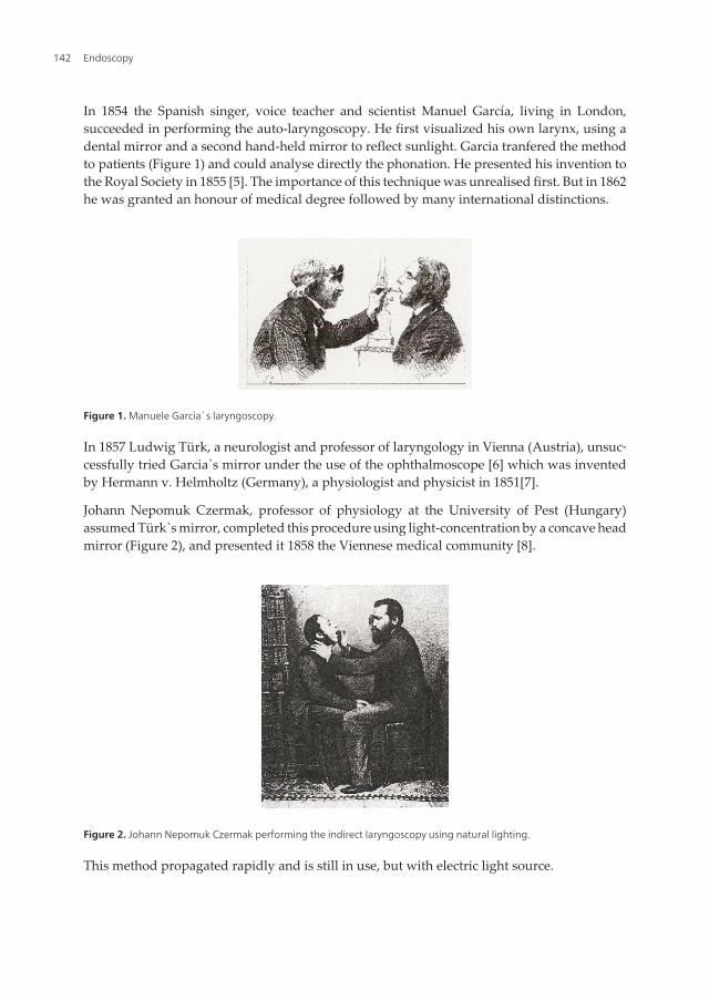
In 1854 the Spanish singer, voice teacher and scientist Manuel García, living in London,succeeded in performing the auto-laryngoscopy. He first visualized his own larynx, using adental mirror and a second hand-held mirror to reflect sunlight. Garcia tranfered the methodto patients (Figure 1) and could analyse directly the phonation. He presented his invention tothe Royal Society in 1855 [5]. The importance of this technique was unrealised first. But in 1862he was granted an honour of medical degree followed by many international distinctions.
Figure 1. Manuele Garcia`s laryngoscopy.
In 1857 Ludwig Türk, a neurologist and professor of laryngology in Vienna (Austria), unsuc‐cessfully tried Garcia`s mirror under the use of the ophthalmoscope [6] which was inventedby Hermann v. Helmholtz (Germany), a physiologist and physicist in 1851[7].
Johann Nepomuk Czermak, professor of physiology at the University of Pest (Hungary)assumed Türk`s mirror, completed this procedure using light-concentration by a concave headmirror (Figure 2), and presented it 1858 the Viennese medical community [8].
Figure 2. Johann Nepomuk Czermak performing the indirect laryngoscopy using natural lighting.
This method propagated rapidly and is still in use, but with electric light source.
Endoscopy142
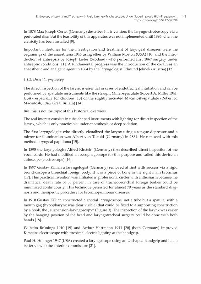
In 1878 Max Joseph Oertel (Germany) describes his invention: the laryngo-stroboscopy via aperforated disc. But the feasibility of this apparatus was not implemented until 1895 when theeletricity has been installed [9].
Important milestones for the investigation and treatment of laryngeal diseases were thebeginnings of the anaesthesia 1846 using ether by William Morton (USA) [10] and the intro‐duction of antisepsis by Joseph Lister (Scotland) who performed first 1867 surgery underantiseptic conditions [11]. A fundamental progress was the introduction of the cocain as ananaesthetic and analgetic agent in 1884 by the laryngologist Edmund Jelinek (Austria) [12].
1.1.2. Direct laryngoscopy
The direct inspection of the larynx is essential in cases of endotracheal intubation and can beperformed by spatulate instruments like the straight Miller-spaculate (Robert A. Miller 1941,USA), especiallly for children [13] or the slightly arcuated Macintosh-spatulate (Robert R.Macintosh, 1943, Great Britain) [14].
But this is not the topic of this historical overview.
The real interest consists in tube-shaped instruments with lighting for direct inspection of thelarynx, which is only practicable under anaesthesia or deep sedation.
The first laryngologist who directly vizualized the larynx using a tongue depressor and amirror for illumination was Albert von Tobold (Germany) in 1864. He removed with thismethod laryngeal papilloma [15].
In 1895 the laryngologist Alfred Kirstein (Germany) first described direct inspection of thevocal cords. He had modified an oesophagoscope for this purpose and called this device anautoscope (electroscope) [16].
In 1897 Gustav Killian a laryngologist (Germany) removed at first with success via a rigidbronchoscope a bronchial foreign body. It was a piece of bone in the right main bronchus[17]. This practical invention was affiliated in professional circles with enthusiasm because thedramatical death rate of 50 percent in case of tracheobronchial foreign bodies could beminimized continuously. This technique persisted for almost 70 years as the standard diag‐nosis and therapeutic procedure for bronchopulmonar diseases.
In 1910 Gustav Killian constructed a special laryngoscope, not a tube but a spatula, with amouth gag (hypopharynx was clear visible) that could be fixed to a supporting constructionby a hook, the „suspension-laryngoscopy” (Figure 3). The inspection of the larynx was easierby the hanging position of the head and laryngotracheal surgery could be done with bothhands [18].
Wilhelm Brünings 1910 [19] and Arthur Hartmann 1911 [20] (both Germany) improvedKirsteins electroscope with proximal electric lighting at the handgrip.
Paul H. Holinger 1947 (USA) created a laryngoscope using an U-shaped handgrip and had abetter view to the anterior commissure [21].
Endoscopy of Larynx and Trachea with Rigid Laryngo-Tracheoscopes Under Superimposed High-Frequency…http://dx.doi.org/10.5772/52996
143

1961 Oskar Kleinsasser (Germany) published [22] his development of a new instrument formagnified endolaryngeal observation and photography. He used a chest holder. A combina‐tion of a wide-angle telescope and a telephoto lens guaranteed excellent depth of field for stilland motion fotography.
In 1970 Geza J. Jako (USA) refered about his development of a laryngosope, a modifiedHolinger-Yankauer tube with an ellipsoidal proximal and a round distal aperture. It is madeof stainless stell with the inside surface matt to decrease glare. On each side of the instrumentthere is a built-in tube for the insertion of fiberoptic light pipes. The laryngoscope with a specialholder is suspended on an instrument table over the patient`s chest and was developed forlaser surgery in the larynx [23].
1970 M. Stuart Strong (USA) coupled the carbon dioxide laser to the surgical microscope. Thelaser provides precise cutting and also haemostasis of small vessels and is manipulated witha joystick and a foot-pedal [24].
In 1979 Hilko Weerda et al. (Germany) designed an expandable laryngoscope which derivedfrom the Kleinsasser-tube and the Killian-Lynch-suspension laryngoscope consisting of anupper and lower blade. Their distance and angle of twist are variable [25].
The rigid laryngotracheal endoscopy made a qualitative leap with the introduction of theHopkins-fibreglass-optics with their unachievable splendour and precision with endoscopicresolutions which allow a hundredfold magnification when used additionally. Prof. Harald HHopkins (Great Britain) developed it the 1960 th with Karl Storz Ltd. Company with limitedliability (Germanny) [26].
1.1.3. Jet ventilation
First attempts to sustain the pulmonary gas exchange without periodical alterations of gasvolumes were done by Franz Volhard in an animal experiment with dogs in 1908 [27]. He
Figure 3. Gustav Killian performing a supension laryngoscopy. The patient`s head is suspended by attaching the lar‐yngoscope to the “gallows”.
Endoscopy144
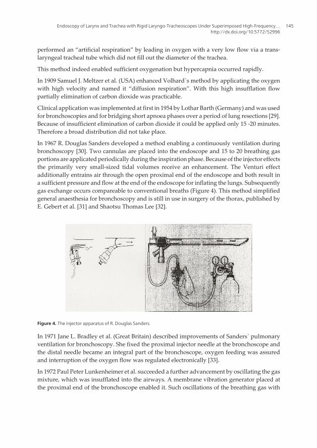
performed an “artificial respiration” by leading in oxygen with a very low flow via a trans‐laryngeal tracheal tube which did not fill out the diameter of the trachea.
This method indeed enabled sufficient oxygenation but hypercapnia occurred rapidly.
In 1909 Samuel J. Meltzer et al. (USA) enhanced Volhard`s method by applicating the oxygenwith high velocity and named it “diffusion respiration”. With this high insufflation flowpartially elimination of carbon dioxide was practicable.
Clinical application was implemented at first in 1954 by Lothar Barth (Germany) and was usedfor bronchoscopies and for bridging short apnoea phases over a period of lung resections [29].Because of insufficient elimination of carbon dioxide it could be applied only 15 -20 minutes.Therefore a broad distribution did not take place.
In 1967 R. Douglas Sanders developed a method enabling a continuously ventilation duringbronchoscopy [30]. Two cannulas are placed into the endoscope and 15 to 20 breathing gasportions are applicated periodically during the inspiration phase. Because of the injector effectsthe primarily very small-sized tidal volumes receive an enhancement. The Venturi effectadditionally entrains air through the open proximal end of the endoscope and both result ina sufficient pressure and flow at the end of the endoscope for inflating the lungs. Subsequentlygas exchange occurs compareable to conventional breaths (Figure 4). This method simplifiedgeneral anaesthesia for bronchoscopy and is still in use in surgery of the thorax, published byE. Gebert et al. [31] and Shaotsu Thomas Lee [32].
Figure 4. The injector apparatus of R. Douglas Sanders.
In 1971 Jane L. Bradley et al. (Great Britain) described improvements of Sanders` pulmonaryventilation for bronchoscopy. She fixed the proximal injector needle at the bronchoscope andthe distal needle became an integral part of the bronchoscope, oxygen feeding was assuredand interruption of the oxygen flow was regulated electronically [33].
In 1972 Paul Peter Lunkenheimer et al. succeeded a further advancement by oscillating the gasmixture, which was insufflated into the airways. A membrane vibration generator placed atthe proximal end of the bronchoscope enabled it. Such oscillations of the breathing gas with
Endoscopy of Larynx and Trachea with Rigid Laryngo-Tracheoscopes Under Superimposed High-Frequency…http://dx.doi.org/10.5772/52996
145
frequences from 1200 to 6000 per minute led in a satisfactory manner to a gas exchange withwidely minimisation of intrathoracic pressure fluctuations [34]. An additional integratedcarbon dioxide absorber increased elimination of carbon doxide [35].
In 1980 Demond J. Bohn et al. published at first a successfully ventilation with this techniquein an animal experiment [36]. With a piston pump sinuoidal vibrations with a frequency from900 to 1500 per minute were tranfered to the breathing gas, elimination of the carbon dioxideis carried out by a cross-flow of fresh mixture. At this way of high frequency oxygenation asuperposition of spontaneous breathing is not feasible.
In 1977 and 1980 Ulf H. Sjöstrand (Sweden) published about his invention of high-frequencypositive pressure ventilation. He modified a conventional lung ventilator by the applicationof special valve configuration and could reduce the compressible gas volume. Tidal volumesfrom 200 to 300 ml with a frequency of 60 to 80 per minute were applicable [37, 38].
Miroslav M. Klain et al. (USA) developed 1976 the high-frequency ventilation. A high pressurejet is conditioned into small-sized single gas quantities by an assessable valve and thenapplicated via a catheter with a low inner diameter. The breathing gas quantities leave thecatheter as a clocked high frequency jet. This jet activates a suction mechanism (entrainment)and the tidal volume is augmented. In a frequency range of 100 to 200 per minute a sufficientgas exchange can be perpetuated in a completely open system towards the atmosphere [39-41].
This high frequency jet ventilation is the initial point of all following developments.
In particular Alexander Aloy and colleagues are engaged practically and scientifically since1990 over more than twenty years in this field of ventilation [42].
His and contemporarely applicatioins and publications are presented in the next followingparts of this chapter.
2. Microlaryngoscopy and endolaryngeal microsurgery
Direct laryngoscopy via the transoral route allows the immediate entry to the inner laryngealand tracheal structures and has now an extreme importance for the special field of laryngo‐logical diagnoses and surgical treatments in the otorhinolaryngologic discipline.
A laryngoscope holder is adhered to the chest selfholding by a mount as a supension similarto a gallow.
The binoccular vision and bi-manual manipualtion via the forward spaced microscopyfacilitates detailed diagnoses and treatment at the vocal cords and circumjacent areas [43-45].
2.1. Indications
Microlaryngocopy is particulary helpful in the need of diagnosis and treatment of the vocalchords or surrounding areas, which are first seen by indirect laryngoscopy. The microlar‐yngoscopy for staging and excision biopsy of tumours is part of a panendoscopy of the upper
Endoscopy146
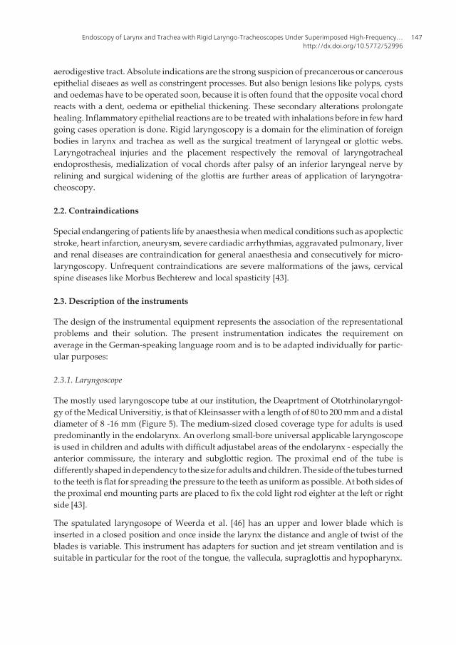
aerodigestive tract. Absolute indications are the strong suspicion of precancerous or cancerousepithelial diseaes as well as constringent processes. But also benign lesions like polyps, cystsand oedemas have to be operated soon, because it is often found that the opposite vocal chordreacts with a dent, oedema or epithelial thickening. These secondary alterations prolongatehealing. Inflammatory epithelial reactions are to be treated with inhalations before in few hardgoing cases operation is done. Rigid laryngoscopy is a domain for the elimination of foreignbodies in larynx and trachea as well as the surgical treatment of laryngeal or glottic webs.Laryngotracheal injuries and the placement respectively the removal of laryngotrachealendoprosthesis, medialization of vocal chords after palsy of an inferior laryngeal nerve byrelining and surgical widening of the glottis are further areas of application of laryngotra‐cheoscopy.
2.2. Contraindications
Special endangering of patients life by anaesthesia when medical conditions such as apoplecticstroke, heart infarction, aneurysm, severe cardiadic arrhythmias, aggravated pulmonary, liverand renal diseases are contraindication for general anaesthesia and consecutively for micro‐laryngoscopy. Unfrequent contraindications are severe malformations of the jaws, cervicalspine diseases like Morbus Bechterew and local spasticity [43].
2.3. Description of the instruments
The design of the instrumental equipment represents the association of the representationalproblems and their solution. The present instrumentation indicates the requirement onaverage in the German-speaking language room and is to be adapted individually for partic‐ular purposes:
2.3.1. Laryngoscope
The mostly used laryngoscope tube at our institution, the Deaprtment of Ototrhinolaryngol‐gy of the Medical Universitiy, is that of Kleinsasser with a length of of 80 to 200 mm and a distaldiameter of 8 -16 mm (Figure 5). The medium-sized closed coverage type for adults is usedpredominantly in the endolarynx. An overlong small-bore universal applicable laryngoscopeis used in children and adults with difficult adjustabel areas of the endolarynx - especially theanterior commissure, the interary and subglottic region. The proximal end of the tube isdifferently shaped in dependency to the size for adults and children. The side of the tubes turnedto the teeth is flat for spreading the pressure to the teeth as uniform as possible. At both sides ofthe proximal end mounting parts are placed to fix the cold light rod eighter at the left or rightside [43].
The spatulated laryngosope of Weerda et al. [46] has an upper and lower blade which isinserted in a closed position and once inside the larynx the distance and angle of twist of theblades is variable. This instrument has adapters for suction and jet stream ventilation and issuitable in particular for the root of the tongue, the vallecula, supraglottis and hypopharynx.
Endoscopy of Larynx and Trachea with Rigid Laryngo-Tracheoscopes Under Superimposed High-Frequency…http://dx.doi.org/10.5772/52996
147
When laser is used the tubes and spatulas are adapted according to the needs. The inner surfacecoating should be non-reflecting, opal or black. On the lateral outside cannulas for suction ofsmoke are mounted.
2.3.2. Laryngoscope holder
The laryngoscope holder is eighter placed on patient`s chest with a broad rubber ring or it isput on a robust instrument table [43, 47] respectively a holding bow [48] just over his chest toavoid pressure on the anaesthetized chest and to minimize movement of the instrument.
2.3.3. Example for a configuration of a microscope combining modern vizualisation technologies witha user friendly platform
1. Light source:
• 2 x 300 W xenon,
• automatic iris control for adjusting the illumunation to the field of view,
• individual light treshhold setting,
• focus light link: working distance controlled light intensity,
• display of remaining lamp life time.
2. Microscope:
• free to move at a tripod or as a ceiling mounted,
• motorized focus,
• working distance 200-500 mm,
• motorized zoom 1:6 zoom ratio,
• 10 x magnetic widefield eyepieces with integrated eyecups,
Figure 5. The Kleinsasser laryngoscope already adapted for jet ventilation (see also part 3 of this chapter).
Endoscopy148

• autofocus with 2 visible laser dots - automatic mode with magnetic brakes,
• binocular observer ocular,
• multifunctional programmable handgrips,
• magnetic clutches for all system axes,
• central user interface,
• XY robotic movement in 3 axes (variable speed),
• system setup: autobalance,
• navigation interface,
• interface for micromanipulator,
• intraoperative fluorescence,
• laseradapter.
3. Video, fotography, Television for decumentation and teaching:
• HD video touchscreen on extended arm,
• integrated video still image capturing on HDD and USB-media,
• integrated HD video camera,
• integrated SD or HD video recording and editing,
• adapatation of consumer (SLR) photo/video camera,
• video-in for external SD video sources,
• patients data transfer from/to PACS.
2.3.4 Instruments
Instruments are small at the tip, thin, long (25 cm) and flexible.
All instruments should be manipulated freehanded, which is easily trainable and backing ofthe arms becomes dispensable. The set of microsurgical instruments should be kept in a basicamount. It includes double-spoon forceps, scissors: straight, laterally and upwards arcuated,acutenaculum, sickle and peeling knifes, suction tubes and coagulation probes.
Instruments for laser microlaryngological surgery are of nonreflecting surface. Protectors areneccessary for the subglottic area when tubeless ventilation is applicated. Additional when thevocal chords are treated the false cords and also the contralateral intact side is to be movedsidewards.
2.4. Beginning and protection activities
The often “poor risk” patients need preoperatively a careful investigation and treatment tomake them capable for anaesthesia. Dental record is essential before insertion of the laryngo‐
Endoscopy of Larynx and Trachea with Rigid Laryngo-Tracheoscopes Under Superimposed High-Frequency…http://dx.doi.org/10.5772/52996
149
scope. When indicated patients have to go for remediation preoperatively. Dental impressiontrays equally spread the pressure of the laryngoscope at the teeth of the upper jaw and canbridge a dental gap.
Patients lie in dorsal position on the operating table and the correct posture of the head is thedorsal flection in flat bedding. Using adequate laryngoscope types it enables even in patientswith a thick and short neck to adjust the larynx. If there are anamnestic indications concerningpathologies at the cervicale spin an orthopaedic specialist has to decide if the head positionduring laryngoscopy will be tolerated well.
In case of laser application all persons in the operating room have to wear special glasses. WhenCO2 laser is in use eyeglass lenses are sufficient. Patients face and eyes are to be protected bya humid green woven fabric. The doors to the operating room where laser is used must besignalized.
2.5. Anaesthesia and ventilation during micro laryngotracheoscopy (basic considerations)
Please see the next part of this chapter.
2.6. Insertion of the laryngoscope
First of all a teeth protector for the lower and upper jaw is placed in position. The insertion ofthe laryngoscope should be performed not until the patient is full relaxed and in sufficientdepth of narcosis. The laryngoscope should be as large as possible enabling best lighting andoverview of the larynx. In cases of preexisting laryngotracheal intubation the ventilation tubeis moved by two fingers to the left side and the laryngoscope is inserted from the right sideunder the illumination of a cold light which is piped inside of the tube to the distal end. Thetongue should not be pinched between the teeth and the laryngoscope at the same time. Theepiglottis is loaded up by the laryngoscope. If the laryngeal skeleton is to be pressed fromoutside to inward in cases of the need to expose the anterior commissure the laryngoscopeshould be inserted only into the area of the false cords. In cases of a small, weak or U-shapedepiglottis it can happen that the epiglottis is enrolled and the suprahyoidal part is folded andcompressed when the laryngoscope is inserted. Consecutively inspection of the anteriorcommissure is hindered and a postoperative oedema can occur. Solving of this problem is toclamp the epiglottis when inserting the laryngoscope. The laryngoscope is positioned fewmillimeters above the anterior commissure. The dorsal lying ventilation tube can be used as aguidance till the exposition of the false and vocal chords.
Next is to underpin the laryngoscope at the mobile laryngoscopy-table and to move it slowlyupwards. When a distending laryngoscope is inserted at maximum aperture laterallybetween the spatulas parts of the tongue can be pinched and it protrudes into the lumenof the laryngoscope. This can be prevented by covering the closed distending-laryngo‐scope with a finger of a medical glove which is forming a lateral wall when the laryngo‐scope is opened [49].
Endoscopy150
2.7. Inspection of the larynx and the circumjacent areas
At the begin of the introduction of the laryngocope the structures are inspected by lookingwith the naked eyes using the lighting inside the laryngoscope: base of the tongue,valleculae, epiglottis: lingual and laryngeal surface area and free margin, aryepiglottic fold,pharyngoepiglottic fold, the processus vocalis and tubercula cuneiformia respectivelycorniculata, the arytaenoid cartilage, the sinus piriformis bilateral, the posterior hypophar‐yngeal wall and postcricoid area. After fixation of the laryngoscope under the use of themicroscope the false and vocal chord, the sinus Morgagni, the anterior and posteriorcommissure as well as the subglottic space and the upper parts of the trachea are to beseen under a magnification of up to 40.
2.8. Microlaryngoscopic diagnoses and surgical therapies
(most frequent clinical symptom is hoarseness)
2.8.1. Tumours
Diagnosis and therapy of precancerous lesions and carcinomas of the larynx, especially thevocal chords are still a central domain of laryngoscopy. Any unclear or suspicious epithelialalteration is to be exstirpated and examined by histology. As followup exam after radiotherapymicrolaryngoscopy is qualified in particular.
2.8.1.1. Precancerous lesions
Chronic hyperplastic laryngitis is an epithelial thickening of laryngeal mucous membrane affectingboth vocal chords with a spread over their complete length. In this dermatoid epithelium oftenleukoplakia lies as milky area, slightly opacity of the surface or as verrucous, thick, white coating.A second group of cornification of the epithelium is the circumscript keratosis which is locatednearly only at one vocal chord. The surface can be plain, tubercular, verrucous or papillary withall transitions. Histological differentiation of the keratosisis is for all benign types is grade I,when few atypic cells are seen grade II and when a carcinoma in situ is diagnosed grade III. Acarcinoma in situ becomes in a high rate an apparent carcinoma and is to be treated as such amalignant tumour. The human papilloma virus induced solitary hyperceratotic “adult”papillo‐ma has a high rate of recurrence after surgery and is to be distinguished from the “juvenile”papilloma which also occurs in adults but never turns to a malignization. Precancerous lesionsare always movable over the muscle of the vocal chord without problems.
2.8.1.2. Squamous scell carcinomas
Carcimomas in situ and apparent carcinomas primarily originate bilateral from an enlargedarea and are named wallpaper carcimomas respectively superficial spreading carcinomas.Additionally, not infrequently, multiple focuses of seperated carcinomas confluence later.Difficult relocatability, solid or swallen areas and superficial exulceration are important signsof deeper invasion. A relatively rare shape of growth is excessive polypus/exophytic like.Because of tumour infiltration the blood supply suffers and necroses occur. To identify the
Endoscopy of Larynx and Trachea with Rigid Laryngo-Tracheoscopes Under Superimposed High-Frequency…http://dx.doi.org/10.5772/52996
151
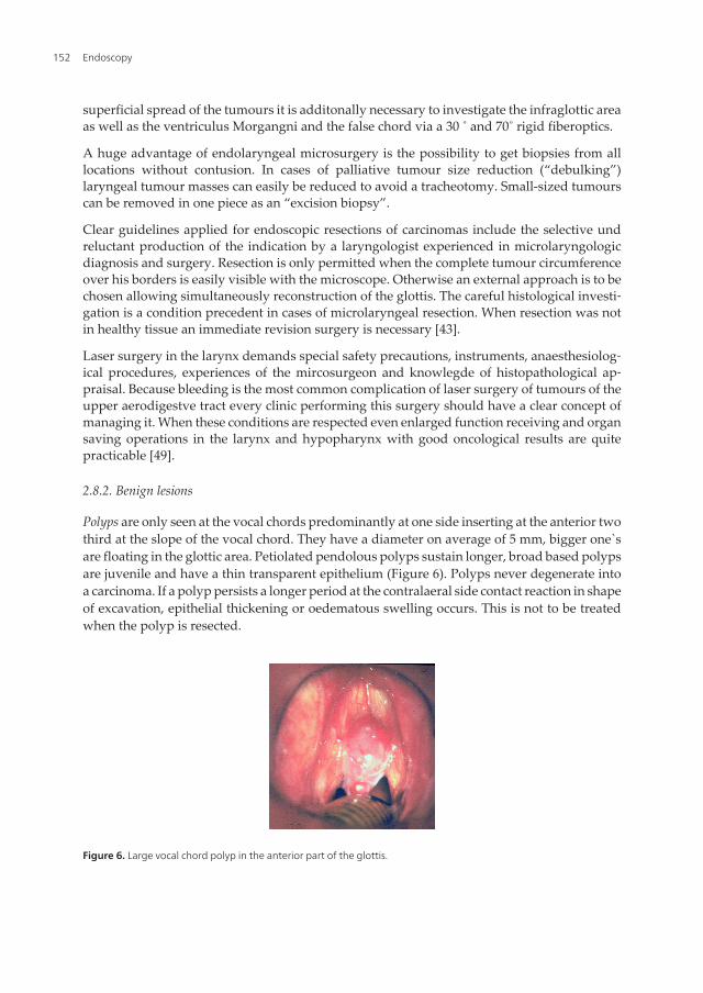
superficial spread of the tumours it is additonally necessary to investigate the infraglottic areaas well as the ventriculus Morgangni and the false chord via a 30 ˚ and 70˚ rigid fiberoptics.
A huge advantage of endolaryngeal microsurgery is the possibility to get biopsies from alllocations without contusion. In cases of palliative tumour size reduction (“debulking”)laryngeal tumour masses can easily be reduced to avoid a tracheotomy. Small-sized tumourscan be removed in one piece as an “excision biopsy”.
Clear guidelines applied for endoscopic resections of carcinomas include the selective undreluctant production of the indication by a laryngologist experienced in microlaryngologicdiagnosis and surgery. Resection is only permitted when the complete tumour circumferenceover his borders is easily visible with the microscope. Otherwise an external approach is to bechosen allowing simultaneously reconstruction of the glottis. The careful histological investi‐gation is a condition precedent in cases of microlaryngeal resection. When resection was notin healthy tissue an immediate revision surgery is necessary [43].
Laser surgery in the larynx demands special safety precautions, instruments, anaesthesiolog‐ical procedures, experiences of the mircosurgeon and knowlegde of histopathological ap‐praisal. Because bleeding is the most common complication of laser surgery of tumours of theupper aerodigestve tract every clinic performing this surgery should have a clear concept ofmanaging it. When these conditions are respected even enlarged function receiving and organsaving operations in the larynx and hypopharynx with good oncological results are quitepracticable [49].
2.8.2. Benign lesions
Polyps are only seen at the vocal chords predominantly at one side inserting at the anterior twothird at the slope of the vocal chord. They have a diameter on average of 5 mm, bigger one`sare floating in the glottic area. Petiolated pendolous polyps sustain longer, broad based polypsare juvenile and have a thin transparent epithelium (Figure 6). Polyps never degenerate intoa carcinoma. If a polyp persists a longer period at the contralaeral side contact reaction in shapeof excavation, epithelial thickening or oedematous swelling occurs. This is not to be treatedwhen the polyp is resected.
Figure 6. Large vocal chord polyp in the anterior part of the glottis.
Endoscopy152
Vocal chords nodules are seen exclusively in women with powerful voice formation. Theypresent as a symmetric swelling of the epithelium of the vocal chords in typical localisation atthe border between the first and second third. In up to 50 % the diagnosis of nodules by indirectlaryngoscopy is wrong – microlaryngoscopy shows cysts or polyps. Morphodiffentiationcompared to polyps is difficult but in any case they are no fibroepithelioma. Microlaryngealresection and postoperative phonatric training is indicated.
In hyperactive children vocal nodes impress as soft spindling swelling in the middle of bothvocal chords. Operative resection is not necessary because by no longer than the puberty theyinvolute spontaneously. Phoniatric therapy is useful. Laryngeal papillomas are to be excluded.
The varix chordis is found in patients with vocal overstressing at the surface or margin in themiddle or posterior part of one vocal chord. Under microscpy one or various capillaries leadto a bloodblaster. Laser coagulation followed by vocal training is a promising therapy.
Laryngeal cysts are frequent and located typically at three areas:
1. vocal chord cysts are with about 50 % the most frequent and in most cases as one chamberat the subglottic slope of the anterior third of a vocal chord with a diameter of about 5mm. Their inner lining is squamous epithelium. The content matter is a muddy aqueous-milky detritus. Under microscopy the subepithelial content glimmers yellowish. They areremoved gentle under protection of the muscle layer.
2. vestibular fold cysts are found nearly exclusively in elderly patients. The first mode,originating from the minor salivary glands, presents with one chamber and is peduncu‐lated at the roof of the ventriculus laryngis (Morgagni) with a prolapse into the glottis.They are neither a real prolapse nor an inner laryngocele and can be mutated oncocytoid,which is a benign age-related metaplastic dyschylic transformation. They can be removedeasily by cutting through the peduncle. The second mode is characterized by multiplechambers, often bilateral, inside the vestibular fold and metaplastic transformed epithe‐lium. The excision is hindered by the deep location and the botryoid extension, whichpromotes relapses.
3. epiglottic cysts are located always at the same position, deep in the vallecula epiglotticaslightly paramedian at the lingual surface of the epiglottis. In ten percent they occurmultiple, only the bigger one cause dysphagia and have to be operated. This can becomplicated by prominent venes. The inner layer of the wall of the cysts is squamousepithelium enclosing a milky pale yellow fluid. The complete sac of the cyst is to beremoved.
The Reinke-oedema or polypoid chorditis is a frequent disease mostly in heavy smokers. Theextention ranges from marginal spindle shaped swelling to bulky floating bulges with stridor.In most cases both vocal chords are involved, but asymmetrically. With longer persistence ofthe Reinke-oedema the epithelium keratinizes. Patients develop a compensatory phonation bythe vestibular folds which are to be reversed by postoperative voice therapy. Surgery beginswith plain and clean cutted margins at the tip of the processus vocalis of the arytaenoidcartilage where the oedema is pronounced. The anteroir commissure is not involved and an
Endoscopy of Larynx and Trachea with Rigid Laryngo-Tracheoscopes Under Superimposed High-Frequency…http://dx.doi.org/10.5772/52996
153
epithelial debridment there should be prevented to avoid synechias. The subepthelial mucousis to be sucked carefully.
Chronical hyperplastic laryngitis is predominantly seen in 90 percent in heavy smoking men.Additional working place associated noxious agents as heat, dust and noice-induced overuseof the voice are said to be at least co-factors. Microlaryngoscopic findings are typical. Thethickening of the epithelium begins always at the anterior third of the vocal chords and spreadsto the whole vocal chords and inner larynx. In advanced cases of expanse leucoplakia and apathological secretion of a yellow viscous mucous adheres. Sometimes the disease progressesper acute exacerbation. Signs of this procedure are slit shaped ulcers at the free margin of thevocal chords and crimson bulges of oedema at the border from squamous to cylindricalepithelium. Carcinoma can follow. Resection of nearly all hyperplastic area via microlayngo‐scope is the symptomatic therapy of choice. It lasts about 4 to 8 weeks till re-epithelisation ofa vocal chord is finished. In this period a stingent ban on smoking is to order.
Contact granuloma and contact pachydermia are two pathogenetic identical but independentdiseases caused by vocal abuse and strike of the processus vocales against each other withfollowing formation of granulation tissue. This can be connected with dysphagia, odynopha‐gia, scratching, sensation of foreign body and haemoptysis. Sometimes granulomas arerejected spontaneously. In other cases the granuloma persists over months and years whichare forming bilateral cranial and caudal from the processus vocalis lip-like bulges (in Americathe Jackson`s “contact ulcer”, which is not realy an ulcer - there is no loss of substance) anddash against each other during phonation. All transitions to the typical dish-shaped epithe‐liazed contact pachyderms are seen. Despite careful microsurgical resection and postoperat‐vive voice training local recurrences frequently arise.
Intubation granulomata are caused by too large sized larynotracheal tubes which excoriate theprocessus vocales almost one-sided (Figure 7). Depending on their growth they get a pendu‐alted form and can be rejected and expectorated spantaneously. Recurrences after resectionare seen not infrequently, additionally applicaion of cortison should minimize them.
Figure 7. Intubation granuloma in the posterior third of the right vocal chord.
“Juvenile” papillomata are to be revoved repeatedly because of their tendency of frequentrecurrences: optionally by cold instruments, laser, ultrasound, cryocautery or electrocoagula‐tion. Additional mitomycin is applied. It is of importance to work organ preserving. The
Endoscopy154
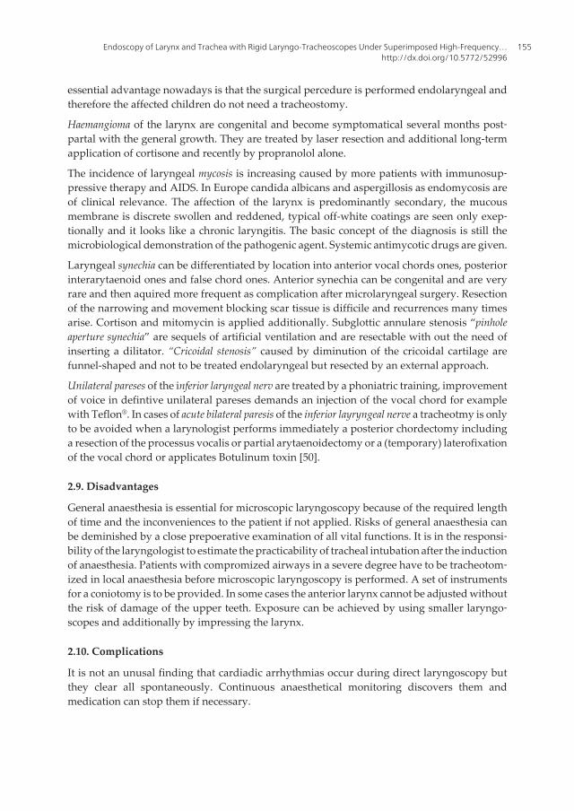
essential advantage nowadays is that the surgical percedure is performed endolaryngeal andtherefore the affected children do not need a tracheostomy.
Haemangioma of the larynx are congenital and become symptomatical several months post‐partal with the general growth. They are treated by laser resection and additional long-termapplication of cortisone and recently by propranolol alone.
The incidence of laryngeal mycosis is increasing caused by more patients with immunosup‐pressive therapy and AIDS. In Europe candida albicans and aspergillosis as endomycosis areof clinical relevance. The affection of the larynx is predominantly secondary, the mucousmembrane is discrete swollen and reddened, typical off-white coatings are seen only exep‐tionally and it looks like a chronic laryngitis. The basic concept of the diagnosis is still themicrobiological demonstration of the pathogenic agent. Systemic antimycotic drugs are given.
Laryngeal synechia can be differentiated by location into anterior vocal chords ones, posteriorinterarytaenoid ones and false chord ones. Anterior synechia can be congenital and are veryrare and then aquired more frequent as complication after microlaryngeal surgery. Resectionof the narrowing and movement blocking scar tissue is difficile and recurrences many timesarise. Cortison and mitomycin is applied additionally. Subglottic annulare stenosis “pinholeaperture synechia” are sequels of artificial ventilation and are resectable with out the need ofinserting a dilitator. “Cricoidal stenosis” caused by diminution of the cricoidal cartilage arefunnel-shaped and not to be treated endolaryngeal but resected by an external approach.
Unilateral pareses of the inferior laryngeal nerv are treated by a phoniatric training, improvementof voice in defintive unilateral pareses demands an injection of the vocal chord for examplewith Teflon®. In cases of acute bilateral paresis of the inferior layryngeal nerve a tracheotmy is onlyto be avoided when a larynologist performs immediately a posterior chordectomy includinga resection of the processus vocalis or partial arytaenoidectomy or a (temporary) laterofixationof the vocal chord or applicates Botulinum toxin [50].
2.9. Disadvantages
General anaesthesia is essential for microscopic laryngoscopy because of the required lengthof time and the inconveniences to the patient if not applied. Risks of general anaesthesia canbe deminished by a close prepoerative examination of all vital functions. It is in the responsi‐bility of the laryngologist to estimate the practicability of tracheal intubation after the inductionof anaesthesia. Patients with compromized airways in a severe degree have to be tracheotom‐ized in local anaesthesia before microscopic laryngoscopy is performed. A set of instrumentsfor a coniotomy is to be provided. In some cases the anterior larynx cannot be adjusted withoutthe risk of damage of the upper teeth. Exposure can be achieved by using smaller laryngo‐scopes and additionally by impressing the larynx.
2.10. Complications
It is not an unusal finding that cardiadic arrhythmias occur during direct laryngoscopy butthey clear all spontaneously. Continuous anaesthetical monitoring discovers them andmedication can stop them if necessary.
Endoscopy of Larynx and Trachea with Rigid Laryngo-Tracheoscopes Under Superimposed High-Frequency…http://dx.doi.org/10.5772/52996
155
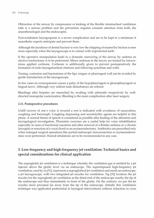
Obtruction of the airway by compression or kinking of the flexible intratracheal ventilationtube is a serious problem and the prevention requires constant attention from both, theanaesthesiologist and the endoscopist.
Post-extubation laryngospasm is a severe complication and are to be kept to a minimum ifanaesthetic experts anticipate and prevent them.
Although the incidence of dental fracture is very low the chipping of enamel by friction is seenmore especially when the laryngoscope is in contact with unprotected teeth.
If the operative manipulation leads to a dramatic narrowing of the airway by oedema anelective tracheotomy is to be performed. Minor oedemas in the larynx are treated by intrave‐nious applied cortisone. Cortisone is additionally given to prevent postoperatively theformation of endo-laryngotracheal citatrices and following synechiae and webs.
Tearing, contusion and haematoma of the lips, tongue or pharyngeal wall can be avoided bygentle introduction of the laryngoscope.
In few cases an overexpansion causes a palsy of the hypopharyngeal or glossopharyngeal orlingual nerve. Although very seldom taste disturbances are refered.
Bleedings after biopsies are staunched by swabing with adrenalin resepctively by well-directed monopolar cauterization. Bleeding is the main complication after laser surgery.
2.11. Postoperative procedures
Untill recover of one`s voice is recured a rest is indicated with avoidance of susurration,coughing and harrumph. Coughing depressing and secretolythic agents are helpful in thisphase. A normal theme of speech is considered as possible after healing of the alteration andlaryngological investigation. Phoniatric exercises are a useful help for voice rehabilitationespecially in cases of functional causation and after removal of a Reinke-oedema or a chroniclaryngitis or resection of a vocal chord or an arytaenoidectomy. Antibiotics are prescribed onlywhen enlarged surgical operations like partial endoscopic tumorresection or arytaenoidecto‐mies were performed. Humid inhalations are to be recommended in any case.
3. Low-frequency and high-frequency jet ventilation: Technical basics andspecial considerations for clinical application
The supraglottic jet ventilation is a technique whereby the ventilation gas is emitted by a jetinjector above the glottic level via an endoscope. The superimposed high-frequency jetventilation, used by us [51], represents a supraglottical jet ventilation and needs an endoscope,a jet laryngoscope, with two integrated jet nozzles for ventilation. Ng [52] localizes the jetnozzles for the supraglottic jet ventilation at the distal end of the endoscope nearby the tip ofthe endoscope and thus immediately in front of the glottis. On the contrary we put the jetnozzles more proximal far away from the tip of the endoscope. Initially this ventilationtechnique was applicated preferential in laryngeal interventions without reduction in cross
Endoscopy156
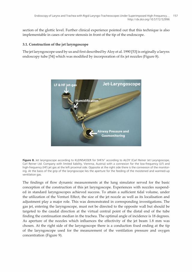
section of the glottic level. Further clinical experience pointed out that this technique is alsoimplementable in cases of severe stenosis in front of the tip of the endoscope.
3.1. Construction of the jet laryngoscope
The jet laryngoscope used by us and first described by Aloy et al. 1990 [53] is originally a larynxendoscopy tube [54] which was modified by incorporation of fix jet nozzles (Figure 8).
Figure 8. Jet laryngoscope according to KLEINSASSER for SHFJV® according to ALOY (Carl Reiner Jet Laryngoscope,Carl Reiner Ltd. Company with limited liability, Viennna, Austria) with a connexion for the low-frequency (LF) andhigh-frequency (HF) jet gas at the left proximal side. Opposite at the right side there is the connexion of the monitor‐ing. At the basis of the grip of the laryngoscope lies the aperture for the feeding of the moistened and warmed-upventilation gas.
The findings of flow dynamic measurements at the lung simulator served for the basicconception of the construction of this jet laryngoscope. Experiences with nozzles suspend‐ed in standard laryngoscopes achieved success. To attain a sufficient tidal volume, underthe utilization of the Venturi Effect, the size of the jet nozzle as well as its localisation andadjustment play a major role. This was demonstrated in corresponding investigations. Thegas jet, entering the laryngoscope, must not be directed to the opposite wall but should betargeted to the caudal direction at the virtual central point of the distal end of the tubefinding the continuation median in the trachea. The optimal angle of incidence is 18 degrees.As aperture of the nozzles which influences the effectivity of the jet beam 1.8 mm waschosen. At the right side of the laryngoscope there is a conduction fixed ending at the tipof the laryngoscope used for the measurement of the ventilation pressure and oxygenconcentration (Figure 9).
Endoscopy of Larynx and Trachea with Rigid Laryngo-Tracheoscopes Under Superimposed High-Frequency…http://dx.doi.org/10.5772/52996
157
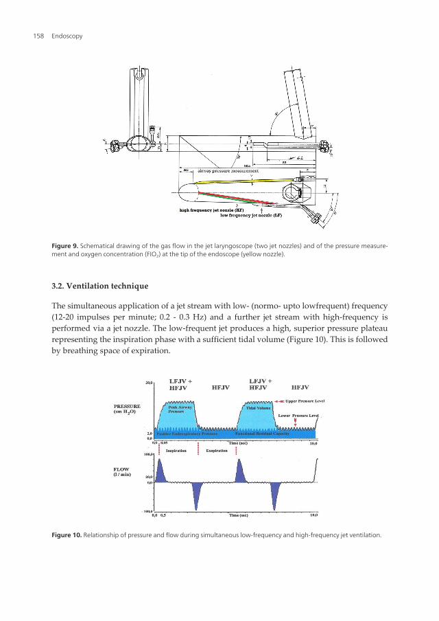
3.2. Ventilation technique
The simultaneous application of a jet stream with low- (normo- upto lowfrequent) frequency(12-20 impulses per minute; 0.2 - 0.3 Hz) and a further jet stream with high-frequency isperformed via a jet nozzle. The low-frequent jet produces a high, superior pressure plateaurepresenting the inspiration phase with a sufficient tidal volume (Figure 10). This is followedby breathing space of expiration.
Figure 10. Relationship of pressure and flow during simultaneous low-frequency and high-frequency jet ventilation.
Figure 9. Schematical drawing of the gas flow in the jet laryngoscope (two jet nozzles) and of the pressure measure‐ment and oxygen concentration (FIO2) at the tip of the endoscope (yellow nozzle).
Endoscopy158

On the other hand via a second nozzle during the low-frequent inspiration and the subsequentexpiration a continous high-frequent gas application is conducted and is additionally super‐imposed to the low-frequent jet ventilation causing a minimal enhancement of the inspiratorypressure plateau. In the expiration sphase of the low-frequent jet ventilation there exists onlythe high-frequent jet. The high-frequent jet portion (frequency from 20 to1500 impulses perminute; 0.3 to 25 Hz) produces a lower pressure plateau corresponding to an end-espiratorypressure plateau, corresponding to an end-expiratory pressure (PEEP).
3.3. Pressure profile in the lung
Ventilation via a complete open system with two different pressure plateaus is generated.
The periods of inspration and exspiration of the normo-frequent as well as the high-frequentjet gases are to be adjusted variable. It is a time- and pressure controlled ventilation at two pressureplateaus with decelerating flow.
3.4. Adjustment of the respirator
Variable parameter of the low-frequency jet ventilation:
Working pressure of the gas leaving the jet nozzle: 0.03-0.04 bar/kg body weight.
Frequency: 12-20 impulses/min (adults), 20-30 impulses/min (children).
Inspiration/Exspiration ratio: primary 1:2 or 1:1.
Variable parameter of the high-frequency jet ventilation:
Working pressure of the gas leaving the jet nozzle: 0.02 bar/kg body weight.
Frequency variable: 100-1500 impulses/min.
Inspiration/Exspiration ratio: primary 1:2 or 1:1.
3.5. Respirator
The development of the jet ventilation with two jet streams made it mandatory to designa specific respirator (Figure 11). An electronic respirator enabling the low- and high-frequent gas application was developed. Simultaneously the pinside of the endoscopemeasured ventilation pressures are digital and graphically represented, just as the adjust‐ed and measured FIO2. Furthermore a laser mode is practicable reducing automatically theFIO2. The ventilation parameters are recorded and an integrated pressure limitation reactsto a too high pressure and also to a pressure drop. Therefore a barotrauma can be avoidedwith greatest certainty. After the input of patients body weight the ventilation is startedwith a default setting of the device. The connection with the jet laryngoscope is made bytwo not confusable jet hose couplings. Moreover the respirator includes different usableventilation modes for the application of bronchoscopy and infraglottic one-lumen cathetertechniques.
Endoscopy of Larynx and Trachea with Rigid Laryngo-Tracheoscopes Under Superimposed High-Frequency…http://dx.doi.org/10.5772/52996
159
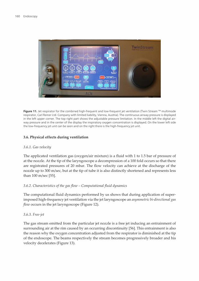
Figure 11. Jet respirator for the combined high-frequent and low-frequent jet ventilation (Twin Stream TM multimoderespirator, Carl Reiner Ltd. Company with limited liability, Vienna, Austria). The continuous airway pressure is displayedin the left upper corner. The top right part shows the adjustable pressure limitation. In the middle left the digital air‐way pressure and in the center of the display the inspiratory oxygen concentration is displayed. On the lower left sidethe low-frequency jet unit can be seen and on the right there is the high-frequency jet unit.
3.6. Physical effects during ventilation
3.6.1. Gas velocity
The applicated ventilation gas (oxygen/air mixture) is a fluid with 1 to 1.5 bar of pressure ofat the nozzle. At the tip of the laryngoscope a decompression of a 100 fold occurs so that thereare registrated pressures of 20 mbar. The flow velocity can achieve at the discharge of thenozzle up to 300 m/sec, but at the tip of tube it is also distinctly shortened and represents lessthan 100 m/sec [55].
3.6.2. Characteristics of the gas flow – Computational fluid dynamics
The computational fluid dynamics performed by us shows that during application of super‐imposed high-frequency jet ventilation via the jet laryngoscope an asymmetric bi-directional gasflow occurs in the jet laryngoscope (Figure 12).
3.6.3. Free-jet
The gas stream emitted from the particular jet nozzle is a free jet inducing an entrainment ofsurrounding air at the rim caused by an occurring discontinuity [56]. This entrainment is alsothe reason why the oxygen concentration adjusted from the respirator is diminished at the tipof the endoscope. The beams respectively the stream becomes progressively broader and hisvelocity decelerates (Figure 13).
Endoscopy160
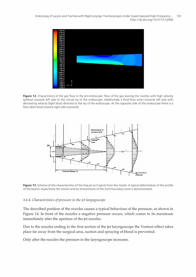
Figure 13. Schema of the characteristics of the free jet as it ejects from the nozzle. A typical deformation of the profileof the beams respectively the stream and an entrainment of the (rim) boundary zone is demonstrated.
3.6.4. Characteristics of pressure in the jet laryngoscope
The described position of the nozzles causes a typical behaviour of the pressure, as shown inFigure 14. In front of the nozzles a negative pressure occurs, which comes to its maximumimmediately after the aperture of the jet nozzles.
Due to the nozzles ending in the first section of the jet laryngoscope the Venturi–effect takesplace far away from the surgical area, suction and spraying of blood is prevented.
Only after the nozzles the pressure in the laryngoscope increases.
Figure 12. Charactristics of the gas flow in the jet endoscope: flow of the gas leaving the nozzles with high velocity(yellow) towards left side to the virtual tip of the endoscope. Additionally a fluid flow exists towards left side withdecreasing velocity (light blue) directed to the tip of the endoscope. At the opposite side of the endoscope there is aflow (dark blue) toward right side outwards.
Endoscopy of Larynx and Trachea with Rigid Laryngo-Tracheoscopes Under Superimposed High-Frequency…http://dx.doi.org/10.5772/52996
161
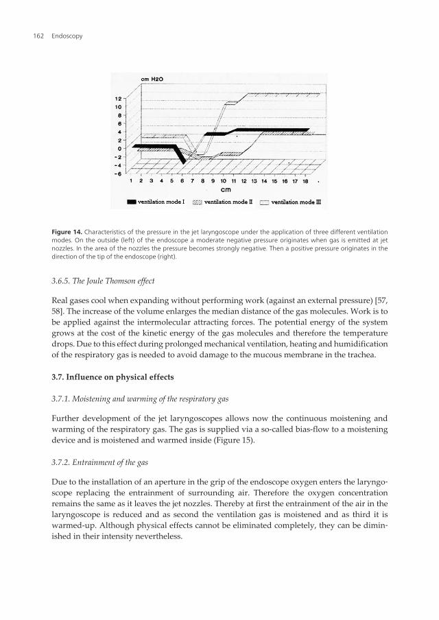
Figure 14. Characteristics of the pressure in the jet laryngoscope under the application of three different ventilationmodes. On the outside (left) of the endoscope a moderate negative pressure originates when gas is emitted at jetnozzles. In the area of the nozzles the pressure becomes strongly negative. Then a positive pressure originates in thedirection of the tip of the endoscope (right).
3.6.5. The Joule Thomson effect
Real gases cool when expanding without performing work (against an external pressure) [57,58]. The increase of the volume enlarges the median distance of the gas molecules. Work is tobe applied against the intermolecular attracting forces. The potential energy of the systemgrows at the cost of the kinetic energy of the gas molecules and therefore the temperaturedrops. Due to this effect during prolonged mechanical ventilation, heating and humidificationof the respiratory gas is needed to avoid damage to the mucous membrane in the trachea.
3.7. Influence on physical effects
3.7.1. Moistening and warming of the respiratory gas
Further development of the jet laryngoscopes allows now the continuous moistening andwarming of the respiratory gas. The gas is supplied via a so-called bias-flow to a moisteningdevice and is moistened and warmed inside (Figure 15).
3.7.2. Entrainment of the gas
Due to the installation of an aperture in the grip of the endoscope oxygen enters the laryngo‐scope replacing the entrainment of surrounding air. Therefore the oxygen concentrationremains the same as it leaves the jet nozzles. Thereby at first the entrainment of the air in thelaryngoscope is reduced and as second the ventilation gas is moistened and as third it iswarmed-up. Although physical effects cannot be eliminated completely, they can be dimin‐ished in their intensity nevertheless.
Endoscopy162
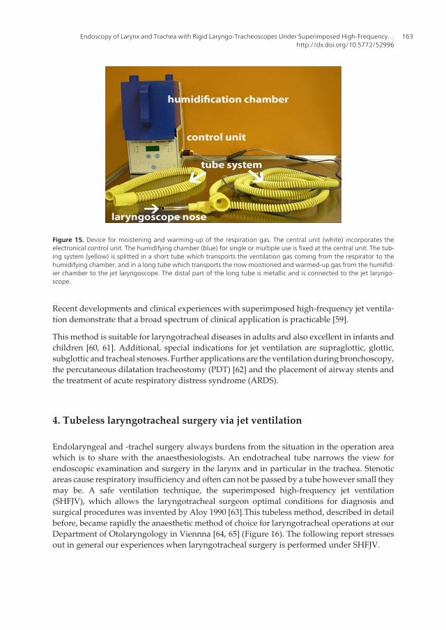
Recent developments and clinical experiences with superimposed high-frequency jet ventila‐tion demonstrate that a broad spectrum of clinical application is practicable [59].
This method is suitable for laryngotracheal diseases in adults and also excellent in infants andchildren [60, 61]. Additional, special indications for jet ventilation are supraglottic, glottic,subglottic and tracheal stenoses. Further applications are the ventilation during bronchoscopy,the percutaneous dilatation tracheostomy (PDT) [62] and the placement of airway stents andthe treatment of acute respiratory distress syndrome (ARDS).
4. Tubeless laryngotracheal surgery via jet ventilation
Endolaryngeal and -trachel surgery always burdens from the situation in the operation areawhich is to share with the anaesthesiologists. An endotracheal tube narrows the view forendoscopic examination and surgery in the larynx and in particular in the trachea. Stenoticareas cause respiratory insufficiency and often can not be passed by a tube however small theymay be. A safe ventilation technique, the superimposed high-frequency jet ventilation(SHFJV), which allows the laryngotracheal surgeon optimal conditions for diagnosis andsurgical procedures was invented by Aloy 1990 [63].This tubeless method, described in detailbefore, became rapidly the anaesthetic method of choice for laryngotracheal operations at ourDepartment of Otolaryngology in Viennna [64, 65] (Figure 16). The following report stressesout in general our experiences when laryngotracheal surgery is performed under SHFJV.
Figure 15. Device for moistening and warming-up of the respiration gas. The central unit (white) incorporates theelectronical control unit. The humidifying chamber (blue) for single or multiple use is fixed at the central unit. The tub‐ing system (yellow) is splitted in a short tube which transports the ventilation gas coming from the respirator to thehumidifying chamber, and in a long tube which transports the now moistioned and warmed-up gas from the humifid‐ier chamber to the jet laryngoscope. The distal part of the long tube is metallic and is connected to the jet laryngo‐scope.
Endoscopy of Larynx and Trachea with Rigid Laryngo-Tracheoscopes Under Superimposed High-Frequency…http://dx.doi.org/10.5772/52996
163
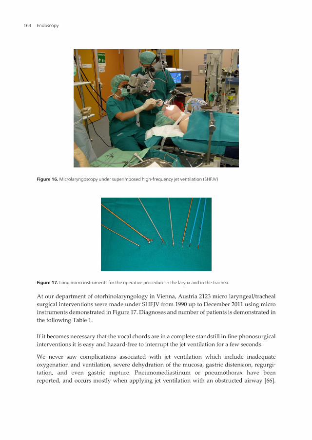
Figure 16. Microlaryngoscopy under superimposed high-frequency jet ventilation (SHFJV)
Figure 17. Long micro instruments for the operative procedure in the larynx and in the trachea.
At our department of otorhinolaryngology in Vienna, Austria 2123 micro laryngeal/trachealsurgical interventions were made under SHFJV from 1990 up to December 2011 using microinstruments demonstrated in Figure 17. Diagnoses and number of patients is demonstrated inthe following Table 1.
If it becomes necessary that the vocal chords are in a complete standstill in fine phonosurgicalinterventions it is easy and hazard-free to interrupt the jet ventilation for a few seconds.
We never saw complications associated with jet ventilation which include inadequateoxygenation and ventilation, severe dehydration of the mucosa, gastric distension, regurgi‐tation, and even gastric rupture. Pneumomediastinum or pneumothorax have beenreported, and occurs mostly when applying jet ventilation with an obstructed airway [66].
Endoscopy164
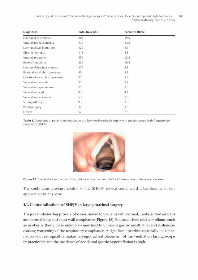
The continuous pressure control of the SHFJV- device could ward a barotrauma in ourapplication in any case.
4.1. Contraindications of SHFJV in laryngotracheal surgery
The jet ventilation has proven to be most suited for patients with normal, unobstructed airwaysand normal lung and chest wall compliance (Figure 18). Reduced chest wall compliance suchas in obesity (body mass index >35) may lead to unmeant gastric insufflation and distensioncausing worsening of the respiratory compliance. A significant overbite especially in combi‐nation with retrognathia makes laryngotracheal placement of the ventilation laryngoscopeimpracticable and the incidence of accidental gastric hyperinflation is high.
Diagnoses Total (n=2123) Percent (100%)
Laryngeal carcinoma
Vocal chord leucoplakia
Laryngeal papillomatosis
Chronic laryngitis
Vocal chord polyp
Reinke`s oedema
Laryngeal/tracheal stenosis
Bilateral vocal chord paralysis
Unilateral vocal chord paralysis
Vocal chord nodule
Vocal chord granuloma
Vocal chord cyst
Vocal chords synechia
Supraglottic cyst
Phonosurgery
Others
403
272
122
116
259
221
172
45
72
57
71
93
53
83
32
52
19.0
12.8
5.7
5.5
12.2
10.4
8.1
2.1
3.4
2.7
3.3
4.4
2.5
3.9
1.5
2.5
Table 1. Diagnoses of patients undergoing micro laryngeal/tracheal surgery with superimposed high-frequency jetventilation (SHFJV).
Figure 18. Cyst at the free margin of the right vocal chord anterior half with free access to the operation area.
Endoscopy of Larynx and Trachea with Rigid Laryngo-Tracheoscopes Under Superimposed High-Frequency…http://dx.doi.org/10.5772/52996
165
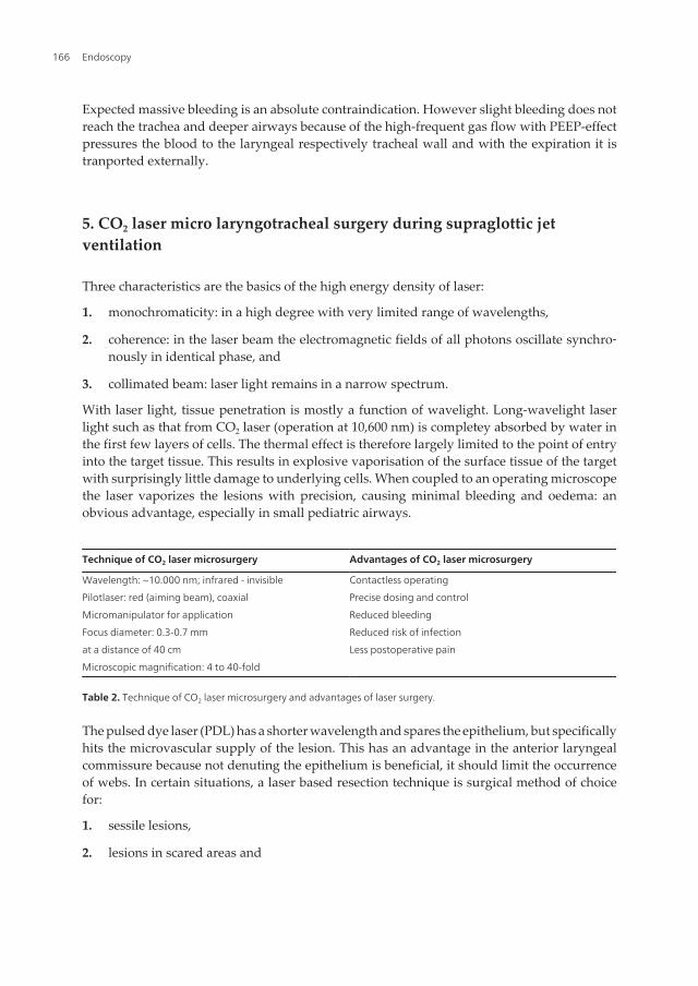
Expected massive bleeding is an absolute contraindication. However slight bleeding does notreach the trachea and deeper airways because of the high-frequent gas flow with PEEP-effectpressures the blood to the laryngeal respectively tracheal wall and with the expiration it istranported externally.
5. CO2 laser micro laryngotracheal surgery during supraglottic jetventilation
Three characteristics are the basics of the high energy density of laser:
1. monochromaticity: in a high degree with very limited range of wavelengths,
2. coherence: in the laser beam the electromagnetic fields of all photons oscillate synchro‐nously in identical phase, and
3. collimated beam: laser light remains in a narrow spectrum.
With laser light, tissue penetration is mostly a function of wavelight. Long-wavelight laserlight such as that from CO2 laser (operation at 10,600 nm) is completey absorbed by water inthe first few layers of cells. The thermal effect is therefore largely limited to the point of entryinto the target tissue. This results in explosive vaporisation of the surface tissue of the targetwith surprisingly little damage to underlying cells. When coupled to an operating microscopethe laser vaporizes the lesions with precision, causing minimal bleeding and oedema: anobvious advantage, especially in small pediatric airways.
Technique of CO2 laser microsurgery Advantages of CO2 laser microsurgery
Wavelength: ~10.000 nm; infrared - invisible
Pilotlaser: red (aiming beam), coaxial
Micromanipulator for application
Focus diameter: 0.3-0.7 mm
at a distance of 40 cm
Microscopic magnification: 4 to 40-fold
Contactless operating
Precise dosing and control
Reduced bleeding
Reduced risk of infection
Less postoperative pain
Table 2. Technique of CO2 laser microsurgery and advantages of laser surgery.
The pulsed dye laser (PDL) has a shorter wavelength and spares the epithelium, but specificallyhits the microvascular supply of the lesion. This has an advantage in the anterior laryngealcommissure because not denuting the epithelium is beneficial, it should limit the occurrenceof webs. In certain situations, a laser based resection technique is surgical method of choicefor:
1. sessile lesions,
2. lesions in scared areas and
Endoscopy166
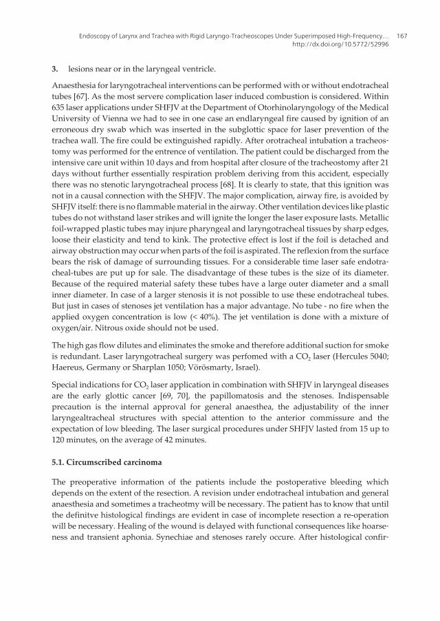
3. lesions near or in the laryngeal ventricle.
Anaesthesia for laryngotracheal interventions can be performed with or without endotrachealtubes [67]. As the most servere complication laser induced combustion is considered. Within635 laser applications under SHFJV at the Department of Otorhinolaryngology of the MedicalUniversity of Vienna we had to see in one case an endlaryngeal fire caused by ignition of anerroneous dry swab which was inserted in the subglottic space for laser prevention of thetrachea wall. The fire could be extinguished rapidly. After orotracheal intubation a tracheos‐tomy was performed for the entrence of ventilation. The patient could be discharged from theintensive care unit within 10 days and from hospital after closure of the tracheostomy after 21days without further essentially respiration problem deriving from this accident, especiallythere was no stenotic laryngotracheal process [68]. It is clearly to state, that this ignition wasnot in a causal connection with the SHFJV. The major complication, airway fire, is avoided bySHFJV itself: there is no flammable material in the airway. Other ventilation devices like plastictubes do not withstand laser strikes and will ignite the longer the laser exposure lasts. Metallicfoil-wrapped plastic tubes may injure pharyngeal and laryngotracheal tissues by sharp edges,loose their elasticity and tend to kink. The protective effect is lost if the foil is detached andairway obstruction may occur when parts of the foil is aspirated. The reflexion from the surfacebears the risk of damage of surrounding tissues. For a considerable time laser safe endotra‐cheal-tubes are put up for sale. The disadvantage of these tubes is the size of its diameter.Because of the required material safety these tubes have a large outer diameter and a smallinner diameter. In case of a larger stenosis it is not possible to use these endotracheal tubes.But just in cases of stenoses jet ventilation has a major advantage. No tube - no fire when theapplied oxygen concentration is low (< 40%). The jet ventilation is done with a mixture ofoxygen/air. Nitrous oxide should not be used.
The high gas flow dilutes and eliminates the smoke and therefore additional suction for smokeis redundant. Laser laryngotracheal surgery was perfomed with a CO2 laser (Hercules 5040;Haereus, Germany or Sharplan 1050; Vörösmarty, Israel).
Special indications for CO2 laser application in combination with SHFJV in laryngeal diseasesare the early glottic cancer [69, 70], the papillomatosis and the stenoses. Indispensableprecaution is the internal approval for general anaesthea, the adjustability of the innerlaryngealtracheal structures with special attention to the anterior commissure and theexpectation of low bleeding. The laser surgical procedures under SHFJV lasted from 15 up to120 minutes, on the average of 42 minutes.
5.1. Circumscribed carcinoma
The preoperative information of the patients include the postoperative bleeding whichdepends on the extent of the resection. A revision under endotracheal intubation and generalanaesthesia and sometimes a tracheotmy will be necessary. The patient has to know that untilthe definitve histological findings are evident in case of incomplete resection a re-operationwill be necessary. Healing of the wound is delayed with functional consequences like hoarse‐ness and transient aphonia. Synechiae and stenoses rarely occure. After histological confir‐
Endoscopy of Larynx and Trachea with Rigid Laryngo-Tracheoscopes Under Superimposed High-Frequency…http://dx.doi.org/10.5772/52996
167

mation of the initial diagnosis the surgery by CO2 laser with the soft super pulsed mode anearly char-free cutting via vaporisation is performed.
Dissecting the soft tissue of the tumour is has proven to perform with the micro manipulatorfine serrated hither and thihter movements. With this method laser`s physical properties forcutting and coagulation are utilized most effective [71]. Like the study group of our departmentcould show excellent oncological results can be expected in T1a-glottic cancer in comparisonto radiotherapy and conventional surgery [72].
Postoperative stridor is mostly due to a swelling of the local mucous membrane and is easy tobe treated by cortisone.
5.2. Laryngo/tracheal stenosis
Congenital and aquired stenoses in all regions of the larynx are a prefered indication for CO2
laser surgery (Figures 19, 20, 21). Tubeless ventilation allows the surgeon to operate in analready narrow area.
Figure 19. View through the ventilation laryngoscope in position. Tumour caused massive stenosis. In the area of theposterior commissure a residual lumen exists, enabling patient`s spontaneous but stridulous breathing.
Figure 20. High grade subglottic stenosis with a substantially reduced lumen.
Endoscopy168
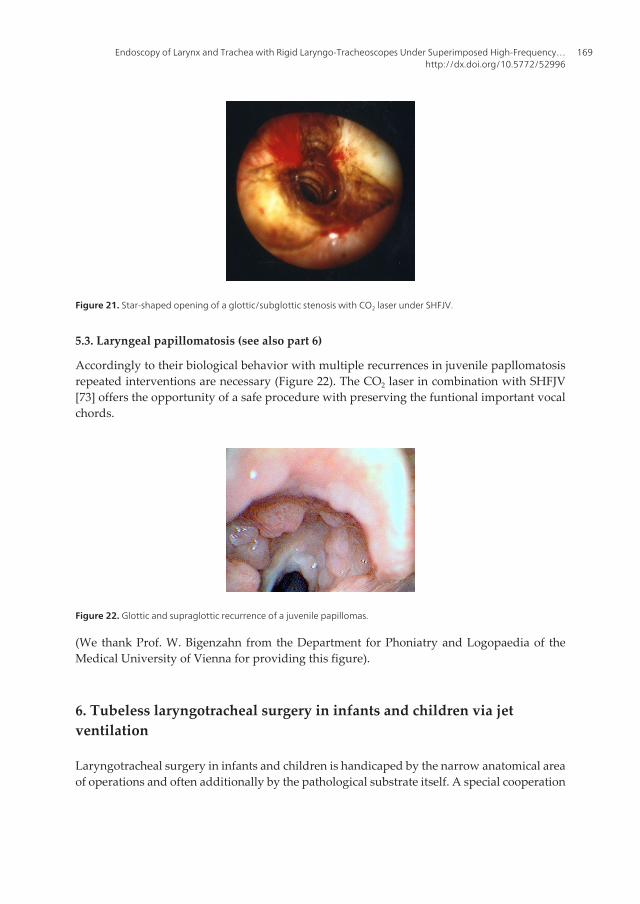
Figure 21. Star-shaped opening of a glottic/subglottic stenosis with CO2 laser under SHFJV.
5.3. Laryngeal papillomatosis (see also part 6)
Accordingly to their biological behavior with multiple recurrences in juvenile papllomatosisrepeated interventions are necessary (Figure 22). The CO2 laser in combination with SHFJV[73] offers the opportunity of a safe procedure with preserving the funtional important vocalchords.
Figure 22. Glottic and supraglottic recurrence of a juvenile papillomas.
(We thank Prof. W. Bigenzahn from the Department for Phoniatry and Logopaedia of theMedical University of Vienna for providing this figure).
6. Tubeless laryngotracheal surgery in infants and children via jetventilation
Laryngotracheal surgery in infants and children is handicaped by the narrow anatomical areaof operations and often additionally by the pathological substrate itself. A special cooperation
Endoscopy of Larynx and Trachea with Rigid Laryngo-Tracheoscopes Under Superimposed High-Frequency…http://dx.doi.org/10.5772/52996
169

between the surgeon and the anaesthesist is indispensable. Ventilation via an endotrachealtube with a cuff is most safe to operate. But even this endotracheal tube blocks on the one handthe unobstructed view of the operating field and on the other hand the necessary space forsurgical activity and additionally mutates the anatomical structures. In cases of pronouncedstenoses in the laryngotracheal area an endotracheal intubation cannot be applied. Analternative procedure is the tracheotomy, especially for infants and children and the postop‐erative care an enormous burden.
A rigid bronchoscope used for ventilation restricts the visibility of the working space and itmay occur that it cannot pass a stenosis. Laryngotracheal surgery in the apnea technique isstill in use but is associated with the heightened risk of hypoxemia and hypercapnia.
An improvement of ventilation of the patient during laryngotracheal surgery was establishedby single-frequency jet ventilation techniques applied either per percutaneous insertion of aneedle into the trachea or endotracheal tubes or catheters, or application of a jet nozzle intothe endocopy tube [74, 75]. All these techniques have the disadvantage of the risk of hypo‐xaemia and hypercapnia especially in patients underlying pulmonary and cardiadic aggra‐vated risks and operations with a long continuance. Needles placed transtracheal bear theelevated risk of barotrauma because the gas supply takes place below the stenosis [76].
6.1. Ventilation
As an alternative ventilation technique the jet ventilation presents themselves with 3 modalities:
1. transtracheal jet ventilation (Figure 23): for endoscopic laryngeal surgery, also with laser,excellent visibility for the surgeon, even in glottic or supraglottic stenoses with minorcomplications up to 20 % [77-79]. Very important is the bedding of the child with maxi‐mum extension of the head. This allows fixing the mobile trachea between middle fingerand thumb. The cricoid membrane is narrow and at best to be felt by a fingernail. Thepunction with ventilation catheter is to be directed in a caudal direction.
Figure 23. Percutaneous transtracheal jet ventilation catheter (VBM®-Medizintechnik, Germany) for children with steelpunction needle.
Endoscopy170
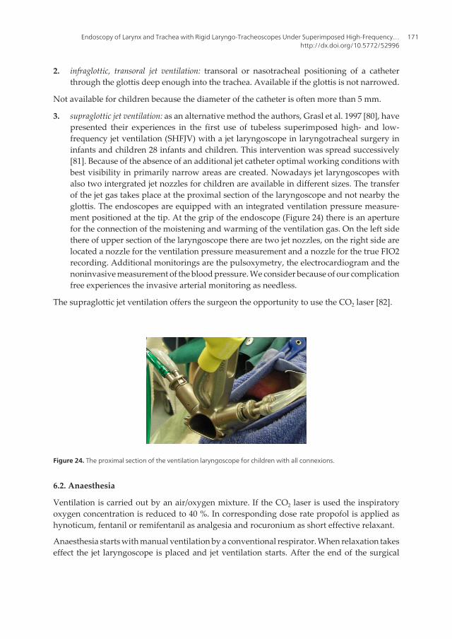
2. infraglottic, transoral jet ventilation: transoral or nasotracheal positioning of a catheterthrough the glottis deep enough into the trachea. Available if the glottis is not narrowed.
Not available for children because the diameter of the catheter is often more than 5 mm.
3. supraglottic jet ventilation: as an alternative method the authors, Grasl et al. 1997 [80], havepresented their experiences in the first use of tubeless superimposed high- and low-frequency jet ventilation (SHFJV) with a jet laryngoscope in laryngotracheal surgery ininfants and children 28 infants and children. This intervention was spread successively[81]. Because of the absence of an additional jet catheter optimal working conditions withbest visibility in primarily narrow areas are created. Nowadays jet laryngoscopes withalso two intergrated jet nozzles for children are available in different sizes. The transferof the jet gas takes place at the proximal section of the laryngoscope and not nearby theglottis. The endoscopes are equipped with an integrated ventilation pressure measure‐ment positioned at the tip. At the grip of the endoscope (Figure 24) there is an aperturefor the connection of the moistening and warming of the ventilation gas. On the left sidethere of upper section of the laryngoscope there are two jet nozzles, on the right side arelocated a nozzle for the ventilation pressure measurement and a nozzle for the true FIO2recording. Additional monitorings are the pulsoxymetry, the electrocardiogram and thenoninvasive measurement of the blood pressure. We consider because of our complicationfree experiences the invasive arterial monitoring as needless.
The supraglottic jet ventilation offers the surgeon the opportunity to use the CO2 laser [82].
Figure 24. The proximal section of the ventilation laryngoscope for children with all connexions.
6.2. Anaesthesia
Ventilation is carried out by an air/oxygen mixture. If the CO2 laser is used the inspiratoryoxygen concentration is reduced to 40 %. In corresponding dose rate propofol is applied ashynoticum, fentanil or remifentanil as analgesia and rocuronium as short effective relaxant.
Anaesthesia starts with manual ventilation by a conventional respirator. When relaxation takeseffect the jet laryngoscope is placed and jet ventilation starts. After the end of the surgical
Endoscopy of Larynx and Trachea with Rigid Laryngo-Tracheoscopes Under Superimposed High-Frequency…http://dx.doi.org/10.5772/52996
171
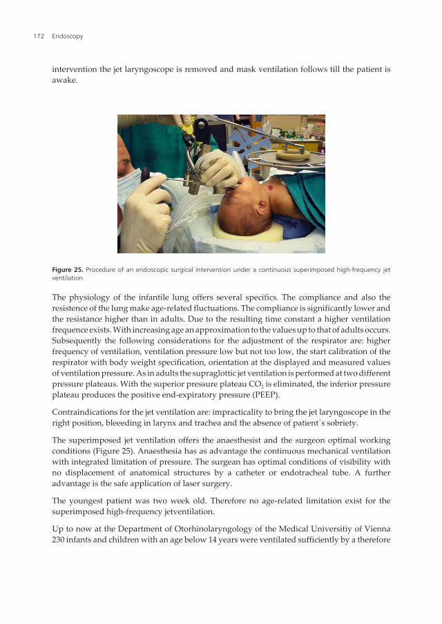
intervention the jet laryngoscope is removed and mask ventilation follows till the patient isawake.
Figure 25. Procedure of an endoscopic surgical intervention under a continuous superimposed high-frequency jetventilation.
The physiology of the infantile lung offers several specifics. The compliance and also theresistence of the lung make age-related fluctuations. The compliance is significantly lower andthe resistance higher than in adults. Due to the resulting time constant a higher ventilationfrequence exists. With increasing age an approximation to the values up to that of adults occurs.Subsequently the following considerations for the adjustment of the respirator are: higherfrequency of ventilation, ventilation pressure low but not too low, the start calibration of therespirator with body weight specification, orientation at the displayed and measured valuesof ventilation pressure. As in adults the supraglottic jet ventilation is performed at two differentpressure plateaus. With the superior pressure plateau CO2 is eliminated, the inferior pressureplateau produces the positive end-expiratory pressure (PEEP).
Contraindications for the jet ventilation are: impracticality to bring the jet laryngoscope in theright position, bleeeding in larynx and trachea and the absence of patient`s sobriety.
The superimposed jet ventilation offers the anaesthesist and the surgeon optimal workingconditions (Figure 25). Anaesthesia has as advantage the continuous mechanical ventilationwith integrated limitation of pressure. The surgean has optimal conditions of visibility withno displacement of anatomical structures by a catheter or endotracheal tube. A furtheradvantage is the safe application of laser surgery.
The youngest patient was two week old. Therefore no age-related limitation exist for thesuperimposed high-frequency jetventilation.
Up to now at the Department of Otorhinolaryngology of the Medical Universitiy of Vienna230 infants and children with an age below 14 years were ventilated sufficiently by a therefore
Endoscopy172
extra designed rigid endoscopy tube derived from the Kleinsasser tube [79] during laryngo-tracheal surgery. Diagnoses were: papillomatosis, subglottic stenoses, laryngeal inspection,web, foreign body, vocal chord cyst, vocal chords granuloma and miscellaneous. Movementof foreign bodies becomes much more easy and elegant during the tubeless ventilation: aFogarty-Catheters© guided behind the foreign body helps to bring him more proximal wherehe can be gripped and extracted with a forceps [83]. With this SHFJV technique laryngealstenoses and cardiopulmonal insufficiences do not present special risks. Laryngotrachealsurgery under SHFJV can be applied in any child except special general contraindications.
As in adults before effective jet ventilation can start, the laryngoscope tube, adjusted to size ofthe childlike larynx, is inserted and evaluated for suitability for jet ventilation. Immediatelyafterward the suspension system is installed and the jet injector needles are attached to thelaryngoscope. The steadily ventilation can begin. Up to this point of time the manipulationhappens in an anaesthetized but apnoeic patient.
The procedure of the jet ventilation took from 5 up to 130 minutes, on average about 35 minutes.The ventilation assured in any case a sufficient oxygenation and CO2 elimination.
Only in a few cases during introduction or recovery of the anaesthesia an endotracheal tubehad to replace the ventilation tube temporarily caused by adverse anatomical characteristics.A serve laryngospasm never resulted from SHFJV. To eliminate the danger of pneumothoraxin the application of SHFJV it is to state that it is an open system with an air supply alwaysabove the existing stenoses. Additionally an intergrated monitoring of pressure in thelaryngoscope is positioned. The surgical procedure could be performed in any case.
Contraindication for SHFJV in infants und children are in principle the same as in grownups.
The technique of SHFJV has helped us to handle the naturally difficult surgery of the larynxand trachea in infants and children and simplified it considerably.
A very special laryngeal disease in children is the recurrent respiratory papillomatosis (RRP)[84-86].
Only a minority of Human papilloma virus (HPV) carrying mothers will become symptomatic.The route of transmission to the infant in not yet completely understood.
There is no cure for RRP at present and no therapy modality that might eradicate the virusfrom the respiratory mucosa. Local recurrences are therefore frequently seen.
Vaccination against HPV 6 and 11 across the board should decrease the incidence of childrenin future.
RRP requires a protracted and repeatedly therapy over years.
The surgical therapy with a removal of the papillomas as much as possible und preservationof normal structures is the procedure of (Figure 26). Scaring from overagressive resectionseffects dysphonia and airway comprise. Incomplete resections are accepted under theseaspects especially in the anterior commissure. It is not practicable to eradicate all virus particleseven when clinically all evident papillomas had been removed.
Endoscopy of Larynx and Trachea with Rigid Laryngo-Tracheoscopes Under Superimposed High-Frequency…http://dx.doi.org/10.5772/52996
173
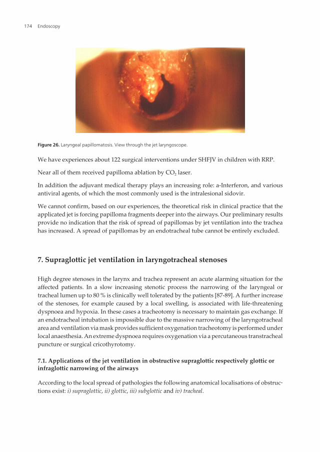
We have experiences about 122 surgical interventions under SHFJV in children with RRP.
Near all of them received papilloma ablation by CO2 laser.
In addition the adjuvant medical therapy plays an increasing role: a-Interferon, and variousantiviral agents, of which the most commonly used is the intralesional sidovir.
We cannot confirm, based on our experiences, the theoretical risk in clinical practice that theapplicated jet is forcing papilloma fragments deeper into the airways. Our preliminary resultsprovide no indication that the risk of spread of papillomas by jet ventilation into the tracheahas increased. A spread of papillomas by an endotracheal tube cannot be entirely excluded.
7. Supraglottic jet ventilation in laryngotracheal stenoses
High degree stenoses in the larynx and trachea represent an acute alarming situation for theaffected patients. In a slow increasing stenotic process the narrowing of the laryngeal ortracheal lumen up to 80 % is clinically well tolerated by the patients [87-89]. A further increaseof the stenoses, for example caused by a local swelling, is associated with life-threateningdyspnoea and hypoxia. In these cases a tracheotomy is necessary to maintain gas exchange. Ifan endotracheal intubation is impossible due to the massive narrowing of the laryngotrachealarea and ventilation via mask provides sufficient oxygenation tracheotomy is performed underlocal anaesthesia. An extreme dyspnoea requires oxygenation via a percutaneous transtrachealpuncture or surgical cricothyrotomy.
7.1. Applications of the jet ventilation in obstructive supraglottic respectively glottic orinfraglottic narrowing of the airways
According to the local spread of pathologies the following anatomical localisations of obstruc‐tions exist: i) supraglottic, ii) glottic, iii) subglottic and iv) tracheal.
Figure 26. Laryngeal papillomatosis. View through the jet laryngoscope.
Endoscopy174
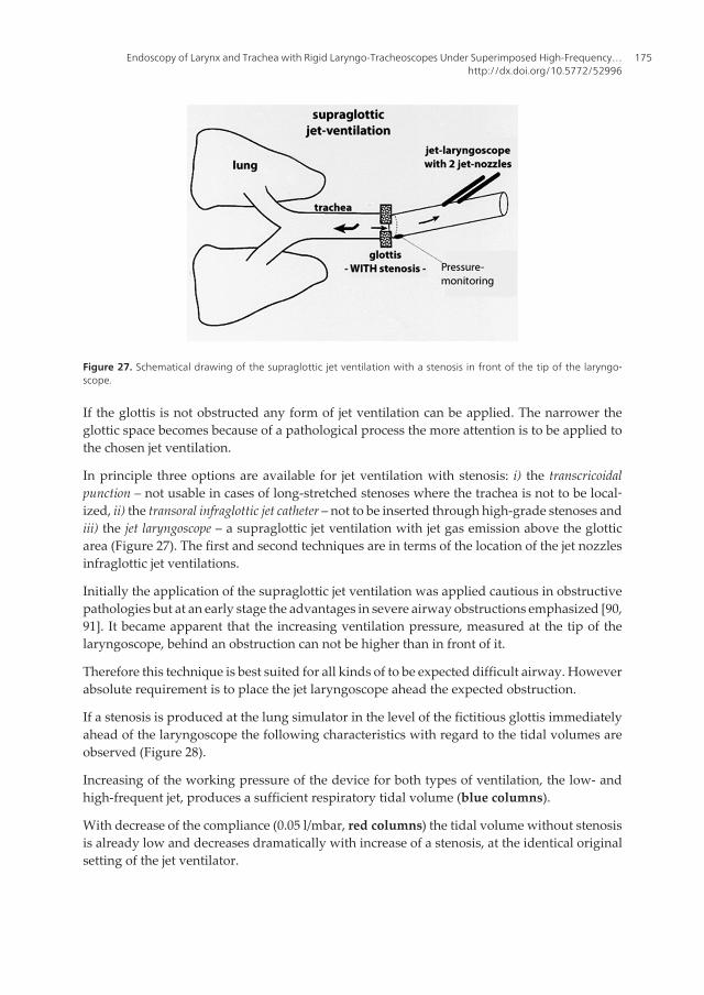
If the glottis is not obstructed any form of jet ventilation can be applied. The narrower theglottic space becomes because of a pathological process the more attention is to be applied tothe chosen jet ventilation.
In principle three options are available for jet ventilation with stenosis: i) the transcricoidalpunction – not usable in cases of long-stretched stenoses where the trachea is not to be local‐ized, ii) the transoral infraglottic jet catheter – not to be inserted through high-grade stenoses andiii) the jet laryngoscope – a supraglottic jet ventilation with jet gas emission above the glotticarea (Figure 27). The first and second techniques are in terms of the location of the jet nozzlesinfraglottic jet ventilations.
Initially the application of the supraglottic jet ventilation was applied cautious in obstructivepathologies but at an early stage the advantages in severe airway obstructions emphasized [90,91]. It became apparent that the increasing ventilation pressure, measured at the tip of thelaryngoscope, behind an obstruction can not be higher than in front of it.
Therefore this technique is best suited for all kinds of to be expected difficult airway. Howeverabsolute requirement is to place the jet laryngoscope ahead the expected obstruction.
If a stenosis is produced at the lung simulator in the level of the fictitious glottis immediatelyahead of the laryngoscope the following characteristics with regard to the tidal volumes areobserved (Figure 28).
Increasing of the working pressure of the device for both types of ventilation, the low- andhigh-frequent jet, produces a sufficient respiratory tidal volume (blue columns).
With decrease of the compliance (0.05 l/mbar, red columns) the tidal volume without stenosisis already low and decreases dramatically with increase of a stenosis, at the identical originalsetting of the jet ventilator.
Figure 27. Schematical drawing of the supraglottic jet ventilation with a stenosis in front of the tip of the laryngo‐scope.
Endoscopy of Larynx and Trachea with Rigid Laryngo-Tracheoscopes Under Superimposed High-Frequency…http://dx.doi.org/10.5772/52996
175

The increase of the working pressures of the ventilators succeeds in achievement of sufficienttidal volumes even in 80 % stenosis (green column).
7.2. Computational fluid dynamic
In a jet laryngoscope a flow simulation with ANSYS fluent® was carried out. First, the creationof a three-dimensional image of the jet laryngoscope with the preprocessor Gambit wasperformed. This was followed by the definition of boundary conditions and input parametersin the solver and the iterative calculation in fluent.
Figure 29. Distribution of pressure in the jet laryngoscope in case of a stenosis. Note the impact pressure in front ofthe stenosis (Figures 29, 30). The pressure behind the stenosis is lower than the pressure before the stenosis.
Figure 28. Diagram of the respiratory tidal volume (ml) to be achieved by a supraglottic jet ventilation via a jet lar‐yngoscope in a default setting on the lung simulator: with a narrowing of the cross section for ventilation, at first with‐out stenosis (0 %, left), then with a 50 % stenosis (centre) and at last with a 80 % stenosis (right) all yellow columns.The lung compliance was adjusted at 0.1 liter per millibar (l/mbar).
Endoscopy176
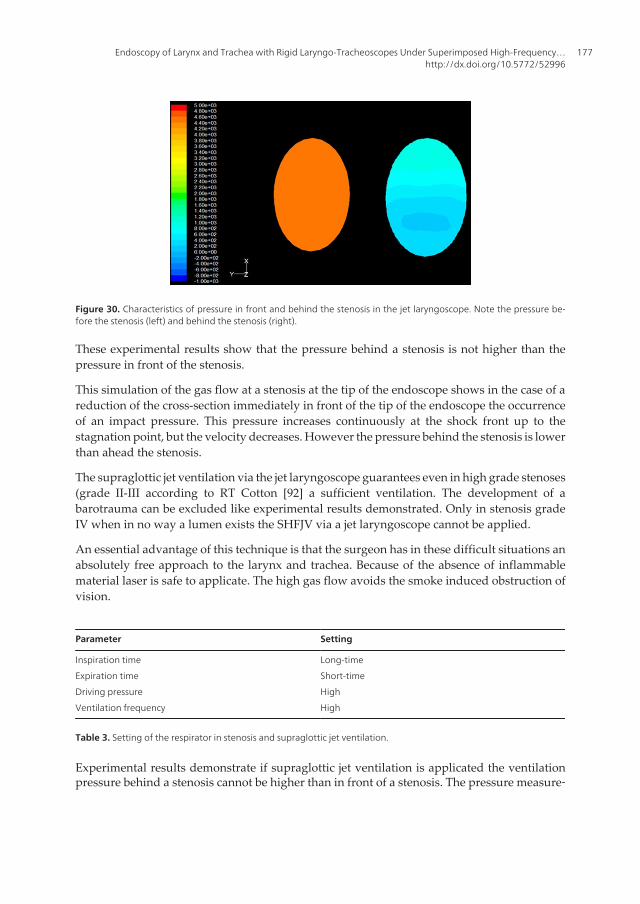
Figure 30. Characteristics of pressure in front and behind the stenosis in the jet laryngoscope. Note the pressure be‐fore the stenosis (left) and behind the stenosis (right).
These experimental results show that the pressure behind a stenosis is not higher than thepressure in front of the stenosis.
This simulation of the gas flow at a stenosis at the tip of the endoscope shows in the case of areduction of the cross-section immediately in front of the tip of the endoscope the occurrenceof an impact pressure. This pressure increases continuously at the shock front up to thestagnation point, but the velocity decreases. However the pressure behind the stenosis is lowerthan ahead the stenosis.
The supraglottic jet ventilation via the jet laryngoscope guarantees even in high grade stenoses(grade II-III according to RT Cotton [92] a sufficient ventilation. The development of abarotrauma can be excluded like experimental results demonstrated. Only in stenosis gradeIV when in no way a lumen exists the SHFJV via a jet laryngoscope cannot be applied.
An essential advantage of this technique is that the surgeon has in these difficult situations anabsolutely free approach to the larynx and trachea. Because of the absence of inflammablematerial laser is safe to applicate. The high gas flow avoids the smoke induced obstruction ofvision.
Parameter Setting
Inspiration time
Expiration time
Driving pressure
Ventilation frequency
Long-time
Short-time
High
High
Table 3. Setting of the respirator in stenosis and supraglottic jet ventilation.
Experimental results demonstrate if supraglottic jet ventilation is applicated the ventilationpressure behind a stenosis cannot be higher than in front of a stenosis. The pressure measure‐
Endoscopy of Larynx and Trachea with Rigid Laryngo-Tracheoscopes Under Superimposed High-Frequency…http://dx.doi.org/10.5772/52996
177
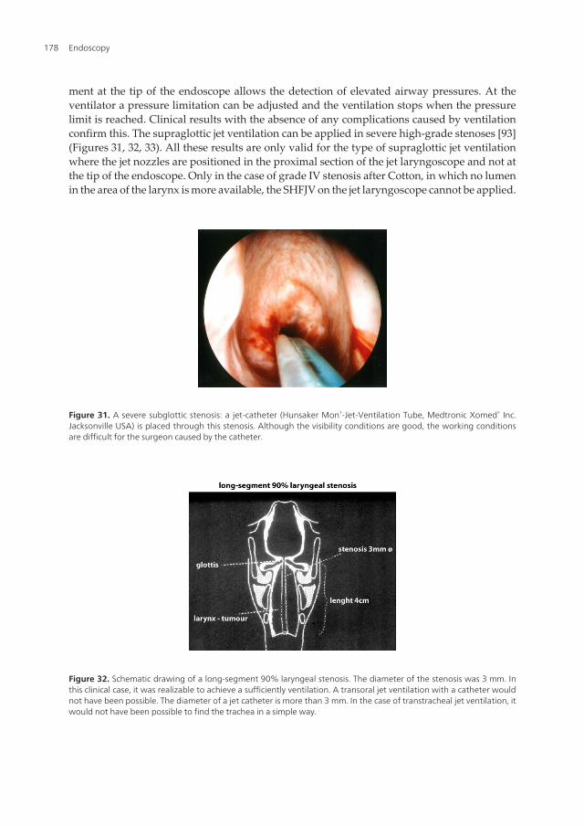
ment at the tip of the endoscope allows the detection of elevated airway pressures. At theventilator a pressure limitation can be adjusted and the ventilation stops when the pressurelimit is reached. Clinical results with the absence of any complications caused by ventilationconfirm this. The supraglottic jet ventilation can be applied in severe high-grade stenoses [93](Figures 31, 32, 33). All these results are only valid for the type of supraglottic jet ventilationwhere the jet nozzles are positioned in the proximal section of the jet laryngoscope and not atthe tip of the endoscope. Only in the case of grade IV stenosis after Cotton, in which no lumenin the area of the larynx is more available, the SHFJV on the jet laryngoscope cannot be applied.
Figure 31. A severe subglottic stenosis: a jet-catheter (Hunsaker Mon®-Jet-Ventilation Tube, Medtronic Xomed® Inc.Jacksonville USA) is placed through this stenosis. Although the visibility conditions are good, the working conditionsare difficult for the surgeon caused by the catheter.
Figure 32. Schematic drawing of a long-segment 90% laryngeal stenosis. The diameter of the stenosis was 3 mm. Inthis clinical case, it was realizable to achieve a sufficiently ventilation. A transoral jet ventilation with a catheter wouldnot have been possible. The diameter of a jet catheter is more than 3 mm. In the case of transtracheal jet ventilation, itwould not have been possible to find the trachea in a simple way.
Endoscopy178
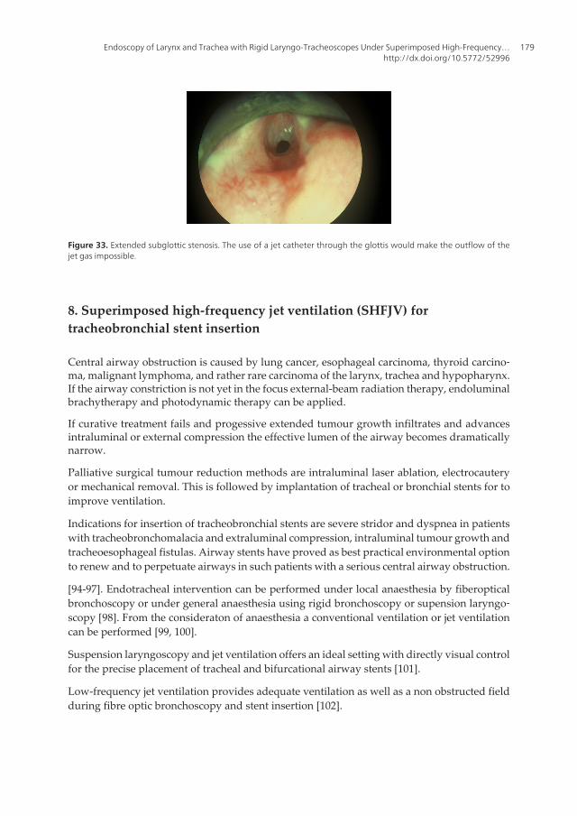
Figure 33. Extended subglottic stenosis. The use of a jet catheter through the glottis would make the outflow of thejet gas impossible.
8. Superimposed high-frequency jet ventilation (SHFJV) fortracheobronchial stent insertion
Central airway obstruction is caused by lung cancer, esophageal carcinoma, thyroid carcino‐ma, malignant lymphoma, and rather rare carcinoma of the larynx, trachea and hypopharynx.If the airway constriction is not yet in the focus external-beam radiation therapy, endoluminalbrachytherapy and photodynamic therapy can be applied.
If curative treatment fails and progessive extended tumour growth infiltrates and advancesintraluminal or external compression the effective lumen of the airway becomes dramaticallynarrow.
Palliative surgical tumour reduction methods are intraluminal laser ablation, electrocauteryor mechanical removal. This is followed by implantation of tracheal or bronchial stents for toimprove ventilation.
Indications for insertion of tracheobronchial stents are severe stridor and dyspnea in patientswith tracheobronchomalacia and extraluminal compression, intraluminal tumour growth andtracheoesophageal fistulas. Airway stents have proved as best practical environmental optionto renew and to perpetuate airways in such patients with a serious central airway obstruction.
[94-97]. Endotracheal intervention can be performed under local anaesthesia by fiberopticalbronchoscopy or under general anaesthesia using rigid bronchoscopy or supension laryngo‐scopy [98]. From the consideraton of anaesthesia a conventional ventilation or jet ventilationcan be performed [99, 100].
Suspension laryngoscopy and jet ventilation offers an ideal setting with directly visual controlfor the precise placement of tracheal and bifurcational airway stents [101].
Low-frequency jet ventilation provides adequate ventilation as well as a non obstructed fieldduring fibre optic bronchoscopy and stent insertion [102].
Endoscopy of Larynx and Trachea with Rigid Laryngo-Tracheoscopes Under Superimposed High-Frequency…http://dx.doi.org/10.5772/52996
179
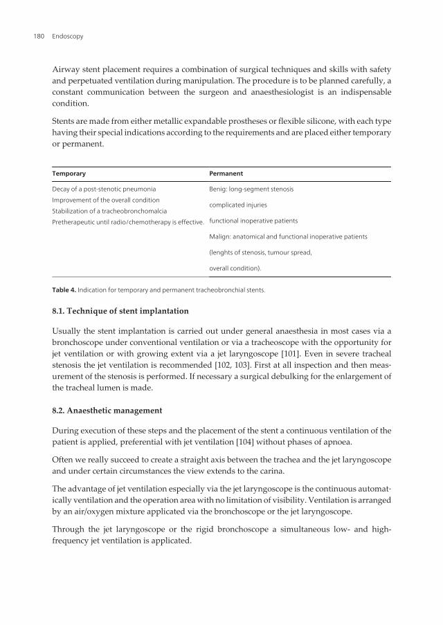
Airway stent placement requires a combination of surgical techniques and skills with safetyand perpetuated ventilation during manipulation. The procedure is to be planned carefully, aconstant communication between the surgeon and anaesthesiologist is an indispensablecondition.
Stents are made from either metallic expandable prostheses or flexible silicone, with each typehaving their special indications according to the requirements and are placed either temporaryor permanent.
Temporary Permanent
Decay of a post-stenotic pneumonia
Improvement of the overall condition
Stabilization of a tracheobronchomalcia
Pretherapeutic until radio/chemotherapy is effective.
Benig: long-segment stenosis
complicated injuries
functional inoperative patients
Malign: anatomical and functional inoperative patients
(lenghts of stenosis, tumour spread,
overall condition).
Table 4. Indication for temporary and permanent tracheobronchial stents.
8.1. Technique of stent implantation
Usually the stent implantation is carried out under general anaesthesia in most cases via abronchoscope under conventional ventilation or via a tracheoscope with the opportunity forjet ventilation or with growing extent via a jet laryngoscope [101]. Even in severe trachealstenosis the jet ventilation is recommended [102, 103]. First at all inspection and then meas‐urement of the stenosis is performed. If necessary a surgical debulking for the enlargement ofthe tracheal lumen is made.
8.2. Anaesthetic management
During execution of these steps and the placement of the stent a continuous ventilation of thepatient is applied, preferential with jet ventilation [104] without phases of apnoea.
Often we really succeed to create a straight axis between the trachea and the jet laryngoscopeand under certain circumstances the view extends to the carina.
The advantage of jet ventilation especially via the jet laryngoscope is the continuous automat‐ically ventilation and the operation area with no limitation of visibility. Ventilation is arrangedby an air/oxygen mixture applicated via the bronchoscope or the jet laryngoscope.
Through the jet laryngoscope or the rigid bronchoscope a simultaneous low- and high-frequency jet ventilation is applicated.
Endoscopy180
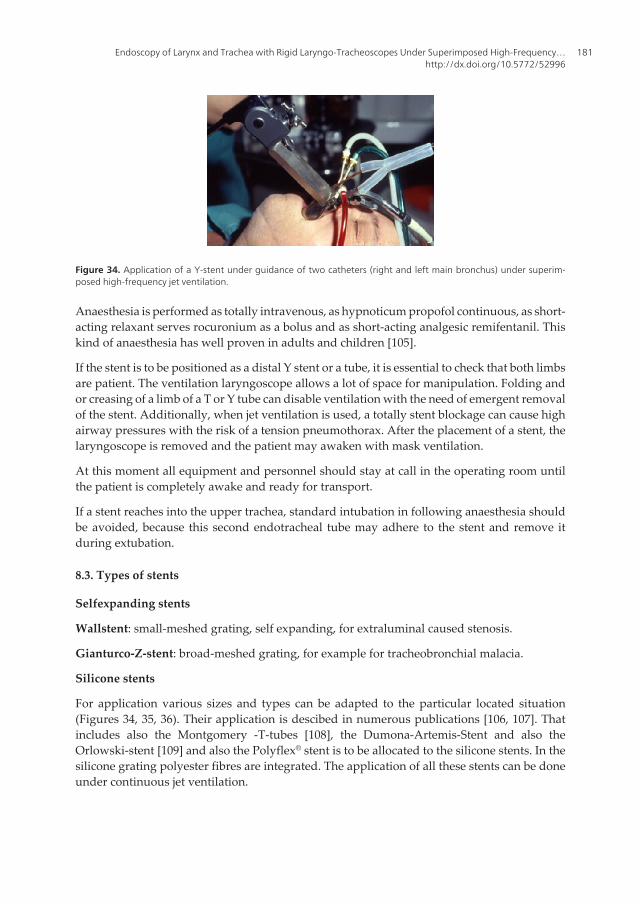
Anaesthesia is performed as totally intravenous, as hypnoticum propofol continuous, as short-acting relaxant serves rocuronium as a bolus and as short-acting analgesic remifentanil. Thiskind of anaesthesia has well proven in adults and children [105].
If the stent is to be positioned as a distal Y stent or a tube, it is essential to check that both limbsare patient. The ventilation laryngoscope allows a lot of space for manipulation. Folding andor creasing of a limb of a T or Y tube can disable ventilation with the need of emergent removalof the stent. Additionally, when jet ventilation is used, a totally stent blockage can cause highairway pressures with the risk of a tension pneumothorax. After the placement of a stent, thelaryngoscope is removed and the patient may awaken with mask ventilation.
At this moment all equipment and personnel should stay at call in the operating room untilthe patient is completely awake and ready for transport.
If a stent reaches into the upper trachea, standard intubation in following anaesthesia shouldbe avoided, because this second endotracheal tube may adhere to the stent and remove itduring extubation.
8.3. Types of stents
Selfexpanding stents
Wallstent: small-meshed grating, self expanding, for extraluminal caused stenosis.
Gianturco-Z-stent: broad-meshed grating, for example for tracheobronchial malacia.
Silicone stents
For application various sizes and types can be adapted to the particular located situation(Figures 34, 35, 36). Their application is descibed in numerous publications [106, 107]. Thatincludes also the Montgomery -T-tubes [108], the Dumona-Artemis-Stent and also theOrlowski-stent [109] and also the Polyflex® stent is to be allocated to the silicone stents. In thesilicone grating polyester fibres are integrated. The application of all these stents can be doneunder continuous jet ventilation.
Figure 34. Application of a Y-stent under guidance of two catheters (right and left main bronchus) under superim‐posed high-frequency jet ventilation.
Endoscopy of Larynx and Trachea with Rigid Laryngo-Tracheoscopes Under Superimposed High-Frequency…http://dx.doi.org/10.5772/52996
181
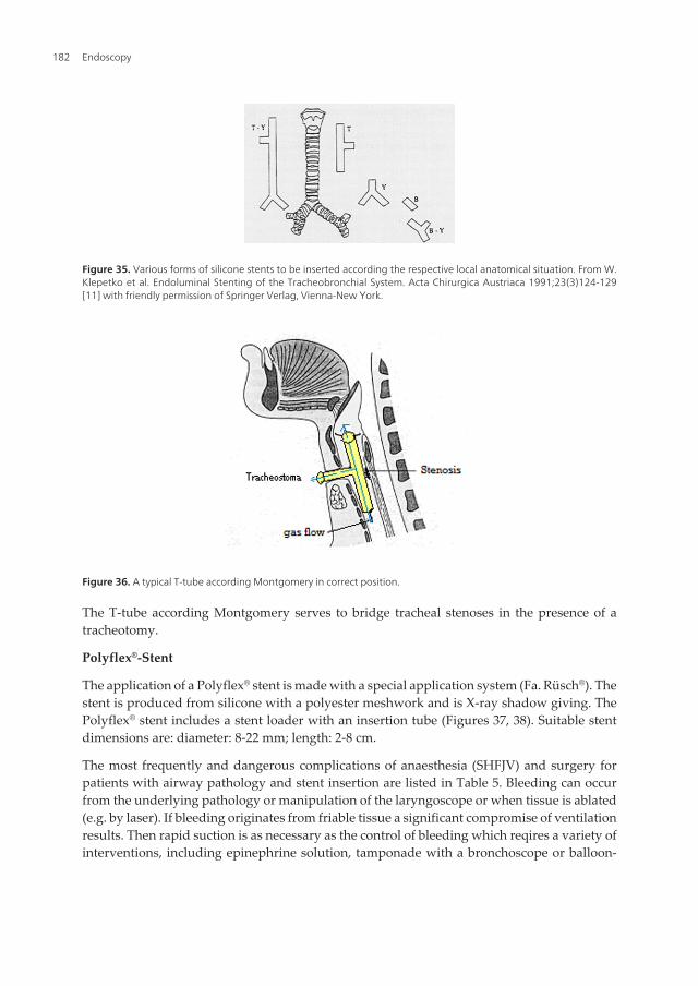
Figure 36. A typical T-tube according Montgomery in correct position.
The T-tube according Montgomery serves to bridge tracheal stenoses in the presence of atracheotomy.
Polyflex®-Stent
The application of a Polyflex® stent is made with a special application system (Fa. Rüsch®). Thestent is produced from silicone with a polyester meshwork and is X-ray shadow giving. ThePolyflex® stent includes a stent loader with an insertion tube (Figures 37, 38). Suitable stentdimensions are: diameter: 8-22 mm; length: 2-8 cm.
The most frequently and dangerous complications of anaesthesia (SHFJV) and surgery forpatients with airway pathology and stent insertion are listed in Table 5. Bleeding can occurfrom the underlying pathology or manipulation of the laryngoscope or when tissue is ablated(e.g. by laser). If bleeding originates from friable tissue a significant compromise of ventilationresults. Then rapid suction is as necessary as the control of bleeding which reqires a variety ofinterventions, including epinephrine solution, tamponade with a bronchoscope or balloon-
Figure 35. Various forms of silicone stents to be inserted according the respective local anatomical situation. From W.Klepetko et al. Endoluminal Stenting of the Tracheobronchial System. Acta Chirurgica Austriaca 1991;23(3)124-129[11] with friendly permission of Springer Verlag, Vienna-New York.
Endoscopy182

Figure 37. Polyflex®-stent in regular size (left) and after potential expansion with the stent-loader (right). The stentloader can be placed through each jet laryngoscope.
Figure 38. Application of a Polyflex® stent with a stent loader under superimposed jet ventilation. The patient is venti‐lated continuously by the jet laryngoscope. The stent loader can be adapted and placed into the trachea passing thejet laryngoscope.
Figure 39. Tracheal stent (Polyflex®-stent) in the trachea.
Endoscopy of Larynx and Trachea with Rigid Laryngo-Tracheoscopes Under Superimposed High-Frequency…http://dx.doi.org/10.5772/52996
183
tipped catheter. Pieces of necrotic or fragmentary tissue as well as clots from bleeding can blockthe distal airway. SHFJF is not be to be interrupted when they are removed. Airway perforationcan be caused by the manipulation at the walls of the trachea or bronchii when the stent isinserted, a subcutaneous emphysema results. An injury of the inner larynx, especially the vocalchords, happens prevalently when difficulties of insertion of the laryngosope tube occur andis rarely severe.
A barotauma occurs suddenly when a mass causes a ball-valve effect or in a distal airway theroute of air exit is blocked by a mass and the air pushed behind causes a tension pneumothorax.Typical signs are absence of chest excursion, sudden tachycardia and hypotension. A rapiddecompression to prevent a cardiavascular collapse is essential.
The occurrence of inadequate oxygenation and ventilation are always to be observed and thisis best to be done by strictly attention to a loud pulsoxymeter which allows quickly an adequatereaction and correction.
Complications of tracheobronchial stent insertion
Acute complications
Bleeding
Occlusion
Perforation
Vocal chord injury
Subcutaneous emphysema
Pneumothorax
Hypoxaemia
Hypercapnia
Long-term complications
Displacement of the stent
Granulation tissue
Mucosa impaction of the stent
Oesophagotracheal fistula
Table 5. Complications of tracheobronchial stent insertion.
The application of the described stents like silicone stents, wall stents and Polyflex® stents viathe jet laryngoscope enables the surgeon due to the excellent field conditions a fast and safeintervention (Figure 39). The anaesthesiologist takes care of an unproblematic ventilation. Aventilation caused barotrauma under supraglottic jet ventilation did not occur in any case atour department. Inside the jet laryngoscope a continuous ventilation pressure measurementwith pressure limitation under connection to the respirator is conducted.
9. High-frequency ventilation techniques in adult respiratory distresssyndrom (ARDS)
The high frequency ventilation is characterized as a type of artificial respiration where low tidalvolume is applicated with a hyper-physiological frequency. Different types of high-frequencyjet ventilations were developed and used in the last 30 years [110]. From the numerous potential
Endoscopy184
applicable types of high-frequency ventilations like High-Frequency-Pulsation (HFP), ForcedDiffusion Ventilation (FDV) [111, 112] and High Frequency Jet Ventilator (HFJV) only the HighFrequency Positive Pressure Ventilation (HFPP), the High Frequency Jet Ventilation (HFJ), theHigh Frequency Oscillation (HFO) and combined high-frequency ventilation techniques becamewidely accepted and are used in clinical field (Table 7).
9.1. Theoretical advantages of the high-frequency ventilation
From the aspect of a lung protective ventilation with the option to reduce the end-inspiratorylung volume the risk of ventilation-induced damage of lungs [113] when conventionalventilation is performed should be reduced by the high- frequency ventilation.
However higher end-exspiratory lung volumes can be applied [114]. Simultaneously thesetidal volumes are transferred with only marginally pressure variations at a higher frequencyand thus the average airway pressure is to be kept at a higher level as it is in conventionalventilation. The high average airway pressure seems to optimize the end-exspiratory lungvolume and is protective against the periodic collapse and therefore also avoiding an atalectatictrauma. The perfect mode of application of high-frequency ventilation should permit lungrecruitment manoeuvre and thereby shifting the lung under optimizing of the compliance andoxygenation to the exspiratory arm of the pressure/volume relationship. The opened lung isventilated then with small-sized tidal volumes and slight fluctuation of pressure. This resultsalso in a diminished alveolar distension and a reduced collapse of alveolar tissue. In anexperimental laboratory animal study the high-frequency oscillation shows in comparison toa lung protective conventional ventilation an attenuation of activation of alveolar macrophagesand neutrophils in lung injury [115].
Clinical indications for high frequency ventilation
Bronchopulmonary fistula
All types of pulmonary reduction of ventilation with no improvement under conventional ventilation therapy
Atalectasis
Pneumonia
Acute lung insuffiency (ALI)
Adult respiratory distress syndrome (ARDS)
Inhalation injury
Table 6. Clinical indications for high-frequency ventilation in adult distress syndrom (ARDS).
9.2. High-frequency oscillatory ventilation (HFO)
Lunkenheimer and co-workers observed already 1994 [116] that normocapnea can be achievedwhen small gas volumes rates are applicated into the airway of animals with a ventilationfrequency of more than 40 Hertz. Using a piston pump sinus-like variations of pressure are
Endoscopy of Larynx and Trachea with Rigid Laryngo-Tracheoscopes Under Superimposed High-Frequency…http://dx.doi.org/10.5772/52996
185
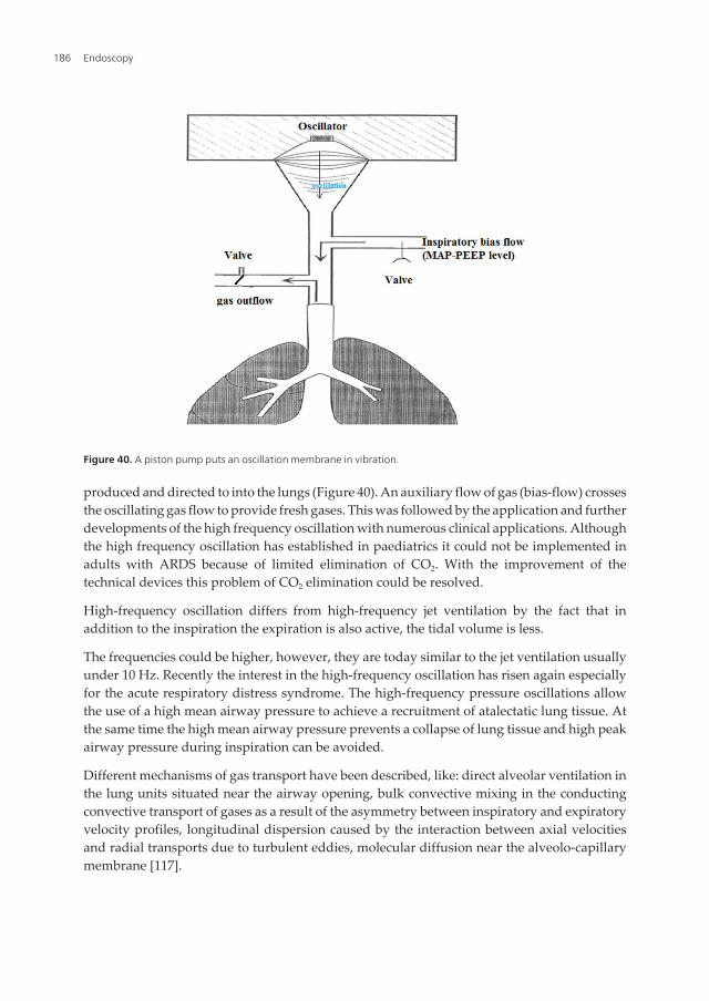
produced and directed to into the lungs (Figure 40). An auxiliary flow of gas (bias-flow) crossesthe oscillating gas flow to provide fresh gases. This was followed by the application and furtherdevelopments of the high frequency oscillation with numerous clinical applications. Althoughthe high frequency oscillation has established in paediatrics it could not be implemented inadults with ARDS because of limited elimination of CO2. With the improvement of thetechnical devices this problem of CO2 elimination could be resolved.
High-frequency oscillation differs from high-frequency jet ventilation by the fact that inaddition to the inspiration the expiration is also active, the tidal volume is less.
The frequencies could be higher, however, they are today similar to the jet ventilation usuallyunder 10 Hz. Recently the interest in the high-frequency oscillation has risen again especiallyfor the acute respiratory distress syndrome. The high-frequency pressure oscillations allowthe use of a high mean airway pressure to achieve a recruitment of atalectatic lung tissue. Atthe same time the high mean airway pressure prevents a collapse of lung tissue and high peakairway pressure during inspiration can be avoided.
Different mechanisms of gas transport have been described, like: direct alveolar ventilation inthe lung units situated near the airway opening, bulk convective mixing in the conductingconvective transport of gases as a result of the asymmetry between inspiratory and expiratoryvelocity profiles, longitudinal dispersion caused by the interaction between axial velocitiesand radial transports due to turbulent eddies, molecular diffusion near the alveolo-capillarymembrane [117].
Figure 40. A piston pump puts an oscillation membrane in vibration.
Endoscopy186
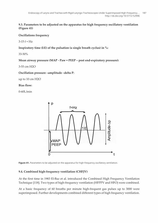
9.3. Parameters to be adjusted on the apparatus for high frequency oscillatory ventilation(Figure 41)
Oscillations frequency
3-15 f = Hz
Inspiratory time (I:E) of the pulsation (a single breath cyclus) in %:
33-50%
Mean airway pressure (MAP - Paw = PEEP – post end-expiratory pressure):
3-55 cm H2O
Oscillation pressure –amplitude –delta P:
up to 10 cm H2O
Bias flow:
0-60L/min
Figure 41. Parameters to be adjusted on the apparatus for high-frequency oscillatory ventilation.
9.4. Combined high-frequency ventilation (CHFJV)
At the first time in 1983 El-Baz et al. introduced the Combined High Frequency VentilationTechnique [118]. Two types of high-frequency ventilation (HFPPV and HFO) were combined.
At a basic frequency of 60 breaths per minute high-frequent gas pulses up to 3000 weresuperimposed. Further developments combined different types of high frequency ventilation.
Endoscopy of Larynx and Trachea with Rigid Laryngo-Tracheoscopes Under Superimposed High-Frequency…http://dx.doi.org/10.5772/52996
187
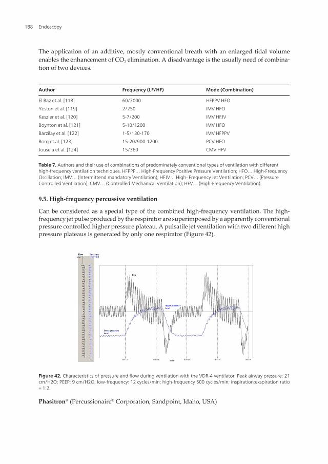
The application of an additive, mostly conventional breath with an enlarged tidal volumeenables the enhancement of CO2 elimination. A disadvantage is the usually need of combina‐tion of two devices.
Author Frequency (LF/HF) Mode (Combination)
El Baz et al. [118]
Yeston et al. [119]
Keszler et al. [120]
Boynton et al. [121]
Barzilay et al. [122]
Borg et al. [123]
Jousela et al. [124]
60/3000
2/250
5-7/200
5-10/1200
1-5/130-170
15-20/900-1200
15/360
HFPPV HFO
IMV HFO
IMV HFJV
IMV HFO
IMV HFPPV
PCV HFO
CMV HFV
Table 7. Authors and their use of combinations of predominately conventional types of ventilation with differenthigh-frequency ventilation techniques. HFPPP… High-Frequency Positive Pressure Ventilation; HFO… High-FrequencyOscillation; IMV… (Intermittend mandatory Ventilation); HFJV… High- Frequency Jet Ventilation; PCV… (PressureControlled Ventilation); CMV… (Controlled Mechanical Ventilation); HFV… (High-Frequency Ventilation).
9.5. High-frequency percussive ventilation
Can be considered as a special type of the combined high-frequency ventilation. The high-frequency jet pulse produced by the respirator are superimposed by a apparently conventionalpressure controlled higher pressure plateau. A pulsatile jet ventilation with two different highpressure plateaus is generated by only one respirator (Figure 42).
Figure 42. Characteristics of pressure and flow during ventilation with the VDR-4 ventilator. Peak airway pressure: 21cm/H2O; PEEP: 9 cm/H2O; low-frequency: 12 cycles/min; high-frequency 500 cycles/min; inspiration:exspiration ratio= 1:2.
Phasitron® (Percussionaire® Corporation, Sandpoint, Idaho, USA)
Endoscopy188
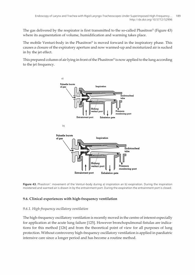
The gas delivered by the respirator is first transmitted to the so-called Phasitron® (Figure 43)where its augmentation of volume, humidification and warming takes place.
The mobile Venturi-body in the Phasitron® is moved forward in the inspiratory phase. Thiscauses a closure of the expiratory aperture and now warmed-up and moisturized air is suckedin by the jet effect.
This prepared column of air lying in front of the Phasitron® is now applied to the lung accordingto the jet frequency.
a)
b)
Figure 43. Phasitron®: movement of the Venturi-body during a) inspiration an b) exspiration. During the inspirationmoistened and warmed air is drawn in by the entrainment port. During the exspiration the entrainment port is closed.
9.6. Clinical experiences with high-frequency ventilation
9.6.1. High-frequency oscillatory ventilation
The high-frequency oscillatory ventilation is recently moved in the centre of interest especiallyfor application at the acute lung failure [125]. However bronchopulmonal fistulas are indica‐tions for this method [126] and from the theoretical point of view for all purposes of lungprotection. Without controversy high-frequency oscillatory ventilation is applied in paediatricintensive care since a longer period and has become a routine method.
Endoscopy of Larynx and Trachea with Rigid Laryngo-Tracheoscopes Under Superimposed High-Frequency…http://dx.doi.org/10.5772/52996
189
The best references for their indicated application at ARDS results from two prospectivestudies [127, 128] and a retrospective clinical trial with application of high-frequency oscilla‐tion in combination with a recruitment manoeuvre [129], all showing significantly increase ofoxygenation in comparison to conventional ventilation.
9.6.2. Combined high-frequency jet ventilation (CHFJV)
The use of the conventional part of ventilation with low ventilation frequencies with conven‐tional PEEP but higher volume tides guarantees a sufficient CO2 elimination.
A high-frequent ventilation mode superimposes the conventional mode. Predominantly theoxygenation is enhanced by the high-frequent pulsatile fraction of ventilation.
9.6.3. High-frequency percussive ventilation
In literature concerning the implementation of the high-frequency percussive ventilation inpatients with acute lung failure was reduced over a long period to paediatric and adult. Moredescriptions exist about successful application in patients with ARDS, after inhalation traumaand taumata [130]. In paediatric patient polpulation with an inhalation trauma a lower rate ofinfection and mortality could be observed. Surprisingly a greater extent of departments applythe high-frequency percussive ventilation in clinical use even though they do not publizisetheir results.
9.6.4. High-frequency jet ventilation
The high-frequency jet ventilation has become widely accepted in operative interventions inthe larynx and trachea. Optimal working conditions with best view for the surgeons areachieved under the application of thin jet catheter and the absence of an endotracheal tube.
Application of high-frequency ventilation in intensive care units in premature patients withrespiratory distress syndrome and interstitial pulmonary emphysema is a safe procedure anddimishes peak pressures. In relation to the outcome and mortality from the high-frequenecyjet ventilation did not derive a benefit in comparison to conventional ventilation.
The exclusive application of high-frequency jet ventilation in adults did not prevail. Usuallyis is applicated as combined high-frequency jet ventilation in cases when the conventionalventilation fails. The literature refers only about case reports or not randomized trials with fewcases. However it demonstrates that these ventilation techniques are in use, although expen‐sive, safe in application with a high potential to increase the oxygenation.
9.6.5. Superimposed high-frequency jet-ventilation (SHFJV)
SHFJV is a special type of combined high-frequency ventilation. A respirator produces a low-frequent jet ventilation with a higher pressure level. Simultaneously a superposition with ahigh-frequent jet ventilation takes place which ensures for itself alone a lower plateau ofpressure analogous a positive end-expiraratory pressure (PEEP). Only one respirator generatesthese two plateaus of pressure. This ventilation technique is primarily used in the operative
Endoscopy190
area of otorinolaryngology, but this Ventilation can be applied also due to ventilation techni‐que of the respirator in the intensive care unit. So we could show in a clinical study [131] thatthis ventilation technique can lead to a quicker recruitment of the lungs in patients with arespiratory insuffiency. However, further studies are necessary to make more precise anddefinitive statements.
9.6.6. Potential effects of high-frequent ventilation techniques
First clinical results show that under high-frequency ventilation potentially a fast recruitmentof dependent areas of the lung occurs without a simultaneous massive overexpansion of non-dependent areas of the lungs. It is conceivable that the under high-frequency ventilationobserved enhanced gas exchange is not so much to be explained by mechanism of increasingdiffusion but by pulsatile mechanism leading to a fast recruitment of lung tissue.
Acknowledgements
The authors thank Mr. Heinrich Reiner from the Carl Reiner Corp. in Vienna, Austria, for hisunderstandingful and dynamic help in implementation and propagation of our jet ventilationprogram at the Medical University of Vienna. Mr. Reiner supported not only the clinical andpractical issues but also the scientific ones.
Author details
Alexander Aloy1* and Matthaeus Grasl2
*Address all correspondence to: [email protected], [email protected]
1 Department of Anaesthesia and General Intensive Care Medicine, Vienna, Austria
2 Department of Otorhinolaryngology, Head & Neck Surgery, Vienna, Austria
This chapter deals with the history of laryngoscopy and the fruitful scientific and clinicalcollaboration of anaesthesiologists and laryngologists in development and application of jetventilation for surgical laryngotracheal interventions via a rigid laryngoscope.
References
[1] Bozzini, Ph. Der Lichtleiter oder die Beschreibung einer einfachen Vorrichtung inner‐er Höhlen und Zwischenräume des lebenden animalischen Körpers. Weimar: Verlagdes Industrie Compoir; (1807).
Endoscopy of Larynx and Trachea with Rigid Laryngo-Tracheoscopes Under Superimposed High-Frequency…http://dx.doi.org/10.5772/52996
191
[2] Mende, L. Von der Bewegung der Stimmritze beym Athemholen; eine neue Entdeck‐ung; mit beygefügten Bemerkungen über den Nutzen und die Verrichtung des Kehl‐deckels. Greifswald; (1916).
[3] Mackenzie, M. The Use of the Laryngoscope in Diseases of the Throat. Philadelphia:Lindsay and Blaciston; (1865).
[4] Babington, B. Report of the Huntarian Society. The London Medical Gazette. (1829);3:565.
[5] Garcia, M. Observations on the Human Voice. Proceedings of the Royal Society ofLondon, (1855);7:397-410.
[6] Türk, L. Kehlkopfspiegel und die Methode seines Gebrauchs. Z Ges Ärzte zu Wien.(1858);26:401-409.
[7] Helmholtz von H. Beschreibung eines Augenspiegls zur Untersuchung der Netzhautim lebenden Auge; Berlin.. Translation in Arch Ophthalmol. (1951);46:565-583.
[8] Czermak JN. Über den Kehlkopfspiegel. Wiener Med Wschr. (1858);8:196-198.
[9] Oertel MJ. Das Laryngo-stroboskop und die laryngostroboskopische Untersuchung.Arch Laryngorhinologie. (1985);3:1-16.
[10] Morton W. Remarks on the Proper Mode of Admininstering Sulfuric Ether by Inhala‐tion. Dutton and Wentworth; (1847).
[11] Lister J. On the Antiseptic Principle of Surgery. The British Medical Journal (1867);351:245-260.
[12] Jelinek E. Das Cocain als Anästhetikum für den Phaynx und den Larynx. WienerMed Wschr. (1934);34:1334-1337 and 1364-1367.
[13] Miller RA. A new laryngoscope. Anaesthesiology. (1941);7:205-206.
[14] Macintosh RR. New Inventions. A new laryngoscope. Lancet. (1943);241:205.
[15] Tobold A. Lehrbuch der Laryngoskopie. Berlin; August Hirschwald Verlag, (1869).
[16] Kirstein, A. Autoskopie des Larynx und der Trachea (Laryngoscopia directa, Euthy‐skopie, Besichtigung ohne Spiegel). Archiv für Laryngologie und Rhinologie. (1895);3:156-164.
[17] Kollofrath O. Entfernung eines Knochenstücks aus dem rechten Bronchus auf natür‐lichem Wege und unter Anwendung der directen Laryngoskopie. Münch MedWschr. (1897);38: 1038-1039.
[18] Killian G. Die Schwebelaryngoscopie. Archiv für Laryngologie und Rhinologie.(1912);26:277-317.
Endoscopy192
[19] Brünings W. Direct Laryngoscopy: Criteria determinating the appliccability of auto‐scopy. Direct laryngoscopy, brochoscopy and esophagoscopy. London: Bailliere, Tin‐dall, Cox; (1912);93-95.
[20] Hartmann A. Zur Behandlung der Larynxtuberkulose. In: Hoffmann R (Hrsg) Verh dVereins dt Laryngologen 1908-1912. Würzburg: Kabitsch; (1913).
[21] Holinger P. A new anterior commissure laryngoscope. Ann Otol Rhinol Laryngol.(1947);56:437-440.
[22] Kleinsasser O. Larynx-Microscope for Early Diagnosis and Differential-Diagnosis forCarcinoma of the Larynx, Pharynx and Oral Cavity. Z Laryng Rhinol. (1961);40:276-279.
[23] Jako GJ. Laryngoscope for microscopic observation, surgery and photography. Thedevelopment of an instrument. Arch Otolaryngol (1970);91:196-199.
[24] Strong MS. Microscopic laryngoscopy. A review and appraisal. Laryngoscope.(1970);80:1540-1552.
[25] Weerda H, Pedersen P, Meuret G, Wehmer H, Braune H. A new laryngoscope for en‐dolaryngeal microsurgery. A contribution to inject respiration. Arch Otolaryngol.(1979);225:103-106.
[26] Hopkins, HH. The Application of Frequency Response Techniques in Optics. Pro‐ceedings of the Physical Society. (1962);79(5): 889-896.
[27] Volhard F. Über die künstliche Beatmung durch Ventilation der Trachea und eineeinfache Vorrichtung zur rhytmischen künstlichen Atmung. Münch Med Wschr.(1908);55:209-211.
[28] Meltzer SJ, Auer J. Continuous respiration without respiration movements. J ExpMed. (1909);11:622-625.
[29] Barth L. Thearpeutic use of diffusion breathing in bronchoscopy. Anaesthesist.(1954);3(5) 227-229.
[30] Sanders RD. Two ventilatory attachments for bronchocopes. Del Med J. (1967);39:170-176.
[31] Gebert E, Deilman M, Pedersen P. Inject-ventilation in brochoscopy. Anaesthesist.(1979);28(8) 378-389.
[32] Lee S. A ventilating laryngoscope for inhalation anaestesia and augmented ventila‐tion during laryngoscopic procedures. Br. J Anaesth. (1972);44(8) 874-878.
[33] Bradley JL, Moyes RN, Parke FW. Modifications of Sanders` techique of ventilationduring bronchoscopy. Thorax. (1971);26(1) 112-114.
Endoscopy of Larynx and Trachea with Rigid Laryngo-Tracheoscopes Under Superimposed High-Frequency…http://dx.doi.org/10.5772/52996
193
[34] Lunkenheimer PP, Raffenbeul W, Keller H, Frank I, Dickhut HH, Fuhrmann C. Ap‐plication of transtracheal pressure oscillations as modification of “diffusion respira‐tion”. Br Anaesth. (1972);44(6) 627-633 .
[35] Lunkenheimer PP, Frank I, Ising H, Keller H, Dickhut HH, Fuhrmann C. Intrapulmo‐nal gas exchange during simulated apnea due to transtracheal periodic intrathoracicpressure changes. Anaesthesist (1973);22(5):232-238.
[36] Bohn DJ, Mijasaka K, Marchak BE, Thomson WK, Froese AB, Bryan AC. Ventilationby high-frequency oscillation. J Appl Physiol (1980);48(4) 710-716.
[37] Sjöstrand U. Review of the physiological rationale for and development of high-fre‐quency positive pressure ventilation. Acta Anaesth Scand [Suppl.] (1977);64:7-27.
[38] Sjöstrand U. High-frequency positive pressure ventilation (HFPPV): a review. CritCare Med (1980);8(6) 345-364.
[39] Smith RB, Lindholm CE, Klain M. Jet ventilation for fiberoptic bronchoscopy undergeneral anaesthesia. Acta Anaesthesiol Scand. (1976);20(2) 111-116.
[40] Babinski M, Smith RB, Klain M. High-frequency jet ventilation for laryngoscopy. An‐esthesiology. (1980);52(2) 178-180.
[41] Klain M, Kezler H. High-frequency jet ventilation. Surg Clin North Am. (1985);65(4)917-930.
[42] Aloy A, Schachner M, Spiss CK, Cancura W. Tube-free translaryngeal superposed jetventialtion. Anaesthesist. (1990);39(19) 493-498.
[43] Kleinsaser O. Microlaryngoscopy and endolaryngeal microsurgery. Philadelphia:WB Saunders & Co; (1968).
[44] Jako GJ. Laryngoscope for microscopic obeservation, surgery and photography. ArchOtol. (1970);91(2) 196-199.
[45] Strong MS. Microscopic laryngoscopy. A review and appraisal. Laryngoscope.(1970);80(10) 1540-1552.
[46] Weerda H, Pedersen P, Meuret G, Wehmer H, Braune H. A new laryngoscope for en‐dolaryngeal microsurgery. A contribution to inject respiration. Arch Otolaryngol.(1979);225:103-106.
[47] Ono J, Saito S. Endoscopic microsurgery of the larynx. Ann Otol Rhinol Laryngol.(1971);80(4) 479-86.
[48] Thomas GK. Suspension apparatus for laryngeal microsurgery. Arch Otolaryngol.(1971);94(3) 258-259.
[49] Steiner W, Ambrosch P. Endoscopic Laser surgery of the upper aerodigestiv tract.With special emphasis on cancer surgery. Stuttgart/New York: Thieme; (1997).
Endoscopy194
[50] Cohen SR, Thompson JW. Use of botulinum toxin to lateralize true vocal cords: a bio‐chemical method to relieve bilateral abductor vocal cord paralysis. Ann Otol RhinolLaryngol. (1987);96(4) 534-541.
[51] Aloy A, Schachner M, Cancura W. Tubeless translaryngeal superimposed jet-ventila‐tion. Eur Arch Otorhinolaryngol. (1991);248(8) 475-478.
[52] Ng A, Russell WC, Harvey N, Thompson JP. Comparing methods of administeringhigh frequency jet ventilation in a model of laryngotracheal srenosis. Anesth Analg.(2002);95(3): 764-769.
[53] Aloy A, Schachner M, Spiss CK, Cancura W. Tube-free translaryngeal superimposedjet ventilation. Anaesthesist. (1990);39(10) 493-498.
[54] Kleinsasser Operations Laryngoskop; Zepf. Medical Instruments; Germany Tuttlin‐gen
[55] Aloy A, Schragl E, Neth H, Donner A, Kluwick A. Flow pattern of respiratory gasesin superimposed high-frequncy jet ventilation (SHFJV) with the jet laryngoscope.Anaesthesist (1995);44(6) 558-565.
[56] Piquet J. Turbulent Flows: Models and Physics. Berlin-Heidelber-New York: SpringerVerlag; (2001).
[57] Halder G. Introduction to chemical engineering thermodynamics. New Dehli: BookVistas, PHI:learning; (2009).
[58] Winterbone D.E. Advanced Thermodynamics for Engineers; Oxford: ButterworthHeinemann; An imprint of Elsevier Science (2002).
[59] Leiter R, Aliverti A, Priori R, Staun P, Lo Mauro A, Larsson A, Frykholm P. Compari‐son of superimposed high-frequency jet ventilation with conventional jet ventilationfor laryngeal surgery. Br J Anaesth. (2012);108(4):690-698.
[60] Lanzenberger-Schragl E, Donner A, Grasl M, Zimpfer M, AloyA. SuperimposedHigh-Frequency Jet Ventilation for Laryngeal and Tracheal Surgery. Arch Otolaryng‐ol Head Neck Surg. (2000);126:40-44.
[61] Rezaie-Majd A, Bigenzahn W, Denk DM, Burian M, Kornfehl J, Grasl MCh, Ihra G,Aloy A. Superimposed high-frequency jet ventilation (SHFJV) for endoscopic laryng‐otracheal surgery in more than 1500 patients. Br J Anaesth. (2006);96(5):650-9. Epub2006 Mar 30.
[62] Nowak A, Langebach R, Klemm E, HellerW. Percutaneous dilational tracheostomy(PDT) and prevention of blood aspiration with superimposed high-frequency jet ven‐tilation (SHFJV) using the tracheotomy-endoscope (TED): results of numerical andexperimental simulations. Biomed Tech, Berlin. (2012);57(2):107-111.
[63] Aloy A, Schachner M, Spiss CK, Cancura W. Tube-free translaryngeal superposed jetventialtion. Anaesthesist. (1990);39(10) 493-498.
Endoscopy of Larynx and Trachea with Rigid Laryngo-Tracheoscopes Under Superimposed High-Frequency…http://dx.doi.org/10.5772/52996
195
[64] Lanzenberger-Schragl E, Donner A, Grasl MC, Zimpfer M, Aloy A. Superimposedhigh-frequency jet ventilation for laryngeal and tracheal surgery. Arch OtolaryngolHead Neck Surg. (2000);126(1) 40-44.
[65] Rezaie-Majd A, Bigenzahn W, Denk DM, Burian M, Kornfehl J, Grasl MCh, Ihra G,Aloy A. Superimposed high-frequncy jet ventilation (SHFJV) for endoscopic laryngo‐tracheal surgery in more than 1500 patients. Br J Anaesth. (2006);96(5) 650-569.
[66] Jaquet Y, Monnier P, Van Melle G, Ravussin P, Spahn DR, Chollet-Rivier M. Compli‐cations of different ventilation strategies in endoscopic laryngeal surgery. A 10-yearreview. Anaesthesiology. (2006);104(1) 52-59.
[67] Rampii IJ. Anaesthesia for laser surgery. In Miller RD, ed. Miller`s Anesthesia. 7 thed. Philadelphia: Chruchill Livingston; (2010); p2405-2418.
[68] Wöllmer W, Schade G, Kessler G. Endotracheal tube fires still happen – a short over‐view. Med Laser Appl. (2010);25(2) 118-25.
[69] Burian K, Höfler H. On microsurgical treatment of vocal cord carcinomas with CO2-laser. Laryngol Rhinol Otol. (1979);58(7) 551-556.
[70] Strong MS. Laser excision o fcarcinoma of the larynx. Laryngoscope. (1975);85:1286-1289.
[71] Steiner W, Ambrosch P. Laser microsurgery for laryngeal carcinoma. In: Steiner W,Ambrosch P. (ed.) Endoscopic Laser Surgery of the Upper Aerodigestive Tract. NewYork: Thieme; (2000); p47-82.
[72] Thurnher D, Erovic BM, Frommlet F, Brannath W, Ehrenberger K, Jansen B, Selzer E,Grasl MC. Challenging a dogma--surgery yields superior long-term results for T1asquamous cell carcinoma of the glottic larynx compared to radiotherapy. Eur J SurgOncol. (2008);34(6) 692-698.
[73] Grasl M.C, Donner A, Schragl E, Aloy A. Tubeless laryngotracheal surgery in infantsand children via jet ventilation laryngoscope. Laryngoscope. 1997;107(2) 277-281.
[74] Aloy A, Schachner M, Cancura W. Tubeless translaryngeal superimposed jet ventila‐tion. Eur Arch Otorhinolaryngol. (1991);248(8):475-478.
[75] Ossoff RH, Tucker JA, Werkhaven JA. Neonatal and pediatric microsubglottiscopeset. Otol Rhinol Laryngol. (1991);100(4) 325-326.
[76] Bourgain JL, Desruennes E, Fischler M, Ravussin P. Transtracheal high frequency jetventilation for endoscopic airway surgery: a multicentre study. Br J Anaesth. (2001);87(6) 870-875.
[77] Ravussin P, Bayer-Berger M, Monnier P, Savary M, Freeman J. Percutaneous trans‐tracheal ventilation for laser endoscopic procedures in infants and small childrenwith laryngeal obstruction: report of two cases. Can J Anaesth. (1987);34(1) 83-86.
Endoscopy196
[78] Depierraz B, Ravussin P, Brossad E, Monnier P. Percutaneous transtracheal jet venti‐lation for paediatric endoscopic laser treatment of laryngeal and subglottic lesions.Can J Anaesth. (1994);41(12) 1200-1207.
[79] Ross-Anderson D.J, Ferguson C, Patel A. Transtracheal jet ventilation in 50 patientswith severe airway compromise and stridor. Br J Anaesth. (2011);106(1) 140-144.
[80] Grasl M.C, Donner A, Schragl E, Aloy A (1997) Tubeless laryngotracheal surgery ininfants and children via jet ventilation laryngoscope. Laryngoscope. (1997);107(2)277-281.
[81] Mausser G, Friedrich G, Schwarz G. Airway mangement and anesthesia in neonates,infants and children during endolaryngotracheal surgery. Paediatr Anaesth. (2007);17(10) 942-947.
[82] Ihra G, Hieber C, Adel S, Kashanipour A, Aloy A. Tubeless combined high-frequen‐cy jet ventilation for laryngotracheal laser surgery in paediatric anaesthesia. Acta An‐aesthesiol Scand. (2000);44(4) 475-479.
[83] Tan SS, Dhara SS, Sim CK. Removal of a laryngeal foreign body using high frequencyjet ventilation. Anasthesia. (1991);46(9) 741-3.
[84] Derkay CS, Faust RA. Recurrent respiratory papillomatosis. In: Flint PW, HaugheyBH, Lund VJ, et al. eds. Cummings Otolaryngology: Head & Neck Surgery. 5 th ed.Philadelphia: Mosby; (2010); p2884-2885.
[85] Derkay CS. Recurrent respiratory papillomatosis. Laryngoscope. (2001);111(1) 57-69.
[86] Weisberger EC, Miner JD. Apneic anaesthesia for improved endoscopic removal oflaryngeal papillomata. Laryngoscope. (1988);98(7) 693-697.
[87] Drenger B, Zidenbaum M, Reifen E, Leitersdorf E. Severe upper airway obstructionand difficult intubation in cicatricial pemphigoid. Anaesthesia (1986);41(10)1029-1031.
[88] Hallenborg C, Rowe LD, Gamsu G, Boushev HA, Golden JA. Severe upper airwayobstruction caused by bullous pemphigoid: diagnostic usefulness of the flow-volumecurve.Otolaryngol Head Neck Surg. (1982);90(1):20-24.
[89] Monniere Ph, editor. Pediatric Airway surgery. Management of laryngotrachealstenosis in infants and children. New York: Springer; (2011).
[90] Schragl E, Donner A, Kashanipour A, Gradwohl I, Ullrich R, Aloy A. Anesthesia inacute respiratory tract obstructions caused by high grade degree laryngeal and tra‐cheobronchial stenosis. Anaesthesiol Intensivmed Notfallmed Schmerzther. (1994);29(5) 269-277.
[91] Schragl E, Donner A, Grasl MC, Kashanipour A, Aloy A. Beatmung während einerTracheotomie bei langstreckiger 90% iger laryngealer Stenose mittels superponierter
Endoscopy of Larynx and Trachea with Rigid Laryngo-Tracheoscopes Under Superimposed High-Frequency…http://dx.doi.org/10.5772/52996
197
Hochfrequenz Jet-Ventilation über das Jet-Laryngoskop. Laryngorhinootologie.(1995);74(4) 223-226.
[92] Cotton RT. Management of subglottic stenosis. Otolaryngol Clin North Am (2000);33(1) 111-130.
[93] Aloy A, Kimla T, Schragl E, Donner A, Grasl M. (1994) Tubeless superimposed highfrequency jet ventilation in high grade laryngeal stenosis. Laryngorhinootologie(1994);73(8) 405-411.
[94] Olak J, Rosenberg S. Simple technique for sizing and positioning tracheal stents. AnnThorac Surg. (2000);70(4) 1389-1390.
[95] Wassermann K, Eckel HE, Michel O, Mueller RP. Emergency stenting of malignantobstruction of the upper airways: long-term follow-up with two types of siliconeprosteses. Ann Otol Rhinol Laryngol (1998);107(2) 149-154.
[96] Saito Y, Imamura H. Airway stenting. Surg Today (2005);35(4) 265-270.
[97] Wood DE. Airway stenting. Chest Surg Clin N Am (2001);11(4) 841–860.
[98] Shin JH. Interventional management of tracheobronchial strctures. World Journal ofRadiology. (2010);2(8) 323-328.
[99] Bourgain JL, Desruennes E, Fischler M, Ravussin.Transtracheal high frequency jetventilation for endoscopic airway surgery: a multcenter study. Br J Anaesth (2001);87(6) 870-875.
[100] Aloy A, Donner A, Strasser K, Klepetko W, Schragl E, Taslimi R, Rotheneder E, Ka‐shanipour A. Jet ventilation superimposed on a special jet laryngoscope for endolu‐minal stent insertion in the tracheobronchial system. Anaesthesist. (1994);43(4)262-269.
[101] Eckel HE, Berendes Sh, Damm M, Klusmann JP, Wassermann K. Suspension lar‐yngoscopy for endotracheal stenting. Laryngoscope (2003);113(1) 11-15.
[102] Baraka AS, Siddik SS, Taha SK, Jalbout MI, Massouh FM. Low frequency jet ventila‐tion for stent insertion in a patient with tracheal stenosis. Can J Anaesth (2001);48(7)701-704.
[103] Brodsky JB. Anesthesia for pulmonary stent insertion. Curr Opin Anaesthesiol.(2003);16(1) 65-67.
[104] Klepetko W, Müller MR, Grimm M, Aloy A, Kashanipour A, Wisser W, EckersbergerF, Wolner E. doluminale Schienung (Stenting) bei Stenosen des Tracheobronchialsys‐tems. Acta Chirurgica Austriaca. (1991);23(3)124-129 .
[105] Choudhury M, Saxena N. Total intravenous anaesthesia for tracheobronchial stent‐ing in children. Anaesth Intensive Care (2002);30(3) 376-379.
Endoscopy198
[106] Wassermann K, Eckel HE, Michel O, Mueller RP. Emergency stenting of malignantobstruction of the upper airways: long-term follow-up with two types of siliconeprostheses. J Thorac Cardiovasc Surg (1996);112(4) 859-66.
[107] Ernst A, Majid A, Feller-Kopman D, Guerrero J, Boisele P, Loring SH, O`Donnel C,Decamp M, Herth FJ, Gangadharan S, Ashiku S. Airway stabilisation with siliconestents for treating adult tracheoronchomalacies. Chest (2007);132(2) 609-916.
[108] Montgomery WW. T-tube tracheal stent. Arch Otolaryngol (1965);82(5) 320-321.
[109] Grillo HC. Surgery of the trachea and bronchi. London: B.C. Decker Inc.; (2004).
[110] Froese AB, Bryan AC, High frequency ventilation. Am Rev Respir Dis (1987);135(6)1363-1374.
[111] Baum M, Benzer H, Geyer A, Haider W, Mutz N, Forced diffusion ventilation (FDV).Bases and clinical application. Anaesthesist. (1980);29(11) 586-591.
[112] Baum M, Benzer H, Mutz N, Pauser G, Tonczar L. Inversed ratio ventilation (FDV).Role of the respiratory time ratio in artifcial respiration in ARDS. Anaesthesist (1980);29(11) 592-596.
[113] Slutsky AS, Drazen JM, Ventilation with small tidal volumes. N Engl J Med (2002);347(9) 630-631.
[114] Krishnan JA, Brower RG. High-frequency ventilation for acute lung injury andARDS. Chest (2000);118(3) 795-807.
[115] Shimaoku M, Fujino Y, Taenaka N, Hiroi T, Kiyono H, Yoshiya II. High frequencyoscillatory ventilation attenuates the activation of alveolar macrophages and neutro‐phils in lung injury. Crit Care (1998);2(1) 35-39.
[116] Lunkenheimer PP, Redmann K, Krebs S, Gleich C, Brasselet M, Scheld HH. Ventila‐tion by high frequency oscillations in adults. An experimental study of conditionsand methods.Can Anesthesiol (1994);42(3): 303-314.
[117] Chang HK. Mechanisms of gas transport during ventilation by high-frequency oscil‐lation J.App.Physiol. (1984) 56(3):553-563.
[118] El-Baz N, Penfield Faber L, Doolas A. Combined high-frequency ventilation for man‐agement of terminal respiratory failure: a new technique. Anesth Analg. (1983);62(1)39-49.
[119] Yeston NS, Grasberger RC, McCormick JR. Severe combined respiratory and myocar‐dial failure treated with high-frequency jet ventilation. Crit Care Med. (1985);13(3):208-209.
[120] Keszler M, Donn SM, Spitzer AR. High-frequency jet ventilation in respiratory dis‐tress syndrome. J Pediatr. (1991);119(2):340-341.
Endoscopy of Larynx and Trachea with Rigid Laryngo-Tracheoscopes Under Superimposed High-Frequency…http://dx.doi.org/10.5772/52996
199
[121] Boynton BR, Mannino FL, Davis RF, Kopotic RJ, Friederichsen G. Combined high-frequency oscillatory ventilation and intermittent mandatory ventilation in criticallyill neonates. J Pediatr. (1984);105(2):297-302.
[122] Barzilay E, Kessler D, Raz R. Superimposed high frequency ventilation with conven‐tional mechanical ventilation. Chest. (1989);95(3):681-682.
[123] Borg UR, Stoklosa JC, Siegel JH, Wiles CE 3rd, Belzberg H, Blevins S, Cotter K, LaghiF, Rivkind A. Prospective evaluation of combined high-frequency ventilation in post-traumatic patients with adult respiratory distress syndrome refractory to optimizedconventional ventilatory management. Crit Care Med. (1989);17(11):1129-1142.
[124] Jousela I, Mäkeläinen A, Linko K. The effect of combined high frequency ventilationwith and without continuous positive airway pressure in experimental lung injury.Acta Anaesthesiol Scand. (1992);36(6):508-512.
[125] McLuckie A, Editorial II: High-frequency oscillation in acute respiratory distress syn‐drome (ARDS). Brit J Anaesth (2004);93(3) 322-324.
[126] Ha DV, Johnson D. High frequency oscillatory ventilation in the management of ahigh output bronchopleural fistula: a case report. Can J of Anesth (2004);51(1) 78-83.
[127] Mehta S, Lapinsky SE, Hallett DC, Merker D, Groll RJ, Cooper AB, MacDonald RJ,Stewart TE. Prospective trial of high-frequency oscillation in adults with acute repir‐atory distress syndrome. Crit Care Med (2001);29(7) 1360-1369.
[128] Derdak S, Metha S, Steward T, Smith T, Rogers M, Buchmann T, Carlin B, Lowson S,Granton J and the multicenter oscillatory ventilation for acute respiratory distresssyndrome TRIAL (MOAT) study investigators. High frequency oscillatory ventila‐tion for acute respiratory distress syndrome in adults. Am J Respir Crit Care Med(2002);166(6) 801-808.
[129] Ferguson ND, Chiche JD, Kacmarek RM, Hallett DC, Metha S, Findlay GP, GrantonJT, Slutsky AS, Stewart TE, Combining high-frequency oscillatory ventilation and re‐cruitment maneuvers in adults with early acute respiratory distress syndrome: thetreatment with oscillation and an Open Lung Strategy (TOOLS) trial pilot study. CritCare Med (2005);33(3): 479-486.
[130] Cortiella J, Mlcak R, Herndon D. High frequency percussive ventilation in pediatricpatients with inhalation injury. J Burn Care Rehabil (1999);20(3) 232-235.
[131] Kraincuk P, Körmöczi G, Prokop M, Ihra G, Aloy A. Alveolar recruitment of atelecta‐sis under combined high-frequency jet ventilation: a computed tomography study.Intensive Care Med (2003);29:1265-1272.
Endoscopy200
Chapter 10
Bronchology – A Well Branched Tree
Mihai Olteanu, Costin Teodor Streba,Bogdan Oprea, Raluca Marinas, Mimi Floarea Nitu,Emilia Crisan and Tudorel Ciurea
Additional information is available at the end of the chapter
http://dx.doi.org/10.5772/52748
1. Introduction
1.1. Lung cancer – Stadialization
Lung cancer is responsible for 1.3 million deaths worldwide annually, and it is the mostcommon cause of cancer-related death in men and the second most common in women.Lung cancer staging is the assessment of the degree to which a lung cancer has spread fromits original source. As with most cancers, for lung cancer staging is of paramount impor‐tance for the treatment planning process and prognosis. Two primary methods of lung can‐cer staging are available: clinical staging and pathologic staging. In clinical staging,information is provided by noninvasive or minimally invasive techniques, such as physicalexamination, radiologic examination, endoscopic ultrasound, bronchoscopy, mediastinosco‐py, and thoracoscopy. In pathologic staging, information obtained from clinical staging iscombined with findings from both the invasive surgical procedure and the pathologic evalu‐ation of excised tissue. Clinical staging is important and can help to determine the next ap‐propriate step in therapy, such as the decision to proceed with pathologic staging, whichremains the reference standard because the overall level of agreement between the two sys‐tems only ranges from 35% to 55% [1].
Definition of the stage is an essential part of the approach to patients with lung cancer, andit has led to the development of a universally accepted stage classification systems for mosttumors. The Union Internationale Contre le Cancer (UICC) and the American Joint Commit‐tee on Cancer (AJCC) periodically define, review, and refine the stage classification systems.Nearly half of all patients with lung cancer have mediastinal disease at diagnosis, and thisimplies metastases to ipsilateral or subcarinal nodes (N2) that are classified as stage IIIA dis‐
© 2013 Olteanu et al.; licensee InTech. This is an open access article distributed under the terms of theCreative Commons Attribution License (http://creativecommons.org/licenses/by/3.0), which permitsunrestricted use, distribution, and reproduction in any medium, provided the original work is properly cited.

ease. Management of stage IIIA disease is more controversial, but many centers treat it withradiation and chemotherapy, with surgery performed under investigational protocols. Di‐rect mediastinal invasion (T4) or metastasis to contralateral mediastinal nodes (N3) is classi‐fied as stage IIIB disease. The 5-year survival rate in this case is at best 5%, and patients aregenerally offered treatment with chemoradiotherapy without surgery.
The previous tumor, node, and metastasis (TNM) classification, the sixth edition of the TNMClassification of Malignant Tumors, was published in 2002. The last revision of the stagingsystem for lung cancer has been presented by Dr. Clifton Mountain in 1997 [2], after his pro‐posal had been adopted in 1973 by the AJCC in the same year and by the UICC in 1974, andthis revision remained unaltered on the 2002 manual [3]. The 1973 original system wasbased on a database of 2,155 patients from the MD Anderson Cancer Center in Houston, TX.Subsequent revisions of the TNM staging system continued to be based on this database,which grew to include 5,319 cases at the time the lung cancer staging system was revised in1997 and it was also validated by Dr. Naruke from Japan in 2001. Mountain’s revision wasbased on a relatively small population of patients who underwent surgical treatment in asingle geographic area, and no validation was presented to justify the individual descriptors[4]. In order to overcome the limitations of the sixth TNM classification, the InternationalAssociation for the Study of Lung Cancer (IASLC) launched the Lung Cancer StagingProject in 1996, and in 1998 created an International Staging Committee (ISC) of multidisci‐plinary members to conduct revisions [5]. Changes to the sixth edition were proposed by theIASLC based on an international collection and review of 100,869 patients from 46 sources of20 countries. Data were drawn from lung cancer cases treated by all modalities between1990 and 2000. After exclusion of ineligible cases, 81,015 patients (67,725 NSCLC and 13,290SCLC) remained for investigation [6]. Proposals for revision were submitted to the AJCCand UICC for consideration in the new edition of the staging manual, the 7th, and both ac‐cepted the recommendations. In the last months of 2009, the Seventh Edition of the TNMClassification of Malignant Tumors was published, with a new lung cancer staging system.The new edition took effect on January 1, 2010 [7].
The changes recommended by the IASLC for the 7th edition of TNM classification for lungcancer were based on differences in survival [8], and the results of the data analysis wereinternally and externally validated [9]. These changes include new T and M definitions andconsequent new stage groupings, a new lymph node map, a novel definition on pleural in‐vasion, as well as recommendations to apply the TNM system to broncho-pulmonary carci‐noid tumors and SCLC.
The success story of EBUS-TBNA starts in 2003 with a publication in Thorax by MarkKrasnik and Peter Vilmann from Gentofte University Hospital, Denmark [10]. This articlegave the first description of the principle of EBUS-TBNA. In the same journal, in 2006, thesame group, together with a group from the Thoraxklinik in Heidelberg and Harvad Med‐ical School’s Beth Israel Deaconess MC, published their study on 502 patients that showedthat EBUS-TBNA resulted in 93% diagnostic yield, a sensitivity of 94%, specificity of 100%and accuracy 94%, with PPV at 100% and NPV at 11% [11]. In 2006, an internationalEBUS-TBNA focus group was formed by Felix J.F. Herth (Copenhagen), Kazuhiro Yasufu‐
Endoscopy202
ku (Chiba), Robert Rintoul (Cambridge) and Armin Ernst (Boston) that published in theJournal of Bronchology a description of how to do an EBUS-TBNA thus offering a de‐tailed description of local lymph node position and orientation within the mediastinum[12]. EBUS-TBNA has been studied and compared also in relation with existing modalitieslike EUS –FNA (by Vilmann et al. in 2005 on 33 pts. and by Herth et al. in 2005 on 160pts.), PET-CT (by Yasufuku et al. in 2006 on 102 pts.), classical TBNA and EUS-FNA (byWallace et al. in 2008 on 138 pts.).
EBUS-TBNA was proven in 2007 by Wong and Yasufuku et al. on 65 patients to be a safemethod allowing a high yield also for the diagnosis of sarcoidosis [13].
It soon became widely accepted that EBUS-TBNA is a reliable diagnostic tool for enlargedlymph nodes in patients with NSCLC and that lymph nodes below the one centimeter rangecould also be sampled. This led to a study with 100 patients published in 2006 in EuropeanRespiratory Journal [14] that showed that every sixth patient with no evidence of mediasti‐nal disease on CT was diagnosed positive using EBUS-TBNA. Thus, EBUS-TBNA showedpotential to avoid unnecessary exploratory thoracoscopies. Following the same idea, in 2008Hwangbo et al. showed that in cases with both CT- and PET-negative and –positive scans,EBUS-TBNA is an excellent tool for detecting mediastinal metastasis, thereby confirmingthat EBUS-TBNA is an effective invasive method following CT and PET scanning [15].
In the same year, Armin Ernst et al. showed in Journal of Thoracic Oncology on 66 patientsthat EBUS-TBNA can have a superior yield compared to cervical mediastinoscopy, whichintuitively suggested that mediastinoscopy is not necessarily of additional diagnostic benefitin evaluating negative EBUS-TBNA staged lymph nodes [16]. However, mediastinoscopyhas an important role especially in operable patients with non-enlarged lymph nodes for as‐sessing local mediastinal invasion and the exclusion of metastatic disease.
Herth et al. evaluated EBUS-TBNA for re-staging in 124 patients with tissue-proven IIIA-N2disease after induction chemotherapy (Journal of Clinical Oncology 2008) and concludedthat EBUS-TBNA is a valuable and practical tool for re-staging with a sensitivity of 76%,specificity of 100%, PPV of 100%, NPV of 20% and diagnostic accuracy of 77% [17]. Theseresults imply that a negative EBUS-TBNA for re-staging should be surgically re-staged.
In 2006, the compatibility between EBUS-TBNA and the Aloka Prosound alpha5 ultrasoundprocessor made additional Doppler modes available, and this led to a consecutive study byHerth et al. on 89 patients that described changes in flow resistance parameters (resistanceindex by Pourcelot) in malignant lymph nodes [18].
Yasufuku et al. from Chiba University have shown great dedication to evaluating the ben‐efits of EBUS-TBNA samples for immunohistochemical analysis and reported encouragingresults with cell cycle-related proteins in chemotherapy patients in Thorax 2008, and ayear earlier in Chest the same group showed that epidermal growth factor receptor(EGFR) mutation can be easily detected in metastatic lymph node samples for EBUS-TBNA and so the samples gained by EBUS-TBNA allowed genetic evaluations of tumourcells from lymph nodes [19, 20].
Bronchology – A Well Branched Treehttp://dx.doi.org/10.5772/52748
203
In 2009 Tournoy et al. provided a detailed analysis of endosonographic landmarks (whereavailable), describing the anatomic borders of the lymph node stations as defined in the 7thedition of the IASLC’s TNM-staging nomenclature, which is relevant for correctly stagingpatients with lung cancer [21].
Building on the strong results of combined EUS-FNA and EBUS-TBNA procedures – a studypublished by Vilman et al. in 2005 had already indicated their complementary nature – An‐nema et al. challenged in 2010 the pre-dominant surgical staging algorithm by comparingthe combined EBUS-TBNA and EUS-TNA with surgical staging alone [22] and showed thatcombining endosonographic and surgical staging resulted in greater sensitivity for mediasti‐nal nodal metastases and fewer unnecessary thoracotomies. These results indicated that thecombination of both procedures may be able to replace surgical stages as the primary stag‐ing method for patients with lung cancer.
Instead of using different scopes for EBUS-TBNA and EUS-TNA, two separate studies pub‐lished in Chest in 2010 by Hwangbo et al. and Herth et al. [23] used only one bronchoscopefor both procedures, starting via the trachea and continuing via the oesophageal route. Theycame to the conclusion that EBUS-TBNA and EUS-TNA are complementary methods andshowed that both procedures can be performed with a single EBUS echoendoscope in onesitting by one operator. A further study in 2011, “Nonsurgical staging of the mediastinum:EBUS and EUS” conducted by Herth, stated that the combination of both procedures ach‐ieves a complete and accurate mediastinal staging. Therefore it can be expected that the im‐plementation of combined EBUS-TBNA and EUS-FNA will reduce the need for surgicalstaging of lung cancer significantly [24].
The summary of scientific studies on EBUS-TBNA provided about clearly shows the proce‐dure’s power in helping to improve mediastinal staging of lung cancer during the past 10years. The technological development of less invasive staging and sampling devices contin‐ues endoscopists using endosonography, we can expect further exciting developments inclinical practice in the years to come.
Endobronchial ultrasound with real-time-guided transbronchial fine-needle aspiration(EBUS-TBNA) is a minimally invasive outpatient procedure by which mediastinal [25] andhilar lymph nodes [26] as well as centrally located primary lung lesions [27] can be visual‐ised and sampled under ultrasound guidance. In a systematic review, EBUS-TBNA hasshown a pooled sensitivity of 93% in the staging of non-small-cell lung cancer. In a directcomparison with surgical staging, EBUS-TBNA even showed to be superior [28]. Therefore,EBUS-TBNA has been adopted in the most recent lung cancer staging guidelines as a mini‐mally invasive alternative to surgical staging [29]
EBUS-TBNA is an accurate, minimally invasive and safe staging procedure and can be con‐sidered the procedure of choice for patients with extrathoracic malignancies in whom hilaror mediastinal lesions are observed. In patients with (concurrent or previously treated) ex‐trathoracic malignancy, EBUS-TBNA has a sensitivity of 85% to demonstrate metastaticspread. Implementation of EBUS-TBNA in these patients obviates invasive surgical diagnos‐tic procedures in 61%. [30, 31]
Endoscopy204
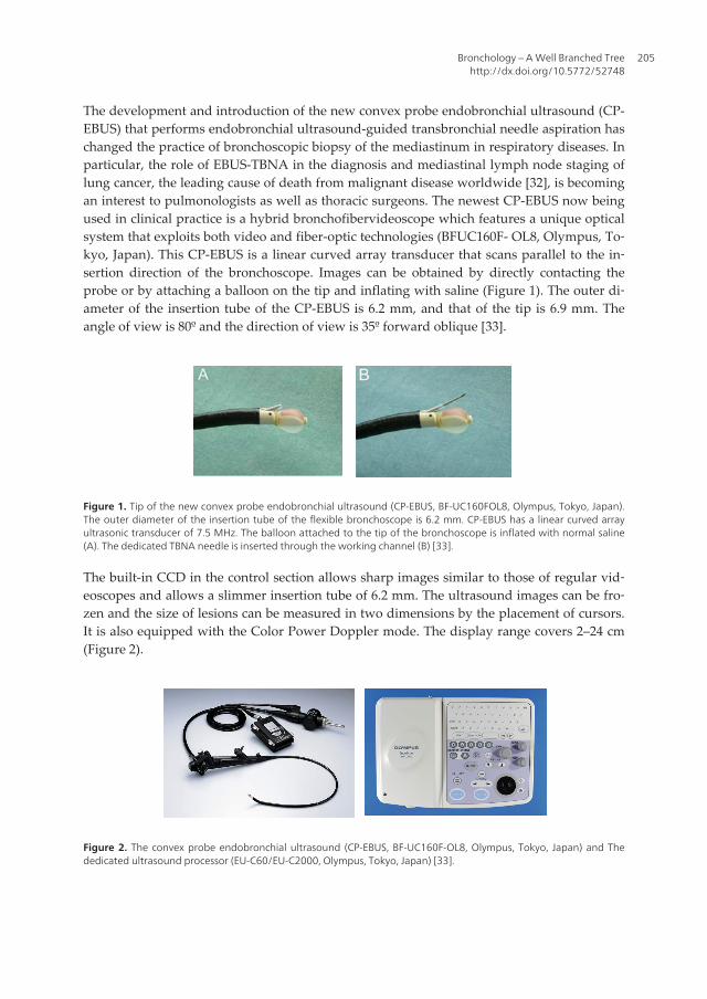
The development and introduction of the new convex probe endobronchial ultrasound (CP-EBUS) that performs endobronchial ultrasound-guided transbronchial needle aspiration haschanged the practice of bronchoscopic biopsy of the mediastinum in respiratory diseases. Inparticular, the role of EBUS-TBNA in the diagnosis and mediastinal lymph node staging oflung cancer, the leading cause of death from malignant disease worldwide [32], is becomingan interest to pulmonologists as well as thoracic surgeons. The newest CP-EBUS now beingused in clinical practice is a hybrid bronchofibervideoscope which features a unique opticalsystem that exploits both video and fiber-optic technologies (BFUC160F- OL8, Olympus, To‐kyo, Japan). This CP-EBUS is a linear curved array transducer that scans parallel to the in‐sertion direction of the bronchoscope. Images can be obtained by directly contacting theprobe or by attaching a balloon on the tip and inflating with saline (Figure 1). The outer di‐ameter of the insertion tube of the CP-EBUS is 6.2 mm, and that of the tip is 6.9 mm. Theangle of view is 80º and the direction of view is 35º forward oblique [33].
Figure 1. Tip of the new convex probe endobronchial ultrasound (CP-EBUS, BF-UC160FOL8, Olympus, Tokyo, Japan).The outer diameter of the insertion tube of the flexible bronchoscope is 6.2 mm. CP-EBUS has a linear curved arrayultrasonic transducer of 7.5 MHz. The balloon attached to the tip of the bronchoscope is inflated with normal saline(A). The dedicated TBNA needle is inserted through the working channel (B) [33].
The built-in CCD in the control section allows sharp images similar to those of regular vid‐eoscopes and allows a slimmer insertion tube of 6.2 mm. The ultrasound images can be fro‐zen and the size of lesions can be measured in two dimensions by the placement of cursors.It is also equipped with the Color Power Doppler mode. The display range covers 2–24 cm(Figure 2).
Figure 2. The convex probe endobronchial ultrasound (CP-EBUS, BF-UC160F-OL8, Olympus, Tokyo, Japan) and Thededicated ultrasound processor (EU-C60/EU-C2000, Olympus, Tokyo, Japan) [33].
Bronchology – A Well Branched Treehttp://dx.doi.org/10.5772/52748
205
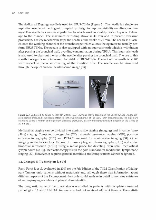
The dedicated 22-gauge needle is used for EBUS-TBNA (Figure 3). The needle is a single useaspiration needle with echogenic dimpled tip design to improve visibility on ultrasound im‐ages. This needle has various adjuster knobs which work as a safety device to prevent dam‐age to the channel. The maximum extruding stroke is 40 mm and to prevent excessiveprotrusion, a safety mechanism stops the needle at the stroke of 20 mm. The needle is attach‐ed onto the working channel of the bronchoscope which allows the operator to actually per‐form EBUS-TBNA. The needle is also equipped with an internal sheath which is withdrawnafter passing the bronchial wall, avoiding contamination during TBNA. This internal sheathis also used to clear out the tip of the needle after passing the bronchial wall. The use of thissheath has significantly increased the yield of EBUS-TBNA. The exit of the needle is at 20°with respect to the outer covering of the insertion tube. The needle can be visualizedthrough the optics and on the ultrasound image [33].
Figure 3. A Dedicated 22-gauge needle (NA-201SX-4022, Olympus, Tokyo, Japan) and the Vaclok syringe used to cre‐ate negative pressure. B The needle attached to the working channel of the EBUS-TBNA bronchoscope. The maximumextruding stroke is 40 mm and to prevent excessive protrusion, a safety mechanism stops the needle at the stroke of20 mm [33].
Mediastinal staging can be divided into noninvasive staging (imaging) and invasive (sam‐pling) staging. Computed tomography (CT), magnetic resonance imaging (MRI), positronemission tomography (PET) and PET-CT are used for noninvasive imaging [34]. Otherimaging modalities include the use of transesophageal ultrasonography (EUS) and endo‐bronchial ultrasound (EBUS) using a radial probe for detecting even small mediastinallymph nodes [35-36]. Mediastinoscopy is still the gold standard for mediastinal lymph nodestaging [37]. However, it requires general anesthesia and complications cannot be ignored.
1.2. Changes to T descriptors [38-39]
Rami-Porta R et al. evaluated in 2007 for the 7th Edition of the TNM Classification of Malig‐nant Tumors only patients without metastasis and, although there was information aboutdifferent aspects of the T component, they only could analyze in detail tumor size, existenceof accompanying nodules and pleural dissemination.
The prognostic value of the tumor size was studied in patients with completely resectedpathological T1 and T2 N0 M0 tumors who had not received adjuvant therapy. The statisti‐
Endoscopy206
cal calculations determined three cut-points at 2, 5 and 7cm, which, in addition to the 3cm,the border between T1 and T2, gave rise to 5 groups of tumors with significantly worse sur‐vival with larger tumor diameters. The groups and their 5-year survival rates were: T1≤2cm,77%; T1>2cm and ≤3cm, 71%; T2>3cm and ≤5cm, 58%; T2>5cm and ≤7cm, 49%, and T2>7cm,35%. This prognostic gradation was maintained when less selective patient populationswere evaluated: clinical staging, incomplete resection and different lymph node affectation.With such arguments, it was decided to subdivide the T1 tumors into T1a (≤2cm) and T1b(>2cm and ≤3cm), and T2 tumors into T2a (>3cm and ≤5cm) and T2b (>5cm and ≤7cm). Like‐wise, the 5-year survival was compared between patients with T2>7cm tumors and T3 tu‐mors. Similar results were found in the different populations, except in the N0 cases withcomplete resection, in which it was verified that the survival was even higher in the T3(41%) than in the T2>7cm (35%), therefore it was decided to reclassify the latter as T3. Whenthey analyzed the tumors that, with pathological staging, presented additional nodules, itwas observed that: (a) the 5-year survival of the T3 (31%) was similar to the T4 classified assuch due to the existence of an additional nodule or nodules in the same lobe as the primarytumor (28%); (b) the T4 due to other factors had the same survival as those classified as M1due to an additional nodule(s) in a different homolateral lobe than the primary tumor (22%);and (c) the T4 due to pleural dissemination had a clearly worse prognosis (11% 5-year sur‐vival). For the new classification, it was therefore recommended to consider as T3 those tu‐mors with additional nodule(s) in the same lobe as the primary tumor, to consider as T4those tumors with additional nodule(s) in a homolateral lobe other than that of the primarytumor, and to include in the M category those tumors with pleural dissemination (Table 1).
T descriptor 6th ed. 7th ed.
T size Tumors = 2 cmT1
T1a
Tumors > 2 cm and = 3 cm T1b
Tumors > 3cm and = 5cm
T2
T2a
Tumors > 5 cm and = 7 cm T2b
Tumors > 7 cm T3
Separate nodule(s) in the primary lobe. T4 T3
Separate nodule(s) in a different ipsilateral lobe. M1 T4
Malignant pericardial effusion. T4 M1a
Pleural dissemination. T4 M1a
Table 1. T descriptor changes: comparison between 6th and 7th edition of the TNM Classification of MalignantTumors [38]
Bronchology – A Well Branched Treehttp://dx.doi.org/10.5772/52748
207
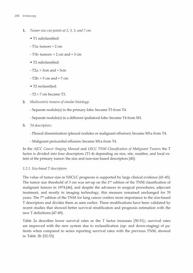
1. Tumor size cut points at 2, 3, 5, and 7 cm:
• T1 subclassified:
- T1a: tumors = 2 cm
- T1b: tumors: > 2 cm and = 3 cm
• T2 subclassified:
- T2a: > 3cm and = 5cm
- T2b: > 5 cm and = 7 cm
• T2 reclassified:
- T2 > 7 cm became T3.
2. Multicentric tumors of similar histology:
- Separate nodule(s) in the primary lobe: became T3 from T4.
- Separate nodule(s) in a different ipsilateral lobe: became T4 from M1.
3. T4 descriptors:
- Pleural dissemination (pleural nodules or malignant effusion): became M1a from T4.
- Malignant pericardial effusion: became M1a from T4.
In the AJCC Cancer Staging Manual and UICC TNM Classification of Malignant Tumors the Tfactor is divided into four descriptors (T1-4) depending on size, site, number, and local ex‐tent of the primary tumor: the size and non-size based descriptors [40].
1.2.1. Size-based T descriptors
The value of tumor size in NSCLC prognosis is supported by large clinical evidence [41-45].The tumor size threshold of 3 cm was set-up on the 2nd edition of the TNM classification ofmalignant tumors in 1974.[46], and despite the advances in surgical procedures, adjuvanttreatment, and mostly in imaging technology, this measure remained unchanged for 35years. The 7th edition of the TNM for lung cancer confers more importance to the size-basedT descriptors and divides them as seen earlier. These modifications have been validated byrecent studies that showed better survival stratification and prognosis estimation with thenew T definitions [47-49].
Table 2a describes lower survival rates as the T factor increases [50-51].; survival ratesare improved with the new system due to reclassification (up- and down-staging) of pa‐tients when compared to series reporting survival rates with the previous TNM, showedin Table 2b [52-53].
Endoscopy208
T component5-year survival (%)
Kameyama et al [25] Li et al [24]
T1a 82.6 75.49
T1b 73.3 74.58
T2a 63.5 60.87
T2b 50.1 55.63
T3 40.6 46.15
T4 34.6 NS
T component5-year survival (%)
Mountain CF [26] Naruke et al. [27]
T1 67 68.9
T2 57 42.5
T3 38 31.9
T4 7 (cT) 18.9
Table 2. a. Five year survival by pT classification with the 7th edition. b. Five year survival by pT classification with the6th edition [40].
1.2.2. Non–size-based T descriptors
1.2.2.1. Multiple nodules
The existence of multiple primary cancers (MPC) was initially reported by Warren andGates in 1932 [54], but in spite of these past 80 years, to date accurate diagnosis ofMPC is not yet clearly established due to a lack of consensus on definition and diagnos‐tic criteria [55]
In 1975, Martini and Melamed were the first to propose clinical and histopathologic criteriafor the differential diagnosis of second lung cancers [56]. MPLCs are defined synchronous, ifdetected simultaneously, or metachronous, if tumors are separated in time [57-58]. Synchro‐nous nodules may represent a MPLC (second primary), a metastasis, or an extension fromthe primary (satellite nodule) [59] (Table 3).
Deslauriers et al. described in 1989 intrapulmonary nodular metastasis in patients withNSCLC as satellite nodules, and the 5- year survival rates for these patients with satellitenodules were 21.6% compared to 44% for patients without satellite nodules. They concludedthat patients with satellite nodules should be classified as stage IIIA [60].
Bronchology – A Well Branched Treehttp://dx.doi.org/10.5772/52748
209
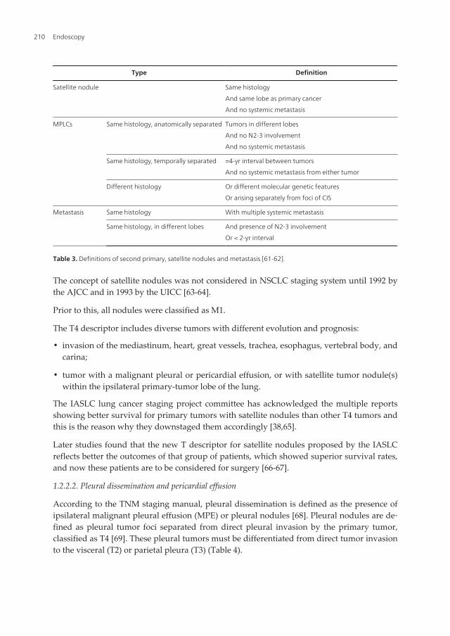
Type Definition
Satellite nodule Same histology
And same lobe as primary cancer
And no systemic metastasis
MPLCs Same histology, anatomically separated Tumors in different lobes
And no N2-3 involvement
And no systemic metastasis
Same histology, temporally separated =4-yr interval between tumors
And no systemic metastasis from either tumor
Different histology Or different molecular genetic features
Or arising separately from foci of CIS
Metastasis Same histology With multiple systemic metastasis
Same histology, in different lobes And presence of N2-3 involvement
Or < 2-yr interval
Table 3. Definitions of second primary, satellite nodules and metastasis [61-62].
The concept of satellite nodules was not considered in NSCLC staging system until 1992 bythe AJCC and in 1993 by the UICC [63-64].
Prior to this, all nodules were classified as M1.
The T4 descriptor includes diverse tumors with different evolution and prognosis:
• invasion of the mediastinum, heart, great vessels, trachea, esophagus, vertebral body, andcarina;
• tumor with a malignant pleural or pericardial effusion, or with satellite tumor nodule(s)within the ipsilateral primary-tumor lobe of the lung.
The IASLC lung cancer staging project committee has acknowledged the multiple reportsshowing better survival for primary tumors with satellite nodules than other T4 tumors andthis is the reason why they downstaged them accordingly [38,65].
Later studies found that the new T descriptor for satellite nodules proposed by the IASLCreflects better the outcomes of that group of patients, which showed superior survival rates,and now these patients are to be considered for surgery [66-67].
1.2.2.2. Pleural dissemination and pericardial effusion
According to the TNM staging manual, pleural dissemination is defined as the presence ofipsilateral malignant pleural effusion (MPE) or pleural nodules [68]. Pleural nodules are de‐fined as pleural tumor foci separated from direct pleural invasion by the primary tumor,classified as T4 [69]. These pleural tumors must be differentiated from direct tumor invasionto the visceral (T2) or parietal pleura (T3) (Table 4).
Endoscopy210
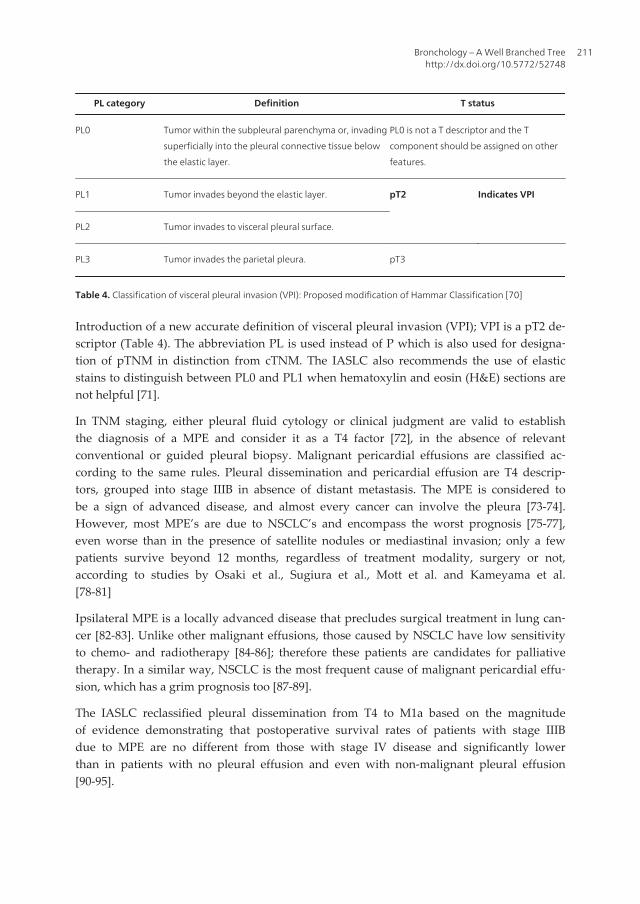
PL category Definition T status
PL0 Tumor within the subpleural parenchyma or, invading
superficially into the pleural connective tissue below
the elastic layer.
PL0 is not a T descriptor and the T
component should be assigned on other
features.
PL1 Tumor invades beyond the elastic layer. pT2 Indicates VPI
PL2 Tumor invades to visceral pleural surface.
PL3 Tumor invades the parietal pleura. pT3
Table 4. Classification of visceral pleural invasion (VPI): Proposed modification of Hammar Classification [70]
Introduction of a new accurate definition of visceral pleural invasion (VPI); VPI is a pT2 de‐scriptor (Table 4). The abbreviation PL is used instead of P which is also used for designa‐tion of pTNM in distinction from cTNM. The IASLC also recommends the use of elasticstains to distinguish between PL0 and PL1 when hematoxylin and eosin (H&E) sections arenot helpful [71].
In TNM staging, either pleural fluid cytology or clinical judgment are valid to establishthe diagnosis of a MPE and consider it as a T4 factor [72], in the absence of relevantconventional or guided pleural biopsy. Malignant pericardial effusions are classified ac‐cording to the same rules. Pleural dissemination and pericardial effusion are T4 descrip‐tors, grouped into stage IIIB in absence of distant metastasis. The MPE is considered tobe a sign of advanced disease, and almost every cancer can involve the pleura [73-74].However, most MPE’s are due to NSCLC’s and encompass the worst prognosis [75-77],even worse than in the presence of satellite nodules or mediastinal invasion; only a fewpatients survive beyond 12 months, regardless of treatment modality, surgery or not,according to studies by Osaki et al., Sugiura et al., Mott et al. and Kameyama et al.[78-81]
Ipsilateral MPE is a locally advanced disease that precludes surgical treatment in lung can‐cer [82-83]. Unlike other malignant effusions, those caused by NSCLC have low sensitivityto chemo- and radiotherapy [84-86]; therefore these patients are candidates for palliativetherapy. In a similar way, NSCLC is the most frequent cause of malignant pericardial effu‐sion, which has a grim prognosis too [87-89].
The IASLC reclassified pleural dissemination from T4 to M1a based on the magnitudeof evidence demonstrating that postoperative survival rates of patients with stage IIIBdue to MPE are no different from those with stage IV disease and significantly lowerthan in patients with no pleural effusion and even with non-malignant pleural effusion[90-95].
Bronchology – A Well Branched Treehttp://dx.doi.org/10.5772/52748
211
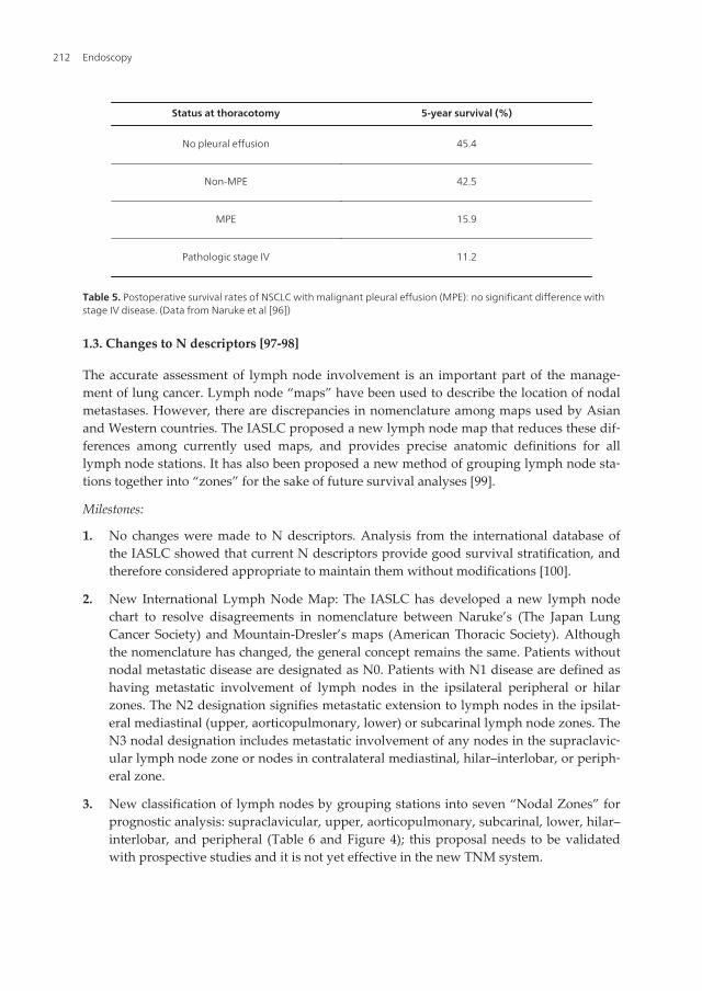
Status at thoracotomy 5-year survival (%)
No pleural effusion 45.4
Non-MPE 42.5
MPE 15.9
Pathologic stage IV 11.2
Table 5. Postoperative survival rates of NSCLC with malignant pleural effusion (MPE): no significant difference withstage IV disease. (Data from Naruke et al [96])
1.3. Changes to N descriptors [97-98]
The accurate assessment of lymph node involvement is an important part of the manage‐ment of lung cancer. Lymph node “maps” have been used to describe the location of nodalmetastases. However, there are discrepancies in nomenclature among maps used by Asianand Western countries. The IASLC proposed a new lymph node map that reduces these dif‐ferences among currently used maps, and provides precise anatomic definitions for alllymph node stations. It has also been proposed a new method of grouping lymph node sta‐tions together into “zones” for the sake of future survival analyses [99].
Milestones:
1. No changes were made to N descriptors. Analysis from the international database ofthe IASLC showed that current N descriptors provide good survival stratification, andtherefore considered appropriate to maintain them without modifications [100].
2. New International Lymph Node Map: The IASLC has developed a new lymph nodechart to resolve disagreements in nomenclature between Naruke’s (The Japan LungCancer Society) and Mountain-Dresler’s maps (American Thoracic Society). Althoughthe nomenclature has changed, the general concept remains the same. Patients withoutnodal metastatic disease are designated as N0. Patients with N1 disease are defined ashaving metastatic involvement of lymph nodes in the ipsilateral peripheral or hilarzones. The N2 designation signifies metastatic extension to lymph nodes in the ipsilat‐eral mediastinal (upper, aorticopulmonary, lower) or subcarinal lymph node zones. TheN3 nodal designation includes metastatic involvement of any nodes in the supraclavic‐ular lymph node zone or nodes in contralateral mediastinal, hilar–interlobar, or periph‐eral zone.
3. New classification of lymph nodes by grouping stations into seven “Nodal Zones” forprognostic analysis: supraclavicular, upper, aorticopulmonary, subcarinal, lower, hilar–interlobar, and peripheral (Table 6 and Figure 4); this proposal needs to be validatedwith prospective studies and it is not yet effective in the new TNM system.
Endoscopy212
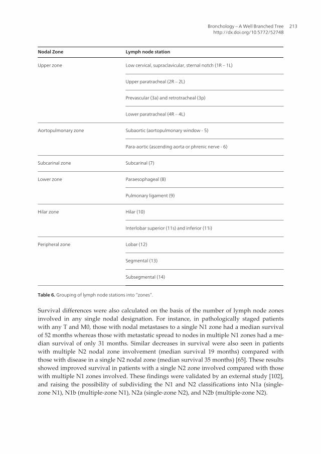
Nodal Zone Lymph node station
Upper zone Low cervical, supraclavicular, sternal notch (1R – 1L)
Upper paratracheal (2R – 2L)
Prevascular (3a) and retrotracheal (3p)
Lower paratracheal (4R – 4L)
Aortopulmonary zone Subaortic (aortopulmonary window - 5)
Para-aortic (ascending aorta or phrenic nerve - 6)
Subcarinal zone Subcarinal (7)
Lower zone Paraesophageal (8)
Pulmonary ligament (9)
Hilar zone Hilar (10)
Interlobar superior (11s) and inferior (11i)
Peripheral zone Lobar (12)
Segmental (13)
Subsegmental (14)
Table 6. Grouping of lymph node stations into “zones”.
Survival differences were also calculated on the basis of the number of lymph node zonesinvolved in any single nodal designation. For instance, in pathologically staged patientswith any T and M0, those with nodal metastases to a single N1 zone had a median survivalof 52 months whereas those with metastatic spread to nodes in multiple N1 zones had a me‐dian survival of only 31 months. Similar decreases in survival were also seen in patientswith multiple N2 nodal zone involvement (median survival 19 months) compared withthose with disease in a single N2 nodal zone (median survival 35 months) [65]. These resultsshowed improved survival in patients with a single N2 zone involved compared with thosewith multiple N1 zones involved. These findings were validated by an external study [102],and raising the possibility of subdividing the N1 and N2 classifications into N1a (single-zone N1), N1b (multiple-zone N1), N2a (single-zone N2), and N2b (multiple-zone N2).
Bronchology – A Well Branched Treehttp://dx.doi.org/10.5772/52748
213
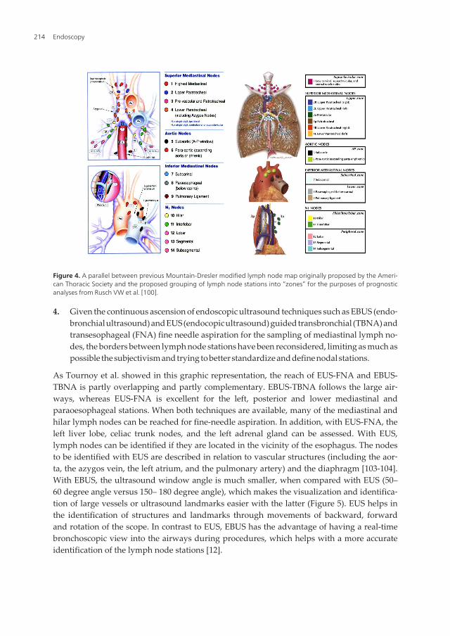
Figure 4. A parallel between previous Mountain-Dresler modified lymph node map originally proposed by the Ameri‐can Thoracic Society and the proposed grouping of lymph node stations into “zones” for the purposes of prognosticanalyses from Rusch VW et al. [100].
4. Given the continuous ascension of endoscopic ultrasound techniques such as EBUS (endo‐bronchial ultrasound) and EUS (endocopic ultrasound) guided transbronchial (TBNA) andtransesophageal (FNA) fine needle aspiration for the sampling of mediastinal lymph no‐des, the borders between lymph node stations have been reconsidered, limiting as much aspossible the subjectivism and trying to better standardize and define nodal stations.
As Tournoy et al. showed in this graphic representation, the reach of EUS-FNA and EBUS-TBNA is partly overlapping and partly complementary. EBUS-TBNA follows the large air‐ways, whereas EUS-FNA is excellent for the left, posterior and lower mediastinal andparaoesophageal stations. When both techniques are available, many of the mediastinal andhilar lymph nodes can be reached for fine-needle aspiration. In addition, with EUS-FNA, theleft liver lobe, celiac trunk nodes, and the left adrenal gland can be assessed. With EUS,lymph nodes can be identified if they are located in the vicinity of the esophagus. The nodesto be identified with EUS are described in relation to vascular structures (including the aor‐ta, the azygos vein, the left atrium, and the pulmonary artery) and the diaphragm [103-104].With EBUS, the ultrasound window angle is much smaller, when compared with EUS (50–60 degree angle versus 150– 180 degree angle), which makes the visualization and identifica‐tion of large vessels or ultrasound landmarks easier with the latter (Figure 5). EUS helps inthe identification of structures and landmarks through movements of backward, forwardand rotation of the scope. In contrast to EUS, EBUS has the advantage of having a real-timebronchoscopic view into the airways during procedures, which helps with a more accurateidentification of the lymph node stations [12].
Endoscopy214
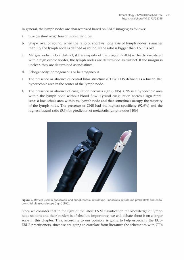
In general, the lymph nodes are characterized based on EBUS imaging as follows:
a. Size (in short axis): less or more than 1 cm.
b. Shape: oval or round; when the ratio of short vs. long axis of lymph nodes is smallerthan 1.5, the lymph node is defined as round; if the ratio is bigger than 1.5, it is oval.
c. Margin: indistinct or distinct; if the majority of the margin (>50%) is clearly visualizedwith a high echoic border, the lymph nodes are determined as distinct. If the margin isunclear, they are determined as indistinct.
d. Echogenecity: homogeneous or heterogeneous
e. The presence or absence of central hilar structure (CHS); CHS defined as a linear, flat,hyperechoic area in the center of the lymph node.
f. The presence or absence of coagulation necrosis sign (CNS). CNS is a hypoechoic areawithin the lymph node without blood flow. Typical coagulation necrosis sign repre‐sents a low echoic area within the lymph node and that sometimes occupy the majorityof the lymph node. The presence of CNS had the highest specificity (92.6%) and thehighest hazard ratio (5.6) for prediction of metastatic lymph nodes [106]
Figure 5. Devices used in endoscopic and endobronchial ultrasound. Endoscopic ultrasound probe (left) and endo‐bronchial ultrasound scope (right) [105].
Since we consider that in the light of the latest TNM classification the knowledge of lymphnode stations and their borders is of absolute importance, we will debate about it on a largerscale in this chapter. This, according to our opinion, is going to help especially the EUS-EBUS practitioners, since we are going to correlate from literature the schematics with CT’s
Bronchology – A Well Branched Treehttp://dx.doi.org/10.5772/52748
215
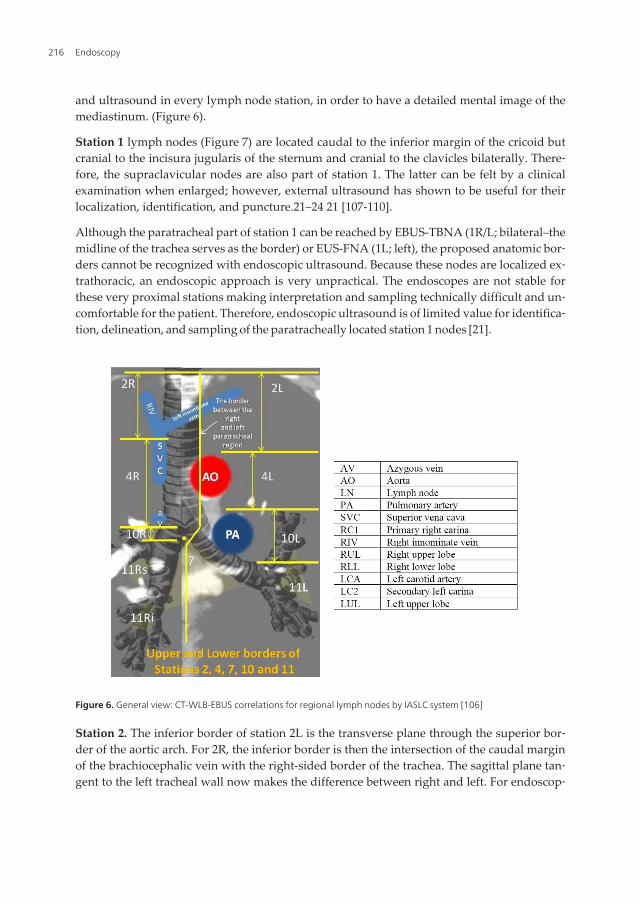
and ultrasound in every lymph node station, in order to have a detailed mental image of themediastinum. (Figure 6).
Station 1 lymph nodes (Figure 7) are located caudal to the inferior margin of the cricoid butcranial to the incisura jugularis of the sternum and cranial to the clavicles bilaterally. There‐fore, the supraclavicular nodes are also part of station 1. The latter can be felt by a clinicalexamination when enlarged; however, external ultrasound has shown to be useful for theirlocalization, identification, and puncture.21–24 21 [107-110].
Although the paratracheal part of station 1 can be reached by EBUS-TBNA (1R/L; bilateral–themidline of the trachea serves as the border) or EUS-FNA (1L; left), the proposed anatomic bor‐ders cannot be recognized with endoscopic ultrasound. Because these nodes are localized ex‐trathoracic, an endoscopic approach is very unpractical. The endoscopes are not stable forthese very proximal stations making interpretation and sampling technically difficult and un‐comfortable for the patient. Therefore, endoscopic ultrasound is of limited value for identifica‐tion, delineation, and sampling of the paratracheally located station 1 nodes [21].
Figure 6. General view: CT-WLB-EBUS correlations for regional lymph nodes by IASLC system [106]
Station 2. The inferior border of station 2L is the transverse plane through the superior bor‐der of the aortic arch. For 2R, the inferior border is then the intersection of the caudal marginof the brachiocephalic vein with the right-sided border of the trachea. The sagittal plane tan‐gent to the left tracheal wall now makes the difference between right and left. For endoscop‐
Endoscopy216
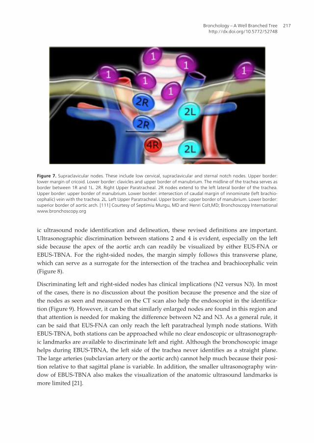
ic ultrasound node identification and delineation, these revised definitions are important.Ultrasonographic discrimination between stations 2 and 4 is evident, especially on the leftside because the apex of the aortic arch can readily be visualized by either EUS-FNA orEBUS-TBNA. For the right-sided nodes, the margin simply follows this transverse plane,which can serve as a surrogate for the intersection of the trachea and brachiocephalic vein(Figure 8).
Discriminating left and right-sided nodes has clinical implications (N2 versus N3). In mostof the cases, there is no discussion about the position because the presence and the size ofthe nodes as seen and measured on the CT scan also help the endoscopist in the identifica‐tion (Figure 9). However, it can be that similarly enlarged nodes are found in this region andthat attention is needed for making the difference between N2 and N3. As a general rule, itcan be said that EUS-FNA can only reach the left paratracheal lymph node stations. WithEBUS-TBNA, both stations can be approached while no clear endoscopic or ultrasonograph‐ic landmarks are available to discriminate left and right. Although the bronchoscopic imagehelps during EBUS-TBNA, the left side of the trachea never identifies as a straight plane.The large arteries (subclavian artery or the aortic arch) cannot help much because their posi‐tion relative to that sagittal plane is variable. In addition, the smaller ultrasonography win‐dow of EBUS-TBNA also makes the visualization of the anatomic ultrasound landmarks ismore limited [21].
Figure 7. Supraclavicular nodes. These include low cervical, supraclavicular and sternal notch nodes. Upper border:lower margin of cricoid. Lower border: clavicles and upper border of manubrium. The midline of the trachea serves asborder between 1R and 1L. 2R. Right Upper Paratracheal. 2R nodes extend to the left lateral border of the trachea.Upper border: upper border of manubrium. Lower border: intersection of caudal margin of innominate (left brachio‐cephalic) vein with the trachea. 2L. Left Upper Paratracheal. Upper border: upper border of manubrium. Lower border:superior border of aortic arch. [111] Courtesy of Septimiu Murgu, MD and Henri Colt,MD; Bronchoscopy Internationalwww.bronchoscopy.org
Bronchology – A Well Branched Treehttp://dx.doi.org/10.5772/52748
217
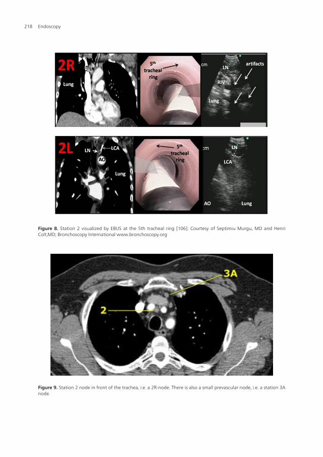
Figure 8. Station 2 visualized by EBUS at the 5th tracheal ring [106]. Courtesy of Septimiu Murgu, MD and HenriColt,MD; Bronchoscopy International www.bronchoscopy.org
Figure 9. Station 2 node in front of the trachea, i.e. a 2R-node. There is also a small prevascular node, i.e. a station 3Anode.
Endoscopy218
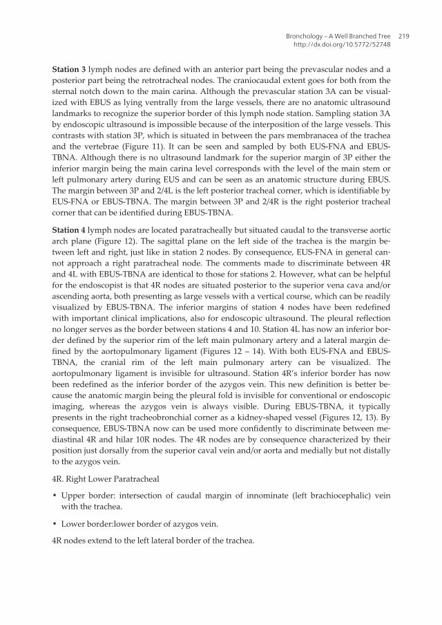
Station 3 lymph nodes are defined with an anterior part being the prevascular nodes and aposterior part being the retrotracheal nodes. The craniocaudal extent goes for both from thesternal notch down to the main carina. Although the prevascular station 3A can be visual‐ized with EBUS as lying ventrally from the large vessels, there are no anatomic ultrasoundlandmarks to recognize the superior border of this lymph node station. Sampling station 3Aby endoscopic ultrasound is impossible because of the interposition of the large vessels. Thiscontrasts with station 3P, which is situated in between the pars membranacea of the tracheaand the vertebrae (Figure 11). It can be seen and sampled by both EUS-FNA and EBUS-TBNA. Although there is no ultrasound landmark for the superior margin of 3P either theinferior margin being the main carina level corresponds with the level of the main stem orleft pulmonary artery during EUS and can be seen as an anatomic structure during EBUS.The margin between 3P and 2/4L is the left posterior tracheal corner, which is identifiable byEUS-FNA or EBUS-TBNA. The margin between 3P and 2/4R is the right posterior trachealcorner that can be identified during EBUS-TBNA.
Station 4 lymph nodes are located paratracheally but situated caudal to the transverse aorticarch plane (Figure 12). The sagittal plane on the left side of the trachea is the margin be‐tween left and right, just like in station 2 nodes. By consequence, EUS-FNA in general can‐not approach a right paratracheal node. The comments made to discriminate between 4Rand 4L with EBUS-TBNA are identical to those for stations 2. However, what can be helpfulfor the endoscopist is that 4R nodes are situated posterior to the superior vena cava and/orascending aorta, both presenting as large vessels with a vertical course, which can be readilyvisualized by EBUS-TBNA. The inferior margins of station 4 nodes have been redefinedwith important clinical implications, also for endoscopic ultrasound. The pleural reflectionno longer serves as the border between stations 4 and 10. Station 4L has now an inferior bor‐der defined by the superior rim of the left main pulmonary artery and a lateral margin de‐fined by the aortopulmonary ligament (Figures 12 – 14). With both EUS-FNA and EBUS-TBNA, the cranial rim of the left main pulmonary artery can be visualized. Theaortopulmonary ligament is invisible for ultrasound. Station 4R’s inferior border has nowbeen redefined as the inferior border of the azygos vein. This new definition is better be‐cause the anatomic margin being the pleural fold is invisible for conventional or endoscopicimaging, whereas the azygos vein is always visible. During EBUS-TBNA, it typicallypresents in the right tracheobronchial corner as a kidney-shaped vessel (Figures 12, 13). Byconsequence, EBUS-TBNA now can be used more confidently to discriminate between me‐diastinal 4R and hilar 10R nodes. The 4R nodes are by consequence characterized by theirposition just dorsally from the superior caval vein and/or aorta and medially but not distallyto the azygos vein.
4R. Right Lower Paratracheal
• Upper border: intersection of caudal margin of innominate (left brachiocephalic) veinwith the trachea.
• Lower border:lower border of azygos vein.
4R nodes extend to the left lateral border of the trachea.
Bronchology – A Well Branched Treehttp://dx.doi.org/10.5772/52748
219
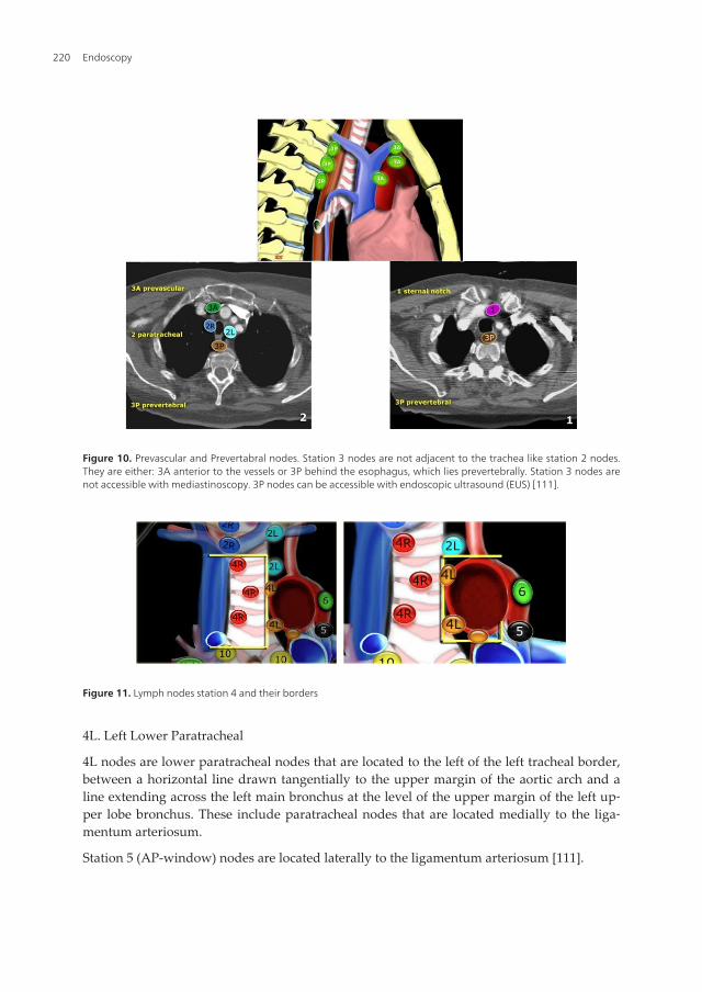
4L. Left Lower Paratracheal
4L nodes are lower paratracheal nodes that are located to the left of the left tracheal border,between a horizontal line drawn tangentially to the upper margin of the aortic arch and aline extending across the left main bronchus at the level of the upper margin of the left up‐per lobe bronchus. These include paratracheal nodes that are located medially to the liga‐mentum arteriosum.
Station 5 (AP-window) nodes are located laterally to the ligamentum arteriosum [111].
Figure 10. Prevascular and Prevertabral nodes. Station 3 nodes are not adjacent to the trachea like station 2 nodes.They are either: 3A anterior to the vessels or 3P behind the esophagus, which lies prevertebrally. Station 3 nodes arenot accessible with mediastinoscopy. 3P nodes can be accessible with endoscopic ultrasound (EUS) [111].
Figure 11. Lymph nodes station 4 and their borders
Endoscopy220
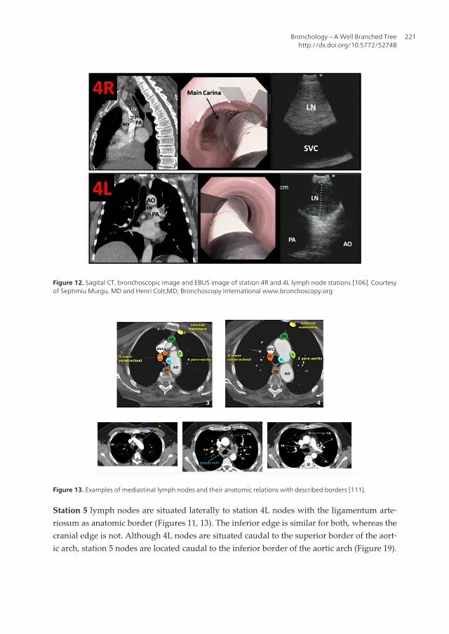
Figure 12. Sagital CT, bronchoscopic image and EBUS image of station 4R and 4L lymph node stations [106]. Courtesyof Septimiu Murgu, MD and Henri Colt,MD; Bronchoscopy International www.bronchoscopy.org
Figure 13. Examples of mediastinal lymph nodes and their anatomic relations with described borders [111].
Station 5 lymph nodes are situated laterally to station 4L nodes with the ligamentum arte‐riosum as anatomic border (Figures 11, 13). The inferior edge is similar for both, whereas thecranial edge is not. Although 4L nodes are situated caudal to the superior border of the aort‐ic arch, station 5 nodes are located caudal to the inferior border of the aortic arch (Figure 19).
Bronchology – A Well Branched Treehttp://dx.doi.org/10.5772/52748
221
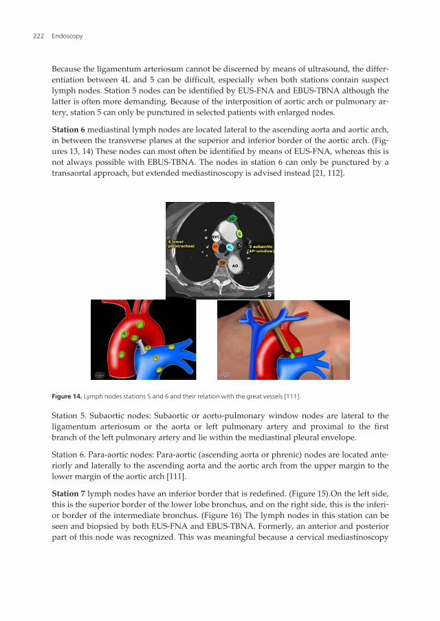
Because the ligamentum arteriosum cannot be discerned by means of ultrasound, the differ‐entiation between 4L and 5 can be difficult, especially when both stations contain suspectlymph nodes. Station 5 nodes can be identified by EUS-FNA and EBUS-TBNA although thelatter is often more demanding. Because of the interposition of aortic arch or pulmonary ar‐tery, station 5 can only be punctured in selected patients with enlarged nodes.
Station 6 mediastinal lymph nodes are located lateral to the ascending aorta and aortic arch,in between the transverse planes at the superior and inferior border of the aortic arch. (Fig‐ures 13, 14) These nodes can most often be identified by means of EUS-FNA, whereas this isnot always possible with EBUS-TBNA. The nodes in station 6 can only be punctured by atransaortal approach, but extended mediastinoscopy is advised instead [21, 112].
Figure 14. Lymph nodes stations 5 and 6 and their relation with the great vessels [111].
Station 5. Subaortic nodes: Subaortic or aorto-pulmonary window nodes are lateral to theligamentum arteriosum or the aorta or left pulmonary artery and proximal to the firstbranch of the left pulmonary artery and lie within the mediastinal pleural envelope.
Station 6. Para-aortic nodes: Para-aortic (ascending aorta or phrenic) nodes are located ante‐riorly and laterally to the ascending aorta and the aortic arch from the upper margin to thelower margin of the aortic arch [111].
Station 7 lymph nodes have an inferior border that is redefined. (Figure 15).On the left side,this is the superior border of the lower lobe bronchus, and on the right side, this is the inferi‐or border of the intermediate bronchus. (Figure 16) The lymph nodes in this station can beseen and biopsied by both EUS-FNA and EBUS-TBNA. Formerly, an anterior and posteriorpart of this node was recognized. This was meaningful because a cervical mediastinoscopy
Endoscopy222

cannot reach the posterior part of this station. With EUS-FNA and EBUS-TBNA, the entiresubcarinal area can be approached. Although identification of the nodes in this station iseasy for both EUS-FNA (the nodes lay just dorsally to the origin of the left pulmonary arteryand cranial to the left atrium) and EBUS-TBNA (by means of the endoscopic view), the de‐lineation of the inferior border by means of endoscopic ultrasound is, however, not easy.With EUS-FNA, the left atrium is generally seen as the anatomic border above which thesubcarinal nodes are situated, although this is with the new definition probably too restric‐tive. The relation of the left atrium or pulmonary artery to the bronchus intermedius and theleft lower lobe bronchus, the latter being the newly defined inferior borders of station 7, canbe variable. With EBUS-TBNA, the delineation of the inferior border is possible because thisinvestigation allows a simultaneous bronchoscopic view of the bronchial tree although thereare no distinct corresponding ultrasound landmarks.
Figure 15. Virtual borders between lymph node stations and their relation with the great vessels of mediastinum[113].
Figure 16. The subcarinal station shown on CT scan (left panel) can easily be visualized with both EUS (right panel) butof course also with EBUS. During EUS, the subcarinal station identifies as a round, hypoechogenic sharply edged struc‐ture located in between the esophagus and the pulmonary artery. The upper rim of this artery corresponds in generalwith the main carina and is helpful during EUS. The main carina is visualized by means of endoscopy during EBUS. Theinferior border is the roof of the lower lobe bronchus on the left and the bottom of the bronchus intermedius on theright. There are no unique ultrasound features that correspond to this definition although endoscopy during EBUS ishelpful [21]. Courtesy of Septimiu Murgu, MD and Henri Colt,MD; Bronchoscopy International www.bronchoscopy.org
Bronchology – A Well Branched Treehttp://dx.doi.org/10.5772/52748
223
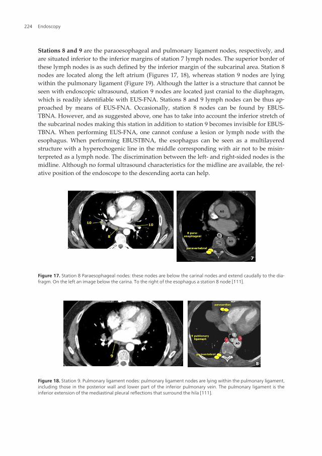
Stations 8 and 9 are the paraoesophageal and pulmonary ligament nodes, respectively, andare situated inferior to the inferior margins of station 7 lymph nodes. The superior border ofthese lymph nodes is as such defined by the inferior margin of the subcarinal area. Station 8nodes are located along the left atrium (Figures 17, 18), whereas station 9 nodes are lyingwithin the pulmonary ligament (Figure 19). Although the latter is a structure that cannot beseen with endoscopic ultrasound, station 9 nodes are located just cranial to the diaphragm,which is readily identifiable with EUS-FNA. Stations 8 and 9 lymph nodes can be thus ap‐proached by means of EUS-FNA. Occasionally, station 8 nodes can be found by EBUS-TBNA. However, and as suggested above, one has to take into account the inferior stretch ofthe subcarinal nodes making this station in addition to station 9 becomes invisible for EBUS-TBNA. When performing EUS-FNA, one cannot confuse a lesion or lymph node with theesophagus. When performing EBUSTBNA, the esophagus can be seen as a multilayeredstructure with a hyperechogenic line in the middle corresponding with air not to be misin‐terpreted as a lymph node. The discrimination between the left- and right-sided nodes is themidline. Although no formal ultrasound characteristics for the midline are available, the rel‐ative position of the endoscope to the descending aorta can help.
Figure 17. Station 8 Paraesophageal nodes: these nodes are below the carinal nodes and extend caudally to the dia‐fragm. On the left an image below the carina. To the right of the esophagus a station 8 node [111].
Figure 18. Station 9. Pulmonary ligament nodes: pulmonary ligament nodes are lying within the pulmonary ligament,including those in the posterior wall and lower part of the inferior pulmonary vein. The pulmonary ligament is theinferior extension of the mediastinal pleural reflections that surround the hila [111].
Endoscopy224

Figure 19. Hilar nodes Hilar nodes are proximal lobar nodes, distal to the mediastinal pleural reflection and nodesadjacent to the intermediate bronchus on the right. Nodes in station 10 - 14 are all N1-nodes, since they are not locat‐ed in the mediastinum [111].
Station 10 or hilar lymph nodes are situated immediately adjacent to the main stem bron‐chus but caudal to the inferior border of azygos vein on the right and superior rim pulmona‐ry veins and artery on the left. These nodes can be seen and sampled by EBUS-TBNA(Figures 20, 21). The inferior margin of station 10R is the interlobar region. There is nounique ultrasound feature that defines that border, but because a bronchoscopic view isavailable during EBUS-TBNA, the secondary carina or the upper lobe split off can serve assurrogate here. EUS-FNA has been thought to be unable to see and sample hilar stations.However, there is no doubt that in certain cases, station 10 nodes located medially from themain stem bronchi can be assessed. Endoscopists should be aware of this because misinter‐pretation could lead to overstaging [114].
Figure 20. Station 10 lymph nodes: sagittal CT, endobronchial view and corresponding EBUS [106]. Courtesy of Septi‐miu Murgu, MD and Henri Colt,MD; Bronchoscopy International www.bronchoscopy.org
Bronchology – A Well Branched Treehttp://dx.doi.org/10.5772/52748
225
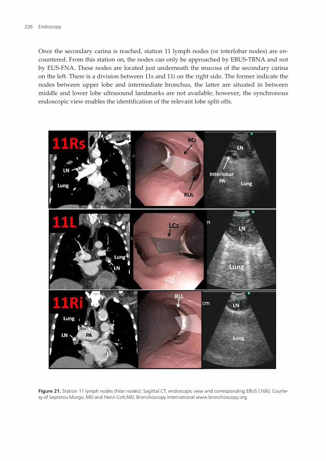
Once the secondary carina is reached, station 11 lymph nodes (or interlobar nodes) are en‐countered. From this station on, the nodes can only be approached by EBUS-TBNA and notby EUS-FNA. These nodes are located just underneath the mucosa of the secondary carinaon the left. There is a division between 11s and 11i on the right side. The former indicate thenodes between upper lobe and intermediate bronchus, the latter are situated in betweenmiddle and lower lobe ultrasound landmarks are not available; however, the synchronousendoscopic view enables the identification of the relevant lobe split offs.
Figure 21. Station 11 lymph nodes (hilar nodes): Sagittal CT, endoscopic view and corresponding EBUS [106]. Courte‐sy of Septimiu Murgu, MD and Henri Colt,MD; Bronchoscopy International www.bronchoscopy.org
Endoscopy226
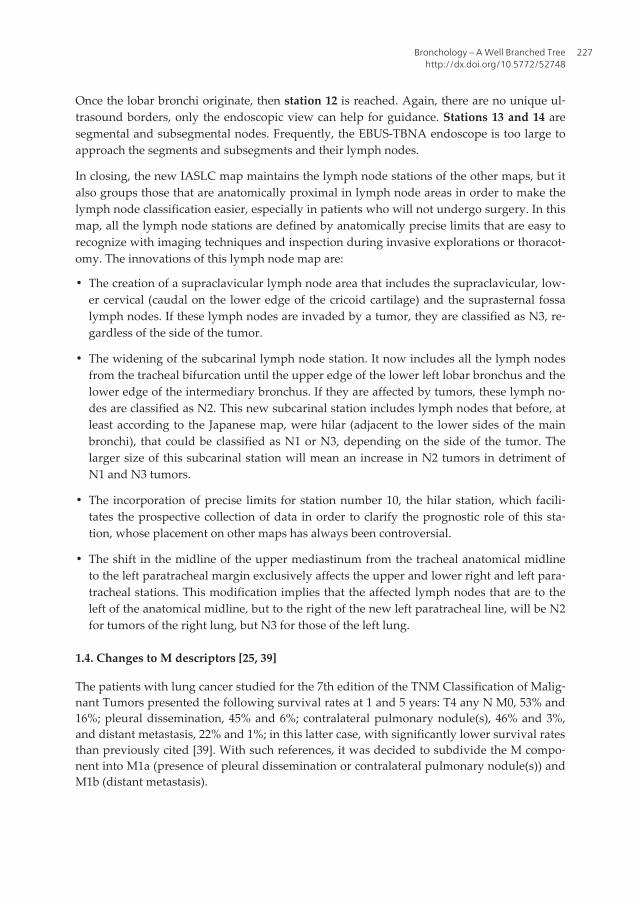
Once the lobar bronchi originate, then station 12 is reached. Again, there are no unique ul‐trasound borders, only the endoscopic view can help for guidance. Stations 13 and 14 aresegmental and subsegmental nodes. Frequently, the EBUS-TBNA endoscope is too large toapproach the segments and subsegments and their lymph nodes.
In closing, the new IASLC map maintains the lymph node stations of the other maps, but italso groups those that are anatomically proximal in lymph node areas in order to make thelymph node classification easier, especially in patients who will not undergo surgery. In thismap, all the lymph node stations are defined by anatomically precise limits that are easy torecognize with imaging techniques and inspection during invasive explorations or thoracot‐omy. The innovations of this lymph node map are:
• The creation of a supraclavicular lymph node area that includes the supraclavicular, low‐er cervical (caudal on the lower edge of the cricoid cartilage) and the suprasternal fossalymph nodes. If these lymph nodes are invaded by a tumor, they are classified as N3, re‐gardless of the side of the tumor.
• The widening of the subcarinal lymph node station. It now includes all the lymph nodesfrom the tracheal bifurcation until the upper edge of the lower left lobar bronchus and thelower edge of the intermediary bronchus. If they are affected by tumors, these lymph no‐des are classified as N2. This new subcarinal station includes lymph nodes that before, atleast according to the Japanese map, were hilar (adjacent to the lower sides of the mainbronchi), that could be classified as N1 or N3, depending on the side of the tumor. Thelarger size of this subcarinal station will mean an increase in N2 tumors in detriment ofN1 and N3 tumors.
• The incorporation of precise limits for station number 10, the hilar station, which facili‐tates the prospective collection of data in order to clarify the prognostic role of this sta‐tion, whose placement on other maps has always been controversial.
• The shift in the midline of the upper mediastinum from the tracheal anatomical midlineto the left paratracheal margin exclusively affects the upper and lower right and left para‐tracheal stations. This modification implies that the affected lymph nodes that are to theleft of the anatomical midline, but to the right of the new left paratracheal line, will be N2for tumors of the right lung, but N3 for those of the left lung.
1.4. Changes to M descriptors [25, 39]
The patients with lung cancer studied for the 7th edition of the TNM Classification of Malig‐nant Tumors presented the following survival rates at 1 and 5 years: T4 any N M0, 53% and16%; pleural dissemination, 45% and 6%; contralateral pulmonary nodule(s), 46% and 3%,and distant metastasis, 22% and 1%; in this latter case, with significantly lower survival ratesthan previously cited [39]. With such references, it was decided to subdivide the M compo‐nent into M1a (presence of pleural dissemination or contralateral pulmonary nodule(s)) andM1b (distant metastasis).
Bronchology – A Well Branched Treehttp://dx.doi.org/10.5772/52748
227
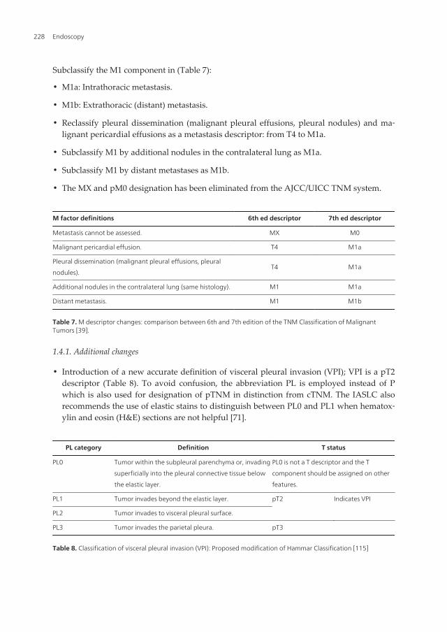
Subclassify the M1 component in (Table 7):
• M1a: Intrathoracic metastasis.
• M1b: Extrathoracic (distant) metastasis.
• Reclassify pleural dissemination (malignant pleural effusions, pleural nodules) and ma‐lignant pericardial effusions as a metastasis descriptor: from T4 to M1a.
• Subclassify M1 by additional nodules in the contralateral lung as M1a.
• Subclassify M1 by distant metastases as M1b.
• The MX and pM0 designation has been eliminated from the AJCC/UICC TNM system.
M factor definitions 6th ed descriptor 7th ed descriptor
Metastasis cannot be assessed. MX M0
Malignant pericardial effusion. T4 M1a
Pleural dissemination (malignant pleural effusions, pleural
nodules).T4 M1a
Additional nodules in the contralateral lung (same histology). M1 M1a
Distant metastasis. M1 M1b
Table 7. M descriptor changes: comparison between 6th and 7th edition of the TNM Classification of MalignantTumors [39].
1.4.1. Additional changes
• Introduction of a new accurate definition of visceral pleural invasion (VPI); VPI is a pT2descriptor (Table 8). To avoid confusion, the abbreviation PL is employed instead of Pwhich is also used for designation of pTNM in distinction from cTNM. The IASLC alsorecommends the use of elastic stains to distinguish between PL0 and PL1 when hematox‐ylin and eosin (H&E) sections are not helpful [71].
PL category Definition T status
PL0 Tumor within the subpleural parenchyma or, invading
superficially into the pleural connective tissue below
the elastic layer.
PL0 is not a T descriptor and the T
component should be assigned on other
features.
PL1 Tumor invades beyond the elastic layer. pT2 Indicates VPI
PL2 Tumor invades to visceral pleural surface.
PL3 Tumor invades the parietal pleura. pT3
Table 8. Classification of visceral pleural invasion (VPI): Proposed modification of Hammar Classification [115]
Endoscopy228
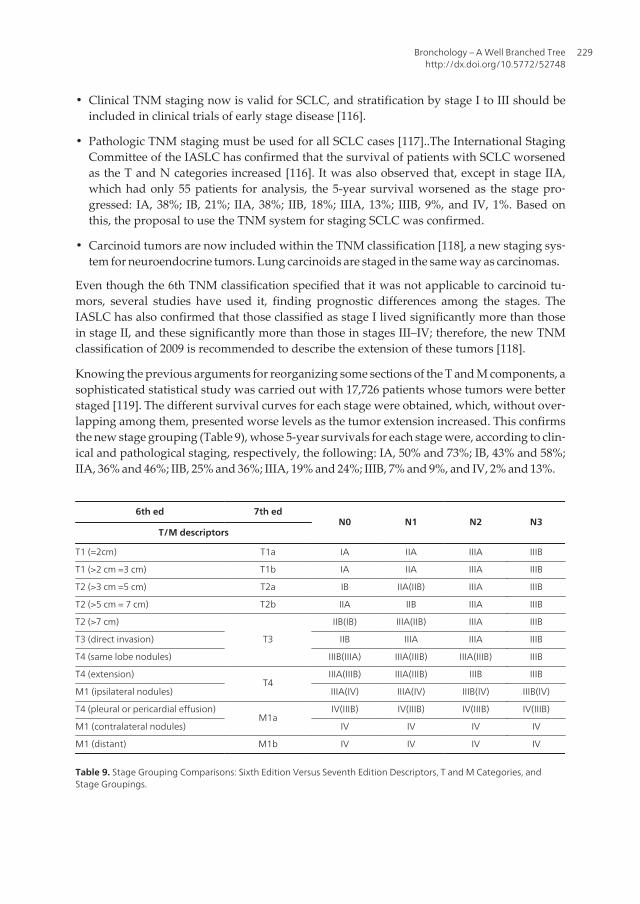
• Clinical TNM staging now is valid for SCLC, and stratification by stage I to III should beincluded in clinical trials of early stage disease [116].
• Pathologic TNM staging must be used for all SCLC cases [117]..The International StagingCommittee of the IASLC has confirmed that the survival of patients with SCLC worsenedas the T and N categories increased [116]. It was also observed that, except in stage IIA,which had only 55 patients for analysis, the 5-year survival worsened as the stage pro‐gressed: IA, 38%; IB, 21%; IIA, 38%; IIB, 18%; IIIA, 13%; IIIB, 9%, and IV, 1%. Based onthis, the proposal to use the TNM system for staging SCLC was confirmed.
• Carcinoid tumors are now included within the TNM classification [118], a new staging sys‐tem for neuroendocrine tumors. Lung carcinoids are staged in the same way as carcinomas.
Even though the 6th TNM classification specified that it was not applicable to carcinoid tu‐mors, several studies have used it, finding prognostic differences among the stages. TheIASLC has also confirmed that those classified as stage I lived significantly more than thosein stage II, and these significantly more than those in stages III–IV; therefore, the new TNMclassification of 2009 is recommended to describe the extension of these tumors [118].
Knowing the previous arguments for reorganizing some sections of the T and M components, asophisticated statistical study was carried out with 17,726 patients whose tumors were betterstaged [119]. The different survival curves for each stage were obtained, which, without over‐lapping among them, presented worse levels as the tumor extension increased. This confirmsthe new stage grouping (Table 9), whose 5-year survivals for each stage were, according to clin‐ical and pathological staging, respectively, the following: IA, 50% and 73%; IB, 43% and 58%;IIA, 36% and 46%; IIB, 25% and 36%; IIIA, 19% and 24%; IIIB, 7% and 9%, and IV, 2% and 13%.
6th ed 7th edN0 N1 N2 N3
T/M descriptors
T1 (=2cm) T1a IA IIA IIIA IIIB
T1 (>2 cm =3 cm) T1b IA IIA IIIA IIIB
T2 (>3 cm =5 cm) T2a IB IIA(IIB) IIIA IIIB
T2 (>5 cm = 7 cm) T2b IIA IIB IIIA IIIB
T2 (>7 cm)
T3
IIB(IB) IIIA(IIB) IIIA IIIB
T3 (direct invasion) IIB IIIA IIIA IIIB
T4 (same lobe nodules) IIIB(IIIA) IIIA(IIIB) IIIA(IIIB) IIIB
T4 (extension)T4
IIIA(IIIB) IIIA(IIIB) IIIB IIIB
M1 (ipsilateral nodules) IIIA(IV) IIIA(IV) IIIB(IV) IIIB(IV)
T4 (pleural or pericardial effusion)M1a
IV(IIIB) IV(IIIB) IV(IIIB) IV(IIIB)
M1 (contralateral nodules) IV IV IV IV
M1 (distant) M1b IV IV IV IV
Table 9. Stage Grouping Comparisons: Sixth Edition Versus Seventh Edition Descriptors, T and M Categories, andStage Groupings.
Bronchology – A Well Branched Treehttp://dx.doi.org/10.5772/52748
229
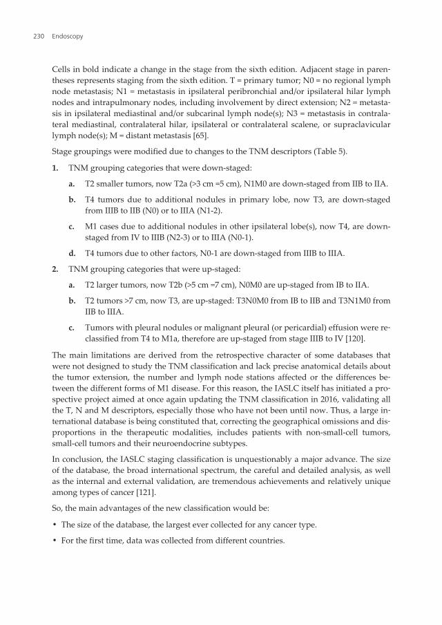
Cells in bold indicate a change in the stage from the sixth edition. Adjacent stage in paren‐theses represents staging from the sixth edition. T = primary tumor; N0 = no regional lymphnode metastasis; N1 = metastasis in ipsilateral peribronchial and/or ipsilateral hilar lymphnodes and intrapulmonary nodes, including involvement by direct extension; N2 = metasta‐sis in ipsilateral mediastinal and/or subcarinal lymph node(s); N3 = metastasis in contrala‐teral mediastinal, contralateral hilar, ipsilateral or contralateral scalene, or supraclavicularlymph node(s); M = distant metastasis [65].
Stage groupings were modified due to changes to the TNM descriptors (Table 5).
1. TNM grouping categories that were down-staged:
a. T2 smaller tumors, now T2a (>3 cm =5 cm), N1M0 are down-staged from IIB to IIA.
b. T4 tumors due to additional nodules in primary lobe, now T3, are down-stagedfrom IIIB to IIB (N0) or to IIIA (N1-2).
c. M1 cases due to additional nodules in other ipsilateral lobe(s), now T4, are down-staged from IV to IIIB (N2-3) or to IIIA (N0-1).
d. T4 tumors due to other factors, N0-1 are down-staged from IIIB to IIIA.
2. TNM grouping categories that were up-staged:
a. T2 larger tumors, now T2b (>5 cm =7 cm), N0M0 are up-staged from IB to IIA.
b. T2 tumors >7 cm, now T3, are up-staged: T3N0M0 from IB to IIB and T3N1M0 fromIIB to IIIA.
c. Tumors with pleural nodules or malignant pleural (or pericardial) effusion were re‐classified from T4 to M1a, therefore are up-staged from stage IIIB to IV [120].
The main limitations are derived from the retrospective character of some databases thatwere not designed to study the TNM classification and lack precise anatomical details aboutthe tumor extension, the number and lymph node stations affected or the differences be‐tween the different forms of M1 disease. For this reason, the IASLC itself has initiated a pro‐spective project aimed at once again updating the TNM classification in 2016, validating allthe T, N and M descriptors, especially those who have not been until now. Thus, a large in‐ternational database is being constituted that, correcting the geographical omissions and dis‐proportions in the therapeutic modalities, includes patients with non-small-cell tumors,small-cell tumors and their neuroendocrine subtypes.
In conclusion, the IASLC staging classification is unquestionably a major advance. The sizeof the database, the broad international spectrum, the careful and detailed analysis, as wellas the internal and external validation, are tremendous achievements and relatively uniqueamong types of cancer [121].
So, the main advantages of the new classification would be:
• The size of the database, the largest ever collected for any cancer type.
• For the first time, data was collected from different countries.
Endoscopy230
• The timeframe of 10 years allowed for 5 years follow-up.
• Cases included all treatment modalities.
• Pathologic and clinical staging where considered in survival analysis.
• The statistical analysis included meticulous internal and external validation.
• Changes to the TNM descriptors and stage groups were derived strictly from the outcomemeasure of overall survival.
The new TNM system is less intuitive and more complex than the 6th edition, and will bemore difficult to learn. The oncology community needs to overcome the higher learningcurve in order to offer patients the most appropriate treatment choices based on more accu‐rate prognostic information [120].
2. Electromagnetic navigation bronchoscopy
It is known that diagnostic yield of flexible bronchoscopy is limited by its inability to guidebiopsy instruments directly to the lesion. As it was expected, diagnosis success rate is de‐pendent on the size and location of the lesion. The diagnostic yield of flexible bronchoscopyis expected to be between 20 and 84%. However, current nonsurgical techniques available todiagnose small peripheral lung lesions (SPLL) are limited either by low accuracy [122–125]or by potential complications [126–129].
For lesions less than 2 cm in diameter, the diagnostic yield of flexible bronchoscopy is 14%for peripheral lesions in the outer third of the chest and as high as 31% if in the proximaltwo-thirds [130]. The diagnostic yield of flexible bronchoscopy for mediastinal lymph nodesusing transbronchial needle aspiration (TBNA) is reported to be between 15 and 83% [131].Also, diagnostic yield of TBNA in staging of bronchogenic carcinoma is reported to be be‐tween 50 and 60% [132].
Within the past several years, electromagnetic navigation guided bronchoscopy (EGB) hasproven very effective at assessing pulmonary nodules accurately with very low complica‐tion rates. EGB consists of four elements:
1. Computer software that utilizes thin-slice CT to create a three-dimensional rendering ofthe lung and tracheobronchial tree, which can then be used for virtual bronchoscopy.
2. A sensor probe that fits through the small working/suction channel of a bronchoscopewith a steering mechanism. Because the sensor probe is recognizable within the electro‐magnetic field, it can be navigated through the small airways of the lung toward pe‐ripheral lesions not reachable by conventional bronchoscopy (Figure 22).
3. An electromagnetic field encompassing the patient’s thorax, so that the real anatomycan be merged with the computer generated (virtual) anatomy by use of standard “reg‐istration points” (e.g., carina, takeoff of the right upper lobe bronchus, bifurcation be‐tween the left upper and lower lobes).
Bronchology – A Well Branched Treehttp://dx.doi.org/10.5772/52748
231
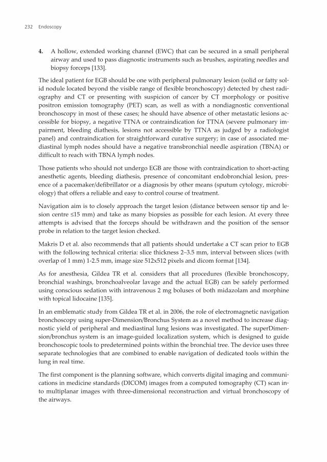
4. A hollow, extended working channel (EWC) that can be secured in a small peripheralairway and used to pass diagnostic instruments such as brushes, aspirating needles andbiopsy forceps [133].
The ideal patient for EGB should be one with peripheral pulmonary lesion (solid or fatty sol‐id nodule located beyond the visible range of flexible bronchoscopy) detected by chest radi‐ography and CT or presenting with suspicion of cancer by CT morphology or positivepositron emission tomography (PET) scan, as well as with a nondiagnostic conventionalbronchoscopy in most of these cases; he should have absence of other metastatic lesions ac‐cessible for biopsy, a negative TTNA or contraindication for TTNA (severe pulmonary im‐pairment, bleeding diathesis, lesions not accessible by TTNA as judged by a radiologistpanel) and contraindication for straightforward curative surgery; in case of associated me‐diastinal lymph nodes should have a negative transbronchial needle aspiration (TBNA) ordifficult to reach with TBNA lymph nodes.
Those patients who should not undergo EGB are those with contraindication to short-actinganesthetic agents, bleeding diathesis, presence of concomitant endobronchial lesion, pres‐ence of a pacemaker/defibrillator or a diagnosis by other means (sputum cytology, microbi‐ology) that offers a reliable and easy to control course of treatment.
Navigation aim is to closely approach the target lesion (distance between sensor tip and le‐sion centre ≤15 mm) and take as many biopsies as possible for each lesion. At every threeattempts is advised that the forceps should be withdrawn and the position of the sensorprobe in relation to the target lesion checked.
Makris D et al. also recommends that all patients should undertake a CT scan prior to EGBwith the following technical criteria: slice thickness 2–3.5 mm, interval between slices (withoverlap of 1 mm) 1-2.5 mm, image size 512x512 pixels and dicom format [134].
As for anesthesia, Gildea TR et al. considers that all procedures (flexible bronchoscopy,bronchial washings, bronchoalveolar lavage and the actual EGB) can be safely performedusing conscious sedation with intravenous 2 mg boluses of both midazolam and morphinewith topical lidocaine [135].
In an emblematic study from Gildea TR et al. in 2006, the role of electromagnetic navigationbronchoscopy using super-Dimension/Bronchus System as a novel method to increase diag‐nostic yield of peripheral and mediastinal lung lesions was investigated. The superDimen‐sion/bronchus system is an image-guided localization system, which is designed to guidebronchoscopic tools to predetermined points within the bronchial tree. The device uses threeseparate technologies that are combined to enable navigation of dedicated tools within thelung in real time.
The first component is the planning software, which converts digital imaging and communi‐cations in medicine standards (DICOM) images from a computed tomography (CT) scan in‐to multiplanar images with three-dimensional reconstruction and virtual bronchoscopy ofthe airways.
Endoscopy232
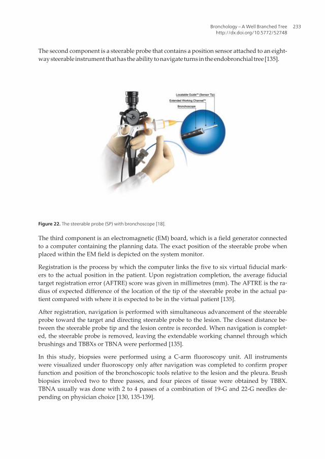
The second component is a steerable probe that contains a position sensor attached to an eight-way steerable instrument that has the ability to navigate turns in the endobronchial tree [135].
Figure 22. The steerable probe (SP) with bronchoscope [18].
The third component is an electromagnetic (EM) board, which is a field generator connectedto a computer containing the planning data. The exact position of the steerable probe whenplaced within the EM field is depicted on the system monitor.
Registration is the process by which the computer links the five to six virtual fiducial mark‐ers to the actual position in the patient. Upon registration completion, the average fiducialtarget registration error (AFTRE) score was given in millimetres (mm). The AFTRE is the ra‐dius of expected difference of the location of the tip of the steerable probe in the actual pa‐tient compared with where it is expected to be in the virtual patient [135].
After registration, navigation is performed with simultaneous advancement of the steerableprobe toward the target and directing steerable probe to the lesion. The closest distance be‐tween the steerable probe tip and the lesion centre is recorded. When navigation is complet‐ed, the steerable probe is removed, leaving the extendable working channel through whichbrushings and TBBXs or TBNA were performed [135].
In this study, biopsies were performed using a C-arm fluoroscopy unit. All instrumentswere visualized under fluoroscopy only after navigation was completed to confirm properfunction and position of the bronchoscopic tools relative to the lesion and the pleura. Brushbiopsies involved two to three passes, and four pieces of tissue were obtained by TBBX.TBNA usually was done with 2 to 4 passes of a combination of 19-G and 22-G needles de‐pending on physician choice [130, 135-139].
Bronchology – A Well Branched Treehttp://dx.doi.org/10.5772/52748
233
Eberhardt et al. [18] reported in 2007 92 peripheral lung lesions biopsies from 89 consecutivesubjects. The diagnostic yield of EGB was 67%, which was independent of lesion size. Totalprocedure time ranged from 16.3 to 45.0 min (mean [± SD] procedure time, 26.9 ± 6.5 min).The mean navigation error was 9 ± 6 mm(range, 1 to 31 mm). They reported two incidencesof pneumothorax for which no intervention was required. When analyzed by lobar distribu‐tion, there was a trend toward a higher ENB yield in diagnosing lesions in the right middlelobe (88%). They concluded that EGB can be used as an independent bronchoscopic techni‐que without the need for fluoroscopy when compared with other available studies. Therewas no increased risk of pneumothorax (2 of 89 patients; 2%). The upper lobes tend to havesharper angles in the bronchial tree that may be challenging to navigate even with a steera‐ble sensor probe. The EWC ends close to the tip of the sensor probe and makes it less flexi‐ble. This reduces the range of deflection and, consequently, the ability to navigate. It canalso make the probe flip into a different position when negotiating some tight angles in thebronchi. Navigation in the lower lobes is more affected by diaphragmatic movement duringbreathing and could result in larger errors than recorded. This is because the planning dataare based on CT scan images acquired in a single breathhold [140, 141].
Eberhardt et al. also showed that the improved yield of EGB compared to conventionaltransbronchial lung biopsy in small lesions (diameter ≤ 2 cm) can be attributed to the im‐proved precision in navigation. This study has shown the yield, safety, and timesaving withuse of the EGB system without the need for fluoroscopy. This system eliminates radiationexposure and could reduce procedure costs. The diagnostic utility of the use of EGB in thebiopsy of peripheral lung lesions appears to be equivalent to other advanced techniques likeendobronchial ultrasound [142-144].
Apparently, these techniques have pushed bronchoscopic biopsy yields closer to those ach‐ieved by CT scan-guided transthoracic needle biopsy and surgery. Given the relative com‐fort [145] and safety [146] of flexible bronchoscopy, and the recognized risks of both CTscan-guided [147-149] and surgical biopsies [150], there is growing need to develop and re‐fine these techniques. The expanded role of lung cancer screening, in which the vast majori‐ty of lesions is benign, makes this all the more important [151]. Multimodality diagnosis bycombining ENB with other bronchoscopic and imaging techniques may further enhance thediagnostic yield [140].
3. Autoflorescence Bronchoscopy (AFB)
Visible light perceived by the human eye comprises the whole wavelength range, between400–700 nm. Conventional bronchoscopy illuminates mucosal structures of the airwaveswith the full wave spectrum; therefore, light gets reflected, backscattered or absorbed by thestructures it encounters, thus providing the human eye with an image [152]. Tissues show anatural autoflorescence when excited in the 200-460 nm range; however, only the visiblepart of this range is used in medical applications [153].
Endoscopy234
However, the light source attached to the bronchoscope can emit only blue light, as a pro‐portion of the blue spectrum excites the cellular chromophores, such as collagen, elastin orkeratin, contained in the layers of the submucosa, especially within the connective tissuecomprised in the elastic fiber bundles, or on the exterior surface of the cartilages (perichon‐drium). When excited by blue-violet light in the 400-450 nm spectrum, the excited chromo‐phores of normal cellular lines found in the mucosa display a green tint. Another aspect ofthe autoflorescence theory refers to the thickness and cellular morphology of the examinedtissue. Tumors and premalignant lesions have an increased mucosal thickness and thereforeabsorb more of the excitation occurring from fluorescence light, in what is called “the archi‐tectural effect” [153, 154]. Tissues that underwent a morphological change due to variouspathological conditions, predominantly premalignant dysplasia or metaplasia-like phenom‐enon, are colored differently as the green fluorescence is affected by either alterations of cel‐lular chemistry, morphology or epithelial thickness [155–157]. A wide range of AFB devicesare currently available on the market: the Storz D-light system [158], Pentax SAFE-1000[159], the Xilix LIFE system [158] or the DAFE system by Richard Wolf [160].
The main advantage of AFB is that progressive dysplasia of the mucosal layers also resultsin a progressive transition from normal green autoflorescence to a red-brown color, specificto precancerous or malignant lesions. This makes early premalignant lesions far easier tospot during bronchoscopy as compared to regular white-light based techniques. Inflamma‐tory reactions, granulomas, scars, dysplasia/metaplasia and early malignant lesions whichare hard to spot due to their submucosal confinement are therefore easily spotted due totheir dark red-brown appearance, surrounded by normal green tissue [155–157].
The addition of AFB to conventional endoscopy can significantly increase diagnostic rate ofearly malignant or premalignant lesion, without the need of tumor sensitizers and thereforewith no additional complications to standard bronchoscopy techniques. The detection rateof high-grade dysplasia and carcinoma in-situ (CIS) is increased to 88% after using AFB, upfrom the median 40% detection rate that conventional white-light bronchoscopy provides[161, 162]. However, as already stated, both premalignant lesions and various inflammatoryconditions have similar appearances in AFB. Therefore, the specificity is rather low, with ahigh rate of false positive investigations [163, 164].
Since diffuse reflectance spectroscopy measures directly the changes in the path of whitelight through tissue scattering and absorption levels, some have theorized that combiningautofluorescence with diffuse reflectance probes may enhance the specificity of this method.Preliminary findings have shown that a combination between the two can significantly im‐prove the positive predictive value of AFB without altering the already attained high sensi‐tivity for premalignant and early malignant lesions [165] (Bard MPL et al, 2005). Otherattempts to improve specificity by local spectroscopical measurements have shown promis‐ing results, making way for one-stop techniques which would combine two types of wave‐length measurement and fluorophore weighting [153, 165].
One study investigated the use of AFB in primary lung cancer, after treatment of head andneck cancer, detecting 29% (12/44 patients) of all second primary lung cancers, while detect‐ing early lesions in two patients which would otherwise be missed by conventional imaging
Bronchology – A Well Branched Treehttp://dx.doi.org/10.5772/52748
235
methods [166]. Early reports of Lam and his team in asbestos and diesel-exposed individu‐als showed an 86% sensitivity for moderate to severe dysplasia and CIS in the case of AFB,compared to just 52% for white light bronchoscopy [167]. A multicenter study performed byLam in 1998 with the LIFE system, on 173 patients (142 biopsy-proven cases of severe dys‐plasia, CIS or invasive cancer) showed that AFB has a relative sensitivity of 2.71 comparedto conventional bronchoscopy, successfully detecting 91 cases in comparison to only 35 withbronchoscopy alone. When considering intraepithelial lesions alone, the relative sensitivityincreases to 6.1 for AFB compared to standard bronchoscopy [164].
A correlation between the loss of fluorescence and the grade of dysplasia; therefore, atrained endoscopist can differentiate these gradations to some extent, being even able toidentify inflammatory or granulomatous lesions [155]. This would In turn greatly improvethe specificity of AFB. A different approach consists in the usage of computer assisted analy‐sis of signals of different wavelengths, represented in a spectrogram which in turn can clas‐sify benign from malignant lesions.
4. Narrow-Band Imaging (NBI)
NBI is a novel imaging technique capable of improving the visualization of superficial struc‐tures of the respiratory mucosa [168, 169]. NBI is used for an accurate classification of le‐sions, even though it does not have an established role during routine bronchoscopy [170–172]. High magnification bronchoscopy in combination with NBI can visualize the alteredmicro-vascularization which is formed in dysplastic or neoplastic lesions [173]. As the tumorprogresses, it requires an adequately enlarged blood supply, therefore in most cancers, in‐cluding those of the lungs and airways, neo angiogenesis is a constant phenomenon throughwhich newly formed vessels are constantly produced in order to supplement local needs.These vessels form irregular aberrant patterns, having unequal diameters, tortuous configu‐rations and uneven segment lengths.
The principle of NBI is simple: three optical filters segment the RGB (red-green-blue) lightspectrum in sequence, thus narrowing the bandwidth of the spectral transmittance [174].This allows for visual marking of capillary structures of the sub-mucosa in deep red color,allowing an easier identification and characterization, as different wavelengths penetrate thetissue at different depths. The filtering system is placed in the optical illumination systemand basically contains two narrow wavelength components: NBI-B corresponding to the400-430 nm range (blue and green visualization) and NBI-G for the 530-550 nm range (redvisualization), while a third filter can be installed for an accurate segmentation of the blue-green features, operating in the 430-460 nm range; the optical light-source usually operatesin the 400-700 nm wavelength range. All current models include a 2-filter system, which em‐phasizes capillaries in the superficial mucosal layers by the 400-430 nm light, coloring themin shades of brown, while deeper mucosal or submucosal vessels are displayed in cyan byusing the 530-550 nm filter. Modern systems allow for easy switch between the normal WLIand the enhanced NBI operations [168].
Endoscopy236
Limitations of the technology arise from the fact that not all lung malignancies develop sub‐mucosal neo vascularization visible from the airways, therefore potential false-negative NBIfindings can be frequent when used in combination with WLB. Its sensitivity may be how‐ever increased when used in conjunction with other specific techniques such as AFB; how‐ever, its role in the screening process for lung malignancies is yet to be determined. A stepfurther in this direction would be the standardization of imaging findings, based on clinicaldescriptions and classification of abnormal airway vascularity. High magnification broncho‐scopy can improve the specificity of the technique, as it may allow a more clear descriptionof the vascular patterns [168]. Good prospects come from new techniques such as probe-based confocal laser endomicroscopy which can provide an in situ diagnosis of malignancyif used in conjunction with any endoscopic technique.
5. Probe-based Confocal Laser Endomicroscopy (pCLE)
Gastrointestinal applications of pCLE are numerous, the system being successfully testedin real-life conditions [175]. It can be used for identifying premalignant lesions otherwiseinvisible to classic endoscopic techniques, such as Barrett’s esophagus [176], classificationof polipous lesions at colonic level [177], gastric metaplasia or early stage lesions at alllevels of the digestive tracts [178, 179]. The technique requires injection of fluorescein oranother fluorophore, followed by endoscopic imaging of the dye to identify mucosal de‐tails such as mucosal and vessel architecture to distinguish among normal, dysplastic, andneoplastic tissue. The use of fluorescein as an in-vivo contrast agent for detecting vascularstructures has been deemed safe by several studies [180]. It thus allows for better visuali‐zation of vascular structures, serving as an overall tissue contrasting agent as it may leakbeyond capillary confinement.
The imaging microprobe is connected to 30,000 fiber-optic threads that enable point-to-point real-time detection at 12 frames/sec. The microprobe’s flexibility and size (1.5 mm indiameter) allow for great user maneuverability to scan tissues in situ at angles that wouldnot be possible with any available confocal microscope objective. The mini-probe is insert‐ed through a working channel of any standard bronchoscope and can reach the alveolarduct. A laser generator emits 488 nm blue argon laser light which is transmitted throughthe optical fibers and excites the elastin scaffold of the acinus. In turn, a real time imageof the elastic fibers which sustain the alveoli, as well as the microvessel architecture is re‐vealed. The probe can be translated over a larger tissue sample, and the images later re‐constructed in order to extend the field of view covered [181]. A monitor attached to animaging unit is available for real-time visualization of cellular images, the device being ca‐pable of recording full length movies which can be recorded and later on analyzed withdedicated software [182].
This technology is still in its early stages; however, it was deemed safe by regulatory organ‐isms in the United States of America for applications in patient settings. In conjunction withsafe contrasting agents such as fluorescein [183] or methilen blue [184], it can be used for the
Bronchology – A Well Branched Treehttp://dx.doi.org/10.5772/52748
237
in vivo histologic assessment of the bronchial epithelium at all levels of the airway system[185–187]. Several studies reported good initial results in control groups of healthy smokersor patients with chronic obstructive pulmonary disease for diagnosing various forms of pa‐renchymal lung disease [188, 189]. A pCLE investigation seems to be a quick and safe proce‐dure for patients with various pulmonary conditions, with minimal side-effects related toboth the technique and the contrasting fluorescent agents used for imaging. General consen‐sus is that further studies are needed in order to extent the diagnostic capabilities and en‐hance its sensibility in comparison with standard cytological and histological techniques.
Author details
Mihai Olteanu1, Costin Teodor Streba1, Bogdan Oprea1, Raluca Marinas1,Mimi Floarea Nitu1, Emilia Crisan1,2 and Tudorel Ciurea1
1 University of Medicine and Pharmacy of Craiova, Romania
2 "Marius Nasta" Pulmonology Institute, Bucharest, Romania
References
[1] Lopez-Encuentra A, Garcia-Lujan R, Rivas JJ, Rodriguez-Rodriguez J, Torres-Lanza J,Varela- Simo G. Comparison between clinical and pathologic staging in 2,994 cases oflung cancer. Ann Thorac Surg 2005; 79:974–979; discussion, 979.
[2] Mountain CF. Revisions in the international system for staging lung cancer. Chest1997; 111 (6): 1710-1717.
[3] Lung. In: American Joint Committee on Cancer. AJCC Cancer Staging Manual. 6thed. New York, NY: Springer, 2002, pp 167-181.
[4] Silvestri GA. A seismic shift in staging. J Thorac Oncol 2007; 2 (8): 682-683.
[5] Goldstraw P Crowley JJ; on behalf of the IASLC International Staging Project. The In‐ternational Association for the Study of Lung Cancer International Staging Project onLung Cancer. J Thorac Oncol 2006; 1 (4): 281-286.
[6] Rami Porta R. Nueva clasificación TNM del cáncer de pulmón. Arch Bronconeumol2009; 45 (4): 159-161.
[7] Sobin LH, Gospodarowicz MK, Wittekind C. eds. TNM Classification of MalignantTumours, 7th ed., 2010, Wiley-Blackwell.
[8] Rami-Porta R, Crowley JJ, Goldstraw P. The revised TNM staging system for lungcancer. Ann Thorac Cardiovasc Surg 2009; 15 (1): 4-9.
Endoscopy238
[9] Groome PA, Bolejack V, Crowley JJ, Kennedy C, Krasnik M, Sobin LH, Goldstraw P;IASLC International Staging Committee; Cancer Research and Biostatistics; Observ‐ers to the Committee; Participating Institutions. The IASLC Lung Cancer StagingProject: validation of the proposals for revision of the T, N, and M descriptors andconsequent stage groupings in the forthcoming (seventh) edition of the TNM classifi‐cation of malignant tumours. J Thorac Oncol 2007; 2 (8): 694-705.
[10] Krasnik M, Vilmann P, Larsen S, Jacobsen G, Preliminary experience with a newmethod of endoscopic transbronchial real time ultrasound guided biopsy for diagno‐sis of mediastinal and hilar lesions Thorax. 2003 December; 58(12): 1083–1086.
[11] Herth F J F, Eberhardt R, Vilmann P, Krasnik M, Ernst A, Real‐time endobronchialultrasound guided transbronchial needle aspiration for sampling mediastinal lymphnodes, Thorax. 2006 September; 61(9): 795–798.
[12] Herth FJF, Krasnik M, Yasufuku K, R Rintoul R, Ernst A, Endobronchial Ultrasound-guided Transbronchial Needle Aspiration, J Bronchol 2006;13:84–91
[13] Wong M, Yasufuku K, Nakajima T, Herth FJ, Sekine Y, Shibuya K, Iizasa T, Hiroshi‐ma K, Lam WK, Fujisawa T, Endobronchial ultrasound: new insight for the diagnosisof sarcoidosis, Eur Respir J. 2007 Jun; 29(6):1182-6.
[14] Herth FJF, Ernst A, Eberhardt R, Vilmann P, Dienemann H, Krasnik M, Endobron‐chial Ultrasound-guided Transbronchial Needle Aspiration of Lymph Nodes in theRadiologically Normal Mediastinum. Eur Respir J 2006; 28: 910-914.
[15] Hwangbo B, Kim SK, Lee HS, Lee HS, Kim MS, Lee JM, Kim HY, Lee GK, Nam BH,Zo JI, Application of endobronchial ultrasound-guided transbronchial needle aspira‐tion following integrated PET/CT in mediastinal staging of potentially operable non-small cell lung cancer. Chest. 2009 May;135(5):1280-7. Epub 2008 Dec 31.
[16] Ernst A, Anantham D, Eberhardt R, Krasnik M, Herth FJ. Diagnosis of mediastinaladenopathy-real-time endobronchial ultrasound guided needle aspiration versus me‐diastinoscopy. J Thorac Oncol. 2008 Jun;3(6):577-82.
[17] Herth FJ, Yasufuku K, Eberhardt R, Hoffmann H, Krasnik M, Ernst A. Resistance in‐dex in mediastinal lymph nodes: a feasibility study. J Thorac Oncol. 2008 Apr;3(4):348-50.
[18] Herth F, Eberhardt R, Mulay T, Anantham D, Ernst A, Resistance index throughEBUS in enlarged mediastinal lymph nodes correlates with malignant involvement,CHEST. October 2007;132(4_MeetingAbstracts):465c-466.
[19] Nakajima T, Yasufuku K, Suzuki M, et al. Assessment of epidermal growth factor re‐ceptor mutation by endobronchial ultrasound-guided transbronchial needle aspira‐tion CHEST 2007;132(2):597-602.
Bronchology – A Well Branched Treehttp://dx.doi.org/10.5772/52748
239
[20] Nakajima T, Yasufuku K, Suzuki M, et al. Chemosensitivity-related aberrant methyl‐ation profiling of non-small cell lung cancer by endobronchial ultrasound-guidedtransbronchial needle aspiration CHEST 2007;132(4_MeetingAbstracts):466a-466.
[21] Tournoy KG, Annema JT, Krasnik M, Herth FJ, van Meerbeeck JP Endoscopic andendobronchial ultrasonography according to the proposed lymph node map defini‐tion in the seventh edition of the tumor, node, metastasis classification for lung can‐cer. J Thorac Oncol. 2009 Dec;4(12):1576-84.
[22] Annema JT, van Meerbeeck JP, Rintoul RC, Dooms C, Deschepper E, Dekkers OM,De Leyn P, Braun J, Carroll NR, Praet M, de Ryck F, Vansteenkiste J, Vermassen F,Versteegh MI, Veseliç M, Nicholson AG, Rabe KF, Tournoy KG. Mediastinoscopy vsendosonography for mediastinal nodal staging of lung cancer: a randomized trial.JAMA. 2010 Nov 24;304(20):2245-52.
[23] Hwangbo B, Lee G, Lee H, et al. Transbronchial and transesophageal fine-needle as‐piration using an ultrasound bronchoscope in mediastinal staging of potentially op‐erable lung cancer CHEST 2010;138(4):795-802, Herth FF, Krasnik M, Kahn N,Eberhardt R, Ernst A. Combined endoscopic-endobronchial ultrasound-guided fine-needle aspiration of mediastinal lymph nodes through a single bronchoscope in 150patients with suspected lung cancer CHEST 2010;138(4):790-794.
[24] Herth FJ Nonsurgical staging of the mediastinum: EBUS and EUS. Semin Respir CritCare Med. 2011 Feb;32(1):62-8. Epub 2011 Apr 15
[25] Edge SB, Byrd DR, Compton CC, Fritz AG, Greene FL, Trotti A. eds. Part I. Generalinformation on cancer staging and end-results reporting. 1. Purposes and principlesof cancer staging. In: Edge SB, Byrd DR, Compton CC, Fritz AG, Greene FL, Trotti A.eds. AJCC Cancer Staging Manual. 7th ed., 2010, Springer; pp. 3-14.
[26] Herth FJ, Eberhardt R, Vilmann P et al. Real-time endobronchial ultrasound guidedtransbronchial needle aspiration for sampling mediastinal lymph nodes. Thorax2006; 61: 795–798.
[27] Ernst A, Eberhardt R, Krasnik M, Herth FJ. Efficacy of endobronchial ultrasoundg‐uided transbronchial needle aspiration of hilar lymph nodes for diagnosing and stag‐ing cancer. J Thorac Oncol 2009; 4: 947–950.
[28] Tournoy KG, Rintoul RC, van Meerbeeck JP et al. EBUS-TBNA for the diagnosis ofcentral parenchymal lung lesions not visible at routine bronchoscopy. Lung Cancer2009; 63: 45–49.
[29] Ernst A, Anantham D, Eberhardt R et al. Diagnosis of mediastinal adenopathy real-time endobronchial ultrasound guided needle aspiration versus mediastinoscopy. JThorac Oncol 2008; 3: 577–582.
Endoscopy240
[30] Detterbeck FC, Jantz MA, Wallace MB et al. Invasive mediastinal staging of lung can‐cer. ACCP evidence based clinical practice guidelines, 2nd edition. Chest 2007; 132:202S–220S..
[31] Tournoy KG, Govaerts E, Malfait T, Dooms C. Endobronchial ultrasound-guidedtransbronchial needle biopsy for M1 staging of extrathoracic malignancies. Ann On‐col first published online July 5, 2010 doi:10.1093/annonc/mdq311
[32] American Cancer Society. Cancer facts and figures, 1998. Atlanta, GA: AmericanCancer Society, 1998.
[33] A. Ernst, F.J.F. Herth (eds.), Endobronchial Ultrasound, DOI10.1007/978-0-387-09437-3_1, ª Springer ScienceþBusiness Media, LLC 2009
[34] Silvestri GA, Gould MK, Margolis ML, et al. Noninvasive staging in non-small celllung cancer: ACCP evidenced-based clinical practice guidelines (2nd edition). Chest.2007; 132: 178–201
[35] Bhutani MS, Hawes RH, Hoffmann BJ. A comparison of the accuracy of echo featuresduring endoscopic ultrasound (EUS) and EUS-guided fine-needle aspiration for di‐agnosis of malignant lymph node invasion. Gastrointest Endosc. 1997; 31: 550–553.
[36] Okamoto H, Watanabe K, Nagatomo A, et al. Endobronchial ultrasonography formediastinal and hilar lymph node metastases of lung cancer. Chest. 2002; 121: 1498–1506.
[37] Yasufuku node staging in non-small cell carcinoma. Do we have to accept the com‐promise? Eur J Cardiothorac Surg. 2001; 20: 652–654.
[38] Rami-Porta R, Ball D, Crowley J, Giroux DJ, Jett J, Travis WD, Tsuboi M, Vallières E,Goldstraw P; International Staging Committee; Cancer Research and Biostatistics;Observers to the Committee; Participating Institutions. The IASLC Lung CancerStaging Project: proposals for the revision of the T descriptors in the forthcoming(seventh) edition of the TNM classification for lung cancer. J Thorac Oncol 2007; 2 (7):593-602.
[39] Postmus PE, Brambilla E, Chansky K, Crowley J, Goldstraw P, Patz EF Jr, YokomiseH; International Association for the Study of Lung Cancer International StagingCommittee; Cancer Research and Biostatistics; Observers to the Committee; Partici‐pating Institutions. The IASLC Lung Cancer Staging Project: proposals for revision ofthe M descriptors in the forthcoming (seventh) edition of the TNM classification oflung cancer. J Thorac Oncol 2007; 2 (8): 686-693.
[40] Dr. A. De la Guerra. New TNM Classification for Lung Cancer. Part II: A review.Doctors Lounge Website. Available at: http://www.doctorslounge.com/index.php/articles/page/342.
Bronchology – A Well Branched Treehttp://dx.doi.org/10.5772/52748
241
[41] Strand TE, Rostad H, Møller B, Norstein J. Survival after resection for primary lungcancer: a population based study of 3211 resected patients. Thorax 2006; 61 (8):710-715.
[42] Suzuki K, Nagai K, Yoshida J, Nishimura M, Takahashi K, Yokose T, Nishiwaki Y.Conventional clinicopathologic prognostic factors in surgically resected nonsmall celllung carcinoma. A comparison of prognostic factors for each pathologic TNM stagebased on multivariate analyses. Cancer 1999; 86 (10): 1976-1984.
[43] van Rens MT, de la Rivière AB, Elbers HR, van Den Bosch JM. Prognostic assessmentof 2,361 patients who underwent pulmonary resection for non-small cell lung cancer,stage I, II, and IIIA. Chest 2000; 117 (2): 374-379.
[44] Hung JJ, Hsu WH, Hsieh CC, Huang BS, Huang MH, Liu JS, Wu YC. Post-recurrencesurvival in completely resected stage I non-small cell lung cancer with local recur‐rence. Thorax 2009; 64 (3): 192-196.
[45] Casali C, Storelli E, Morandi U. The prognostic impact of tumor size in resected stageI non-small cell lung cancer: evidence for a two thresholds tumor diameters classifi‐cation. Lung Cancer 2006; 54 (2): 185-191.
[46] Mountain CF, Carr DT, Anderson WA. A system for the clinical staging of lung can‐cer. Am J Roentgenol Radium Ther Nucl Med 1974; 120 (1): 130-138.
[47] Ruffini E, Filosso PL, Bruna MC, Coni F, Cristofori RC, Mossetti C, Solidoro P, OliaroA. Recommended changes for T and N descriptors proposed by the International As‐sociation for the Study of Lung Cancer - Lung Cancer Staging Project: a validationstudy from a single-centre experience. Eur J Cardiothorac Surg 2009; 36 (6):1037-1044.
[48] Fukui T, Mori S, Hatooka S, Shinoda M, Mitsudomi T. Prognostic evaluation basedon a new TNM staging system proposed by the International Association for theStudy of Lung Cancer for resected non-small cell lung cancers. J Thorac CardiovascSurg 2008; 136 (5): 1343-1348.
[49] Kassis ES, Vaporciyan AA, Swisher SG, Correa AM, Bekele BN, Erasmus JJ, Hofstet‐ter WL, Komaki R, Mehran RJ, Moran CA, Pisters KM, Rice DC, Walsh GL, Roth JA.Application of the revised lung cancer staging system (IASLC Staging Project) to acancer center population. J Thorac Cardiovasc Surg 2009; 138 (2): 412-418.
[50] Li Z, Yu Y, Lu J, Luo Q, Wu C, Liao M, Zheng Y, Ai X, Gu L, Lu S. Analysis of the Tdescriptors and other prognosis factors in pathologic stage I non-small cell lung can‐cer in China. J Thorac Oncol 2009; 4 (6): 702-709.
[51] Kameyama K, Takahashi M, Ohata K, Igai H, Yamashina A, Matsuoka T, NakagawaT, Okumura N. Evaluation of the new TNM staging system proposed by the Interna‐tional Association for the Study of Lung Cancer at a single institution. J Thorac Car‐diovasc Surg 2009; 137 (5): 1180-1184.
Endoscopy242
[52] Mountain CF. Revisions in the international system for staging lung cancer. Chest1997; 111 (6): 1710-1717.
[53] Naruke T, Tsuchiya R, Kondo H, Asamura H. Prognosis and survival after resectionfor bronchogenic carcinoma based on the 1997 TNM staging classification: the Japa‐nese experience. Ann Thorac Surg 2001; 71(6): 1759-1764.
[54] Warren S, Gates O. Multiple primary malignant tumors: a survey of the literatureand a statistical study. Am J Cancer 1932; 16: 1358-1414.
[55] Howe HL (ed). A review of the definition for multiple primary cancers in the UnitedStates. Workshop proceedings from December 4-6, 2002, in Princeton, New Jersey.Springfield (IL): North American Association of Central Cancer Registries, May 2003.
[56] Martini N, Melamed MR. Multiple primary lung cancers. J Thorac Cardiovasc Surg1975; 70 (4): 606-612.
[57] Facility Oncology Registry Data Standards (FORDS): Revised for 2009. Chicago, Illi‐nois: Commission on Cancer, American College of Surgeons; 2002.
[58] Johnson CH, Adamo M (eds.), SEER Program Coding and Staging Manual 2007. Na‐tional Cancer Institute, NIH Publication number 07-5581, Bethesda, MD 2008 revi‐sion. Appendix C - Site-Specific Coding Modules. Part 3 C30.0 -C39.9: pgs 375-526.
[59] Shen KR, Meyers BF, Larner JM, Jones DR; American College of Chest Physicians.Special treatment issues in lung cancer: ACCP evidence-based clinical practice guide‐lines (2nd edition). Chest 2007; 132 (3 Suppl): 290S-305S.
[60] Deslauriers J, Brisson J, Cartier R, Fournier M, Gagnon D, Piraux M, Beaulieu M. Car‐cinoma of the lung. Evaluation of satellite nodules as a factor influencing prognosisafter resection. J Thorac Cardiovasc Surg 1989; 97 (4): 504-512.
[61] Martini N, Melamed MR. Multiple primary lung cancers. J Thorac Cardiovasc Surg1975; 70 (4): 606-612.
[62] Shen KR, Meyers BF, Larner JM, Jones DR; American College of Chest Physicians.Special treatment issues in lung cancer: ACCP evidence-based clinical practice guide‐lines (2nd edition). Chest 2007; 132 (3 Suppl): 290S-305S.
[63] Urschel JD, Urschel DM, Anderson TM, Antkowiak JG, Takita H. Prognostic implica‐tions of pulmonary satellite nodules: are the 1997 staging revisions appropriate?Lung Cancer 1998; 21 (2): 83-87.
[64] Rao J, Sayeed RA, Tomaszek S, Fischer S, Keshavjee S, Darling GE. Prognostic factorsin resected satellite-nodule T4 non-small cell lung cancer. Ann Thorac Surg 2007; 84(3): 934-938.
[65] Goldstraw P, Crowley J, Chansky K, Giroux DJ, Groome PA, Rami-Porta R, PostmusPE, Rusch V, Sobin L; International Association for the Study of Lung Cancer Inter‐national Staging Committee; Participating Institutions. The IASLC Lung Cancer Stag‐
Bronchology – A Well Branched Treehttp://dx.doi.org/10.5772/52748
243
ing Project: proposals for the revision of the TNM stage groupings in theforthcoming (seventh) edition of the TNM Classification of malignant tumours. JThorac Oncol 2007; 2 (8): 706-714.
[66] Molina JR. The case of a good satellite: outcomes of resected ipsilateral same-lobe sat‐ellite pulmonary nodules. Chest 2009; 136 (3): 660-662.
[67] Pennathur A, Lindeman B, Ferson P, Ninan M, Quershi I, Gooding WE, Schuchert M,Christie NA, Landreneau RJ, Luketich JD. Surgical resection is justified in non-smallcell lung cancer patients with node negative T4 satellite lesions. Ann Thorac Surg2009; 87 (3): 893-899.
[68] Sobin LH, Wittekind C eds. Lung and pleural tumours. In: Sobin LH, Wittekind Ceds. UICC International Union Against Cancer, TNM classification of malignant tu‐mours, 6th ed. New York: Wiley-Liss, 2002; pp. 97-107.Even so, some authors use theterm as a synonym for pleural nodules.
[69] Sobin LH, Wittekind C eds. Lung and pleural tumours. In: Sobin LH, Wittekind Ceds. UICC International Union Against Cancer, TNM classification of malignant tu‐mours, 6th ed. New York: Wiley-Liss, 2002; pp. 97-107.Even so, some authors use theterm as a synonym for pleural nodules.
[70] Hammar SP. Common tumors. In: Dail DH, Hammar SP, eds. Pulmonary pathology,2nd ed. New York: Springer-Verlag, 1994; pp. 1123–1278.
[71] Travis WD, Brambilla E, Rami-Porta R, Vallières E, Tsuboi M, Rusch V, Goldstraw P;International Staging Committee. Visceral pleural invasion: pathologic criteria anduse of elastic stains: proposal for the 7th edition of the TNM classification for lungcancer. J Thorac Oncol 2008; 3 (12): 1384-1390.
[72] Mountain CF. Revisions in the international system for staging lung cancer. Chest1997; 111 (6): 1710-1717.
[73] Antony VB, Loddenkemper R, Astoul P, Boutin C, Goldstraw P, Hott J, RodriguezPanadero F, Sahn SA. Management of malignant pleural effusions. Eur Respir J 2001;18 (2): 402-419.
[74] American Thoracic Society. Management of malignant pleural effusions. Am J RespirCrit Care Med 2000; 162 (5): 1987-2001.
[75] Antunes G, Neville E, Duffy J, Ali N; Pleural Diseases Group, Standards of CareCommittee, British Thoracic Society. BTS guidelines for the management of malig‐nant pleural effusions. Thorax 2003; 58 (Suppl 2): ii29-ii38.
[76] Sahn SA. Pleural diseases related to metastatic malignancies. Eur Respir J 1997; 10(8): 1907-1913.
[77] Heffner JE, Nietert PJ, Barbieri C. Pleural fluid pH as a predictor of survival for pa‐tients with malignant pleural effusions. Chest 2000; 117 (1): 79-86.
Endoscopy244
[78] Osaki T, Sugio K, Hanagiri T, Takenoyama M, Yamashita T, Sugaya M, Yasuda M,Yasumoto K. Survival and prognostic factors of surgically resected T4 non-small celllung cancer. Ann Thorac Surg 2003; 75 (6): 1745-1751.
[79] Sugiura S, Ando Y, Minami H, Ando M, Sakai S, Shimokata K. Prognostic value ofpleural effusion in patients with non-small cell lung cancer. Clin Cancer Res 1997; 3(1): 47-50.
[80] Mott FE, Sharma N, Ashley P. Malignant pleural effusion in non-small cell lung can‐cer--time for a stage revision? Chest 2001; 119 (1): 317-318.
[81] Kameyama K, Huang CL, Liu D, Okamoto T, Hayashi E, Yamamoto Y, Yokomise H.Problems related to TNM staging: patients with stage III non-small cell lung cancer. JThorac Cardiovasc Surg 2002; 124 (3): 503-510.
[82] Sawabata N, Matsumura A, Motohiro A, Osaka Y, Gennga K, Fukai S, Mori T; Japa‐nese National Chest Hospital Study group for Lung Cancer. Malignant minor pleuraleffusion detected on thoracotomy for patients with non-small cell lung cancer: is tu‐mor resection beneficial for prognosis? Ann Thorac Surg 2002; 73 (2): 412-415.
[83] Osaki T, Sugio K, Hanagiri T, Takenoyama M, Yamashita T, Sugaya M, Yasuda M,Yasumoto K. Survival and prognostic factors of surgically resected T4 non-small celllung cancer. Ann Thorac Surg 2003; 75 (6): 1745-1751.
[84] Rodriguez Panadero F. Lung cancer and ipsilateral pleural effusion. Ann Oncol 1995;6 (Suppl 3): S25-S27.
[85] Ohta Y, Tanaka Y, Hara T, Oda M, Watanabe S, Shimizu J, Watanabe Y. Clinicopa‐thological and biological assessment of lung cancers with pleural dissemination. AnnThorac Surg 2000; 69 (4): 1025-1029.
[86] Sugiura S, Ando Y, Minami H, Ando M, Sakai S, Shimokata K. Prognostic value ofpleural effusion in patients with non-small cell lung cancer. Clin Cancer Res 1997; 3(1): 47-50.
[87] Maruyama R, Yokoyama H, Seto T, Nagashima S, Kashiwabara K, Araki J, Semba H,Ichinose Y. Catheter drainage followed by the instillation of bleomycin to managemalignant pericardial effusion in non-small cell lung cancer: a multi-institutionalphase II trial. J Thorac Oncol 2007; 2 (1): 65-68.
[88] Kunitoh H, Tamura T, Shibata T, Imai M, Nishiwaki Y, Nishio M, Yokoyama A, Wa‐tanabe K, Noda K, Saijo N; JCOG Lung Cancer Study Group, Tokyo, Japan. A rando‐mised trial of intrapericardial bleomycin for malignant pericardial effusion with lungcancer (JCOG9811). Br J Cancer 2009; 100 (3): 464-469.
[89] Kaira K, Takise A, Kobayashi G, Utsugi M, Horie T, Mori T, Imai H, Inazawa M,Mori M. Management of malignant pericardial effusion with instillation of mitomy‐cin C in non-small cell lung cancer. Jpn J Clin Oncol 2005; 35 (2): 57-60.
Bronchology – A Well Branched Treehttp://dx.doi.org/10.5772/52748
245

[90] Naruke T, Tsuchiya R, Kondo H, Asamura H, Nakayama H. Implications of stagingin lung cancer. Chest 1997; 112 (4 Suppl): 242S-248S.
[91] Osaki T, Sugio K, Hanagiri T, Takenoyama M, Yamashita T, Sugaya M, Yasuda M,Yasumoto K. Survival and prognostic factors of surgically resected T4 non-small celllung cancer. Ann Thorac Surg 2003; 75 (6): 1745-1751.
[92] Alon BN, Anson BL. Pleural effusion in patients with non-small cell carcinoma--stageIV and not T4. Lung Cancer 2007; 57 (1): 123.
[93] Mott FE, Sharma N, Ashley P. Malignant pleural effusion in non-small cell lung can‐cer-time for a stage revision? Chest 2001; 119 (1): 317-318.
[94] Kameyama K, Huang CL, Liu D, Okamoto T, Hayashi E, Yamamoto Y, Yokomise H.Problems related to TNM staging: patients with stage III non-small cell lung cancer. JThorac Cardiovasc Surg 2002; 124 (3): 503-510.
[95] Leong SS, Rocha Lima CM, Sherman CA, Green MR. The 1997 International StagingSystem for non-small cell lung cancer: have all the issues been addressed? Chest1999; 115 (1): 242-248.
[96] Naruke T, Tsuchiya R, Kondo H, Asamura H, Nakayama H. Implications of stagingin lung cancer. Chest 1997; 112 (4 Suppl): 242S-248S.
[97] Rusch VW, Asamura H, Watanabe H, Giroux DJ, Rami-Porta R, Goldstraw P; Mem‐bers of IASLC Staging Committee. The IASLC lung cancer staging project: a proposalfor a new international lymph node map in the forthcoming seventh edition of theTNM classification for lung cancer. J Thorac Oncol 2009; 4 (5): 568-577.
[98] Rusch VW, Crowley J, Giroux DJ, Goldstraw P, Im JG, Tsuboi M, Tsuchiya R, Van‐steenkiste J; International Staging Committee; Cancer Research and Biostatistics; Ob‐servers to the Committee; Participating Institutions. The IASLC Lung Cancer StagingProject: proposals for the revision of the N descriptors in the forthcoming seventh ed‐ition of the TNM classification for lung cancer. J Thorac Oncol 2007; 2 (7): 603-612.
[99] Rusch VW, Asamura H, Watanabe H, Giroux DJ, Rami-Porta R, Goldstraw, on Behalfof the Members of the IASLC Staging Committee. The IASLC lung cancer stagingproject: a proposal for a new international lymph node map in the forthcoming sev‐enth edition of the TNM classification for lung cancer. J Thorac Oncol 2009;4:568-577.
[100] Rusch VW, Crowley J, Giroux DJ, Goldstraw P, Im JG, Tsuboi M, Tsuchiya R, Van‐steenkiste J; International Staging Committee; Cancer Research and Biostatistics; Ob‐servers to the Committee; Participating Institutions. The IASLC Lung Cancer StagingProject: proposals for the revision of the N descriptors in the forthcoming seventh ed‐ition of the TNM classification for lung cancer. J Thorac Oncol 2007; 2 (7): 603-612.
[101] The International Association for the Study of Lung Cancer (IASLC) lymph nodemap, including the proposed grouping of lymph node stations into “zones” for thepurposes of prognostic analyses. (Reprinted from Rusch VW, on Behalf of the Mem‐
Endoscopy246
bers of the IASLC Staging Committee. The IASLC lung cancer staging project: a pro‐posal for a new international lymph node map in the forthcoming seventh edition ofthe TNM classification for lung cancer. J Thorac Oncol. 2009;4:568-577.
[102] Lee JG, Lee CY, Bae MK, et al. Validity of International Association for the Study ofLung Cancer proposals for the revision of N descriptors in lung cancer. J Thorac On‐col2008; 3:1421 –1426.
[103] Annema JT, Rabe KF. EUS in non-small cell lung cancer. In RH Hawes, P Fockens(Eds.), Endosonography. Philadelphia: WB Saunders, 2006:61–72.
[104] Kramer H, van Putten JW, Douma WR, Smidt AA, van Dullemen HM, Groen HJ.Technical description of endoscopic ultrasonography with fine-needle aspiration forthe staging of lung cancer. Respir Med 2005; 99:179-185.
[105] Navani N, Spiro SG, Janes SM, Mediastinal staging of NSCLC with endoscopic andendobronchial ultrasound, Nat Rev Clin Oncol. 2009 May; 6(5): 278–286. doi: 10.1038/nrclinonc.2009.39
[106] Murgu S, Colt H, University of California, Irvine, The EBUS Bronchoscopist: Explor‐ing the mediastinum with endobronchial ultrasound, Module 1, 2011, available athttp://www.bronchoscopy.org/education.
[107] van Overhagen H, Brakel K, Heijenbrok MW, et al. Metastases in supraclavicularlymph nodes in lung cancer: assessment with palpation, US, and CT. Radiology2004;232:75– 80.
[108] Kumaran M, Benamore RE, Vaidhyanath R, et al. Ultrasound guided cytological as‐piration of supraclavicular lymph nodes in patients with suspected lung cancer.Thorax 2005;60:229 –233.
[109] Sihoe AD, Lee TW, Ahuja AT, Yim AP. Should cervical ultrasonography be a routinestaging investigation for lung cancer patients with impalpable cervical lymph nodes?Eur J Cardiothorac Surg 2004;25: 486–491.
[110] Prosch H, Strasser G, Sonka C, et al. Cervical ultrasound (US) and US-guided lymphnode biopsy as a routine procedure for staging of lung cancer. Ultraschall Med2007;28:598–603.
[111] Robin Smithuis Radiology department of the Rijnland Hospital in Leiderdorp, theNetherlands, available at http://www.radiologyassistant.nl/en/4646f1278c26f#.
[112] von Bartheld MB, Rabe KF, Annema JT. Transaortic EUS-guided FNA in the diagno‐sis of lung tumors and lymph nodes. Gastrointest Endosc 2009;69:345–349.
[113] Malay Sharma, Vishal Arya and CS RameshBabu (2011). Techniques of Linear Endo‐bronchial Ultrasound, Ultrasound Imaging - Medical Applications, Igor V. Mininand Oleg V. Minin (Ed.), ISBN: 978-953-307-279-1, InTech, Available from: http://
Bronchology – A Well Branched Treehttp://dx.doi.org/10.5772/52748
247
www.intechopen.com/books/ultrasound-imaging-medical-applications/techniques-of-linear-endobronchial-ultrasound
[114] Dooms C, Vansteenkiste J, Van RD, De LP. Esophageal ultrasoundcontrolled fineneedle aspiration for staging of mediastinal lymph nodes in patients with resectablelung cancer: do we always see the reality? J Thorac Oncol 2009;4:1043–1045.
[115] Hammar SP. Common tumors. In: Dail DH, Hammar SP, eds. Pulmonary pathology,2nd ed. New York: Springer-Verlag, 1994; pp. 1123–1278.
[116] Shepherd FA, Crowley J, Van Houtte P, Postmus PE, Carney D, Chansky K, ShaikhZ, Goldstraw P; International Association for the Study of Lung Cancer InternationalStaging Committee and Participating Institutions. The International Association forthe Study of Lung Cancer lung cancer staging project: proposals regarding the clini‐cal staging of small cell lung cancer in the forthcoming (seventh) edition of the tu‐mor, node, metastasis classification for lung cancer. J Thorac Oncol 2007; 2 (12):1067-1077.
[117] Vallières E, Shepherd FA, Crowley J, Van Houtte P, Postmus PE, Carney D, ChanskyK, Shaikh Z, Goldstraw P; International Association for the Study of Lung Cancer In‐ternational Staging Committee and Participating Institutions. The IASLC Lung Can‐cer Staging Project: proposals regarding the relevance of TNM in the pathologicstaging of small cell lung cancer in the forthcoming (seventh) edition of the TNMclassification for lung cancer. J Thorac Oncol 2009; 4 (9): 1049-1059.
[118] Travis WD, Giroux DJ, Chansky K, Crowley J, Asamura H, Brambilla E, Jett J, Ken‐nedy C, Rami-Porta R, Rusch VW, Goldstraw P; International Staging Committeeand Participating Institutions. The IASLC Lung Cancer Staging Project: proposals forthe inclusion of broncho-pulmonary carcinoid tumors in the forthcoming (seventh)edition of the TNM Classification for Lung Cancer. J Thorac Oncol 2008; 3 (11):1213-1223.
[119] Goldstraw P., editors. Staging manual in thoracic oncology. Orange Park, FL, USA:Rx Press; 2009.
[120] Dr. A. De la Guerra. New TNM Classification for Lung Cancer - Part I: The changes.Doctors Lounge Website. Available at: http://www.doctorslounge.com/index.php/articles/page/340.
[121] Detterbeck FC, Boffa DJ, Tanoue LT. The new lung cancer staging system. Chest2009; 136 (1): 260-271.
[122] Torrington KC, Kern JD. The utility of fiberoptic bronchoscopy in the evaluation ofthe solitary pulmonary nodule. Chest 1993;104:1021–1024.
[123] Fletcher EC, Levin DC. Flexible fiberoptic bronchoscopy and fluoroscopically guidedtransbronchial biopsy in the management of solitary pulmonary nodules. West J Med1982;136:477–483.
Endoscopy248
[124] Shure D, Fedullo PF. Transbronchial needle aspiration of peripheral masses. Am RevRespir Dis 1983;128:1090–1092.
[125] Geraghty PR, Kee ST, McFarlane G, Razavi MK, Sze DY, Dake MD. CT-guided trans‐thoracic needle aspiration biopsy of pulmonary nodules: needle size and pneumo‐thorax rate. Radiology 2003;229:475–481.
[126] Kazerooni EA, Lim FT, Mikhail A, Martinez FJ. Risk of pneumothorax in CT-guidedtransthoracic needle aspiration biopsy of the lung. Radiology 1996;198:371–375.
[127] Ohno Y, Hatabu H, Takenaka D, et al. CT-guided transthoracic needle aspiration bi‐opsy of small (, or520 mm) solitary pulmonary nodules. AJR Am J Roentgenol2003;180:1665–1669.
[128] Kato R, Katada K, Anno H, Suzuki S, Ida Y, Koga S. Radiation dosimetry at CT fluo‐roscopy: physician’s hand dose and development of needle holders. Radiology1996;201:576–578.
[129] Baaklini W, Reinoso M, Gorin A, et al. Diagnostic yield of fiberoptic bronchoscopy inevaluating solitary pulmonary nodules. Chest 2000;117:1049-1054.
[130] Rajamani S, Mehta AC. Transbronchial needle aspiration of central and peripheralnodules. Monaldi Arch Chest Dis 2001;56:436–445.
[131] Horrow EM, Wajdy A, Blum J, Harkin T, Gasparini S, Addrizzo-Harris DJ, ArroligaAC, Wight G, Mehta A. The utility of transbronchial needle aspiration in the stagingof bronchogenic carcinoma. Am J Respir Crit Care Med 2000;161:601–607.
[132] Daryl P. Pearlstein, MD, Society of Thoracic Surgeons in San Diego, Calif., February2011.
[133] Markis D, Scherpereel A, Leroy S, et al. Electromagnetic navigation diagnostic bron‐choscopy for small peripheral lung lesions. Eur Respir J 2007;29:1187-1192.
[134] Gildea TR, Mazzone PJ, Karnak D, Meziane M, Mehta AC. Electromagnetic naviga‐tion diagnostic bronchoscopy: a prospective study. Am J Respir Crit Care Med2006;174:982–989.
[135] Goldberg SN, Raptopoulos V, Boiselle PM, Edinburgh KJ, Ernst A. Mediastinal lym‐phadenopathy: diagnostic yield of transbronchial mediastinal lymph node biopsywith CT fluoroscopic guidance-initial experience. Radiology 2000;216:764–767.
[136] Schenk DA, Chambers SL, Dardak S, Komadina KH, Pickard JS, Strollo PJ, Lewis RE,Patefield AJ, Henderson JH, Tomski SM. Comparison of Wang 19-gauge and 22-gauge needles in the mediastinal staging of lung cancer. Am Rev Respir Dis1993;147:1251–1258.
[137] Wang KP, Britt EJ. Needle brush in the diagnosis of lung mass or nodule throughflexible bronchoscopy. Chest 1991;100:1148–1150.
Bronchology – A Well Branched Treehttp://dx.doi.org/10.5772/52748
249

[138] Reichenberger F, Weber J, Tamm M, Bolliger CT, Dalquen P, Perruchoud AP, SolerM. The value of transbronchial needle aspiration in the diagnosis of peripheral pul‐monary lesions. Chest 1999; 116:704–708.
[139] Eberhardt R, Anantham D, Herth F, Feller-Kopman D, Ernst A, Electromagnetic Nav‐igation Diagnostic Bronchoscopy in Peripheral Lung Lesions, CHEST 2007;131:1800–1805.
[140] Becker HC, Herth F, Ernst A, et al. Bronchoscopic biopsy of peripheral lung lesionsunder electromagnetic guidance: a pilot study. J Bronchol 2005;12:9–13.
[141] Paone G, Nicastri E, Lucantoni G, et al. Endobronchial ultrasound-driven biopsy inthe diagnosis of peripheral lung lesions. Chest 2005;128:3551–3557.
[142] Kikuchi E, Yamazaki K, Sukoh N, et al. Endobronchial ultrasonography with guide-sheath for peripheral pulmonary lesions. Eur Respir J 2004;24:533–537.
[143] Kurimoto N, Miyazawa T, Okimasa S, et al. Endobronchial ultrasonography using aguide sheath increases the ability to diagnose peripheral pulmonary lesions endo‐scopically. Chest 2004;126:959–965.
[144] Herth FJ, Ernst A, Becker HD. Endobronchial ultrasoundguided transbronchial lungbiopsy in solitary pulmonary nodules and peripheral lesions. Eur Respir J2002;20:972–974.
[145] Herth FJ, Eberhardt R, Becker HD, et al. Endobronchial ultrasound-guided trans‐bronchial lung biopsy in fluoroscopically invisible solitary pulmonary nodules: aprospective trial. Chest 2006;129:147–150.
[146] Lechtzin N, Rubin HR, White P Jr, et al. Patient satisfaction with bronchoscopy. Am JRespir Crit Care Med 2002; 166:1326–1331.
[147] Suratt PM, Smiddy JF, Gruber B. Deaths and complications associated with fiberopticbronchoscopy. Chest 1976;69:747–751.
[148] Sawabata N, Ohta M, Maeda H. Fine-needle aspiration cytologic technique for lungcancer has a high potential of malignant cell spread through the tract. Chest2000;118:936–939.
[149] Gupta S, Krishnamurthy S, Broemeling LD, et al. Small (</=2-cm) subpleural pulmo‐nary lesions: short- versus longneedle-path CT-guided biopsy; comparison of diag‐nostic yields and complications. Radiology 2005;234:631–637.
[150] DeCamp MM Jr, Jaklitsch MT, Mentzer SJ, et al. The safety and versatility of video-thoracoscopy: a prospective analysis of 895 consecutive cases. J Am Coll Surg1995;181:113–120.
[151] International Early Lung Cancer Action Program Investigators, Henschke CI, Yanke‐levitz DF, et al. Survival of patients with stage I lung cancer detected on CT screen‐ing. N Engl J Med 2006;355:1763–1771.
Endoscopy250
[152] Hirsch FR, Prindiville SA, Miller YE, et al. Fluorescence versus white-light broncho‐scopy for detection of preneoplastic lesions: a randomized study. JNatl Cancer Inst2001;93:1385—91.
[153] Hüttenberger D, Gabrecht T, Wagnières G et al. Autofluorescence detection of tu‐mors in the human lung—–Spectroscopical measurements in situ, in an in vivo mod‐el and in vitro. Photodiagnosis Photodyn Ther. 2008;5(2):139-47.
[154] Qu J, MacAulay S, Lam S, Palcic B. Laser-induced fluorescence spectroscopy at en‐doscopy: tissue optics, Monte-Carlo modeling and in vivo measurements. OpticalEngineering 1995;34(11):3334—43.
[155] Nakhosteen JA, Khanavkar B. Autofluorescence Bronchoscopy: The Laser ImagingFluorescence Endoscope. In: Interventional Bronchoscopy. Prog Respir Res. BolligerCT, Mathur PN (eds). Basel, Karger, 2000, 30:236–242.
[156] Becker HD. Bronchoscopy and computer technology. In: Thoracic Endoscopy: Ad‐vances in Interventional Pulmonology. Simoff MJ, Sterman DH, Ernst A (Eds). Mas‐sachusetts, USA; Blackwell Publishing, 2006: 88–118.
[157] Morgan R, Ernst A. Advanced diagnostic bronchoscopy. In: Introduction to broncho‐scopy. Ernst A (Ed). New York, USA. Cambridge University Press, 2009: 134–141.
[158] Herth FJ, Ernst A, Becker HD. Autofluorescence bronchoscopy—a comparison of twosystems (LIFE and D-Light). Respiration 2003;70:395—8.
[159] Homasson JP, Capron F, Angebault M, Nguyen Bich N. Lung autofluorescence. Pre‐liminary study of two systems without laser illumination or photosensitization. Re‐view of Pneumological Clinics 2001;57:202—7.
[160] Goujon D, Zellweger M, Radu A, et al. In vivo autofluorescence imaging of early can‐cers in the human tracheobronchial tree with a spectrally optimized system. Journalof Biomedical Optics 2003;8:17—25.
[161] Kurie JM, Lee JS, Morice RC, et al. Autofluorescence bronchoscopy in the detection ofsquamous metaplasia and dysplasia in current and former smokers. J Natl CancerInst 1998;90:991—5.
[162] Wagnieres GA, Star WM, Wilson BC. In vivo fluorescence spectroscopy and imagingfor oncological applications. Photochem Photobiol 1998;68:603—32.
[163] Lam S, MacAulay C, Hung J, et al. Detection of dysplasia and carcinoma in situ witha lung imaging fluorescence endoscope device. J Thorac Cardiovasc Surg1993;105:1035–1040.
[164] Lam S, Kennedy T, Unger M, et al. Localization of bronchial intraepithelial neoplasticlesions by fluorescence bronchoscopy. Chest 1998;113:696–702.
[165] Bard MP, Amelink A, Skurichina M, den Bakker M et al. Improving the specificity offluorescence bronchoscopy for the analysis of neoplastic lesions of the bronchial tree
Bronchology – A Well Branched Treehttp://dx.doi.org/10.5772/52748
251
by combination with optical spectroscopy: preliminary communication. Lung Can‐cer. 2005;47(1):41-7.
[166] Lee P, de Bree R, Brokx HA, et al. Primary lung cancer after treatment of head andneck cancer without lymph node metastasis: is there a role for autofluorescence bron‐choscopy? Lung Cancer. 2008;62(3):309-15.
[167] Lam S, Hung J, Kennedy SM, et al: Detection of dysplasia and carcinoma in situ byratio fluorometry. Am Rev Respir Dis 1992;146: 1458–1461.
[168] Yamada G, Kitamura Y, Kitada J et al. Increased microcirculation in subepithelial in‐vasion of lung cancer. Intern Med. 2011;50:839-43.
[169] Vincent BD, Fraig M, SilvestriGA. Apilot study of narrow band imaging compared towhite light bronchoscopy for evaluation of normal airways, pre-malignant and ma‐lignant airways disease. Chest. 2007;131(6):1794–1799.
[170] Kaltenbach T, Sano Y, Friedland S et al. American Gastroenterological Association(AGA) institute technology assessment on image-enhanced endoscopy. Gastroenter‐ology 2008; 134: 327–40.
[171] Tajiri H, Niwa H. Proposal for a consensus terminology in endoscopy: how shoulddifferent endoscopic imaging techniques be grouped and defined? Endoscopy 2008;40: 775–8.
[172] Ohtani K, Lee AM, Lam S. Frontiers in bronchoscopic imaging. Respirology.2012;17(2):261-9.
[173] Shibuya K, Hoshino H, Chiyo M, Iyoda A, Yoshida S, et al. High magnification bron‐chovideoscopy combined with narrow band imaging could detect capillary loops ofangiogenic squamous dysplasia in heavy smokers at high risk for lung cancer. Thor‐ax 2003;58:989-995.
[174] Gono K, Obi T, Yamaguchi M, Ohyama N, et al. Appearance of enhanced tissue fea‐tures in narrow-band endoscopic imaging. J Biomed Opt. 2004;9(3):568-77.
[175] Liu H, Li YQ, Yu T, Zhao YA, Zhang JP, et al. Confocal endomicroscopy for in vivodetection of microvascular architecture in normal and malignant lesions of uppergastrointestinal tract. J Gastroenterol Hepatol 2008;23:56e61.
[176] Pohl H, Rosch T, Vieth M, Koch M, Becker V, et al. Miniprobe confocal laser micro‐scopy for the detection of invisible neoplasia in patients with Barrett’s oesophagus.Gut 2008;57: 1648e53.
[177] Buchner AM, Shahid MW, Heckman MG, Krishna M, Ghabril M, et al. Comparisonof probe-based confocal laser endomicroscopy with virtual chromoendoscopy forclassification of colon polyps. Gastroenterology 2009;138:834e42.
[178] Hoffman A, Goetz M, Vieth M, et al. Confocal laser endomicroscopy: technical statusand current indications. Endoscopy. 2006;38:1275–1283.
Endoscopy252
[179] Hsiung PL, Hardy J, Friedland S, et al. Detection of colonic dysplasia in vivo using atargeted heptapeptide and confocal microendoscopy. Nat Med. 2008;14:454–458.
[180] Wallace MB, Meining A, Canto MI, Fockens P, Miehlke S, et al. The safety of intrave‐nous fluorescein for confocal laser endomicroscopy in the gastrointestinal tract. Ali‐ment Pharmacol Ther 2010;31:548e52.
[181] Thiberville L, Salaun M, Lachkar S, Dominique S, Moreno-Swirc S, et al. Human invivo fluorescence microimaging of the alveolar ducts and sacs during bronchoscopy.Eur Respir J 2009;33:974e85.
[182] Newton RC, Kemp SV, Yang GZ et al. Imaging parenchymal lung diseases with con‐focal endomicroscopy. Respir Med. 2012;106(1):127-37.
[183] Fuchs FS, Zirlik S, Hildner K, Frieser M, Ganslmayer M, et al. Fluorescein-aided con‐focal laser endomicroscopy of the lung. Respiration 2011;81:32e8.
[184] Thiberville L, Salaun M, Lachkar S, Moreno-Swirc S, Bourg-Heckly G. In-vivo confo‐cal endomicroscopy of peripheral lung nodules using 488nm/660nm induced fluores‐cence and topical methylene blue. Eur Respir Soc Berlin 2008:263se4s
[185] Thiberville L, Moreno-Swirc S, Vercauteren T, Peltier E, Cave C, et al. In vivo imag‐ing of the bronchial wall microstructure using fibered confocal fluorescence micro‐scopy. Am J Respir Crit Care Med 2007;175:22e31.
[186] Musani AI, Sims M, Sareli C, Russell W, McLaren WJ, et al. A pilot study of the feasi‐bility of confocal endomicroscopy for examination of the human airway. J Broncholo‐gy Interv Pulmonology 2010;17:126e30.
[187] Lane PM, Lam S, McWilliams A, Leriche JC, Anderson MW, et al. Confocal fluores‐cence microendoscopy of bronchial epithelium. J Biomed Opt 2009;14:024008.
[188] Newton RC, Kemp S, Elson DS, Yang G-Z, Thomas CMR, et al. Confocal endomicro‐scopy in diffuse lung diseases – initial results and future directions. Am J Respir CritCare Med 2010; 181:A6620.
[189] Newton RC, Kemp S, Yang GZ, Darzi A, Sheppard M, et al. Tracheobronchial amy‐loidosis and confocal endomicroscopy. Respiration 2011;82:209e11.
Bronchology – A Well Branched Treehttp://dx.doi.org/10.5772/52748
253

Chapter 11
Diagnostic and Therapeutic Approaches in RespiratoryEndoscopy
R. Peric, M. de Mol, N. van Walree and J.G. Aerts
Additional information is available at the end of the chapter
http://dx.doi.org/10.5772/52740
1. Introduction
The field of respiratory endoscopy has developed over the last decade. Endoscopy is now usedfor early detection, diagnostic procedures, endoscopic ultrasound and endobronchial inter‐ventions. Bronchoscopy is a standard procedure for endoscopists but recently new techniqueslike autofluoresence bronchoscopy have been introduced. It is not known how these techni‐ques should be incorporated in standard clinical care. Autofluorescence bronchoscopy can beused for detection of premalignant lesions but it is known that the used histological classifi‐cation does not correlate to biological behaviour. Premalignant lesions may regress or veryearly abnormal lesions may progress into tumor. Therefore the work up of these lesions is notknown. To overcome this problem new techniques are introduced like optical coherencetomography or incorporation of spectroscopy. The value of these techniques for daily practiceand research will be reviewed. We will discuss all the presently available techniques and thereindications.
Endobronchial ultrasound is a new technique used for staging in lung cancer or diagnosticspurposes. This technique offers impressive opportunities for endoscopists to perform minimalinvasive staining of the mediastinum and lung lesions adjacent to the bronchus. However theexact role of the endobronchial ultrasound in staging of lung cancer has to be established. Alsoalthough quite limited the technique can have complications which should be kept in mindbefore performing this procedure. The indications and major drawbacks of this technique willbe discussed.
Endobronchial interventions have proven to be of mostly palliative value for individualpatients. As large randomised trials are lacking, lots of techniques are applied mostly depend‐ing on local habits. However from published data some guideline can be given. We will reviewthe main endoscopic interventions and advise which technique to be used in which indication.
© 2013 Peric et al.; licensee InTech. This is an open access article distributed under the terms of the CreativeCommons Attribution License (http://creativecommons.org/licenses/by/3.0), which permits unrestricted use,distribution, and reproduction in any medium, provided the original work is properly cited.
2. EUS and EBUS in non-small cell lung cancer
The development of transesophageal endoscopic ultrasound-guided fine needle aspiration(EUS-FNA) and transbronchial ultrasound real time guided needle aspiration (EBUS-TBNA)have drastically altered lung cancer staging algorithms. Both are minimally invasive techni‐ques that enable ultrasound controlled tissue sampling in addition or as an alternative tosurgical procedures. Tissue proof of presumed mediastinal spread is mandatory for accuratediagnosis and staging as well as treatment planning. Imaging modalities such as computedtomography (CT), positron emission tomography (PET) provide information regarding sizeand metabolic activity, respectively, but are not accurate enough to be used in clinical practice.Consequently, tissue diagnosis remains necessary. Mediastinoscopy is considered the stand‐ard method for mediastinal lymph node staging, however drawbacks are its invasiveness,requirement for general anesthesia, clinical admission and costs. The role of endosonographyis evolving rapidly, the aim of the following is to present the status of E(B)US based on aliterature survey.
2.1. EUS-FNA
Linear echo-endoscopes were originally developed for gastrointestinal diseases [1]. In 1995 itbecame apparent that a considerable part of the middle and posterior mediastinum could bereached and sampled [2] including lymph nodes paratracheal on the left (station 4L), in theaortopulmonary window (station 5), para-aortal (station 6), subcarinally (station 7), lowerparaesophageal (station 8), pulmonary ligament (station 9), as well as the left adrenal gland.Levels 2 and 4R are not always accessible. Specific sonographic features of lymph nodes (shortaxis size >1cm, round shape, distinct margins, homogeneous echogenicity, absent central hilarstructure, coagulation necrosis) are more likely to contain metastasis [3] for which EUS has asensitivity, specificity, positive and negative predictive value of 78%, 71%, 75% and 79%respectively [4]. EUS in combination with FNA however is more accurate, with a pooledsensitivity of 83% and a pooled specificity of 97% [5]. Rapid on site evaluation by a cytopa‐thologist (ROSE) has been shown to improve the diagnostic yield [6], if however on- sitecytology isn’t available, the optimal number of needle passes needed to obtain an optimal yieldis three [7]. EUS-FNA is usually performed in an ambulatory setting under local anaesthesiaand conscious sedation using midazolam. The procedure is considered safe as no seriouscomplications have been reported. FNA of a cystic lesion, however, should be avoided due tothe risk of mediastinitis [8, 9].
2.2. EBUS-TBNA
Sampling mediastinal lymph nodes through the tracheal carina using a rigid bronchoscopewas first described in 1949 [10]. In 1983 Wang reported the use of TBNA for lung cancer staging[11]. The sensitivity varies between 39% and 78% depending on the prevalence of mediastinalmetastasis [12]. Despite being an available technique for over 50 years, TBNA has beenunderused. The main reason for its limited use is the lack of real time needle visualizationcausing numerous false negative results and complications [13]. Radial endobronchial
Endoscopy256
ultrasound first described in 1992 has been shown to increase the yield of TBNA, however,due to the nature of the probe it does not allow real time needle visualization [14]. Conversely,the linear endobronchial ultrasound developed in 2002 could reach and sample para-tracheal(stations 2 and 4), subcarinal (station 7), hilar and intrapulmonary nodes (stations 10 and 11)[15]. Specific sonographic features of lymph nodes (short axis size >1cm, round shape, distinctmargins, homogeneous echogenicity, absent central hilar structure, coagulation necrosis) aremore likely to contain metastasis [16] for which EBUS-TBNA has a pooled sensitivity of 88%and a pooled specificity of 100% [17]. Optimal results can be obtained in three aspirations perlymph node. When at least one tissue core aspiration is obtained, two aspirations per lymphnode can be acceptable [18]. EBUS-TBNA is generally performed in a ambulatory setting underlocal anaesthesia and conscious sedation using midazolam. To date, no major complicationshave been reported.
2.3. Comparison of staging methods
Non-invasive methods such as CT and PET have limited sensitivity (57%) and specificity(82%), with a positive predictive value of only 79% for detection of mediastinal lymph nodemetastasis [4]. The combination of lymph node size and metabolic activity with PET/CTimproves accuracy, but does not eliminate the need for invasive testing [19]. For primarymediastinal lymph node staging, the American College of Chest Physicians (ACCP) andthe European Society of Thoracic Surgery (ESTS) consider mediastinoscopy the goldstandard with a sensitivity of 78% and a negative predictive value of 88% [20, 21]. Althoughconsidered a standard, there are limitations to its diagnostic reach. Stations 5, 6, posteriorpart of 7, 8 and 9 are not accessible by cervical mediastinoscopy. Further limitations arethe requirement for general anesthesia, clinical admission, costs, as well as a reported 2%risk morbidity [22]. Less invasive methods have emerged such as TBNA, EUS-FNA andEBUS-TBNA. TBNA has a variable yield, is a ‘blind’ technique, and the results depend onthe size of the lymph node. In contrast, real-time ultrasound-guided nodal aspiration byEUS or EBUS has a higher sensitivity for mediastinal metastasis. In a comparison of EUS-FNA and mediastinoscopy, both methods are just as accurate (91% versus 90%), but dueto their complementary reach, the combination of EUS-FNA and mediastinoscopy detectsignificantly more patients with lymph node metastasis than either method alone [23]. EUS-FNA prevented 50-70% of scheduled surgical procedures [24, 25]. Furthermore, in combin‐ing EUS-FNA and EBUS-TBNA a near complete minimally invasive mediastinal stagingcan be achieved with a higher sensitivity (93%) and negative predictive value (97%)compared with either method alone [26]. Strategies with an EBUS-TBNA bronchoscopeplaced first in the airway and then in the oesophagus can be just as useful with a sensitiv‐ity of 96% and a negative predictive value of 95% [27]. Finally, a staging strategy combin‐ing endosonography and surgical staging compared with surgical staging alone resultedin greater sensitivity (94% versus 79%) for mediastinal metastasis and fewer unnecessarythoracotomies [28]. Cost–minimization models for assessment of mediastinal nodalmetastasis demonstrate that the pretest probability of nodal metastasis determines the mostcost effective strategy. EUS alone or combined with EBUS is less costly compared withsurgical staging [29]. In a strategy using PET, EUS-FNA reduced staging costs by 40% by
Diagnostic and Therapeutic Approaches in Respiratory Endoscopyhttp://dx.doi.org/10.5772/52740
257
preventing surgical staging [30]. Given that the sensitivity of EUS-FNA and EBUS-TBNAis similar to that of mediastinoscopy and that endosonography is less invasive, safe, andmore cost- effective, endosonography is now regarded as the initial staging procedure ofchoice and incorporated in guidelines.
2.4. Conclusion
EUS-FNA and EBUS-TBNA enable complete ultrasound controlled mediastinal tissuesampling in addition or as an alternative to surgical procedures and as such have drasticallyaltered lung cancer staging algorithms.
3. Therapeutic bronchoscopy in thoracic malignancies: Endobronchialinterventions
In the last decades multiple endobronchial intervention techniques have been developed forthe treatment of various pulmonary conditions and regaining airway patency. Especially inpatients with pulmonary malignant diseases and significant partial airway obstruction,ensuring an open airway on a short notice is mandatory due to an increased risk of completeobstruction and suffocation. A surgical approach is usually not feasible in these patients, as itis mostly a palliative intervention. Also, significant comorbidities (i.e. COPD, heart failure)often do not permit a surgical approach as such a procedure is accompanied with an increasedrisk of severe complications. Therefore techniques are required that ensure a patent airwaywith a minimal risk of complications.
Obstruction of the airways in patients with thoracic malignancies is caused by threemechanisms: 1. endoluminal growth of neoplasms, 2. compression of the airway by amalignant process, and 3. a combination of these two. Interventions in patients are aimedat debulking of tumorous tissue and regaining an open airway. This can be best per‐formed with rigid or flexible bronchoscopy and offers in most cases instant relief for thepatient. Beneficial features of flexible bronchoscopy compared to rigid bronchoscopy arethe lack of general anaesthesia and the easy access of the distal bronchi and the airwaysof the upper lobes. However, rigid bronchoscopy enables control of ventilation during theprocedure, the removal of large fragments of tumorous tissue and stenting of the affect‐ed airways [31]. Examples of the applied techniques during these interventions are lasertherapy, diathermia, argon plasma coagulation and the application of endobronchial stents.Application of brachytherapy, cryotherapy and photodynamic therapy are in most casescontraindicated for the treatment of lesions obstructing the airways, since they require timeto achieve their effects.
We will discuss the different advantages and disadvantages of each technique and also theirindications and contraindications. We will review their use in a palliative setting, but alsoevaluate their potential as a curative intervention.
Endoscopy258
3.1. Interventional techniques
3.1.1. Electrocautery
Electrocautery uses an electrical current to achieve its effects. The voltage difference betweenprobe and target tissue generates a flow of electrons. Due to resistance of the target tissue forthe electrons, heat is generated which is used for coagulation and tissue necrosis. The extendof the effect of electrocautery on the target tissue depends of several factors [32]. First, thesmaller the contact area between probe and tissue, the more the current density is increasedand thereby the effect on the tissue. Second, the time the electrons are allowed to flow throughthe tissue. Longer duration of application results in an increased effect. Third, the wattage orvoltage difference between probe and tissue. An increased wattage corresponds with anincreased flow of electrons and thus an increased effect. Reduction of the effect of electrocau‐tery is due to the leakage of electrons via fluids and the metallic segments of the bronchoscope.
Compared with the YAG laser, electrocautery combines low costs [33] with easy to use featuresin daily practice and a lower risk of airway perforation [34]. Contact of the probe with thetarget tissue results in a similar effect as the YAG laser, although the extent of the effect isdifferent. As with argon plasma coagulation (see below), the electrons do not reach the deepertissue layers as compared with the photons of the YAG laser and therefore cause superficialnecrosis. Due to these properties, electrocautery is very suitable for regaining airway patencyin combination with mechanical debulking and stenting.
Since electrocautery causes superficial necrosis, it may be useful for the treatment of endo‐bronchial carcinoma in situ and endoluminal superficial lung cancer [35]. Electrocauteryshould also be considered for the treatment of granulomatous tissue and various other benignlesions [36, 37].
Complications of electrocautery are endobronchial fires, especially when the fraction ofinspired oxygen is more than 0.4 and high wattage settings are used, and perforation of thebronchial wall [34]. Contraindications for electrocautery are the same as for lasertherapy (seebelow) [32].
3.1.2. Argon plasma coagulation
Argon plasma coagulation allows coagulation without making contact with tissue. It usesionised argon gas (plasma) to conduct electrons. These electrons do not reach the deeper layersof tissue, in contrast with the scattering photons emitted by the Nd-YAG laser, and therebycause superficial coagulation/necrosis. A similar effect is achieved by the carbon dioxide laser.Argon plasma coagulation can be used for coagulation, vaporisation and cutting. In patientswith airway obstruction by endobronchial tumour growth it should be combined withmechanical debulking. Since its effect is superficial, it minimizes the risk of bleeding in areasof the bronchial tree were major vessels rally just beneath the surface of the airway.
Due to its ability in achieving superficial necrosis, Argon plasma jet coagulation may be usedin a curative setting for the treatment of superficial lungcancer [38, 39].
Diagnostic and Therapeutic Approaches in Respiratory Endoscopyhttp://dx.doi.org/10.5772/52740
259
Complications of argon plasma coagulation are the same as with electrocautery. They includeendobronchial fires and bronchial wall perforation [40]. Contraindications are comparablewith the ones for laser surgery and electrocautery.
3.1.3. Laser resection
Laser resection of endobronchial lesions can be performed using rigid or flexible bronchosco‐py. To date, the neodymium- yttrium aluminium garnet (Nd-YAG) laser is the most commonlyapplied laser. Toty et al were one of the firsts to describe the use of the Nd-YAG laser forpatients with tracheal and bronchogenic cancer [41]. Since then several studies have evaluatedits role as an instrument to relieve obstruction and achieve haemostasis [42, 43].
During procedures the power setting should be limited to 40 W to prevent complications [31].The tumorous tissue is first devascularised after which it can be removed. In relatively smalllesions a good strategy may be vaporisation of the whole lesion.
In contrast with the Nd-YAG laser the carbon dioxide laser has limited coagulation abilities.Its most important advantage lies in its quality as a precise cutting instrument.
When laser surgery is performed during flexible bronchoscopy, general anaesthesia is in mostcases not required.
Advantages of the Nd-YAG laser are its ability to vaporise tissue and its excellence in coagu‐lation. It disadvantages are the increased risk of perforation and the costs [33, 42]. Alsolocalisation of the tumor, especially in the upper bronchi, has a negative influence on thesuccess of the procedure [42].
Complications associated with endobronchial laser surgery are perforation, intraoperativeventilation problems, post and intraoperative bleeding, postoperative infections, fistulaformation, endobronchial fire and even death [32, 42].
It is clear that laser therapy offers excellent opportunities for palliative care. Rapid relief ofdyspnoea and haemoptysis can be achieved in patients with endobronchial growth of amalignancy. The only contraindication is external compression of the airways. Relativecontraindications are coagulopathy and hypoxemia [32]. Laser surgery can also be consideredin multiple non-malignant conditions [36]. Examples are stenosis due to trauma, sarcoidosis,radiation therapy, granulation tissue and benign tumors.
3.1.4. Endobronchial stenting
To date, endobronchial and tracheal stent placement is being performed for already multipledecades [44]. In these years various new stents have been developed ranging from bare metalstents to covered metal stents and silicone stents. Due to the high rate of complicationsassociated with the use of bare metal stents, covered metal stents and silicone stents are nowbeing preferred by most interventional pulmonologists [45]. Unfortunately, comparative dataof the different stents is lacking. In almost all cases the choice of the applied stent is made bythe personal experience of the performing interventional pulmonologist.
Endoscopy260
Some of the silicone stents, such as the Dumon stent [46], need to be placed by rigid broncho‐scopy. This procedure requires general anestheisa. The self-expanding metal stents however,do not require general anaesthesia and can be inserted during flexible bronchoscopy.
Stents are being used for ensuring an open airway in cases with extrinsic compression due toneoplasms or after debulking of endobronchial malignancies. Furthermore, they can beapplied to cover malignant fistulas or iatrogenic fistulas after surgery and for fistulas betweenthe oesophagus and trachea.
Complications associated with endobronchial stenting are the increased production of mucoidsecretions, migration of the stent, the development of granulation tissue and the increasedchance of respiratory tract infections. Applying endobronchial stents is contraindicated if non-viable lung is present beyond the obstruction [45].
4. Conclusion
We summarised the different endobronchial interventional tools for patients with pulmonarymalignancy. All the discussed methods have in common that they offer rapid relief of symp‐toms. Differences exist in the costs and the ease of use of the various methods. Unfortunately,no comparing data is available. Therefore the level of expertise of the interventional pulmo‐nologist with a certain tool, the nature of the lesion and the associated risks should determinewhich tool to use. When not available and intervention is mandatory, patients should bereferred to hospitals in which these procedures are being performed. It is clear that in the groupof patients with malignancies, the risk of severe complications exists. Patients should beaccordingly informed about these risks with in mind that in some cases time does not permitless radical interventions.
Author details
R. Peric, M. de Mol, N. van Walree and J.G. Aerts
Department of Pulmonary Diseases Erasmus MC Rotterdam and Amphia Hospital Breda,The Netherlands
References
[1] Wiersema MJ, Vilmann P, Giovannini M et al. Endosonography-guided fine-needleaspiration biopsy: diagnostic accuracy and complication assessment. Gastroenterolo‐gy 1997; 112: 1087-1095
Diagnostic and Therapeutic Approaches in Respiratory Endoscopyhttp://dx.doi.org/10.5772/52740
261
[2] Pedersen BH, Vilmann P, Milman N et al. Endoscopic ultrasonography with guidedfine needle aspiration biopsy of a mediastinal mass lesion. Acta Radiol 1995; 36:326-328
[3] Bhutani MS, Hawes RH, Hoffman BJ. A comparison of the accuracy of echo featuresduring endoscopic ultrasound (EUS) and EUS-guided fine- needle aspiration for di‐agnosis of malignant lymph node invasion. Gastrointest Endosc 1997; 45: 474-479
[4] Toloza EM, Harpole L, McCrory DC. Noninvasive staging of non-small cell lung can‐cer: a review of the current evidence. Chest 2003; 123(Suppl): 137S-146S
[5] Micames CG, McCrory DC, Pavey DA et al. Endoscopic ultrasound-guided fine- nee‐dle aspiration for non-small cell lung cancer staging: a systematic review and metaa‐nalysis. Chest 2007; 131: 539-548
[6] Tournoy KG, Praet MM, Van Maele G et al. Esophageal endoscopic ultrasound withfine-needle aspiration with an on-site-cytopathologist: high accuracy for the diagno‐sis of mediastinal lymphadenopathy. Chest 2005; 128(4): 3004-3009
[7] Leblanc JK, Ciaccia D, Al Assi MT et al. Optimal number of EUS-guided fine needlepasses needed to obtain a correct diagnosis. Gastrointest Endosc 2004; 59: 475-481
[8] Annema JT, Veselic M, Versteegh MI et al. Mediastinitis caused by EUS-FNA of abronchogenic cyst. Endoscopy 2003; 35:791-793
[9] Aerts J.G, Kloover J, Los J et al. EUS-FNA of enlarged necrotic lymph nodes maycause infectious mediastinitis. J Thorac Oncol. 2008; 3: 1191-1193
[10] Schieppati E. Mediastinal lymph node puncture through the tracheal carina. SurgGynecol Obstet 1958; 107: 243-246
[11] Wang KP, Terry PB. Transbronchial needle aspiration in the diagnosis and staging ofbronchogenic carcinoma. Am Rev Respir Dis 1983; 127: 344-347
[12] Holty JEC, Kuschner WG, Gould MK. Accuracy of transbronchial needle aspirationfor mediastinal staging of non-small cell lung cancer: a meta-analysis. Thorax 2005;60: 945-955
[13] Haponik EF, Sture D. Underutilization of transbronchial needle aspiration: experi‐ence of current pulmonary fellows. Chest 1997; 112: 251-253
[14] Hurter T, Hanrath P. Endobronchial sonography: feasibility and preliminary results.Thorax 1992; 47: 565-567
[15] Yasufuku K, Chiyo M, Sekine Y et al. Real-time endobronchial ultrasound-guidedtransbronchial needle aspiration of mediastinal and hilar lymph nodes. Chest 2004;126(1): 122-128
Endoscopy262
[16] Fujiwara T, Yasufuku K, Nakajima T. et al. The utility of sonographic features duringendobronchial ultrasound-guided transbronchial needle aspiration for lymph nodestaging in patients with lung cancer. Chest 2010; 138(3): 641-647
[17] Adams K, Shah PL, Edmonds L et al. Test performance of endobronchial ultrasoundand transbronchial needle aspiration biopsy for mediastinal staging in patients withlung cancer: systematic review and meta-analysis. Thorax 2009; 64: 757-762
[18] Lee HS, Lee GK, Hwangbo B et al. Real-time endobronchial ultrasound-guided trans‐bronchial needle aspiration in mediastinal staging of non-small cell lung cancer.Chest 2008; 134: 368-374
[19] Tournoy KG, Maddens S, Gosselin R et al. Integrated FDG-PET/CT does not make in‐vasive staging of the intrathoracic lymph nodes in non-small cell lung cancer redun‐dant: a prospective study. Thorax 2007; 62: 696-701
[20] Detterbeck FC, Jantz MA, Wallace MB et al. Invasive mediastinal staging of lung can‐cer: ACCP evidence based clinical practice guidelines (2nd edn). Chest 2007; 132:202S-220S
[21] De Leyn P, Lardinois D, Van Schil PE et al. ESTS guidelines for preoperative lymphnode staging for non-small cell lung cancer. Eur J Cardiothorac Surg 2007; 32:1-8
[22] Hammoud ZT, Anderson RC, Meyers BF et al. The current role of mediastinoscopyin the evaluation of thoracic disease. J Thorac Cardiovasc Surg 1999; 118(5):894-899
[23] Annema JT, Versteegh MI, Veselic M et al. Endoscopic ultrasound added to mediasti‐noscopy for preoperative staging of patients with lung cancer. JAMA 2005; 294:931-936
[24] Tournoy KG, De Ryck F, Vanwalleghem LR et al. Endoscopic ultrasound reducessurgical mediastinal staging in lung cancer: a randomized trial. Am J Respir CritCare Med 2008; 177: 531-535
[25] Annema JT, Versteegh MI, Veselic M et al. Endoscopic ultrasound guided FNA in thediagnosis and staging of lung cancer and its impact on surgical staging. J Clin Oncol2005; 23: 8357-8361
[26] Wallace MB, Pascual JM, Raimondo M et al. Minimally invasive endoscopic stagingof suspected lung cancer. JAMA 2008; 299: 540-546
[27] Herth FJ, Krasnik M, Kahn N et al. Combined endoscopic-endobronchial ultrasound-guided fine-needle aspiration of mediastinal lymph nodes through a single broncho‐scope in 150 patients with suspected lung cancer. Chest 2010; 138: 790-794
[28] Annema JT, van Meerbeek JP, Rintoul RC et al. Mediastinoscopy vs endosonographyfor mediastinal nodal staging of lung cancer: a randomized trial. JAMA 2010; 304:2245-2252
Diagnostic and Therapeutic Approaches in Respiratory Endoscopyhttp://dx.doi.org/10.5772/52740
263
[29] Harewood GC, Pascual J, Raimondo M et al. Economic analysis of combined endo‐scopic and endobronchial ultrasound in the evaluation of patients with suspectednon-small cell lung cancer. Lung Cancer 2010; 67: 366-371
[30] Kramer H, van Putten JW, Post WJ et al. Oesophageal endoscopic ultrasound withfine needle aspiration improves and simplifies the staging of lung cancer. Thorax2004; 59: 596-601
[31] Du Rand IA, Barber PV, Goldring J, Lewis RA, Mandal S, Munavvar M, Rintoul RC,Shah PL, Singh S, Slade MG, Woolley A; British Thoracic Society Interventional Bron‐choscopy Guideline Group. Britisch Thoracic Society guideline for advanced diag‐nostic and therapeutic flexible bronchoscopy in adults. Thorax, 2011; 66(3); iii1-21
[32] Bolliger CT, Sutedja TG, Strausz J, Freitag L.Therapeutic bronchoscopy with immedi‐ate effect: laser, electrocautery, argon plasma coagulation and stents. Eur Respir J,2006; 27; 1258-1271
[33] Van boxem T, Muller M, Venmans B, Postmus P, Sutedja T, Nd-YAG laser vs bron‐choscopic electrocautery for palliation of symptomatic airway obstruction : A cost-effectiveness study., 1999; 116(4):1108-12
[34] Tremblay A, Marquette C. Endobronchial electrocautery and argon plasma coagula‐tion: A practical approach. Can Respir J, 2004; 11;305- 310
[35] Van Boxem T, Venmans BJ, Schramel FM, et al. Radiographically occult lung cancertreated with fiberoptic bronchoscopic electrocautery: A pilot study of a simple andinexpensive technique. Eur Respir J, 1998; 11; 169-172
[36] Marel M, Pekarek Z, Spasova I, Pafko P, Schutzner J, Betka J, Pospisil R. Managementof benign stenoses of the large airways in the university hospital in Prague, Czechrepublic, in 1998-2003. Respiration, 2005; 72(6); 622-628
[37] Wahidi MM, Unroe MA, Adlakha N, Beyea M, Shofer SL. The use of electrocauteryas the primary ablation modality for malignant and benign airway obstruction. JThorac Oncol, 2011; 6(9); 1516-1520
[38] Sutedja TG, van Boxem AJ, Postmus PE. The curativepotential of intraluminal bron‐choscopic treatment forearly-stage non-small-cell lung cancer. Clin Lung Cancer,2001; 2; 264–270
[39] Mathur PN, Edell E, Sutedja G, Vergnon JM. Treatment of early stage non-small celllung cancer. American College of Chest Physicians. Chest 2003; 123: Suppl. 1, 176S–180S
[40] Reichle G, Freitag H, kullmann J, Prenzel R, Macha HN, Farin G. Argon plasma coag‐ulation in bronchology: A new method – alternative or complementary? J Bronchol,2000; 7; 109-117
Endoscopy264
[41] Toty L, Personne C, Colchen A. Vourc’h G. Bronchoscopic management of tracheallesions using the neodynium yttrium aluminium garnet laser. Thorax, 1981; 36(3);175-178
[42] Hujala K, Sipilä J, Grenman R. Endotracheal and bronchial laser surgery in the treat‐ment of malignant and benign lower airway obstructions. Eur Arch Otorhinolaryng‐ol, 2003; 260(4); 219-222
[43] Han CC, Prasetyo D, Wright GM. Endobronchial palliation using Nd:YAG laser is as‐sociated with improved survival when combined with multimodal adjuvant treat‐ment. J Thorac Oncol, 2007; 2; 59-64
[44] Montgomery WW. T-tube tracheal stent. Arch otolaryngol, 1965; 82; 320-321
[45] Bolliger CT, Mathur PN, Beamis JF, Becker HD, Cavaliere S, Colt H, Diaz-Jimenez JP,Dumon JF, Edell E, Kovitz KL, Macha HN, Mehta AC, Marel M, Noppen M, StrauszJ, Sutedja TG; European Respiratory Society/American Thoracic Society. ERS/ATSstatement on interventional pulmonology. European Respiratory Society/AmericanThoracic Society. Eur Respir J, 2002 Feb; 19(2) ; 356-73
[46] Dumon JF, Cavaliere S, Diaz-Jimenez JP, et al. Seven-year experience with the Du‐mon prosthesis. J. Bronchol, 1996; 3; 6-10
[47] References by our group related to the subject:
[48] Optical detection of preneoplastic lesions of the central airways. van der Leest C,Amelink A, van Klaveren RJ, Hoogsteden HC, Sterenborg HJ, Aerts JG.
[49] ISRN Oncol. 2012;2012:957835. Epub 2012 Mar 22
[50] Endoscopic ultrasound fine needle aspiration in the diagnosis of lymphoma. Creem‐ers K, van der Heiden O, Los J, van Esser J, Newhall D, Djamin RS, Aerts JG. J Oncol.2011;2011:785425. Epub 2011 Apr 10.
[51] Characterization of mediastinal lymph node physiology in vivo by optical spectro‐scopy during endoscopic ultrasound-guided fine needle aspiration. Kanick SC, vander Leest C, Djamin RS, Janssens AM, Hoogsteden HC, Sterenborg HJ, Amelink A,Aerts JG. J Thorac Oncol. 2010 Jul;5(7):981-7.
[52] Integration of single-fiber reflectance spectroscopy into ultrasound-guided endo‐scopic lung cancer staging of mediastinal lymph nodes. Kanick SC, van der Leest C,Aerts JG, Hoogsteden HC, Kascáková S, Sterenborg HJ, Amelink A. J Biomed Opt.2010 Jan-Feb;15(1):017004.
[53] EUS-FNA of enlarged necrotic lymph nodes may cause infectious mediastinitis.Aerts JG, Kloover J, Los J, van der Heijden O, Janssens A, Tournoy KG. J Thorac On‐col. 2008 Oct;3(10):1191-3.
[54] Endoscopic ultrasound reduces surgical mediastinal staging in lung cancer: arandomized trial. Tournoy KG, De Ryck F, Vanwalleghem LR, Vermassen F, Praet M,
Diagnostic and Therapeutic Approaches in Respiratory Endoscopyhttp://dx.doi.org/10.5772/52740
265
Aerts JG, Van Maele G, van Meerbeeck JP. Am J Respir Crit Care Med. 2008 Mar1;177(5):531-5. Epub 2007 Oct 25.
[55] HIF1a expression in bronchial biopsies correlates with tumor microvascular satura‐tion determined using optical spectroscopy. Aerts JG, Amelink A, van der Leest C,Hegmans JP, Hemmes A, den Hamer B, Sterenborg HC, Hoogsteden HC, LambrechtBN. Lung Cancer. 2007 Sep;57(3):317-21. Epub 2007 May 7.
[56] Optical spectroscopy for the classification of malignant lesions of the bronchial tree.Bard MP, Amelink A, Skurichina M, Noordhoek Hegt V, Duin RP, Sterenborg HJ,Hoogsteden HC, Aerts JG. Chest. 2006 Apr;129(4):995-1001.
[57] Measurement of hypoxia-related parameters in bronchial mucosa by use of opticalspectroscopy. Bard MP, Amelink A, Hegt VN, Graveland WJ, Sterenborg HJ, Hoog‐steden HC, Aerts JG. Am J Respir Crit Care Med. 2005 May 15;171(10):1178-84.
[58] Improving the specificity of fluorescence bronchoscopy for the analysis of neoplasticlesions of the bronchial tree by combination with optical spectroscopy: preliminarycommunication.
[59] Bard MP, Amelink A, Skurichina M, den Bakker M, Burgers SA, van Meerbeeck JP,Duin RP, Aerts JG, Hoogsteden HC, Sterenborg HJ. Lung Cancer. 2005 Jan; 47(1):41-7.
Endoscopy266

Section 4
Miscellaneous

Chapter 12
Innovative Uses andEmerging Technologiesin Endoscopy
J. Van Den Bogaerde and D. Sorrentino
Additional information is available at the end of the chapter
http://dx.doi.org/10.5772/52552
1. Introduction
Endoscopy is a fast moving field, and new techniques are constantly emerging. In recentdecades, gastrointestinal endoscopy has evolved and branched out from a visual diagnosticmodality, using fibreoptic bundles, to enhanced video and computer assisted imaging, withimpressive interventional capabilities. Some new endoscopic techniques will be too complexor expensive to make the leap into general gastroenterology practice, others already showmajor progress in the management of digestive diseases. In this chapter we will discusssome of the emerging techniques and technologies used to increase the diagnostic yield inthe colon and small intestine including third eye retroscopes, colon capsule endoscopy, bal‐loon and spiral enteroscopy and confocal laser endomicroscopy. We will also discuss overthe scope clip (OTSC) devices, a relatively simple and inexpensive tool potentially capableof closing noninvasively intestinal perforations and allowing the removal of infiltrating tu‐mors. Experimental modalities such as natural orifice translumenal endoscopic surgery(NOTES) will also be discussed, with emphasis on their future clinical use. We will also fo‐cus on endoscopic ultrasonography (EUS), which has moved from an experimental techni‐que to a valuable established diagnostic modality which not only competes with modernimaging modalities such as MRI, but is also particularly useful in the interventional settingespecially in pancreatic and hepatobiliary pathology. We will also discuss the importance oftraining endoscopists in the use of these new techniques and we will offer some speculationon which of them may become really useful in routine patient care or remain restricted tolarge teaching hospitals.
2. Improving adenoma detection rate during colonoscopy
The most important task of endoscopists is the early detection and timely removal of co‐lonic polyps. After completing a colonoscopy, the endoscopist should be confident thatall polyps have been removed, including proximal flat lesions. Polyps are howevermissed in up to 35% of colonoscopies, and proximal adenomas are more frequentlymissed [1]. Proximal sessile serrated adenomas are particularly difficult to diagnose, butare present in up to 13% of screening colonoscopies, and result in colorectal malignan‐cies [2]. Variation in detection rates is related to endoscopic skill and training. Real lifedata show that colonoscopy reduced mortality from colorectal cancer by 65% [3]. Can‐cers developing 3 years after a colonoscopy almost certainly represent missed lesions,and as many as 14.4% of right sided neoplasms are not detected during conventional co‐lonoscopy [4]. The data show that endoscopists should focus on improving detection ofright sided colonic lesions, where missed lesions and reduction in mortality is not opti‐mal [5,6].
2.1. Retroflexion in cecum
The folds of the colon hide neoplastic lesions, and the majority (93.3%) of undiagnosedpolyps hide behind folds [7]. Endoscopists know that rectal lesions close to the analverge are difficult to see, and experienced operators do a thorough rectal examinationand retroflex in the rectum to avoid missing these lesions. According to these principlescecalretroflexion with withdrawal and evaluation of the folds of the ascending colonshould also be useful. A recent report has documented that cecalretroflexion was safe,achievable in 94.4%, and increased adenoma detection rate by 9.8% [8]. The advantage ofthis technique is that conventional equipment is used, all competent endoscopists canperform this, and no perforations were documented in this study. The study is howeveruncontrolled and the authors report that similar results may be achievable by a carefulantegrade second look of the ascending colon. The obvious difficulty of examining theproximal colon has resulted in the development of third eye retroscopes.
2.2. Third eye devices
Third eye retroscopes are thin fibreoptic probes that fit into the working channel of a colono‐scope and can examine folds in the ascending colon, which are not easily visible with for‐ward viewing instruments. In controlled studies adenoma detection rates were improved,with better detection of larger rather than smaller lesions [9-11]. Adenomas larger than 6mm in size were detected at a 25% higher rate, and 10mm or larger lesions at 33.3% [9]. Thereason for this preferential visualization of larger and therefore more important lesions withthe retroscope was documented by other authors [10], and no clear explanation for thissomewhat surprising finding has been advanced.
In expert hands, use of retroscopes increases withdrawal time from 7.58 to 9.52 minutes [11],but increased adenoma detection of approximately 20% seems worthwhile. A potential
Endoscopy270
problem is that once a polyp has been detected the retroscope needs to be withdrawn, tomake way for the polyp snare, thus losing sight of the polyp. Cost issues are crucial, andadding an expensive piece of equipment to every colonoscopy will increase costs. Indeed,cost issues may be the most important limiting factor in the universal acceptance of thispromising new technology.
2.3. Mucosal enhancement techniques
Changing the appearance of the mucosa is now accepted as a method of increasing ade‐noma detection. Two methods exist: dye staining and equipment settings such as narrowband imaging. Blue dyes such as indigo carmine and methylene blue are useful in thecolon, where mucosal definition and vascular pattern changes are emphasized. Lugol’siodine is useful in the esophagus, but can cause patient discomfort and allergic reactions.
Dye stains increase precision of diagnosis in Barrett’s esophagus [12] and ulcerative coli‐tis [13]. Chromoendoscopy using indigo carmine increased adenoma detection rate in alarge and well designed study, from 36.3% to 46.2%, with a marginal increase in with‐drawal times [14]. It is surprising that dye spray is not used more to increase adenomadetection. Personal experience and reports from working endoscopists suggest that theeffort involved with dye spraying reduces enthusiasm for this technique as time goes by.Dye stains have the advantage of being cheap, but also dirty, time inefficient, and pro‐duce variable results [15].
Narrow band imaging (NBI) uses filters to emphasize blue coloured light, thus accentuatingvascular structures. The touch button activation of this makes it user friendly, and NBI canaccurately discriminate between hyperplastic and adenomatous polyps, according to pitpatterns with a high sensitivity and specificity [16]. Dysplasia in ulcerative colitis [17] andearly gastric cancer [18] are amenable to more precise analysis using NBI. In Barrett’s muco‐sa, NBI has been shown to diagnose high grade dysplasia with a very high sensitivity (96%)and specificity (94%) [19].
The problem with NBI, is that the field depth is much reduced when compared to whitelight. Inevitably white light is used to see an abnormality, and then interrogation withNBI assists in confirming the pathology. It certainly is a “nice to have” technique for en‐doscopists who use it regularly, and training endoscopists to analzyse NBI enhanced mu‐cosal pathology is not daunting [20]. Somewhat disappointingly there is little evidencethat NBI increases adenoma detection rate, which is really the point of new equipment[21-24].
The diagnosis of difficult-to-see flat or depressed lesions will remain a challenge, In somestudies flat lesions are documented in 9.4% of patients, and 33% of depressed lesions aremalignant [25]. Endoscopist skill, training, and continued vigilance, in conjunction with on‐going technical advances will increase adenoma detection rate, and hopefully reduce rightsided interval cancers.
Innovative Uses and Emerging Technologies in Endoscopyhttp://dx.doi.org/10.5772/52552
271
2.4. CO2 insufflation
One technical advance which has improved colonoscopy, both for patients and procedural‐ists is CO2 insufflation. Although described more than 30 years ago recent data have un‐equivocally shown superiority to room air insufflation [26-28]. In particular, patientrecovery and distention after the procedure are markedly reduced. In our opinion, even thesmallest unit should strive to change to CO2 insufflation.
3. Colon capsule endoscopy
Although impressive advances have been made in colonic screening programs, the uptakeof colonoscopy, which is the definitive screening tool is still disappointing [29,30]. In a largecommunity based study the uptake of fecal occult blood testing was low (43%, 20.79% men),with obvious limitations of outcome [31].
The perception of what a colonoscopy is, and the perceived danger and invasiveness of theprocedure contributes to poor patient uptake [32]. Other less invasive tests such as CT colo‐nography have been suggested, but radiation and inability to detect flat polyps limit theusefulness of this study [33].
The establishment of small bowel capsule endoscopy has resulted in the development ofcolon capsule endoscopy (CCE), as an alternative to colonoscopy for diagnosis of colonicpathology.
CCE is technically more challenging than small bowel capsule evaluation, since the capsulehas to travel through the small bowel, and then into the colon, which requires an increase inbattery life. Lesions hidden in folds are difficult to visualize, as they are for conventional co‐lonoscopy, and the bowel has to be even cleaner than for a normal colonoscopy, since mu‐cosal washing is not possible [34].
Technical modifications of the original colon capsule, such as larger batteries, image captur‐ing at both ends of the capsule, and increased image capture rate to accommodate faster co‐lonic transit have improved diagnostic accuracy [35]. Sensitivity for polyp detection withsecond generation capsules is 89% [35], using colonoscopy as the gold standard.
Bowel preparation has to be rigorous [36], and some issues with capsule battery life remainchallenging. Recent evidence-based guidelines for CCE have been produced by the Europe‐an Society for Gastrointestinal Endoscopy [37].
CCE will probably be used in the same way as CT colonography, as an useful adjunct tocolonoscopy. In patients who are at high risk for colonoscopy, or where completion ofcolonoscopy is not possible, evaluation by CCE may be useful. A small percentage of pa‐tients may be put off by colonoscopy and for them, the non invasiveness of CCE wouldbe attractive. Cost comparisons would be essential in determining exactly where the fu‐ture of CCE lies. Adenoma detection rates in colonoscopy screening populations approxi‐mate 50% or more [14,31], which suggests that successful screening procedures would
Endoscopy272
necessitate colonoscopy in a majority of patients anyway, to remove visualized adeno‐mas. Flat lesions remain a problem, regardless of the screening modality employed.
4. Small bowel evaluation and spiral enteroscopy
Small bowel evaluation has become precise and relatively easy with small bowel capsuleevaluation. MR enterography has added excellent diagnostic capability, particularly in pa‐tients with Crohn’sdisease [38,39].
The different radiological and capsule techniques are complementary, and evaluation of thebowel wall and extraintestinal structures is a particular strength of MR enterography [40].
The challenge of the small bowel is intervention once pathology has been found. A pa‐tient with iron deficiency anemia may have small bowel vascular ectasia which areamenable to Argon plasma coagulation, or a polyp which can be removed endoscopical‐ly. In the last decade double balloon enteroscopy (DBE) has become an established tech‐nique with reported complete small bowel evaluation possible in 40 to 80 % [41].Subsequently the single balloon enteroscopy (SBE) technique was introduced with low‐er rates of complete enteroscopy (up to 25% of cases) and a diagnostic yield of 40 -60% [42,43]. Both DBE and SBE need up to 90 minutes to be completed and aredemanding procedures. Complications include perforation (2.3% in SBE) and pancreati‐tis (0.3% in DBE) [42,43]. Interventions are sometimes difficult due to the unstable endo‐scope position.
Spiral enterography is a new technique whereby an overtube with a distal thread isplaced over a conventional colonoscope and twisted into the small bowel [44]. The in‐sertion time for spiral enterography appears to be shorter than double balloon enterog‐raphy, but depth of insertion is considerably less [45]. Stent insertion and therapeuticmaneuvers may be easier with the spiral technique due to overtubestabilization [46].DBE uses a Fujinon platform, while the SBE uses an Olympus platform. Spiral entero‐scopy has the advantage of using different endoscopic platforms, but its role has notbeen sufficiently defined to make recommendations yet.
Small bowel pathology is an important part of the work up in a substantial propor‐tion of patients with an undefined iron deficiency anemia. The chosen diagnostic mo‐dality depends on availability and expertise, but small bowel capsule is probably thechoice examination for the time being. Once pathology has been identified, the depthof the lesion in the small bowel determines which of the three interventional modali‐ties is optimal. DBE is the established technology, but in more proximal small bowellesions, particularly if stenting is required, spiral enterography may be the procedureof choice. The role of this technology outside teaching hospitals awaits good compara‐tive studies.
Innovative Uses and Emerging Technologies in Endoscopyhttp://dx.doi.org/10.5772/52552
273
5. ERCP and endoscopic ultrasound
Ironically the greatest advance in ERCP in the last 10 years is the development of MRCP,which has almost completely dispensed with the need for diagnostic ERCP. Techniqueshave not really changed in 10 years, although some useful stent modifications have occur‐red. Novel stenting devices include stents impregnated with radioactive seeds, which notonly can palliatively drain obstructed common bile ducts, but also irradiate the contiguouspancreatic malignancy [47].
The interplay between endoscopic ultrasound (EUS) and ERCP in challenging patients isan interesting new development. ERCP drainage of malignant biliary strictures oftenfails, and EUS drainage bypassing the papilla is feasible, and in expert hands has a highsuccess rate [48,49].
Patients with altered anatomy after surgery present a special challenge, and ERCP may beimpossible. In expert hands EUS can assist in placing stents, but the authors of an authora‐tive review point out that the technical difficulties and the specialized nature of these inter‐ventions are best left to experts in referral centres [50]. Certainly these technologies are notgoing to enter community based departments soon.
Exciting applications of EUS based interventions include the now standard celiac plexusblocks and drainage of pseudocysts, as well as inplantation of radioactive seeds and evenviral vectors in tumours, ablation of cysts, variceal cyanoacrylate injection, and vascular coilplacement [47,51-53]. Obviously these techniques are at the moment very far from main‐stream gastroenterology.
6. Confocal laser endomicroscopy
Confocal laser endomicroscopy (CLE) is a technology which allows real time histology ofthe mucosa during upper and lower endoscopy. Laser illumination of the mucosa combinedwith fluorescent dye illumination enables immediate and precise “microscopic” evaluationof mucosal lesions [54]. Fluorescent dye injection is essential for this technique, and tissueuptake occurs within seconds of injection. There is a very extensive literature of fluorescentdye injection in ophtalmology, confirming its excellent safety profile. The limitation of fluo‐rescein is that it highlights cells, connective tissue and vessels but not nuclear material. Topi‐cal application of acroflavine stains cell nuclei, and can be used separately or in addition tofluorescein.
Depth of view of the endoscopic CLE system is up to 250 µm, while the probe system whichis inserted down the working channel of any endoscope has a more limited depth of view[55]. The area which can be examined is limited - no more than 700 µm2 in the endoscopybased system and even less in the smaller probe based system so precise targeting is impor‐tant. New generation probes can be placed through needles allowing novel approaches to
Endoscopy274
endoultrasonographic tissue sampling, hepatobiliary assessment, and even laparoscopicallyassisted real time hepatic tissue histology [56].
CLE has been used extensively in evaluating Barrett’s mucosa. It has also been used in pa‐tients with colonic neoplasia, gastric metaplasia, and celiac disease.
When used in combination with conventional endoscopy, CLE allows excellent prediction ofhigh grade dysplasia and malignancy in Barrett’s mucosa [57]. In addition to targeting biop‐sies, assessment of submucosal tissue, which may be particularly important in patients whohave undergone ablation of Barrett’s dysplasia can be performed. CLE predicted malignan‐cy in Barrett’s lesions with a specificity of 96%, and sensitivity of 88% [58]. CLE combinedwith four quadrant biopsies was twice as effective in detecting neoplasia, and the majorityof patients in the CLE arm did not need biopsies at all [59].
Gastric metaplasia or malignancy are amenable to CLE evaluation, with high accuracy andreproducibility, and significantly better accuracy than conventional endoscopy [60-62].
During colonoscopy CLE polyp evaluation, when compared with standard histology produ‐ces sensitivity for adenoma of 97.3%, while high and low grade dysplasia was analyzed ac‐curately in 96.7% [63]. CLE evaluation in patients with UC resulted in far higher detection ofintra-epithelial neoplasia (4.75 fold), as well as reducing the number of biopsies by half [64].
The future focus of CLE is the use of specific labeled markers in a method similar to immu‐nohistochemistry, to light up pre malignant or malignant mucosa [65,66] The application ofthis methodology beyond the research setting is however still unclear.
The concept of optical biopsies [67] has been well established for colonic and gastric neo‐plastic lesions, particularly by Japanese endoscopists assessing flat colonic lesions, but mi‐croscopic in vivo biopsies using CLE technology advances this concept to a new level. Whathas not been addressed is the medico legal issue related to this technique. How confidentcan an endoscopist be when making a diagnosis by CLE of high grade dysplasia in a patientwith Barrett’s esophagus, and use only this information to guide subsequent therapy? Inthese cases the gold standard will remain conventional histopathology, with its establishedand extensive guidelines.
Finally, this technology does not improve detection rate of suspicious lesions, but relies onconventional endoscopic evaluation to target the optical biopsy. As discussed at the begin‐ning of this chapter, the greatest problem with endoscopy is the missed lesion. Although thereduced number of biopsies is mentioned as an advantage this is a tenuous advantage atmost. The time taken to analyze tissue by CLE, would be easily spent taking more biopsies ifclinically indicated. In a recent study the advantage of optical biopsy in patients on anticoa‐gulation is brought forward as a reason to pursue in vivohistology [68] but current guide‐lines do not exclude patients on anticoagulation from undergoing biopsy [69].
Although CLE appears glamorous and exciting as a technology, it has been around for al‐most a decade, and has not really expanded its reach beyond the research setting. The timeconstraints, expense and technical difficulties probably will keep this as a “nice to have”technology in selected tertiary hospitals where enthusiasts will use it.
Innovative Uses and Emerging Technologies in Endoscopyhttp://dx.doi.org/10.5772/52552
275

7. Over the scope clip devices
Colonic perforation remains an important complication of colonoscopy, with a large recentseries reporting an alarming 0.33% rate [70].
Both diagnostic and therapeutic interventions can cause perforation, and even argon plasmacoagulation (APC) can result in perforations. Even in the best hands, perforation occurswhen complex polyps are removed, and indeed increased polyp detection and removal ofincreasingly large polyps will result in more rather than less colonic endoscopic complica‐tions [71].
Over the scope clip devices (OTSC) are pre armed on a transparent silicone cap, and are re‐leased by winding up a pre loaded thread similar to band ligators. A large pair of forceps ispassed through the working channel to approximate the defect and pull the tissue into thecap, followed by release of the bear trap-like device. Even deep lesions penetrating into theserosa can be closed. OTSC devices have been successfully used in animal models to closefull thickness perforations [72]. Perforation closure strength has been shown to approximateconventional surgical techniques [73].
In a small clinical case report study perforation closure was achieved in 6 of 7 cases,and avoidance of any surgery achieved in 4 of 7 [74]. Perforations of up to 20 mm weremanaged using these clips in a clinical setting [75], and surgery was performed in onlyone of 10 patients.
These clips have been shown to close colonic fistulae, without surgical intervention [76]. Inanother report, 11 of 12 patients were treated successfully for chronic fistulae and colonicperforations with no reported complications [77]. Placing clips is technically challenging,but a mean procedure time of 54 minutes for fistula closure is not dauntingly long whencompared to other difficult endoscopic techniques [78].
Even refractory chronic duodenal fistula and esophageal anastomotic perforation after gas‐trectomy have been managed by OTSC devices [78,79].
The bear trap structure of these clips does however demands caution when placing, andvery careful consideration of clip removal if placement is incorrect.
Large defects greater than 2.5 cm are not amenable to treatment with these clips, but appli‐cation of more than one clip may be helpful. Severe fibrosis over a large area is also not ame‐nable to clip application in patients with long standing ulcers or fistulae. Reports ofcomplications are scarce. One paper reports no complications [77] but post clip pain may bedue to grasping of visceral fat or peritoneum [75]. Strictures can also develop, particularlywhen large portions of mucosa are grasped and clipped.
In addition, when drawing back the mucosa with forceps, the clip must not grasp the for‐ceps during deployment, since loosening of the clip is impossible. This then results in theclip and forceps being stuck in the channel of the endoscope, and stuck to the mucosa. Sur‐gical removal is the only option: a true endoscopic nightmare.
Endoscopy276
OTSC devices can control bleeding in animal models [80]. A recent study of upper gastroin‐testinal bleeding documented a 13% re-bleed rate after initial endoscopy, and a mortality of10% [81]. Although the study does not precisely detail the endoscopic appearance of pepticulcers in re-bleeding patients, a substantial percentage of these patients probably had chron‐ic fibrotic ulceration, with endoscopically difficult to control visible vessels in the ulcer base.It is well recognized that these patients often re-bleed and need surgical intervention. Plac‐ing conventionally available clipping devices on these vessels is challenging, dangerous andoften unsuccessful. In theory, the OTSC device offers a far better approach to these patients,and this indication may actually become the most important of these devices.
7.1. OTSC and endoscopic submucosal dissection
Endoscopic submucosal dissection (ESD) allows en block removal of gastric and colonic neo‐plasia. Perforation rates of 4.1% in gastric [82] and as high as 20.4% in colon lesions havebeen reported [83]. OTSC devices may assist in closing some of the larger perforations inthese patients. Full thickness endoscopic resection of tumours or polyps is possible with acombination of OTSC and snare devices [84]. In those patients where endoultrasonographyhas shown invasion of the muscular wall, and endoscopic mucosal resection (EMR) or endo‐scopic submucosal dissection (ESD) is not feasible, the OTSC device may offer a non surgicalapproach for removal of these neoplastic and invasive lesions in selected patients. In partic‐ular, this technique could be potentially useful in treating gastrointestinal stroma tumors,granulosa cell tumours, carcinoids, or any other slowly growing neoplasms which invadethe muscular wall and are therefore not amenable to other endoscopic techniques.
7.2. NOTES
One of the problems of natural orifice translumenal endoscopic surgery (NOTES) proce‐dures is the gastrostomy, which may be amenable to OTSC closure [85]. However, NOTESremains a modality that has yet to find its place outside the experimental sphere. The techni‐cal challenges of these procedures, with the relatively minimal gain of avoiding minor en‐trance wounds in laparoscopic surgery, would suggest that these techniques may not begoing to become routine.
8. Endoscopic control of bleeding
Techniques for controlling bleeding have not substantially changed in the last decade, but anew method of nanopowder spray seems to be both effective and easy to apply [86,87]. Thispowder could be of potential benefit in difficult to control arterial bleeds, where visibility isan issue, or as a bridge to surgery. The major advantage of this would possibly be that lessexperienced endoscopists could obtain control of bleeding, without performing technicallydifficult procedures. In our opinion, most senior endoscopy consultants would appreciatethis modality if it would mean that more junior consultants could safely handle emergencybleeds.
Innovative Uses and Emerging Technologies in Endoscopyhttp://dx.doi.org/10.5772/52552
277
Author details
J. Van Den Bogaerde1,2* and D. Sorrentino1,2,3
*Address all correspondence to: [email protected]
1 Department of Gastroenterology, Nambour General Hospital, University of Queensland,Australia
2 Sunshine Coast Clinical School, Australia
3 University of Udine Medical School, Udine, Italy
References
[1] Laiyemo AO, Doubeni C, Sanderson AK, Pinsky PF, Badurdeen DS, Doria-Rose VP,et al. Likelihood of missed and recurrent adenomas in the proximal versus the distalcolon. GastrointestEndosc 2011, Aug;74(2):253-61.
[2] Kahi CJ, Hewett DG, Norton DL, Eckert GJ, Rex DK. Prevalence and variable detec‐tion of proximal colon serrated polyps during screening colonoscopy. ClinGastroen‐terolHepatol 2011, Jan;9(1):42-6.
[3] Kahi CJ, Imperiale TF, Juliar BE, Rex DK. Effect of screening colonoscopy on colorec‐tal cancer incidence and mortality. ClinGastroenterolHepatol 2009, Jul;7(7):770-5;quiz 711.
[4] Singh H, Nugent Z, Demers AA, Bernstein CN. Rate and predictors of early/missedcolorectal cancers after colonoscopy in manitoba: A population-based study. Am JGastroenterol 2010, Dec;105(12):2588-96.
[5] Singh H, Nugent Z, Demers AA, Kliewer EV, Mahmud SM, Bernstein CN. The re‐duction in colorectal cancer mortality after colonoscopy varies by site of the cancer.Gastroenterology 2010, Oct;139(4):1128-37.
[6] Brenner H, Hoffmeister M, Arndt V, Stegmaier C, Altenhofen L, Haug U. Protectionfrom right- and left-sided colorectal neoplasms after colonoscopy: Population-basedstudy. J Natl Cancer Inst 2010, Jan 1;102(2):89-95.
[7] Pickhardt PJ, Nugent PA, Mysliwiec PA, Choi JR, Schindler WR. Location of adeno‐mas missed by optical colonoscopy. Ann Intern Med 2004, Sep 9;141(5):352-9.
[8] Hewett DG, Rex DK. Miss rate of right-sided colon examination during colonoscopydefined by retroflexion: An observational study. GastrointestEndosc 2011, Aug;74(2):246-52.
Endoscopy278
[9] Waye JD, Heigh RI, Fleischer DE, Leighton JA, Gurudu S, Aldrich LB, et al. A retro‐grade-viewing device improves detection of adenomas in the colon: A prospectiveefficacy evaluation (with videos). GastrointestEndosc 2010, Mar;71(3):551-6.
[10] DeMarco DC, Odstrcil E, Lara LF, Bass D, Herdman C, Kinney T, et al. Impact of ex‐perience with a retrograde-viewing device on adenoma detection rates and with‐drawal times during colonoscopy: The third eye retroscope study group.GastrointestEndosc 2010, Mar;71(3):542-50.
[11] Leufkens AM, DeMarco DC, Rastogi A, Akerman PA, Azzouzi K, Rothstein RI, et al.Effect of a retrograde-viewing device on adenoma detection rate during colonoscopy:The TERRACE study. GastrointestEndosc 2011, Mar;73(3):480-9.
[12] Kouklakis GS, Kountouras J, Dokas SM, Molyvas EJ, Vourvoulakis GP, MinopoulosGI. Methylene blue chromoendoscopy for the detection of barrett's esophagus in agreek cohort. Endoscopy 2003, May;35(5):383-7.
[13] Kiesslich R, Fritsch J, Holtmann M, Koehler HH, Stolte M, Kanzler S, et al. Methyleneblue-aided chromoendoscopy for the detection of intraepithelial neoplasia and coloncancer in ulcerative colitis. Gastroenterology 2003, Apr;124(4):880-8.
[14] Pohl J, Schneider A, Vogell H, Mayer G, Kaiser G, Ell C. Pancolonicchromoendosco‐py with indigo carmine versus standard colonoscopy for detection of neoplastic le‐sions: A randomised two-centre trial. Gut 2011, Apr;60(4):485-90.
[15] Kwan V. Advances in gastrointestinal endoscopy. Intern Med J 2012, Feb;42(2):116-26.
[16] East JE, Suzuki N, Bassett P, Stavrinidis M, Thomas HJ, Guenther T, et al. Narrowband imaging with magnification for the characterization of small and diminutivecolonic polyps: Pit pattern and vascular pattern intensity. Endoscopy 2008, Oct;40(10):811-7.
[17] Esaki M, Kubokura N, Kudo T, Matsumoto T. Endoscopic findings under narrowband imaging colonoscopy in ulcerative colitis. Dig Endosc 2011, May;23Suppl1:140-2.
[18] Zhang J, Guo S-B, Duan Z-J. Application of magnifying narrow-band imaging endos‐copy for diagnosis of early gastric cancer and precancerous lesion. BMC Gastroenter‐ol 2011;11:135.
[19] Mannath J, Subramanian V, Hawkey CJ, Ragunath K. Narrow band imaging forcharacterization of high grade dysplasia and specialized intestinal metaplasia in bar‐rett's esophagus: A meta-analysis. Endoscopy 2010, May;42(5):351-9.
[20] Higashi R, Uraoka T, Kato J, Kuwaki K, Ishikawa S, Saito Y, et al. Diagnostic accura‐cy of narrow-band imaging and pit pattern analysis significantly improved for less-experienced endoscopists after an expanded training program. GastrointestEndosc2010, Jul;72(1):127-35.
Innovative Uses and Emerging Technologies in Endoscopyhttp://dx.doi.org/10.5772/52552
279
[21] Sabbagh LC, Reveiz L, Aponte D, de Aguiar S. Narrow-band imaging does not im‐prove detection of colorectal polyps when compared to conventional colonoscopy: Arandomized controlled trial and meta-analysis of published studies. BMC Gastroen‐terol 2011;11:100.
[22] Ikematsu H, Saito Y, Tanaka S, Uraoka T, Sano Y, Horimatsu T, et al. The impact ofnarrow band imaging for colon polyp detection: A multicenter randomized control‐led trial by tandem colonoscopy. J Gastroenterol 2012, Mar 3.
[23] Nagorni A, Bjelakovic G, Petrovic B. Narrow band imaging versus conventionalwhite light colonoscopy for the detection of colorectal polyps. Cochrane DatabaseSyst Rev 2012;1:CD008361.
[24] Pasha SF, Leighton JA, Das A, Harrison ME, Gurudu SR, Ramirez FC, et al. Compari‐son of the yield and miss rate of narrow band imaging and white light endoscopy inpatients undergoing screening or surveillance colonoscopy: A meta-analysis. Am JGastroenterol 2012, Mar;107(3):363-70; quiz 371.
[25] Soetikno RM, Kaltenbach T, Rouse RV, Park W, Maheshwari A, Sato T, et al. Preva‐lence of nonpolypoid (flat and depressed) colorectal neoplasms in asymptomatic andsymptomatic adults. JAMA 2008, Mar 3;299(9):1027-35.
[26] Hsu W-H, Sun M-S, Lo H-W, Tsai C-Y, Tsai Y-J. Carbon dioxide insufflation duringwithdrawal of the colonoscope improved postprocedure discomfort: A prospective,randomized, controlled trial. Kaohsiung J Med Sci 2012, May;28(5):265-9.
[27] Geyer M, Guller U, Beglinger C. Carbon dioxide insufflation in routine colonoscopyis safe and more comfortable: Results of a randomized controlled double-blinded tri‐al. DiagnTherEndosc 2011;2011:378906.
[28] Wang WL, Wu ZH, Sun Q, Wei JF, Chen XF, Zhou DK, et al. Meta-analysis: The useof carbon dioxide insufflation vs. Room air insufflation for gastrointestinal endos‐copy. Aliment PharmacolTher 2012, May;35(10):1145-54.
[29] McGregor SE, Hilsden RJ, Li FX, Bryant HE, Murray A. Low uptake of colorectal can‐cer screening 3 yr after release of national recommendations for screening. Am J Gas‐troenterol 2007, Aug;102(8):1727-35.
[30] Hoffman-Goetz L, Thomson MD, Donelle L. Reasons for declining colorectal cancerscreening by older canadians: A pilot study. J Cancer Educ 2008;23(1):32-6.
[31] Gupta S, Saunders BP, Fraser C, Kennedy RH, Ignjatovic A, Sala S, et al. The first 3years of national bowel cancer screening at a single UK tertiary centre. Colorectal Dis2012, Feb;14(2):166-73.
[32] Marshall DA, Johnson FR, Kulin NA, Ozdemir S, Walsh JME, Marshall JK, et al. Howdo physician assessments of patient preferences for colorectal cancer screening testsdiffer from actual preferences? A comparison in canada and the united states using astated-choice survey. Health Econ 2009, Dec;18(12):1420-39.
Endoscopy280
[33] Hock D, Ouhadi R, Materne R, Mancini I, Nchimi A. Screening for colorectal cancerin asymptomatic average risk patients: Role of imaging. ActaGastroenterolBelg 2011,Mar;74(1):70-6.
[34] Sieg A. Colon capsule endoscopy compared with conventional colonoscopy for thedetection of colorectal neoplasms. Expert Rev Med Devices 2011, Mar;8(2):257-61.
[35] Eliakim R, Yassin K, Niv Y, Metzger Y, Lachter J, Gal E, et al. Prospective multicenterperformance evaluation of the second-generation colon capsule compared with colo‐noscopy. Endoscopy 2009, Dec;41(12):1026-31.
[36] Spada C, Hassan C, Ingrosso M, Repici A, Riccioni ME, Pennazio M, et al. A new reg‐imen of bowel preparation for pillcam colon capsule endoscopy: A pilot study. DigLiver Dis 2011, Apr;43(4):300-4.
[37] Spada C, Hassan C, Galmiche JP, Neuhaus H, Dumonceau JM, Adler S, et al. Coloncapsule endoscopy: European society of gastrointestinal endoscopy (ESGE) guide‐line. Endoscopy 2012, May;44(5):527-36.
[38] Smith EA, Dillman JR, Adler J, Dematos-Maillard VL, Strouse PJ. MR enterographyof extraluminal manifestations of inflammatory bowel disease in children and ado‐lescents: Moving beyond the bowel wall. AJR Am J Roentgenol 2012, Jan;198(1):W38-45.
[39] Herraiz Hidalgo L, Alvarez Moreno E, CarrascosoArranz J, Cano Alonso R, Martínezde Vega Fernández V. [Magnetic resonance enterography: Review of the techniquefor the study of crohn's disease]. Radiologia 2011;53(5):421-33.
[40] Tennyson CA, Semrad CE. Advances in small bowel imaging. CurrGastroenterol Rep2011, Oct;13(5):408-17.
[41] Yamamoto H, Sekine Y, Sato Y, Higashizawa T, Miyata T, Iino S, et al. Total entero‐scopy with a nonsurgical steerable double-balloon method. GastrointestEndosc 2001,Feb;53(2):216-20.
[42] Kawamura T, Yasuda K, Tanaka K, Uno K, Ueda M, Sanada K, Nakajima M. Clinicalevaluation of a newly developed single-balloon enteroscope. GastrointestEndosc2008, Dec;68(6):1112-6.
[43] Tsujikawa T, Saitoh Y, Andoh A, Imaeda H, Hata K, Minematsu H, et al. Novel sin‐gle-balloon enteroscopy for diagnosis and treatment of the small intestine: Prelimina‐ry experiences. Endoscopy 2008, Jan;40(1):11-5.
[44] Akerman PA, Cantero D. Spiral enteroscopy and push enteroscopy. GastrointestEn‐doscClin N Am 2009, Jul;19(3):357-69.
[45] Frieling T, Heise J, Sassenrath W, Hülsdonk A, Kreysel C. Prospective comparisonbetween double-balloon enteroscopy and spiral enteroscopy. Endoscopy 2010, Nov;42(11):885-8.
Innovative Uses and Emerging Technologies in Endoscopyhttp://dx.doi.org/10.5772/52552
281
[46] Lennon AM, Chandrasekhara V, Shin EJ, Okolo PI. Spiral-enteroscopy-assisted enter‐al stent placement for palliation of malignant small-bowel obstruction (with video).GastrointestEndosc 2010, Feb;71(2):422-5.
[47] Du Y-Q, Li Z-S, Jin Z-D. Endoscope-assisted brachytherapy for pancreatic cancer:From tumor killing to pain relief and drainage. J IntervGastroenterol 2011;1(1):23-7.
[48] Kim TH, Kim SH, Oh HJ, Sohn YW, Lee SO. Endoscopic ultrasound-guided biliarydrainage with placement of a fully covered metal stent for malignant biliary obstruc‐tion. World J Gastroenterol 2012, May 5;18(20):2526-32.
[49] Park DH, Koo JE, Oh J, Lee YH, Moon S-H, Lee SS, et al. EUS-guided biliary drainagewith one-step placement of a fully covered metal stent for malignant biliary obstruc‐tion: A prospective feasibility study. Am J Gastroenterol 2009, Sep;104(9):2168-74.
[50] Will U, Meyer F. [Endoscopic ultrasonography (EUS)-guided transluminalcholangio‐drainage (EUCD) - a novel option of interventional endoscopy in the interdiciplinarymanagement of obstructive jaundice]. ZentralblChir 2012, Feb;137(1):20-31.
[51] Cho CM, Dewitt J, Al-Haddad M. Echo-endoscopy: New therapeutic frontiers. Min‐erva GastroenterolDietol 2011, Jun;57(2):139-58.
[52] Ponnudurai R, Sachithanandan S, George A. Endoscopic ultrasound-guided injectiontherapy for hepatobiliary disease. J HepatobiliaryPancreatSci 2011, May;18(3):311-8.
[53] Trevino JM, Varadarajulu S. Endoscopic ultrasonography-guided ablation therapy. JHepatobiliaryPancreatSci 2011, May;18(3):304-10.
[54] Kiesslich R, Goetz M, Neurath MF. Confocal laser endomicroscopy for gastrointesti‐nal diseases. GastrointestEndoscClin N Am 2008, Jul;18(3):451-66, viii.
[55] Matloff JL, Abidi W, Richards-Kortum R, Sauk J, Anandasabapathy S. High-resolu‐tion and optical molecular imaging for the early detection of colonic neoplasia. Gas‐trointestEndosc 2011, Jun;73(6):1263-73.
[56] Wallace MB, Fockens P. Probe-based confocal laser endomicroscopy. Gastroenterolo‐gy 2009, May;136(5):1509-13.
[57] Canto MI. Endomicroscopy of barrett's esophagus. GastroenterolClin North Am2010, Dec;39(4):759-69.
[58] Wallace MB, Sharma P, Lightdale C, Wolfsen H, Coron E, Buchner A, et al. Prelimi‐nary accuracy and interobserver agreement for the detection of intraepithelial neo‐plasia in barrett's esophagus with probe-based confocal laser endomicroscopy.GastrointestEndosc 2010, Jul;72(1):19-24.
[59] Dunbar KB, Okolo P, Montgomery E, Canto MI. Confocal laser endomicroscopy inbarrett's esophagus and endoscopicallyinapparentbarrett'sneoplasia: A prospective,randomized, double-blind, controlled, crossover trial. GastrointestEndosc 2009, Oct;70(4):645-54.
Endoscopy282
[60] Guo Y-T, Li Y-Q, Yu T, Zhang T-G, Zhang J-N, Liu H, et al. Diagnosis of gastric intes‐tinal metaplasia with confocal laser endomicroscopy in vivo: A prospective study.Endoscopy 2008, Jul;40(7):547-53.
[61] Li C-Q, Li Y-Q. Endomicroscopy of intestinal metaplasia and gastric cancer. Gastro‐enterolClin North Am 2010, Dec;39(4):785-96.
[62] Lim LG, Yeoh KG, Salto-Tellez M, Khor CJL, Teh M, Chan YH, et al. Experiencedversus inexperienced confocal endoscopists in the diagnosis of gastric adenocarcino‐ma and intestinal metaplasia on confocal images. GastrointestEndosc 2011, Jun;73(6):1141-7.
[63] Sanduleanu S, Driessen A, Gomez-Garcia E, Hameeteman W, de Bruïne A, MascleeA. In vivo diagnosis and classification of colorectal neoplasia by chromoendoscopy-guided confocal laser endomicroscopy. ClinGastroenterolHepatol 2010, Apr;8(4):371-8.
[64] Kiesslich R, Goetz M, Lammersdorf K, Schneider C, Burg J, Stolte M, et al. Chromo‐scopy-guided endomicroscopy increases the diagnostic yield of intraepithelial neo‐plasia in ulcerative colitis. Gastroenterology 2007, Mar;132(3):874-82.
[65] Goetz M, Ziebart A, Foersch S, Vieth M, Waldner MJ, Delaney P, et al. In vivo molec‐ular imaging of colorectal cancer with confocal endomicroscopy by targeting epider‐mal growth factor receptor. Gastroenterology 2010, Feb;138(2):435-46.
[66] Hsiung P-L, Hsiung P-L, Hardy J, Friedland S, Soetikno R, Du CB, et al. Detection ofcolonic dysplasia in vivo using a targeted heptapeptide and confocal microendosco‐py. Nat Med 2008, Apr;14(4):454-8.
[67] Hurlstone DP, Sanders DS. Recent advances in chromoscopic colonoscopy and endo‐microscopy. CurrGastroenterol Rep 2006, Oct;8(5):409-15.
[68] Minami H, Inoue H, Yokoyama A, Ikeda H, Satodate H, Hamatani S, et al. Recent ad‐vancement of observing living cells in the esophagus using CM double staining: En‐docytoscopicatypia classification. Dis Esophagus 2011, Sep 9.
[69] Anderson MA, Ben-Menachem T, Gan SI, Appalaneni V, Banerjee S, Cash BD, et al.Management of antithrombotic agents for endoscopic procedures. GastrointestEn‐dosc 2009, Dec;70(6):1060-70.
[70] Hagel A, Boxberger F, Dauth W, Kessler H, Neurath M, Raithel M. Colonoscopy-as‐sociated perforation: A 7-year survey of in-hospital frequency, treatment and out‐come in a german university hospital. Colorectal Dis 2011, Nov 11.
[71] Moss A, Bourke MJ, Williams SJ, Hourigan LF, Brown G, Tam W, et al. Endoscopicmucosal resection outcomes and prediction of submucosal cancer from advanced co‐lonic mucosal neoplasia. Gastroenterology 2011, Jun;140(7):1909-18.
Innovative Uses and Emerging Technologies in Endoscopyhttp://dx.doi.org/10.5772/52552
283
[72] Schurr MO, Hartmann C, Ho C-N, Fleisch C, Kirschniak A. An over-the-scope clip(OTSC) system for closure of iatrogenic colon perforations: Results of an experimen‐tal survival study in pigs. Endoscopy 2008, Jul;40(7):584-8.
[73] Voermans RP, Vergouwe F, Breedveld P, Fockens P, van Berge Henegouwen MI.Comparison of endoscopic closure modalities for standardized colonic perforationsin a porcine colon model. Endoscopy 2011, Mar;43(3):217-22.
[74] Seebach L, Bauerfeind P, Gubler C. "Sparing the surgeon": Clinical experience withover-the-scope clips for gastrointestinal perforation. Endoscopy 2010, Dec;42(12):1108-11.
[75] Parodi A, Repici A, Pedroni A, Blanchi S, Conio M. Endoscopic management of GIperforations with a new over-the-scope clip device (with videos). GastrointestEndosc2010, Oct;72(4):881-6.
[76] Grossmann J, Diening C, Althoff C. [Endoscopic closure of a chronic colonic fistulausing the over-the-scope clip (OTSC)]. Dtsch Med Wochenschr 2011, Nov;136(44):2245-8.
[77] Manta R, Manno M, Bertani H, Barbera C, Pigò F, Mirante V, et al. Endoscopic treat‐ment of gastrointestinal fistulas using an over-the-scope clip (OTSC) device: Caseseries from a tertiary referral center. Endoscopy 2011, Jun;43(6):545-8.
[78] von Renteln D, Denzer UW, Schachschal G, Anders M, Groth S, Rösch T. Endoscopicclosure of GI fistulae by using an over-the-scope clip (with videos). GastrointestEn‐dosc 2010, Dec;72(6):1289-96.
[79] Bini R, Coppola F, Recchia S, Fusca M, Gaia S, Leli R. Endoscopic treatment of post‐gastrectomy duodenal fistula with an over-the-scope clip. SurgInnov 2011, Mar;18(1):102-4.
[80] Naegel A, Bolz J, Zopf Y, Matthes K, Mueller B, Kraus F, et al. Hemodynamic efficacyof the over-the-scope clip in an established porcine cadaveric model for spurtingbleeding. GastrointestEndosc 2012, Jan;75(1):152-9.
[81] Hearnshaw SA, Logan RFA, Lowe D, Travis SPL, Murphy MF, Palmer KR. Acute up‐per gastrointestinal bleeding in the UK: Patient characteristics, diagnoses and out‐comes in the 2007 UK audit. Gut 2011, Oct;60(10):1327-35.
[82] Akasaka T, Nishida T, Tsutsui S, Michida T, Yamada T, Ogiyama H, et al. Short-termoutcomes of endoscopic submucosal dissection (ESD) for early gastric neoplasm:Multicenter survey by osaka university ESD study group. Dig Endosc 2011, Jan;23(1):73-7.
[83] Kim ES, Cho KB, Park KS, Lee KI, Jang BK, Chung WJ, Hwang JS. Factors predictiveof perforation during endoscopic submucosal dissection for the treatment of colorec‐tal tumors. Endoscopy 2011, Jul;43(7):573-8.
Endoscopy284
[84] Schurr MO, Baur F, Ho C-N, Anhoeck G, Kratt T, Gottwald T. Endoluminal full-thickness resection of GI lesions: A new device and technique. Minim Invasive TherAllied Technol 2011, May;20(3):189-92.
[85] Voermans RP, van Berge Henegouwen MI, Bemelman WA, Fockens P. Novel over-the-scope-clip system for gastrotomy closure in natural orifice transluminal endo‐scopic surgery (NOTES): An ex vivo comparison study. Endoscopy 2009, Dec;41(12):1052-5.
[86] Giday SA, Kim Y, Krishnamurty DM, Ducharme R, Liang DB, Shin EJ, et al. Long-term randomized controlled trial of a novel nanopowder hemostatic agent (TC-325)for control of severe arterial upper gastrointestinal bleeding in a porcine model. En‐doscopy 2011, Apr;43(4):296-9.
[87] Sung JJY, Luo D, Wu JCY, Ching JYL, Chan FKL, Lau JYW, et al. Early clinical experi‐ence of the safety and effectiveness of hemospray in achieving hemostasis in patientswith acute peptic ulcer bleeding. Endoscopy 2011, Apr;43(4):291-5.
Innovative Uses and Emerging Technologies in Endoscopyhttp://dx.doi.org/10.5772/52552
285

Chapter 13
The Use of Endoscopic Ultrasound-Guided Fine NeedleAspiration (EUS-FNA) in Cytopathology Diagnosis
Lee-Ching Zhu, Qinghua Feng and Verena S. Grieco
Additional information is available at the end of the chapter
http://dx.doi.org/10.5772/52549
1. Introduction
Since the introduction of endoscopic ultrasound (EUS) in the 1980s, EUS-guided fine needleaspiration (EUS-FNA) has become increasingly popular for the diagnosis and staging of gas‐trointestinal diseases, and peri-gastrointestinal lesions, especially in the areas of pancreaticor peri-pancreatic mass-like lesions. The application of FNA has dramatically expanded theclinical utility of EUS[1-10].
The aims of this chapter are to review:
1. The advantage of EUS-FNA in cytopathology diagnosis over other diagnostic modalities.
2. The impact of on-site cytological interpretation on the diagnostic yield of EUS-FNA.
3. The difference of application of ancillary techniques in cytological as compared to sur‐gical specimens. What are the drawbacks but also the advantages of applying immuno‐cytochemistry (ICC) in EUS-FNA obtained specimens.
4. Why EUS-FNA material can be an ideal source of material for molecular studies.
5. In summary, the full value of EUS-FNA can only be achieved with an integrated approach.We will illustrate this approach by one of our interesting EUS-FNA case studies.
2. Aim 1: The advantage of EUS-FNA in cytopathology diagnosis overother diagnostic modalities
In the absence of EUS-FNA, there are many alternatives for obtaining biopsy specimens ofmass lesions in the chest and abdomen [11]. This is done most frequently by transcutaneous
© 2013 Zhu et al.; licensee InTech. This is an open access article distributed under the terms of the CreativeCommons Attribution License (http://creativecommons.org/licenses/by/3.0), which permits unrestricted use,distribution, and reproduction in any medium, provided the original work is properly cited.
ultrasound (TUS), or CT-guided biopsy. Pathology specimens of obstructing lung massesand/or large paratracheal lymph nodes often are obtained at bronchoscopy with forceps orby transbronchial needle aspiration. Pathology specimens of pancreatic masses, especiallythose causing obstructive jaundice are still commonly obtained at the time of diagnostic ortherapeutic ERCP (endoscopic retrograde cholangiopancreatography) with brushing, needleaspiration, or intraductal biopsy forceps. When lesions cannot be accessed by the abovetechniques, more invasive methods, such as mediastinoscopy or operative biopsy (laparo‐scopic or open) are used. The primary limitations of TUS and CT-guided aspiration includedifficulty in accurately targeting small lesions and finding a safe-skin-to-lesion route, espe‐cially for deep-seated retroperitoneal, mediastinal or perirectal lesions. The diagnostic yieldof pancreatic malignancies with ERCP biopsy is particularly problematic, with reported sen‐sitivities ranging from 20% to 80%, and most series having yields of a definitive malignantdiagnosis of only around 50%. The complication rate for ERCP cytologic brushing is report‐edly as high as 11% for the biliary tree and 21% for pancreatic strictures [12-14].
Commercial radial echoendoscopes were introduced by the Olympus Corporation in 1987.Although EUS-FNA was performed successfully with radial echoendoscopes, it was not un‐til 1991, when the linear-array echoendoscopes by Pentax Precision Instruments were intro‐duced, that EUS-FNA has been extensively utilized for a variety of lesions. Fundamentally,EUS-FNA involves passing an 18- to 25-gauge aspiration needle through the biopsy port ofan echoendoscope under real-time guidance into a EUS visualized mass lesion, lymph node,lesion within another organ, or fluid collection.
Although EUS is considered superior to MRI or CT with cross-sectional imaging for tumordetection smaller than 2 to 3 cm [15, 16], it is the ability to target and place a needle intosuspicious lesions at the closest proximity between the tip of the echoendoscope and the tar‐geted lesion that has made EUS-FNA indispensable in the pre-operative diagnosis, especial‐ly in situations where neoadjuvant therapies or non-surgical management might be theclinical choice. Determining the role of EUS-FNA when alternative diagnostic modalities areavailable is difficult to assess in clinical trials; however, decision analysis models have beenused to study the impact of EUS-FNA in lesions of many sites, including non-small-cell lungcancer with mediastinaladenopathy [17, 18], esophageal cancer and pancreatic cancer[19-21]. Commonly employed diagnostic modalities including CT-guided or US-guidedFNA, ERCP with brushing, laparoscopic surgical biopsy and EUS-FNA have been analyzedfor their costs, failure rate, testing characteristics and complication rate. In each of theseanalyses, EUS-FNA is the most cost-effective approach as the primary diagnostic modalityand the preferred secondary alternative method after a failed initial diagnostic method asthe least costly follow-up method.
The overall complication rate of EUS-FNA appears to be about 1-2% [22, 23], comparable tothat reported with CT or US-guided FNA or biopsy. However, another significant factor fa‐voring EUS-FNA over transcutaneous biopsy is avoiding the possible risk of needle-tractseeding[24]. During EUS-FNA the aspiration needle travels from the gut lumen to the lesion,a pathway that usually does not involve significant crossing of peritoneal or pleural surfa‐ces. In addition, endosonographers may have multiple options for the most accessible ap‐
Endoscopy288
proach to a lesion, such as through esophageal, gastric or duodenal path. In the case ofpancreatic neoplasms, because of EUS-FNA imaging advantages, high diagnostic yields andconcern over needle-tract seeding with transcutaneous aspiration, the 6th edition of thehandbook on cancer staging by the American Joint Committee on Cancer has recommendedEUS-FNA as the preferred sampling technique in pancreatic masses if available[25].
Lastly, EUS-FNA has also opened the era of interventional endoscopic ultrasound (IEUS). Inthe same way as FNA, the close proximity between the needle tip and the targeted organallows therapeutic procedures, such as injection therapies, to be performed safely and effec‐tively. This includes EUS-guided celiac plexus neurolysis and block for pain relief, drainageof pancreatic pseudocysts and pelvic fluid collections, and implantation of fiducial markersand radioactive seeds into malignant tumors. Other emerging EUS-guided experimentaltechniques include antitumor injection, ablation of tumors, and vascular access. IEUS is avery promising technique with many potential applications [26-28].
On a different note, performance quality in endoscopy is becoming an important issue forpatient care. Currently there is no universally accepted method for performance quality in‐dicators in EUS, mostly because cancer staging accuracy cannot be verified without surgicalresection. In addition, FNA yields of many sites (such as mediastinal lymph nodes) varygreatly based on pretest probabilities. Because most pancreatic masses that undergo EUS-FNA have a very high pretest probability of being malignant and most endosonographersagree that pancreatic neoplasms are among the most difficult lesions on which to performEUS-FNA, the diagnostic yield of EUS-FNA of solid pancreatic masses has become a bench‐mark for EUS-FNA quality. Much of the data for performance quality has come from serieson pancreatic EUS-FNA, which is also the approach of most of our studies discussed in thischapter. In a multicenter retrospective study of 1075 patients who underwent EUS-guidedFNA of solid pancreatic masses, the overall diagnostic rate of malignancy was 71%, the me‐dian rate per center was 78% and the median rate per endoscopist was 75% [29], remarkablyhigher than any other aforementioned diagnostic modalities.
The ideal benchmark for pancreatic EUS-FNA performance, however, would be the actualsensitivity and specificity of diagnosing malignancy and require the criterion standard of ei‐ther surgical pathology or long-term follow-up, which is the focus of our studies in the nextsection.
3. Aim 2: Efficacy and utility of immediate cytologic interpretation on thediagnostic yield of EUS-FNA of solid and cystic pancreatic and peri-pancreatic lesions
Intra-procedural on-site immediate interpretation by a cytopathologist is not performed insome clinical settings. Having a cytopathologist on site is time-consuming and diverts thepathologist from other duties, and the cytopathologist time is not compensated at the samerate as routine surgical pathology [30, 31]. In the setting of service time pressure, there is a
The Use of Endoscopic Ultrasound-Guided Fine Needle Aspiration (EUS-FNA) in Cytopathology Diagnosishttp://dx.doi.org/10.5772/52549
289
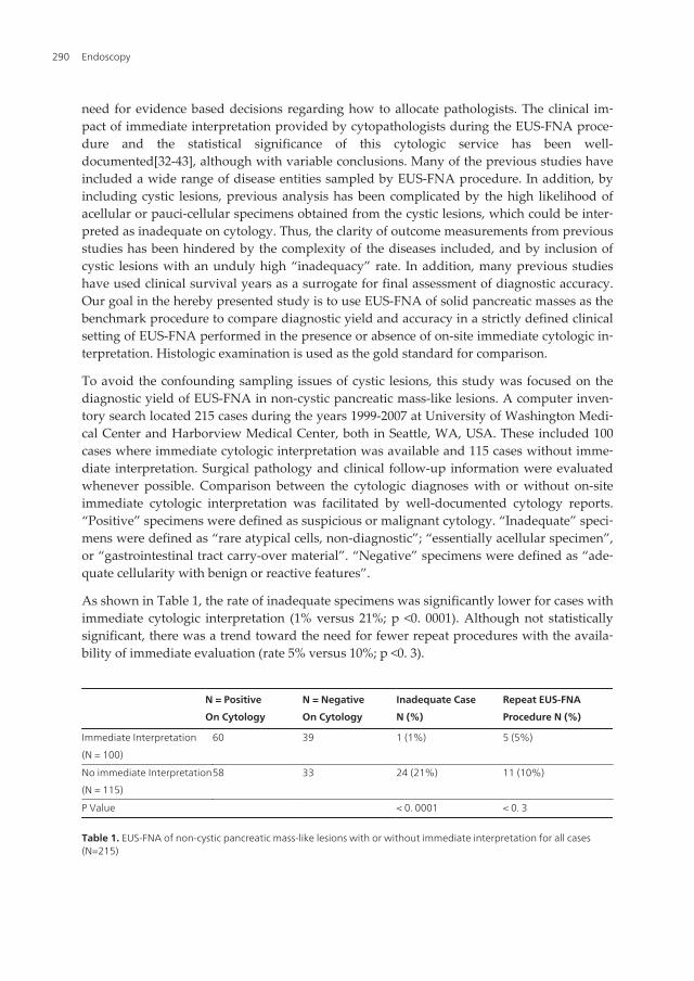
need for evidence based decisions regarding how to allocate pathologists. The clinical im‐pact of immediate interpretation provided by cytopathologists during the EUS-FNA proce‐dure and the statistical significance of this cytologic service has been well-documented[32-43], although with variable conclusions. Many of the previous studies haveincluded a wide range of disease entities sampled by EUS-FNA procedure. In addition, byincluding cystic lesions, previous analysis has been complicated by the high likelihood ofacellular or pauci-cellular specimens obtained from the cystic lesions, which could be inter‐preted as inadequate on cytology. Thus, the clarity of outcome measurements from previousstudies has been hindered by the complexity of the diseases included, and by inclusion ofcystic lesions with an unduly high “inadequacy” rate. In addition, many previous studieshave used clinical survival years as a surrogate for final assessment of diagnostic accuracy.Our goal in the hereby presented study is to use EUS-FNA of solid pancreatic masses as thebenchmark procedure to compare diagnostic yield and accuracy in a strictly defined clinicalsetting of EUS-FNA performed in the presence or absence of on-site immediate cytologic in‐terpretation. Histologic examination is used as the gold standard for comparison.
To avoid the confounding sampling issues of cystic lesions, this study was focused on thediagnostic yield of EUS-FNA in non-cystic pancreatic mass-like lesions. A computer inven‐tory search located 215 cases during the years 1999-2007 at University of Washington Medi‐cal Center and Harborview Medical Center, both in Seattle, WA, USA. These included 100cases where immediate cytologic interpretation was available and 115 cases without imme‐diate interpretation. Surgical pathology and clinical follow-up information were evaluatedwhenever possible. Comparison between the cytologic diagnoses with or without on-siteimmediate cytologic interpretation was facilitated by well-documented cytology reports.“Positive” specimens were defined as suspicious or malignant cytology. “Inadequate” speci‐mens were defined as “rare atypical cells, non-diagnostic”; “essentially acellular specimen”,or “gastrointestinal tract carry-over material”. “Negative” specimens were defined as “ade‐quate cellularity with benign or reactive features”.
As shown in Table 1, the rate of inadequate specimens was significantly lower for cases withimmediate cytologic interpretation (1% versus 21%; p <0. 0001). Although not statisticallysignificant, there was a trend toward the need for fewer repeat procedures with the availa‐bility of immediate evaluation (rate 5% versus 10%; p <0. 3).
N = Positive
On Cytology
N = Negative
On Cytology
Inadequate Case
N (%)
Repeat EUS-FNA
Procedure N (%)
Immediate Interpretation
(N = 100)
60 39 1 (1%) 5 (5%)
No immediate Interpretation
(N = 115)
58 33 24 (21%) 11 (10%)
P Value < 0. 0001 < 0. 3
Table 1. EUS-FNA of non-cystic pancreatic mass-like lesions with or without immediate interpretation for all cases(N=215)
Endoscopy290
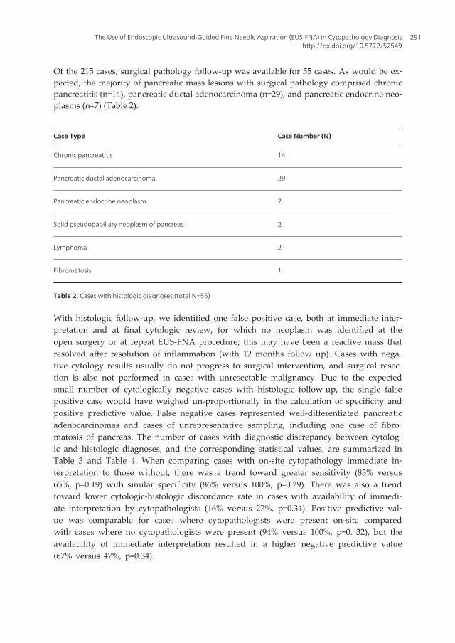
Of the 215 cases, surgical pathology follow-up was available for 55 cases. As would be ex‐pected, the majority of pancreatic mass lesions with surgical pathology comprised chronicpancreatitis (n=14), pancreatic ductal adenocarcinoma (n=29), and pancreatic endocrine neo‐plasms (n=7) (Table 2).
Case Type Case Number (N)
Chronic pancreatitis 14
Pancreatic ductal adenocarcinoma 29
Pancreatic endocrine neoplasm 7
Solid pseudopapillary neoplasm of pancreas 2
Lymphoma 2
Fibromatosis 1
Table 2. Cases with histologic diagnoses (total N=55)
With histologic follow-up, we identified one false positive case, both at immediate inter‐pretation and at final cytologic review, for which no neoplasm was identified at theopen surgery or at repeat EUS-FNA procedure; this may have been a reactive mass thatresolved after resolution of inflammation (with 12 months follow up). Cases with nega‐tive cytology results usually do not progress to surgical intervention, and surgical resec‐tion is also not performed in cases with unresectable malignancy. Due to the expectedsmall number of cytologically negative cases with histologic follow-up, the single falsepositive case would have weighed un-proportionally in the calculation of specificity andpositive predictive value. False negative cases represented well-differentiated pancreaticadenocarcinomas and cases of unrepresentative sampling, including one case of fibro‐matosis of pancreas. The number of cases with diagnostic discrepancy between cytolog‐ic and histologic diagnoses, and the corresponding statistical values, are summarized inTable 3 and Table 4. When comparing cases with on-site cytopathology immediate in‐terpretation to those without, there was a trend toward greater sensitivity (83% versus65%, p=0.19) with similar specificity (86% versus 100%, p=0.29). There was also a trendtoward lower cytologic-histologic discordance rate in cases with availability of immedi‐ate interpretation by cytopathologists (16% versus 27%, p=0.34). Positive predictive val‐ue was comparable for cases where cytopathologists were present on-site comparedwith cases where no cytopathologists were present (94% versus 100%, p=0. 32), but theavailability of immediate interpretation resulted in a higher negative predictive value(67% versus 47%, p=0.34).
The Use of Endoscopic Ultrasound-Guided Fine Needle Aspiration (EUS-FNA) in Cytopathology Diagnosishttp://dx.doi.org/10.5772/52549
291
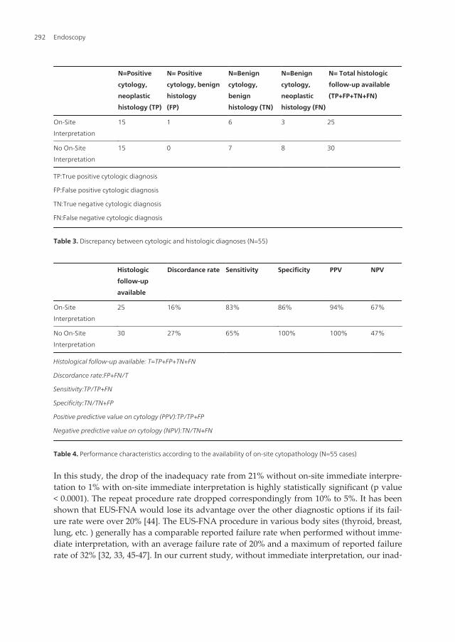
N=Positive
cytology,
neoplastic
histology (TP)
N= Positive
cytology, benign
histology
(FP)
N=Benign
cytology,
benign
histology (TN)
N=Benign
cytology,
neoplastic
histology (FN)
N= Total histologic
follow-up available
(TP+FP+TN+FN)
On-Site
Interpretation
15 1 6 3 25
No On-Site
Interpretation
15 0 7 8 30
TP:True positive cytologic diagnosis
FP:False positive cytologic diagnosis
TN:True negative cytologic diagnosis
FN:False negative cytologic diagnosis
Table 3. Discrepancy between cytologic and histologic diagnoses (N=55)
Histologic
follow-up
available
Discordance rate Sensitivity Specificity PPV NPV
On-Site
Interpretation
25 16% 83% 86% 94% 67%
No On-Site
Interpretation
30 27% 65% 100% 100% 47%
Histological follow-up available: T=TP+FP+TN+FN
Discordance rate:FP+FN/T
Sensitivity:TP/TP+FN
Specificity:TN/TN+FP
Positive predictive value on cytology (PPV):TP/TP+FP
Negative predictive value on cytology (NPV):TN/TN+FN
Table 4. Performance characteristics according to the availability of on-site cytopathology (N=55 cases)
In this study, the drop of the inadequacy rate from 21% without on-site immediate interpre‐tation to 1% with on-site immediate interpretation is highly statistically significant (p value< 0.0001). The repeat procedure rate dropped correspondingly from 10% to 5%. It has beenshown that EUS-FNA would lose its advantage over the other diagnostic options if its fail‐ure rate were over 20% [44]. The EUS-FNA procedure in various body sites (thyroid, breast,lung, etc. ) generally has a comparable reported failure rate when performed without imme‐diate interpretation, with an average failure rate of 20% and a maximum of reported failurerate of 32% [32, 33, 45-47]. In our current study, without immediate interpretation, our inad‐
Endoscopy292

equacy rate was 21%, a rate at which the procedure may not be cost effective over other di‐agnostic options. On-site immediate cytology interpretation of EUS-FNA of solid pancreaticlesions thus results in cost savings, reduced intervals between diagnosis and therapeutic in‐tervention, and enhanced patient care.
An important task at the EUS-FNA procedure is representative sampling of the lesion. Withon-site immediate microscopic evaluation of part of the sample, an experienced cytologistshould be able to assess the cellularity of the sampling and to differentiate gastrointestinaltract carryover material from moderately to poorly differentiated malignant cells. The pres‐ence of dense numbers of inflammatory cells, necrotic debris, and fibrous stromal elementsprovides good evidence that the endosonographer is actually sampling the targeted lesion.In our experience, on-site communication with immediate feedback is invaluable in guidingthe next needle pass. Another indispensable value is that immediate evaluation gives thepathologists the best opportunity to triage the sampled material for its optimal use, includ‐ing sending fresh specimen for flow cytometry analysis for possible lymphoproliferative dis‐ease, sending cyst contents for microbiology tests, or chemical analysis for tumor markers,such as amylase, CEA, CA19-9, etc. All those commonly available laboratory tests cannot beperformed once the specimen is fixed.
What is the impact of EUS-FNA on-site evaluation in situations of non-solid pancreatic le‐sions [48-52]? In our daily practice of cytology, EUS-FNA specimens comprise almost half thevolume of our on-site immediate assessment service. There are occasions when the aspiratedmaterial has presented with an unusual or unexpected, and yet remarkable gross appearance.On-site availability gives us the advantage of observing the gross appearance of freshly aspi‐rated material. With increased experience, we feel these grossly “unusual” aspirates can ac‐tually help to raise our suspicion towards a relatively specific diagnosis and triage thematerial for certain helpful laboratory tests. In our 2008 publication, we conducted 10-year in‐stitutional case review and summarized three different patterns of gross appearance of aspi‐rated material from non-neoplastic pancreatic and peri-pancreatic cystic lesions [53].
3.1. Pattern 1: Grossly yellowish-green pasty material
Case 1: we encountered two patients who presented with almost identical gross and micro‐scopic findings at EUS-FNA. The first patient was a 52-year-old male with a past medicalhistory of acute gallstone pancreatitis 4 years prior, treated by cholecystectomy. He has hadno other episodes of pancreatitis after the surgery. During a workup for his nephrolithiasis,CT scan incidentally identified a 3 cm unilocular cyst adjacent to the neck and body of hispancreas. Endoscopic ultrasound was requested in order to evaluate this peri-pancreaticcystic lesion. On site, EUS revealed a 3.4 cm primarily hypoechoic lesion. Aspirated materialhad an unusual yellowish-green gross appearance with a pasty texture. Microscopically, thesmears contain rare squamous cells of gut luminal origin, abundant amorphous materialand acellular debris (Figure 1, A). A Hall’s stain for bilirubin showed focal staining, sugges‐tive of bile pigment (Figure 1, B). Serum amylase assay was within normal range during thisperiod. In light of the cytological finding and clinical presentation, it was felt that the lesionwas most consistent with a small, chronic biloma, which likely developed at the time of his
The Use of Endoscopic Ultrasound-Guided Fine Needle Aspiration (EUS-FNA) in Cytopathology Diagnosishttp://dx.doi.org/10.5772/52549
293
cholecystectomy. Follow up CT scan revealed an unchanged cyst and no further evaluationwas recommended.
Figure 1. Representative cytologic findings in a case of biloma. Panel A:High-power view showing abundant amor‐phous material and acellular debris (Papanicolaou stain, 400x). Panel B: A Hall’s stain for bilirubin showed focal stain‐ing, suggestive of bile pigment (Hall’s stain, 400x).
Case 2: the second patient was a 39-year-old male with a history of chronic pancreatitis.During the workup for his intermittent upper abdominal pain, CT scan revealed a 2 cm hy‐podense mass off the tail of his pancreas. The patient underwent EUS-FNA twice. Each timeEUS revealed a 2cm hypoechoic mass off the tail of the pancreas and grossly similar copiousyellowish-green material was aspirated and submitted for cytology. Microscopically, bothEUS-FNA specimens from this patient revealed almost identical findings as our previouscase with abundant bile-stained cyst contents and amorphous debris (Data not shown). Noinflammation was present. Concurrent serum CA19-9 assay was within normal limits. Al‐though a specific diagnosis of biloma was not rendered on the original report, findings ar‐gue for a benign cystic lesion and a CT follow-up for the stability of the lesion wasrecommended without further intervention.
3.2. Pattern 2: Grossly tan-white cheesy material
Case 3: this was a 42-year-old male with chronic alcohol use and chronic hepatitis B and HIVco-infection. CT scan found a cystic lesion near the pancreatic tail, which had been stable insize. EUS identified a 2.5 cm complex cystic lesion adjacent to the pancreatic tail with both cyst‐ic and solid components. Aspirated material was grossly tan-white in color and semi-solid,cheesy in texture. Most of the material was lost during processing when air-dried or alcohol-fixed slides with Diff-Quik or Papanicolaou staining was attempted. Microscopic examinationonly revealed abundant amorphous degenerate material (Figure 2, A). Given the suspicion thatmaterial was dissolved by alcohol, Oil-Red O stain was performed on air-dried slides andshowed variable fat staining (Figure 2, B). Because of the unusual gross appearance, the aspi‐rated material on site was also sent for microbiology (negative findings) and assayed for CEA(164971 ng/ml), and amylase (2357 u/L). The significantly elevated CEA level was concerning
Endoscopy294
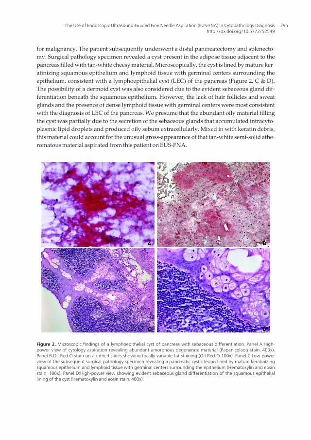
for malignancy. The patient subsequently underwent a distal pancreatectomy and splenecto‐my. Surgical pathology specimen revealed a cyst present in the adipose tissue adjacent to thepancreas filled with tan-white cheesy material. Microscopically, the cyst is lined by mature ker‐atinizing squamous epithelium and lymphoid tissue with germinal centers surrounding theepithelium, consistent with a lymphoepithelial cyst (LEC) of the pancreas (Figure 2, C & D).The possibility of a dermoid cyst was also considered due to the evident sebaceous gland dif‐ferentiation beneath the squamous epithelium. However, the lack of hair follicles and sweatglands and the presence of dense lymphoid tissue with germinal centers were most consistentwith the diagnosis of LEC of the pancreas. We presume that the abundant oily material fillingthe cyst was partially due to the secretion of the sebaceous glands that accumulated intracyto‐plasmic lipid droplets and produced oily sebum extracellularly. Mixed in with keratin debris,this material could account for the unusual gross-appearance of that tan-white semi-solid athe‐romatous material aspirated from this patient on EUS-FNA.
Figure 2. Microscopic findings of a lymphoepithelial cyst of pancreas with sebaceous differentiation. Panel A:High-power view of cytology aspiration revealing abundant amorphous degenerate material (Papanicolaou stain, 400x).Panel B:Oil-Red O stain on air-dried slides showing focally variable fat staining (Oil-Red O 100x). Panel C:Low-powerview of the subsequent surgical pathology specimen revealing a pancreatic cystic lesion lined by mature keratinizingsquamous epithelium and lymphoid tissue with germinal centers surrounding the epithelium (Hematoxylin and eosinstain, 100x). Panel D:High-power view showing evident sebaceous gland differentiation of the squamous epitheliallining of the cyst (Hematoxylin and eosin stain, 400x).
The Use of Endoscopic Ultrasound-Guided Fine Needle Aspiration (EUS-FNA) in Cytopathology Diagnosishttp://dx.doi.org/10.5772/52549
295
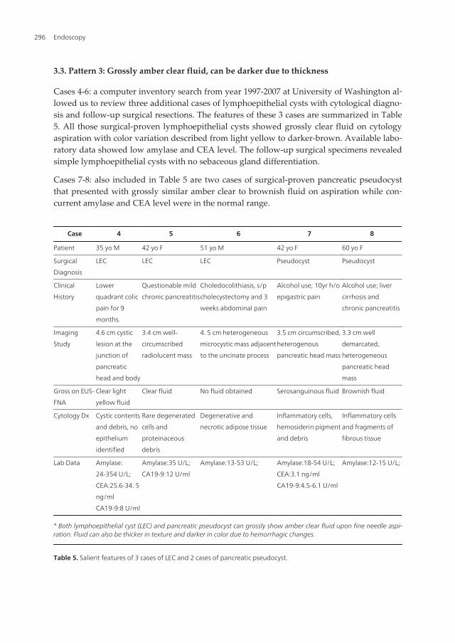
3.3. Pattern 3: Grossly amber clear fluid, can be darker due to thickness
Cases 4-6: a computer inventory search from year 1997-2007 at University of Washington al‐lowed us to review three additional cases of lymphoepithelial cysts with cytological diagno‐sis and follow-up surgical resections. The features of these 3 cases are summarized in Table5. All those surgical-proven lymphoepithelial cysts showed grossly clear fluid on cytologyaspiration with color variation described from light yellow to darker-brown. Available labo‐ratory data showed low amylase and CEA level. The follow-up surgical specimens revealedsimple lymphoepithelial cysts with no sebaceous gland differentiation.
Cases 7-8: also included in Table 5 are two cases of surgical-proven pancreatic pseudocystthat presented with grossly similar amber clear to brownish fluid on aspiration while con‐current amylase and CEA level were in the normal range.
Case 4 5 6 7 8
Patient 35 yo M 42 yo F 51 yo M 42 yo F 60 yo F
Surgical
Diagnosis
LEC LEC LEC Pseudocyst Pseudocyst
Clinical
History
Lower
quadrant colic
pain for 9
months.
Questionable mild
chronic pancreatitis
Choledocolithiasis, s/p
cholecystectomy and 3
weeks abdominal pain
Alcohol use; 10yr h/o
epigastric pain
Alcohol use; liver
cirrhosis and
chronic pancreatitis
Imaging
Study
4.6 cm cystic
lesion at the
junction of
pancreatic
head and body
3.4 cm well-
circumscribed
radiolucent mass
4. 5 cm heterogeneous
microcystic mass adjacent
to the uncinate process
3.5 cm circumscribed,
heterogenous
pancreatic head mass
3.3 cm well
demarcated,
heterogeneous
pancreatic head
mass
Gross on EUS-
FNA
Clear light
yellow fluid
Clear fluid No fluid obtained Serosanguinous fluid Brownish fluid
Cytology Dx Cystic contents
and debris, no
epithelium
identified
Rare degenerated
cells and
proteinaceous
debris
Degenerative and
necrotic adipose tissue
Inflammatory cells,
hemosiderin pigment
and debris
Inflammatory cells
and fragments of
fibrous tissue
Lab Data Amylase:
24-354 U/L;
CEA:25.6-34. 5
ng/ml
CA19-9:8 U/ml
Amylase:35 U/L;
CA19-9:12 U/ml
Amylase:13-53 U/L; Amylase:18-54 U/L;
CEA:3.1 ng/ml
CA19-9:4.5-6.1 U/ml
Amylase:12-15 U/L;
* Both lymphoepithelial cyst (LEC) and pancreatic pseudocyst can grossly show amber clear fluid upon fine needle aspi‐ration. Fluid can also be thicker in texture and darker in color due to hemorrhagic changes.
Table 5. Salient features of 3 cases of LEC and 2 cases of pancreatic pseudocyst.
Endoscopy296
Our experience tells us that being fully aware of the limitations on cytology diagnosis andthe high possibility of non-representative sampling of the lesions, cytologists should beready to reject or classify the specimens as non-diagnostic in many situations of acellularcytological material and thus avoid misleading the clinical decisions with an erroneous di‐agnosis. On the other hand, based on the individual institute settings and the availabilitiesof cytopathologists for on-site immediate assessment, cytology diagnosis tends to ignorethe gross appearance of the aspirated material and the background material is also oftenoverlooked microscopically, especially in an acellular cytology specimen. Incorporatingthose gross material observations can sometimes add valuable information towards apathological diagnosis.
We presented three entities (8 cases) here in an attempt to highlight the diagnostic value ofgross appearance in fine needle aspiration cytology that can provide clues to the nature ofa pancreatic or a peripancreatic cystic lesion. These three entities: biloma, pancreatic lym‐phoepithelial cyst and pancreatic pseudocyst, although considered uncommon and notwidely represented in the cytology literature, are actually encountered more commonlynow with the rapid advance of our imaging and imaging guided-sampling practices. Oncytology specimens, those are the lesions that generally produce acellular or sparsely cellu‐lar material (not including carryover material from gastrointestinal luminal origin). How‐ever by carefully combining the clinical, radiological, laboratory and cytological findingsgrossly and microscopically, cytology can help lead to or confirm clinical judgments andaid in patient care.
A biloma is an encapsulated bile collection outside the biliary tree. The underlying causesinclude iatrogenic, traumatic, and spontaneous injury of the biliary tree causing bile leaks.Its diagnosis can be established upon clinical history, imaging studies, and needle aspirationcytology and chemical analysis of the aspirated fluid. The symptomatic biloma if left un‐treated may result in significant morbidity and mortality. However, non-surgical interven‐tion is considered the first choice of treatment for biloma, especially in many asymptomaticsituations. Alternatively symptomatic biloma may be treated successfully with intervention‐al radiologic techniques instead of open surgery [54, 55]. It was recently reported that endo‐scopic ultrasound-guided fine-needle aspiration of an infected biloma, together withendoscopic biliary stent placement, resulted in complete resolution of a patient’s biloma[56]. Thus, cytological diagnosis of biloma upon EUS-FNA can help a great deal in manag‐ing this potentially complex problem.
A wide variety of cystic lesions can arise within or adjacent to the pancreas. They can begenerally placed into non-neoplastic and neoplastic categories. The non-neoplastic cystic le‐sions can be both congenital and acquired. EUS-FNA has emerged as the primary choice forobtaining diagnostic material on pancreatic cystic lesions. Aspirated cystic fluid analysis forpancreatic enzymes, tumor markers, and fluid viscosity can be of great help in the differen‐tial diagnosis. Non-neoplastic cysts generally are high on amylase level and low on tumormarkers (CEA, CA 19-9) while these tumor markers are generally elevated in malignantcystic neoplasms, but low in non-neoplastic or benign neoplastic cysts. Having said that, ex‐ceptions do occur and no standardized values exist among institutions. On cytology speci‐
The Use of Endoscopic Ultrasound-Guided Fine Needle Aspiration (EUS-FNA) in Cytopathology Diagnosishttp://dx.doi.org/10.5772/52549
297
men, neoplastic cystic lesions usually produce a more cellular aspirate compared to non-neoplastic cysts if appropriately sampled.
The acquired non-neoplastic pancreatic cysts include dermoid cysts, LECs and pseudocysts.Both dermoid cysts and LECs are squamous-lined non-neoplastic cysts. LECs are benigncystic lesions [57] seen predominantly in males, in the fifth to sixth decades of life. They maybe unilocular or multilocular. The cyst contents may vary from serous to cheesy/casseous-appearing material depending on the degree of keratin formation. Microscopically, the cystsare lined by well-differentiated stratified squamous epithelium, which may or may not haveprominent keratinization. In some areas, the lining may appear more transitional, and inothers, flat, cuboidal, or focally denuded. The squamous epithelium is surrounded by aband of dense lymphoid tissue composed of mature T-lymphocytes with intervening germi‐nal centers formed by B cells. The representative aspirate findings are non-specific, showinga mixed population of lymphocytes, histiocytes in a background of keratin debris and pro‐teinaceous debris. Squamous lining cells may be seen. The occurrence of sebaceous glands inLEC of pancreas is well documented [58]. It is unclear whether the florid sebaceous glandu‐lar differentiation correlates with the exceedingly elevated CEA and amylase level in our pa‐tient. LECs of the pancreas do not appear to be associated with any autoimmune conditions,human immunodeficiency virus infection, lymphoma, or carcinoma. All of these lesionshave been documented to occur in their salivary gland counterparts [59]. Dermoid cysts arealso rare in the pancreas region. They are reported in younger patients (2nd-3rd decades). Thepresence of sebaceous glands or hair follicles and absence of closely associated lymphoid tis‐sue is more typical for dermoid cysts and differentiate them from LECs.
Pancreatic pseudocysts account for the vast majority (75-90%) of pancreatic cysts [60]. Pseu‐docyst lacks an epithelial lining. It develops when a focus of peri-pancreatic fat necrosis isresorbed, producing a debris-filled space rich in pancreatic exocrine enzymes. It is in generalcomposed of an inflammatory fibrous capsule surrounding a region of necrosis. However,the pathologic findings may vary depending on the stage of the process. The cyst contents,originally necrotic fat, transform into a mixture of necrotic cells, enzymes, scavenger cells,cholesterol clefts and sometimes neutrophils. The tissue that surrounds the necrotic materialfirst produces granulation tissue, and eventually becomes a fibrotic pseudocapsule. FNAusually yields clear to dark brown fluid, which often but not always (as in our cases) showselevated amylase level on fluid analysis. Aspirate smears are again non-specific and com‐posed of granular necrotic debris and mixed inflammatory cells [61]. A clinical history ofpancreatitis and confirmatory gross evidence of pancreatitis on imaging should confirm thediagnosis. The diagnosis of pseudocyst on cytology should be considered one of exclusion.
4. Aim 3: Application of immunocytochemistry (ICC) studies in EUS-FNA obtained cytology material
When applying ancillary techniques in cytologic specimens, cytopathologists often encoun‐ter the difficulties of limited material, lack of negative or positive controls or lack of internal
Endoscopy298

controls. On the other hand, the presence of internal control material can sometimes addconfusion rather than reassurance in ICC-stained material. Nevertheless, by carefully andstrategically using the cytology specimen, immunocytochemical studies are often achievablewith limited aspiration material. We have used some of the techniques that have been de‐scribed by others, such as immunostaining over Papanicolaou-stained monolayer slides; ap‐plying different antibodies to different areas of one smear slide; or making cell blockpreparations whenever possible. When unstained material is not available and no cell blockis initially prepared, Dr. V. Grieco and her colleagues at Harborview Medical Center, Seattle,Washington, have used a scraped slide technique that transfers smear material on Papanico‐laou stained slides into paraffin embedded cell blocks. The cell block can then be used forICC with appropriate controls. If successful, this may avoid the necessity of obtaining addi‐tional tissue. We have routinely used this scraped cell block technique in our daily practiceas well as in research projects. We hereby briefly discuss our mucin study project with ar‐chival cytology material from 1997-2007 at University of Washington, presented at DigestiveDisease Week [62]. Schema of Scraping Technique is illustrated in Figure 3: scraping the ma‐terial from rehydrated direct smear slides into a cell block preparation.
Figure 3. Schema of Scrapped Cell Block Technique:Papanicolaou-stained cellular direct smears are placed in xylene,coverslip removed, and re-hydrated in sequentially graded ethanol to tap water. The cellular material is then scrapedfrom the glass slides using a razor blade, embedded in Histogel and processed with routine cell block preparation.
The Use of Endoscopic Ultrasound-Guided Fine Needle Aspiration (EUS-FNA) in Cytopathology Diagnosishttp://dx.doi.org/10.5772/52549
299
Cytology diagnosis of mucin-producing pancreatic neoplasms including mucinous non-cystic carcinoma (colloid carcinoma), mucinous cystic neoplasm, and intraductal papillary-mucinous neoplasm (IPMN) is important because of the speculation that disruption of theintegrity of these mucinous tumors by an incisional biopsy may cause dissemination of tu‐mor cells with mucin and worsen the prognosis. Mucin-detection is not always an easy taskon EUS-FNA specimen. Reactive/inflammatory pancreatitis can cause dilated ducts and‐pseudocyst formation with proteinaceous precipitate that can be mistaken as mucinous ma‐terial. Sheets of carry-over gastric foveolar cells can mimic cells from mucinous neoplasia[ 63-67]. By applying conventional cytochemistry including PAS (periodic acid-Schiff reac‐tion), PASD (PAS with diastase), alcian blue and mucicarmin, in conjunction withmucinprotein (MUC1, MUC2) immunostaining on surgical-proven cytology specimens, this studyaims at selecting a practical diagnostic tool to enhance the diagnostic accuracy of detectingmucin-producing pancreatic neoplasms.
Studied material include: 1) Bench FNA specimens performed on autopsy or surgical-ob‐tained normal tissue that are likely to be carried-over during an endoscopic procedure in‐cluding pancreas, gastric body, pylorus and duodenum; 2) EUS-FNA (with scraped cellblock preparation by scraping archival smears into a cell block), along with subsequent sur‐gical specimens on mucinous cystadenoma, borderline mucinous cystic neoplasm, IPMN,colloid carcinoma and ductal adenocarcinma with mucin production; 3) Non-mucin produc‐ing lesions including solid pseudopapillary tumor and chronic pancreatitis with dilatedduct. Our data demonstrate that mucicarmin is very insensitive in detecting mucin produc‐tion. We are unable to show reliable mucicarmin staining on both surgical and cytologyspecimen of IPMN(Figure 4, Panel D-F) as well as mucinous cystic neoplasia cases. MUC1and MUC 2 immunostains do not consistently pick up mucin production by pancreatic neo‐plasia on cytology specimens either. In contrast, PAS/PAS-Diastase are a reliable and sensi‐tive marker to stain background mucin material and mucin-producing epithelial cells oncytology, yet specific enough to distinguish proteinaceous precipitate in a pancreatitis case(Figure 4, Panel A-C, precipitate in a dilated duct that was mistaken as possible mucinousmaterial on cytology). The results have directed our daily practice on the use of mucin stainson cytologic diagnosis of pancreatic mucinous neoplasia.
Concomitant use of ICC can greatly enhance the diagnostic accuracy on cytology specimens,especially when it comes to the diagnostic entities invariably requiring immunophenotypicidentification, such as in the diagnosis of pancreatic endocrine neoplasia (PENs) [68-72].However, due to the initial unanticipated nature of the lesion, lack of cytopathologist on siteor difficulties of the procedure, sufficient aspirate/biopsy material for cellblock preparationis not always available. By using the scraped cell block technique, a select panel of immuno‐cytochemical studies can be achieved to aid in the diagnosis of PEN. Using the aforemen‐tioned scraped cell block technique, figure 5 shows strong expression of the neuroendocrineimmuno-marker, chromogranin, by the neoplastic cells. Figure 6 shows neoplastic cellsstained positively with the neuroendocrine marker, synaptophysin by directly applying theantibody onto a Papanicolaou-stained charged monolayer slide.
Endoscopy300
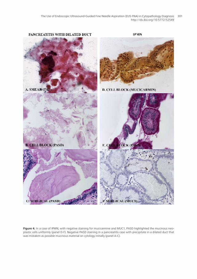
Figure 4. In a case of IPMN, with negative staining for mucicarmine and MUC1, PASD highlighted the mucinous neo‐plastic cells uniformly (panel D-F). Negative PASD staining in a pancreatitis case with precipitate in a dilated duct thatwas mistaken as possible mucinous material on cytology initially (panel A-C).
The Use of Endoscopic Ultrasound-Guided Fine Needle Aspiration (EUS-FNA) in Cytopathology Diagnosishttp://dx.doi.org/10.5772/52549
301
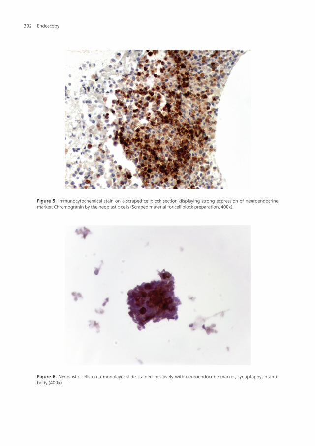
Figure 5. Immunocytochemical stain on a scraped cellblock section displaying strong expression of neuroendocrinemarker, Chromogranin by the neoplastic cells (Scraped material for cell block preparation, 400x).
Figure 6. Neoplastic cells on a monolayer slide stained positively with neuroendocrine marker, synaptophysin anti‐body (400x)
Endoscopy302
Before leaving this section, it should also be stated that in cytology, as in surgical pathology,the light microscopic appearance of the tumor is more important than a particular stainingresult and that immunocytochemistry should be used for confirmation of the cytomorpho‐logical diagnosis. Be aware of that the staining of cytology specimens is often focal andsometimes difficult to interpret, because of various factors, including limited material, highbackground staining, and the presence of three-dimensional clusters with non-specific stain‐ing and edge artifact. A combined clinical, radiographic and pathologic approach should al‐ways be emphasized in rendering each diagnosis.
5. Aim 4: EUS-FNA as a source of material for molecular studies
We are at the beginning of the era of clinical application of molecular probes to assess genet‐ic alterations. Many assays have become clinically available in identifying genetic alterationsthat serve as prognostic markers and therapeutic targets. EGFR expression in head and neckcancers, non-small cell lung carcinomas, colon cancers, and recently in pancreatic cancers[73-82]; microsatellite instability tests in colon cancer serve as prognostic marker, therapeu‐tic predictive marker and screening test for Lynch syndrome [83-88] are among the moststudied areas. Immunodetection of molecular markers on cytology specimen, especially incancer staging for metastatic disease will be playing a major role as a guide for molecular-targeted therapy. EGFR expression has now been routinely assessed on cell block cytologyspecimens by many of the commercial or academic laboratories. A prominent example inthis regard is FNA diagnosis of thyroid nodules. Given that the cytology reports on thyroidFNA diagnoses are often indeterminate or suspicious, the potential use of diagnostic molec‐ular markers as adjunct methods for cytologic diagnosis appears to be among the mostpromising area of research [89-98]. Fluorescent (FISH) and chromogenic (CISH) in situ hy‐bridization have recently become part of the diagnostic armamentarium of breast patholo‐gists. HER2 gene amplification testing by FISH and/or CISH has become an integral part ofthe diagnostic workup for patients with breast cancer [99-105 ]. FISH or CISH is now used inFNA material with success in many situations, including HER2 assessment in FNA with ex‐cellent correlation with the histologic specimens [106-108 ]. Many other applications, such asN-myc amplication on FNA from neuroblastomas [109, 110] and potential use of multiplexFISH for detection of recurrence of transitional carcinomas on urine specimens [111-114] arealso well documented molecular tests using cytology specimens.
Compared to paraffin-embedded tissue, cytology specimens offer the advantage of disag‐gregated, intact cells, with fewer problems associated with tumor cell homogenization. FISHor CISH probe for diagnosis should be expected to be detected more accurately in cytologicspecimen as it contains whole cells and whole nuclei. For the patient, the advantages includelow cost, prompt access and the avoidance of surgery.
In carcinogenesis, the majority of molecular alterations occur prior to progressive tumorgrowth. It has been well documented that resection of small (early) pancreatic tumors and tu‐mors of low histological or cytological grade and stage are correlated with improved survival.
The Use of Endoscopic Ultrasound-Guided Fine Needle Aspiration (EUS-FNA) in Cytopathology Diagnosishttp://dx.doi.org/10.5772/52549
303
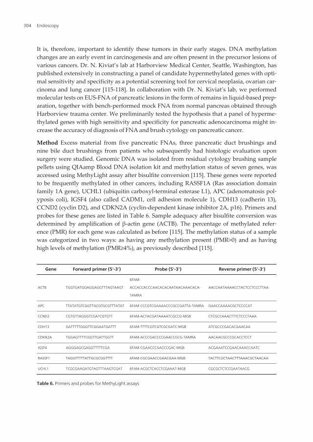
It is, therefore, important to identify these tumors in their early stages. DNA methylationchanges are an early event in carcinogenesis and are often present in the precursor lesions ofvarious cancers. Dr. N. Kiviat’s lab at Harborview Medical Center, Seattle, Washington, haspublished extensively in constructing a panel of candidate hypermethylated genes with opti‐mal sensitivity and specificity as a potential screening tool for cervical neoplasia, ovarian car‐cinoma and lung cancer [115-118]. In collaboration with Dr. N. Kiviat’s lab, we performedmolecular tests on EUS-FNA of pancreatic lesions in the form of remains in liquid-based prep‐aration, together with bench-performed mock FNA from normal pancreas obtained throughHarborview trauma center. We preliminarily tested the hypothesis that a panel of hyperme‐thylated genes with high sensitivity and specificity for pancreatic adenocarcinoma might in‐crease the accuracy of diagnosis of FNA and brush cytology on pancreatic cancer.
Method Excess material from five pancreatic FNAs, three pancreatic duct brushings andnine bile duct brushings from patients who subsequently had histologic evaluation uponsurgery were studied. Genomic DNA was isolated from residual cytology brushing samplepellets using QIAamp Blood DNA isolation kit and methylation status of seven genes, wasaccessed using MethyLight assay after bisulfite conversion [115]. These genes were reportedto be frequently methylated in other cancers, including RASSF1A (Ras association domainfamily 1A gene), UCHL1 (ubiquitin carboxyl-terminal esterase L1), APC (adenomatosis pol‐yposis coli), IGSF4 (also called CADM1, cell adhesion molecule 1), CDH13 (cadherin 13),CCND2 (cyclin D2), and CDKN2A (cyclin-dependent kinase inhibitor 2A, p16). Primers andprobes for these genes are listed in Table 6. Sample adequacy after bisulfite conversion wasdetermined by amplification of β-actin gene (ACTB). The percentage of methylated refer‐ence (PMR) for each gene was calculated as before [115]. The methylation status of a samplewas categorized in two ways: as having any methylation present (PMR>0) and as havinghigh levels of methylation (PMR≥4%), as previously described [115].
Gene Forward primer (5'-3') Probe (5'-3') Reverse primer (5'-3')
ACTB TGGTGATGGAGGAGGTTTAGTAAGT
6FAM-
ACCACCACCCAACACACAATAACAAACACA-
TAMRA
AACCAATAAAACCTACTCCTCCCTTAA
APC TTATATGTCGGTTACGTGCGTTTATAT 6FAM-CCCGTCGAAAACCCGCCGATTA-TAMRA GAACCAAAACGCTCCCCAT
CCND2 CGTGTTAGGGTCGATCGTGTT 6FAM-ACTACGATAAAATCGCCG-MGB CTCGCCAAACTTTCTCCCTAAA
CDH13 GATTTTTGGGTTCGGAATGATTT 6FAM-TTTTCGTCGTCGCGATC-MGB ATCGCCCGACACGAACAA
CDKN2A TGGAGTTTTCGGTTGATTGGTT 6FAM-ACCCGACCCCGAACCGCG-TAMRA AACAACGCCCGCACCTCCT
IGSF4 AGGGAGCGAGGTTTTTCGA 6FAM-CGAACCCAACCCGAC-MGB ACGAAATCCGAACAAACCAATC
RASSF1 TAGGTTTTTATTGCGCGGTTTT 6FAM-CGCGAACCGAACGAA-MGB TACTTCGCTAACTTTAAACGCTAACAA
UCHL1 TCGCGAAGATGTAGTTTAAGTCGAT 6FAM-ACGCTCACCTCGAAAT-MGB CGCGCTCTCCGAATAACG
Table 6. Primers and probes for MethyLight assays
Endoscopy304
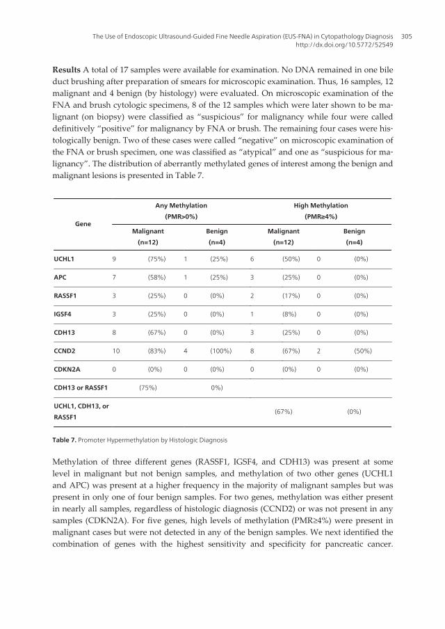
Results A total of 17 samples were available for examination. No DNA remained in one bileduct brushing after preparation of smears for microscopic examination. Thus, 16 samples, 12malignant and 4 benign (by histology) were evaluated. On microscopic examination of theFNA and brush cytologic specimens, 8 of the 12 samples which were later shown to be ma‐lignant (on biopsy) were classified as “suspicious” for malignancy while four were calleddefinitively “positive” for malignancy by FNA or brush. The remaining four cases were his‐tologically benign. Two of these cases were called “negative” on microscopic examination ofthe FNA or brush specimen, one was classified as “atypical” and one as “suspicious for ma‐lignancy”. The distribution of aberrantly methylated genes of interest among the benign andmalignant lesions is presented in Table 7.
Gene
Any Methylation
(PMR>0%)
High Methylation
(PMR≥4%)
Malignant
(n=12)
Benign
(n=4)
Malignant
(n=12)
Benign
(n=4)
UCHL1 9 (75%) 1 (25%) 6 (50%) 0 (0%)
APC 7 (58%) 1 (25%) 3 (25%) 0 (0%)
RASSF1 3 (25%) 0 (0%) 2 (17%) 0 (0%)
IGSF4 3 (25%) 0 (0%) 1 (8%) 0 (0%)
CDH13 8 (67%) 0 (0%) 3 (25%) 0 (0%)
CCND2 10 (83%) 4 (100%) 8 (67%) 2 (50%)
CDKN2A 0 (0%) 0 (0%) 0 (0%) 0 (0%)
CDH13 or RASSF1 (75%) 0%)
UCHL1, CDH13, or
RASSF1(67%) (0%)
Table 7. Promoter Hypermethylation by Histologic Diagnosis
Methylation of three different genes (RASSF1, IGSF4, and CDH13) was present at somelevel in malignant but not benign samples, and methylation of two other genes (UCHL1and APC) was present at a higher frequency in the majority of malignant samples but waspresent in only one of four benign samples. For two genes, methylation was either presentin nearly all samples, regardless of histologic diagnosis (CCND2) or was not present in anysamples (CDKN2A). For five genes, high levels of methylation (PMR≥4%) were present inmalignant cases but were not detected in any of the benign samples. We next identified thecombination of genes with the highest sensitivity and specificity for pancreatic cancer.
The Use of Endoscopic Ultrasound-Guided Fine Needle Aspiration (EUS-FNA) in Cytopathology Diagnosishttp://dx.doi.org/10.5772/52549
305
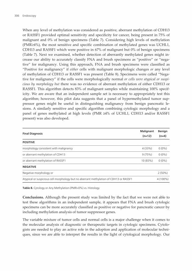
When any level of methylation was considered as positive, aberrant methylation of CDH13or RASSF1 provided optimal sensitivity and specificity for cancer, being present in 75% ofmalignant and 0% of benign specimens (Table 7). Considering high levels of methylation(PMR≥4%), the most sensitive and specific combination of methylated genes was UCHL1,CDH13 and RASSF1 which were positive in 67% of malignant but 0% of benign specimens(Table 7). Next we examined, whether detection of aberrantly methylated genes might in‐crease our ability to accurately classify FNA and brush specimens as “positive” or “nega‐tive” for malignancy. Using this approach, FNA and brush specimens were classified as“Positive for malignancy” if either cells with malignant morphologic changes or any levelof methylation of CDH13 or RASSF1 was present (Table 8). Specimens were called “Nega‐tive for malignancy” if the cells were morphologically normal or cells were atypical or suspi‐cious by morphology but there was no evidence of aberrant methylation of either CDH13 orRASSF1. This algorithm detects 83% of malignant samples while maintaining 100% specif‐icity. We are aware that an independent sample set is necessary to appropriately test thisalgorithm; however, this pilot data suggests that a panel of hypermethylated tumor sup‐pressor genes might be useful in distinguishing malignancy from benign pancreatic le‐sions. A similarly sensitive and specific algorithm combining cytologic morphology and apanel of genes methylated at high levels (PMR ≥4% of UCHL1, CDH13 and/or RASSF1present) was also developed.
Final DiagnosisMalignant
(n=12)
Benign
(n=4)
POSITIVE
morphology consistent with malignancy 4 (33%) 0 (0%)
or aberrant methylation of CDH13 9 (75%) 0 (0%)
or aberrant methylation of RASSF1 10 (83%) 0 (0%)
NEGATIVE
Negative morphology or 2 (50%)
Atypical or suspicious cell morphology but no aberrant methylation of CDH13 or RASSF1 4 (100%)
Table 8. Cytology or Any Methylation (PMR>0%) vs. Histology
Conclusions. Although the present study was limited by the fact that we were not able totest these algorithms in an independent sample, it appears that FNA and brush cytologicspecimens can be more accurately classified as positive or negative for pancreatic cancer byincluding methylation analysis of tumor suppressor genes.
The variable mixture of tumor cells and normal cells is a major challenge when it comes tothe molecular analysis of diagnostic or therapeutic targets in cytologic specimens. Cytolo‐gists are needed to play an active role in the adoption and application of molecular techni‐ques, since we are able to interpret the results in the light of cytological morphology. Our
Endoscopy306
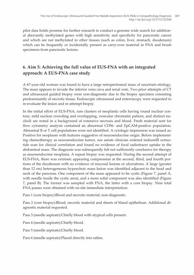
pilot data holds promise for further research to conduct a genome wide search for addition‐al aberrantly methylated genes with high sensitivity and specificity for pancreatic cancerand which are not methylated in other tissues (such as colon, liver, stomach, duodenum)which can be frequently or incidentally present as carry-over material in FNA and brushspecimens from pancreatic lesions.
6. Aim 5: Achieving the full value of EUS-FNA with an integratedapproach: A EUS-FNA case study
A 67-year-old woman was found to have a large retroperitoneal mass of uncertain etiology.The mass appears to invade the inferior vena cava and renal vein. Two prior attempts of CTand ultrasound guided biopsy were non-diagnostic due to the biopsy specimen consistingpredominantly of necrotic tissue. Endoscopic ultrasound and enteroscopy were requested tore-evaluate the lesion and re-attempt biopsy.
In the initial effort of EUS-FNA, rare clusters of neoplastic cells having round nuclear con‐tour, mild nuclear crowding and overlapping, vesicular chromatin pattern, and distinct nu‐cleoli are noted in a background of extensive necrosis and blood. Fresh material sent forflow cytometry analysis contained an abnormal CD56- and EpCAM-positive population.Abnormal B or T cell populations were not identified. A cytologic impression was issued as:Positive for neoplasm with features suggestive of neuroendocrine origin. Before implement‐ing chemotherapy as neuroendocrine tumor, our astute clinician ordered indiumIII octreo‐tide scan for clinical correlation and found no evidence of focal radiotracer uptake in theabdominal mass. The diagnosis was subsequently felt not sufficiently conclusive for therapyas neuroendocrine neoplasia. A repeat biopsy was requested. During the second attempt ofEUS-FNA, there was extrinsic appearing compression at the second, third, and fourth por‐tions of the duodenum with no evidence of mucosal lesions or ulcerations. A large (greaterthan 12 cm) heterogeneous hypoechoic mass lesion was identified adjacent to the head andneck of the pancreas. One component of the mass appeared to be cystic (Figure 7, panel A,with needle inside the cystic area), and a more solid component was also identified (Figure7, panel B). The former was sampled with FNA, the latter with a core biopsy. Nine totalFNA passes were obtained with on-site immediate interpretation:
Pass 1 (core biopsy):Blood and necrotic material, non-diagnostic.
Pass 2 (core biopsy):Blood, necrotic material and sheets of bland epithelium. Additional di‐agnostic material requested.
Pass 3 (needle aspirate):Chiefly blood with atypical cells present.
Pass 4 (needle aspirate):Chiefly blood.
Pass 5 (needle aspirate):Chiefly blood.
Pass 6 (needle aspirate):Placed directly into saline.
The Use of Endoscopic Ultrasound-Guided Fine Needle Aspiration (EUS-FNA) in Cytopathology Diagnosishttp://dx.doi.org/10.5772/52549
307
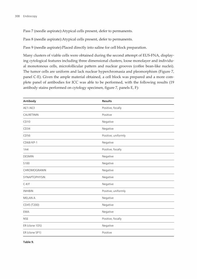
Pass 7 (needle aspirate):Atypical cells present, defer to permanents.
Pass 8 (needle aspirate):Atypical cells present, defer to permanents.
Pass 9 (needle aspirate):Placed directly into saline for cell block preparation.
Many clusters of viable cells were obtained during the second attempt of EUS-FNA, display‐ing cytological features including three dimensional clusters, loose monolayer and individu‐al monotonous cells, microfollicular pattern and nuclear grooves (coffee bean-like nuclei).The tumor cells are uniform and lack nuclear hyperchromasia and pleomorphism (Figure 7,panel C-E). Given the ample material obtained, a cell block was prepared and a more com‐plete panel of antibodies for ICC was able to be performed, with the following results (19antibody stains performed on cytology specimen, figure 7, panels E, F):
Antibody Results
AE1/AE3 Positive, focally
CALRETININ Positive
CD10 Negative
CD34 Negative
CD56 Positive, uniformly
CD68/KP-1 Negative
1A4 Positive, focally
DESMIN Negative
S100 Negative
CHROMOGRANIN Negative
SYNAPTOPHYSIN Negative
C-KIT Negative
INHIBIN Positive, uniformly
MELAN A Negative
CD45 (T200) Negative
EMA Negative
NSE Positive, focally
ER (clone 1D5) Negative
ER (clone SP1) Positive
Table 9.
Endoscopy308
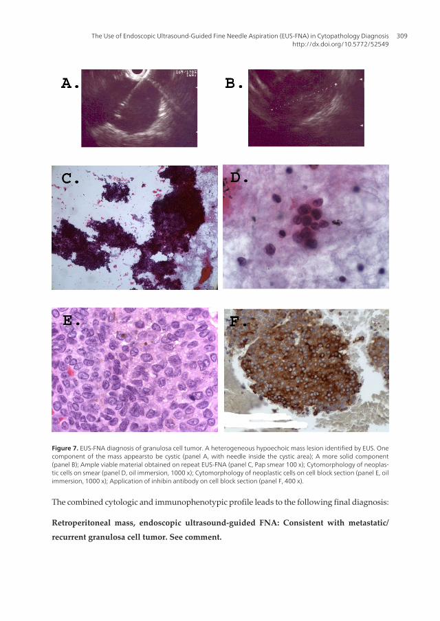
Figure 7. EUS-FNA diagnosis of granulosa cell tumor. A heterogeneous hypoechoic mass lesion identified by EUS. Onecomponent of the mass appearsto be cystic (panel A, with needle inside the cystic area); A more solid component(panel B); Ample viable material obtained on repeat EUS-FNA (panel C, Pap smear 100 x); Cytomorphology of neoplas‐tic cells on smear (panel D, oil immersion, 1000 x); Cytomorphology of neoplastic cells on cell block section (panel E, oilimmersion, 1000 x); Application of inhibin antibody on cell block section (panel F, 400 x).
The combined cytologic and immunophenotypic profile leads to the following final diagnosis:
Retroperitoneal mass, endoscopic ultrasound-guided FNA: Consistent with metastatic/
recurrent granulosa cell tumor. See comment.
The Use of Endoscopic Ultrasound-Guided Fine Needle Aspiration (EUS-FNA) in Cytopathology Diagnosishttp://dx.doi.org/10.5772/52549
309
Comment: The cytologic appearance of this tumor, along with the overall immuno-pheno‐type demonstrated by calretinin and inhibin positivity, supports the diagnosis of granulo‐sa cell tumor. Additional studies performed at PhenoPath Laboratory using the moresensitive anti-ER antibody SP1 document expression of estrogen receptor (in contrast tothe negative anti-ER studies using the 1D5 clone), further supporting the diagnosis ofgranulosa cell tumor.
After the diagnosis, patient recalls that she had a procedure for granulosa cell tumor about30 years ago in Mexico.
Granulosa cell tumor may pose a diagnostic challenge in cytology preparations. First, thereis some similarity between the granulosa tumor cells and reactive mesothelial cells. Indis‐tinct cell borders, high nuclear cytoplasmic ratio and indentation of the nuclear membraneare helpful features in distinguishing granulosa tumor cells from mesothelial cells. Secondly,although nuclear features, especially nuclear grooves, are one of the classic features on gran‐ulosa cell tumor, they can also be seen in other ovarian tumors such as Brenner tumor andother sex cord-stromal tumor. Other characteristic histological features for granulosa cell tu‐mor, such as Call-Exner bodies, a second population of elongated theca cells, are rarely evi‐dent on cytology specimens. Lastly, as occurred in our case, cells of carcinoid, especiallypancreatic neuroendocrine tumors in this anatomic site, share many features of granulosacells. Judicious use of a panel of immunocytochemistry (with the availability of sufficientsampling material) can be of great help in this scenario. Granulosa cell tumor is positive forinhibin, CD99, calretinin, vimentin, CD56, is generally negative for cytokeratin, EMA, Ber-Ep4, and is negative for neuroendocrine markers synaptophysin and chromogranin. Brennertumor and other sex cord-stromal tumor usually are positive for cytokeratin and EMA.
As illustrated in this case, clinical-pathologic correlation is essential with octreotide scanpreventing a diagnostic pitfall of neuroendocrine neoplasia. The on-site cytology interpreta‐tion performed for each of 9 EUS-FNA passes played a pivotal role in obtaining adequateviable diagnostic material, considering the sampling difficulty due to extensive tumor ne‐crosis despite the apparent large size of the tumor by imaging study. A more complete ICCpanel was thus able to be performed. The ICC results together with cytomorphology led to adiagnosis consistent with the subsequently retrieved clinical history.
Thus, our final note: the full value of FNA is only achieved with the integrated approach:the integration of clinical information, light microscopic analysis, results of ancillary studies,and even the gross appearance of the aspiration material will inform and lead us to a moreaccurate pathology diagnosis that can help tremendously in directing patient care.
Acknowledgements
The authors want to express their sincere gratitude to Drs. Nancy B. Kiviat (University ofWashington, Seattle, WA), Melissa P. Upton (University of Washington, Seattle, WA) andStephen J. Rulyak (Evergreen Medical Center, Gastroenterology, Seattle, WA), for their pro‐fessional guidance, insightful discussions and tremendous technical support.
Endoscopy310

Author details
Lee-Ching Zhu1*, Qinghua Feng2 and Verena S. Grieco3
*Address all correspondence to: [email protected]
1 Anatomic Pathology, Group Health Permanente, Seattle, WA, USA
2 Department of Pathology, School of Medicine, University of Washington, Seattle, WA,USA
3 Harborview Medical Center, Anatomic Pathology, Seattle, WA, USA
References
[1] Rosch T, Lightdale CJ, Botet JF, et al:Localization of pancreatic endocrine tumors byendoscopic ultrasonography. N Engl J Med.1992;326(26):1721-26.
[2] Voss M, Hammel P, Molas G, et al:Value of endoscopic ultrasound guided fine nee‐dle aspiration biopsy in the diagnosis of solid pancreatic masses. Gut.Feb 2000;46(2):244-9.
[3] Yoshinaga S, Suzuki H, Oda I, Saito Y:Role of endoscopic ultrasound-guided fineneedle aspiration (EUS-FNA) for diagnosis of solid pancreatic masses.Dig Endosc;2011 May;23Suppl 1:29-33.
[4] Zhang S, Defrias DV, Alasadi R, Nayar R:Endoscopic ultrasound-guided fine needleaspiration (EUS-FNA): experience of an academic centre in the USA. Cytopathology;2010 Feb;21(1):35-43.
[5] Hwang CY, Lee SS, Song TJ, Moon SH, Lee D, Park do H, Seo DW, Lee SK, KimMH:Endoscopic ultrasound guided fine needle aspiration biopsy in diagnosis of pan‐creatic and peripancreatic lesions: a single center experience in Korea. Gut Liver;2009 Jun;3(2):116-21.
[6] Raddaoui E:Clinical utility and diagnostic accuracy of endoscopic ultrasound-guidedfine needle aspiration of pancreatic lesions: Saudi Arabian experience.ActaCytol;2011;55(1):26-9.
[7] Fisher L, Segarajasingam DS, Stewart C, Deboer WB, Yusoff IF:Endoscopic ultra‐sound guided fine needle aspiration of solid pancreatic lesions: Performance andoutcomes.J GastroenterolHepatol; 2009 Jan;24(1):90-6.
[8] Nakahara O, Yamao K, Bhatia V, Sawaki A, Mizuno N, Takagi T, Shimizu Y, Koshi‐kawa T, Yatabe Y, Baba H:Usefulness of endoscopic ultrasound-guided fine needle
The Use of Endoscopic Ultrasound-Guided Fine Needle Aspiration (EUS-FNA) in Cytopathology Diagnosishttp://dx.doi.org/10.5772/52549
311
aspiration (EUS-FNA) for undiagnosed intra-abdominal lymphadenopathy.J Gastro‐enterol; 2009;44(6):562-7.
[9] Moehler M, Voigt J, Kastor M, Heil M, Sengespeick C, Biesterfeld S, Dippold W, Kan‐zler S, Galle PR:Endoscopic ultrasonography-guided fine-needle aspiration (EUS-FNA) as primary diagnostic tool for unclear lesions in the upper gastrointestinaltract.Dtsch Med Wochenschr; 2011 Feb;136(7):303-8.
[10] Fritscher-Ravens A, Izbicki JR, Sriram PV, et al:Endosonography-guided, fine-needleaspiration cytology extending the indication for organ-preserving pancreatic sur‐gery.Am J Gastroenterol. Sep 2000;95(9):2255-2260.
[11] Erickson, RA: EUS-guided FNA.GastrointestEndosc. 2004 Aug;60(2):267-79.
[12] De Bellis M, Sherman S, et al:Tissue sampling at ERCP in suspected malignant biliarystrictures, part 1, GastrointestEndosc2002;56:552-61.
[13] De Bellis M, Sherman S, et al:Tissue sampling at ERCP in suspected malignant biliarystrictures, part 2, GastrointestEndosc2002;56:720-30.
[14] Vandervoort J, Soetikno RM, et al:Accuracy and complication rate of brush cytologyfrom bile duct versus pancreatic duct.GastrointestEndosc 1999;49:322-7.
[15] Rösch T, Lorenz R, Braig C et al: Endoscopic ultrasound in small pancreatic tumors.Z Gastroenterol 1991;29:110-5.
[16] Vilmann P, Saftoiu A:Endoscopic ultrasound-guided fine needle aspiration biop‐sy:equipment and technique.J GastroenterolHepatol.Nov2006;21(11):1646-1655.
[17] AabakkenL, Silvestri GA, Hawes R, et al:Cost-efficacy of endoscopic ultrasonogra‐phy with fine-needle aspiration vs. mediastinotomy in patients with lung cancer andsuspected mediastinaladenopathy.Endoscopy 1999;31:707-11.
[18] Harewood GC, Wiersema MJ, Edell ES, et al: Cost-minimization analysis of alterna‐tive diagnostic approaches in a modeled patient with non-small cell lung cancer andsubcarinal lymphadenopathy.Mayo ClinProc 2002;77:155-64.
[19] Harewood GC, Wiersema MJ:A cost analysis of endoscopic ultrasound in the evalua‐tion of esophageal cancer.Am J Gastroenterol 2002;97:452-8.
[20] Harewood GC, Wiersema MJ:A cost analysis of endoscopic ultrasound in the evalua‐tion of pancreatic head adenocarcinoma.Am J Gastroeneerol 2001;96:2651-6.
[21] Chen VK, Arguedas MR, Kilgore ML, et al:A cost-minimization of analysis of alterna‐tive strategies in diagnosing pancreatic cancer.Am J Gastroenterol 2004;99:2223-34.
[22] O'Toole D, Palazzo L, Arotcarena R et al:Assessment of complications of EUS-guidedfine-needle aspiration.GastrointestEndosc 2001;53:470-4.
[23] Chieng DC, JhalaD, Jhala N et al:Endoscopic ultrasound –guided fine-needle aspira‐tion biopsy: a study of 103 cases. Cancer 2002;96:232-9.
Endoscopy312
[24] Dumonceau JM, Polkowski M, Larghi A, Vilmann P, Giovannini M, Frossard JL,Heresbach D, Pujol B, Fernández-EsparrachG, Vazquez-Sequeiros E, Ginès A:Euro‐pean Society of Gastrointestinal Endoscopy.Indications, results, and clinical impactof endoscopic ultrasound (EUS)-guided sampling in gastroenterology: European So‐ciety of Gastrointestinal Endoscopy (ESGE) Clinical Guideline.Endoscopy; 2011 Oct;43(10):897-912.
[25] Exocrine pancreas.In: Greene FL, Page DL, Fleming ID, et al. editors. AJCC cancerstaging handbook. 6thed. New York: Springer-Verlag;2002. p.182.
[26] Klapman JB, Chang KJ:Endoscopic ultrasound-guided fine-needle injection. Gastro‐intestEndoscClin N Am. 2005 Jan;15(1):169-77.
[27] Tarantino I, Barresi, L:Interventional endoscopic ultrasound: Therapeutic capabilityand potential.World GastrointestEndosc 2009 Oct 15;1(1):39-44.
[28] Cho CM, Dewitt J, Ai-Haddad M:Echo-endoscopy: new therapeutic frontiers. Miner‐va GastroenterolDietol. 2011 Jun;57(2):139-58.
[29] Savides TJ, Donohue M, Hunt G, et al:EUS-guided FNA diagnostic yield of malig‐nancy in solid pancreatic masses:a benchmark for quality performance measure‐ment.GastrointestEndosc 2007;66:277-82.
[30] Layfield LJ, Bentz JS, and Gopez EV:Immediate on-site interpretation of fine-needleaspiration smears: a cost and compensation analysis.Cancer 2001;93319-322.
[31] Schwartz MR: Endoscopic ultrasound-guided fine-needel aspiration-time, diagnosticchallenges and clnical impact. Cancer Cytopathology 2004;102(4):203-205.
[32] Klapman JB, Logrono R, Dye CE, et al:Clinical impact of on-site cytopathology inter‐pretation on endoscopic ultrasound-guided fine needle aspiration.Am J Gastroenter‐ol 2003;98:1289-94.
[33] Alsohaibani F, Girgis S, SandhaGS:Does onsite cytotechnology evaluation improvethe accuracy of endoscopic ultrasound-guided fine-needle aspiration biopsy?Can JGastroenterol. 2009 Jan;23(1):26-30.
[34] Jhala N, Jhala D,Etoum I, Vickers SM, et a:Endoscopic ultrasound-guided fine-needleaspiration biopsy: a powerful tool to obtain samples from small lesions.Cancer2004;102(4):239-246.
[35] Erickson RA, Sayage-Rable L, Beissner S:Factors predicting the number of EUS-guid‐ed fine-needle passes for diagnosis of pancreatic malignancies. Gastrointestinal En‐doscopy 2000;51(2):184-190.
[36] Jhala NC, Jhala DN, Chhieng DC, et a:Endoscopic ultrasound-guided fine-needle as‐piration-a cytopathologist's perspective.AJCP 2003; 120(3):351-367.
The Use of Endoscopic Ultrasound-Guided Fine Needle Aspiration (EUS-FNA) in Cytopathology Diagnosishttp://dx.doi.org/10.5772/52549
313
[37] JhalaNC,Eltoum IA, Eloubeidi MA, et al:Providing on-site diagnosis of malignancyon endoscopic-ultrasound-guided fine-needle aspiration: should it be done?AnnalsDiagPathol 2007;11:176-181.
[38] Dumonceau JM, Koessler T, vanHooft JE, Fockens P:Endoscopic ultrasonography-guided fine needle aspiration: Relatively low sensitivity in the endosonographerpopulation.World J Gastroenterol; 2012 May 21;18(19):2357-63.
[39] Iqbal S, Friedel D, Gupta M, Ogden L, Stavropoulos SN:Endoscopic-ultrasound-guided fine-needle aspiration and the role of the cytopathologist in solid pancreaticlesion diagnosis.Patholog Res Int; 2012;2012:317167.
[40] Cherian PT, Mohan P, Douiri A, Taniere P, Hejmadi RK, Mahon BS:Role of endo‐scopic ultrasound-guided fine-needle aspiration in the diagnosis of solid pancreaticand peripancreatic lesions: is onsite cytopathology necessary?HPB (Oxford); 2010Aug;12(6):389-95.
[41] HikichiT, Irisawa A, Bhutani MS, et al:Endoscopic ultrasound-guided fine-needle as‐piration of solid pancreatic masses with rapid on-site cytological evaluation by endo‐sonographers without attendance of cytopathologists.J Gastroenterol2009;44(4):322-8.
[42] Nguyen YP, Maple, JT, Zhang, Q, et al:Reliability of gross visual assessment of speci‐men adequacy during EUS-guided FNA of pancreatic masses.GastrointestEndosc2009;69(7):1264-70.
[43] Boujaoude J:Role of endoscopic ultrasound in diagnosis and therapy of pancreaticadenocarcinomas. World J Gastro 2007;13(27):3662-3666.
[44] Eltoum IA, Jhala CD, Crowe DR, et al:Cumulative sum procedure in evaluation ofEUS-guided FNA cytology: the learning curve and diagnostic performance beyondsensitivity and specificity.Cytopathology 2007;18:143-50.
[45] Nasuti JF, Gupta PK and Baloch ZW:Diagnostic value and cost-effectiveness of on-site evaluation of fine-needle aspiration specimens: review of 5,688 cases.Diagn. Cy‐topathol. 2002;27:1-4.
[46] Shin HJC, Lahoti S, and Sneige N:Endoscopic ultrasound-guided fine-needle aspira‐tion in 179 cases.Cancer (Cancer Cytopathol) 2002;96:174-80.
[47] Savoy AD, Raimondo M, Woodward TA, et al:Can endosonographers evaluate on-site cytologic adequacy?A comparison with cytotechnologists.GastrointestEndosc2007;65(7):953-7.
[48] de Jong K, Poley JW, van Hooft JE, Visser M, Bruno MJ, Fockens P:Endoscopic ultra‐sound-guided fine-needle aspiration of pancreatic cystic lesions provides inadequatematerial for cytology and laboratory analysis: initial results from a prospectivestudy.Endoscopy; 2011 Jul;43(7):585-90.
Endoscopy314
[49] Karim Z, Walker B, Lam E:Lymphoepithelial cysts of the pancreas: the use of endo‐scopic ultrasound-guided fine-needle aspiration in diagnosis.Can J Gastroenterol;2010 Jun;24(6):348-50.
[50] Barresi L, Tarantino I, Granata A, Curcio G, Traina M:Pancreatic cystic lesions: Howendoscopic ultrasound morphology and endoscopic ultrasound fine needle aspira‐tion help unlock the diagnostic puzzle. World J GastrointestEndosc; 2012 Jun 16;4(6):247-59.
[51] Bhatia V, Rastogi A, Saluja SS, Kumar M, Bihari C, Kalayarasan R, Gupta NL:Cysticpancreatic lymphangioma. The first report of a preoperative pathological diagnosisby endoscopic ultrasound-guided cyst aspiration. JOP; 2011 Sep;12(5):473-6.
[52] Coe AW, Evans J, Conway J:Pancreas cystic lymphangioma diagnosed with EUS-FNA. JOP; 2012 May;13(3):282-4.
[53] ZhuLC and Grieco V:Diagnostic value of unusual gross appearance of aspirated ma‐terial from endoscopic ultrasound-guided fine needle aspiration of pancreatic andperipancreatic cystic lesions.ActaCytol. 2008 Sep-Oct;52(5):535-40.
[54] Dev V, Shah D, Gaw F, Lefor AT:Gastric outlet obstruction secondary to post chole‐cystectomy biloma:case report and review of the literature. JSLS. 1998;2(2):185-8.
[55] Chang ML, Lin DY:Symptomless cyst formation at the location of a biloma resolvedwith a single aspiration:case report. Chang Gung Med J. 2000;23(12):794-8.
[56] Ponnudurai, R:Endoscopic ultrasound-guided drainage of a biloma: A novel ap‐proach. Endoscopy 2006;38:199.
[57] Policarpio-Nicolas,ML, Shami VM, Kahaleh M, Adams RB, Mallery S, Stanley MW,Bardales RH, Stelow EB:Fine-needle aspiration cytology of pancreatic lymphoepithe‐lial cysts. Cancer (Cancer Cytopathol) 2006;108:501-6.
[58] Fujiwara H, Kohno N, Nakaya S, Ishikawa Y:Lymphoepithelial cyst of the pancreaswith sebaceous differentiation. J Gastroenterol 2000;35:396-401.
[59] Adsay NV:Cystic lesions of the pancreas.Modern Pathology, 2007;20:S71-S93.
[60] Solcia EN, Capella C, Kloppel G:Tumors of the pancreas. Fascicle 20 in Atlas of Tu‐mor Pathology, 3rd series. Armed Forces Institute of Pathology, Washington DC:1997,215-223.
[61] Weinstein LJ: Pancreas, in Cytology, Diagnostic principles and clinical correlates. Ci‐bas ES and Ducatman BS, Saunders, Philadelphia 2003, 367-382.
[62] Zhu L and GriecoV:Identifying mucin on cytology specimens from EUS-FNA proce‐dures.Digestive Disease Week, Washington, DC, 2007.
[63] Ringel J, Löhr M:The MUC gene family: their role in diagnosis and early detection ofpancreatic cancer.Mol Cancer. 2003 Jan 7;2:9
The Use of Endoscopic Ultrasound-Guided Fine Needle Aspiration (EUS-FNA) in Cytopathology Diagnosishttp://dx.doi.org/10.5772/52549
315
[64] Hruban RH, Maitra A, Kern SE, Goggins M:Precursors to pancreatic cancer.Gastro‐enterolClin North Am. 2007 Dec;36(4):831-49.
[65] Albores-Saavedra J, Simpson K, Dancer YJ, Hruban :.Intestinal type adenocarcinoma:a previously unrecognized histologic variant of ductal carcinoma of the pan‐creas.Ann DiagnPathol. 2007 Feb;11(1):3-9.
[66] Furukawa T, Klöppel G, VolkanAdsay N, et al:Classification of types of intraductalpapillary-mucinous neoplasm of the pancreas: a consensus study.Virchows Arch.2005 Nov;447(5):794-9. Epub2005 Aug 9.
[67] Giorgadze TA, Peterman H, Baloch ZW, Furth EE, Pasha T, Shiina N, Zhang PJ, Gup‐ta PK:Diagnostic utility of mucin profile in fine-needle aspiration specimens of thepancreas: an immunohistochemical study with surgical pathology correlation.2006Jun 25;108(3):186-97.
[68] Zhu L and Peck A: Endoscopic ultrasound guided fine needle aspiration of pancreat‐ic endocrine neoplasms:Diagnostic challenges and the role of immunocytochemis‐try.Check Sample, ASCP, 2008.
[69] Eloubeidi MA, Tamhane AR, Buxbaum J:Unusual, metastatic, or neuroendocrine tu‐mor of the pancreas:a diagnosis with endoscopic ultrasound-guided fine-needle aspi‐ration and immunohistochemistry.Saudi J Gastroenterol;2012 Mar-Apr;18(2):99-105.
[70] Chang F, Vu C, Chandra A, Meenan J, Herbert A:Endoscopic ultrasound-guided fineneedle aspiration cytology of pancreatic neuroendocrine tumours: cytomorphologi‐cal and immunocytochemical evaluation. Cytopathology. Feb 2006;17(1):10-17.
[71] NotoharaK,Hamazaki S, Tsukayama C, et al:Solid-pseudopapillary tumor of the pan‐creas: immunohistochemical localization of neuroendocrine markers and CD10. Am JSurgPathol. Oct 2000;24(10):1361-1371.
[72] Portela-Gomes GM, Hacker GW, Weitgasser R:Neuroendocrine cell markers for pan‐creatic islets and tumors.ApplImmunohistochemMolMorphol. 2004 Sep;12(3):183-92.
[73] Carlsson J:Potential for clinical radionuclide-based imaging and therapy of commoncancers expressing EGFR-family receptors.Tumour Biol. 2012 Jun;33(3):653-9. Epub2012 Jan 7.
[74] Matthaios D, Zarogoulidis P, Balgouranidou I, Chatzaki E, Kakolyris S:Molecularpathogenesis of pancreatic cancer and clinical perspectives.Oncology. 2011;81(3-4):259-72. Epub 2011 Nov 23.
[75] Stella GM, Luisetti M, Inghilleri S, CemmiF,Scabini R, Zorzetto M, Pozzi E:TargetingEGFR in non-small-cell lung cancer: lessons, experiences, strategies.Respir Med. 2012Feb;106(2):173-83. Epub 2011 Nov 21
[76] Bohanes P, LaBonte MJ, Winder T, Lenz HJ:Predictive molecular classifiers in color‐ectal cancer.SeminOncol. 2011 Aug;38(4):576-87.
Endoscopy316
[77] Cervera P, Fléjou JF:Changing pathology with changing drugs: tumors of the gastro‐intestinal tract.Pathobiology.2011;78(2):76-89. doi: 10.1159/000315535. Epub 2011 Jun15.
[78] Saijo N:Critical comments for roles of biomarkers in the diagnosis and treatment ofcancer.Cancer Treat Rev. 2012 Feb;38(1):63-7. Epub 2011 Jun 8.
[79] William WN Jr:Oral premalignant lesions: any progress with systemic therapies?Cur‐rOpinOncol. 2012 May;24(3):205-10.
[80] Smith RA, Tang J, Tudur-Smith C, Neoptolemos JP, Ghaneh P:Meta-analysis of im‐munohistochemical prognostic markers in resected pancreatic cancer.Br J Cancer.2011 Apr 26;104(9):1440-51. Epub 2011 Mar 29. Review.
[81] Larsen AK, Ouaret D, El Ouadrani K, Petitprez A:Targeting EGFR and VEGF(R)pathway cross-talk in tumor survival and angiogenesis.PharmacolTher. 2011 Jul;131(1):80-90. Epub 2011 Mar 23.
[82] Murphy M, Stordal B:Erlotinib or gefitinib for the treatment of relapsed platinumpretreated non-small cell lung cancer and ovarian cancer: a systematic review.DrugResist Updat. 2011 Jun;14(3):177-90. Epub 2011 Mar 24.
[83] Iacopetta B, Grieu F, Amanuel B:Microsatellite instability in colorectalcancer.Asia PacJ ClinOncol. 2010 Dec;6(4):260-9. doi: 10.1111/j.1743-7563.2010.01335.x. Epub 2010 Oct26.
[84] Imai K, Yamamoto H:Carcinogenesis and microsatellite instability: the interrelation‐ship between genetics and epigenetics.Carcinogenesis. 2008 Apr;29(4):673-80. Epub2007 Oct 17.
[85] Lawes DA, SenGupta S, BoulosPB:The clinical importance and prognostic implica‐tions of microsatellite instability in sporadic cancer.Eur J SurgOncol. 2003 Apr;29(3):201-12.
[86] Chapusot C, Martin L, Puig PL, Ponnelle T, Cheynel N, Bouvier AM, Rageot D,Roignot P, Rat P, Faivre J, Piard F:What is the best way to assess microsatellite insta‐bility status in colorectal cancer? Study on a population base of 462 colorectalcanc‐ers.Am J SurgPathol. 2004 Dec;28(12):1553-9.
[87] Shia J, Ellis NA, Paty PB, Nash GM, Qin J, Offit K, Zhang XM, Markowitz AJ, Nafa K,Guillem JG, Wong WD, Gerald WL, KlimstraDS:Value of histopathology in predict‐ing microsatellite instability in hereditarynonpolyposiscolorectal cancer and sporadiccolorectalcancer.Am J SurgPathol. 2003 Nov;27(11):1407-17.
[88] Edmonston TB, Cuesta KH, Burkholder S, Barusevicius A, Rose D, Kovatich AJ, Bo‐man B, Fry R, Fishel R, Palazzo JP:Colorectal carcinomas with high microsatellite in‐stability: defining a distinct immunologic and molecular entity with respect toprognosticmarkers.Hum Pathol. 2000 Dec;31(12):1506-14.
The Use of Endoscopic Ultrasound-Guided Fine Needle Aspiration (EUS-FNA) in Cytopathology Diagnosishttp://dx.doi.org/10.5772/52549
317
[89] Kouniavsky G, Zeiger MA:The quest for diagnostic molecular markers for thyroidnodules with indeterminate or suspicious cytology.J SurgOncol. 2012 Apr 1;105(5):438-43.
[90] Bartolazzi A, Bellotti C, Sciacchitano S:Methodology and technical requirements ofthe galectin-3 test for the preoperative characterization of thyroidnodules.ApplIm‐munohistochemMolMorphol. 2012 Jan;20(1):2-7. Review.
[91] Freitas BC, Cerutti J:.Genetic markers differentiating follicular thyroid carcinomafrom benign lesions.Mol Cell Endocrinol. 2010 May 28;321(1):77-85. Epub 2009 Nov20.
[92] Nikiforova MN, NikiforovYE:Molecular diagnostics and predictors in thyroid can‐cer.Thyroid. 2009 Dec;19(12):1351-61.
[93] Arora N, Scognamiglio T, Zhu B, Fahey TJ 3rd:Do benign thyroid nodules have malig‐nant potential? An evidence-based review.World J Surg. 2008 Jul;32(7):1237-46.
[94] Carpi A, Nicolini A, Marchetti C, Iervasi G, Antonelli A, Carpi F:Percutaneous large-needle aspiration biopsy histology of palpable thyroid nodules: technical and diag‐nostic performance.Histopathology. 2007 Aug;51(2):249-57.
[95] Kapur U, WojcikEM:Follicular neoplasm of the thyroid--vanishing cytologicdiagno‐sis?DiagnCytopathol. 2007 Aug;35(8):525-8.
[96] Ogilvie JB, Piatigorsky EJ, Clark OH:Current status of fine needle aspiration for thy‐roidnodules.Adv Surg. 2006;40:223-38.
[97] Carpi A, Mechanick JI, Nicolini A, Rubello D, Iervasi G, Bonazzi V, Giardino :Thy‐roid nodule evaluation: what have we really learned from recent clinical guidelines?BiomedPharmacother. 2006 Sep;60(8):393-5. Epub 2006 Aug 4.
[98] Mechanick JI, Carpi A:Progress in the preoperative diagnosis of thyroid nodules:managing uncertainties and the ultimate role for molecularinvestigation.Biomed‐Pharmacother. 2006 Sep;60(8):396-404. Epub 2006 Aug 1
[99] Lambros MB, Natrajan R, Reis-FilhoJS:Chromogenic and fluorescent in situ hybridi‐zation in breast cancer.HumPathol. 2007 Aug;38(8):1105-22.
[100] Lee JA, Shaheen M, Walke T, Daly M:Clinical and health economic outcomes of alter‐native HER2 test strategies for guiding adjuvant trastuzumabtherapy.Expert RevPharmacoecon Outcomes Res. 2011 Jun;11(3):325-41.
[101] Ross JS:Update on HER2 testing for breast and upper gastrointestinal tract can‐cers.Biomark Med. 2011 Jun;5(3):307-18.
[102] Penault-Llorca F, Bilous M, Dowsett M, Hanna W, OsamuraRY, Rüschoff J, van deVijver M:Emerging technologies for assessing HER2amplification.Am J ClinPathol.2009 Oct;132(4):539-48.
Endoscopy318
[103] Ross JS:Breast cancer biomarkers and HER2 testing after 10 years of anti-HER2thera‐py.Drug News Perspect. 2009 Mar;22(2):93-106.
[104] Sauter G, Lee J, Bartlett JM, Slamon DJ, Press MF:Guidelines for human epidermalgrowth factor receptor 2 testing: biologic and methodologicconsiderations.JClinOn‐col. 2009 Mar 10;27(8):1323-33. Epub 2009 Feb 9.
[105] Cuadros M, Villegas R:Systematicreview of HER2breast cancer testing.ApplImmuno‐histochemMolMorphol. 2009 Jan;17(1):1-7.
[106] Bayani J, Squire JA:Fluorescence in situ Hybridization (FISH).CurrProtocCell Biol.2004 Sep;Chapter 22:Unit 22.4.
[107] Shabaik A, Lin G, Peterson M, HastehF, Tipps A, Datnow B, Weidner N:Reliability ofHer2/neu, estrogen receptor, and progesterone receptor testing by immunohisto‐chemistry on cell block of FNA and serous effusions from patients with primary andmetastatic breast carcinoma.DiagnCytopathol. 2011 May;39(5):328-32.
[108] Hanley KZ, Birdsong GG, Cohen C, SiddiquiMT:Immunohistochemical detection ofestrogen receptor, progesterone receptor, and human epidermal growth factor recep‐tor 2 expression in breast carcinomas: comparison on cell block, needle-core, and tis‐sue block preparations.Cancer. 2009 Aug 25;117(4):279-88.
[109] Barroca H, Carvalho JL, da Costa MJ,Cirnes L, Seruca R, Schmitt FC:Detection of N-myc amplification in neuroblastomas using Southern blotting on fine needle aspi‐rates.ActaCytol. 2001 Mar-Apr;45(2):169-72.
[110] Fröstad B, Martinsson T, Tani E, Falkmer U, Darnfors C, Skoog L, Kogner P:The useof fine-needle aspiration cytology in the molecular characterization of neuroblastomain children.Cancer. 1999 Apr 25;87(2):60-8.
[111] Constantinou M, Binka-Kowalska A, Borkowska E, Zajac E, Jałmuzna P, Matych J,Nawrocka A, Kałuzewski B:Application of multiplexFISH, CGH and MSSCP techni‐ques for cytogenetic and molecular analysis of transitional cell carcinoma (TCC) cellsin voided urine specimens.J Appl Genet. 2006;47(3):273-5.
[112] Halling KC, KippBR:Bladder cancer detection using FISH (UroVysion assay).Ad‐vAnatPathol. 2008 Sep;15(5):279-86.
[113] vanRhijn BW, van der Poel HG, van der KwastTH:Urine markers for bladder cancersurveillance: a systematic review.Eur Urol. 2005 Jun;47(6):736-48. Epub 2005 Mar 23.
[114] Dey P:Urinary markers of bladder carcinoma.ClinChimActa. 2004 Feb;340(1-2):57-65.
[115] Feng Q, Balasubramanian, A, Hawes SE et al:Detection of hypermethylated genes inwomen with and without cervical neoplasia.J Natl Cancer Inst 2005;97:273-82.
[116] Hawes SE, Stern JE, Feng Q, Wiens LW, Rasey JS, Lu H, Kiviat NB, Vesselle H:DNA‐hypermethylation of tumors from non-small cell lung cancer (NSCLC) patients is as‐
The Use of Endoscopic Ultrasound-Guided Fine Needle Aspiration (EUS-FNA) in Cytopathology Diagnosishttp://dx.doi.org/10.5772/52549
319

sociated with gender and histologic type.Lung Cancer. 2010 Aug;69(2):172-9. Epub2009 Nov 28.
[117] Feng Q, Deftereos G, Hawes SE, Stern JE, Willner JB, Swisher EM, Xi L, Drescher C, Ur‐ban N, Kiviat NB:DNA hypermethylation, Her-2/neu overexpression and p53 muta‐tions in ovarian carcinoma.GynecolOncol. 2008 Nov;111(2):320-9. Epub 2008 Aug 30.
[118] Feng Q, Hawes SE, Stern JE, Dem A, Sow PS, Dembele B, Toure P, Sova P, Laird PW, Kivi‐at NB:Promoter hypermethylation of tumor suppressor genes in urine from patientswith cervical neoplasia.CancerEpidemiol Biomarkers Prev. 2007 Jun;16(6):1178-84.
Endoscopy320

Chapter 14
Endoscopy in Pregnant Patients
Nurten Akyurek Savas
Additional information is available at the end of the chapter
http://dx.doi.org/10.5772/53242
1. Introduction
Although gastrointestinal endoscopy is generally safe, its safety must be separately analyzedduring pregnancy. Though it is prudent to postpone the investigation to the third trimester orrather to the postpartum period; in certain clinical situations where therapeutic intervention isnecessary, it offers a relatively safe alternative to radiologic or surgical intervention. There area number of potential risks associated with endoscopy during pregnancy [1]:
1. Oversedation may cause maternal hypotension and hypoxia, which in turn may lead tofetal hypoxia, with potentially fatal consequences.
2. The fetus may be exposed to potentially teratogenic drugs and radiaton.
3. Care must be taken with maternal positioning to avoid inferior vena caval compression bythe pregnant uterus, which can lead to decreased uterine blood flow and fetal hypoxia.
The American Society for Gastrointestinal Endoscopy (ASGE) guidelines [2] list the follow‐ing general principles guiding endoscopy in pregnancy (Table1).
Fetal risks from endoscopic medications are minimized by avoiding FDA category D drugs,minimizing endoscopic medications, and anesthesiologist attendance at endoscopy. Esopha‐gogastroduedenoscopy (EGD) seems to be relatively safe for the fetus and may be per‐formed when strongly indicated during pregnancy. Flexible sigmoidoscopy duringpregnancy also appears to be relatively safe for the fetus and may be performed whenstrongly indicated. Colonoscopy may be considered in pregnant patients during the secondtrimester if there is a strong indication. Data on colonoscopy during the other trimesters arelimited. Therapeutic endoscopic retrograde cholangiopancreatography (ERCP) seems to berelatively safe during pregnancy and should be performed for strong indications (for exam‐ple, complicated choledocholithiasis). Endoscopic safety precautions during pregnancy in‐clude the performance of endoscopy in hospital by an expert endoscopist and only when
© 2013 Savas; licensee InTech. This is an open access article distributed under the terms of the CreativeCommons Attribution License (http://creativecommons.org/licenses/by/3.0), which permits unrestricted use,distribution, and reproduction in any medium, provided the original work is properly cited.
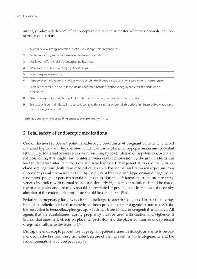
strongly indicated, deferral of endoscopy to the second trimester whenever possible, and ob‐stetric consultation.
1 Always have a strong indication, particularly in high-risk pregnancies
2 Defer endoscopy to second trimester whenever possible
3 Use lowest effective dose of sedative medications
4 Whenever possible, use category A or B drugs
5 Minimize procedure time
6 Position pregnant patients in left pelvic tilt or left lateral position to avoid vena cava or aortic compression
7 Presence of fetal heart sounds should be confirmed before sedation is begun and after the endoscopic
procedure
8 Obstetric support should be available in the event of a pregnancy-related complication
9 Endoscopy is contraindicated in obstetric complications such as placental abruption, imminent delivery, ruptured
membranes, or eclampsia
Table 1. General Principles guiding endoscopy in pregnancy (ASGE)
2. Fetal safety of endoscopic medications
One of the most important point in endoscopic procedures of pregnant patients is to avoidmaternal hypoxia and hypotension which can cause placental hypoperfusion and potentialfetal injury. Maternal oversedation with resulting hypoventilation or hypotension, or mater‐nal positioning that might lead to inferior vena caval compression by the gravid uterus canlead to decreased uterine blood flow and fetal hypoxia. Other potential risks to the fetus in‐clude teratogenesis (both from medication given to the mother and radiation exposure fromfluoroscopy) and premature birth [1-6]. To prevent hypoxia and hypotension during the in‐tervention, pregnant patients should be positioned in the left lateral position, prompt intra‐venous hydration with normal saline or a similarly high osmolar solution should be made,use of analgesics and sedatives should be restricted if possible and in the case of necessityabortion of the endoscopic procedure should be considered [5,6].
Sedation in pregnancy has always been a challenge to anesthesiologists. No anesthetic drug,inhaled anesthetics, or local anesthetic has been proven to be teratogenic in humans. A nota‐ble exception is benzodiazepine group, which has been linked to congenital anomalies. Allagents that are administered during pregnancy must be used with caution and vigilence. Itis clear that anesthetic effects on placental perfusion and the placental transfer of depressantdrugs may influence the fetus [5,6,7].
During the endoscopic procedures in pregnant patients, anesthesiologic asistance is recom‐mended in the first and third trimester because of the increaed risk of teratogenecity and therisk of premature labor, respectively [5].
Endoscopy322
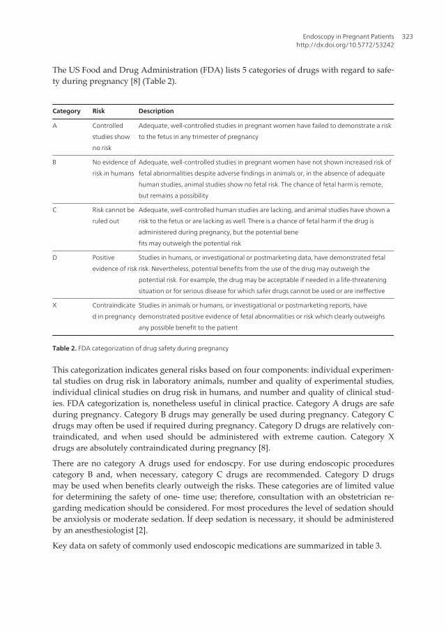
The US Food and Drug Administration (FDA) lists 5 categories of drugs with regard to safe‐ty during pregnancy [8] (Table 2).
Category Risk Description
A Controlled
studies show
no risk
Adequate, well-controlled studies in pregnant women have failed to demonstrate a risk
to the fetus in any trimester of pregnancy
B No evidence of
risk in humans
Adequate, well-controlled studies in pregnant women have not shown increased risk of
fetal abnormalities despite adverse findings in animals or, in the absence of adequate
human studies, animal studies show no fetal risk. The chance of fetal harm is remote,
but remains a possibility
C Risk cannot be
ruled out
Adequate, well-controlled human studies are lacking, and animal studies have shown a
risk to the fetus or are lacking as well. There is a chance of fetal harm if the drug is
administered during pregnancy, but the potential bene
fits may outweigh the potential risk
D Positive
evidence of risk
Studies in humans, or investigational or postmarketing data, have demonstrated fetal
risk. Nevertheless, potential benefits from the use of the drug may outweigh the
potential risk. For example, the drug may be acceptable if needed in a life-threatening
situation or for serious disease for which safer drugs cannot be used or are ineffective
X Contraindicate
d in pregnancy
Studies in animals or humans, or investigational or postmarketing reports, have
demonstrated positive evidence of fetal abnormalities or risk which clearly outweighs
any possible benefit to the patient
Table 2. FDA categorization of drug safety during pregnancy
This categorization indicates general risks based on four components: individual experimen‐tal studies on drug risk in laboratory animals, number and quality of experimental studies,individual clinical studies on drug risk in humans, and number and quality of clinical stud‐ies. FDA categorization is, nonetheless useful in clinical practice. Category A drugs are safeduring pregnancy. Category B drugs may generally be used during pregnancy. Category Cdrugs may often be used if required during pregnancy. Category D drugs are relatively con‐traindicated, and when used should be administered with extreme caution. Category Xdrugs are absolutely contraindicated during pregnancy [8].
There are no category A drugs used for endoscpy. For use during endoscopic procedurescategory B and, when necessary, category C drugs are recommended. Category D drugsmay be used when benefits clearly outweigh the risks. These categories are of limited valuefor determining the safety of one- time use; therefore, consultation with an obstetrician re‐garding medication should be considered. For most procedures the level of sedation shouldbe anxiolysis or moderate sedation. İf deep sedation is necessary, it should be administeredby an anesthesiologist [2].
Key data on safety of commonly used endoscopic medications are summarized in table 3.
Endoscopy in Pregnant Patientshttp://dx.doi.org/10.5772/53242
323

Drug FDA category in
pregnancy
key points regarding drug safety
Narcotics
Meperidine B, but D at term Repeated high dose and prolonged administration can cause respiratory depression
and seizures.
Fentanyl C In low doses it is safe.
Propofol B Generally suggested to be used in difficult to sedate and complicated clinical
situations.
General anesthetics
Ketamine B Human data is limited; animal data suggests prolonged usage is unsafe.
Sedatives
Diazepam D Some of congenital malformations and mental retardations are possibly associated
with diazepam usage so its usage is discouraged during pregnancy.
Midazolam D As a benzodiazepine member, its usage is restricted during pregnancy especially in
first trimester.
Reversal agents
Naloxone B It is probably safe and should be used only in respiratory depression, systemic
hypotension or unresponsiveness in a closely monitored pregnant after endoscopy.
Flumazenil C Its fetal risks are largely unknown, it should be given in small doses carefully.
Table 3. Safety of anesthetics commonly used in gastrointestinal endoscopy
Meperidine, an opiate analgesic, was commonly used for gastrointestinal endoscopy in thegeneral population, but has been replaced by short-acting analgesics due to theoretical con‐cerns about toxicity, manifested as respiratory depression and seizures. Meperidine is rapid‐ly transferred across the human placenta after intravenous administration, after thatmetabolized to normeperidine which has a long half-life. Repeated high dose and prolongedadministration of meperidine can cause progressive accumulation of normeperidine, andproduce toxic effects of maternal respiratory depression and seizures. Meperidine is rated acategory B drug during pregnancy, but rated category D when used for prolonged periods(>36 h) at high doses at term. Meperidine use should be limited to 50–75 mg during routineendoscopy during pregnancy [4,5,9].
Fentanyl is a potent narcotic agonist with a rapid onset of action and a shorter patient recoverytime than meperidine. It is an FDA category C drug during pregnancy. It appears safe whengiven in low doses (<125 mg) in patients undergoing endoscopy during pregnancy [2,4,5].
Benzodiazepines (diazepam and midazolam) are commonly administrated before gastroin‐testinal endoscopy to reduce anxiety, induce brief amnesia, and produce muscle relaxation.Prolonged use of diazepam during early pregnancy has been associated with cleft palatemalformations [4,11] but several large studies have not shown this association [12,13]. Some
Endoscopy324
studies have raised possible associations between diazepam administration and mental re‐tardation or neurologic defects,[14] cardiac defects [11] and Mobius syndrome (sixth andseventh nerve palsies) [15]. Diazepam is rated a category D drug during pregnancy, becauseof these potential associations its use during pregnancy is discouraged, particularly in thefirst trimester. Clinical data concerning fetal safety of midazolam are limited; however, thedrug has not been associated with oral cleft palates. Administration of midazolam duringlabor has been reported to transiently depress neonatal neurobehavioral responsiveness[16,17]. Although rated a category D drug, since it has not been reported to be associatedwith congenital abnormalities, midazolam is the preferred benzodiazepine when meperi‐dine is inadequate. Due to a similar mechanism of action as diazepam, midazolam useshould be restricted during pregnancy, especially during the first trimester [1,2,4,5].
Propofol is increasingly used for anesthesia at endoscopy in the general population. It isshort –acting with a recovery period much shorter than with standard agents. However, as aresult of a relatively narrow therapeutic index (defined as a ratio given by average toxicdose divided by average therapeutic dose) and the potential for respiratory depressionwhich can cause respiratory arrest and even death if improperly monitored, it is generallyadministrated by anesthesiologists. Endoscopy socities have recommended the usage of thisagent for difficult to sedate and complicated clinical situations. Propofol is rated a categoryB drug and it is considered relatively safe to use during pregnancy, although its safety dur‐ing the first trimester is inadequately studied [4,5,18].
Ketamine is useful for endoscopy in patients who are expected to experience insufficient seda‐tion from propofol. Like propofol, ketamine has a rapid onset of action and a short duration ofeffect. Although rated a category B drug during pregnancy, ketamine carries the caveats thatfetal safety during the first trimester is unstudied and unknown in humans, and an extremelyhigh dose or prolonged administration of it during pregnancy may be unsafe [4,19].
Naloxone, a rapidly acting narcotic antagonist, crossing the placenta within 2 minutes of in‐travenous administration, is occasionally administered during endoscopy to reverse narcoticoverdose [2,4,20]. The drug is rated a category B drug during pregnancy. It should only beused in respiratory depression, systemic hypotension or unresponsiveness in a closely moni‐tored setting during or after endoscopy. It should be administered during pregnancy insmall, graded doses titrated to the desired effect. It should not be routinely administered af‐ter endoscopy during pregnancy,since one neonatal fatality has been attributed to its use[21]. Naloxone is contraindicated in pregnant patients who are dependent on narcotics be‐cause it can precipitate opiate withdrawal syndrome [22]. Flumazenil, a benzodiazepine an‐tagonist, is used to reverse oversedation from benzodiazepines administered duringendoscopy. It is rated a category C drug during pregnancy. The drug should only be used toreverse benzodiazepine overdose during pregnancy because its fetal risks are largely un‐known, with only few case reports of use during pregnancy. Flumazenil overdose can causematernal seizures, particularly when administrated to patients who are chronically habituat‐ed to benzodiazepines. This overdose can be prevented by careful and slow titration and ad‐ministration of minimal dosage of benzodiazepines required for endoscopy. [4,19,20,23].
Endoscopy in Pregnant Patientshttp://dx.doi.org/10.5772/53242
325
3. Upper gastrointestinal endoscopy
Although it would be ideal to postpone all endoscopic procedures until after delivery, preg‐nant patients can develop conditions that require urgent upper endoscopy. The most commonindications for EGD in pregnant patients are significant or continued gastrointestinal hemor‐rhage, dysphagia and refractory nausea and vomiting (Table 4). In a multicenter retrospectivestudy of 83 pregnant women on safety and clinical efficacy of EGD in pregnant patients; endos‐copy indications included gastrointestinal bleeding, abdominal pain and vomiting in decreas‐ing order [24]. The Mallory-Weiss tear was an important cause of upper gastrointestinalbleeding in 14% of patients, the peptic ulcer was also responsible for 14% of gastrointestinalbleeding in those patients which is significantly lower than the reported frequencies in nonpregnant patients. The diagnostic yield for upper gastrointestinal bleeding was 95% and therewas no cases of premature labour or congenital fetal malformation. Labor was not induced inany of the patients and 95% of the patients gave birth to healthy infants. The four fetal deaths inthe study were in all high risk patients and were unrelated to EGD. The mean Apgar scores oflive born infants were not significantly different than control groups.
In an other study conducted by Debby et al [25], 60 pregnant women underwent EGD forrecurrent and intractable nausea and vomiting in their first trimester. In that study only 11patients had the upper gastrointestinal bleeding, the other majority had only intractablenausea and vomiting. The diagnostic yield of EGD in those patients appeared higher forgastrointestinal bleeding than for intractable vomiting but the difference was not statisticallysignificant. Since the endoscopic findings only minimally changed the clinical managementof patients with nausea and vomiting the authors suggested the necessity of EGD for uppergastrointestinal bleeding but not nausea and vomiting or hyperemesis gravidarum. Amailed survey of over 3000 members of the American College of Gastroenterology, whichincluded information over 73 upper endoscopies performed during pregnancy. Endoscopicdiagnoses from these procedures included esophagitis, gastritis, ulcers, Mallory-Weis tearsand normal findings in descending order. This survey reported no significant complicationsadversly affecting pregnancy [26].
The relationship between hyperemesis gravidarum and Helicabacter Pylori (H.Pylori) infec‐tion is estimated by Bagis et all, in their study, H Pylori infection was histologically demon‐strated in 95% of pregnant patients with hyperemesis gravidarum and 50% of controlpatients. In that study patients with hyperemesis gravidarum had more severe H. Pylori in‐fection compared to controls with H.Pylori infection, as measured histologically by densityof bacterial infiltration. Authors suggested the usage of H.Pylori diagnostic tests to be partof hyperemesis gravidarum investigation [27]. Upper gastrointestinal complaints, especiallynause and vomiting, are very common in pregnants. During pregnancy with the effect ofprogesteron and estrogen and with a lesser effect of motilin hormone the lower esophagealsphincter (LES) tone, gastric and intestinal motility decrease, causing gastroesophageal re‐flux disease (GERD) symptoms. As pregnancy progresses, the frequency of and intensity ofGERD symptoms increase which is caused by gastrointestinal motility changes during preg‐nancy and with a small contribution of physical effects of gravid uterus [28,29]. So as a con‐clusion endoscopy is rarely helpful and rarely indicated for nausea and vomiting, or even
Endoscopy326
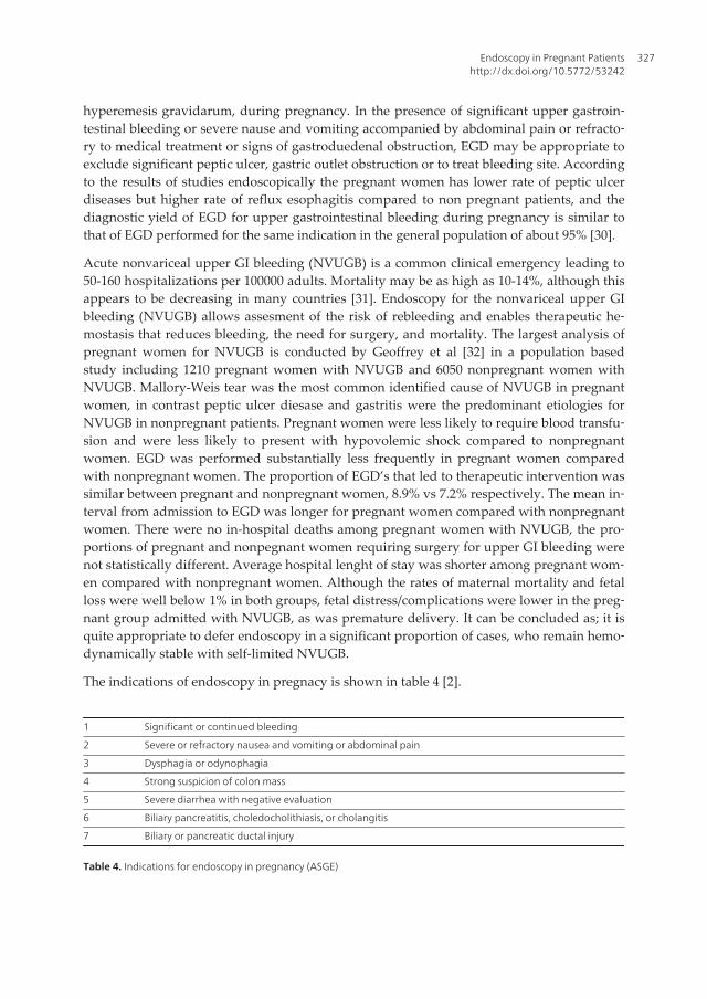
hyperemesis gravidarum, during pregnancy. In the presence of significant upper gastroin‐testinal bleeding or severe nause and vomiting accompanied by abdominal pain or refracto‐ry to medical treatment or signs of gastroduedenal obstruction, EGD may be appropriate toexclude significant peptic ulcer, gastric outlet obstruction or to treat bleeding site. Accordingto the results of studies endoscopically the pregnant women has lower rate of peptic ulcerdiseases but higher rate of reflux esophagitis compared to non pregnant patients, and thediagnostic yield of EGD for upper gastrointestinal bleeding during pregnancy is similar tothat of EGD performed for the same indication in the general population of about 95% [30].
Acute nonvariceal upper GI bleeding (NVUGB) is a common clinical emergency leading to50-160 hospitalizations per 100000 adults. Mortality may be as high as 10-14%, although thisappears to be decreasing in many countries [31]. Endoscopy for the nonvariceal upper GIbleeding (NVUGB) allows assesment of the risk of rebleeding and enables therapeutic he‐mostasis that reduces bleeding, the need for surgery, and mortality. The largest analysis ofpregnant women for NVUGB is conducted by Geoffrey et al [32] in a population basedstudy including 1210 pregnant women with NVUGB and 6050 nonpregnant women withNVUGB. Mallory-Weis tear was the most common identified cause of NVUGB in pregnantwomen, in contrast peptic ulcer diesase and gastritis were the predominant etiologies forNVUGB in nonpregnant patients. Pregnant women were less likely to require blood transfu‐sion and were less likely to present with hypovolemic shock compared to nonpregnantwomen. EGD was performed substantially less frequently in pregnant women comparedwith nonpregnant women. The proportion of EGD’s that led to therapeutic intervention wassimilar between pregnant and nonpregnant women, 8.9% vs 7.2% respectively. The mean in‐terval from admission to EGD was longer for pregnant women compared with nonpregnantwomen. There were no in-hospital deaths among pregnant women with NVUGB, the pro‐portions of pregnant and nonpegnant women requiring surgery for upper GI bleeding werenot statistically different. Average hospital lenght of stay was shorter among pregnant wom‐en compared with nonpregnant women. Although the rates of maternal mortality and fetalloss were well below 1% in both groups, fetal distress/complications were lower in the preg‐nant group admitted with NVUGB, as was premature delivery. It can be concluded as; it isquite appropriate to defer endoscopy in a significant proportion of cases, who remain hemo‐dynamically stable with self-limited NVUGB.
The indications of endoscopy in pregnacy is shown in table 4 [2].
1 Significant or continued bleeding
2 Severe or refractory nausea and vomiting or abdominal pain
3 Dysphagia or odynophagia
4 Strong suspicion of colon mass
5 Severe diarrhea with negative evaluation
6 Biliary pancreatitis, choledocholithiasis, or cholangitis
7 Biliary or pancreatic ductal injury
Table 4. Indications for endoscopy in pregnancy (ASGE)
Endoscopy in Pregnant Patientshttp://dx.doi.org/10.5772/53242
327
4. Therapeutic endoscopy
4.1. Endoscopic hemostasis for variceal bleeding
Patients with cirrhosis are not likely to become pregnant due to endocrine and metabolic im‐balances. On the other hand, women with non-cirrhotic portal hypertension have normalfertility rates. The incidence of variceal bleeding during pregnancy may reach up to 45%with a mortality rate of 18-50% The variceal bleeding frequently occurs during the last twotrimester of gestation. The possible explanation for the high severity of variceal bleeding inpregnancy seems to be related to the increase in water retention and cardiac output, typicalof both pregnancy and cirrhosis. Women with esophageal varices or severe liver diseaseshould be advised, in case of planning the pregnancy, about the high risk of both varicealbleeding and hepatic decompensation during pregnancy. Also patients who have esopha‐geal varices should be informed about the benefits of β-adrenergic receptor antagonist ther‐apy during pregnancy to reduce portal pressure.
Both endoscopic injectional sclerotheraphy (EIS) and endoscopic band ligation (EBL) eitherprophylactic or urgent seem to be safe procedures during pregnancy. When bleeding is notarrested endoscopically in cirrhotic patients, an emergency transjugular intrahepatic porto‐systemic shunt (TIPS) is indicated, but data regarding pregnant cirrhotic women are scarce[33-39]. Since there are only a few case reports regarding the treatment options for this clini‐cal condition, the management of esophageal varices and their major life-threatining compli‐cation – hemorhage during pregnancy is still under evaluation. In the early 80’s EIS wasgenerally accepted as the first line treatment procedure for bleeding esophageal varices. De‐spite this fact, only few cases of EIS with conventional sclerosants (polidocanol, absolute al‐chocol, sodium tetradecyl sulphate) were reported during pregnancy [36,38,39]. There areno studies regarding the effect of the conventional sclerosants on the fetus published in theliterature, although the procedure is considered safe and effective to control active varicealbleeding. Vasoactive drugs used to achieve hemostasis are contraindicated during pregnan‐cy, since these (vasopressin and terlipressin) may induce labor or fetal malformations [36].Recently EBL was reported as an effective treatment option for active variceal hemorrhageas well as prophylaxis of this severe complication during pregnancy [36-39].
In early 90’s EBL has been proven effective in controlling active hemorrhage and in longterm prevention of recurrent bleeding. There are several case reports that describe successfulhemostasis without fetal complications [40,41]. When EBL is used there is no risk of migra‐tion of a toxic substance to placenta. Studies of EBL versus EST in nonpregnant patientshave shown improved reduction in rebleeding and mortality with EBL [40,42]. But there areno studies directly comparing EBL to EST in pregnant patients.
In a study conducted by Aggarwal et al, 17 patients with noncirrhotic portal hypertensioncaused by extrahepatic portal vein obstruction or portal fibrosis, underwent EIS with eitherabsolute alcohol or sodium tetradecyl sulphate for acute variceal bleeding without compli‐cations during pregnancy. In that retrospective analysis pregnancy outcomes included sixhealthy full-term infants, two preterm deliveries, three stillbirths, one neonatal death, and
Endoscopy328
five voluntary abortions. In that report two patients required EBL after failure of EIS to ob‐literate esophageal varices [43].
In an another report 10 patients underwent EIS with absolute alcohol, 5 patients for activevariceal bleeding and another 5 patients for the profilaxis of variceal bleeding. Hemostasiswas achieved in 5 actively variceal bleeding patients. All 10 patients delivered healthy in‐fants [44]. According to the reported studies as in the nonpregnant population, EBL seems tobe a reasonable option for acute variceal bleeding as well as for the profilaxis of varicealbleeding. EIS could be a secondary choice for the acute variceal bleeding due to the probableeffects on fetal safety.
When the endoscopic and pharmacological theraphy fails; TIPS may be a rescue procedurein pregnant women with recurrent and difficult-to treat variceal bleeding. Several papersconfirm the utility and efficacy of TIPS for variceal bleeding unresponsive to endoscopic andpharmacological treatment, however since adequate controlled trials are lacking, it shouldbe limited to a selected group of patients. TIPS placement is associated with radiation expo‐sure of both the patient and the medical staff, it usually requires prolonged fluoroscopy. Inthe literature there is a few case of TIPS placement in pregnancy in which between 5.2 mSvand 2.1 mGy fetal dose of radiation reported [35,45,46]. So pregnancy is not an absolute con‐traindication for TIPS placement in the treatment of relapsing bleeding varices and may be arescue procedure when medical and endoscopic treatments have failed.
4.2. Endoscopic hemostasis for nonvariceal bleeding
Endoscopic hemostatic techniques for nonvariceal bleeding include injection therapy withsaline, with adrenaline, sclerosants, thrombin, fibrin, cyanoacrilate, ablative therapy withthermocoagulation, electrocoagulation, photocoagulation, argon plasma coagulation, andmechanical compression with hemoclips, detachable snares, graspers, or sutures [47]. Inspite of these numerous techniques, there are a few case reports on fetal safety of endoscopichemostasis for NVUGB. In those case reports adrenalin injection, thermocoagulation andelectrocoagulation was used for hemostasis. In only one of the pregnant patient the hemo‐static procedures were unsuccesful so patient required surgery. The fetal outcomes werehealthy infants without fetal malformations [24,25,47,48,49].
Adrenalin is category C drug and can cause a decrease in uterine blood flow. Althoughthere is limited data of case reports, no advers events from adrenaline injection were report‐ed, in this case the benefits (cessation of hemorhage, prevention of rebleeding) would seemto outweigh the risks [1,2,29,50].
Amniotic fluid can conduct electrical current to the fetus. During the electrocoagulation thegrounding pad should be placed in such a position that the uterus is not between the electri‐cal catheter and grounding pad. Bipolar electrocautery should be used to minimize the riskof ‘stray’ currents going through the fetus. Electrocautery is relatively safe when used forhemostasis[2]. Due to the limited data on hemostasis for nonvariceal bleeding in pregnantpatients, the therapeutic technique choice is based on expert opinion and generally made ac‐cording to the results of clinical studies of nonpregnant patients.
Endoscopy in Pregnant Patientshttp://dx.doi.org/10.5772/53242
329
4.3. Percutaneous endoscopic gastrostomy
During pregnancy, optimal nutrition is essential in order to minimize maternal and neonatalmorbidity [51]. Nause and vomiting are seen in 80% of pregnancies but usually symptomsare mild and self limited. In case of severe hyperemesis gravidarum with dehydration andketonuria, patients should be hospitalized and receive intravenous hydration and anti-emet‐ic agents. When the hospitalization duration prolongs without oral intake then supportivenutrition with enteral feeding or total parenteral nutrition should be considereed [29]. Longterm nasogastric feeding is limited by patient intolerability and nasal septal necrosis. Sideeffects of long term total parental nutrition limit its usage during pregnancy[52]. Thus per‐cutaneous endoscopic gastrostomy (PEG) becomes an important option for long term enter‐al feeding. However, concerns about uterine damage, fetal injury, premature labor, andinfections have restricted the application of PEG tube placement in pregnant women. Therewere no major complications with PEG tube placement in the several reported cases in theliterature [53-61]. PEG enteral nutritional support was provided for an average of 14 weeksin the literature. During the pregnancy PEG tube placement is a feasible procedure for opti‐mal enteral nutrition in the critical care setting. It is also feasible to perform PEG tube place‐ment in the third trimester of pregnancy. A potential problem with PEG during pregnancyis puncture of uterus or fetus instead of the stomach during transabdominal neddle inser‐tion. This risk is reduced by demarcating the upper border of uterus before PEG by abdomi‐nal ultrasonography and by inserting the PEG needle ≥5cm cephaled.
PEG tube placement should be reserved only for severe refractory cases where the nutritionof the mother and the fetus is at risk. The pregnant should be informed about the risks of theprocedure and potential placental injury. If possible less invasive alternative techniques asnasoenteric feeding tube or peripherally inserted catheter for parenteral nutrition should beattempted if this is not succesful or is refused by the patient, PEG tube placement should beconsidered.
4.4. Percutaneous endoscopic gastrojejunostomy
Percutaneous endoscopic gastrojejunostomy( PEGJ) is a feeding tube placement in to the je‐jenum via a gastrostomy. This placement enables food to be delivered more distally to de‐crease the sensation of nausea, vomiting and risk of aspiration. PEGJ indications are similarto PEG indications with additional risk of aspiration. In case of refractory nause and vomit‐ing in the presence of PEG tube feeding, PEG tube can be converted to PEGJ. There are onlyfew case reports of PEGJ for hyperemesis gravidarum and one patient with coma from mas‐sive stroke [62-65]. As in the PEG, PEGJ can be considered in very severe hyperemesis gravi‐darum refractory to medical treatment and the other noninvasive treatment modalities.
5. Sigmoidoscopy/ colonoscopy
Most pregnant patients are young, healthy women and the gestational period is only 40weeks in duration, because of that; it is unusual for them to need flexible sigmoidoscopy or
Endoscopy330
colonoscopy during pregnancy. Lower endoscopy should be avoided for weak indicationsduring pregnancy and should be deferred until after the first trimester, or if possible, untilthe postpartum period [66].
Although flexible sigmoidoscopy will be sufficient for most pregnant women, colonoscopymay be required when there is life-threatening colonic bleeding and when a cause has notbeen established by sigmoidoscopy. In late pregnancy, patients should not be placed in thedecubitus or prone position during colonoscopy. External abdominal pressure should gener‐ally be avoided; if pressure is required, it should be minimal and directed away from theuterus.
All avilable evidence suggests that sigmoidoscopy is safe during pregnancy and the indica‐tions include rectal bleeding, chronic diarrhea, abdominal pain and rectal pain. Guidelinesfor colonoscopy in pregnancy are not readily available due to insufficient data, althoughstudies which have been done demonstrate safety and efficacy of the procedure providedthat obstetrical consultation and close monitoring take place. Colonoscopy is indicated forsuspected colon cancer, uncontrolled severe hemorrhage or when necessary before colonicsurgery in pregnant women as well as in general population [67,68].
The safety and efficacy of the flexible sigmoidoscopy during pregnancy has been studied inone case-controlled study of 45 patients undergoing 48 sigmoidoscopies [69]. The most com‐mon clinical indication was hematochezia in 29 patients, diarrhea was the indication in 10patients and abdominal pain was in 4 patients. The most common sigmoidoscopic diagnosisincluded reactivated or newly diagnosed inflamatory bowel disease, bleeding internal hem‐orroids and other colitis. Among the 29 patients with hematochezia, 8 patients were de novodiagnosis or flare of ulcerative colitis, 7 patients were de novo diagnosis or flare of Crohn’sdisease, 3 patients with acute proctosigmoitidis, 2 patients were bleeding internal hemor‐rhoids, 1 patient was pseudomembraneous colitis, and 1 patient was sigmoid adenoma.Hematochezia gave the highest diagnostic yield compared with the other clinical indica‐tions. Excluding one unknown pregnancy outcome and four voluntary abortions, 38 (93%)of 41 pregnant patients who underwent sigmoidoscopy delivered healthy infants, including27 at term. The mean Apgar score of live-born infants in this study was not statistically dif‐ferent from the mean national Apgar score of live born infants.
Therapeutic changes because of the sigmoidoscopic findings occured in 24 patients, includ‐ing changing or instituting medication for inflamatory bowel disease in 15 patients, steroidenemas for nonspesific proctitis in two patients, avoiding surgery in two patients, and hem‐orrhoidal treatment in two patients.
Other than this study there are individual case reports of sigmoidoscopy performed duringpregnancy and a mailed survey study [26, 70-74]. Multiple case reports describing flexiblesigmoidoscopy in pregnant patients showed the safety of this procedure. In the mailed sur‐vey study; after contacting 3300 gastroenterologists, their responses indicated that therewere no endoscopic complications in 13 pregnant women who underwent flexible sigmoi‐doscopy. In addition all pregnancies resulted in healthy infants. Common themes amongthese reports include a relatively high diagnostic yield of flexible sigmoidoscopy when per‐
Endoscopy in Pregnant Patientshttp://dx.doi.org/10.5772/53242
331
formed during pregnancy for strong indications, clinically important changes in therapy re‐sulting from sigmoidoscopic diagnosis, a relatively high rate of favorable fetal outcomes,poor fetal outcomes generally occuring only in very sick mothers and unrelated to sigmoi‐doscopy etiologically or temporally, and a low rate of congenital anomalies.
Studies suggested that sigmoidoscopy during pregnancy does not induce labor or causecongenital malformations, it is not contraindicated, and should be considered in medicallystable patients with important indications. Sigmoidoscopy should be performed with mater‐nal monitoring by electrocardiography and pulse oximetry, after obstetrical consultationand after medical stabilization. Medical stabilization may require blood transfusions andsupplemental oxygenation [4,66,68]. Sigmoidoscopy is not recommended during pregnancyfor indications of a change in bowel habits, abdominal pain, a family history of colon cancer,and routine screening or surveillance. In these cases, sigmoidoscopy is best deferred until atleast 6 weeks postpartum [67, 68].
Little is known about the safety or otherwise of bowel cleansing agents during pregnancy.Studies have shown that the systemic absorbtion of Polyethylene glycol is minimal and theproblems with abdominal bloating and gas are less common as compared to other laxatives[75] But since polyethylene glycol solutions have not been studied during pregnancy, there‐fore it is a category C drug during pregnancy. Sodium phosphate solutions (also category C)may cause fluid and electrolyte disturbance, and therefore probably best avoided duringpregnancy. Also it was published that newborns were shown to manifest bone deminerali‐zation and bone growth failure because of maternal phosphate overload [76], although a onetime use in pregnancy has not shown to be detrimental. Another consideration when usingphosphosoda preperations is the risk of phosphate nephropathy [77] which has been report‐ed in selected cases. As for bowel preperation for flexible sigmoidoscopy, phosphate enemasshould be relatively safe, but have not been studied in pregnancy. Tap water enemas may besufficient. In a study [78] of the preference of gastroenterologist and obstetricians of bowelcleansing agent for sigmoidoscopy and colonoscopy in the same hospital, it was shown that50% of gastroenterologist prefer to use polyethylene glycol solutions and 50% avoid use offleet phosphosoda. Twent percent of obstetricians seem to prefer fleet phosphosoda, and26% avoid polyethylene glycol solutions which is exact opposite of gastroenterologist. Bothgroups prefer fleet enema the most (51%), while magnesium citrate is used least often (38%).
6. Colonoscopy
There are insufficient data regarding the safety of performing colonoscopy during pregnan‐cy. The largest case control study of 20 patients [79]; there were 20 pregnant patients under‐going colonoscopy and 20 pregnant controls who were matched for colonoscopy but whodid not undergo colonoscopy. Colonoscopy indications in the study patients included diar‐rhea, hematochezia, bloody diarrhea, abdominal pain, and other reasons. Colonoscopy wasperformed in the second trimester in the majority of patients (n=16) with only 4 patients un‐dergoing the procedure in the first and third trimester. Colonoscopic diagnoses were; ulcer‐
Endoscopy332
ative colitis, ischemic colitis, Crohns colitis, and lymphocytic colitis. Colonoscopy led tosignificant therapeutic changes in 7 (35%) patients. Two mothers experienced minor colono‐scopic complications of mild, transient hypotension. Study patients had relatively favorablefetal outcomes: 18 relatively healthy infants, 1 involuntary abortion, and 1 infant born with acongenital defect (septum secundum cardiac defect).
In a mailed survey of 3300 gastroenterologists 13 colonoscopies were performed withoutcomplications [26]. This study was retrospective, and subject to biases relating to recall anddependence on voluntary reporting. In an other study Cappell and co workers [69] retro‐spectively examined eight pregnant women undergoing colonoscopies at 10 different medi‐cal centers. Excluding one elective abortion and one fetal demise unrelated to thecolonoscopy occuring 4 months later, six healthy infants were born. There was no differencein outcomes based on the trimester during which colonoscopy was performed.
There are several case reports about colonoscopy during pregnancy [80-88], fetal outcomesafter colonoscopy included 8 healthy babies, 2 stillbirth unrelated to the colonoscopy (inmothers with either metastatic colon cancer or massive lower gastrointestinal hemorrhagerequiring emergency colonic surgery), and one unknown fetal outcome.
Given the limited data on its safety and the potential to cause significant adverse events, co‐lonoscopy should be reserved to strong indications or life-threatining emergencies duringsecond trimester. But in case of suspicion of colon cancer, evaluation of colonic mass or co‐lonic stricture of unknown etiology, for severe uncontrolled colonic hemorrhages, for colon‐ic pseudoobstruction when the alternative is surgical decompression and when requiredbefore urgent colonic surgery, colonoscopy should be considered even in the first and thirdtrimester. Colonoscopy should generally be deferred in any trimester of pregnancy until af‐ter delivery for elective indications, such as surveillance for prior history of colon cancer orcolonic polyps.
7. Therapeutic colonoscopy
Therapeutic colonoscopy includes hemostasis of lower gastrointestinal bleeding, colono‐scopic polypectomy, and colonic stenting. As mentioned in the hemostasis for NVUGB sec‐tion,injection therapy with saline, with adrenaline, sclerosants, thrombin, fibrin,cyanoacrilate, ablative therapy with thermocoagulation, electrocoagulation, photocoagula‐tion, argon plasma coagulation, and mechanical compression with hemoclips, detachablesnares, graspers, or sutures are used during lower gastrointestinal bleeding [47].
Epinephrine is commonly used to treat gastrointestinal bleeding and achieves hemostasisthrough its vasoconstrictive effects. There are numerous studies about the fetal safety of epi‐nephrine administration during labor which established its fetal safety so it is commonlyadded to spinal epidural anesthesia. Although in the report of Briggs and colleagues’ [10],there was no congenital defect in 35 infants with first trimester in utero exposure of epi‐nephrine, there is a case report of fatal intracranial hemorrhage in an infant during child‐
Endoscopy in Pregnant Patientshttp://dx.doi.org/10.5772/53242
333
birth after excessive in utero epinephrine [89]. Because of the epinephrine’s α adrenergiceffect of decreasing uterine blood flow, it is a category C during pregnancy and its dosageshould be kept low during pregnancy.
Electrocautery is also another method of providing hemostasis during lower gastrointestinalbleeding, also used for performing polypectomy or hot biopsy. Using electrocautery to le‐sions may occasionally be required during pregnancy and has been safely performed with‐out detectable advers effcts to fetus. However, because amniotic fluid has beendemonstrated to conduct electrical current, externally placed grounding pad should beplaced close to the electrical catheter, devices should use bipolar currents. Polyp removal ifnot bleeding, should be postponed after pregnancy [4, 5, 66].
Colonic tattooing is done with Indian ink and methylene blue in nonpregnant patients. Indi‐an ink has been shown to persist for the entire life of patient. There have ben no reports oflong term complications of Indian ink tattooing. Methylene blue tattooing during pregnancyis not been studied but there are reports of examination of Metylene blue during amniocent‐esis and in detection of ruptured membranes. In these reports fetal death, jejunal atresia, isreported which labels Metylene blue as teratogenic. Although safety of colonic injection isnot studied; its usage should be avoided during pregnancy [66, 90, 91].
8. Enteroscopy
Enteroscopy takes a very long time of procedure with a long time of anesthesia. There is nocase report of enteroscopy during pregnancy, so the fetal safety of enteroscopy could not bepredicted.
9. Video capsule endoscopy
Video capsule endoscopy (VCE) represents a significant advance in the investigation ofsmall bowel diseases. The main indications are obscure gastrointestinal hemorrhages,Crohn’s disease, celiac disease, small bowel tumors and polyposis syndromes. The maincontraindications are known or suspected gastrointestinal obstruction, strictures, fistulas,cardiac pacemakers and swallowing disorders [92]. During pregnancy the growing graviduterus pushes and compresses the gastrointestinal tract, and gastrointestinal motility de‐creases due to inhibition of intestinal smooth muscle by gestational progestin. These effectsraise the theoretical concerns regarding capsule impaction during pregnancy [4,93]. Al‐though according to the FDA, pregnancy is a relative contraindication for VCE [92]; there isa report of VCE usage in a young acute bleeding pregnant patient in whom endoscopy andcolonoscopy revealed no lesion other than fresh blood exiting the terninal ileum. On VCE anactively bleeding jejunal lesion was shown of which pathology was jejunal carcinoid tumor.Patient and fetus did well after surgery [94]. From this report, it can be concluded that VCEmay be considerable during pregnancy for strong indications, it is not absolutely contraindi‐
Endoscopy334
cated during pregnancy. But it is more likely to be incomplete when done during pregnancybecause of slowed intestinal transit time during pregnancy.
9.1. Endoscopic retrograde cholangiopancreatography
Pregnancy is associated with an increased risk of gallstone formation. Fortunately, compli‐cations due to cholelithiasis, such as cholecystitis, choledocholithiasis and pancreatitis arerelatively uncommon and in many cases can be managed conservatively. However occasion‐ally patients develop complications related to gallstones that require intervention duringpregnancy. Although there are no precise estimates of the incidence, several reports havefound that biliary tract disease (most commonly cholecystitis) represented one of the mostfrequent indications for non-obstetrical surgery during pregnancy [95-99].
A subset of patients requires ERCP, most commonly for choledocholithiasis or presumedgallstone pancreatitis. Opinions regarding the safety of ERCP during pregnancy differ invarious reports, reflecting the relatively limited data. Major concerns surround issues relat‐ed to radiation exposure to the fetus and the risk of procedure on pregnancy outcome.
A general principle in the care of the women with an acute biliary tract disorder duringpregnancy is to provide the most conservative management possible with the hope of delay‐ing intervention until after pregancy or until the second trimester, when surgical interven‐tion is relatively safer. There are numerous reports about ERCP during pregnancy especiallyfor the last ten years.
The largest series in the literature included 65 pregnant patients [100]; the most common in‐dications for ERCP during pregnancy were recurrent biliary colic, abnormal liver functiontests, and a dilated bile duct on ultrasound. Sixty eight ERCP was performed on 65 pregnantpatients, 17 pregnants were in the first trimester, 20 pregnants were in the second trimesterand 31 pregnants were in the third trimester. The median fluoroscopy time was 1.45 mi‐nutes. Almost all patients underwent a therapeutic procedure. Post ERCP pancreatitis devel‐oped in 11 patients (16%), none of whom had a severe course. Most patients achieved a termpregnancy (89%), there were no fetal deaths, perinatal deaths, or evident congenital malfor‐mations. Only 5 babies (8%) were born prematurely or with low birth weight.
Another series of 23 patients included 20 of whom underwent a therapeutic ERCP whilethree underwent a diagnostic ERCP only [101]. One patient developed post ERCP pancreati‐tis (after each of her three ERCP). There was one spontaneous abortion and one neonataldeath 26 hours after delivery. The neonatal death and post-ERCP pancreatitis were in thesame patient who undergone three ERCPs (twice during the first and one during the thirdtrimesters) with pancreatic duct stenting for stenosis of pancretaic orifice after a previoussurgical sphincteroplasty.
Shelton et al in a retrospective series of 21 cases of ERCP with sphincterotomy reported suc‐cessful extraction of stones in 14 women and successful removal of sludge in 7 women.There was one maternal complication of pancreatitis. There were 17 healthy babies deliv‐ered at term; One preterm, low birth weight baby, and 3 unknown fetal outcomes [102].
Endoscopy in Pregnant Patientshttp://dx.doi.org/10.5772/53242
335
Another series described long term follow up of 18 pregnant women who underwent biliarysphincterotomy for common bile duct stones during pregnancy (first trimester 4, second 6,third 8) [103]. Despite short term complications (one with postsphincterotomy bleedings,one with mild post ERCP pancreatitis and preterm labor), no long term maternal complica‐tions were seen after a median of six years (range 1-11 year). Only 11 of the 18 families wereretrospectively contacted; all 11 babies were healthy on follow up at a mean of 6 years post‐partum. Only one mother had preterm delivery.
Kahaleh et al reported a total of 17 pregnant patients underwent ERCP; of which 15 had ra‐diation dosimetry measured. Mean fluoroscopy time was 14 seconds (1-48seconds), meanestimated fetal radiation exposure was 40mrad (1-180mrad). Complications were reportedas; postsphincterotomy bleeding in one patient, which was controlled by placement of he‐moclip, and post-ERCP pancreatitis in one patient. All infants delivered had Apgar scores of8 or greater. Thirteen of 15 patients who delivered were contacted, and they confirmed thattheir child was in good health. [104].
In an another series of 15 patients, the incidence of complications (7%) was no different thanthe rate of complications observed in nonpregnant patients. [105]. There were no serious ad‐verse outcomes to the fetus or mother.
Different from the aforementioned reports, Farca et al reported a prospective study ≥10 ther‐apeutic ERCP during pregnancy [106]. In this study a single 10F stent was placed withoutsphincterotomy, all patients had uncomplicated pregnancies and delivered healthy infants.All underwent ERCP with sphincterotomy and stent extraction postpartum; 8 had stones ex‐tracted. In two patients, the single 10F stent remained in place for 7 and 8 months, respec‐tively; no one developed cholangitis.
During the ERCP in pregnancy the perceived risk of radiation exposure is much greaterthan the actual risk, but a full explanation of these risks to the pregnant patient and her fam‐ily is more credible if given prior to exposure. Patients should be fully informed, back‐ground population risks for miscarriage, congenital anomalies, genetic disease, and thegrowth restriction are approximately 20, 4, 10 and 10 percent, respectively. Potential radia‐tion exposure risks to the fetus can be divided into four categories: intrauterine fetal death,malformations, disturbance of growth and development, mutagenic and carcinogenic ef‐fects.
Ionizing radiation is measured in special units, rad (radiation absorbed dose) and rem (radi‐ation equivalent man) and in the international units, gray (Gy) and sievert (Sv)
(1 rad = 1rem = 0.01Gy = 0.01Sv). The avarage person in United States receives about 360mrem of ionizing radiation annually, of which about 60 mrem comes from man-made sour‐ces, including medical exposures such as diagnostic radiographs. The rem is a unit used tomeasure the effect of radiation on the human body is referred to as the ‘ effective doseequivalent’. Fetal radiation exposure can result in developmental abnormalities, particularlyif exposure is during the first trimester when organogenesis occurs. High radiation exposuremay result in fetal wastage. Although estimates vary, it is recommended that fetal radiationexposure not exceed 100mrem during the first trimester [104].
Endoscopy336
Data from studies of animals and nuclear bomb survivors suggest that the period of majororganogenesis, between the 8th and 15th weeks of gestation, is the most sensitive for growthretardation, which may be observed with exposures of 200-250mrem. Exposures greaterthan 100mrem occuring later, during neuron development and migration, may be associatedwith microcephaly, seizures, decline in mental ability, and childhood cancer [107]. Duringthe whole of gestation, the maxium permitted dose of ionizing radiation to the fetus is500rems [108].
Lead shielding should be used to minimize radiation exposure to the uterus. The lead apronshield must be placed underneath the patient and not draped over the abdomen since theradiation source is underneath the patient when using the standard fluoroscopy C-arm[109]. External shielding can not elimineta fetal exposure due to internally scattered radia‐tion. Even though the fetus can be shielded, efforts should be made to avoid performingERCP during the first trimester. While harmful effects of radiation exposure are unlikely todevelop below a certain threshold of radiation dose, the threshold associated with a risk ofchildhood cancers such as leucemia is not known precisely.
Though a majority of investigators have reported no immediate complication in newborn,because of low dose radiation exposure, no one has looked 10-20 years after the exposure,from that point; ERCP without fluoroscopy is investigated.
ERCP without fluoroscopy has been reported in some studies [102,111] in the last years.Sharma and Maharshi [110] described a two step procedure with biliary sphincterotomy andstenting without fluoroscopy or ultrasound (US) asistance as a first step and definitive ERCPwith stone extraction after delivery. In another series [102] it is described as; endoscopistcontrols wire –guided cannulation first; then the cannule is not advanced in to the duct un‐less the endoscopist is confident that the bile duct has been cannulated, as assesssed by thepresence of bile flowing around the wire from the papillary orifice. Once biliary cannulationis confirmed, a standard wire-guided biliary sphincterotomy is performed using the papillo‐tome. While the bile is not seen flowing around the guidewire, instead of advancing thecatheter to aspirate fluid, a 5F 2 cm stent is inserted over the wire and the drainage from thestent is observed. The color of draining fluid is used to assess whether the stent is in the bileduct or the pancreatic duct. If the stent reveals bile flow, a stent –guided biliary sphincterot‐omy using a needdle-knife is performed. The stent is removed after biliary sphincterotomy.
Even though the introduction of ERCP without fluoroscopy, ERCP should be avoided forweak indications such as preoperative cholangiography in patients with a low probability ofhaving choledocholithiasis. Women of childbearing age should be asked about the possibili‐ty of pregnancy and a pregnancy test should be ordered based on clinical history. Othermethods of diagnosis that do not involve radiation should be considered, magnetic reso‐nance cholangiopancreatography (MRCP) can provide diagnostic information for a varietyof hepatobiliary conditions while endoscopic ultrasonography (EUS) is highly sensitive andspesific for choledocholithiasis. MRCP for the detection of common bile duct stone and sub‐sequent extraction without using fluoroscopy has been reported in some newly publishedcase reports. In a study conducted by Oto et al [111], the role of MRCP in the evaluation ofpregnant patients with acute pancreaticobiliary disease is investigated; 18 pregnant patients
Endoscopy in Pregnant Patientshttp://dx.doi.org/10.5772/53242
337
underwent MRCP for the indications of right upper quadrant pain, cholangitis, jaundice andpancreatitis. Fifteen of the 18 patients were also evaluated with abdominal US. Biliary dilata‐tion was detected in 8 patients with US, but the cause of biliary dilatation could not be deter‐mined by US in 7 patients. MRCP demonstrated the etiology in four of these patients(choledocholithiasis, Mirizzi syndrome, choledochal cysts, and intrahepatic biliary stones)and excluded obstructive pathology in the other four patients. MRCP was unremarkable inthe 7 patients who had no biliary dilatation on US. Three patients underwent only MRCP;two had choledocholithiasis, and one had cholelithiasis and pancreatitis. While this studysuggests that MRCP may be very helpful diagnostically for acute pancreatobiliary disease inthe pregnant population, this study does not give any information about the fetal outcomes,including the incidence of congenital amlformations after MRCP during pregnancy.
So, in the case of choledocholithiasis, biliary pancreatitis, cholangitis and findings of coledo‐chal dilatation on abdomianl US with abnormal serum liver function tests and known gall‐stones, ERCP with / without fluoroscopy should be made.
9.2. Electrocautery and hemostasis
Using cutting and cauterizing current to lesions may occasioanlly be required during preg‐nancy and has been safely performed without detectable adverse consequences to the devel‐oping fetus. However, because amniotic fluid has been demonstrated to conduct electricalcurrent [112], a number of precautions are appropriate. These include ensuring that the ex‐ternally placed graunding pad is placed close to the interventional electrical catheter so thatthe uterus does not lie between them. Devices using only bipolar currents should be used tominimize this risk of ‘stray’ currents going through the fetus. Electrocautery is relativelysafe when used for sphincterotomy and hemostasis[2,5].
Epinephrine is category C drug during pregnancy and causes a decrease in uterine bloodflow. Its safety, when used as an endoscopic injectant, has not been studied, although, whengiven in low dose combinations for analgesia, it is safe. Its use for hemostasis should balancethe benefits with the potential risks [2].
10. Endoscopic ultrasonography
Endoscopic ultrasonography (EUS) is a widely accepted modality for the diagnosis of gas‐trointestinal and pancreatobiliary diseases. EUS has been shown to reduce unnecessary in‐terventions in patients with low or moderate probabilities for choledocholithiasis. It is a safealternative to fluoroscopy for the evaluation of biliary disorders during pregnancy. Howev‐er, there are only case reports on EUS for pregnant patients in the literature. The largest ser‐ies consist of 6 cases of EUS performed for suspected choledocholithiasis in pregnantpatients [102]. EUS findings revealed choledocholithiasis in two patients, biliary sludge intwo patients and nonsignificant findings in two patients.
All six patients were underwent ERCP after EUS, there were no maternal complications. Fe‐tal outcome was favorable for 5 infants but one infant outcome was unknown. In another
Endoscopy338
report, EUS was performed for acute pancreatitis of unknown etiology in 3 pregnant pa‐tients. Biliary pancreatitis without common bile duct stones found in 2 patients and pancrea‐titis due to unspecified pancreatic anomaly was found in one pregnant patient. There wereno reported maternal complications in this study with 2 healthy infants but one fetal death10 weeks after EUS probably due to recurrent cholangitis [113].
One may argue that EUS will prolong the overall time for a procedure. However, when EUSis normal, ERCP interventions can be avoided. In addition, EUS may provide other usefulinformation. In experienced hands, the added time for EUS can be only a few minutes. Sofurther studies are required to assess the role of EUS in the management of pregnant pa‐tients. However, it would seem acceptable to perform EUS when choledocholithiasis is apossible but unproven diagnosis and MRCP is an undesirable alternative.
11. Endoscopic spyscopy
The SpyGlass system is a recently developed system for performing cholangioscopy, pan‐creatoscopy. The main advantage over standard ERCP is that with the SpyGlass system thescope can be inserted directly into the bile duct and the pathology can be directly visualized,rather than using radiographs to visualize the bile ducts. This direct visualization allows theendoscopists to obtain a targetted biopsy, if needed, or to use electrohydraulic lithotripsy tocrush stones under direct vision. This technique has been applied to 7 pregnant patients inthe literature, five patients underwent choledochoscopy using spyscopy after ERCP, sphinc‐terotomy, and balon sweeps; this procedure confirmed the removal of all choledochal stonesin those patients. In one patient this technique is used to show residual 2 mm common bileduct stones after balon sweeping at ERCP, and in the remainig patient it has been used toshow the sludge coming from the cystic duct. Choledochoscopy produced no maternal com‐plications in those reports. This technique allows for the limitation or elimination of ionizingradiation through direct intraductal visualization and stone clearence confirmation. The di‐agnostic and therapeutic capability of ERCP is increased in a manner that contributes to pa‐tient safety and hopefully better maternal and fetal outcomes. So more studies are needed ofthis technique during pregnancy to asses the fetal outcomes [4, 114-116].
12. Endoscopic cystogastrostomy
Endoscopic cystogastrostomy with or without endosonographic guidance for drainage ofpseudocyst has been demostrated to be an acceptable alternative to radiologic or surgicaldrainage. For the pregnant patient who has a pancreatic pseudocyst, this technique wouldbe ideal because it would eliminate the risk of radiation incurred by a radiologic drainageand would involve less risk to the fetus than an intraabdominal surgical procedure. Thereare only two cases of endoscopic cystogastrostomy procedure applied to pregnant patients.In one patient it was successful; the pseudocyst was punctured percutaneously under con‐
Endoscopy in Pregnant Patientshttp://dx.doi.org/10.5772/53242
339
ventional ultrasound guidance, than it is aspirated and filled with contrast agent. Then en‐doscopically a cystogastrostomy was created and a stent was inserted. The patient got welland gave birth to a healthy infant. In the second patient unfortunately the stent was migrat‐ed making the procedure partially unsuccessful, but with other techniques patient again gotwell and gave birth to a healthy infant too. So according to these two cases endoscopic cys‐togastrostomy is still a little bit experimental during pregnancy, it should be considered inpregnants who have symptomatic pseudocysts and cannot delay the procedure until deliv‐ery [4,29,116-118].
Author details
Nurten Akyurek Savas
Baskent University, Department of Gastroenterology, Istanbul Hospital, Turkey
References
[1] O’Mahony S. Endoscopy in pregnancy. Best Practice & Research Clinic Gastroenter‐ol. 2007; 21 (5): 893-9.
[2] ASGE Guideline: guidelines for endoscopy in pregnant and lactating women Gastro‐intest Endoscopy 2005; 61 (3):357-62.
[3] Kammerer WS. Non-obstetric surgery during pregnancy. Med Clin North Am 1979;63: 1157-64.
[4] Cappell MS. Risks versus benefits of gasrointestinal endoscopy during pregnancy.Nat Rev Gastroenterol Hepatol 2011; 8: 610-34.
[5] 5.Gilinsky NH, Muthunayagam N. Gastrointestinal endoscopy in pregnant and lac‐tating women: Emerging Standard of care to guide decision-making. Obstet and Gy‐necol Survey 2006;61: 791-9.
[6] Glosten B. Anesthesia for Obstetrics. Miller RD (ed) Anesthesia Churchill Living‐stone. New York. 2000; 2025-68.
[7] Morgan GE, Mikhail SM, Murray JM. Clinical anesthesiology . McGrow-hill, NewYork 2000; 819-46.
[8] Food and Drug Administration. Federal Register 1980; 44: 37434-67
[9] Jiraki K. Lethal effects of normeperidine. Am J Forensic Med. Pathol. 1992; 13(1):42-3.
Endoscopy340
[10] 10. Briggs GC, Freeman RK, Yaffe SJ. Drugs in pregnancy and lactation: a referenceguide to fetal and maternal risks, 8th edn (Lippincott, Williams & Wilkins, Philadel‐phia, 2008)
[11] Rothman KJ, Fyler DC, Goldblatt A, Kreidberg MB. Exogenous hormones and otherdrug exposures of children with congenital heart disease. Am J Epidemiol 1979; 109:433-9.
[12] Ornoy A, Arnon J, Shechtman S, Moerman L, Lukashova I. Is benzodiazepine useduring pregnancy really teratogenic? Reprod Toxicol. 1998; 12: 511-5.
[13] Czeizel A. Lack of evidence of teratogenicity of benzodiazepine drugs in Hungary.Reprod. Toxicol. 1987; 1(3): 183-8.
[14] Laegreid L, Olegard R, Walström J& Conradi N. Teratogenic effects of benzodiaze‐pine use during pregnancy. J Pediatr. 1989; 114: 126-31.
[15] Arduini D, Rizzo G, Dell’Acqua S, Mancuso S, Romanini C. Effect of naloxone on fe‐tal behavior near term. Am J Obstet Gynecol. 1987; 156: 474-8.
[16] Bland BA, Lawest EG, Duncan PW, Wanell I, Downing JW Comparison of midazo‐lam and thiopental for rapid sequence anesthetic induction for elective cesarean sec‐tion. Anesth Analg. 1987; 66(11): 1165-8.
[17] Ravlo O, Carl P, Crawford ME et al. A randomized comparison between midazolamand thiopental for elective cesarean section anesthesia: II. Neonates Anesth Analg.1989, 68, 234-7.
[18] Lazzaroni M, Bianchi Porro G. Preparation, premedication, and surveillance. Endos‐copy 2005; 37 (2): 101-9.
[19] Cappell MS. Sedation and analgesia for gastrointestinal endoscopy during pregnan‐cy. Gastrointest Endosc Clin N Am 2006; 16: 1-31.
[20] Fassoulaki A, Theodoraki K, Melemeni A. Pharmacology of sedation agents and re‐versal agents. Digestion 2010; 82: 80-3.
[21] Goodlin R.C. Naloxone and its possible relationship to fetal endorphin levels and fe‐tal distress. Am J Obstet Gynecol. 1981; 139: 16-9.
[22] Gibbs J, Newson T, Williams J, Davidson D.C. Naloxone hazard in infant of opioidabuser. Lancet. 1989; 2 (8655): 159-160.
[23] Brogden RN, Goa KL. Flumazenil. A reappraisal of its pharmacological propertiesand therapeutic efficacy as a benzodiazepine antagonist. Drugs1991; 42:1061-89.
[24] Cappell MS, Colon VJ, Sidhom O A. A study at eight medical centers of the safetyand clinical efficacy of esophagogastroduedenoscopy in 83 pregnant females withfollow-up of fetal outcome and with comparison to control groups. Am J Gastroen‐terol 1996; 91: 348-54.
Endoscopy in Pregnant Patientshttp://dx.doi.org/10.5772/53242
341
[25] Debby A, Golan A, Sadan O, Glezerman M, Shirin H. Clinical utility of esophagogas‐troduodenoscopy in the management of recurrent and intractable vomiting in preg‐nancy. J Reprod Med. 2008; 53: 347-51.
[26] Frank B. Endoscopy in pregnancy. In: Karlstadt RG, Surawicz CM, Croitoru R., edi‐tors. Gastrointestinal disorders during pregnancy. Arlington, VA: American Collegeof Gastroenterology:1994. 24-9.
[27] Bagis T, Gumurdulu Y, Kayaselcuk F, et al. Endoscopy in hyperemesis gravidarumand Helicobacter pyleri infection. Int J Gynacol Obstetr 2002; 79:105-9.
[28] Baron TH, Ramirez B, Richter JE. Gastrointestinal motility disorders during pregnan‐cy. Ann Intern Med 1993;118 (5):366-75.
[29] Bruno JM, Kroser J. Efficacy and safety of upper endoscopy procedures during preg‐nancy Gastrointest Endoscopy Clin N Am 2006; 16: 33-40.
[30] Chak A, Cooper GS, Lloyd LE, et all. Effectiveness of endoscopy in patients admittedto the intensive care unit with upper GI hemorrhage. Gastrointest Endosc. 2001;53:6-13.
[31] Barkun AN, Bardou M, Kuipers EJ, et al. International consensus recommendationson the management of patients with nonvariceal upper gastrointestinal bleeding.Ann Intern Med 2010; 152(2): 101-13.
[32] Nguyen GC, Dinani AM, Pivovarov K. Endoscopic management and outcomes ofpregnant women hospitalized for nonvariceal upper GI bleeding: a nationwide anal‐ysis. Gastrointest Endoscopy 2010; 72: 954-9.
[33] Russell MA, Craigo SD. Cirrhosis and portal hypertension in pregnancy. Semin peri‐natol 1998; 22: 156-65.
[34] Homburg R, Bayer I, Lurie B. Bleeding esophageal varices in pregnancy. A report oftwo cases. J. Reprod Med 1988, 33: 784-6.
[35] Lodato F, Cappelli A, Montagnani M, et al. Transjugular intrahepatic portosystemicshunt: A case report of rescue management of unrestrainable variceal bleeding in apregnant woman. Digestive and Liver Dis 2008; 40: 387-90.
[36] Starkel P, Horsmans Y, Geubel A. Endoscopic band ligation: a safe technique to con‐trol bleeding esophageal varices in pregnancy. Gastrointest Endosc 1998; 48: 212-4.
[37] Dhiman RK, Biswas R, Aggarwal N, Sawhney H, Chawla Y. Management of varicealbleeding with endoscopic variceal ligation and N-butyl-2 cyanoacrylate: report ofthese cases. Gastrointest Endosc 2000; 51: 91-3.
[38] Iwase H, Morise K, Kawase T, Horiuchi Y. Endoscopic injection sclerotherapy foresophageal varices during pregnancy. J. Clin Gastroenterol 1994; 18: 80-3.
[39] Ghidirim G, Mishin I, Dolghii A, Lupashcu A. Prophylactic endoscopic band ligationof esophageal varices during pregnancy. J Gastrointestin Liver D. 2008;17: 236-7.
Endoscopy342
[40] Stiegmann GV, Goff JS, Michaletz-Onady PA, ET AL.. Endoscopic sclerotherapy ascompared with endoscopic ligation for bleeding esophageal varices. N eng J Med1992; 326: 1527-32.
[41] Gimson AE, Ramage JK, Panos MZ, et al. Randomised trial of variceal banding liga‐tion versus injection sclerotherapy for bleeding esophageal varices. Lancet 1993; 342:391-4.
[42] de la Pena J, Rivero M, Sanchez E, etal. Variceal ligation compared with endoscopicsclerotherapy for variceal hemorrhage: prospective randomized trial. GastrointestEndosc 1999; 49: 417-23.
[43] Aggarwal N, Sawhney H, Vasishta K, Dhiman RK, Chawla Y. Non-cirrhotic portalhypertension in pregnancy. Int J Gynaecol Obstet. 2001; 72: 1-7.
[44] Kochhar R, Kuma S, Goel RC., et al. Pregnancy and its outcome in patients with non‐cirrhotic portal hypertension. Dig Dis Sci 1999; 44:1356-61.
[45] Sanyal AJ, Freedman AM, Luketic VA, et al. Transjugular intrahepatic portosystemicshunts for patients with active variceal hemorrhage unresponsive to sclerotherapy.Gastroenterolgy 1996; 111: 138-46.
[46] Tesdal IK, Filser T, Weiss C, et al. Transjugular intrahepatic portosystemic shunts:adjunctive embolotherapy of gastroesophageal collateral vessels in the prevention ofvariceal rebleeding. Radiology 2005; 236: 360-7.
[47] Cappell MS. Therapeutic endoscopy for acute upper gastrointestinal bleeding. NatRev Gastroenterol Hepatol. 2010; 7: 214-29.
[48] Brunner G, Meyer H, Athmann C. Omeprazole for peptic ulcer disease in pregnancy.Digestion 1998; 59; 651-4.
[49] Macedo G, Carvalho L, Ribeiro T. Endoscopic sclerotherapy for upper gastrointesti‐nal bleeding due to Mallory-Weiss syndrome. Am J Gastroenterol. 1995; 90, 1364-5.
[50] Marcus MA, Vertommen JD, Van Aken H, Wouters PF. Hemodynamic effects of in‐travenous isoproterenol versus epinephrine in the chronic maternal-fetal sheep prep‐eration. Anesth Analg 1996; 82: 1023-6.
[51] Villar J, Merialdi M, Gülmezoğlu AM, et al. Nutritional interventions during preg‐nancy for the prevention or treatment of maternal morbidity and preterm delivery:an overview of randomized controlled trials. J Nutr 2003; 133: 1606-25.
[52] Wong M, Apodaca CC, Markenson MG, Yancey M. Nutrition management in a preg‐nant comatose patient. Nutr Clin Pract.1997; 12: 63-6.
[53] Koh ML, Lipkin EW. Nutrition support of a pregnant comatose patient via percuta‐neous endoscopic gastrostomy. JPEN J Parenter Enteral Nutr 1993; 17: 384-7.
[54] Shaheen NJ, Crosby MA, Grimm IS, Isaacs K. The use of percutaneous endoscopicgastrostomy in pregnancy. Gastrointest Endosc 1997; 46: 564-5.
Endoscopy in Pregnant Patientshttp://dx.doi.org/10.5772/53242
343
[55] Godil A, Chen YK. Percutaneous endoscopic gastrostomy for nutrition support inpregnancy associated with hyperemesis gravidarum and anorexia nervosa. JPEN JParenter Enteral Nutr 1998; 22: 238-41.
[56] Serrano P, Velloso A, Garcia-Luna PP, et al. Enteral nutrition by percutaneous endo‐scopic gastrojejunostomy in severe hyperemesis gravidarum: a report of two cases.Clin Nutr 1998; 17: 135-9.
[57] O’Connell MP, Wilson OF, Masson EA, Lindow SW. Pregnancy outcome in a patientwith chronic malnutrition. Hum Reprod 2000; 15: 2443-5.
[58] Wejda BU, Soennichsen B, Huchzermeyer H et al. Successful jejunal nutrition thera‐py in a pregnant patient with apallic syndrome. Clin Nutr 2003; 22: 209-11.
[59] Irwing PM, Howell RJ, Shidrawi RG. Percutaneous endoscopic gastrostomy with ajejunal port for severe hyperemesis gravidarum. Eur J Gastroenterol Hepatol 2004,16: 937-9.
[60] Ceccaldi PF, Bazin A, Gomis P, et al. Persistent vegatative state with encephalitis in apregnant woman with successful fetal outcome. BJOG. 2005 ; 112(6): 843-4.
[61] Senadhi V, Chaudhary J, Dutta S. Percutaneous endoscopic gastrostomy placementduring pregnancy in the critical care setting. Endoscopy 2010; 42: E358-359.
[62] Pereira JL Velloso A, Parejo J, et al. Percutaneous endoscopic gastrostomy and gas‐trojejunostomy. Experience and its role in domiciliary enteral nutrition. Nutr Hosp1998; 13:50-56. .
[63] Saha S, Loranger D, Pricolo V, Degli-Esposti S. Feding jejunostomy for the treatmentof severe hyperemesis gravidarum: a case series. JPEN J Parenter Enteral Nutr. 200933(5):529-34. .
[64] Belda O, Serrano P, Bozada JM, et aI. Percutaneous endoscopic gastrostomy. Realityin the intra and extra community clinical nutritional practice. Rev Clin Esp 2005; 205:472-7.
[65] Schrag SP, Sharma R, Jaik NP, et al. Complications related to percutaneous endo‐scopic gastrostomy (PEG) tube. A comprehensive clinical review. J GastrointestineLiver Dis 2007; 16: 407-18.
[66] Siddiqui U, Proctor D.D. Flexible sigmoidoscopy and colonoscopy during pregnancy.Gastrointest Endoscopy Clin N Am. 2006; 16: 59-9.
[67] Cappell MS, Sidhom O. Multicenter, multiyear study of safety and efficacy of flexiblesigmoidoscopy during pregnancy in 24 females with follow-up fetal outcome. DigDis Sci 1995; 40: 472-9.
[68] Cappell MS. The fetal safety and clinical efficacy of gastrointestinal endoscopy dur‐ing pregnancy Gastroenterol Clin North Am 2003; 32: 123-179.
[69] Cappell MS, Colon VJ, Sidhom OA A study at 10 medical centers of the safety andefficacy of 48 flexible sigmoidoscopies and 8 colonoscopies during pregnancy with
Endoscopy344
follow up of fetal outcome and with comparison to control groups. Dig Dis Sci 1996;41:2353-61.
[70] Huang WS, Lin PY, Wang JY, Chin CC, Hsieh CC. Urgent colectomy and cesareansection of a pregnant familial adenomatous polyposis: a case report. Int J ColorectalDis 2007; 22: 847-8.
[71] Ishijima N, Ojima E, Tonouchi H, Suzuki H, Fukunishi S. Delivery of a normal new‐born after intensive medical treatment for an acute exacerbation of ulcerative colitisduring pregnancy; a case report. Surg Today 1999; 29:1257-9.
[72] Minter A, Malik R, Ledbetter L, et al. Colon cancer in pregnancy. Cancer control2005; 12: 196-202.
[73] Mirza M S, Mulla M., Hall RI, Large bowel obstruction in pregnancy: a rare entity, anunusual case. Arch. Gynecol. Obstet.2009; 279: 171-178.
[74] Seubert DE, Puder K, Goldmeier P, Gonik B. Colonoscopic release of the incarceratedgravid uterus. Obstet. Gynecol. 1999; 94: 792-4.
[75] Prather CM. Pregnancy-related constipation. Curr Gastroenterol Rep 2004; 6: 402-4.
[76] Rimensberger P, Schubiger G, Willi U. Connatal rickets following repeated adminis‐tration of phosphate enemas in pregnancy: a case report. Eur J. Pediatr 1992; 151:54-6.
[77] Desmeules S, Bergeron MJ, Isenring P. Acute phosphate nephropathy and renal fail‐ure. N Eng J Med 2003; 349: 1006-7.
[78] Vinod J, Bonheur J, Korelitz BI, Panagopoulos G. Choice of laxatives and colonoscop‐ic preperation in pregnant patients from the viewpoint of obstetricians and gastroen‐terolgists. World J Gastroenterol 2007; 13 (48): 6549-6552.
[79] Cappell MS, Fox SR, Gorrepati N. Safety and efficacy of colonoscopy during preg‐nancy: an analysis of pregnancy outcome in 20 patients. J Reprod Med 2010; 55:115-23.
[80] Bashir RM, Montgomery EA, Gupta PK, et al. Massive gastrointestinal hemorrhageduring pregnancy caused by ectopic decidua of the terminal ileum and colon. Am JGastroenterol 1995; 90: 1325-7.
[81] Gonsoulin W, Mason B,Carpenter RJ. Colon cancer in pregnancy with elevated ma‐ternal serum alpha-fetoprotein level at presentation. Am J Obstet Gynecol. 1990; 163:1172–3.
[82] Rojansky, N. Shushan A, Livni N, et al. Pregnancy associated with colon carcinomaoverexpressing p53. Gynecol. Oncol. 1997; 64: 516–20.
[83] Van Voorhis, B, Cruikshank, D P. Colon carcinoma complicating pregnancy. A reportof two cases. J Reprod Med. 1989; 34: 923–7.
Endoscopy in Pregnant Patientshttp://dx.doi.org/10.5772/53242
345

[84] Woods, JB, Martin JN, Ingram FH, et al. Pregnancy complicated by carcinoma of thecolon above the rectum. Am J Perinatol. 1992; 9: 102–10.
[85] Chan, YM, Ngai SW, Lao T T Colon cancer in pregnancy. A case report. J ReprodMed. 1999; 44: 733–6.
[86] Montes H, Wolf J. Cecal volvulus in pregnancy. Am J Gastroenterol. 1999; 94: 2554–6.
[87] Rausch, ME, Troiano NH, Rosen T. Use of neostigmine to relieve a suspected colonicpseudoobstruction in pregnancy. J Perinatol. 2007; 27, 244–6.
[88] Rozen, P., Schreiber, L. & Brazowski, E. Endometriosis, pregnancy, and colonoscopy.Endoscopy 2003; 35: 975
[89] Entmann SS, Moise KJ. Anaphylaxis in pregnancy. South Med J.1984; 77: 402.
[90] Kidd SA, Lancaster PA, Anderson JC, et al. A cohort study of pregnancy outcomesafter amniocentesis in twin pregnancy. Pediatr Perinat Epidemiol 1997; 11: 200-13.
[91] Cragan JD. Teratogen update: metylene blue. Teratology 1999; 60: 42-8.
[92] Waterman M, Eliakim R. Capsule enteroscopy of the small intestine. Abdom Imaging2009; 34: 452-8.
[93] Lawson M, Kern F, Jr, Everson GT. Gastrointestinal transit time in human pregnancy:prolongation in the second and third trimesters followed by postpartum normaliza‐tion. Gastroenterology 1985; 89: 996-9.
[94] Hogan RB, Ahmad N, Hogan RBIII, et al. Video capsule endoscopy detection of jeju‐nal carcinoid in life threatening hemorrhage, first trimester pregnancy GastrointestEndosc 2007; 66: 205-7.
[95] Glenn F, McSherry CK. Gallstones and pregnancy among 300 young women treatedby cholecystectomy. Surg Gynecol Obstet 1968; 127: 1067-72.
[96] Printen KJ, Ott RA. Cholecystectomy during pregnancy. Am Surg 1978; 44: 432-4.
[97] Amos JD, Schorr SJ, Norman PF, et al. Laparoscopic surgery during pregnancy. Am JSurg 1996; 171: 435-7.
[98] Curet MJ, Allen D, Josloff RK, et al. Laparoscopy during pregnancy. Arch Surg 1996;131: 546-50.
[99] Graham G, Baxi L, Tharakan T. Laparoscopic cholecystectomy during pregnancy: acase series and review of the literature. Obstet Gynecol Surv. 1998; 53: 566-74.
[100] Tang SJ, Mayo MJ, Rodriguez –Frias E, et al. Safety and utility of ERCP during preg‐nancy. Gastrointest Endosc 2009; 69: 453-61.
[101] Jamidar PA, Beck GJ, Hoffman BJ, et al. Endoscopic retrograde cholangiopancreatog‐raphy in pregnancy. Am J Gastroenterol 1995; 90: 1263-7.
Endoscopy346
[102] Shelton J, Linder JD, Rivera-Alsina M. E., Tarnasky P.R. Commitment, confirmation,and clearence: new techniques for nonradiation ERCP during pregnancy. Gastroint‐est. Endosc. 2008; 67: 364-368.
[103] Gupta R, Tandan M, Lakhtakia S , et al. Safety of therapeutic ERCP in pregnancy-anindian experience. Indian J Gastroenterol 2005; 24: 161-3.
[104] Kahaleh M, Hartwell GD, Arseneau KO, et al. Safety and efficacy of ERCP in preg‐nancy. Gastrointest Endosc. 2004; 60: 287-292.
[105] Tham TC, Vandervoort J, Wong RC, et al. Safety of ERCP in pregnancy. Am JGas‐troenterol 2003; 98: 308-11.
[106] Farca A, Aguilar ME, Rodriquez G, de la Mora G., Arango L. Biliary stents as tempo‐rary treatment for choledocholithiasis in pregnant patients. Gastrointest. Endosc.1997; 46: 99-101.
[107] Wagner L, Lester R, Saldana L. Exposure of the pregnant patient to diagnostic radia‐tions: a guide to medical management. 2nd ed. Madison (WI): medical Physics Pub‐lishing. 1997.
[108] Campbell N, Sparrow K, Fortier M, Ponich T. Practical radiation safety and protec‐tion for the endoscopist during ERCP. Gastrointest. Endosc 2002; 55: 552-7.
[109] Barthel JS, Chowdhury T, Miedema BW. Endoscopic sphincterotomy for the treat‐ment of gallstone pancreatitis during pregnancy. Surg Endosc 1998; 12: 394-9.
[110] Shelton J, Linder JD, Rivera Alsina ME, Tarnasky PR. Commitment, confirmation,and clearence: new techniques for nonradiation ERCP during pregnancy. GastrintestEndosc 2008; 67: 364.
[111] Oto A, Ernst R, Ghulmiyyah L, et al. The role of MR cholangiopancreatography inthe evaluation of pregnant patients with acute pancreatobiliary diesase. The Br J Ra‐diol. 2009; 82: 279-85.
[112] Einarson A, Bailey B, Inocencion G, et al. Accidental electric shock in pregnancy; aprospective cohort study. Am J Obstet Gynecol 1997; 176: 678-81.
[113] Roumieu F, Ponchon T, Audra P, Gaucherand P. Acute pancreatitis in pregnancy:place of the different explorations (magnetic resonance cholangiopancreatography,endoscopic ultrasonography) and their therapeutic consequences. Eur J Obstet Gyne‐col Reprod Biol 2008; 140: 141-2.
[114] Draganov P. The SpyGlass Direct Visualization system for cholangioscopy. Gastroen‐terol & Hepatol 2008; 4: 469-70.
[115] Uradomo L, Pandolfe F, Aragon G, Borum ML. SpyGlass cholangioscopy for man‐agement of choledocholithiasis during pregnancy. Hepatobiliary Pancreat Dis Int2011; 10: 107-8.
Endoscopy in Pregnant Patientshttp://dx.doi.org/10.5772/53242
347

[116] Girotra M, Jani N. Role of endoscopic ultrasound / SpyScope in diagnosis and treat‐ment of choledocholithiasis in pregnancy. World J Gastroenterol 2010; 16: 3601-2.
[117] Ryan ME. Endoscopic management of a pancretaic pseudocyst during pregnancy.Gastrointest Endosc 1992; 38: 605-8.
[118] Gyokores T, Topa L, Marton I, Pap A. Endoscopic cystogastrostomy during pregnan‐cy. Gastrointest Endosc 2001; 53: 516-8.
Endoscopy348
Chapter 15
Endoscopy in Pregnancy
Paul Mitrut, Anca Oana Docea,Cornelia - Daniela Calina and Liliana Streba
Additional information is available at the end of the chapter
http://dx.doi.org/10.5772/52550
1. Introduction
In most of gastrointestinal disorders, endoscopy has a major diagnostic and therapeutic role,but though its clinical efficacy and safety have been established, it is not that well knownwhen performing it in pregnant patients, due to the potential harm of the foetus like hypo‐xia, teratogenesis, trauma, placental abruption or induction of premature labor.
Endoscopy is generally considered to be a low risk procedure, the most frequently beingperformed at the patient`s request in ambulatory but also in hospitals [1]. Nevertheless, thesafety and effectiveness of gastrointestinal endoscopy in particular circumstances at thepregnant woman has not been yet well studied. During the pregnancy the risks for the fetusand mother are various and the magnitude of this risk is different, according to the trimes‐ter. Taking into consideration the difficulties in the performance of those studies to pregnantwomen these risks have not been enough evaluated.
The main concern is the fetal safety and endoscopic medication and use of sedative and an‐algesics represent a significant risk if they are not chosen properly. It is strongly indicatedthe presence of an anesthesiologist and an obstetrician, in selected cases when a pregnancy-related complication is most probable. Before the procedure, the pregnant patient should bewell evaluated by the gastroenterologist, anesthesiologist and obstetrician and should be in‐formed about the potential risks of sedation and analgesia.
Because of the fact that there are potential risks for the fetus and for the pregnantwoman, the indications of endoscopy to pregnant women limit to superior gastrointesti‐nal hemorrhages, dysphagia, uncontrolled nausea/vomits, rectal bleeding, diarrhea, bili‐ary lithiasis or biliary pancreatitis [2]. For that matter, endoscopy for pregnant womenis considered to be a very rare procedure; in USA only 19000 pregnant women do thistype of annual investigation [3].
© 2013 Mitrut et al.; licensee InTech. This is an open access article distributed under the terms of the CreativeCommons Attribution License (http://creativecommons.org/licenses/by/3.0), which permits unrestricted use,distribution, and reproduction in any medium, provided the original work is properly cited.
It is being performed during the pregnancy only when there are no other ways of diagnosisor therapy less invasive, the indications for the procedure are clear and as much as possiblethe gastroscopy should be postponed until third trimester.
The safety of gastrointestinal endoscopy during the pregnancy was evaluated by Cappell etal [4] who performed esophago-gastro-duodenoscopies on a lot of 83 pregnant women andconcluded on the fact that the procedure is safe and does not induce birth or congenital mal‐formations. The same authors investigated the safety of sigmoidscopy to 46 patients and es‐tablished the safety of the procedure to the pregnant women who had small gastrointestinalbleeding and avoided to induce early labor and the appearance of congenital malformations.Nevertheless, as a result of data which are rather limited and incomplete in what concernsthe safety of the procedure, fetal risk to endoscopy in the pregnancy has not been complete‐ly excluded. In that effect, in Qureshi`s work et al. which has been approved by the Ameri‐can Society of Endoscopic Gastrointestinal Endoscopy (ASGE, 2005) there had beendistinguished the principles that the doctor must comply with when he decides to make en‐doscopies to a pregnant woman [5].
2. Physiological modifications in the pregnancy and the endoscopy
Pregnant women are susceptible to gastrointestinal reflux disease, biliary tract complica‐tions and gastrointestinal bleeding, thus needing to be performed upper endoscopy, ERCPor colonoscopy. For example, only in the United States, upper endoscopy was necessary inover 12,000 pregnant patients per year and colonoscopy or sigmoidoscopy in more than6000 pregnant patients per year. ERCP is also needed, as cholelithiasis has a 12% incidencein pregnant patients. Latest studies suggest that, if having good indication (upper gastroin‐testinal bleeding, refractory nausea or vomiting), upper endoscopy is relatively safe in preg‐nant patients.
Increase in size of pregnant uterus determines the lift of stomach along with the modifica‐tion of intra abdominal segment of esophagus that gets into the thorax. As a result it is beingreduced the tonus of the inferior esophagian sphincter and increases intragastric pressurewhich predisposes to gastroesophageal reflux. Even if gastric volume and acidity of gastricjuice does not modify during the pregnancy, the pregnant women show a regurgitating riskthrough the decrease of pressure barrier of inferior eshophageal sphincter. This risk justifiesthe pharmacological methods of reduction of gastric secretion. 50-80% from the pregnantwomen show “retrosternal burning-pyrosis” which is the clinical correspondent of gastro-esophageal reflux. On those grounds, the pregnant women are considered as having a fullstomach [6].
During the last months of pregnancy, the pregnant uterus modifies anatomic relations be‐tween abdominal and pelvic organs. These modifications can extend the time of perform‐ance of the procedure, they can increase the quantity of anaesthesic medicines that are beingadministered and the air quantity inoculated in order to facilitate the intraluminal visualiza‐tion. In order to diminish the compression of vena cava by the pregnant uterus there should
Endoscopy350
be adopted the lying down position of the pregnant woman, position that was used in a nor‐mal way for colonoscopy. For those grounds, the endoscopist must be accustomed with en‐doscopic procedures to pregnant women and fully evaluate anatomic and pathologicalmodifications of superior and interior gastrointestinal tract.
Biliary oversaturation and biliary hypomotility are as well frequently seen as physiologicalmodifications that appear during the pregnancy. Unfortunately, this can lead to the forma‐tion of biliary sludge and biliary calculi, which at their turn can widen biliary colics, chole‐cystitis and even pancreatitis.
Airway mucosa is edematiated during the pregnancy, showing a reduction of airways.Moreover, there is a compensatory increase of ventilation on each minute in order to answerto demands of maternal and fetal oxygen. Nevertheless, pulmonary capacity is reduced as aresult of ascension of the diaphragm.
As well, during the pregnancy there appear hemodynamic modifications. Cardiac frequencyincreases from 90 to 100 beats a minute. Systolic arterial pressure can increase. Pregnantwomen cannot often tolerate the supine position, especially after 30 pregnancy weeks, givenreduced arterial pressure because of big weight of the uterus.
3. Endoscopy indications during pregnancy
Endoscopy should be performed during pregnancy only when the indication for the proce‐dure is clear and there are no less invasive or therapeutical diagnosis ways. In order to per‐form this operation it is necessary the patient`s informed consent. It should be taken intoaccount that the procedure should be postponed until second trimester. The specific indica‐tions for endoscopy during pregnancy, as ASGE recommends are the following:
• Significant or continued GI bleeding
• Severe or refractory nausea and vomiting or abdominal pain
• Dysphagia or odynophagia
• Strong suspicion of colon mass
• Severe diarrhea with negative evaluation
• Biliary pancreatitis, choledocholithiasis, or cholangitis
• Biliary or pancreatic ductal injury
Also, there are a few principles (ASGE) to respect:
• have a strong indication
• preferably perform endoscopy during the second trimester of pregnancy
• use lowest effective dose of sedative drugs and of category A or B, if available
Endoscopy in Pregnancyhttp://dx.doi.org/10.5772/52550
351
• minimize procedure time
• position pregnant patients in left pelvic tilt or left lateral position to avoid vena cava oraortic compression
• presence of fetal heart sounds should be confirmed before sedation is begun and after theendoscopic procedure
• obstetric support should be available in the event of a pregnancy-related complication
• endoscopy is contraindicated in obstetric complications such as placental abruption, im‐minent delivery, ruptured membranes, or eclampsia.
The decision of performing an endoscopy to a pregnant patient must be taken by a team:obstetrician – endoscopist – anaesthesist, because there must be analyzed: the implicationsfor the fetus, for the mother, the emergency of the situation and the possibility of therapeuti‐cal alternatives in order to solve the issue in safety conditions or postponement after labor[7]. An obstetrician must be available during the procedure if there are complications relatedto the pregnancy.
4. Fetal and maternal risks in endoscopic explorations
4.1. Endoscopic risks for the fetus
They can be: hypoxy given to oversedation, fetal hyperperfusion given to maternal position,teratogenity given to the medicines administration, uterine trauma along with the impact onthe fetus through endoscopic trauma, preterm labor through uterine compression. That iswhy it is advisable to avoid the performance of endoscopies in first trimester, excepting clin‐ical emergency cases.
Among these risks, potential risk to dermine a malformation in fetal development throughpills administration, early labor or giving mechanical uterine trauma seriously need evalua‐tion when it is being analyzed an endoscopic procedure to pregnant people. Moreover, med‐ical and ethical problems require a reticence for doctors and pregnant people in whatconcerns endoscopic studies and require the patient`s informed consent. Fetal normal statusmust be confirmed before starting the endoscopy and it must be revaluated as soon as possi‐ble after its completion.
There is no evidence that certifies increased fetal morbidity for pregnant women that aresubject to endoscopic procedures in comparison with pregnant women that had not beensubject at any investigations. On the contrary, a case control study that enclosed a number of83 superior digestive endoscopies performed to pregnant women showed that there had notbeen any preterm labors and any new born children of these women had not different Ap‐gar scores to birth in comparison with those women that had not performed these kind ofinvestigations [4].
A survey performed by 300 specialist gastroenterologists doctors, which included informa‐tion regarding 73 digestive superior endoscopies and 13 colonoscopies performed during
Endoscopy352
the pregnancy, did not succeed to report any significant complication for those pregnancies[8]. Just as well, a series of groups from 10 medical centers reported an experience of 48 flex‐ible sigmoidoscopies and 8 colonoscopies performed during the pregnancy [9] and dit notreport any negative result that could affect the fetus and that could be assigned to those en‐doscopic procedures.
From the point of view of gestation development, fetal complications are: fetal abnormali‐ties, labor induction, preterm labor that could be determined by endoscopic procedures,having the greatest risk to appear in the first trimester of pregnancy [10]. In general, the sec‐ond trimester is considered to have the lowest risk for endoscopic interventions.
Even if the studies that had been performed did not seem to indicate a negative result forthe fetus and which could have been assigned directly to endoscopy, the procedure itself re‐quires a great caution from the doctors. Endoscopy, which has a diagnostic or therapeuticalrole must be taken into consideration only when, without its execution, the risks are higher.General rules are to postpone the procedure until labor or at least after first trimester ofpregnancy. Pre-procedure training of the pregnant woman must include a special examina‐tion from the obstetrician and his availability in care of eventual complications. There willbe monitored the fetal heart beats before starting the procedure that will be revaluated assoon as possible after the completion.
The risk of fetal hypoxy can be reduced through minimum sedation of the pregnant woman.The procedure is not performed with the mother in position of lying down, because thepregnant uterus can compress the aorta and/inferior vena cava that causes maternal hypo‐tension and therefore it could interfere with the placental perfusion. Therefore it is advisableleft lateral position. American Society for Gastrointestinal Endoscopy has recently publishedthe guidelines that must be fulfilled for the endoscopy to pregnant women, based on availa‐ble data and the consent of specialist doctors [11].
Fetal monitoring is made through the hearts beats with the help of a monitor and it allows tothe doctor to detect any fatal suffering. This difficulty can be improved through the correc‐tion of maternal hypoxy or hypotension.
In 2009 ASA and American College of obstetrics and gynecology (ACOG) issued a commonstatement regarding the endoscopic interventions performed during pregnancy [12]. Thedocument contains the following mentions:
• endoscopist must obtain a pre-operative examination together with the obstetrician be‐fore performing each endoscopic procedure. This must be achieved no matter the gesta‐tion age of the fetus.
• when the fetus is pre-viable, Doppler detection of fetal heart beats immediately beforeand after the procedure is enough.
• when the fetus is viable, continuous intra-operative monitoring of fetal heart beats andthe presence of a gynecology surgeon is ideal, if it appears an emergency labor.
The decision to use a monitor for fetal heart beats should be individualized, according to theavailability of resources.
Endoscopy in Pregnancyhttp://dx.doi.org/10.5772/52550
353
4.2. Endoscopic risks for the pregnant woman
Endoscopic risks for the pregnant woman can be:
• precipitated aspiration through abdominal dystensia
• hypotension through the compression of inferior vena cava
• uterine trauma through the anatomic modification
Maternal hypotension appears through the compression of inferior vena cava and the reduc‐tion of venous return. That is why there must be avoided the supine position during theprocedures.
Compression of abdominal organs by the pregnant uterus can modify the digestive lumenand therefore the duration of endoscopy can be extended, and for the vizualization it is nec‐essary a higher amount of air breathed, the endoscope can jump-up (especially to colonos‐copies) and it can appear an abdominal dystensia accompanied by a discomfort of thepregnant woman.
Endoscopy for pregnant women is made only in carefully selected cases. It can’t be made anendoscopy in case of an imminent abortion or if there are some obstetrical problems. Theprocedure is better performed during day in a specialized endoscopy office. It can be admin‐istered oxygen, but it is not compulsory.
5. Sedative medication used for endoscopy in pregnancy
As well the mother and the fetus are subject to some potential risks in case of endoscopicsedation. These risks vary according to pregnancy trimester.
Sedative and analgesic agents should always be used in that smaller dose in order to mini‐mize the potential risk of teratogenic effects. The highest risk for the fetus takes place duringthe first trimester when it is the most vulnerable to possible teratogenic effects.
As there are no well controlled studies regarding the safety of the fetus towards the pharma‐cological agents that are used for sedation in the pregnant women endoscopy, no medicinehas been framed by FDA in class A, without teratogenity [13].
Data for safe medicines, not teratogenic, are reduced, because the clinical studies to preg‐nant women are very rarely performed, they are expensive and require long term monitor‐ing [14]. This point of view led to the recommendation of sedative medicines for endoscopyduring the pregnancy in very small quantities and only if it is strictly necessary.
A good cooperation between the pregnant woman and gastroenterologist should bring tothe performance of a fast endoscopic procedure, simple and without sedation. But most ofthem are made in emergency situations and with therapeutical intentions. In these situationsit is necessary the sedation, and the endoscopist should consult with obstetrician doctor andwith anasthesist about the anesthesic medicines, taking into account the FDA classification
Endoscopy354
of teratogenity of medicines. It is very important that the doctor be familiar with the stagesof fetal development and teratogenesis. At two weeks from the conception, embrionary cellsare subject to the law „all or nothing”. In this period the exposure to toxic drugs will leadeither to a normal and healthy fetus or the embryo will not be viable. In the next eight weeksand until the second trimester it appears the difference of cells and organogenesis. At themoment, the exposure to teratogenic medicines will have as a result severe congenital mal‐formations. In the second and third trimester the medicines can still generate fetal toxicity,especially from the neurological point of view.
In order to classify their safety when using in pregnancy, drugs were divided by FDA into 5categories:
1. category A – defined as adequate and well controlled studied in pregnant women;
2. category B - animal studies have revealed no evidence of harm to the fetus, but thereare no adequate studies in pregnant women or animal studies have shown an adverseeffect, but adequate studies in humans have failed to demonstrate a risk to the fetus;
3. category C - animal studies have shown an adverse effect, and there are no adequatestudies in pregnant women or no animal studies have been conducted and there are noadequate studies in pregnant women;
4. category D - studies in pregnant women have demonstrated a risk to the fetus (howev‐er, the benefits of therapy may outweigh the risk);
5. category X - studies in both animals and humans have demonstrated evidence of fetalabnormalities; use is contraindicated in women who are or may be pregnant.
Because there are no well controlled studies regarding fetus safety when using pregnant pa‐tients sedation in endoscopy, no drug was classified as category A by the FDA.
Most of the drugs used in pregnant patients sedation are category B or C. Category D drugsshould be avoided and used only when the benefit outweighs the risks, while category X itis not used at all. Sedation should be moderate or anxiolisis; in case of deep sedation is need‐ed, it should be only under the surveillance of a specialised anesthesiologist. It should beused the lowest efficacious dose of sedation or, if possible, the endoscopy should be per‐formed without any sedation: most upper endoscopies and sigmoidoscopies can be accom‐plished without sedation.
The most common medicines that are used for endoscopic sedation and analgesia areMeperidine, Fentanyl, benzodiazepines and Propofol. Meperidine (FDA category B) doesnot seem to be teratogenic. It is better than choosing Fentanyl during pregnancy. Fentan‐yl (FDA category C) as well, can be relatively safe in small doses, but there are fewavailable data referring to its safety. The use of some benzodiazepines (FDA category D)is not yet controversial.
The use of Diazepam is not recommended during pregnancy. The use of Diazepam dur‐ing pregnancy had been associated with split of the hard palate to new born infants, ac‐cording to some old studies [15] but other two meta-analyses did not succeed to confirm
Endoscopy in Pregnancyhttp://dx.doi.org/10.5772/52550
355
these results [16]. Midazolam has not been associated with fetal malformations, but itsuse must be avoided in the first trimester. The safety of Propofol (FDA category B) hasnot yet been established, but taking into account the short duration of action, with mini‐mum secondary effects, it could be a drug of choice when it is necessary a more pro‐found sedation.
In the first trimester endoscopic procedures must rather be achieved without sedation or byusing Meperidine all alone. In the second and third trimester Meperidine remains the firstchoice drug, but little doses of Midazolam can be added according to needs.
If it is necessary a more profound sedation, it is recommended the examination with the an‐aesthesist. The pregnancy in trimesters two and three represents a great challenge for endos‐copies, taking into account the management of airways, sedation and monitoring cardiacand respiratory functions and the fluids balance. Approaching must be made by a team thatincludes the obstetrician, anaesthesist and the endoscopist working together in a hospital.
There are certain drugs commonly used in gastrointestinal endoscopy: meperidine, fentanyl,naloxone, benzodiazepines, pethidine, flumazenil, propofol, simethicone, glucagon, topicalanesthetics, colon-cleansing agents that need to be discussed extensively, according to theircategory.
5.1. Meperidine (category B)
Of all opioidagonists that have teratogenic effects on animals, meperidine appears to be safewhen being used for endoscopy in pregnant women[17; 18]. This is one of the standarddrugs used for analgesia and sedation and is the preferred opiate during pregnancy. It doesnot cross the blood brain barriers or a pidasmorphineandis often used by obstetricians foranalgesia during labor. However, using high doses close to birth time, may cause neonatalrespiratory depression. Meperidine may also be responsible for transient fetal heart rate ab‐normalities, but in the absence of other fetal changes it can not be considered an indicator ofpoor prognosis in this situation[19].
To obtain the minimum sedation effect during endoscopy in pregnant women it should beused the lowest dose, limited to a maximum of 75 mg meperidine during routine explora‐tion. In case of occasional occurrence of respiratory depression or hypotension secondary toopiate use, there can be used rapid acting opiate antagonists such as naloxone (B category)[20]. It crosses the placenta shortly after administered to the mother and was not shown tobe associated with teratogenicity. It is preferred compared to fentanyl and morphine.
5.2. Fentanyl (category C)
It is considered that fentanyl is a safe opiate when used in low doses if administered duringpregnancy. It has a faster onset of action than meperidine and is generally indicated in preg‐nant patients with a previous history of seizures. Although it was found as being embryoci‐dal to rats, clinical experience in pregnant women was very similar to meperidine [21].
Endoscopy356
5.3. Naloxone (category B)
It doesn’t seem to be teratogenic. It is contraindicatedin mothers who are dependent on opi‐ates because it may precipitate opiate-withdrawal symptoms. It is being used only in casesof respiratory depression, hypotension, or lack of response and under strict monitoring. Itshould be noted that there is a risk of resedation due to metabolising the drug.
5.4. Flumazenil (category C)
It is a benzodiazepine antagonist, not very well studied, but it seems it might determineneurobehavioral changes in rats if exposed to it in utero.
5.5. Benzodiazepines (category D)
Diazepam accumulates rapidly into fetal circulation after maternal administration and wasassociated with congenital defects in mice. As concerning the occurrence of similar anoma‐lies in humans, opinions are divided. The data suggest that administration of diazepam inearly pregnancy appears to be a risk factor, although using it during the second and thirdtrimester could theoretically determine neurobehavioral disorders in neonates [25]. There‐fore, the conclusion is that diazepam should not be used during pregnancy.
Midazolam is the most commonly used sedative for endoscopic procedures. Although itcrosses the placentaandcanbedetected to the fetus, unlike diazepam, it is not concentrated inthe fetal circulation and was not associated with congenital defects. However, belonging tobenzodiazepines, it has the neonatal respiratory depressant potential. Therefore it should beused in small doses, with great care under the supervision of ananesthesiologist, when seda‐tion with meperidineisin sufficient [26].A meta-analysis of studies on the risks of anesthesiafor gastroscopies during pregnancy concluded that the only potential problem is a slight in‐creased incidence of abortion in the first or second trimester of pregnancy[27]. It is not rec‐ommended during the first trimester of pregnancy [28].
5.6. Propofol (category B)
Propofolis an increasingly used for endoscopy sedative drug, especially in the United States.It is short acting, with a much shorter recovery period than other sedative pharmacologicalagents[22]. However, following an arrow therapeutic index and potential respiratory de‐pression, itis generally administered only by anesthesiologists. Gastroenterologists’ medicalsocieties have recommended that this agent should be reserved for deeper sedation and oth‐er complicated clinical situations in a highly monitored for vital functions environment [23].It is considered relatively safe for use during pregnancy, although few data are available re‐garding its use in the first trimester of pregnancy[24].
5.7. Simethicone (category C)
It is frequently used in pregnant patients, with no reported addverse effects, but it is not ex‐tensively studied yet so it belongs to category C drugs.
Endoscopy in Pregnancyhttp://dx.doi.org/10.5772/52550
357
5.8. Glucagon (category B)
It is a safe to use antispasmodic, especially in ERCP.
5.9. Topical anesthetics (category B)
Lidocaine is used to provide pharingeal anesthesia and it was reported as being safe to useeven in the first trimester of pregnancy.
5.10. Colon cleansing-agents
Most of them are category C, due to lack of studies during pregnancy, and this is the case forPEG solutions (polyethylene glycol) and sodium phosphate solutions which can cause fluiddisturbance. It is considered that for flexible sigmoidoscopy tap water enemas are sufficient.
6. Endoscopy and variceal bleeding in pregnancy
Variceal gastrointestinal bleedings during pregnancy are true emergencies. A delay in theirtreatment could lead to serious complications and even death of the mother and fetus. Ther‐apeutic endoscopic procedures (sclerotherapy) are recommended as first line treatment. Ef‐fectiveness and relative safety of these approaches during pregnancy is documented in theliterature [29]. Octreotide is used to treat variceal bleeding but its safety in pregnancy hasnot been established.Non-variceal gastrointestinal bleeding were successfully treated withendoscopic therapy during pregnancy using an epinephrine injection plus either placementof hemostatic clips or thermo-coagulation.
7. Endoscopic Retrograde Cholangiopancreatography (ERCP)
It is strongly recommended to be performed by very well trained endoscopists and is indi‐cated in pregnant patients with pancreatitis, cholangitis, choledocholithiasis (complicatedwith jaundice, impacted or not). If possible, ERCP should be postponed from the first tri‐mester to the second in order to minimize irradiation’s potential teratogenic effect. Thereshould be used a lead shielding for the patient’s abdomen, a guidewire as opposed to injec‐tion of contrast and minimal use of fluoroscopy and spot radiographs.
8. Lower endoscopy
It is indicated in important lower intestinal bleeding, severe unexplained diarrhea and colonneoplasm suspicion. Colorectal cancer screening or change in bowel habits are indicationsthat may be postponed for the postpartum period.
Most of the studies specify sigmoidoscopy as a safely procedure during pregnancy (in sta‐ble pregnant patients, having strong indication: sigmoid or rectal mass, severe diarrhea –prolonged and not responding to specific treatment, lower intestinal hemorrhage). It shouldbe postponed until after birth for patients complaining abdominal pain or change in bowel
Endoscopy358
habits. Sigmoidoscopy is not causing congenital abnormalities and isn’t inducing labor, allstudies performed so far reporting delivery of healthy new borns at term.
Colonoscopy safety during pregnancy is not yet properly studied, thus guidelines are notavailable. It is recommended only for life-threatening situations. Maneuvers like placing thepatient in the prone position or exerting external abdominal compression should be avoid‐ed, especially in the third trimester of pregnancy.
9. Nonendoscopic imaging modalities of the digestive tract duringpregnancy
There can be used endoscopic videocapsules which offer theoretical advantages of gastroin‐testinal mucosa inspection. They do not need sedation of the patient or mechanical pressureon the abdomen, but safety of their use during pregnancy is unknown. Originally designedto view the small intestine, a change in the original design is now available to inspect theesophagus. It remains to be seen if technical adjustments of this technology will allow futureuse for the colonas well.
Virtual colonoscopy by MRI is safer during pregnancy than using classic CT. Althoughstill classified as "experimental", virtual technologies are increasingly used as a screeningtool for colorectal neoplasia. However, its safety has not been studied in pregnancy al‐though there are some reports showing the usefulness and lack of teratogenicity from ab‐dominal MRI with gadolinium as contrast agent in different clinical situations duringpregnancy [30; 31; 32].
10. Conclusions
Performing endoscopies in pregnant patients has not only to be done by a physician experi‐enced in general gastroenterology and endoscopic procedures but also highly experiencedin performing such procedures in pregnant women. It is not recommended for these endos‐copies to be performed by a beginner, as they carry the unique issue of fetal safety. Endo‐scopic interventions in pregnancy must be performed quickly and with caution. Theendoscopist should always be prepared to discontinue the action at any time for safety rea‐sons. An anesthesiologist and an obstetrician must be part of the working team, especiallyready for emergencies.
A number of studies concluded that esophagogastroduodenoscopy and sigmoidoscopy arenot contraindicated during pregnancy. If for instance significant upper gastrointestinalbleeding is suspected, emergency esophagogastroduodenoscopy should be performed. Sig‐moidoscopy is to be considered when the pregnant patient is stabilized, and only if strongindications for this procedure exist.
Endoscopy in Pregnancyhttp://dx.doi.org/10.5772/52550
359
Even if no definitive data exist at the present time, ERCP should be performed when thepossibility for a sphincterotomy exists. It should only be attempted by highly trained per‐sonnel, in a center with extensive experience and the resources to resolve all possible issuesthat may incur.
Author details
Paul Mitrut1, Anca Oana Docea2, Cornelia - Daniela Calina2 and Liliana Streba1*
*Address all correspondence to: [email protected]
1 Department of Internal Medicine, University of Medicine and Pharmacy of Craiova, Romania
2 Department of Pharmacology, University of Medicine and Pharmacy of Craiova, Romania
References
[1] Mitruţ P., Mitrut Anca Oana, Streba Liliana, Calina Daniela, Salplahta D. Sedation re‐lated to gastrointestinal endoscopy. In: Pascu O. (ed.) Gastrointestinal Endoscopy. In‐Tech, 2011; 3: 23-44.
[2] O’MahonyS. Endoscopy in pregnancy. Best Pract Res ClinGastroenterol. 2007; 21:893-899.
[3] Cappell MS. The fetal safety and clinical efficacy of gastrointestinal endoscopy dur‐ing pregnancy. GastroenterolClin North A m 2003; 32: 123-179.
[4] Cappell MS, Colon VJ, SidhomOA . A study of eight medical centers of the safetyand clinical efficacy of esophagogastroduodenoscopy in 83 pregnant females withfollow up of fetal outcome with comparison control groups. A m J Gastroenterol1996; 9: 348-354.
[5] Qureshi WA, Rajan E, Adler DG, Davila RE, Hirota WK, Jacobson BC, Leighton JA,Zuckerman MJ, Hambrick RD, Fanelli RD, Baron T , Faigel DO. American Society forGastrointestinal Endoscopy. ASGE Guideline: Guidelines for endoscopy in pregnantand lactating women. Gastrointest Endosc 2005; 6: 357-362.
[6] Acalovschi I, Anestezie clinică, Ed. Clusium Cluj Napoca, 2001.
[7] Gilinsky NH, Muthunayagam N. Gastrointestinal endoscopy in pregnant and lactat‐ing women: emerging standard of care to guide decision-making. Obstetrical & Gy‐necological Survey 2006; 61:791-799.
Endoscopy360
[8] Frank B. Endoscopy in pregnancy. In: Karlstadt RG, Surawicz CM, Croitoru R, eds.Gastrointestinal Disorders During Pregnancy. American College of Gastroenterolo‐gy, 1994: 24–29
[9] Cappell MS, Colon VJ, SidhomOA . A study at 10 medical centers of the safety andefficacy of 48 flexible sigmoidoscopies and 8 colonoscopies during pregnancy withfollow-up of fetal outcome and with comparison to control groups. Dig Dis Sci 1996;4: 2353-2361.
[10] Gupta R, Tandan M, Lakhtakia S, Santosh D, Rao GV, Reddy DN. Safety of therapeu‐tic ERCP in pregnancy-an Indian experience. Indian J Gastroenterol 2005; 24: 161-163.
[11] American Society for Gastrointestinal Endoscopy. Guidelines for Endoscopy in Preg‐nant and Lactating Women. Gastrointestinal Endoscopy. Vol. 61, No. 3: 2005.
[12] American Society of Anesthesiologists (ASA) and the American College of Obstetri‐cians and Gynecologists (ACOG) Statement on non obstetric surgery during preg‐nancy 10/2009.
[13] Meadows M. Pregnancy and the drug dilemma. FDA Consumer Magazine, May–June 2001. Available at: www.fda.gov/fdac/features/2001/301_preg.html.
[14] Powrie RO. Principles for drug prescribing in pregnancy. Editors: Rosene-MontellaK., Keely E., Barbour LA., Lee RV. Medical care of the pregnant patient. 2nd ed. Phil‐adelphia: ACP press. 2008.
[15] Safra MJ., Okley GP., Association between cleft lip with or without cleft palate andprenatal exposure to Diazepam. Lancet 1975; 2: 478-80.
[16] Dolovich LR., Addis A., Vaillancourt JM., et al. Benzodiazepine use in pregnancy andmajor malformations or oral cleft: metaanalysis of cohort and case-control studies.BMJ 1998; 317: 839-43.
[17] Briggs GG, Freeman RK, Yaffe SJ. Drugs in Pregnancy and Lactation: A ReferenceGuide to Fetal and Neonatal Risk, 7th ed. Philadelphia: Lippincott Williams & Wil‐kins, 2005.
[18] Briggs GG, Wan SR. Drug therapy during labor and delivery, part 1. Am J HealthSyst Pharm 2006; 63: 1038–1047.
[19] Cunningham FG, Grant NF, Leveno KJ, et al. Analgesia and sedation. In: Cunning‐ham FG, Grant NF, Leveno KJ, et al, eds. William’s Obstetrics, 21st Ed. New York:McGraw-Hill, 2001: 537–563.
[20] Cappell MS. The fetal safety and clinical efficacy of gastrointestinal endoscopy dur‐ing pregnancy. GastroenterolClin N Am 2003. 32: 123–179.
[21] Martin LV, Jurand A. The absence of teratogenic effects of some analgesics used inanesthesia: additional evidence from the mouse model. Anesthesia 1992. 47: 473-6.
[22] Lazear SE, Course 1055: Moderate sedation/analgesia. CME resources. P. 44.
Endoscopy in Pregnancyhttp://dx.doi.org/10.5772/52550
361
[23] Augustyn D, Brill JV, Faigel D, et al. National Affairs: Three Gastroenterology Spe‐cialty Groups Issue Joint Statement on Sedation in Endoscopy. March 8, 2004. Availa‐ble at: www.acg.gi.org/members/nataffairs/trisociety.asp.
[24] Gin T. Propofol during pregnancy. ActaAnaesthesiol Sin 1994;32:127–132.
[25] Ornoy A, Arnon J, Shechtman S, et al. Is benzodiazepine use during pregnancy reallyteratogenic? ReprodToxicol 1998; 12: 511–515.
[26] Hawkins JL. Anesthesia for the pregnant patient undergoing nonobstetric surgery.ASA 2005; 33: 137-144.
[27] Boiven JF. Risk of spontaneous abortion in women occupationally exposed to anes‐thetic gases: A meta analysis. OccupEnviron Med 1997; 54: 541-548.
[28] Kost M. Moderate Sedation/Analgesia: Core Competencies for Practice, 2nd Ed. St.Louis, MO: Saunders, St. Louis; 2004; 59.
[29] Starkel P., Horsmans Y., Geubel A. Endoscopic band ligation: a safe technique to con‐trol bleeding esophageal varices in pregnancy. GastrointestEndosc1998; 48: 212-214.
[30] Brown MA, Birchard KR, Seminelka RC. Magnetic resonance evaluation of pregnantpatients with acute abdominal pain. Semin Ultrasound CT MR 2005; 26: 206–211.
[31] Pedrosa I, Levine D, Eyvazzadeh AD. MR imaging evaluation of acute appendicitisin pregnancy. Radiology 2006; 238: 891–899.
[32] Garcia-Bournissen F, Shrim A, Koren G. Safety of gadolinium during pregnancy. CanFam Physician 2006; 52: 309–310.
Endoscopy362
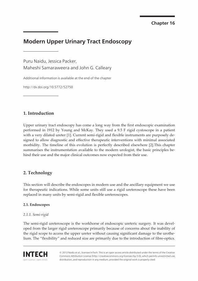
Chapter 16
Modern Upper Urinary Tract Endoscopy
Puru Naidu, Jessica Packer,Maheshi Samaraweera and John G. Calleary
Additional information is available at the end of the chapter
http://dx.doi.org/10.5772/52750
1. Introduction
Upper urinary tract endoscopy has come a long way from the first endoscopic examinationperformed in 1912 by Young and McKay. They used a 9.5 F rigid cystoscope in a patientwith a very dilated ureter [1]. Current semi-rigid and flexible instruments are purposely de‐signed to allow diagnostic and effective therapeutic interventions with minimal associatedmorbidity. The timeline of this evolution is perfectly described elsewhere [2].This chaptersummarises the instrumentation available to the modern urologist, the basic principles be‐hind their use and the major clinical outcomes now expected from their use.
2. Technology
This section will describe the endoscopes in modern use and the ancillary equipment we usefor therapeutic indications. While some units still use a rigid ureteroscope these have beenreplaced in many units by semi-rigid and flexible ureteroscopes.
2.1. Endoscopes
2.1.1. Semi-rigid
The semi-rigid ureteroscope is the workhorse of endoscopic ureteric surgery. It was devel‐oped from the larger rigid ureteroscope primarily because of concerns about the inability ofthe rigid scope to access the upper ureter without causing significant damage to the urothe‐lium. The “flexibility” and reduced size are primarily due to the introduction of fibre-optics.
© 2013 Naidu et al.; licensee InTech. This is an open access article distributed under the terms of the CreativeCommons Attribution License (http://creativecommons.org/licenses/by/3.0), which permits unrestricted use,distribution, and reproduction in any medium, provided the original work is properly cited.
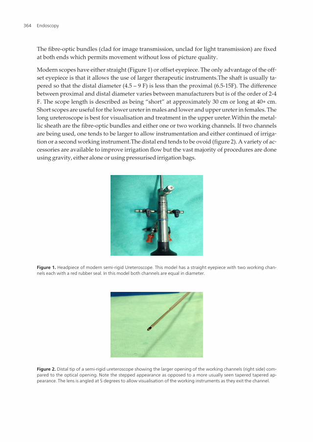
The fibre-optic bundles (clad for image transmission, unclad for light transmission) are fixedat both ends which permits movement without loss of picture quality.
Modern scopes have either straight (Figure 1) or offset eyepiece. The only advantage of the off‐set eyepiece is that it allows the use of larger therapeutic instruments.The shaft is usually ta‐pered so that the distal diameter (4.5 – 9 F) is less than the proximal (6.5-15F). The differencebetween proximal and distal diameter varies between manufacturers but is of the order of 2-4F. The scope length is described as being “short” at approximately 30 cm or long at 40+ cm.Short scopes are useful for the lower ureter in males and lower and upper ureter in females. Thelong ureteroscope is best for visualisation and treatment in the upper ureter.Within the metal‐lic sheath are the fibre-optic bundles and either one or two working channels. If two channelsare being used, one tends to be larger to allow instrumentation and either continued of irriga‐tion or a second working instrument.The distal end tends to be ovoid (figure 2). A variety of ac‐cessories are available to improve irrigation flow but the vast majority of procedures are doneusing gravity, either alone or using pressurised irrigation bags.
Figure 1. Headpiece of modern semi-rigid Ureteroscope. This model has a straight eyepiece with two working chan‐nels each with a red rubber seal. In this model both channels are equal in diameter.
Figure 2. Distal tip of a semi-rigid ureteroscope showing the larger opening of the working channels (right side) com‐pared to the optical opening. Note the stepped appearance as opposed to a more usually seen tapered tapered ap‐pearance. The lens is angled at 5 degrees to allow visualisation of the working instruments as they exit the channel.
Endoscopy364
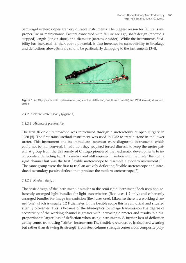
Semi-rigid ureteroscopes are very durable instruments. The biggest reason for failure is im‐proper use or maintenance. Factors associated with failure are age, shaft design (tapered <stepped) length (long > short) and diameter (narrow > wider). While the instruments flexi‐bility has increased its therapeutic potential, it also increases its susceptibility to breakageand deflections above 5cm are said to be particularly damaging to the instruments [3-4].
Figure 3. An Olympus flexible ureteroscope (single active deflection, one thumb handle) and Wolf semi-rigid uretero‐scope.
2.1.2. Flexible ureteroscopy (figure 3)
2.1.2.1. Historical perspective
The first flexible ureteroscope was introduced through a ureterotomy at open surgery in1960 [5]. The first trans-urethral instrument was used in 1962 to treat a stone in the lowerureter. This instrument and its immediate successor were diagnostic instruments whichcould not be manoeuvred. In addition they required forced diuresis to keep the ureter pat‐ent. A group from the University of Chicago pioneered the next major developments to in‐corporate a deflecting tip. This instrument still required insertion into the ureter through arigid channel but was the first flexible ureteroscope to resemble a modern instrument [6].The same group were the first to trial an actively deflecting flexible ureteroscope and intro‐duced secondary passive deflection to produce the modern ureteroscope [7].
2.1.2.2. Modern design
The basic design of the instrument is similar to the semi-rigid instrument.Each uses non-co‐herently arranged light bundles for light transmission (flexi uses 1-2 only) and coherentlyarranged bundles for image transmission (flexi uses one). Likewise there is a working chan‐nel (one) which is usually 3.2 F diameter. In the flexible scope this is cylindrical and situatedslightly off-center. This is because of the fibre-optics for image transmission.The degree ofeccentricity of the working channel is greater with increasing diameter and results in a dis‐proportionate larger loss of deflection when using instruments. A further loss of deflectionability comes from using “stiffer” instruments.The flexible ureteroscope is also hard wearingbut rather than drawing its strength from steel column strength comes from composite poly‐
Modern Upper Urinary Tract Endoscopyhttp://dx.doi.org/10.5772/52750
365
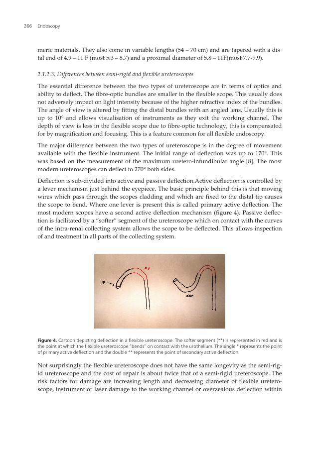
meric materials. They also come in variable lengths (54 – 70 cm) and are tapered with a dis‐tal end of 4.9 – 11 F (most 5.3 – 8.7) and a proximal diameter of 5.8 – 11F(most 7.7-9.9).
2.1.2.3. Differences between semi-rigid and flexible ureteroscopes
The essential difference between the two types of ureteroscope are in terms of optics andability to deflect. The fibre-optic bundles are smaller in the flexible scope. This usually doesnot adversely impact on light intensity because of the higher refractive index of the bundles.The angle of view is altered by fitting the distal bundles with an angled lens. Usually this isup to 10° and allows visualisation of instruments as they exit the working channel. Thedepth of view is less in the flexible scope due to fibre-optic technology, this is compensatedfor by magnification and focusing. This is a feature common for all flexible endoscopy.
The major difference between the two types of ureteroscope is in the degree of movementavailable with the flexible instrument. The initial range of deflection was up to 170°. Thiswas based on the measurement of the maximum uretero-infundibular angle [8]. The mostmodern ureteroscopes can deflect to 270° both sides.
Deflection is sub-divided into active and passive deflection.Active deflection is controlled bya lever mechanism just behind the eyepiece. The basic principle behind this is that movingwires which pass through the scopes cladding and which are fixed to the distal tip causesthe scope to bend. Where one lever is present this is called primary active deflection. Themost modern scopes have a second active deflection mechanism (figure 4). Passive deflec‐tion is facilitated by a “softer” segment of the ureteroscope which on contact with the curvesof the intra-renal collecting system allows the scope to be deflected. This allows inspectionof and treatment in all parts of the collecting system.
Figure 4. Cartoon depicting deflection in a flexible ureteroscope. The softer segment (**) is represented in red and isthe point at which the flexible ureteroscope “bends” on contact with the urothelium. The single * represents the pointof primary active deflection and the double ** represents the point of secondary active deflection.
Not surprisingly the flexible ureteroscope does not have the same longevity as the semi-rig‐id ureteroscope and the cost of repair is about twice that of a semi-rigid ureteroscope. Therisk factors for damage are increasing length and decreasing diameter of flexible uretero‐scope, instrument or laser damage to the working channel or overzealous deflection within
Endoscopy366
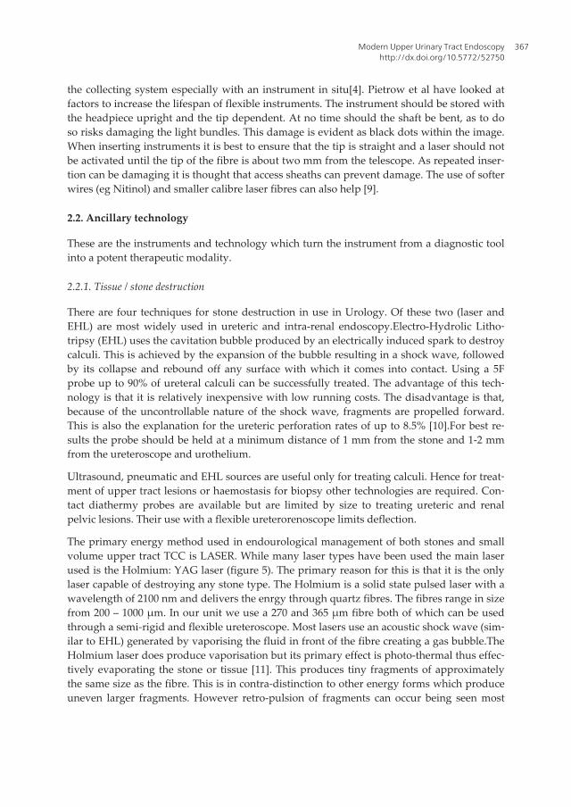
the collecting system especially with an instrument in situ[4]. Pietrow et al have looked atfactors to increase the lifespan of flexible instruments. The instrument should be stored withthe headpiece upright and the tip dependent. At no time should the shaft be bent, as to doso risks damaging the light bundles. This damage is evident as black dots within the image.When inserting instruments it is best to ensure that the tip is straight and a laser should notbe activated until the tip of the fibre is about two mm from the telescope. As repeated inser‐tion can be damaging it is thought that access sheaths can prevent damage. The use of softerwires (eg Nitinol) and smaller calibre laser fibres can also help [9].
2.2. Ancillary technology
These are the instruments and technology which turn the instrument from a diagnostic toolinto a potent therapeutic modality.
2.2.1. Tissue / stone destruction
There are four techniques for stone destruction in use in Urology. Of these two (laser andEHL) are most widely used in ureteric and intra-renal endoscopy.Electro-Hydrolic Litho‐tripsy (EHL) uses the cavitation bubble produced by an electrically induced spark to destroycalculi. This is achieved by the expansion of the bubble resulting in a shock wave, followedby its collapse and rebound off any surface with which it comes into contact. Using a 5Fprobe up to 90% of ureteral calculi can be successfully treated. The advantage of this tech‐nology is that it is relatively inexpensive with low running costs. The disadvantage is that,because of the uncontrollable nature of the shock wave, fragments are propelled forward.This is also the explanation for the ureteric perforation rates of up to 8.5% [10].For best re‐sults the probe should be held at a minimum distance of 1 mm from the stone and 1-2 mmfrom the ureteroscope and urothelium.
Ultrasound, pneumatic and EHL sources are useful only for treating calculi. Hence for treat‐ment of upper tract lesions or haemostasis for biopsy other technologies are required. Con‐tact diathermy probes are available but are limited by size to treating ureteric and renalpelvic lesions. Their use with a flexible ureterorenoscope limits deflection.
The primary energy method used in endourological management of both stones and smallvolume upper tract TCC is LASER. While many laser types have been used the main laserused is the Holmium: YAG laser (figure 5). The primary reason for this is that it is the onlylaser capable of destroying any stone type. The Holmium is a solid state pulsed laser with awavelength of 2100 nm and delivers the enrgy through quartz fibres. The fibres range in sizefrom 200 – 1000 µm. In our unit we use a 270 and 365 µm fibre both of which can be usedthrough a semi-rigid and flexible ureteroscope. Most lasers use an acoustic shock wave (sim‐ilar to EHL) generated by vaporising the fluid in front of the fibre creating a gas bubble.TheHolmium laser does produce vaporisation but its primary effect is photo-thermal thus effec‐tively evaporating the stone or tissue [11]. This produces tiny fragments of approximatelythe same size as the fibre. This is in contra-distinction to other energy forms which produceuneven larger fragments. However retro-pulsion of fragments can occur being seen most
Modern Upper Urinary Tract Endoscopyhttp://dx.doi.org/10.5772/52750
367
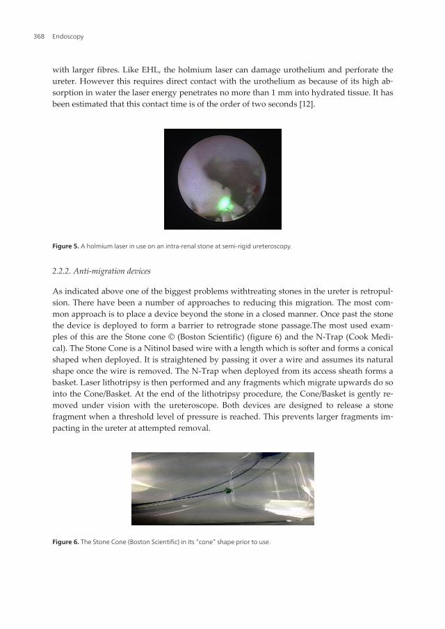
with larger fibres. Like EHL, the holmium laser can damage urothelium and perforate theureter. However this requires direct contact with the urothelium as because of its high ab‐sorption in water the laser energy penetrates no more than 1 mm into hydrated tissue. It hasbeen estimated that this contact time is of the order of two seconds [12].
Figure 5. A holmium laser in use on an intra-renal stone at semi-rigid ureteroscopy.
2.2.2. Anti-migration devices
As indicated above one of the biggest problems withtreating stones in the ureter is retropul‐sion. There have been a number of approaches to reducing this migration. The most com‐mon approach is to place a device beyond the stone in a closed manner. Once past the stonethe device is deployed to form a barrier to retrograde stone passage.The most used exam‐ples of this are the Stone cone © (Boston Scientific) (figure 6) and the N-Trap (Cook Medi‐cal). The Stone Cone is a Nitinol based wire with a length which is softer and forms a conicalshaped when deployed. It is straightened by passing it over a wire and assumes its naturalshape once the wire is removed. The N-Trap when deployed from its access sheath forms abasket. Laser lithotripsy is then performed and any fragments which migrate upwards do sointo the Cone/Basket. At the end of the lithotripsy procedure, the Cone/Basket is gently re‐moved under vision with the ureteroscope. Both devices are designed to release a stonefragment when a threshold level of pressure is reached. This prevents larger fragments im‐pacting in the ureter at attempted removal.
Figure 6. The Stone Cone (Boston Scientific) in its “cone” shape prior to use.
Endoscopy368
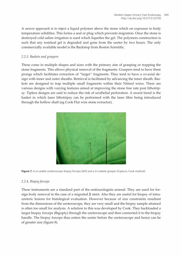
A newer approach is to inject a liquid polymer above the stone which on exposure to bodytemperature solidifies. This forms a seal or plug which prevents migration. Once the stone isdestroyed cold saline irrigation is used which liquefies the gel. The polymers construction issuch that any residual gel is degraded and gone from the ureter by two hours. The onlycommercially available model is the Backstop from Boston Scientific.
2.2.3. Baskets and graspers
These come in multiple shapes and sizes with the primary aim of grasping or trapping thestone fragments. This allows physical removal of the fragments. Graspers tend to have threeprongs which facilitates extraction of “larger” fragments. They tend to have a co-axial de‐sign with inner and outer sheaths. Retrieval is facilitated by advancing the inner sheath. Bas‐kets are designed to trap multiple small fragments within their Nitinol wires. There arevarious designs with varying features aimed at improving the stone free rate post lithotrip‐sy. Tipless designs are said to reduce the risk of urothelial perforation. A recent trend is thebasket in which laser lithotripsy can be performed with the laser fibre being introducedthrough the hollow shaft (eg Cook Flat wire stone extractor).
Figure 7. A re-usable ureteroscopic biopsy forceps (left) and a tri-radiate grasper (Captura, Cook medical)
2.2.4. Biopsy forceps
These instruments are a standard part of the endourologists arsenal. They are used for for‐eign body removal in the case of a migrated JJ stent. Also they are useful for biopsy of intra-ureteric lesions for histological evaluation. However because of size constraints resultantfrom the dimensions of the ureteroscope, they are very small and the biopsy sample attainedis often too small for analysis. A solution to this was developed by Cook. They backloaded alarger biopsy forceps (Bigopty) through the ureteroscope and then connected it to the biopsyhandle. The biopsy forceps thus enters the ureter before the ureteroscope and hence can beof greater size (figure 8).
Modern Upper Urinary Tract Endoscopyhttp://dx.doi.org/10.5772/52750
369
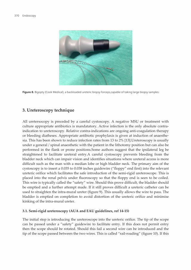
Figure 8. Bigopty (Cook Medical), a backloaded ureteric biopsy forceps,capable of taking large biopsy samples.
3. Ureteroscopy technique
All uretersocopy is preceded by a careful cystoscopy. A negative MSU or treatment withculture appropriate antibiotics is mandatatory. Active infection is the only absolute contra-indication to ureteroscopy. Relative contra-indications are ongoing anti-coagulation therapyor bleeding diatheses. Appropriate antibiotic prophylaxis is given at induction of anaesthe‐sia. This has been shown to reduce infection rates from 13 to 2% [13].Ureteroscopy is usuallyunder a general / spinal anaesthetic with the patient in the lithotomy position but can also beperformed in the flank or prone positions.Some authors suggest that the ipsilateral leg bestraightened to facilitate ureteral entry.A careful cystoscopy prevents bleeding from thebladder neck which can impair vision and identifies situations where ureteral access is moredifficult such as the man with a median lobe or high bladder neck. The primary aim of thecystoscopy is to insert a 0.035 to 0.038 inches guidewire (“floppy” end first) into the relevantureteric orifice which facilitates the safe introduction of the semi-rigid ureteroscope. This isplaced into the renal pelvis under fluoroscopy so that the floppy end is seen to be coiled.This wire is typically called the “safety” wire. Should this prove difficult, the bladder shouldbe emptied and a further attempt made. If it still proves difficult a ureteric catheter can beused to straighten the intra-mural ureter (figure 9). This usually allows the wire to pass. Thebladder is emptied on completion to avoid distortion of the ureteric orifice and minimisekinking of the intra-mural ureter.
3.1. Semi-rigid ureteroscopy (AUA and EAU guidelines, ref 14-15)
The initial step is introducing the ureteroscope into the ureteric orifice. The tip of the scopecan be passed under a ”safety” guidewire to facilitate entry. If this does not permit entrythen the scope should be rotated. Should this fail a second wire can be introduced and thetip of the scope passed between the two wires. This is called “rail-roading” (figure 10). If this
Endoscopy370
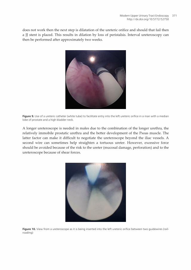
does not work then the next step is dilatation of the ureteric orifice and should that fail thena JJ stent is placed. This results in dilation by loss of peristalsis. Interval ureteroscopy canthen be performed after approximately two weeks.
Figure 9. Use of a ureteric catheter (white tube) to facilitate entry into the left ureteric orifice in a man with a medianlobe of prostate and a high bladder neck.
A longer ureteroscope is needed in males due to the combination of the longer urethra, therelatively immobile prostatic urethra and the better development of the Psoas muscle. Thelatter factor can make it difficult to negotiate the ureteroscope beyond the iliac vessels. Asecond wire can sometimes help straighten a tortuous ureter. However, excessive forceshould be avoided because of the risk to the ureter (mucosal damage, perforation) and to theureteroscope because of shear forces.
Figure 10. View from a ureteroscope as it is being inserted into the left ureteric orifice between two guidewires (rail‐roading)
Modern Upper Urinary Tract Endoscopyhttp://dx.doi.org/10.5772/52750
371
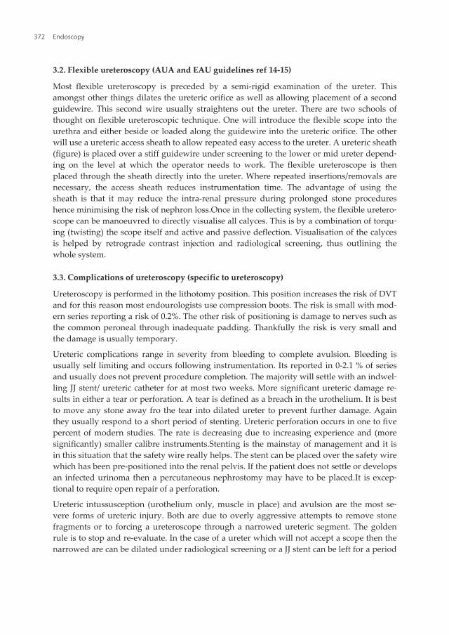
3.2. Flexible ureteroscopy (AUA and EAU guidelines ref 14-15)
Most flexible ureteroscopy is preceded by a semi-rigid examination of the ureter. Thisamongst other things dilates the ureteric orifice as well as allowing placement of a secondguidewire. This second wire usually straightens out the ureter. There are two schools ofthought on flexible ureteroscopic technique. One will introduce the flexible scope into theurethra and either beside or loaded along the guidewire into the ureteric orifice. The otherwill use a ureteric access sheath to allow repeated easy access to the ureter. A ureteric sheath(figure) is placed over a stiff guidewire under screening to the lower or mid ureter depend‐ing on the level at which the operator needs to work. The flexible ureteroscope is thenplaced through the sheath directly into the ureter. Where repeated insertions/removals arenecessary, the access sheath reduces instrumentation time. The advantage of using thesheath is that it may reduce the intra-renal pressure during prolonged stone procedureshence minimising the risk of nephron loss.Once in the collecting system, the flexible uretero‐scope can be manoeuvred to directly visualise all calyces. This is by a combination of torqu‐ing (twisting) the scope itself and active and passive deflection. Visualisation of the calycesis helped by retrograde contrast injection and radiological screening, thus outlining thewhole system.
3.3. Complications of ureteroscopy (specific to ureteroscopy)
Ureteroscopy is performed in the lithotomy position. This position increases the risk of DVTand for this reason most endourologists use compression boots. The risk is small with mod‐ern series reporting a risk of 0.2%. The other risk of positioning is damage to nerves such asthe common peroneal through inadequate padding. Thankfully the risk is very small andthe damage is usually temporary.
Ureteric complications range in severity from bleeding to complete avulsion. Bleeding isusually self limiting and occurs following instrumentation. Its reported in 0-2.1 % of seriesand usually does not prevent procedure completion. The majority will settle with an indwel‐ling JJ stent/ ureteric catheter for at most two weeks. More significant ureteric damage re‐sults in either a tear or perforation. A tear is defined as a breach in the urothelium. It is bestto move any stone away fro the tear into dilated ureter to prevent further damage. Againthey usually respond to a short period of stenting. Ureteric perforation occurs in one to fivepercent of modern studies. The rate is decreasing due to increasing experience and (moresignificantly) smaller calibre instruments.Stenting is the mainstay of management and it isin this situation that the safety wire really helps. The stent can be placed over the safety wirewhich has been pre-positioned into the renal pelvis. If the patient does not settle or developsan infected urinoma then a percutaneous nephrostomy may have to be placed.It is excep‐tional to require open repair of a perforation.
Ureteric intussusception (urothelium only, muscle in place) and avulsion are the most se‐vere forms of ureteric injury. Both are due to overly aggressive attempts to remove stonefragments or to forcing a ureteroscope through a narrowed ureteric segment. The goldenrule is to stop and re-evaluate. In the case of a ureter which will not accept a scope then thenarrowed are can be dilated under radiological screening or a JJ stent can be left for a period
Endoscopy372
of four weeks. The stent causes ureteric dilation and ureteroscopy can usually be completedat the time of stent removal. In the situation where a stone laden basket will not negotiate anarrowed area then it should be gently moved back up to dilated ureter and opened. Thiswill release some fragments and permit a further attempt at removal. If this fails it meansthat the fragments are too large and further laser lithotripsy is warranted. If the basket willnot open then it can be disassembled and laser lithotripsy performed to the fragments with‐in the basket.If either avulsion or intussusception is suspected then an immediate retrogradeexamination is performed. If the renal collecting system is not outlined with contrast thenthe diagnosis is confirmed. Drainage of the collecting system is mandatatory and this is usu‐ally with a percutaneous nephrostomy. Open surgery is required and the technique usedwill depend on the site and extent of injury.
4. Upper tract stones
4.1. Introduction
Between 1200 and 1400 per 100,000 people will develop urinary stones each year with amale:female ratio of 3:1.The majority of stones are composed of calcium oxalate, often mixedwith calcium phosphate, in both adults and children. The acute presentation is usually un‐mistakable with the classical history of loin to groin colicky pain. Evaluation with non-con‐trast CT is advisable for diagnosis. The immediate management usually involves analgesiaand treatment of any infection present, and then determining definitive management [14].Stones smaller than 5 mm will generally pass, but larger stones often require urological in‐tervention [16]. For the purpose of this chapter, the management of upper urinary tractstones will be explored and outlined with the emphasis being on endourological manage‐ment.
4.2. Management
The options available for the management of upper urinary tract stones include observation,Extra-Corporeal Shock wave Lithotripsy (ESWL), Ureteroscopy, Percutaneous Nephro-lith‐otomy (PCNL),laparoscopy and rarely open surgical removal. The appropriate modality foreach individual patient will depend on the interaction between stone (size, location, appear‐ance of the stone on imaging, composition),anatomical abnormalities, the presence of infec‐tion and concomitant co-morbidities which may affect the decision regarding appropriateanaesthetic time.There also is an increasing trend toward intervention because of technologi‐cal improvements and a growing dissatisfaction with the overall success rates with extracor‐poreal shock wave lithotripsy [17].
Most stones less than 5mm in size will pass spontaneously.European Association of Urologyguidelines state that active stone removal is recommended for renal stones >6-7mm in size[14]. However, those of less than 6mm in size, if symptomatic, can be considered for treat‐ment [14-15].
Modern Upper Urinary Tract Endoscopyhttp://dx.doi.org/10.5772/52750
373
4.2.1. Non invasive treatment of upper tract stones
ESWL is entirely non invasive, and it uses super sonic waves to fragment stones into smallpieces that can be easily passed. Shortly after its introduction in 1983, it became widely ac‐cepted as the first line treatment modality for the majority of stones and rapidly replacedinvasive surgical options. ESWL is effective for most renal stones less than 2 cm in size andureteric stones less than 1 cm in size [18].In patients with normal anatomy, and with nonlower pole renal stones < 20mm in size, ESWL is recommended as first line treatment. Lowerpole stones have a higher failure rate with ESWL and so other treatment modalities shouldbe sought ( see below). In one study which compared stone free rates of ureteral stones ofvariable sizes and locations which were treated by ureteroscopy or extra-corporeal shockwave lithotripsy, ESWL was associated with a success rate of 64% ( up to two treatments), ascompared to 96% for ureteroscopy in a single treatment [17].
It is important to highlight that certain stone composition such as cystine or phosphatestones may be resistant to fragmentation.There are also multiple other contra indications tothe use of ESWL which include pregnancy, bleeding diatheses, severe obesity, anatomicalobstruction distal to the stone.
4.2.2. Invasive treatment of upper tract stones
Larger stones, particularly those composed of cystine or struvite, can be approached via es‐tablishing percutaneous access to the collecting system through a small flank incision. Thiswould allow direct visualization and intra-corporeal lithotripsy for stone disruption, and re‐moval of fragments known collectively as Percutaneous nephrolithotomy (PCNL).
PCNL has high success rates of around 90% however there are risks onvolved, and majorintra-operative or post operative complication rates are often reported as 0.03% to 10% [19].However, ureteroscopy is fast becoming the main form of treatment for upper urinary tractstone management and this is what will be discussed for the purpose of this chapter.
4.2.3. Selecting the treatment for a patient with stone management
Intra-renal calculi
Different stone sizes respond better to different therapies and success rates are variable forthe size of stone. The European Urological Association recommends the following manage‐ment plan for kidney stones in the renal pelvis or upper/middle calyx, categorised accordingto size:
Flexible ureteroscopy is used less as a first line treatment for stones > 1.5cm in size. Howeverwith ongoing improvements in newer generation flexible ureteroscopes, there is an increas‐ing trend toward ureteroscopy and laser lithotripsy for intr-renal calculi of all sizes andcompositions. In the available literature, there are very few reported studies on the use ofsemi rigid ureteroscopy to treat renal stones. A prospective analysis performed by Bryniar‐ski and co-workers assessed the safety of PNCL and retrograde intrarenal surgery use semirigid ureteroscopy for the management of renal stones of >2cm in size. Although stone free
Endoscopy374

rates were superior in the PCNL group, the semi rigid ureteroscopy provides advantages foroperating times, haemoglobin loss, post-operative visual analogue scoring by patients andreduced hospital stay [20]. The situation is slightly more complex in lower pole stones as thefollowing table shows.
Stones <1cm in size Stones 1-2cm in size Stones >2cm in size
ESWL ESWL or endourology Endourology (PCNL / flexible
ureteroscopy)
Flexible ureteroscopy ESWL
PCNL Laparoscopy
Table 1. European Association of Urology (EAU) recommendations for the treatment of Renal stones.
Treatment for renal calculi in the inferior calyx is also very dependent on size:
Stones <1cm in size Stones 1-2cm in size Stones >2cm in size
ESWL Favourable factors for ESWL? Endourology ( PCNL or
ureteroscopy)
Flexible ureteroscopy No -> Endourology
PCNL Yes -> ESWL or endourology
Table 2. EAU recommendations for the treatment of Lower pole stones.
Ureteric Calculi
The management of ureteric calculi is also dependent on the size of the stone involved. Thetable below outlines the European Urological Association guidelines for Stone managementdependent on size of stone:
Location Stone size 1st choice 2nd choice
Proximal ureter <10mm
>10mm
ESWL
Ureteroscopy (retrograde or
antegrade) or ESWL
Ureteroscopy
Ureteroscopy (retrograde or
antegrade) or ESWL
Distal ureter <10mm
>10mm
Ureteroscopy or ESWL
Ureteroscopy
Ureteroscopy or ESWL
ESWL
Table 3. EAU recommendations for the treatment of Ureteric stones
The outcomes of ureteroscopy
The European Association of Urology and the American Urological Association guidelinesPanel have published stone free rates for different treatment modalities within their nephro‐lithiasis guidelines.In a cohort comparison group (early 1980s vs 1992) the success of ure‐
Modern Upper Urinary Tract Endoscopyhttp://dx.doi.org/10.5772/52750
375
teroscopic procedures rose from 86% to 96%. In addition, they observed an overall decreasein complications (20% to 12%) in ureteroscopy and 6.6% to 1.5% in ureteroscopic laser litho‐tripsy [21]. Partly, this was thought to be due to greater surgeon experience and that this issignificantly correlated to higher success and lower complication rates in ureteroscopic laserlithotripsy with holmium laser [21].
Renal Calculi
The success rates (stone free or insignificant fragments) reported with PCNL are greaterthan 90% for renal stones >2cm. However, major complications during or after PCNL occurat reported rates of 0.03 % to 10% [22]. The success rates of retrograde intra-renal surgeryhave been reported as 75-95% for intra-renal stones >2cm after the first or second treatment,whereas the major or minor complications vary from 1.5 % to 12% [23].This is less frequentthan rates in PCNL procedures. Major complications in ureteroscopy such as ureteric perfo‐ration or avulsion are extremely rare.
Ureteric Calculi
One study of a two year experience, highlighted that the success rates following ESWL wereheavily influenced by stone size.The overall stone free success rate was 74.7% with one ses‐sion. However, as the size of the stone increased, the success rate reduced. For stones <1cmthe success ratewas 83.6%and when the stone size > 1cm the success rate reduced to 42.1%.The stone free rates also varied according to the site of the stone - 72.4% (proximal), 70%(mid ureter), and 82% (distal) after a single session [14-15].
In ureteroscopy, an overall stone –free rate of 87.8% was obtained irrespective of the size ofthe stone (88.9% for <1cm and 86.6% for >1cm). The success rates did slightly vary in relationto the stone site. The stone-free rates were 75% (proximal), 94.6% (mid ureter) and 84.6%(distal) [24].
The American Urological Association recent 2012 guidelines have published stone free ratesfor Shock Wave lithotripsy and ureteroscopy for the treatment of ureteric calculi and theseare outlined in the tables below:
Proximal ureter:
Treatment Overall Stone size < 10mm Stone size >10mm
ESWL 82% 90% 68%
Ureteroscopy overall
Flexible ureteroscopy
81%
89%
80%
84%
79%
Table 4. Proximal Ureter; stone clearance comparison between Ureteroscopy and ESWL.
Mid Ureter:
The reduced success for stone free rates using ESWL in the mid ureter is likely explained bythe anatomical changes at this site. The mid-ureter is closely related to the transverse proc‐
Endoscopy376
esses of the lumbar verterbrae and focus of the lithotripsy beam is more difficult due to theanatomical relationships to the spine.
Treatment Overall Stone size < 10mm Stone size >10mm
ESWL 73% 84% 76%
Ureteroscopy overall
Flexible ureteroscopy
86%
88%
91%
Not documented
78%
Not documented
Table 5. Mid Ureter; stone clearance comparison between Ureteroscopy and ESWL.
The reduced success for stone free rates using ESWL in the mid ureter is likely explained bythe anatomical changes at this site. The mid-ureter is closely related to the transverse proc‐esses of the lumbar verterbrae and focus of the lithotripsy beam is more difficult due to theanatomical relationships to the spine.
Distal ureter:
Treatment Overall Stone size < 10mm Stone size >10mm
ESWL 74% 86% 74%
Ureteroscopy overall
Rigid ureteroscopy
94%
94%
97%
98%
93%
94%
Table 6. Distal Ureter; stone clearance comparison between Ureteroscopy and ESWL.
The use of holmium laser lithotripsy via ureteroscopy is safe and effective in urinary stonemanagement, particularly for larger calculi. It is associated with success rates of more than90% and with complication rates as low as 10%.
In a study which described 300 procedures of ureteric stone lithotripsy with holmium la‐ser, there was an overall complication rate of 10%. Their overall success rates were 90%and after the first episode, 86% were stone free [21]. In another series of 598 patients, theoverall complication rate was 4%, with an overall the success rate of 97% and 94% afterthe first episode [25].
5. Upper tract Transitional Cell Carcinoma (TCC) / malignancy
Upper tract TCC accounts for approximately 10% of all renal tumors and 5% of all urothelialtumors. It is found to be more common in Caucasian, occurs more often in the sixth to sev‐enth decade of life [26]. Worryingly there is evidence to suggest that the incidence of uppertract TCC is increasing [27]. The presentation is usually with haematuria and approximately30% will have “ureteric colic” secondary to blood clot [28]. They occur more commonly inpeople with a history of bladder cancer.
Modern Upper Urinary Tract Endoscopyhttp://dx.doi.org/10.5772/52750
377
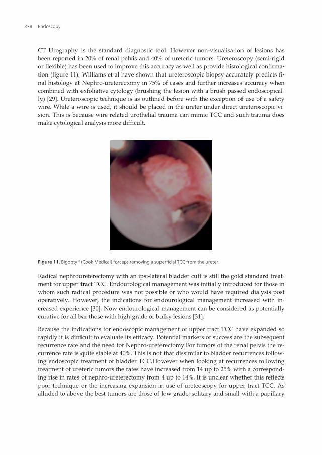
CT Urography is the standard diagnostic tool. However non-visualisation of lesions hasbeen reported in 20% of renal pelvis and 40% of ureteric tumors. Ureteroscopy (semi-rigidor flexible) has been used to improve this accuracy as well as provide histological confirma‐tion (figure 11). Williams et al have shown that ureteroscopic biopsy accurately predicts fi‐nal histology at Nephro-ureterectomy in 75% of cases and further increases accuracy whencombined with exfoliative cytology (brushing the lesion with a brush passed endoscopical‐ly) [29]. Ureteroscopic technique is as outlined before with the exception of use of a safetywire. While a wire is used, it should be placed in the ureter under direct ureteroscopic vi‐sion. This is because wire related urothelial trauma can mimic TCC and such trauma doesmake cytological analysis more difficult.
Figure 11. Bigopty ®(Cook Medical) forceps removing a superficial TCC from the ureter.
Radical nephroureterectomy with an ipsi-lateral bladder cuff is still the gold standard treat‐ment for upper tract TCC. Endourological management was initially introduced for those inwhom such radical procedure was not possible or who would have required dialysis postoperatively. However, the indications for endourological management increased with in‐creased experience [30]. Now endourological management can be considered as potentiallycurative for all bar those with high-grade or bulky lesions [31].
Because the indications for endoscopic management of upper tract TCC have expanded sorapidly it is difficult to evaluate its efficacy. Potential markers of success are the subsequentrecurrence rate and the need for Nephro-ureterectomy.For tumors of the renal pelvis the re‐currence rate is quite stable at 40%. This is not that dissimilar to bladder recurrences follow‐ing endoscopic treatment of bladder TCC.However when looking at recurrences followingtreatment of ureteric tumors the rates have increased from 14 up to 25% with a correspond‐ing rise in rates of nephro-ureterectomy from 4 up to 14%. It is unclear whether this reflectspoor technique or the increasing expansion in use of ureteoscopy for upper tract TCC. Asalluded to above the best tumors are those of low grade, solitary and small with a papillary
Endoscopy378
appearance with negative cytology [32]. From the point of view of laser choice the Holmiumis best for resection and Neodymium YAG is best for fulgaration.
6. Ureteric stricture / Pelvi-Ureteric Junction (PUJ) obstruction
PUJ obstruction is a functional blockage to antegrade flow of urine to the bladder from theupper tract due to a narrowing at the PUJ. Its classically treated by an open (or laterally lap‐aroscopic) procedure by the name of Pyeloplasty. Retrograde endopyelotomy is a minimallyinvasive option.This is performed using a large calibre rigid ureterorenoresectoscope or us‐ing a Holmium laser. The laser procedure is more common and usually linked with subse‐quent balloon dilatation of the incised area. The incision is made laterally at the PUJ tominimise vascular injury. The incision is deepened until peri-ureteric fat is seen. Balloon di‐latation up to 24 F is then performed and a special stent inserted (Tapered with greater di‐ameter at the top end) for six weeks. Longterm success rates of up to 77% have beenreported. Failure usually requires either open or laparoscopic pyeloplasty. A split functionof less than 20% and redundant renal pelvis are factors predictive of failure.
Ureteric strictures can also be treated with laser incision. For distal strictures the successrates are of the order of 75% with an average follow-up of 3 years. Failures tend to occurearly [33]. Similar results are reported for mid-ureteric and proximal ureteric strictures. Thetechnique for ureteroscopic surgery is again incision to peri-ureteric fat but also includes in‐cision into normal tissue either side of the stricture.
An alternative form of endo-urological stricture management is to use a combined balloondilator and Monopolar electrode. Identification of the stricture is radiological as once theballoon is inflated with contrast, the stricture will be identified as an indentation on the bal‐loon ie “waisting”. As the procedure is not visualised an adequate incision is identified ascontrast extravasation.
7. The future
There are now production models of video ureterorenoscopes. Their advantage is that theyare smaller, more manoeuvrable with a better picture and fluid flow rate. These instrumentsuse distal chip technology where the incoming light energy impacts on a digital chip. Thisenergy transfer results in a charge which is transmitted along a single fibre to a processorwhich converts it into a usuable image.The better visual image is primarily due to less trans‐mission loss. Energy (light) loss in fibreoptic ureteroscopes is multi-factorial but involvescladding damage, damage to the fibre-optic bundles, light lead / connection damage andcamera head. It is primarily in the treatment of upper tract TCC where the improved imag‐ing will be of most use. The increased magnification and resolution of the images togetherwith technology such as Narrow Band Imaging should expand the place of ureteroscopy inthe management of low grade upper tract TCC.
Modern Upper Urinary Tract Endoscopyhttp://dx.doi.org/10.5772/52750
379
8. Conclusion
The use of endo-urological techniques in day to day urological practice is increasing. This isfuelled by many factors but amongst them is better technology, a greater number of trainedindividuals and a desire to improve patient experience while maintaining outcomes. The fu‐ture will be even better as better image definition becomes more readily available as video-ureteroscopes become more durable.
Author details
Puru Naidu, Jessica Packer, Maheshi Samaraweera and John G. Calleary
*Address all correspondence to: [email protected].
Department of Urology, Morth Manchester General Hospital, UK
References
[1] Young HH , McKay RE. Congenital valvular obstruction of the prostatic urethra SurgGynecol Obstet 1929; 48:509-510)
[2] Payne DA, Keeley FX . Rigid and Flexible Ureteroscopes:Technical Features. In:Smith`s Textbook of Endourology, 3rd Ed. Eds Smith AD, Badlani GH, PremingerGM, Kavoussi LR. 2012 Blackwell Publishing
[3] Carey RI, Gomez CS, Maurici G et al. Frequency of ureteroscopic damage seen at atertiary care center. J Urol 2006;176:607-610
[4] Sung JC, Springhart WP, Marguet CG et al. Location and aetiology of flexible andsemi-rigid ureteroscope damage. Urology 2005;66:958-963
[5] Marshall VF, Fibreoptics in Urology J Urol 1964; 91: 110-114).
[6] Bagley DH, Ureteral endoscopy with passively deflectable, irrigating irrigating flexi‐ble ureteroscopes. Urology 1987;29:170
[7] Grasso M, Bagley DH A 7.5 / 8.2 actively deflectable ureteroscope: a new device forboth diagnostic and therapeutic upper urinary tract endoscopy Urology 1994;43:435-441
[8] Bagley DH, Rittenburg MH. Intrarenal dimensions. Guidelines for flexible uretero‐pyeloscopes. Surg Endosc 1987;1:119-121
[9] Pietrow PK, Auge BK, DelvecchioFC et al Techniques to maxamise ureteroscope lon‐gevity. Urology 2003;62:218-222)
Endoscopy380
[10] Lingeman JE, Matlaga BR, Evan AP. Surgical management of upper urinary tract cal‐culi. In: Wein AJ, Kavoussi LR, Novick AC, Partin AW, Peters CA eds. CampbellWalsh Urology, 9th ed Philadelphia: WB Saunders 2007, 1431-1506.
[11] Chan KF, Vasser GJ, Pfefer TJ, Teichman JM, Glickman RD, Weintraub ST, Welch AJ.Holmium: YAG laser lithotripsy. A dominant photothermal ablative mechanismwith chemical decomposition of urinary calculi. Lasers Surg Med 1999; 25:22-37
[12] Santa-Cruz RW, Leveille RJ, Krongrad A. Ex vivo comparison of four lithotriptorscommonly used in the ureter: what does it take to perforate? J Endourol 1998;12:417-422)
[13] Knopf HJ, Graff HJ, Schulze H. Perioperative antibiotic prophylaxis in ureteroscopicstone removal. Eur Urol 2003; 44(1):115-118
[14] Turk F, Knoll T, Petrik A, Sarica K, Straub M, Seitz C. European Urology associationguidelines on urolithiasis. http://www.uroweb.org/guidelines accessed 1st August2012.
[15] AUA / EAU guidelines 2007, Ureteral calculi. http://auanet.org/content/clinical-prac‐tise/main-reports/Uretcal07 accessed 1st Aug 2012.
[16] Worcester E, Coe FL. Nephrolithiasis. Prim Care. 2008 June ; 35(2): 369–vii.
[17] Gerber Glenn S. Acharya Sujeet S. Management of ureteral calculi. Journal of endour‐ology. Volume 24 number 6. June 2010. Pp 953-954
[18] Bach C, Buchholz N. Shock wave lithotripsy for renal and ureteric stones.EuropeanUrology Supplements. 2011.10 .423-432
[19] Atis Gokhan, Gurbuz Cenk, Arikan Ozgur, Canat Lutfi, Kiic Mert, Caskurlu Turhan.Ureteroscopic Management with Laser Lithotripsy of renal Pelvic Stones. Journal ofEndourology. Volume 26. Number 00. 2012. pp 1-5
[20] Bryniarski P, Paradysz A, Zyczkowski M et al. A randomised control study to ana‐lyze the safety and efficacy of percutaneous nephrolithotripsy and retrograde intra‐renal surgery in the management of stones more than 2cm in diameter. Journal ofEndourology. 2012. Vol 26. Pp 52-57.
[21] Leijte Joost AP, Odeens Jorg R, Lock Tycho MTW. Holmiun laser lithotripsy for ure‐teric calculi: Predictive factors for complications and success. Journal of endourology.Volume 22. Number 23. February 2008. pp 257- 260
[22] Michel MS, Trojan L, Rassweiler JJ. Complications in percutaneous nephrolithotomy.European Urology. 2007. Volume 51. Pp 899 – 906
[23] Breda A, Ogunyemi O, leppert JT, et al. Flexible ureteroscopy and laser lithotripsyfor single intra-renal stones 2cm or greater – is this the new frontier? Journal of Urol‐ogy. 2008. Vol 179. Pp 981-984
Modern Upper Urinary Tract Endoscopyhttp://dx.doi.org/10.5772/52750
381
[24] Park H, Park M and Park T. Two year experience with ureteral stones: Extracorporealshock wave lithotripsy v Ureteroscopic manipulation. Journal of Endourology. Vol12. Number 6. December 1998. Pp 501-504
[25] Sofer M, Watterson JD, Wollin TA, Nott L, Razvi H, Denstedt JD. Holmium: YAG la‐ser lithotripsy for upper urinary tract calculi in 598 patients. Hournal of Urology.2002. Volume 167. Pp 31-34
[26] Anderstrom C, Johansson SL, Pettersson S, Wahlqvist L. Carcinoma of ureter. J Urol1989; 142:280.
[27] Munoz JJ et al.(2000) Upper tract urothelial neoplasms: incidence and survival dur‐ing last two decades. J Urol 164: 1523-25
[28] Murphy DM, Zincke H, Furlow WI Management of high grade transitional cell can‐cer of the upper urinary tract. J Urol 1981; 125:25-29.)
[29] Williams SK, Denton KJ, Minervini AA, Oxley J, Khastigir J, Timoney AG, Keeley JRFX. Correlation of Upper –Tract Cytology, Retrograde Pyelography, Ureteroscopicappearance and Ureteroscopic biopsy with Histological Examination of Upper-TractTransitional Cell Carcinoma. J Endourol 2008; 22:1, 71-76
[30] Elliot DS, Segura w, Lightner D, Patterson DE, Blute ML. Is nephroureterectomy nec‐essary in all cases of upper tract transitional cell carcinoma? Long-term results ofconservative endourological management of upper tract transitional cell carcinomain individuals with a normal contralateral kidney. Urology 2001; 58 (2): 174-178.
[31] Painter DJ, Denton K, Timoney AG, Keeley FX. Ureteroscopic Management of Up‐per-Tract Urothelial Cancer: An exciting Nephron-sparing option or an UnacceptableRisk? J Endourol 2008; 22(8): 1237-1239).
[32] Bagley DH Ureteroscopic diagnosis and treatment of upper urinary trat neoplasms.In: Smith`s Textbook of Endourology, 3rd Ed. Eds Smith AD, Badlani GH, PremingerGM, Kavoussi LR. 2012 Blackwell Publishing
[33] Lane BR, Desai MM, Hegarty NJ, Streem SB. Long-term efficacy of holmium laser en‐doureterotomy for benign ureteral strictures. Urology 2006; 67: 894-897
Endoscopy382